Article

Coffee By-Products and Chitosan for Preventing Contamination for Botrytis sp. and Rhizopus sp. in Blueberry Commercialization

by
Gonzalo Hernández-López
* and
Laura Leticia Barrera-Necha
*
Centro de Desarrollo de Productos Bióticos, Instituto Politécnico Nacional, Carretera Yautepec-Jojutla Km. 6, Calle CEPROBI No 8, San Isidro, Yautepec C.P. 62731, Morelos, Mexico
*
Authors to whom correspondence should be addressed.
Resources 2025, 14(3), 48; https://doi.org/10.3390/resources14030048
Submission received: 30 January 2025 / Revised: 2 March 2025 / Accepted: 10 March 2025 / Published: 17 March 2025
(This article belongs to the Special Issue Resource Extraction from Agricultural Products/Waste: 2nd Edition)

Abstract

:
In blueberry storage, non-biodegradable synthetic plastic packaging is used for commercializing this product. The fungi Botrytis sp. and Rhizopus sp. can cause significant losses in postharvest blueberry commercialization. Consequently, the formulations of degradable polymeric based on polylactic acid (PLA)/poly(butylene adipate-co-terephthalate) (PBAT) 60/40 (PP) with coffee parchment (CP), green coffee bean oil (GCBO), chitosan solution (Ch), chitosan nanoparticles (ChNp), and nanostructured coating (NC) were used to develop biodegradable polymer matrix (PM). Caffeine and hexadecanoic acid were identified as principal compounds in GCBO, and the principal compounds in CP were flavonoids, terpenes, and lignans. The 100% mycelial growth inhibition to Botrytis sp. and Rhizopus sp. was observed using GCBO, Ch, ChNp, and NC in high concentrations. GCBO inhibited 100% of spore production in both fungi at all evaluated doses. In the in vivo tests, when compared to the control, the better treatments were: CP for Botrytis sp., with an incidence of 46.6% and a severity of 16%; and Ch for Rhizopus sp., with an incidence of 13.3% and a severity of 0.86%. The PM in the culture medium presented a fungistatic effect. The principal inhibition of mycelial growth (63%) on Botrytis sp. was with PLA/PBAT+NC (PP+NC), and (100%) was observed with PLA/PBAT+CP+NC (PPCP+NC), PP, and PP+NC on Rhizopus sp. Coffee by-products and PM have potential for the control of postharvest fungi in fruits and vegetables.

1. Introduction

Blueberry (Vaccinium corymbosum) is a fruit with high nutritional value, low caloric content, high antioxidant content, and anti-inflammatory properties [1]. These attributes make it an attractive product due to its versatility and benefits for human health. For these reasons, its popularity and demand have increased in the last decade, increasing production and cultivated areas in different regions of the world. Fungal diseases are the main causes of the reduction in plant productivity. The most recurrent genera are Botrytis and Rhizopus, which affect the fruit in the postharvest stage [2]. In blueberries, losses of up to 30% in total production have been recorded due to these phytopathogens [3]. Infections caused by Botrytis sp. are probably the most common worldwide, with a wide distribution and a wide range of crops [4]. The disease it causes tends to be more severe in cold and humid environments; this fungus can develop even at 0 °C. Rhizopus sp. causes soft rot in most fleshy fruit during storage and marketing; when growing conditions are favorable, it can cause large losses in a short period of time due to its rapid development [5]. Today, there are several methods for combating disease-causing agents in agriculturally important crops; however, indiscriminate pesticide use has led to the creation of new biological alternatives that help combat this problem [6]. One of these proposals is to use compounds of natural origin; several plants contain bioactive agents that act against disease development in agriculturally important crops [7]. The high production of coffee generates a large quantity of waste that has a wide application due to its physical and chemical properties, bioavailability, and potential in food production, making it useful in various fields [8].
Green coffee bean oil is obtained from unroasted beans and has several applications in both the cosmetic and food industries due to the presence of bioactive compounds, such as polyphenols, tocopherols, and phytosterols [9]. The (CP) is the covering of the bean at the parchment stage and is considered an agro-industrial waste because its use is in low demand. Recently, it has been sought to explore new applications because CP is a by-product rich in lignocellulosic matter and contains antimicrobial compounds, such as alkaloids and flavonoids, that function as a defense against coffee pathogens and pests [10]. Extracts and oils can be obtained by conventional extraction methods and solvents [11]. Gloria et al. [12] attributed the antimicrobial activity of coffee to caffeine, trigoline, and phenolic acids and their derivatives, and after the bean roasting process it was attributed to melanoids and dicarbonyl compounds. Generally, the bioactive compounds present in coffee are chlorogenic acids, quinic acid, malic acid, and caffeine [13]. Another naturally occurring compound is chitosan, which is obtained from the chitin of some mollusks [14]. It has a high impact on the use of biopolymers and green chemistry due to its high antimicrobial activity. This property depends on several factors, such as the type of pathogen, pH of the medium, and the structure and concentration of chitosan [15]. One of the main uses of chitosan is in the preparation of coatings for fruit and vegetable preservation; they help to reduce deterioration due to the presence of microorganisms and maintain fruit quality [16]. Sotelo-Boyás et al. [17] found that using chitosan nanoparticles enhanced their antimicrobial effects because the nanoparticles could more easily penetrate the wall of the microorganisms. Other polymers used to produce packaging are of synthetic origin, such as polyethylene terephthalate, polystyrene, and polypropylene, which have a negative impact on the environment and health [18]. Today, there is great interest in generating polymers of natural origin that are biodegradable and compostable [19]. Polylactic acid (PLA) is a biodegradable polymer because it is a derivative of lactic acid obtained from renewable resources, such as corn starch and sugar cane, which makes it a polymer of great importance today [20]. Poly(butylene adipate-co-terephthalate) (PBAT) is a synthetic polymer obtained from fossil-based resources; however, it has the capacity to be biodegradable and the potential to be used in various applications [21]. More knowledge of the management and handling of plastics is needed since the main problem they have faced is a lack of information on their origin and adequate treatment, either recycling or composting for their reintegration [22]. Currently, in the food industry, there is a search for incorporating bioactive compounds of natural origin to improve the organoleptic characteristics of fruit and vegetable products, extend their shelf life, and prevent their deterioration by external agents, such as microorganisms [23]. The use of chitosan as an antimicrobial has been studied previously, and it was demonstrated that, by synthesizing chitosan nanoparticles, their antimicrobial effect can be improved [24]. Recent work has been carried out on the synthesis of nanoparticles from extracts obtained from coffee waste [25].
This study aimed (i) to identify chemical compounds of GCBO and CP using chromatographic techniques to know the principal compounds in these coffee wastes; (ii) investigate the antifungal activity of individual coffee residues in vitro and in vivo against Botrytis sp. and Rhizopus sp., and be able to use them as bioactive compounds for disease control; and (iii) evaluate the antifungal in vitro effect of the incorporation of CP and GCBP in a biodegradable polymeric matrix (PM) and be able to reduce the cost of production of these polymers and generate a new ecological alternative.

2. Materials and Methods

Figure 1 shows the research methodology, supplemented in the succeeding parts of this article.

2.1. Materials

The CP and green coffee bean residue were provided by the coffee producers of Coatepec Veracruz, Mexico. The CP was cleaned and separated manually to eliminate external agents. They were then subjected to a drying process using an oven (Binder FD 115, Tuttlingen, Germany) at 60 °C for 12 h to eliminate the total content of humidity. After drying, grinding was carried out in a pulverizing mill (INMIMEX M-150, Tlaxcala, Mexico) and the dust particles that passed through a 100-mesh sieve were recovered. The powders were stored in a container until use. To obtain the oil, ground green coffee beans were used and sifted through a 60-mesh sieve. The Soxhlet extraction system used 50 g of sample placed in the cartridge and left for 6 h in the reflux system with hexane. The extracted oil was recovered and then concentrated in a rotary evaporator (BUCHI R-300, Flawil, Switzerland) to eliminate solvent residues. The sample was packaged and stored in a refrigerator.

2.2. Characterization of Coffee Oil by Gas Chromatography and Mass Spectrometry (GC-MS)

To identify compounds from green coffee beans oil using gas–mass chromatography, (Agilent Technologies 890B Gas Chromatograph Los Angeles, CA, USA), with a thermal separation probe (Agilent G4381A-TPS) and 0.6 mg of oil was placed in a 50 µL TSP microvial and analyzed. The equipment was operated under the following conditions: helium as carrier gas, an initial temperature of 80 °C, a final temperature of 300 °C, an injector temperature at 250 °C, and a run time of 45 min. The mass detector operated under the following conditions: mass range of 32–750 atomic mass units (MS), at a transfer temperature of 280 °C, and with a solvent delay of 0.2 min. The identification of the major compounds was carried out based on the spectra of the NIST library.

2.3. Characterization of CP by Liquid Chromatography and Mass Spectrometry (HPLC-MS)

To identify CP compounds, 1 g of CP was deposited in 10 mL of methanol, and 100 µL was diluted in 900 µL of distilled water and directly injected (10 µL) into a Chromatograph (HPLC) Ultimate 3000 (Dionex Corp, CA, USA) equipped with an array of diodes and a micrOTOF Q-IIanalyzer in electrospray ionization (ESI) system mode (Bruker Daltonics, Billerica, MA, USA). Components were separated using reverse-phase Acclaim 120, reverse-phase (RP)-C18 120 Å, 2.1 × 150 mm, and 3.0 μm column (Dionex, Sunnyvale, CA, USA) and held at 50 °C [26]. MzCloud, MassBank and bibliographic databases were used to identify the compounds.

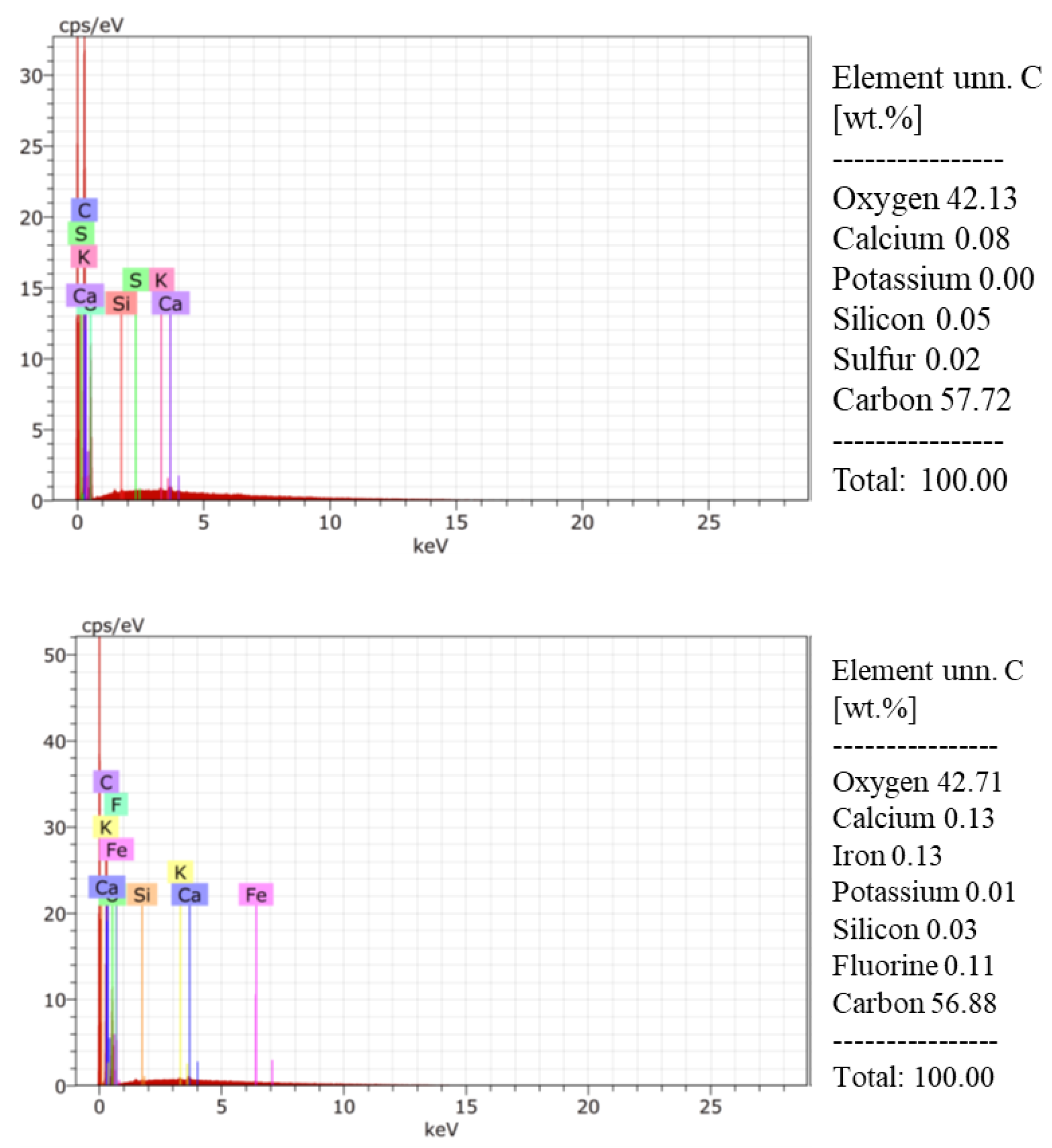
2.4. Environmental Scanning Electron Microscopy (ESEM-EDS)

For elemental analysis of CP, the samples were placed on aluminum stubs with double-sided carbon conductive tape and were observed directly under an environmental scanning electron microscope (Carl Zeiss, EVO LS10, Weimar, Germany) with an acceleration voltage of 30 kV and a pressure of 90 Pa. A backscattered electron detector (NTS BSD) was used, and images were obtained in grayscale and stored in TIFF format with a resolution of 1044 × 756 pixels.

2.5. Nanoparticles and Nanostructured Coating Elaboration

The nanoparticles were prepared using the nanoprecipitation method, which consisted of using a solvent phase composed of 96% ethanol and 0.1 mL of Tween 20 for every 100 mL of ethanol. In addition, a second phase composed of a 0.05% chitosan solution (América Alimentos, Guadalajara, Mexico); 6.25 mL per 100 mL of ethanol, pH 5.6) was used. The chitosan had an intrinsic viscosity of 440.03 ± 6.92 mL∙g−1, a molecular weight of 89,305.66 ± 1850.49 g∙Mol−1, and a degree of deacetylation of 89.44 ± 0.31%. Nanoparticle formation and concentration were determined according to the method proposed by Istúriz-Zapata et al. [27] and were previously characterized by Istúriz-Zapata et al. [28]. To prepare the chitosan coating with the nanoparticle solution, it was mixed in a 9:1 ratio with a 1% chitosan solution (pH 5.6) and a homogenizer (Virtis, Garndier, Warminster, PA, USA) at 20,000 rpm for 20 min.

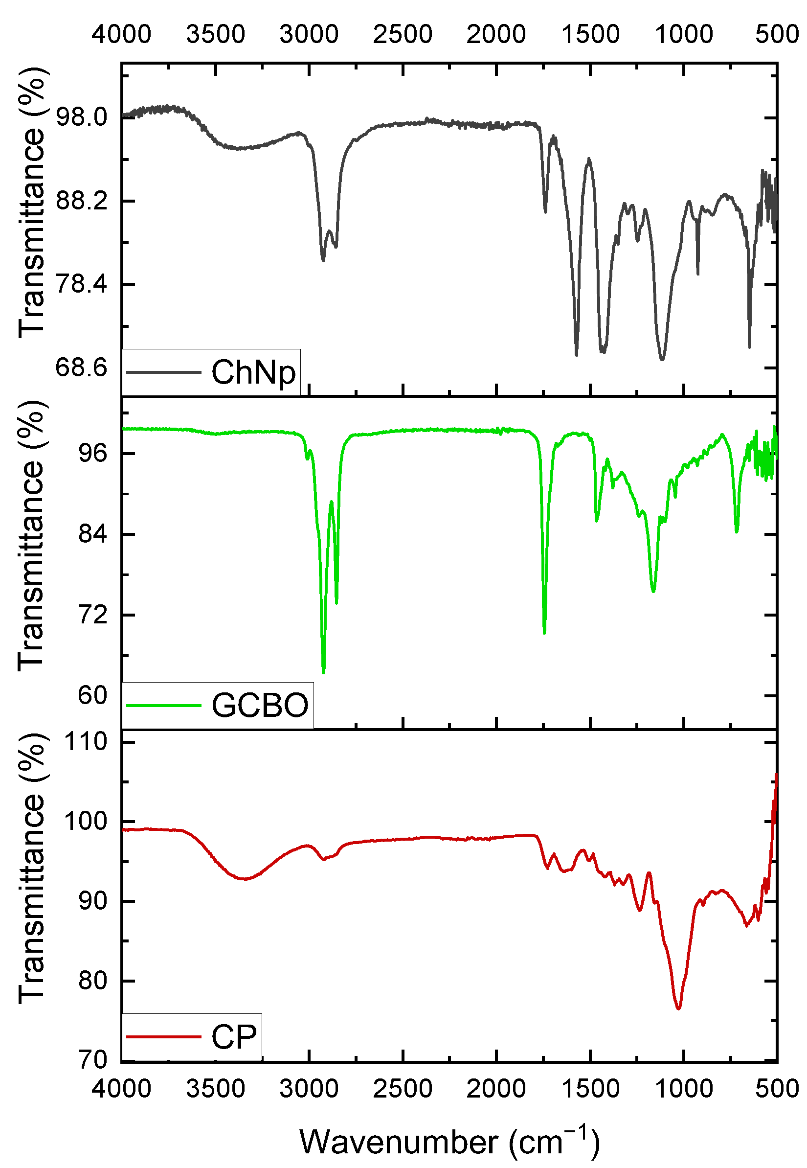
2.6. Funtional Groups of CP, GCBO, and ChNp by FTIR

The functional groups present in the CP, GCBO, and ChNp were obtained using a Confocal Micro-Raman Spectroscopy equipment coupled with Fourier Transformed Infrared (FTIR) (CRAIC Technologies, Whitehall, PA, USA).

2.7. Incorporation of Coffee Waste and NC in a Polymeric Matrix

To produce PM, mixtures of PLA and PBAT polymers were pelletized in a 60/40 ratio according to Correa-Pacheco et al. [29], who mentioned that the mixture of these polymers improved the physical characteristics, such as greater flexibility and an increase in the elongation capacity at break. In addition, 5% CP was used as it was the optimal loading point for better physical characteristics. To obtain the PM, an extrusion process was carried out using a twin-screw extruder (Process 11, Thermo Scientific TM, Waltham, MA, USA), which had eight heating zones, a die, and four feed ports. The following temperatures were used: 160, 180, 180, 180, 180, 170, 170 and 150, and the die at 160 °C. The PLA/PBAT 60/40 pellets with 5% CP were previously dried in a vacuum oven at 60 °C for 24 h to remove moisture. These were placed in the first feed port while the GCBO was incorporated into the second feed port using a peristaltic pump (MasterFlex C/L, Cole-Parmer, Vernon Hills, IL, USA). At an injection speed of 0.2 rpm, the purpose of incorporating GCBO was to take advantage of its antimicrobial and plasticizing properties that provide greater flexibility, lower rigidity, and crystallization, improving the mechanical properties of the polymer [30]. Two variables were used in the cooling bath: one was to use water and the second was to use NC to adhere to the PM.

2.8. Fungal Genera

Botrytis sp. and Rhizopus sp. were obtained from the collection of the CEPROBI-IPN Postharvest Technologies Laboratory, identified by their morphological characteristics and reactivated in potato dextrose agar culture medium. The Petri dishes were incubated at 12 °C ± 2 for 8 days for Botrytis sp. and 28 °C ± 2 for 43 h for Rhizopus sp.

2.9. Mycelial Growth Inhibition

The method reported by Velázquez Silva et al. [31] was used for the evaluation. The treatments consisted of control potato dextrose agar (PDA), CP (1, 5, and 10%), GCBO (1, 3, and 6%), NC (1, 10, and 20%), Ch (1, 10, and 20%), and ChNp (1, 10, and 20%). Then, 6 mL of each mixture was added to Petri dishes of 9 cm diameter. Once the culture medium solidified, 10 μL of a spore suspension (1 × 10⁻⁵) of Botrytis sp. and Rhizopus sp. was placed in the center of the dish and incubated under the conditions mentioned above. The radial growth of the fungus was measured using a Vernier caliper (Cienceware, CA, USA) until the control treatment covered 100% of the Petri dish. The results were reported in millimeters (mm). Six repetitions were carried out per treatment (n = 6) and, at the end of the test, the percentage of mycelial inhibition was calculated using the following equation:
%MI = [(A − B)/A] × 100,
where A is the mycelial growth of the control group and B is the mycelial growth of the pathogen in the applied treatments.
The growth rate was calculated using the equation:
Gr = [(DF−DI)/(FT−IT)],
where Gr is the growth rate, DF is the diameter of the final growth, DI is the diameter of the initial growth, FT is the final time of mycelial growth in days or hours, and IT is the start time.
For percentage data, a Bliss angular transformation was used using the following equation:
y* = arcsen√(y/100)

2.10. Spore Germination Inhibition

The mycelium was scraped with sterile water and filtered to obtain a spore suspension of each treatment. Three discs of 1 cm diameter PDA medium were placed on a sterile slide; the discs were inoculated with 20 µL of the spore suspension. Spore germination was stopped by applying 80% lactic acid at 4, 6, and 8 h, and the number of germinated spores was counted. The percentage of spore germination inhibition was calculated using the equation:
SGI (%) = [(SG Control−SG Treatment)/SG Control] × 100
where SGI is the spore germination inhibition in percentage and SG are the germinated spores.

2.11. In Vivo Evaluation

Blueberry fruit (variety Sweetest batch) collected from Zapopan, Jalisco, Mexico, was used. The fruit was disinfected with a 400 ppm sodium hypochlorite solution for 3 min and rinsed with sterile distilled water. The 1 × 10⁻⁵ spore suspension was prepared for the fungi Botrytis sp. and Rhizopus sp., and 10 µL of the suspension was placed on each fruit, previously punctured with a sterile dissection needle. The fruit was immersed for 1 min in different treatments for evaluation. For ground pure CP, a 1:1 portion of CP and sterile distilled water were used and compared with a commercial fungicide (CAPTAN 500 MezFer) at 3%. Fifteen fruits were used for each treatment, and each treatment was placed in a PET clamshell and stored at room temperature (28 ± 2 °C) under relative humidity conditions (95 ± 2%). Evaluations were made every day, and severity and incidence were determined using the equation:
%Incidence = (Number of infected fruits/Total number of fruits) × 100
Severity was determined by the percentage of fruit damage.

2.12. In Vitro Assay for the PM

The method described by Correa-Pacheco et al. [32] was used with modifications. A 5 mm circle of the PM was placed in the center of the Petri dish with PDA medium, and 10 μL of the 1 × 10⁻⁵ spore suspension of Botrytis sp. and Rhizopus sp. was added. The radial growth of the fungus was measured using a Vernier caliper (Cienceware, CA, USA) until it covered 100% of the Petri dish in the control treatment. Six repetitions were carried out per treatment (n = 6) and, at the end of the incubation time, mycelial growth was reported. The treatments evaluated in this trial were PLA/PBAT (PP), PLA/PBAT with chitosan coating (PP+NC), PLA/PBAT with CP (PPCP), PLA/PBAT with CP and chitosan coating (PPCP+NC), and Polyethylene Terephthalate (PET).

2.13. Statistical Analysis

In vitro results were analyzed by a one-way analysis of variance (ANOVA) with Tukey’s comparison of means (p < 0.05). InfoStat software version 2020 was used.

3. Results

3.1. Characterization of Green Coffee Bean Oil by GC-MS

Six compounds were identified in green coffee bean oil. Figure 2 shows the detection peaks and Table 1 shows the following composition: 10% caffeine alkaloid, 26.5% fatty acids, 30% methylated aromatic compounds, and 20.5% hydroxy-steroid. These compounds have antioxidant and antimicrobial properties that can be widely used in the food industry.

3.2. High-Performance Liquid Chromatography (HPLC) Analysis

A total of 21 major compounds were identified in CP. Most of the identified compounds were phenolic compounds, terpenes, and lignans (Figure 3, Table 2), such as the anthocyanins delphinidin 3-O-galactoside and delphinidin 3-O-arabinoside, the flavonols quercetin 3-O-acetyl-rhamnoside and kaempferol, a biflavonid theaflavin, diterpenoid andrographolide, isoflavone biochanin A, secoiridoid glycoside ligstroside, stilbenoid resveratrol 3-O-glucoside, phenols and alcohol p-HPEA-EDA, lignans secoisolariciresinol-sesquilignan and cyclolariciresinol, disaccharide derivative isorhamnetin 3-O-rutinoside, trihydroxyflavone luteolin 7-O-glucuronide, glycosidal flavonoid dihydromyricetin 3-O-rhamnoside, and sesquiterpene lactone artemisinin (database consulted: PubChem).

3.3. Elemental Analysis of Coffee Parchment

The morphology of the ground and sifted CP is observed in microscopies, with heterogeneous sizes and shapes, obtaining elongated shapes larger than 20 µm (Figure 4).
In the elemental analysis by EDS, oxygen and carbon were observed as major elements within CP (42 and 57%, respectively), and 1% corresponds to minor elements such as calcium (0.08%), silicon (0.05%), sulfur (0.02%), calcium (0.13%), iron (0.13%), potassium (0.01%), and fluorine (0.11%). This study confirms that the CP sample did not contain organic pollutants within its composition (Figure 5).

3.4. Identification of Funtional Groups of CP, GCBO, and ChNp by FTIR

The FTIR spectra shows the functional groups of the CP, GCBO, and ChNp in Figure 6. In the CP, it shows the hydroxyl group (O-H) between 3400–3500 cm−1, that means the stretch of absorbed cellulose bond, between 2800–3000 cm−1 it shows the stretch of the C-H bons, present in hemicellulose, and between 1000–1200 cm−1 it shows the absorption of C-O-C bonds, part of the vibrations of cellulose pyranose ring vibration [33]. In the GCBO, the peaks between 3000 cm−1 represent the symmetric vibration of C-H stretching in aromatic rings, and the peaks between 2800–2900 cm−1 show the methylene group (-CH2) and the methyl groups (-CH3); these groups are presents in lipids [34]. The peaks between 1500–1400 cm-1 represent the chlorogenic acid [25]. ChNp is associated with the hydroxyl group (-OH) between 3400–3600 cm−1 and the carbonyl group of the amino group (-NH2) located between 1600–1500 cm−1 [35].

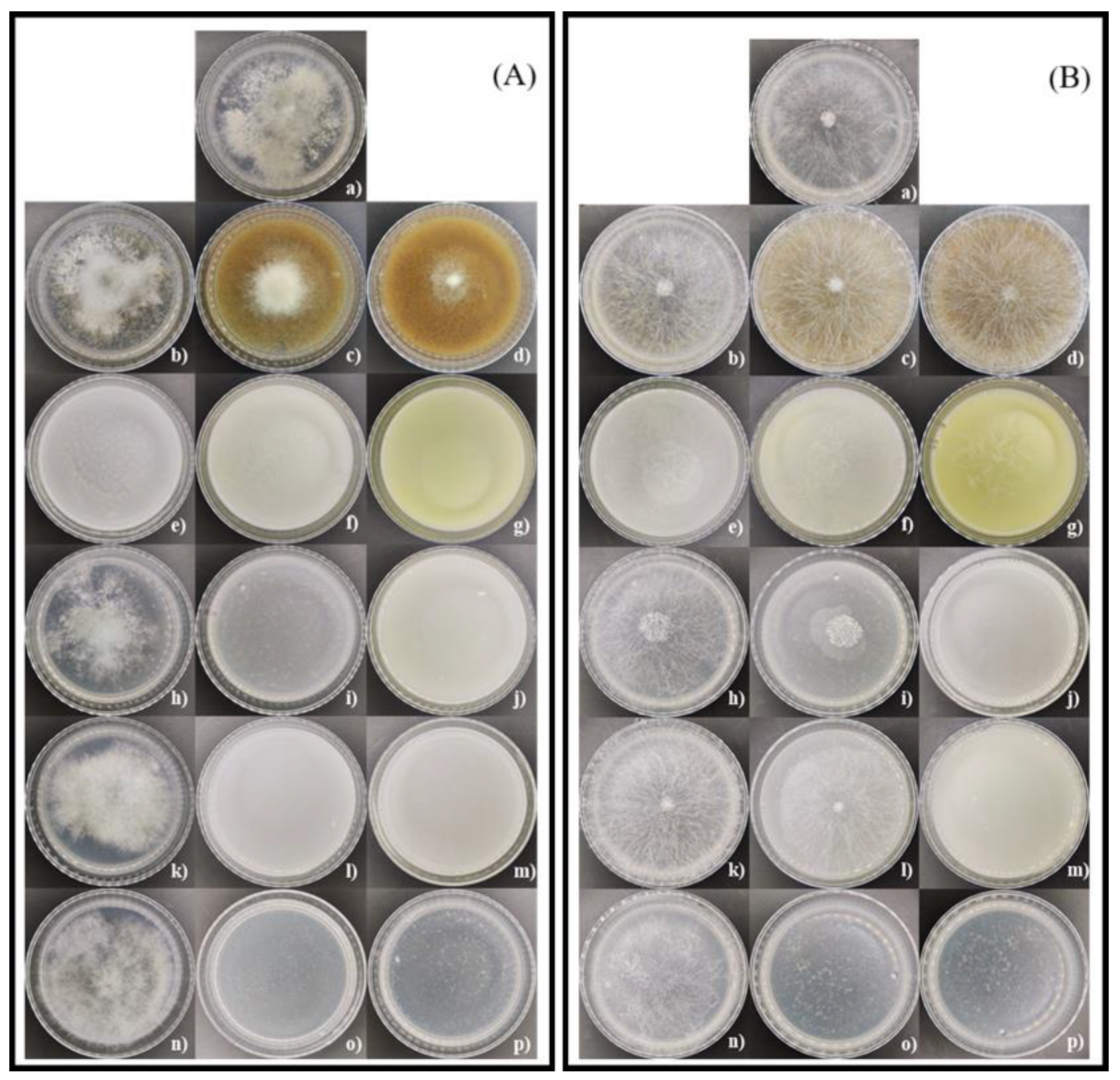
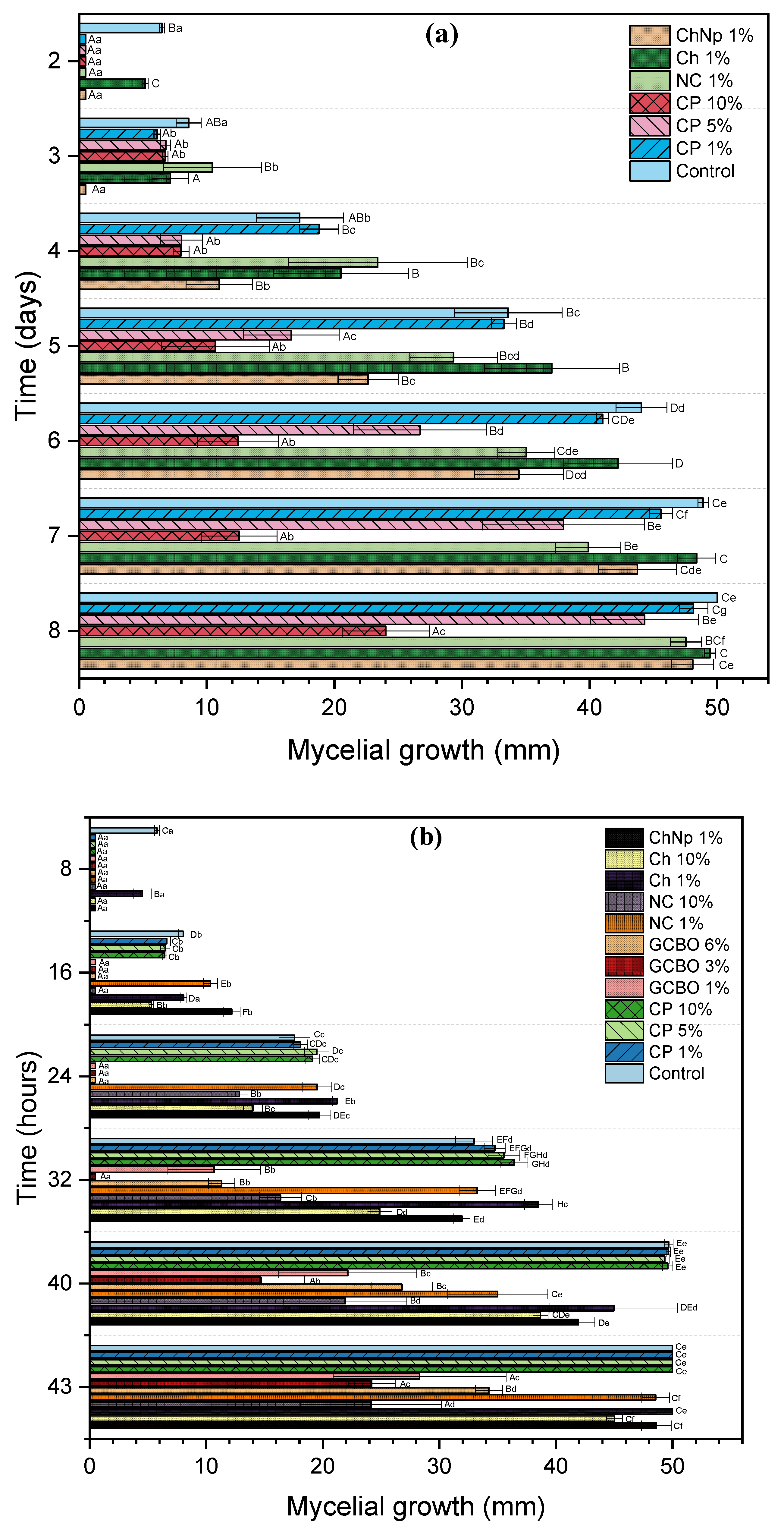
3.5. In Vitro Effect of Coffee Residues, Chitosan, and Chitosan Nanoparticles on Mycelial Growth and Spore Germination Inhibition of Botrytis sp. and Rhizopus sp.

In the case of Botrytis sp., a mycelial growth rate (MGR) of 7.25 mm/day was observed in the control (PDA), similar to the treatments of CP 1% and CP 5% with an MGR of 7.19 and 6.55 mm/day, respectively. These treatments with CP presented a stimulatory effect on mycelial growth; with the exception of CP at 10%, the inhibitory effect was (51.98%). In contrast to GCBO in the three concentrations, which presented 100% inhibition. Additionally, the treatments with NC, Ch, and ChNp at concentrations of 10 and 20% showed a greater inhibitory effect (100%), as shown in Table 3 and Figure 7A. Most treatments inhibited the formation spores, with the exception of the 1% CP, 1% NC, 1% Ch, and 1% ChNp treatments, which presented higher percentages of SGI compared to the control; only the CP 5% treatment presented a slightly positive percentage of germination inhibition of Botrytis spores compared to the control.
In Rhizopus sp., an MGR of 1.26 mm/h was observed in the control (PDA), while in the CP at the different concentrations, a higher rate was observed due to the stimulation of mycelial growth. In the case of GCBO, a decrease in the MGR was observed (0.66 for 1%, 0.54 for 3%, and 0.83 for 6%) and MGI of 43.38 to 31.5%. For NC and Ch at the highest dose of 20%, the inhibition mycelial growth was 100%; however, the 1% dose presented a stimulation with an MGR of 1.28 mm/hour. In ChNp, the lowest dose of 1% presented an MGR of 1.24 mm/hour, but the doses of 10 and 20% did not show mycelial growth. These data can be compared with the inhibition percentages shown in Table 4 and Figure 7B. The treatments that presented a 100% inhibition of the mycelial growth of Rhizopus sp. were 20% NC, 20% Ch, and 10 and 20% ChNp, while treatments with CP did not inhibit mycelial growth. Coffee oil and chitosan nanoparticles at all concentrations inhibited spore formation. The percent inhibition of spore germination presented values from 1.92% to 17.86%, and presented significant differences (p < 0.0001) between the treatments of CP, NC, and Ch. In the germination inhibition of Rhizopus spores, the CP treatments presented the highest percentages of SGI, together with 20% Ch; however, all treatments with GCBO and ChNp presented no spore formation. High concentrations prevented mycelium development, but spores were not formed at low concentrations despite the presence of mycelium.

3.6. In Vitro Assay of Coffee Residues and Incubation Times on Mycelial Growth and Spore Germination Inhibition of Botrytis sp. and Rhizopus sp.
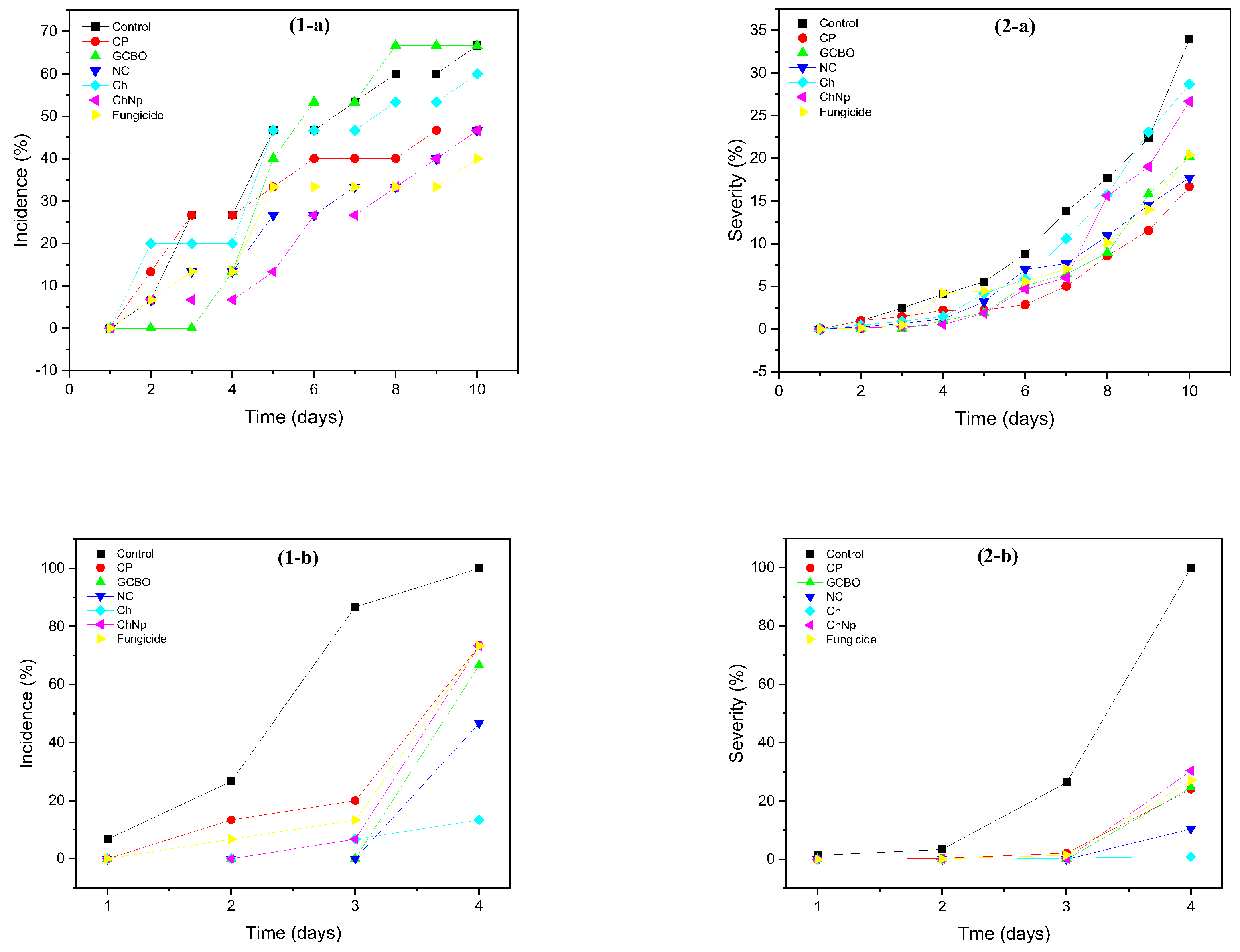
There were no differences in mycelial growth of Botrytis sp. between incubation times or between treatments at different concentrations. The 10% CP treatment showed an initial growth of 5 mm to 24 mm at 8 days compared to the 50 mm control (Figure 8a). In Rhizopus sp., the 1% GCBO, 3% GCBO, 6% GCBO, 10% NC, and 10% CP treatments showed the lowest mycelial growth with significant differences compared to the control (p < 0.001). (Figure 8b).
In the inhibition of spore germination of Botrytis sp. and Rhizopus sp. with respect to time, no significant differences were observed (p < 0.001) between the treatments, except for the treatment with 10% NC at 4 and 6 h on Rhizopus sp., as shown in Figure 9.

3.7. In Vivo Effect

Variations in incidence and severity were observed in blueberry fruits. The in vivo test for Botrytis sp. showed the lowest incidence after 10 days of evaluation (40%) with the commercial fungicide; it was more effective than the other treatments and the control (66.66%). The CP and NC treatments presented an incidence of 46.66%. In terms of severity, the control treatment showed the highest values with 34%, while the NC treatments (17.73%) and CP (16.66%) were more effective in reducing fruit damage (Figure 10 and Figure 12(1-a,2-a)).
For Rhizopus sp., the in vivo test was 4 days (Figure 11 and Figure 12(1-b,2-b). The incidence in the control was the most susceptible with 100%, while the lowest incidence was observed in Ch treatment (13.33%). The severity in the control group reached 100%, while the Ch treatment was the most effective with a severity of 0.86%. All treatments had the lowest incidence and severity on blueberry.

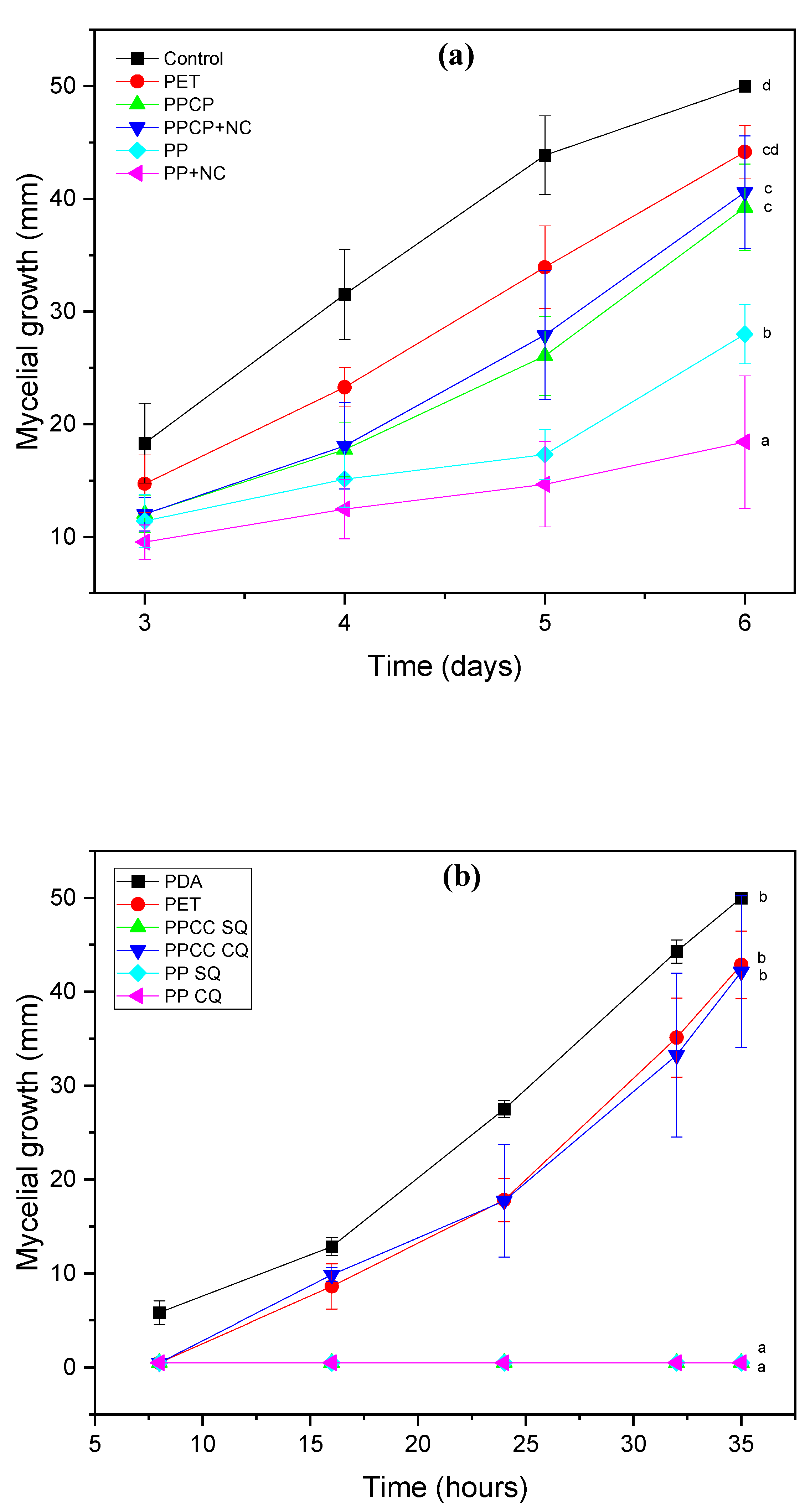
3.8. In Vitro Assay of Coffee Residues Incorporated into the PM

The results are shown in Table 5. A fungistatic effect was obtained in all treatments, with significant differences (p < 0.0001). The PP+NC treatment was the most effective with a mycelial growth inhibition of 63.04%, while the least effective was PET with an inhibition of 11.68% on Botrytis sp. compared to the control. In Rhizopus sp., a fungistatic effect was also obtained. Compared to the control, mycelial growth was inhibited by 14.28% with PET treatments and 15.78% with PPCP treatments. The PPCP+NC, PP, and PP+NC treatments showed 100% inhibition mycelial growth during the time evaluated. However, they presented a fungistatic effect; the fungus continued its development after replanting in PDA medium.
For mycelial growth with respect to time, significant statistical differences were obtained (p < 0.0001), as shown in Figure 13. Lineal mycelial growth was observed in all treatments against Botrytis sp. The lowest mycelial growth occurred with PP and PP+NC treatments (Figure 8a). For Rhizopus sp., significant differences in mycelial growth were observed with respect to time. The control treatment showed mycelial growth at 10 h, and the PET treatment showed growth at 15 h. The PPCP+NC treatment showed growth at 25 h. No mycelial growth was observed in PPCP, PP, and PP+NC treatments at the evaluated times (Figure 8b).

4. Discussion

Regarding the content of volatiles present in green coffee bean oil, caffeine is a typical compound of the coffee plant characterized by having a high antioxidant activity [36]; however, caffeine is also characterized by having a broad antimicrobial activity [37]. It is present in various parts of the coffee plant. Nonthakaew et al. [38] evaluated the antimicrobial activity of caffeine extracts from coffee beans on the mycelial development of Rhizopus oryzae at concentrations of 2.5 to 10 g 100 mL−1. This occurs because caffeine can inhibit fungi by stopping the synthesis of glucose, fructose, and maltose, preventing fungal spread. Other compounds such as hexadecanoic acid and octadecinoic acid with antimicrobial activity have been identified in coffee [39]. Phenolic compounds are abundant in coffee by-products, since they are characterized by their antimicrobial activity [40]. These are abundant in the CP and have the function of covering and protecting the bean from attacks by other microorganisms.
Kaempferol has been reported as a bioactive compound characterized by its high antioxidant activity and is present in various parts of the coffee plant [41]. We found kaempferol 3-O-rhamnoside present at retention time 10.7, kaempferol at retention time 15.4, and kaempferol 3-O-(6″-acetyl-galactoside) 7-O-rhamnoside at retention time 16.3, due to its wide distribution in this residue, acting as defense substances in coffee with antioxidant activity. Periferakis et al. [42], found that kaempferol was a flavonoid widely distributed in several plant species that acts as an antimicrobial agent. The antifungal activity of kaempferol can be enhanced if it is encapsulated in chitosan nanoparticles, as mentioned by IlK et al. [43], who evaluated the antifungal effect of kaempferol and kaempferol loaded lecithin/chitosan nanoparticles on Fusarium oxysporium. Using kaempferol alone resulted in 100% growth on day 20 of evaluation, while using encapsulated kaempferol resulted in a growth greater than 60% on day 60. Quercetin-3-O-galactoside and isorhamnetin inhibit bacterial division and growth during the logarithmic growth period and act as a bacterial inhibitor. Inhibition may be mediated by the disruption of the bacterial cell structure, leading to leakage of contents including sugars, nucleic acids, and proteins [44]. The antibacterial activity of theaflavin showed that epicatechin combinations against Candida albicans were stronger than that of theaflavin alone. Minimum inhibitory concentrations (MICs) of 1024 μg/mL with theaflavin and 128–256 μg/mL with theaflavin-epicatechin combinations were observed by Betts et al. [45]. The compound andrographolide has anti-inflammatory and antioxidant effects; however, it also has properties for the control of infections caused by microorganisms, due to the capacity of intracellular DNA inhibition [46,47]. In grape pomace, 2-S-Glutathionyl caftaric acid has been isolated and antimicrobial activity on three bacterial strains was reported [48]. Ficus eriobotryoides leaves were reported with antifungal activity on six isolates of Fusarium oxysporium and contained myricetin [49]. Although the CP has a wide content of compounds, these mostly act as antioxidants, which is why CP has the lowest antifungal effect compared to other treatments. Mirón-Mérida et al. [10] evaluated ethanolic extracts of CP on the mycelial growth of Colletotrichum gloesporioides and Fusarium verticillioides and found greater inhibition as the extract dose is increased; this is due to the presence of bioactive compounds, most of which are phenolic compounds. In our research the most sensitive fungus to CP was Botrytis sp. at a dose of 10% which can be attributed to the presence of the major phenolic components identified by the HPLC-MS analysis.
In the elemental analysis of CP, values of 57.72 and 56.88% of carbon were obtained, coinciding with results from Coura et al. [50], who reported a carbon content of 60.5 and 62.3% in CP because it is a lignocellulosic material rich in carbon and hydrogen. It has the capacity to release energy due to its high combustion enthalpy and this means that CP could absorb organic compounds on its surface [51].
It was reported by Cai-Ling et al. [52] that the hydroxyl group (O-H) present in the chitosan causes extracellular effects and intercellular effects, and penetrates the cell wall and the cell membrane. The presence of chlorogenic acids in the GCBO can cause early membrane permeabilization of the spores [53].
Several bioactive antifungal compounds have been identified in CP. In the in vitro tests, each treatment presented differing effects due to the differences in its bioactive constituents, but green coffee bean oil showed total inhibition in formation of spores in both fungi due to its volatile phase, which prevents its evaporation when in a closed medium such as the Petri dish. The lipophilic phase of the oil is more easily absorbed by the mycelium [54].
Chitosan has physicochemical properties that provide it with an antifungal effect. It can disintegrate the cell wall membrane of fungi, cause cytoplasm leakage, chelation of essential nutrients, and the binding of nucleic acids that alter the flow of genetic information [55]. By synthesizing chitosan as a nanoparticle, significant inhibitory effects are obtained, with changes occurring in colony formation and spore germination, and nanoparticles induce morphological changes by having a more effective membrane penetration [56]. El-Naggar et al. [57] compared the effect of chitosan and chitosan nanoparticles in inhibiting Botrytis cinerea mycelium, and showed greater inhibition when using nanoparticles. At a concentration of 1 mg/mL with chitosan, they obtained an inhibition of 31.16%, while they obtained an inhibition of 73.58% when using chitosan nanoparticles at the same concentration. This may be explained by the alteration in membrane permeability due to morphological changes that inhibit mycelial growth, sporulation, and spore germination.
In the in vivo test, blueberry fruits contain a high content of phenolic compounds and anthocyanins, which function to preserve the fruits. One way to avoid the loss of these compounds is the application of coatings, such as essential oils or nanoemulsions, that help preserve these compounds and protect the fruits against fungal attacks [58]. The CP and NC treatments presented minor incidence and severity on Botrytis sp., as did the Ch treatments on Rhizopus sp. The CP contains a high content of phenolic compounds, and NC or Ch have solution chitosan and chitosan nanoparticles, which may be responsible for this biological activity in vivo. Sun et al. [59] evaluated a coating of chitosan and carvacrol essential oil and found that the inhibitory effect against Escherichia coli and Penicillium digitatum was greater when using the treatments together than separately. Therefore, mixing a bioactive oil with chitosan can enhance its antimicrobial properties.
To evaluate the PM, the addition of active compounds in polymeric materials can serve as a controlled release vehicle against microorganisms, as reported by Black-Solis et al. [60], who evaluated the addition of cinnamon essential oil and chitosan solution to PLA and PBAT polymeric fibers. They were tested against Alternaria alternata and inhibited its mycelial growth and spore germination at doses of 6.1% cinnamon essential oil.

5. Conclusions

The in vitro effect of coffee by-products occurred mainly on the mycelial growth of Botrytis sp., with 100% inhibition in the presence of GCBO, NC, Ch, and NpCh, while in Rhizopus sp. 20% NC, Ch, and NpCh obtained 100% inhibition. For the inhibition of spore germination, GCBO was the best treatment, since it prevented spore growth at all doses. The incidence and severity in the fruits was 16 and 17% in blueberries treated with CP and NC stored at room temperature. In the in vitro evaluation, the PM showed a significant fungistatic effect on both fungi. In this work, the inhibitory effect on Botrytis sp. and Rhizopus sp. in vitro and in vivo was enhanced by incorporating coffee waste into a polymeric matrix, which could be used to produce biodegradable packaging to extend the shelf life of fruit and vegetables. The PM can be an alternative to producing active packaging since they are made with different natural components CP, NC, and GCBO, whose purpose is to release the compounds within the food packaging to avoid the presence of diseases.
The advantages of this research are that, due to the extensive production of coffee worldwide, there is a high bioavailability of these residues, which contain many compounds with antimicrobial activity. It is important for the use of different extraction methods in the future to know the potential of these residues.

Author Contributions

Research, G.H.-L.; methodology, G.H.-L. and L.L.B.-N.; writing, G.H.-L. and L.L.B.-N.; conceptualization, L.L.B.-N.; writing-review, L.L.B.-N.; editing, L.L.B.-N.; supervision, L.L.B.-N. All authors have read and agreed to the published version of the manuscript.

Funding

This research received no external funding.

Data Availability Statement

The original contributions presented in this study are included in the article. Further inquiries can be directed to the corresponding author.

Acknowledgments

To the National Council of Humanities, Science and Technologies (CONAHCYT) and the National Polytechnic Institute (IPN. To the Center for Nanosciences and Micronanotechnology, of the National Polytechnic Institute for the HPLC analyses and to the Advanced Microscopy Laboratory of the Center for the Development of Biotic Products for Environmental Scanning Electron Microscopy (ESEM-EDS) where they were performed.

Conflicts of Interest

The authors declare no conflicts of interest.

Abbreviations

The following abbreviations are used in this manuscript:
PLAPolylactic acid
PBATPoly(butylene adipate-co-terephthalate)
CPCoffee parchment
GCBOGreen coffee bean oil
ChChitosan solution
ChNpChitosan nanoparticles
PMPolymer matrix
PM+CWPolymer matrix with coffee waste
TLAThree letter acronym
NCNanostructured coating
PPCPPolylactic acid/Poly(butylene adipate-co-terephthalate)+ Coffee parchment
PP+NCPolylactic acid/Poly(butylene adipate-co-terephthalate)+ Nanostructured coating
PPCP+NCPolylactic acid/Poly(butylene adipate-co-terephthalate)+ Coffee parchment+ Nanostructured coating
GC-MSGas chromatography and mass spectrometry
HPLC-MSLiquid chromatography and mass spectrometry
ESEM-EDSEnvironmental scanning electron microscopy
MIMycelial inhibition
GrGrowth rate
DFDiameter of the final growth
DIDiameter of the initial growth
SGISpore germination inhibition
MGRMycelial growth rate
NGNo growth
NSFNo spore germination

References

  1. Banerjee, S.; Nayik, G.A.; Kour, J.; Nazir, N. Blueberries. In Antioxidants in Fruits: Properties and Health Benefits; Nayik, G.A., Gull, A., Eds.; Springer: Singapore, 2020; pp. 593–614. [Google Scholar]
  2. Bell, S.R.; Montiel, L.G.; Estrada, R.R.; Martínez, P. Main diseases in postharvest blueberries, conventional and eco-friendly control methods: A review. LWT 2021, 149, 112046. [Google Scholar] [CrossRef]
  3. Scherm, H.; Savelle, A.T.; Brannen, P.M.; Krewer, G. Occurrence and prevalence of foliar diseases on blueberry in Georgia. Plant Health Prog. 2008, 9, 18. [Google Scholar] [CrossRef]
  4. Garfinkel, A.R. The history of Botrytis taxonomy, the rise of phylogenetics, and implications for species recognition. Phytopathology 2021, 111, 437–454. [Google Scholar] [CrossRef]
  5. Agrios, G.N. Plant Pathology, 5th ed.; Elsevier Academic Press: San Diego, CA, USA, 2012; p. 948. [Google Scholar]
  6. O’Brien, P.A. Biological control of plant diseases. Australas. Plant Pathol. 2017, 46, 293–304. [Google Scholar] [CrossRef]
  7. Gwinn, K.D. Bioactive Natural Products in Plant Disease Control. In Studies in Natural Products Chemistry; Elsevier B.V.: Amsterdam, The Netherlands, 2018; Volume 56, pp. 229–246. [Google Scholar]
  8. Klingel, T.; Kremer, J.I.; Gottstein, V.; Rajcic de Rezende, T.; Schwarz, S.; Lachenmeier, D.W. A Review of Coffee By-Products Including Leaf, Flower, Cherry, Husk, Silver Skin, and Spent Grounds as Novel Foods within the European Union. Foods 2020, 9, 665. [Google Scholar] [CrossRef]
  9. Dong, W.; Chen, Q.; Wei, C.; Hu, R.; Long, Y.; Zong, Y.; Chu, Z. Comparison of the effect of extraction methods on the quality of green coffee oil from Arabica coffee beans: Lipid yield, fatty acid composition, bioactive components, and antioxidant activity. Ultrason. Sonochemistry 2021, 74, 105578. [Google Scholar] [CrossRef]
  10. Mirón-Mérida, V.A.; Yáñez-Fernández, J.; Montañez-Barragán, B.; Barragán Huerta, B.E. Valorization of coffee parchment waste (Coffea arabica) as a source of caffeine and phenolic compounds in antifungal gellan gum films. LWT 2019, 101, 167–174. [Google Scholar] [CrossRef]
  11. Hashemi, B.; Shiri, F.; Švec, F.; Nováková, L. Green solvents and approaches recently applied for extraction of natural bioactive compounds. TrAC 2022, 157, 116732. [Google Scholar] [CrossRef]
  12. Gloria, M.B.A.; Almeida, A.A.P.; Engeseth, N. Antimicrobial activity of coffee. In Coffee: Consumption and Health Implicarions; Farah, A., Ed.; The Royal Society of Chemistry: London, UK, 2019; pp. 234–254. [Google Scholar]
  13. Chaves-Ulate, E.; Esquivel-Rodríguez, P. Ácidos clorogénicos presentes en el café: Capacidad antimicrobiana y antioxidante. Agron. Mesoam. 2019, 30, 299–311. [Google Scholar] [CrossRef]
  14. Verlee, A.; Mincke, S.; Stevens, C.V. Recent developments in antibacterial and antifungal chitosan and its derivatives. Carbohydr. Polym. 2017, 164, 268–283. [Google Scholar] [CrossRef]
  15. Ding, L.; Huang, Y.; Cai, X.; Wang, S. Impact of pH, ionic strength and chitosan charge density on chitosan/casein complexation and phase behavior. Carbohydr. Polym. 2019, 208, 133–141. [Google Scholar] [CrossRef]
  16. Saberi Riseh, R.; Vatankhah, M.; Hassanisaadi, M.; Shafiei-Hematabad, Z.; Kennedy, J.F. Advancements in coating technologies: Unveiling the potential of chitosan for the preservation of fruits and vegetables. Int. J. Biol. Marcromolecules 2024, 254, 127677. [Google Scholar] [CrossRef] [PubMed]
  17. Sotelo-Boyás, M.E.; Correa-Pacheco, Z.N.; Bautista-Baños, S.; Corona-Rangel, M.L. Physicochemical characterization of chitosan nanoparticles and nanocapsules incorporated with lime essential oil and their antibacterial activity against food-borne pathogens. LWT 2017, 77, 15–20. [Google Scholar] [CrossRef]
  18. Moore, C.J. Synthetic polymers in the marine environment: A rapidly increasing, long-term theat. Environ. Res. 2008, 108, 131–139. [Google Scholar] [CrossRef] [PubMed]
  19. Rives-Castillo, S.C.H.; Bautista-Baños, S.; Correa-Pacheco, Z.N.; Ventura-Aguilar, R.I. Situación actual de los envases utilizados para la conservación postcosecha de productos hortofrutícolas. Rev. Iberoam. Tecnol. Postcosecha 2020, 21, 17–34. [Google Scholar]
  20. Pang, X.; Zhuang, X.; Tang, Z.; Chen, X. Polylactic acid (PLA): Research, development and industrialization. Biotechnol. J. 2010, 5, 1125–1136. [Google Scholar] [CrossRef]
  21. Ferreira, F.V.; Cividanes, L.S.; Gouveia, R.F.; Lona, L.M. An overview on properties and applications of poly (butylene adipate-co-terephthalate)—PBAT based composites. Polym. Eng. Sci. 2017, 59 (Suppl. S2), E7–E15. [Google Scholar] [CrossRef]
  22. Castellón-Castro, C.A.; Tejeda-López, L.N.; Tejeda-Benítez, L.P. Evaluación de la degradación ambiental de bolsas plásticas biodegradables. Inf. Técnico 2016, 80, 24–31. [Google Scholar] [CrossRef]
  23. Soquetta, M.B.; Terra, L.D.M.; Bastos, C.P. Green technologies for the extraction of bioactive compounds in fruits and vegetables. CyTA-J. Food 2018, 16, 400–412. [Google Scholar] [CrossRef]
  24. Sultana, T.; Dey, S.C.; Molla, M.A.I.; Hossain, M.R.; Rahman, M.M.; Quddus, M.S.; Moniruzzaman, M.; Rahman, M.M. Facile synthesis of TiO2/Chitosan nanohybrid for adsorption-assisted rapid photodegradation of an azo dye in water. React. Kinet. Mech. Catal. 2021, 133, 1121–1139. [Google Scholar] [CrossRef]
  25. Andrea, M.J.; Mariana, P.G.; Mónica, H.L.; Nacary, C.P.Z.; Silvia, B.B.; Laura, B.N. Nanostructured Chitosan Coating with a Coffee Residue Extract for the Preservation of Tomato and Controlling Pre- and Postharvest Disease Caused by Rhizopus stolonifer. Processes 2025, 13, 220. [Google Scholar] [CrossRef]
  26. González-Quijano, G.; Arrieta, B.D.; Dorantes, A.L.; Aparicio, O.G.; Guerrero, L.I. Effect of extraction method in the content of phytoestrogens and main phenolics in mesquite pod extracts (Prosopis sp.). Rev. Mex. De Ing. Química 2019, 18, 303–312. [Google Scholar] [CrossRef]
  27. Istúriz-Zapata, M.A.; Correa-Pacheco, Z.N.; Bautista-Baños, S.; Acosta-Rodríguez, J.L.; Hernández-López, M.; Barrera-Necha, L.L. Efficacy of extracts of mango residues loaded in chitosan nanoparticles and their nanocoatings on in vitro and in vivo postharvest fungal. J. Phytopathol. 2022, 170, 661–674. [Google Scholar] [CrossRef]
  28. Istúriz-Zapata, M.A.; Hernández-López, M.; Correa-Pacheco, Z.N.; Barrera-Necha, L.L. Quality of cold-stored cucumber as affected by nanostructured coatings of chitosan with cinnamon essential oil and cinnamaldehyde. LWT 2020, 123, 109089. [Google Scholar] [CrossRef]
  29. Correa-Pacheco, Z.N.; Black-Solís, J.D.; Ortega-Gudiño, P.; Sabino-Gutiérrez, M.A.; Benítez-Jiménez, J.J.; Barajas-Cervantes, A.; Bautista-Baños, S.; Hurtado-Colmenares, L.B. Preparation and characterization of bio-based PLA/PBAT and cinnamon essential oil polymer fibers and life-cycle assessment from hydrolytic degradation. Polymers 2019, 12, 38. [Google Scholar] [CrossRef]
  30. Hernández-López, M.; Correa-Pacheco, Z.N.; Bautista-Baños, S.; Zavaleta-Avejar, L.; Benítez-Jiménez, J.J.; Sabino-Gutiérrez, M.A.; Ortega-Gudiño, P. Bio-based composite fibers from pine essential oil and PLA/PBAT polymer blend. Morphological, physicochemical, thermal and mechanical characterization. Mater. Chem. Phys. 2019, 234, 345–353. [Google Scholar] [CrossRef]
  31. Velázquez Silva, A.; Robles Yerena, L.; Barrera Necha, L.L. Chemical profile and antifungal activity of plant extracts on Colletotrichum spp. isolated from fruits of Pimenta dioica (L.) Merr. Pestic. Biochem. Physiol. 2021, 179, 104949. [Google Scholar] [CrossRef]
  32. Correa-Pacheco, Z.N.; Ventura-Aguilar, R.I.; Zavaleta-Avejar, L.; Barrera-Necha, L.L.; Hernández-López, M.; Bautista-Baños, S. Anthracnose Disease Control and Postharvest Quality of Hass Avocado Stored in Biobased PLA/PBAT/Pine Essential Oil/Chitosan Active Packaging Nets. Plants 2022, 11, 2278. [Google Scholar] [CrossRef]
  33. Gebeyehu, B.T.; Bikila, S.L. Determination of caffeine content and antioxidant activity of coffee. Am. J. Appl. Chem. 2015, 3, 69–76. [Google Scholar] [CrossRef]
  34. Reis, R.S.; Tienne, L.G.; Souza, D.H.S.; Maria de Fátima, V.M.; Monteiro, S.N. Characterization of coffee parchment and innovative steam explosion treatment to obtain microfibrillated cellulose as potential composite reinforcement. J. Mater. Res. Technol. 2020, 9, 9412–9421. [Google Scholar] [CrossRef]
  35. Aung Moon, S.; Wongsakul, S.; Kitazawa, H.; Kittiwachana, S.; Saengrayap, R. Application of ATR-FTIR for Green Arabica Bean Shelf-Life Determination in Accelerated Storage. Foods 2024, 13, 2331. [Google Scholar] [CrossRef] [PubMed]
  36. Sathiyabama, M.; Boomija, R.V.; Muthukumar, S.; Gandhi, M.; Salma, S.; Prinsha, T.K.; Rengasamy, B. Green synthesis of chitosan nanoparticles using tea extract and its antimicrobial activity against economically important phytopathogens of rice. Sci. Rep. 2024, 14, 7381. [Google Scholar] [CrossRef] [PubMed]
  37. AlEraky, D.M.; Abuohashinsh, H.M.; Gad, M.M.; Alshuyukh, M.H.; Bugshan, A.S.; Almulhim, K.S.; Mahmoud, M.M. The antifungal and antibiofilm activities of caffeine against Candida albicans on polymethyl methacrylate denture base material. Biomedicines 2022, 10, 2078. [Google Scholar] [CrossRef]
  38. Nonthakaew, A.; Matan, N.; Aewsiri, T.; Matan, N. Caffeine in foods and its antimicrobial activity. Int. Food Res. J. 2015, 22, 9–14. [Google Scholar]
  39. Oliveira, A.L.D.; Cruz, P.M.; Eberlin, M.N.; Cabral, F.A. Brazilian roasted coffee oil obtained by mechanical expelling: Compositional analysis by GC-MS. Food Sci. Technol. 2005, 25, 677–682. [Google Scholar] [CrossRef]
  40. Benyelles, M.; Merzouk, H.; Merzouk, A.Z.; Imessaoudene, A.; Medjdoub, A.; Mebarki, A. Valorization of Encapsulated Coffee Parchment Extracts as Metabolic Control for High Fructose Diet-Induced Obesity, Using Wistar Rat as Animal Model. Waste Biomass Valorization 2024, 15, 265–281. [Google Scholar] [CrossRef]
  41. Machado, M.; Espírito Santo, L.; Machado, S.; Lobo, J.C.; Costa, A.S.; Oliveira, M.B.P.; Ferreira, H.; Alves, R.C. Bioactive potential and chemical composition of coffee by-products: From pulp to silverskin. Foods 2023, 12, 2354. [Google Scholar] [CrossRef]
  42. Periferakis, A.; Periferakis, K.; Badarau, I.A.; Petran, E.M.; Popa, D.C.; Caruntu, A.; Costache, R.S.; Scheau, C.; Caruntu, C.; Costache, D.O. Kaempferol: Antimicrobial properties, sources, clinical, and traditional applications. Int. J. Mol. Sci. 2022, 23, 15054. [Google Scholar] [CrossRef]
  43. Ilk, S.; Saglam, N.; Özgen, M. Kaempferol loaded lecithin/chitosan nanoparticles: Preparation, characterization, and their potential applications as a sustainable antifungal agent. Artif. Cells Nanomed. Biotechnol. 2016, 45, 907–916. [Google Scholar] [CrossRef]
  44. Du, J.; Fu, J.; Chen, T. Investigation of the Antibacterial Properties and Mode of Action of Compounds from Urtica dioica L. Cureus 2024, 16, e52083. [Google Scholar] [CrossRef]
  45. Betts, J.; Wareham, D.; Haswell, S.; Kelly, S. Antifungal synergy of theaflavin and epicatechin combinations against Candida albicans. J. Microbiol. Biotechnol. 2013, 23, 1322–1326. [Google Scholar] [CrossRef] [PubMed]
  46. Banerjee, M.; Parai, D.; Chattopadhyay, S.; Mukherjee, S.K. Andrographolide: Antibacterial activity against common bacteria of human health concern and possible mechanism of action. Folia Microbiol. 2017, 62, 237–244. [Google Scholar] [CrossRef] [PubMed]
  47. Mussard, E.; Cesaro, A.; Lespessailles, E.; Legrain, B.; Berteina-Raboin, S.; Toumi, H. Andrographolide, a natural antioxidant: An update. Antioxidants 2019, 8, 571. [Google Scholar] [CrossRef]
  48. Mollica, A.; Scioli, G.; Della Valle, A.; Cichelli, A.; Novellino, E.; Bauer, M.; Kamysz, W.; Llorent-Martínez, E.L.; Fernández-de Córdova, M.L.; Castillo-López, R.; et al. Phenolic analysis and in vitro biological activity of red wine, pomace and grape seeds oil derived from Vitis vinifera L. cv. Montepulciano d’Abruzzo. Antioxidants 2021, 10, 1704. [Google Scholar] [CrossRef]
  49. Salem, M.Z.; Mohamed, A.A.; Ali, H.M.; Al Farraj, D.A. Characterization of phytoconstituents from alcoholic extracts of four woody species and their potential uses for management of six Fusarium oxysporum isolates identified from some plant hosts. Plants 2021, 10, 1325. [Google Scholar] [CrossRef]
  50. Coura, M.R.; Demuner, A.J.; Demuner, I.F.; Blank, D.E.; Magalhães Firmino, M.J.; Borges Gomes, F.J.; Macedo Ladeira Carvalho, A.M.; Moreira Costa, M.; Henrique dos Santos, M. Technical kraft lignin from coffee parchment. Nord. Pulp Pap. Res. J. 2023, 38, 229–241. [Google Scholar] [CrossRef]
  51. Tomizawa, M.; Kurosu, S.; Konayashi, M.; Kawase, Y. Zero-valent iron treatment of dark brown colored coffee effluent: Contributions of a core-shell structure to pollutant removals. J. Environ. Manag. 2016, 183, 478–487. [Google Scholar] [CrossRef]
  52. Ke, C.-L.; Deng, F.-S.; Chuang, C.-Y.; Lin, C.-H. Antimicrobial Actions and Applications of Chitosan. Polymers 2021, 13, 904. [Google Scholar] [CrossRef]
  53. Calheiros, D.; Dias, M.I.; Calhelha, R.C.; Barros, L.; Ferreira, I.C.F.R.; Fernandes, C.; Gonçalves, T. Antifungal Activity of Spent Coffee Ground Extracts. Microorganisms 2023, 11, 242. [Google Scholar] [CrossRef]
  54. Soylu, E.M.; Kurt, Ş.; Soylu, S. In vitro and in vivo antifungal activities of the essential oils of various plants against tomato grey mould disease agent Botrytis cinerea. Int. J. Food Microbiol. 2010, 143, 183–189. [Google Scholar] [CrossRef]
  55. Poznanski, P.; Hameed, A.; Orczyk, W. Chitosan and chitosan nanoparticles: Parameters enhancing antifungal activity. Molecules 2023, 28, 2996. [Google Scholar] [CrossRef] [PubMed]
  56. Kheiri, A.; Moosawi Jorf, S.A.; Mallihipour, A.; Saremi, H.; Nikkhah, M. Application of chitosan and chitosan nanoparticles for the control of Fusarium head blight of wheat (Fusarium graminearum) in vitro and greenhouse. Int. J. Biol. Macromol. 2016, 93, 1261–1272. [Google Scholar] [CrossRef] [PubMed]
  57. El-Naggar, N.E.A.; Saber, W.I.; Zweil, A.M.; Bashir, S.I. An innovative green synthesis approach of chitosan nanoparticles and their inhibitory activity against phytopathogenic Botrytis cinerea on strawberry leaves. Sci. Rep. 2022, 12, 3515. [Google Scholar] [CrossRef] [PubMed]
  58. Zhang, Y.; Dai, J.; Ma, X.; Jia, C.; Han, J.; Song, C.; Liu, Y.; Wei, D.; Xu, H.; Quin, J.; et al. Nano-emulsification essential oil of Monarda didyma L. to improve its preservation effect on postharvest blueberry. Food Chem. 2023, 417, 135880. [Google Scholar] [CrossRef]
  59. Sun, X.; Narciso, J.; Wang, Z.; Ference, C.; Bai, J.; Zhou, K. Effects of chitosan-essential oil coatings on safety and quality of fresh blueberries. J. Food Sci. 2014, 79, M955–M960. [Google Scholar] [CrossRef]
  60. Black-Solis, J.; Ventura-Aguilar, R.I.; Correa-Pacheco, Z.; Corona-Rangel, M.L.; Bautista-Baños, S. Preharvest use of biodegradable polyester nets added with cinnamon essential oil and the effect on the storage life of tomatoes and the development of Alternaria alternata. Sci. Hortic. 2019, 245, 65–73. [Google Scholar] [CrossRef]
Figure 1. Synthesis scheme of research methodology.
Resources 14 00048 g001
Figure 2. Gas chromatogram and major compounds of green coffee bean oil: (1) caffeine, (2) n-hexadecanoic acid, (3) 17-octadecynoic acid, (4) 1H-indene, 2,3-dihydro-4,7-dimethyl-, (5) benzimidazole, 2-methyl-1-(3-phenylpropylthio)methyl-, and (6) norethindrone. Database consulted: PubChem.
Resources 14 00048 g002
Figure 3. HPLC chromatogram and major compounds of coffee parchment.
Resources 14 00048 g003
Figure 4. Environmental scanning electron microscopy (ESEM-EDS).
Resources 14 00048 g004
Figure 5. Spectra of the EDS elemental analysis of the coffee parchment.
Resources 14 00048 g005
Figure 6. FTIR spectra of ChNp, GCBO, and CP.
Resources 14 00048 g006
Figure 7. In vitro assay on mycelial growth. (A) Botrytis sp. (B) Rhizopus sp. (a) Control (PDA), (b) 1% CP, (c) 5% CP, (d) 10% CP, (e) 1% GCBO, (f) 3% GCBO, (g) 6% GCBO, (h) 1% NC, (i) 10% NC, (j) 20% NC, (k) 1% Ch, (l) 10% Ch, (m) 20% Ch, (n) 1% ChNp, (o) 10% ChNp, and (p) 20% ChNp.
Resources 14 00048 g007
Figure 8. In vitro mycelial growth inhibition with respect to time. Capital letters represent significant differences between treatments and lowercase letters represent differences with respect to time. (a) Botrytis sp. comparison of Tukey means (means ± SD), α = 0.05; DMS = 2.22794; gl = 194; standard error = 9.1862; p < 0.0001. (b) Rhizopus sp. with Tukey’s comparison of means (means ± SD), α = 0.05; DMS = 2.09301; gl = 266; standard error = 4.9471; p< 0.0001.
Resources 14 00048 g008
Figure 9. Inhibition of spore germination. (a) Botrytis sp. One-way ANOVA with Tukey comparison of means (means ± SD) α= 0.05; F = 143.864, p < 0.001. (b) Rhizopus sp. One-way ANOVA with Tukey comparison of means (means ± SD) α = 0.05; F = 252.163, p < 0.001. Bars represent the standard error of the mean and Different letters show a significant difference between the same treatments over time.
Resources 14 00048 g009
Figure 10. Incidence and severity of Botrytis sp. on blueberry.
Resources 14 00048 g010
Figure 11. Incidence and severity of Rhizopus sp. On blueberry.
Resources 14 00048 g011
Figure 12. Effect of coffee residues and chitosan on the incidence (1) and severity (2) of blueberry fruits inoculated with (a) Botrytis sp. and (b) Rhizopus sp.
Resources 14 00048 g012
Figure 13. Effect of the PM on the inhibition of mycelial growth. Control = PDA, PET = Polyethylene Terephthalate, PPCP = PLA/PBAT with CP without NC, PPCP+NC = PLA/PBAT with CP and NC, PP = PLA/PBAT without NC and PP+NC = PLA/PBAT with NC. (a) Botrytis sp. comparison of Tukey means (means ± SD), α = 0.05 lower letters; DMS = 7.05354; gl = 24; standard error = 12.4901; p < 0.0001. Bars represent the standard error of the mean. (b) Rhizopus sp. with Tukey’s comparison of means (means ± SD), α = 0.05 lower letters; DMS = 7.95035; gl = 17; standard error = 10.9018; p < 0.0001. Bars represent the standard error of the mean.
Resources 14 00048 g013
Table 1. Compounds identified by GC-MS in green coffee bean oil.
Peak n°RTCompound% AbundanceAreaCAS
114.94Caffeine10.159506,87358-08-2
216.00n-Hexadecanoic acid14.344715,64657-10-3
317.6417-Octadecynoic acid12.244610,90634450-18-5
422.401H-Indene, 2,3-dihydro-4,7-dimethyl-19.759985,8316682-71-9
522.96Benzimidazole, 2-methyl-1-(3-phenylpropylthio)methyl-10.54010.540615-15-6
623.19Norethindrone20.944020.94468-22-4
Table 2. Chemical composition of coffee parchment, identified by HPLC.
Peak n°RTCompoundAreaCAS Formula
89.6Delphinidin 3-O-galactoside96,352197250-28-5C21H21O12
109.9Delphinidin 3-O-arabinoside115,79828500-01-8C20H19O11
1110.7Kaempferol 3-O-rhamnoside216,143482-39-3C21H19O10
1211.9Isorhamnetin 3-O-galactoside397,7505041-82-7C22H22O12
1313Quercetin 3-O-acetyl-rhamnoside675,166Not availableC23H22O12
1414Theaflavin2,433,1334670-05-7C29H24O12
1515.0Andrographolide1,412,3215508-58-7C20H30O5
2015.4Kaempferol9,936,525520-18-3C15H10O6
2416.3Kaempferol 3-O-(6″-acetyl-galactoside) 7-O-rhamnoside658,288124097-45-6C29H32O16
2817.3Malvidin 3-O-(6″-p-coumaroyl-glucoside)9,548,757158189-28-7C32H31O14
3318.4Biochanin A1,985,709491-80-5C16H12O5
3719.5Ligstroside 958,15635897-92-8C25H32O12
4220.73-Hydroxyphloretin 2′-O-xylosyl-glucoside1,290,713Not availableC26H32O15
4822.6Resveratrol 3-O-glucoside1,275,34338963-95-0C20H22O8
5123.7p-HPEA-EDA8,929,194151194-92-2C17H20O5
5524.4Secoisolariciresinol-sesquilignan446,635Not availableC30H38O10
5624.5Isorhamnetin 3-O-rutinoside801,069Not availableC22H22O11
5724.6Luteolin 7-O-glucuronide462,23729741-10-4C21H18O12
5925.42-[4-(Diethylamino)-2-hydroxybenzoyl]benzoic acid8,324,9255809-23-4C18H19NO4
6025.52-S-Glutathionyl caftaric acid21,393,122Not availableC23H27N3O15S
6125.7Dehydroeburicoic acid48,266,9486879-05-6C31H48O3
6125.9Dihydromyricetin 3-O-rhamnoside4,582,788Not availableC21H22O12
6426.4Cyclolariciresinol390,934548-29-8C20H24O6
7129.2Artemisinin500,29263968-64-9C15H22O5
Table 3. In vitro antifungal effect of coffee residues, chitosan, and chitosan nanoparticles on Botrytis sp.
TreatmentGrow Rate (mm/day)Mycelial Growth on the Last DayInhibition of Mycelial Growth (%)Inhibition of Spore Germination (%)
Control7.2550 ± 0 d0 a0
CP 1%7.1948.14 ± 1.11 d3.72 bc−7.98
CP 5%6.5544.31 ± 4.22 c11.38 c4.19
CP 10%3.1624.01 ± 3.42 b51.98 dNSF
GCBO 1%NG0 a100 fNSF
GCBO 3%NG0 a100 fNSF
GCBO 6%NG0 a100 fNSF
NC 1%7.0947.54 ± 1.20 d4.92 bc−3.39
NC 10%NG0 a100 fNSF
NC 20%NG0 a100 fNSF
Ch 1%7.3749.43 ± 0.45 d1.14 ab−3.39
Ch 10%NG0 a100 fNSF
Ch 20%NG0 a100 fNSF
ChNp 1%7.1848.08 ± 1.640 d3.84 b−8.98
ChNp 10%NG0 a100 fNSF
ChNp 20%NG0 a100 fNSF
CP = coffee parchment; GCBO = green coffee bean oil; NC = nanostructured coating; Ch = chitosan solution; ChNp = chitosan nanoparticles. One-way ANOVA, Tukey α = 0.05; F = 2164.85; gl = 74; standard error = 1.4207; p < 0.0001. Different letters indicate significant differences, NSF = no spore formation, NG = no growth.
Table 4. In vitro antifungal effect of coffee residues, chitosan, and chitosan nanoparticles on Rhizopus sp.
TreatmentGrow Rate (mm/Day)Mycelial Growth on the Last DayInhibition of Mycelial Growth (%)Inhibition of Spore Germination (%)
Control1.2650 ± 0 d0 a0 a
CP 1%1.2850 ± 0 d0 a12.93 ef
CP 5%1.2850 ± 0 d0 a17.86 ef
CP 10%1.2850 ± 0 d0 a16.25 f
GCBO 1%0.6628.31 ± 7.41 b43.38 deNSF
GCBO 3%0.5424.20 ± 2.01 b51.6 eNSF
GCBO 6%0.8334.25 ± 1.17 c31.5 dNSF
NC 1%1.2448.55 ± 1.19 d2.9 b4.72 bcd
NC 10%0.5424.14 ± 6.03 b51.72 e1.92 b
NC 20%NG0 a100 fNSP
Ch 1%1.2850 ± 0 d0 a8.21 cde
Ch 10%1.1445.02 ± 0.69 d9.96 c3.32 def
Ch 20%NG0 a100 f14.51 bc
ChNp 1%1.2448.63 ± 1.28 d2.74 bNSF
ChNp 10%NG0 a100 fNSF
ChNp 20%NG0 a100 fNSF
CP = coffee parchment; GCBO = green coffee bean oil; NC = nanostructured coating; Ch = chitosan solution; ChNp = chitosan nanoparticles. One-way ANOVA, Tukey α = 0.05; F = 379.76; gl = 74; standard error = 6.3158; p < 0.0001. Different letters indicate significant differences. NSF = no spore formation, NG = no growth.
Table 5. In vitro effect of PM on Botrytis sp. and Rhizopus sp.
Botrytis sp.Rhizopus sp.
TreatmentMycelial Growth on the Last Day (mm)Inhibition of Mycelial Growth (%)Mycelial Growth on the Last Day (mm)Inhibition of Mycelial Growth (%)
Control50 ± 00 a50 ± 0 b0 a
PET44.16 ± 1611.68 b42.86 ± 3.68 b14.28 b
PPCP40.61 ± 2.0818.78 b42.11 ± 8.09 b15.78 b
PPCP+NC40.60 ± 4.9918.8 b 0 a100 c
PP27.98 ± 2.6244.04 c0 a100 c
PP+NC18.48 ± 5.8763.04 d0 a100 c
Control = PDA, PET = Polyethylene Terephthalate, PPCP = PLA/PBAT with CP without NC, PPCP+NC = PLA/PBAT with CP and NC, PP = PLA/PBAT without NC and PP+NC = PLA/PBAT with NC.
Disclaimer/Publisher’s Note: The statements, opinions and data contained in all publications are solely those of the individual author(s) and contributor(s) and not of MDPI and/or the editor(s). MDPI and/or the editor(s) disclaim responsibility for any injury to people or property resulting from any ideas, methods, instructions or products referred to in the content.

