Article

Potential Benefits of a Cosmetic Ingredient Combining Thermal Spring Water and Diatom Algae Extract

by
Maria Lourdes Mourelle
1,*,
Jordi Segura de Yebra
2,
Jordi Ayats
2,
Maria Vitale
3 and
Ana López Sánchez
4,*
1
FA2 Research Group, Department of Applied Physics, University of Vigo, Campus Lagoas-Marcosende s/n, 36310 Vigo, Spain
2
ALGAKTIV S.L., Baldiri Reixac 4, 08028 Barcelona, Spain
3
Medical Affairs, Cantabria Labs, 28043 Madrid, Spain
4
R&D, Cantabria Labs, 28043 Madrid, Spain
*
Authors to whom correspondence should be addressed.
Cosmetics 2024, 11(2), 62; https://doi.org/10.3390/cosmetics11020062
Submission received: 6 March 2024 / Revised: 8 April 2024 / Accepted: 11 April 2024 / Published: 17 April 2024

Abstract

:
The development of cosmetic ingredients improving the management of skin with irritation proclivity is an actual need. However, medical recommendations in specific cases, such as sensitive skin, include the use of as few and low-reactive ingredients as possible. In this context, we here describe the development of a new ingredient consisting of a mixture of thermal water and a diatom algae extract. First, we characterized a thermal spring water (La Solia, LS-TSW). Attending to its chemical composition, LS-TSW displayed an exclusive combination of different inorganic elements, with interesting potential properties when compared with other commercial spring waters. Then, LS-TSW hydrobiome was studied, and after finding specimens of Phaeodactylum-like sp., we proposed potentiating LS-TSW benefits with its combination with an oil extract of Phaeodactylum tricornutum (PtOE). Finally, we assessed the potential of the mixture during pro-inflammatory stimulation, in the first instance using an immune cell model, and then in an in vitro system mimicking keratinocytes under skin irritation. In the last-mentioned model, the ingredient of interest effectively attenuated the induced levels of different pro-inflammatory mediators (IL-6, IL-1, TNFα, NF-κB, and CCL1), at the level of gene expression. Thus, our results highlight the potential benefits of this combination in the context of skin irritation, opening roads for its use in new skincare regimens, and addressing an important dermatological concern.

1. Introduction

The daily use of skincare products can provide the protection, maintenance, and improvement of skin health [1]. This is of special interest to individuals with skin irritation tendencies [2]. Skin irritation can originate from a wide range of pathological and non-pathological conditions. In fact, the induction of inflammation in the skin is a fundamental defense mechanism against external aggressions [3]. However, chronic inflammation of skin tissues is a very common and detrimental feature for different pathologies such as psoriasis, rosacea, acne, dermatitis; even photoaging and sensitive skin. In all these individuals, the use of cosmetic products has been demonstrated to have tremendous potential [2,4,5].
Specifically, the skincare recommendations for individuals suffering from what is defined as sensitive skin, appear as certainly complex. The International Forum for the Study of Itch (IFSI) defines sensitive skin as: “Characterized by the occurrence of unpleasant sensations (stinging, burning, pain, pruritus and tingling sensations) in response to stimuli that normally do not provoke such sensations”. The skin can appear normal or be accompanied by erythema in most patients suffering from itch [6]. Thus, for people who suffer from sensitive skin, it is important to have dermocosmetic products that complement or can be an alternative to medications in periods of high irritation, dryness, or skin itching, which can provide relief from their illness; and also to maintain skin hydration, a proper skin barrier function, and comfort during periods of remission. According to IFSI, sensitive skin affects approximately half of the population, is more frequent in women than in men, courses chronically in cycles of relapsing–remitting, and can have a significant impact on quality of life [7,8]. Experts considered that it is beyond doubt that holistic skin care should focus on therapeutic treatment first, but there are uncertainties about practical guidance on skincare routine [9,10], as the use of cosmetics itself has been identified as one of the most important elements in the exposome triggering sensitive skin [11]. Therefore, skincare routines for sensitive skin should consider individual skin differences, and dermatologists and other health professionals should provide the correct dermocosmetic customized advice, depending on the symptomatology and according to the severity of this condition. In terms of cosmetics formulation, experts advise the use of fewer ingredients and water-soluble cosmetics with inert, hypoallergic, fragrance-free, pure ingredients that are specially formulated for sensitive skin [10,12]. Additionally, those cosmetic ingredients that are able to repair and protect the skin barrier should be taken into consideration [2]. Under these premises, it is understandable that there is an increasing interest in the search for new ingredients meeting those multiple requirements.
Thermal spring waters (TSW) emerge as an excellent candidate in the treatment of sensitive skin since they have demonstrated their anti-inflammatory capacity, suppressing pro-inflammatory cytokines production [13]. TSW are mineral-rich waters with specific properties linked to their content of minerals and trace elements, which have been recognized by the competent authorities, and their health benefits have been proven. According to the mineral composition, TSW can be classified as chloride, bicarbonate, sulfide, carbon dioxide, and weakly mineralized waters. They can also contain soluble minerals such as Mg+2, Ca+2, Na+, silicates, SiO2, iron compounds, etc., and trace minerals, such as manganese, selenium, zinc, boron, etc. The role of TWS in the treatment of dermatological illness has been described by several authors. Caccipuoti et al. [14] described recent evidence of major dermatologic diseases that are frequently treated by balneotherapy with a remarkable rate of success. Authors considered that balneotherapy offers several advantages: no chemicals or potentially harmful drugs are needed; there are almost no side effects during and after treatment, and there is a low risk to the patient’s general health and well-being [15]. Thus, TWS are of special interest for their use in the chronic inflammatory diseases of the skin.
On the other hand, the study of the biological communities, the so-called hydrobiome, of TWS has aroused great interest in the last decade, and several cosmetic companies investigated the potential of extracts, lysates, and ferments obtained from them as active cosmetic ingredients [16]. One of these aquatic microorganisms of interest is Phaeodactylum tricornutum, whose extracts are rich in omega-3 fatty acids and fucoxanthin with documented benefits in sensitive skin treatments [17], due to their anti-inflammatory activities [18,19].
In this scenario, the present work aimed to develop and characterize a new cosmetic ingredient with potential benefits for skin problems associated with irritation proclivity. Under this goal, we first characterized La Solia thermal spring water (LS-TSW), which possesses a unique combination of different inorganic elements, and explored its compared composition relative to other commercial spring waters in the market. The LS-TSW hydrobiome was also analysed, unveiling a characteristic diversity, including representation from the groups of Cyanophyta, Chlorophyta, and Heterokontophyta, among which specimens of Phaeodactylum-like sp. were isolated. Accounting for the mentioned benefits of Phaeodactylum tricornutum, we next proposed using oil extracts of P. tricornutum (PtOE) to potentiate the natural benefits of LS-TSW. Under this aim, we developed a new ingredient combining LS-TSW and PtOE, named Aquammunist. Keeping in mind the plausible emerging properties from the LS-TSW composition, the ones previously described for P. tricornutum extracts, and keeping a focus on skin irritation dermatological concerns, Aquammunist was tested for immunomodulatory activities. After a general anti-inflammatory character observed in monocytes, we addressed the effects of Aquammunist treatment in human keratinocytes (HaCaT cells) subjected to a stimulus-mimicking skin irritation. In the selected model, Aquammunist showed a clear control of different immune factors, which are key for different skin inflammatory alterations, including skin reactivity.
Thus, our results highlight the potential benefits of this combination (Aquammunist), laying a scientific foundation for its use in new skincare regimens, and addressing an important dermatological concern.

2. Materials and Methods

2.1. Physicochemical Analysis

Thermal water samples were collected according to the “Standard Methods for the Examination of Water and Wastewater” (23rd edition https://www.standardmethods.org, accessed on 5 March 2024) in clean polythene and sterilized plastic bottles of 2 L and kept at 4 °C. The sampling point was taken at Cantabria Labs facilities, in the north region of Spain, where the natural emergence of the spring’s water occurs, and with the specific collection point at the UTM ETRS89 coordinates X = 430.592 and Y = 4.803.85. Anions analysis was performed with an 850 Professional IC Ion chromatograph equipped with a chemical suppressor module (MSM II) and a digital conductivity detector coupled to a Metrohm 858 Professional Sample Processor with inline ultrafiltration (Metrohm, Herisau, Switzerland). Chromatographic separation was carried out with a Metrosep A Supp 7 (250 × 4 mm; 5 μm; Metrohm, Herisau, Switzerland). A 3.6 mM sodium carbonate in water solution was used as a mobile phase running in isocratic mode at 0.8 mL/min flow. The column temperature was set at 45 °C, the injection volume was 20 uL, and the run time was set at 32 min. The concentrations of metal and metalloid elements were determined by inductively coupled plasma mass spectrometry (ICP-MS) with an X-SERIES 2 Quadrupole ICP-MS with high-performance liquid chromatography (HPLC) coupled to a DIONEX model DGP-3600 A HPLC system (Thermo Scientific, Waltham, MA, USA). The water samples were filtered with syringe filters (0.45 μm) and subsequent acidification in 1% HNO3. Samples are kept in the refrigerator and analyzed with adequate dilution as needed. The silica (SiO2) has been obtained by calculation.

2.2. LS-TSW Microbiome Analysis

Water samples were collected from three distinct water points within LS-TSW. The sampling sites included the natural pond of thermal water, the thermal water source at Cantabria Labs facilities, and the point where thermal water returns to the river. Samples were acquired using sterile bottles, ensuring the preservation of the original microbial community. For microalgae collection from surfaces, sterile brooms were employed. All samples, suspended in thermal spring water, were promptly transported to the laboratory at room temperature, maintaining the integrity of the collected microbiota.
Upon arrival at the laboratory, the collected samples underwent an initial microscopic examination to assess their diversity. Subsequently, samples were subjected to a dilution of 1:10 in various culture media, namely BBM, BG-11 and f2/2 media enriched with nitrate, phosphate, and silicates. Specifically BBM (bold basal media): 0.25 g NaNO3; 0.075 g MgSO4·7H2O; 0.025 g NaCl; 0.075 g K2HPO4; 0.025 g CaCl2·2H2O; 0.0057 g H3BO3; 50 mg EDTANa2; 31 mg KOH; 49.8 mg FeSO4·7H2O; 8.82 mg ZnSO4·7H2O; 1.44 mg MnCl2·4H2O; 1.57 mg CuSO4·5H2O, 0.49 mg Co(NO3)2·6H2O; 0.71 mg MoO3. BG-11: 1.5 g NaNO3; 0.04 g K2HPO4; 0.075 g MgSO4·7H2O; 0.036 g CaCl2·2H2O; 0.006 g citric acid; 0.006 g ferric ammonium citrate; 0.001 g EDTA (disodium salt); 0.02 g Na2CO3; 2.86 mg H3BO3; 1.81 mg MnCl2·4H2O; 0.222 mg ZnSO4·7H2O; 0.39 mg NaMoO4·2H2O; 0.079 mg CuSO4·5H2O; 0.0494 mg Co(NO3)2·6H2O. And f2/2: 0.075 g NaNO3; 5 mg NaH2PO4·H2O; 0,03 g Na2SiO3·9H2O; 4,4 mg Na2EDTA·2H2O; 4,6 mg Fe(NH4)2(SO4)2·6H2O; 6.8 µg CuCl2·2H2O; 152 µg MnSO4·H2O; 23 µg ZnSO4·7H2O; 7,3 µg Na2MoO4·2H2O; 14 µg CoSO4·7H2O. All compounds came from Merck Group, Darmstadt, Germany. This strategic approach aimed to isolate Cyanobacteria, Chlorophyta, and Diatom species, respectively. Once the cultures were established, species were further isolated on enriched agar plates to facilitate the isolation of pure axenic clonal cultures. Colonies originating from single cells were transferred to wells containing enriched isolation growth media. The cultivation conditions were meticulously controlled, maintaining a temperature of 25 ± 1 °C, light intensity at 60 µE.m².s¹, and a continuous 24-h light cycle. The atmosphere within the cultivation environment was enriched with 2% CO2, ensuring optimal growth conditions for the isolated microorganisms (walk-in cabinet, Ibercex, Arganda del Rey, Madrid, n). Microscopic identification of microalgae was conducted using an optical microscope MOTIC BA310 (Motic, Xiamen, China).

2.3. Cell Culture

HaCaT cells were obtained from Eucellbank (Celltech, University of Barcelona, Spain). The cells were grown in Dulbecco′s minimal essential medium high glucose (DMEM-Hi glucose; Gibco; Thermo Fisher Scientific, Waltham, MA, USA) supplemented with 10% fetal bovine serum (FBS; Gibco; Thermo Fisher Scientific, Waltham, MA, USA) and 1% penicillin streptomycin (Gibco; Thermo Fisher Scientific, Waltham, MA, USA). Thereafter, the cells were incubated at 37 °C in a humid atmosphere of 5% CO2. THP1 cells were obtained from ATCC (ATCC:TIB-202™). The cells were grown in RPMI 1640 (Gibco; Thermo Fisher Scientific, Waltham, MA, USA) supplemented with 10% fetal bovine serum (FBS; Gibco; Thermo Fisher Scientific, USA) and 1% penicillin streptomycin (Gibco; Thermo Fisher Scientific, Waltham, MA, USA). Thereafter, cells were incubated at 37 °C in a humid atmosphere of 5% CO2.

2.4. Induction of Inflammation Using LPS

THP-1 cells were seeded at 2.5 × 10⁴ cells/well in 24-well culture plates (6 wells per experimental condition for a total of 1.5 × 10⁶ cells/condition). After 24 h medium was refreshed, and cells were incubated for 1 h with different concentrations of P. tricornutum oil extract and/or La Solia thermal water (1% PtOE, 1% LS-TW, 10% LS-TSW, 1% LS TSW + 1% PtOE, or 10% LS-TSW + 1% PtOE). Inflammation was afterward induced with 1 µg/mL of LPS (O111:B4, Merck Group, Darmstadt, Germany). After 24 h, all supernatants were collected and centrifuged for 5 min at 4 °C. The different treatment conditions were pooled (n = 6) before subsequent analysis using cytokine arrays.

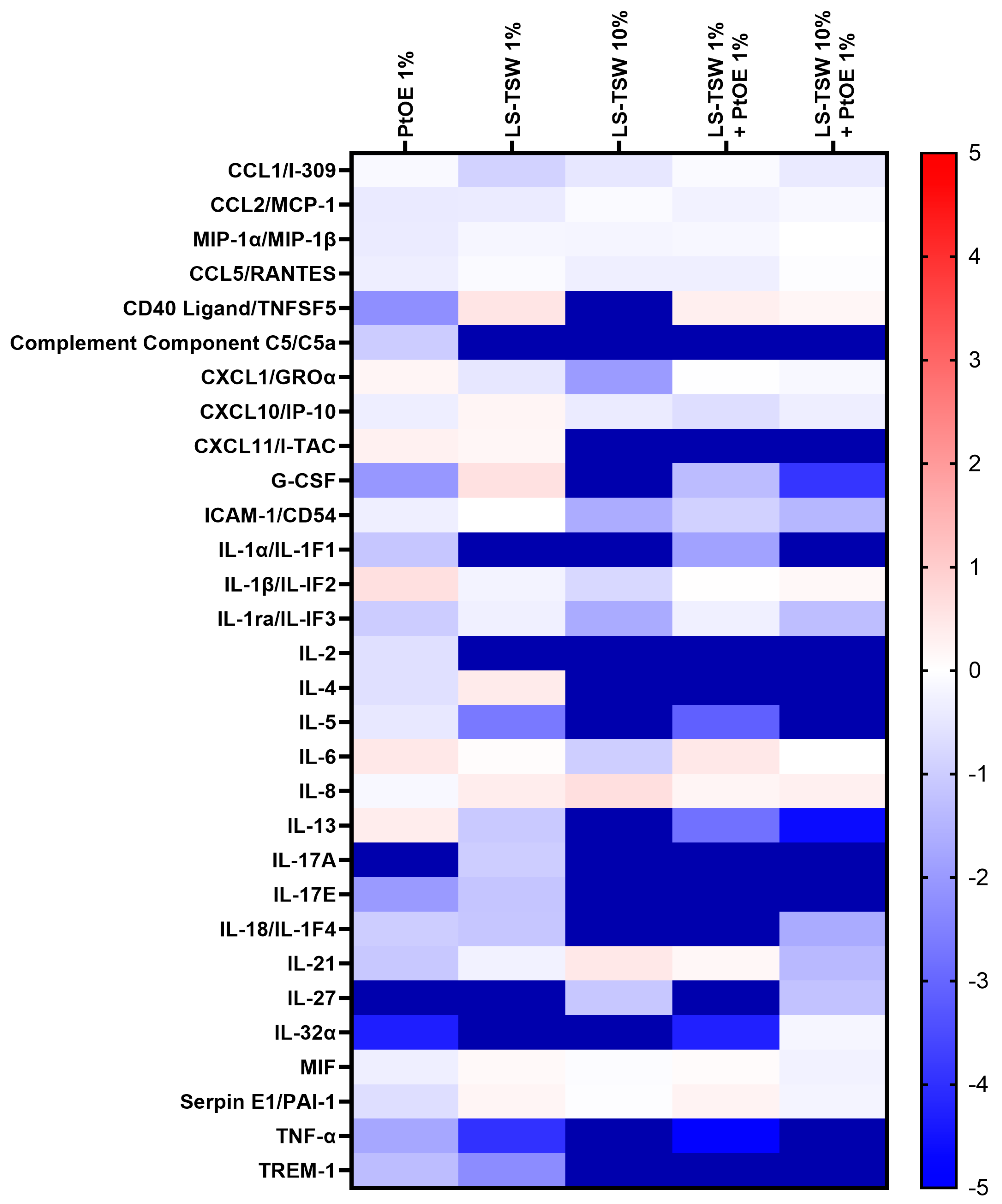
2.5. Cytokine Arrays

Cytokine arrays were performed and developed in accordance with the manufacturer’s protocol (Proteome Profiler Array: Human Cytokine Array Kit, R&D Systems, Minneapolis, MN, USA). Membranes were scanned using ImageQuant LASS 4000 Mini (GE Healthcare Life Sciences, Chicago, IL, USA) and images were analyzed using the MultiGauge V3.0 software (Fujifilm Life Sciences, Tokyo, Japan).

2.6. Induction of Inflammation Using TNF-α and IFN-γ

HaCaT cells were cultured at a concentration of 2.5 × 10⁴ cells/well in 6-well culture plates and incubated for 24 h at 37 °C in a humid atmosphere of 5% CO2. Thereafter, they were treated with different concentrations of P. tricornutum oil extract and/or La Solia thermal water (1% LS TSW + 0.25 PtOE, or 0.5% LS-TSW + 0.5 PtOE). After 24 h of incubation, they were treated simultaneously with TNF-α/IFN-γ mixture (20 ng/mL each, #10291-TA-050, #285-IF-100, R&D Systems, Minneapolis, MN, USA) to induce inflammation. The experiment was repeated twice with similar results.

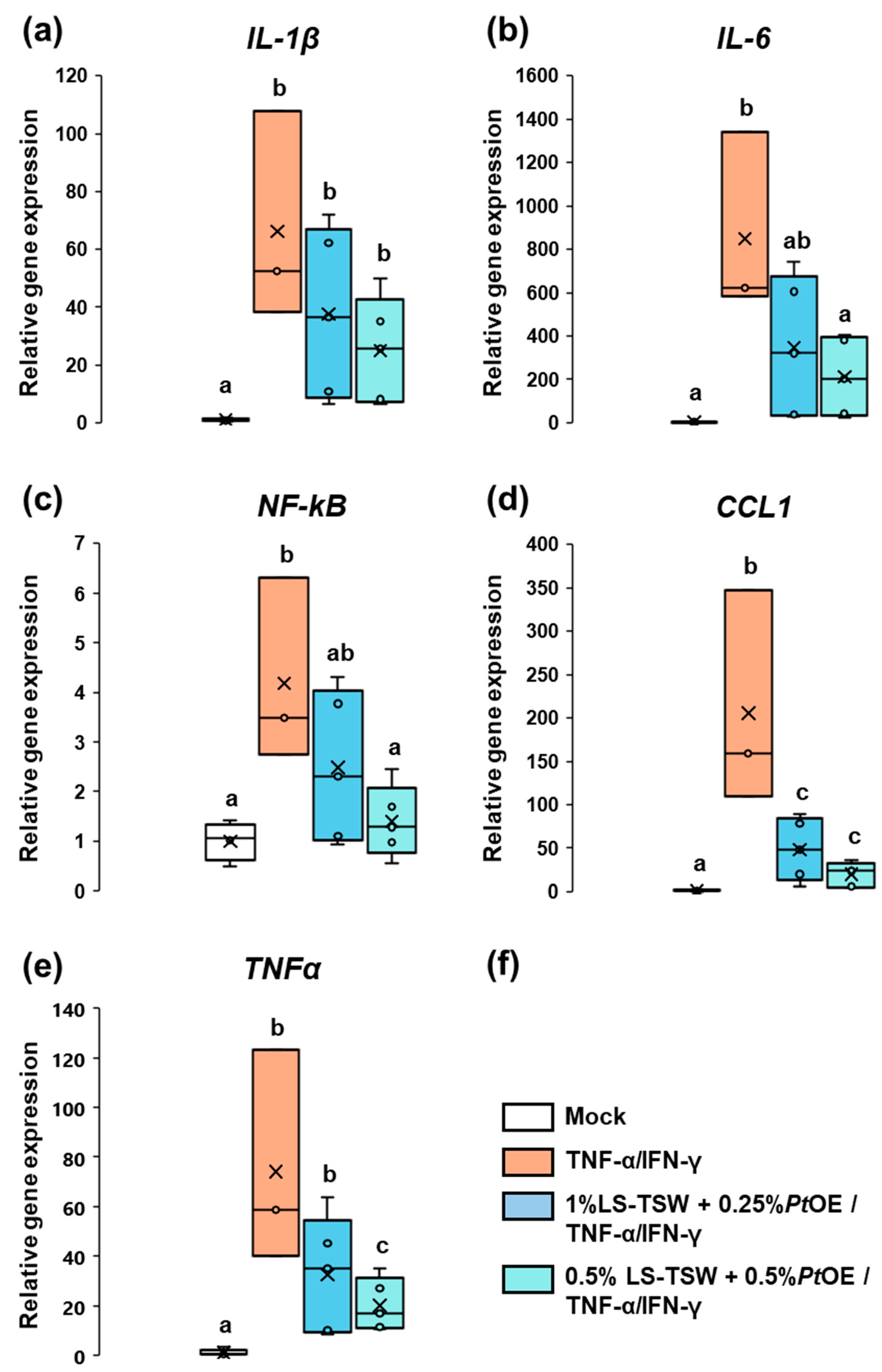
2.7. Total RNA Isolation and Real-Time quantitative Polymerase Chain Reaction (RT-qPCR)

After 24 h of inflammation induction, the total RNA of the HaCaT cells was extracted using the RNeasy kit (Qiagen, Hilden, Germany), according to the manufacturer’s instructions. Thereafter, complementary DNA (cDNA) was synthesized using the Maxima First Strand cDNA Synthesis Kit for RT-qPCR (Thermo Scientific, USA) from 1 µg of purified total RNA. Next, cDNA was amplified with a Light Cycler® 480 with 40 cycles of denaturation (95 °C for 15 s), annealing (60 °C for 60 s), and extension (60 °C for 60 s), using TaqMan probes: TNF-α (ID#: Hs00174128_m1); IL-6 (ID#: Hs00174131_m1); IL-1β (ID#: Hs01555410_m1); NF-κB (ID#: Hs00765730_m1); CCL1 (ID#: Hs00171072_m1). The relative quantification of gene expression was calculated according to the ΔΔCt method, relative to a calibrator sample [20], and using the 18S (ID#: Hs03003631_g1) as a reference gene.

3. Results

3.1. La Solia, Thermal Spring Water Chemical Characterization

Thermal spring waters (also called medicinal mineral waters or natural mineral waters in some countries; TWS) are very well-known for their beneficial effects on different illnesses, mainly rheumatological and respiratory ailments, but also dermatological disorders. Additionally, thermal spring waters are used as raw materials and active ingredients in dermocosmetics to achieve balance, hydration, and the wellbeing of the skin [21]. Given this knowledge, a comprehensive analysis for the characterization of La Solia, a thermal spring water licensed to Cantabria Labs which emerges in the north region of Spain (LS-TSW), was performed.
The composition analysis showed that LS-TSW is an isotonic water particularly rich in sodium, calcium, magnesium, and silica and with trace elements such as zinc, copper, manganese, boron, and selenium (Table 1), which are of interest in skin care. With some of these features, it has been declared as medicinal mineral water by the IGME (Instituto Geológico y Minero de España) and the Department of Industry (Autonomous Government of Cantabria). According to the mineral content, La Solia TSW is classified as chloride, sodium, sulphate, bicarbonate, calcium, and magnesium mineral water [15,21].
Many beneficial properties have been assigned to different spring waters. With the aim of searching for plausible benefits derived by its composition, we performed a comparative analysis of LS-TSW and other commercial spring waters used in the market (Table 2).
Comparing with other thermal spring waters used in dermocosmetics and skin care, its chloride and sodium content is similar to Uriage TSW; its calcium content is similar to La Roche-Posay and Vichy TSW; its magnesium content is similar to Saint-Gervais Mont Blanc TSW; its SiO2 content is similar to La Roche-Posay TSW and Uriage TSW; its boron content is similar to Avène TSW; and its zinc and copper is much higher than any other, though its selenium content is lower than La Roche-Posay TSW [21].

3.2. La Solia Microbiological Characterization

The microbiota of thermal spring waters (hydrobiome) and its relationship with cosmetics formulations has recently gained attention. Thus, we next investigated LS-TSW microbial communities.
As it can be seen in Figure 1, microscopic observations of freshly isolated samples revealed the presence of three main phylogenetic groups commonly named as microalgae: Cyanophyta (blue-green algae, Figure 1a), Chlorophyta (green algae, Figure 1b), and Heterokontophyta (diatoms, Figure 1c).

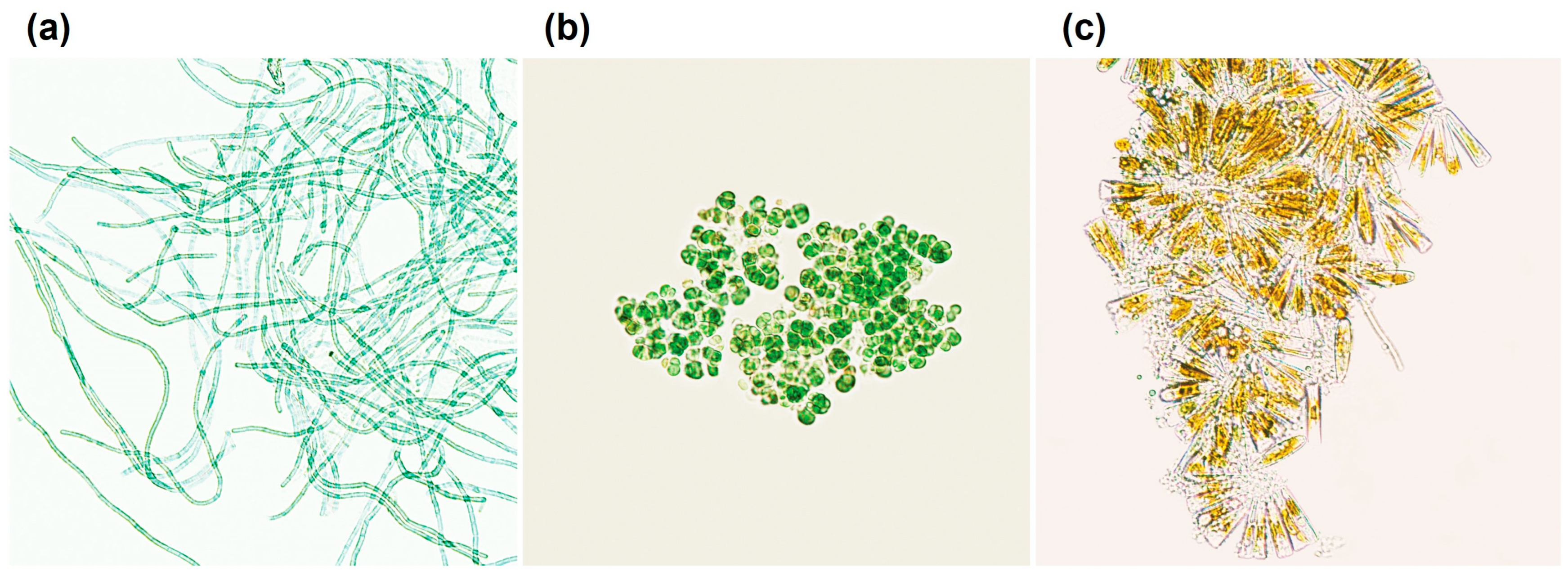
3.2.1. Cyanobacteria

The Cyanophyta phylum, comprising ancient prokaryotic microalgae, showed the presence of different genera in LS-TSW samples: (a) Spirulina, known for its spiral filaments, a cosmopolitan species with thermophilic and alkaline characteristics, making it biologically significant. (b) Oscillatoria, a filamentous Cyanobacteria, often forming mats together with other identified filamentous Cyanobacteria, such as Leptolyngbia, Geitlerinema, or Phormidium [22]. These mats serve as a substrate for other organisms, being of key biological relevance in the ecosystem of TSW. And, (c) Chroococcus, a non-filamentous cyanobacteria with spherical cells [23].

3.2.2. Chlorophyta

A single chlorophyte from the Chlorellaceae family was isolated in the natural thermal water source of LS-TSW (Figure 1b). This species, potentially belonging to the Chlorella genus, represents the Chlorophyta phylum, known for its diversity and unicellular nature.

3.2.3. Diatoms

Diatoms, classified within the Heterokonta phylum, were observed in our isolation efforts from LS-TSW. The identified diatom phylum included individualized genera like Navicula, Phaeodactylum, and Diatoma, each contributing to the rich microbial diversity of the thermal water [24]. Diatoms are sensitive to environmental conditions, and their presence, abundance, and diversity can be used as indicators of water quality, and serve as markers reflecting the unique and ecologically significant water properties of the region [25,26].
Notably, specimens from Phaeodactylum-like sp. were specifically identified in our isolation efforts from the thermal spring. Phaeodactylum is recognized for its adaptability, thriving in both marine and brackish water strains [27], but it has also been reported to grow in freshwater [28], showing a remarkable versatility in aquatic environments.

3.3. Immunomodulatory Activity of a Combination of La Solia Thermal Water and Phaeodactylum Tricornutum Oil Extract: Aquammunist

Many beneficial properties have been previously described for Phaeodactylum tricornutum, being commonly used in the pharmaceutical [29] and cosmetic industries [18]. P. tricornutum extracts are used for their anti-inflammatory and barrier function repair activities [18,19]. Its bioactivities have been associated with its composition, rich in omega-3 fatty acids [30], and the xanthophyl fucoxanthin. Specifically, fucoxanthin exhibits anti-inflammatory properties by controlling the levels of pro-inflammatory cytokines, such as IL-1β, IL-6, and TNF-α [19]. Therefore, Phaeodactylum tricornutum extracts seem to be promising ingredients for dermocosmetic products, particularly in the context of reactive skin treatments.
Having in mind the presence of Phaeodactylum in LS-TSW, we proposed the development of a new cosmetic ingredient potentiating the natural composition of LS-TSW with a lipophilic extract of P. tricornutum, rich in omega-3 fatty acids, standardized to fucoxanthin levels and encapsulated in liposomes. Based on the plausible anti-inflammatory character suggested by the properties of the individual compounds described above, the combination in the form of a single ingredient, subsequently named Aquammunist, was tested.

3.3.1. Aquammunist Showed Potential Anti-Inflammatory Properties in In Vitro Conditions

First, we tested the activity of these two components (LS-TSW and PtOE) for the control of pro-inflammatory factors produced during induced inflammation. To this purpose, we used THP-1 cells (monocytes from peripheral blood) as a common cellular model to address immune modulation [31]. Monocytes were first pre-treated with PtOE extract, LS-TSW, or a combination of both at different proportions (1% PtOE, 1% LS-TW, 10% LS-TSW, 1% LS TSW + 1% PtOE, or 10% LS-TSW + 1% PtOE); and then stimulated by LPS as a general pro-inflammatory stimulus. In this experimental setup, the level of pro-inflammatory molecules was detected by a commercial protein microarray. At it is shown in Figure 2 (normalized data relative to LPS control treatment), the main part of these pro-inflammatory factors seems to reach lower levels (appeared as downregulated) in pre-treated samples, indicating a general anti-inflammatory character of the treatments. Both PtOE and LS-TSW displayed a discrete control over the pro-inflammatory cyto/chemokines analyzed when tested at 1% (a reasonable proportion to be included in cosmetic formulas), and the strongest effect was observed with LS-TSW at 10%. However, the combination of 10% LS-TSW and 1% PtOE did not show a clear benefit over 10% LS-TSW, and the inclusion of 10% LS-TSW in cosmetic formulations is a major challenge due to formula instabilities. Remarkably, the combination of both components in the mixture at 1% each, seemed to have a stronger anti-inflammatory character than both ingredients tested separately, supporting the use of the combination (Figure 2, 4th column vs. columns 1st and 2nd). Thus, this preliminary evidence supported further exploring the immunomodulatory properties of the proposed ingredient, preferably at similar proportions.

3.3.2. Aquammunist Displays an Effective Control of Skin Inflammation in In Vitro Conditions

After the prospective results obtained in THP-1 cells, we next addressed the bioactivity of the proposed combination in skin cells, by using HaCaT cells as a cellular model of human keratinocytes [32]. Under this aim, a gene expression analysis was performed mimicking skin irritation by TNF-α/IFN-γ treatments [33]. HaCaT cell cultures were first pre-treated with Aquammunist. Due to cellular model specificities when compared to monocyte cultures, lower concentrations of Aquammunist were tested. Moreover, to confirm the results of the previous assay, we used two different Aquammunist pre-treatments, one including different proportions of LS-TSW and PtOE (1% LS-TSW:0.25% PtOE), and the other with the ingredients at the same proportions (0.5% LS-TSW:0.5% PtOE). Then, 24 h after pre-treatment, the cell cultures were treated with TNF-α/IFN-γ to induce inflammation. Mock-treated cell cultures (untreated) were used as negative control (non-irritated samples). Non-pre-treated samples but stimulated with TNF-α/IFN-γ were used as a positive intraexperimental control for the induction of inflammation. As pro-inflammatory factors of interest, the gene expression levels of the interleukins IL-1β and IL-6, the transcription factor NF-ϏB, the chemokine CCL1, and the cytokine master regulator TNF-α were analyzed.
As it is shown in Figure 3, a clear induction of all pro-inflammatory factors analyzed was observed in TNF-α/IFN-γ treatments (orange boxes), validating the experimental set up (positive control of induced inflammation). Pre-treatments with the ingredient of interest showed a lower induction of all those pro-inflammatory factors (blue and turquoise boxes). Interestingly, culture cells pre-treated with a combination of LS-TSW/PtOE at the same proportion (0.5% LS-TSW:0.5% PtOE) showed the lowest induction of pro-inflammatory factors, reaching a statistically significant reduction for all tested genes with the exception of IL-1β (turquoise boxes). Specifically in that treatment, for the case of IL-6 and NF-ϏB, the expression levels were not statistically distinguishable from mock samples, highlighting the potential of this combination in the control of induced-inflammation.
Therefore, the combination of the compounds here tested, seems to control skin-induced inflammation, supporting the use of Aquammunist in cosmetic formulations to treat skin conditions involving sensitivity or irritation.

4. Discussion

Individuals with a certain tendency to skin irritation can benefit from cosmetic treatments; however, in specific cases, such as sensitive skin, where the use of cosmetics itself has been demonstrated to trigger and/or worsen the skin condition, the experts find it difficult to agree on specific recommendations without scientific evidence [9,10,11]. This is the reason why the field is currently under an active search for alternative ingredients. In this context, the best candidate compounds should ideally control skin irritation, strengthen skin barrier function, and prevent skin reactivity. Under these premises, we propose the development of a new ingredient based on a thermal spring water and a lipidic extract from diatom algae.
Among the different options for the treatment of reactive skin, thermal spring waters have been recognized as one of the most effective dermocosmetic ingredients to improve sensitive skin condition. Several studies have been carried out in this field during the last decades. Avène thermal spring water (Avène TSW), is a bicarbonate mineral water rich in calcium, magnesium, and boron, and it has been found to exert anti-irritant and anti-inflammatory activity [34,35]; being able to decrease redness and reduce the overall sensitive scale after a dermatological chemical peeling [36]. Other bicarbonate thermal spring waters, such as São Pedro do Sul and Monforthinho, also showed anti-irritant and anti-inflammatory properties [37,38]. Sodium-rich TSW as Uriage TSW (UTSW) was suggested to effectively protect the skin from dehydration, through its effect on the expression of TauT and SVCT1 as it allows a more efficient delivery of taurine and vitamin C to the epidermis to protect it from other issues, such as oxidative stress [39]. Thermal spring waters rich in silica (SiO2) are also of interest in the treatment of sensitive skin as they are able to improve the skin barrier, and have also shown anti-inflammatory properties [40,41]. Several research, both in vitro and clinical studies, have been conducted with La Roche-Posay thermal spring water. This bicarbonate calcium and magnesium water, which also contain strontium and selenium, showed anti-inflammatory and immunomodulatory properties and reduced redness and telangiectasia intensity in rosacea [42,43,44,45,46]. Finally, we must acknowledge hypersaline waters that are rich in magnesium, which have shown to have anti-inflammatory and antioxidant properties; the best-known is Dead Sea mineral water [47,48].
In this scenario, we presented the analysis of a spring water, LS-TSW, which emerges in the region of Cantabria, in the north of Spain. Under our analysis, LS-TSW is classified as chloride, sodium, sulphate, bicarbonate, calcium, and magnesium mineral water [15,21]. Comparing with other thermal spring waters used in dermocosmetics and skin care, due to its high chloride content, it could be of interest in the treatment of different skin disorders, such as psoriasis and sensitive skin. The anti-inflammatory potential of sodium-chlorinated waters has been demonstrated in different studies, with a decrease in PASI being observed in patients with psoriasis [49], and sulphated sodium-chlorinated waters have shown their anti-inflammatory potential in in vivo studies [50]. La Solia TSW is rich in SiO2, which in its amorphous form, as found in thermal spring waters, has been shown to be anti-irritant and to repair the epidermal barrier [37]. In fact, emulsions made with water of very low mineralization but with a certain SiO2 content (16 mg/L), such as Monfortinho, have been shown to be effective in hydration in cases of psoriasis and eczema [51]. Thus, putative anti-irritant properties can be expected in La Solia TSW. Despite the bicarbonate content in La Solia TSW being low (210 mg/L), it is similar to other thermal spring waters, such as Avène TSW, where its bicarbonate and calcium ions intervene in the inhibition of histamine release by mast cells [52]. Other studies showed the role of calcium from Uriage thermal waters in promoting eosinophil apoptosis in cases of hypereosinophilia and, therefore, exerting anti-inflammatory action as well [53]. Both bicarbonate and calcium are present in La Solia TWS. The presence of trace elements such as selenium and zinc are also of great interest in skin care. Roche-Posay TSW and Avène TSW have also shown anti-inflammatory capacity on human keratinocytes, since both suppressed the induction of a prototypical inflammatory cytokine and the formation of ROS after UVB radiation [54]. It seems likely that, in the case of La Roche-Posay TSW, the observed effects are mediated by the high content of selenium (53 μg/L), which is a cofactor of glutathione peroxidase, a key enzyme in the removal of ROS. In the case of Avène TSW, authors considered that the relatively high zinc content (20 μg/L) seems responsible for the anti-inflammatory action. Considering the high zinc content of La Solia TSW and medium the level of selenium content (compared with other thermal spring waters), anti-inflammatory properties should be expected.
There is a rising interest of thermal spring waters microbiota, named as hydrobiome. Beyond the benefits of TSWs associated to its mineral composition, the contribution of the hydrobiome seems to be determinant for its bioactivities. Mourelle et al. (2023) revised the published studies related to the use of the microorganisms present in thermal spring waters in cosmetics, both as postbiotics and paraprobiotics [16]. They found 22 papers focused on extracts, lysates, or ferments of these microorganisms (mainly cyanobacteria and other microorganisms generally considered as microalgae), highlighting its potential as active ingredients in dermocosmetics. One of the most relevant examples is the β-proteobacteria Aquaphilus dolomiae, found in Avène TSW aquifer [16]. Among other evidence, a cosmetic prepared with the extracts from Aquaphilus dolomiae has shown to be effective in reducing pruritus and xerosis in a range of dermatologic and systemic diseases in an open-label, real-world study [55]. Furthermore, exopolysaccharides from Cyanobacterium aponinum (EPS-Ca), a cyanobacteria found in the Blue Lagoon silica-rich thermal water, stimulated dendritic cells to produce the immunosuppressive cytokine IL-10, increased the proportion of dendritic cells expressing CD141, and decreased T cell secretion of IL-17, IL-13, and IL-10 [56]. EPS-Ca. EPS-Ca also demonstrated to reduce keratinocyte secretion of chemokines CCL20 and CXCL10 that are involved in the recruitment of inflammatory cells [57]. In this scenario, this investigation delved into the microbiological profile of LS-TSW. Among the myriad microorganisms isolated, diatoms, took center stage and, specifically, specimens of Phaeodactylum-like sp. were isolated in LS-TSW. In general terms, diatoms are commonly used in the pharmaceutical [29] and cosmetic industries [18]. In particular, Phaeodactylum tricornutum extracts are used for their anti-inflammatory and barrier function repair activities [18,19]. The biomass of P. tricornutum is rich in proteins, carbohydrates, lipids, and essential mineral elements, and no significant amounts of toxic heavy metal elements [58], making it a valuable source for various applications [59]. Amid the spectrum of xanthophylls present in P. tricornutum, fucoxanthin, unique to brown algae and diatoms, has garnered significant attention for its therapeutic properties due to its anti-enzymatic, antimicrobial, photoprotective, and anti-inflammatory capacity; making it suitable for diverse dermocosmetic applications [60]. Additionally, the use of lipid vesicles as a carrier has been found to enhance the bioavailability of fucoxanthin [61]. Accounting on the isolation of Phaeodactylum sp. from LS-TSW, we wondered whether the addition of a PtOE encapsulated in liposomes could potentiate the natural bioactivities of the TSW.
Keeping the focus on addressing the need of alternative ingredients in sensitive skin, we next studied the potential immunomodulatory benefits of both ingredients independently and combined in a single mixture, named Aquammunist. Using monocytes as a cellular model, and induction of unspecific inflammation by LPS, we observed how both ingredients displayed a general anti-inflammatory effect when tested independently. Surprisingly, specific combinations of the ingredients (at same proportions, each −1%), showed a more effective control of pro-inflammatory mediators, suggesting a certain synergistic effect. Going deeper into the characterization of this effect, we analyzed the induction of different pro-inflammatory molecules in cellular skin models. Our gene expression analysis confirmed the reduction in the induced-proinflammatory signals of interest when the keratinocytes were pre-treated with Aquammunist at different proportions of LS-TSW and PtOE. The best results were obtained when both ingredients were mixed at equal concentrations in accordance with the results obtained in monocytes. Consistently as well with the results observed in monocytes, all pro-inflammatory cytokines/chemokines analyzed showed lower levels in samples pre-treated with Aquammunist. These results highlight the potential of the proposed combination (LS-TSW and PtOE), in the form of a single ingredient (Aquammunist) in controlling skin inflammation.
Under external aggression, skin cells are able to produce immune modulator factors to orchestrate the defense response and ensure the skin homeostasis [62]. However, misregulation of these molecules is common in the chronic inflammation of skin tissues, being considered part of the physiopathology for different skin alterations [63,64]. That is the reason why they are considered as molecular markers in diverse inflammatory skin diseases [65]. In fact, due to their key physiopathological role many of the newest treatments using biologics have these molecules as targets [63,64]. Anti-TNFα biologics is maybe the best-known example, being the most important contribution to the treatment of autoimmune skin diseases [66]. Nonetheless, the case of psoriasis treatment blocking TNFα is far from being an isolated case of success, and nowadays many biologics controlling the induction of different cytokines, such as IL-4/13, IL-12/23, IL-17, IL31, IL-36 etc., are being under investigation or already approved for the treatment of psoriasis, atopic dermatitis, chronic spontaneous urticaria, and hidradenitis suppurativa [64]. As exposed at the introduction, it is now clear that cosmetics could bring important benefits not just as adjuvant treatments in diagnosed pathologies, but as part of skin care routines in some conditions like sensitive skin [2,4,9]. Thus, considering that in our in vitro assays Aquammunist seems to control the overexpression of part of this immunomodulatory repertoire of interest, our next steps will focus on exploring the potential benefits of these ingredients in clinical studies. With regards to this, the first analysis addressing Aquammunist smoothing effect on skin discomfort induced by capsaicin have already brought positive results (manuscript under submission). Moreover, keeping in mind the SiO2 levels of LS-TSW, which have been described to have positive effects on hydration [51] and the lipidic composition of PtOE; the protection and improvement of skin barrier function would be another variable of interest to be investigated in the future. In any case, to translate the specific findings on the ingredient properties, the next steps forwards should also consider clinical trials including Aquammunist in complete cosmetic formulations.

5. Conclusions

Thermal spring waters and algae extracts are some of the most promising ingredients for cosmetic purposes, especially for the treatment of reactive skin and other skin alterations with a chronic inflammation component. The results presented here conform the first scientific evidence about the development of an alternative ingredient combining a specific thermal spring water and a diatom alga extract, grounding its following clinical research and dermocosmetological use.

Author Contributions

Conceptualization, M.L.M. and A.L.S.; methodology, A.L.S.; formal analysis, M.LM., J.S.d.Y., and A.L.S.; investigation, M.L.M., J.S.d.Y. and A.L.S.; writing—original draft preparation, M.L.M., J.S.d.Y., J.A. and A.L.S.; writing—review and editing, M.L.M., J.S.d.Y., J.A., M.V. and A.L.S.; supervision, project administration and funding acquisition, A.L.S. All authors have read and agreed to the published version of the manuscript.

Funding

This research was funded by Industrial Farmacéutica Cantabria SA.

Institutional Review Board Statement

Not applicable.

Informed Consent Statement

Not applicable.

Data Availability Statement

Here we present original data not shared before. We consider by publishing this report we are openly releasing this data.

Conflicts of Interest

The authors declare that this study received funding from Cantabria Labs. M.V. and A.L.S. work in Cantabria Labs, and J.S. and J.A. work in Algaktiv SL. The funders participated in the design of the study, as well as in the analyses and interpretation of data.

References

  1. Liu, T.J.; Lin, L.L.; McMeniman, E.; Wu, J.; Kao, Y.-C.; Kumari, S.; Boyle, G.M.; Wells, J.W.; Soyer, H.P.; Gonzalez-Cruz, J.L. Cytokine/Chemokine Assessment as a Complementary Diagnostic Tool for Inflammatory Skin Diseases. Front. Immunol. 2022, 13, 1028435. [Google Scholar] [CrossRef] [PubMed]
  2. Kang, S.-Y.; Um, J.-Y.; Chung, B.-Y.; Lee, S.-Y.; Park, J.-S.; Kim, J.-C.; Park, C.-W.; Kim, H.-O. Moisturizer in Patients with Inflammatory Skin Diseases. Medicina 2022, 58, 888. [Google Scholar] [CrossRef] [PubMed]
  3. Abdallah, F.; Mijouin, L.; Pichon, C. Skin Immune Landscape: Inside and Outside the Organism. Mediat. Inflamm. 2017, 2017, 5095293. [Google Scholar] [CrossRef] [PubMed]
  4. Antelo, D.A.P.; Leta da Costa Rocha, A. Cosmetic Approach in Patients with Acne and Rosacea. In Daily Routine in Cosmetic Dermatology; Issa, M.C.A., Tamura, B., Eds.; Clinical Approaches and Procedures in Cosmetic Dermatology; Springer: Cham, Switzerland, 2016; pp. 1–28. ISBN 978-3-319-20250-1. [Google Scholar]
  5. Purnamawati, S.; Indrastuti, N.; Danarti, R.; Saefudin, T. The Role of Moisturizers in Addressing Various Kinds of Dermatitis: A Review. Clin. Med. Res. 2017, 15, 75–87. [Google Scholar] [CrossRef] [PubMed]
  6. Misery, L.; Ständer, S.; Szepietowski, J.; Reich, A.; Wallengren, J.; Evers, A.; Takamori, K.; Brenaut, E.; Gall-Ianotto, C.; Fluhr, J.; et al. Definition of Sensitive Skin: An Expert Position Paper from the Special Interest Group on Sensitive Skin of the International Forum for the Study of Itch. Acta Derm. Venereol. 2017, 97, 4–6. [Google Scholar] [CrossRef] [PubMed]
  7. Chen, W.; Dai, R.; Li, L. The Prevalence of Self-Declared Sensitive Skin: A Systematic Review and Meta-Analysis. J. Eur. Acad. Dermatol. Venereol. JEADV 2020, 34, 1779–1788. [Google Scholar] [CrossRef]
  8. Legeas, C.; Misery, L.; Fluhr, J.W.; Roudot, A.-C.; Ficheux, A.-S.; Brenaut, E. Proposal for Cut-off Scores for Sensitive Skin on Sensitive Scale-10 in a Group of Adult Women. Acta Derm. Venereol. 2021, 101, 1498. [Google Scholar] [CrossRef] [PubMed]
  9. Goh, C.-L.; Wu, Y.; Welsh, B.; Abad-Casintahan, M.F.; Tseng, C.-J.; Sharad, J.; Jung, S.; Rojanamatin, J.; Sitohang, I.B.S.; Chan, H.N.K. Expert Consensus on Holistic Skin Care Routine: Focus on Acne, Rosacea, Atopic Dermatitis, and Sensitive Skin Syndrome. J. Cosmet. Dermatol. 2023, 22, 45–54. [Google Scholar] [CrossRef] [PubMed]
  10. Guerra-Tapia, A.; Serra-Baldrich, E.; Prieto Cabezas, L.; González-Guerra, E.; López-Estebaranz, J.L. Diagnóstico y tratamiento del síndrome de piel sensible: Un algoritmo para la práctica clínica habitual. Actas Dermo-Sifiliográficas 2019, 110, 800–808. [Google Scholar] [CrossRef] [PubMed]
  11. Chen, X.; Wen, J.; Wu, W.; Peng, Q.; Cui, X.; He, L. A Review of Factors Influencing Sensitive Skin: An Emphasis on Built Environment Characteristics. Front. Public Health 2023, 11, 1269314. [Google Scholar] [CrossRef]
  12. Sinha, S.; Meena, N. The Sensitive Skin: Do’s and Don’ts. In Skin Diseases in Females; Sarkar, R., Sinha, S., Eds.; Springer: Singapore, 2022; pp. 471–486. ISBN 9789811660658. [Google Scholar]
  13. Lee, H.-P.; Choi, Y.-J.; Cho, K.-A.; Woo, S.-Y.; Yun, S.-T.; Lee, J.T.; Kim, H.J.; Lee, K.-H.; Kim, J.-W. Effect of Spa Spring Water on Cytokine Expression in Human Keratinocyte HaCaT Cells and on Differentiation of CD4+ T Cells. Ann. Dermatol. 2012, 24, 324–336. [Google Scholar] [CrossRef] [PubMed]
  14. Cacciapuoti, S.; Luciano, M.A.; Megna, M.; Annunziata, M.C.; Napolitano, M.; Patruno, C.; Scala, E.; Colicchio, R.; Pagliuca, C.; Salvatore, P.; et al. The Role of Thermal Water in Chronic Skin Diseases Management: A Review of the Literature. J. Clin. Med. 2020, 9, 3047. [Google Scholar] [CrossRef]
  15. Mourelle, M.L.; Gómez, C.P.; Legido, J.L. Dermothermal Cosmetics: Added Value for Thermal Centers. In Proceedings of the 1st InternationalCongress on Water Healing Spa and Quality of Life, Ourense, Spain, 23–24 September 2015; pp. 1–7. [Google Scholar]
  16. Mourelle, M.L.; Gómez, C.P.; Legido, J.L. Hydrobiome of Thermal Waters: Potential Use in Dermocosmetics. Cosmetics 2023, 10, 94. [Google Scholar] [CrossRef]
  17. Ferreira, M.S.; Resende, D.I.S.P.; Lobo, J.M.S.; Sousa, E.; Almeida, I.F. Marine Ingredients for Sensitive Skin: Market Overview. Mar. Drugs 2021, 19, 464. [Google Scholar] [CrossRef] [PubMed]
  18. Mosxou, D.; Letsiou, S. Exploring the Protective Effects of Phaeodactylum Tricornutum Extract on LPS-Treated Fibroblasts. Cosmetics 2021, 8, 76. [Google Scholar] [CrossRef]
  19. Lee, A.-H.; Shin, H.-Y.; Park, J.-H.; Koo, S.Y.; Kim, S.M.; Yang, S.-H. Fucoxanthin from Microalgae Phaeodactylum Tricornutum Inhibits Pro-Inflammatory Cytokines by Regulating Both NF-κB and NLRP3 Inflammasome Activation. Sci. Rep. 2021, 11, 543. [Google Scholar] [CrossRef] [PubMed]
  20. Pabinger, S.; Rödiger, S.; Kriegner, A.; Vierlinger, K.; Weinhäusel, A. A Survey of Tools for the Analysis of Quantitative PCR (qPCR) Data. Biomol. Detect. Quantif. 2014, 1, 23–33. [Google Scholar] [CrossRef] [PubMed]
  21. Figueiredo, A.C.; Rodrigues, M.; Mourelle, M.L.; Araujo, A.R.T.S. Thermal Spring Waters as an Active Ingredient in Cosmetic Formulations. Cosmetics 2023, 10, 27. [Google Scholar] [CrossRef]
  22. McGregor, G.B.; Rasmussen, J.P. Cyanobacterial Composition of Microbial Mats from an Australian Thermal Spring: A Polyphasic Evaluation. FEMS Microbiol. Ecol. 2008, 63, 23–35. [Google Scholar] [CrossRef] [PubMed]
  23. Duval, C.; Hamlaoui, S.; Piquet, B.; Toutirais, G.; Yéprémian, C.; Reinhardt, A.; Duperron, S.; Marie, B.; Demay, J.; Bernard, C. Diversity of Cyanobacteria from Thermal Muds (Balaruc-Les-Bains, France) with the Description of Pseudochroococcus Coutei Gen. Nov., Sp. Nov. FEMS Microbes 2021, 2, xtab006. [Google Scholar] [CrossRef] [PubMed]
  24. Leira, M.; Meijide-Failde, R.; Torres, E. Diatom Communities in Thermo-Mineral Springs of Galicia (NW Spain). Diatom Res. 2017, 32, 29–42. [Google Scholar] [CrossRef]
  25. Krajenbrink, H.J.; White, J.C.; Dunbar, M.J.; Wood, P.J. Macroinvertebrate and Diatom Community Responses to Thermal Alterations below Water Supply Reservoirs. River Res. Appl. 2022, 38, 595–612. [Google Scholar] [CrossRef]
  26. Verma, P.; Thapliyal, K. Diatom Analysis in Thermophilic Water Bodies: A Study on Hot Water Springs of Madhya Pradesh. Indian J. Forensic Med. Pathol. 2023, 16, 133–143. [Google Scholar] [CrossRef]
  27. Butler, T.; Kapoore, R.V.; Vaidyanathan, S. Phaeodactylum Tricornutum: A Diatom Cell Factory. Trends Biotechnol. 2020, 38, 606–622. [Google Scholar] [CrossRef] [PubMed]
  28. Yongmanitchai, W.; Ward, O.P. Screening of Algae for Potential Alternative Sources of Eicosapentaenoic Acid. Phytochemistry 1991, 30, 2963–2967. [Google Scholar] [CrossRef]
  29. Dhaouadi, F.; Awwad, F.; Diamond, A.; Desgagné-Penix, I. Diatoms’ Breakthroughs in Biotechnology: Phaeodactylum Tricornutum as a Model for Producing High-Added Value Molecules. Am. J. Plant Sci. 2020, 11, 1632–1670. [Google Scholar] [CrossRef]
  30. Yongmanitchai, W.; Ward, O.P. Growth of and Omega-3 Fatty Acid Production by Phaeodactylum Tricornutum under Different Culture Conditions. Appl. Environ. Microbiol. 1991, 57, 419–425. [Google Scholar] [CrossRef] [PubMed]
  31. Chanput, W.; Mes, J.J.; Wichers, H.J. THP-1 Cell Line: An in Vitro Cell Model for Immune Modulation Approach. Int. Immunopharmacol. 2014, 23, 37–45. [Google Scholar] [CrossRef]
  32. Colombo, I.; Sangiovanni, E.; Maggio, R.; Mattozzi, C.; Zava, S.; Corbett, Y.; Fumagalli, M.; Carlino, C.; Corsetto, P.A.; Scaccabarozzi, D.; et al. HaCaT Cells as a Reliable In Vitro Differentiation Model to Dissect the Inflammatory/Repair Response of Human Keratinocytes. Mediat. Inflamm. 2017, 2017, 7435621. [Google Scholar] [CrossRef] [PubMed]
  33. Tsai, Y.-C.; Chang, H.-H.; Chou, S.-C.; Chu, T.W.; Hsu, Y.-J.; Hsiao, C.-Y.; Lo, Y.-H.; Wu, N.-L.; Chang, D.-C.; Hung, C.-F. Evaluation of the Anti-Atopic Dermatitis Effects of α-Boswellic Acid on Tnf-α/Ifn-γ-Stimulated HaCat Cells and DNCB-Induced BALB/c Mice. Int. J. Mol. Sci. 2022, 23, 9863. [Google Scholar] [CrossRef] [PubMed]
  34. Goldman, M.P.; Merial-Kieny, C.; Nocera, T.; Mery, S. Comparative Benefit of Two Thermal Spring Waters after Photodynamic Therapy Procedure. J. Cosmet. Dermatol. 2007, 6, 31–35. [Google Scholar] [CrossRef] [PubMed]
  35. Casas, C.; Ribet, V.; Alvarez-Georges, S.; Sibaud, V.; Guerrero, D.; Schmitt, A.-M.; Redoulès, D. Modulation of Interleukin-8 and Staphylococcal Flora by Avène Hydrotherapy in Patients Suffering from Chronic Inflammatory Dermatoses. J. Eur. Acad. Dermatol. Venereol. 2011, 25, 19–23. [Google Scholar] [CrossRef] [PubMed]
  36. Mias, C.; Maret, A.; Gontier, E.; Carrasco, C.; Satge, C.; Bessou-Touya, S.; Coubetergues, H.; Bennett-Kennett, R.; Dauskardt, R.H.; Duplan, H. Protective Properties of Avène Thermal Spring Water on Biomechanical, Ultrastructural and Clinical Parameters of Human Skin. J. Eur. Acad. Dermatol. Venereol. 2020, 34, 15–20. [Google Scholar] [CrossRef] [PubMed]
  37. Ferreira, M.O.; Costa, P.C.; Bahia, M.F. Effect of São Pedro Do Sul Thermal Water on Skin Irritation. Int. J. Cosmet. Sci. 2010, 32, 205–210. [Google Scholar] [CrossRef] [PubMed]
  38. Oliveira, A.S.; Vaz, C.V.; Silva, A.; Correia, S.; Ferreira, R.; Breitenfeld, L.; Martinez-de-Oliveira, J.; Palmeira-de-Oliveira, R.; Pereira, C.; Cruz, M.T.; et al. In Vitro Evaluation of Potential Benefits of a Silica-Rich Thermal Water (Monfortinho Thermal Water) in Hyperkeratotic Skin Conditions. Int. J. Biometeorol. 2020, 64, 1957–1968. [Google Scholar] [CrossRef] [PubMed]
  39. Verdy, C.; Branka, J.-E.; Lefeuvre, L. Modulation of Sodium-Dependent Transporters Expression in Normal Human Keratinocytes by a Sodium Rich Isotonic Thermal Water. J. Cosmet. Dermatol. Sci. Appl. 2012, 2, 254–262. [Google Scholar] [CrossRef]
  40. Eysteinsdóttir, J.H.; Ólafsson, J.H.; Agnarsson, B.A.; Lúðvíksson, B.R.; Sigurgeirsson, B. Psoriasis Treatment: Faster and Long-Standing Results after Bathing in Geothermal Seawater. A Randomized Trial of Three UVB Phototherapy Regimens. Photodermatol. Photoimmunol. Photomed. 2014, 30, 25–34. [Google Scholar] [CrossRef] [PubMed]
  41. Eysteinsdóttir, J.H.; Sigurgeirsson, B.; Ólafsson, J.H.; Fridriksson, T.; Agnarsson, B.A.; Davíðsson, S.; Valdimarsson, H.; Lúðvíksson, B.R. The Role of Th17/Tc17 Peripheral Blood T Cells in Psoriasis and Their Positive Therapeutic Response. Scand. J. Immunol. 2013, 78, 529–537. [Google Scholar] [CrossRef] [PubMed]
  42. Moysan, A.; Morlière, P.; Marquis, I.; Richard, A.; Dubertret, L. Effects of Selenium on UVA-Lnduced Lipid Peroxidation in Cultured Human Skin Fibroblasts. Skin Pharmacol. 2009, 8, 139–148. [Google Scholar] [CrossRef] [PubMed]
  43. Celerier, P.; Litoux, P.; Dreno, B.; Richard, A. Modulatory Effects of Selenium and Strontium Salts on Keratinocyte-Derived Inflammatory Cytokines. Arch. Dermatol. Res. 1995, 287, 680–682. [Google Scholar] [CrossRef] [PubMed]
  44. Rougier, A.; Richard, A. Preventing Effect of a Selenium-Rich Thermal Water on UV and Chemically Induced Dermatitis. In Proceedings of the Congress of the International Federation Societies of Cosmetic Chemists, Montreux, Switzerland, 18 September 1995. [Google Scholar]
  45. Rougier, A.; Richard, A. A Selenium-Rich Spring Water Prevents UV and Chemically Induced Inflammation. J. Am. Acad. Dermatol. 2012, 66, AB68. [Google Scholar] [CrossRef]
  46. Seite, S. Thermal Waters as Cosmeceuticals: La Roche-Posay Thermal Spring Water Example. Clin. Cosmet. Investig. Dermatol. 2013, 6, 23–28. [Google Scholar] [CrossRef] [PubMed]
  47. Portugal-Cohen, M.; Oron, M.; Merrik, E.; Ma’or, Z.; Ben-Amitai, D.; Yogev, H.; Zvulunov, A. A Dead Sea Water-Enriched Body Cream Improves Skin Severity Scores in Children with Atopic Dermatitis. J. Cosmet. Dermatol. Sci. Appl. 2011, 1, 71–78. [Google Scholar] [CrossRef]
  48. Portugal-Cohen, M.; Oron, M.; Cohen, D.; Ma’or, Z. Antipollution Skin Protection—A New Paradigm and Its Demonstration on Two Active Compounds. Clin. Cosmet. Investig. Dermatol. 2017, 10, 185–193. [Google Scholar] [CrossRef] [PubMed]
  49. Tsoureli-Nikita, E.; Menchini, G.; Ghersetich, I.; Hercogova, J. Alternative Treatment of Psoriasis with Balneotherapy Using Leopoldine Spa Water. J. Eur. Acad. Dermatol. Venereol. JEADV 2002, 16, 260–262. [Google Scholar] [CrossRef] [PubMed]
  50. Hercogova, J.; Stanghellini, E.; Tsoureli-Nikita, E.; Menchini, G. Inhibitory Effects of Leopoldine Spa Water on Inflammation Caused by Sodium Lauryl Sulphate. J. Eur. Acad. Dermatol. Venereol. JEADV 2002, 16, 263–266. [Google Scholar] [CrossRef] [PubMed]
  51. Almeida, C.; Madeira, A.; Marto, J.; Graça, A.; Pinto, P.; Ribeiro, H. Monfortinho Thermal Water-Based Creams: Effects on Skin Hydration, Psoriasis, and Eczema in Adults. Cosmetics 2019, 6, 56. [Google Scholar] [CrossRef]
  52. Joly, F.; Galoppin, L.; Bordat, P.; Cousse, H.; Neuzil, E. Calcium and Bicarbonate Ions Mediate the Inhibition of Mast Cell Histamine Release by Avène Spa Water. Fundam. Clin. Pharmacol. 2000, 14, 611–613. [Google Scholar] [CrossRef] [PubMed]
  53. Beauvais, F.; Garcia-Mace, J.L.; Joly, F. In Vitro Effects of Uriage Spring Water on the Apoptosis of Human Eosinophils. Fundam. Clin. Pharmacol. 1998, 12, 446–450. [Google Scholar] [CrossRef] [PubMed]
  54. Zöller, N.; Valesky, E.; Hofmann, M.; Bereiter-Hahn, J.; Bernd, A.; Kaufmann, R.; Meissner, M.; Kippenberger, S. Impact of Different Spa Waters on Inflammation Parameters in Human Keratinocyte HaCaT Cells. Ann. Dermatol. 2015, 27, 709–714. [Google Scholar] [CrossRef] [PubMed]
  55. Deleuran, M.; Georgescu, V.; Jean-Decoster, C. An Emollient Containing Aquaphilus Dolomiae Extract Is Effective in the Management of Xerosis and Pruritus: An International, Real-World Study. Dermatol. Ther. 2020, 10, 1013–1029. [Google Scholar] [CrossRef] [PubMed]
  56. Gudmundsdottir, A.B.; Brynjolfsdottir, A.; Olafsdottir, E.S.; Hardardottir, I.; Freysdottir, J. Exopolysaccharides from Cyanobacterium aponinum Induce a Regulatory Dendritic Cell Phenotype and Inhibit SYK and CLEC7A Expression in Dendritic Cells, T Cells and Keratinocytes. Int. Immunopharmacol. 2019, 69, 328–336. [Google Scholar] [CrossRef] [PubMed]
  57. Gudmundsdottir, A.B.; Omarsdottir, S.; Brynjolfsdottir, A.; Paulsen, B.S.; Olafsdottir, E.S.; Freysdottir, J. Exopolysaccharides from Cyanobacterium Aponinum from the Blue Lagoon in Iceland Increase IL-10 Secretion by Human Dendritic Cells and Their Ability to Reduce the IL-17+RORγt+/IL-10+FoxP3+ Ratio in CD4+ T Cells. Immunol. Lett. 2015, 163, 157–162. [Google Scholar] [CrossRef]
  58. Rebolloso-Fuentes, M.M.; Navarro-Pérez, A.; Ramos-Miras, J.J.; Guil-Guerrero, J.L. Biomass Nutrient Profiles of the Microalga Phaeodactylum Tricornutum. J. Food Biochem. 2001, 25, 57–76. [Google Scholar] [CrossRef]
  59. Geider, R.; La Roche, J. Redfield Revisited: Variability of C:N:P in Marine Microalgae and Its Biochemical Basis. Eur. J. Phycol. 2002, 37, 1–17. [Google Scholar] [CrossRef]
  60. Mumu, M.; Das, A.; Emran, T.B.; Mitra, S.; Islam, F.; Roy, A.; Karim, M.M.; Das, R.; Park, M.N.; Chandran, D.; et al. Fucoxanthin: A Promising Phytochemical on Diverse Pharmacological Targets. Front. Pharmacol. 2022, 13, 929442. [Google Scholar] [CrossRef] [PubMed]
  61. Koo, S.Y.; Hwang, K.T.; Hwang, S.; Choi, K.Y.; Park, Y.J.; Choi, J.-H.; Truong, T.Q.; Kim, S.M. Nanoencapsulation Enhances the Bioavailability of Fucoxanthin in Microalga Phaeodactylum tricornutum Extract. Food Chem. 2023, 403, 134348. [Google Scholar] [CrossRef] [PubMed]
  62. Uchi, H.; Terao, H.; Koga, T.; Furue, M. Cytokines and Chemokines in the Epidermis. J. Dermatol. Sci. 2000, 24, S29–S38. [Google Scholar] [CrossRef] [PubMed]
  63. Veilleux, M.S.; Shear, N.H. Biologics in Patients with Skin Diseases. J. Allergy Clin. Immunol. 2017, 139, 1423–1430. [Google Scholar] [CrossRef]
  64. Yao, Y.; Ravn Jørgensen, A.-H.; Thomsen, S.F. Biologics for Chronic Inflammatory Skin Diseases: An Update for the Clinician. J. Dermatol. Treat. 2020, 31, 108–130. [Google Scholar] [CrossRef] [PubMed]
  65. Lee, H.Y.; Stieger, M.; Yawalkar, N.; Kakeda, M. Cytokines and Chemokines in Irritant Contact Dermatitis. Mediat. Inflamm. 2013, 2013, 916497. [Google Scholar] [CrossRef] [PubMed]
  66. Ganzetti, G.; Campanati, A.; Molinelli, E.; Offidani, A. Biologic Therapy in Inflammatory and Immunomediated Skin Diseases: Safety Profile. Curr. Drug Saf. 2016, 11, 12–21. [Google Scholar] [CrossRef] [PubMed]
Figure 1. La Solia thermal spring water hydrobiome characterization. Representative pictures of the different microorganisms isolated from LS-TSW. All pictures were taken at 40× amplification. (a) Cyanobacteria; (b) chlorophyta; (c) diatoms.
Cosmetics 11 00062 g001
Figure 2. PtOE and LS-TSW downregulate pro-inflammatory signals induced by LPS. Cell supernatants of THP-1 cells were assayed using a proteome profiler cytokine array. Samples treated with 1 µg/mL of LPS were used as positive control samples (irritated). The samples of interest were first subjected to the different proportions of the ingredients (1% PtOE, 1% LS-TW, 10% LS-TSW, 1% LS TSW + 1% PtOE, or 10% LS-TSW + 1% PtOE) for 1 h and then stimulated with 1 µg/mL of LPS for 23 h additional hours. Results are presented as a heatmap, representing the log2 fold change of treatment vs. positive control, for the 30 most downregulated pro-inflammatory cyto/chemokines.
Cosmetics 11 00062 g002
Figure 3. Aquammunist controls the induction of pro-inflammatory signals in HaCaT cells. The box and whisker plots represent gene expression analysis in human keratinocytes (HaCaT cells) under irritation simulated from TNF-α/INF-γ treatments. Boxes represent interquartile range (50% of the data around the median, Q1–Q3), median is marked as a line inside the boxes (Q2). Mean is expressed as “X”. Whiskers extend to the maximum and minimum value of the dataset. Middle data points are overlapped on the boxes as opened circles. Mock samples were not treated and used as negative control. Samples treated with TNF-α and INF-γ (no-pre-treated) were used as positive control samples (irritated). The samples of interest were first subjected to Aquammunist at different proportions of the mixture components (1% LS TSW + 0.25 PtOE, or 0.5% LS-TSW + 0.5 PtOE) and then stimulated (irritated) by treatment with TNF-α and INF-γ. (a) IL-1β; (b) IL-6; (c) NF-ϏB; (d) CCL1; (e) TNF-α; (f) figure key. Letters above the boxes represent different statistical groups (Student’s t-test p-value ≤ 0.05).
Cosmetics 11 00062 g003
Table 1. La Solia thermal spring water composition.
Anions/Cations/Trace Elements and Other Compounds
Chloride (mg/L)3200
Sulphate (mg/L)300
Bicarbonate (mg/L)210
Nitrate (mg/L)2
Fluoride (mg/L)<0.5
Phosphate (mg/L)<0.04
Silica SiO2 (mg/L)27.1
Calcium (mg/L)184
Magnesium (mg/L)36
Potassium (mg/L)19
Sodium (mg/L)1956
Iron (mg/L)<0.1
Manganese (μg/L)67
Boron (μg/L)214
Cadmium (μg/L)6.57
Zinc (μg/L)174.0
Copper (μg/L)427.0
Selenium (μg/L)11.97
Barium (μg/L)96
Table 2. Comparison between La Solia TSW composition and other commercial TSW.
A/C/T/OtherLa Solia
(Cantabria Labs)
UriageAvèneLa Roche-PosayVichySaint-Gervais
Chloride (mg/L) 320035005.422.6--530
Sulphate (mg/L) 300286013.156.1182.391812
Bicarbonate (mg/L) 210390226.73874818.63247
Nitrate (mg/L) 2--1.41.6----
Fluoride (mg/L) <0.5--0.10.27.67--
Phosphate (mg/L) <0.04--0.3<0.10.210--
Silica SiO2 (mg/L) 27.1421431.611.78 *--
Calcium (mg/L) 18460042.7149.0165.61234
Magnesium (mg/L) 3612521.24.412.0826.8
Potassium (mg/L) 1945.50.81.9103.5629
Sodium (mg/L) 195623604.81.31862.88944
Iron (mg/L) <0.1--<0.1<0.0050.810<30
Manganese (μg/L) 670.154<0.1<0.0030.2080.327
Boron (μg/L) 214--220--9705030
Cadmium (μg/L) 6.57--2------
Zinc (μg/L) 174.016020<5--57
Copper (μg/L) 427.075<5<5--<2
Selenium (μg/L) 11.97--<553----
Barium (μg/L) 96 --220----17
A: anions; C: cations; T: trace elements; Other: other compounds; --: no data available. * Expressed in silicon (Si).
Disclaimer/Publisher’s Note: The statements, opinions and data contained in all publications are solely those of the individual author(s) and contributor(s) and not of MDPI and/or the editor(s). MDPI and/or the editor(s) disclaim responsibility for any injury to people or property resulting from any ideas, methods, instructions or products referred to in the content.

