

Limonene Exerts Anti-Inflammatory Effect on LPS-Induced Jejunal Injury in Mice by Inhibiting NF-κB/AP-1 Pathway


Abstract
1. Introduction
2. Materials and Methods
2.1. Reagents
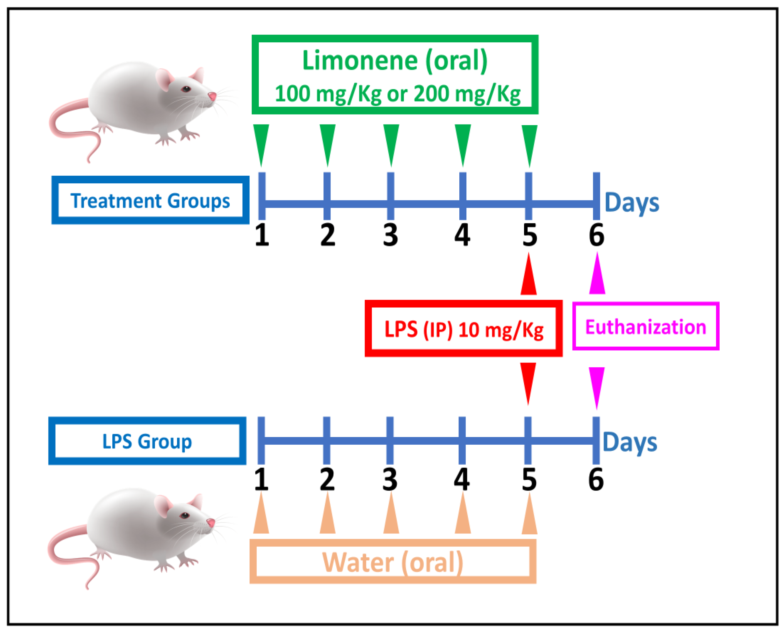
2.2. Animal Experiments
2.3. Blood Collection
2.4. Biochemical Measurements
2.5. The Modified Murine Sepsis Score (MSS) System
2.6. Gene Expression Analysis
2.7. Statistical Analysis
3. Results
3.1. Effects of Limonene on Pro-Inflammatory Cytokines in LPS-Induced Intestinal Injury
3.2. Effects of Limonene on Inflammatory Pathways in LPS-Induced Intestinal Injury
3.3. Effect of Limonene on Oxidative Stress State in LPS-Induced Intestinal Injury
3.4. Effects of Limonene on Murine Sepsis Score (MMS) and Mortality Rate in LPS-Induced Intestinal Injury
3.5. Effect of Limonene on Urea and Creatinine in LPS-Induced Intestinal Injury
4. Discussion
5. Conclusions
Author Contributions
Funding
Institutional Review Board Statement
Data Availability Statement
Conflicts of Interest
References
- Woźniak, D.; Cichy, W.; Przysławski, J.; Drzymała-Czyż, S. The role of microbiota and enteroendocrine cells in maintaining homeostasis in the human digestive tract. Adv. Med. Sci. 2021, 66, 284–292. [Google Scholar] [CrossRef]
- Barbara, G.; Barbaro, M.R.; Fuschi, D.; Palombo, M.; Falangone, F.; Cremon, C.; Marasco, G.; Stanghellini, V. Inflammatory and microbiota-related regulation of the intestinal epithelial barrier. Front. Nutr. 2021, 8, 718356. [Google Scholar] [CrossRef]
- Al-hoshary, D.; Zalzala, M.H. Assessment of ellagic acid action in 5-fluorouracil induced intestinal mucositis: Assessment of ellagic acid action in 5-fluorouracil induced intestinal mucositis. Iraqi J. Pharm. Sci. 2023, 32, 33–41. [Google Scholar] [CrossRef]
- Jung, C.Y.; Bae, J.M. Pathophysiology and protective approaches of gut injury in critical illness. Yeungnam Univ. J. Med. 2021, 38, 27–33. [Google Scholar] [CrossRef]
- Redha, S.M.; Kathem, S.H. Citronellol exerts lung protective and anti-inflammatory effects through hampering NF-κB in lipopolysaccharide-induced acute lung injury in mice. FASEB J. 2022, 36, S1. [Google Scholar] [CrossRef]
- Ghosh, S.S.; Wang, J.; Yannie, P.J.; Ghosh, S. Intestinal barrier dysfunction, LPS translocation, and disease development. J. Endocr. Soc. 2020, 4, bvz039. [Google Scholar] [CrossRef]
- Li, Q.; von Ehrlich-Treuenstätt, V.; Schardey, J.; Wirth, U.; Zimmermann, P.; Andrassy, J.; Bazhin, A.V.; Werner, J.; Kühn, F. Gut Barrier Dysfunction and Bacterial Lipopolysaccharides in Colorectal Cancer. J. Gastrointest. Surg. 2023, 27, 1466–1472. [Google Scholar] [CrossRef]
- Shareef, S.M.; Kathem, S.H. Gentiopicroside ameliorates acute inflammatory kidney injury in a mouse model induced by LPS through hampering NF-κB, AP-1 and IRF3 pathways. FASEB J. 2022, 36, S1. [Google Scholar] [CrossRef]
- Nasrawi, Y.; Kadhim, S.H. Protective Effects of Citronellol Against Rhabdomyolysis-Induced Acute Kidney Injury in Mice by Inhibiting NF-κB and IL-1β. Iraqi J. Pharm. Sci. 2023, 32, 85–90. [Google Scholar] [CrossRef]
- Zimmerman, J.D. Immunoprotective Effects of Lipopolysaccharide Detoxification by a Novel Microbial-Derived Alkaline Phosphatase. Ph.D. Thesis, University of Illinois at Urbana-Champaign, Urbana, IL, USA, 2019. [Google Scholar]
- Zielińska-Błajet, M.; Pietrusiak, P.; Feder-Kubis, J. Selected monocyclic monoterpenes and their derivatives as effective anticancer therapeutic agents. Int. J. Mol. Sci. 2021, 22, 4763. [Google Scholar] [CrossRef] [PubMed]
- Dhureja, M. Neuroprotective Potential of Limonene in LPS Induced Parkinson’s Disease in Rats. Alzheimer’s Dement. 2023, 19, e067741. [Google Scholar] [CrossRef]
- Lin, W.T.; He, Y.H.; Lo, Y.H.; Chiang, Y.T.; Wang, S.Y.; Bezirganoglu, I.; Kumar, K.S. Essential Oil from Glossogyne tenuifolia Inhibits Lipopolysaccharide-Induced Inflammation-Associated Genes in Macro-Phage Cells via Suppression of NF-κB Signaling Pathway. Plants 2023, 12, 1241. [Google Scholar] [CrossRef] [PubMed]
- Uedo, N.; Tatsuta, M.; Iishi, H.; Baba, M.; Sakai, N.; Yano, H.; Otani, T. Inhibition by d-limonene of gastric carcinogenesis induced by N-methyl-N′-nitro-N-nitrosoguanidine in Wistar rats. Cancer Lett. 1999, 137, 131–136. [Google Scholar] [CrossRef] [PubMed]
- Vigushin, D.M.; Poon, G.K.; Boddy, A.; English, J.; Halbert, G.W.; Pagonis, C.; Jarman, M.; Coombes, R.C.; Cancer Research Campaign Phase I/II Clinical Trials Committee. Phase I and pharmacokinetic study of D-limonene in patients with advanced cancer. Cancer Chemother. Pharmacol. 1998, 42, 111–117. [Google Scholar] [CrossRef]
- Rozza, A.L.; de Mello Moraes, T.; Kushima, H.; Tanimoto, A.; Marques, M.O.; Bauab, T.M.; Hiruma-Lima, C.A.; Pellizzon, C.H. Gastroprotective mechanisms of Citrus lemon (Rutaceae) essential oil and its majority compounds limonene and β-pinene: Involvement of heat-shock protein-70, vasoactive intestinal peptide, glutathione, sulfhydryl compounds, nitric oxide and prostaglandin E2. Chem.-Biol. Interact. 2011, 189, 82–89. [Google Scholar] [CrossRef] [PubMed]
- Shrum, B.; Anantha, R.V.; Xu, S.X.; Donnelly, M.; Haeryfar, S.M.; McCormick, J.K.; Mele, T. A robust scoring system to evaluate sepsis severity in an animal model. BMC Res. Notes 2014, 7, 233. [Google Scholar] [CrossRef] [PubMed]
- Yu, Y.; Xu, X.; Liu, L.; Mao, S.; Feng, T.; Lu, Y.; Cheng, Y.; Wang, H.; Zhao, W.; Tang, W. Progranulin deficiency leads to severe inflammation, lung injury and cell death in a mouse model of endotoxic shock. J. Cell. Mol. Med. 2016, 20, 506–517. [Google Scholar] [CrossRef]
- Kathem, S.H. The Protective Effect of Ethanolic Extract of Mentha spicata Against Irinotecan-Induced Mucositis in mice. Iraqi J. Pharm. Sci. 2019, 28, 37–43. [Google Scholar]
- Mahmood, Y.S.; Kathem, S.H. Protective effect of citronellol in rhabdomyolysis-induced acute kidney injury in mice. J. Med. Life 2023, 16, 1057. [Google Scholar] [CrossRef]
- Mahde, S.; Haishm, S. Carvone exerts lung anti-inflammatory effect through hampering NF-KB pathway in lipopolysaccharide-induced acute lung injury in mice. Iraqi J. Pharm. Sci. 2023, 32, 125–132. [Google Scholar] [CrossRef]
- Shareef, M.S.; Kathem, H.S. Gentiopicroside ameliorates lipopolysaccharide-induced acute kidney injury by inhibiting TLR4/NF-κB signaling in mice model. J. Pharm. Negat. Results 2022, 13, 135–145. [Google Scholar]
- Cortese, F.; Loponte, M.; Rossi, S.; Picardi, B.; Rossi Del Monte, S.; Fransvea, P. Acute Gastrointestinal Injury. In Infections in Surgery: Prevention and Management; Springer: Cham, Switzerland, 2021; pp. 179–199. [Google Scholar]
- Lin, D.; Yang, L.; Wen, L.; Lu, H.; Chen, Q.; Wang, Z. Crosstalk between the oral microbiota, mucosal immunity, and the epithelial barrier regulates oral mucosal disease pathogenesis. Mucosal Immunol. 2021, 14, 1247–1258. [Google Scholar] [CrossRef]
- Wu, S.; Pan, L.; Liao, H.; Yao, W.; Shen, N.; Chen, C.; Liu, D.; Ge, M. High-fat diet increased NADPH-oxidase-related oxidative stress and aggravated LPS-induced intestine injury. Life Sci. 2020, 253, 117539. [Google Scholar] [CrossRef]
- Xiong, W.; Ma, H.; Zhang, Z.; Jin, M.; Wang, J.; Xu, Y.; Wang, Z. The protective effect of icariin and phosphorylated icariin against LPS-induced intestinal epithelial cells injury. Biomed. Pharmacother. 2019, 118, 109246. [Google Scholar] [CrossRef]
- Kim, J.Y.; Leem, J.; Hong, H.L. Melittin ameliorates endotoxin-induced acute kidney injury by inhibiting inflammation, oxidative stress, and cell death in mice. Oxidative Med. Cell. Longev. 2021, 2021, 8843051. [Google Scholar] [CrossRef] [PubMed]
- Ioanna, Z.; Katerina, B.; Irene, A. Immunotherapy-on-chip against an experimental sepsis model. Inflammation 2021, 44, 2333–2345. [Google Scholar] [CrossRef]
- Makjaroen, J.; Thim-Uam, A.; Dang, C.P.; Pisitkun, T.; Somparn, P.; Leelahavanichkul, A. A comparison between 1 day versus 7 days of sepsis in mice with the experiments on LPS-activated macrophages support the use of intravenous immunoglobulin for sepsis attenuation. J. Inflamm. Res. 2021, 14, 7243. [Google Scholar] [CrossRef]
- Ozer, E.K.; Goktas, M.T.; Toker, A.; Bariskaner, H.; Ugurluoglu, C.; Iskit, A.B. Effects of carvacrol on survival, mesenteric blood flow, aortic function and multiple organ injury in a murine model of polymicrobial sepsis. Inflammation 2017, 40, 1654–1663. [Google Scholar] [CrossRef] [PubMed]
- Guo, Y.; Li, H.; Liu, Z.; Li, C.; Chen, Y.; Jiang, C.; Yu, Y.; Tian, Z. Impaired intestinal barrier function in a mouse model of hyperuricemia. Mol. Med. Rep. 2019, 20, 3292–3300. [Google Scholar] [CrossRef]
- Xue, X.; Falcon, D.M. The role of immune cells and cytokines in intestinal wound healing. Int. J. Mol. Sci. 2019, 20, 6097. [Google Scholar] [CrossRef] [PubMed]
- Kathem, S.H.; Abdulsahib, W.K.; Zalzala, M.H. Berbamine and thymoquinone exert protective effects against immune-mediated liver injury via NF-κB dependent pathway. Front. Vet. Sci. 2022, 9, 960981. [Google Scholar] [CrossRef]
- Soares, C.L.; Wilairatana, P.; Silva, L.R.; Moreira, P.S.; Barbosa, N.M.; da Silva, P.R.; Coutinho, H.D.; de Menezes, I.R.; Felipe, C.F. Biochemical aspects of the inflammatory process: A narrative review. Biomed. Pharmacother. 2023, 168, 115764. [Google Scholar] [CrossRef]
- Singh, M.; Thakur, M.; Mishra, M.; Yadav, M.; Vibhuti, R.; Menon, A.M.; Nagda, G.; Dwivedi, V.P.; Dakal, T.C.; Yadav, V. Gene regulation of intracellular adhesion molecule-1 (ICAM-1): A molecule with multiple functions. Immunol. Lett. 2021, 240, 123–136. [Google Scholar] [CrossRef]
- Ferrer, M.D.; Busquets-Cortés, C.; Capó, X.; Tejada, S.; Tur, J.A.; Pons, A.; Sureda, A. Cyclooxygenase-2 inhibitors as a therapeutic target in inflammatory diseases. Curr. Med. Chem. 2019, 26, 3225–3241. [Google Scholar] [CrossRef]
- Jang, S.Y.; Kim, S.Y.; Song, H.A.; Kim, H.; Chung, K.S.; Lee, J.K.; Lee, K.T. Protective effect of hydrangenol on lipopolysaccharide-induced endotoxemia by suppressing intestinal inflammation. Int. Immunopharmacol. 2023, 125, 111083. [Google Scholar] [CrossRef]
- Cazanga, V.; Palma, C.; Casanova, T.; Rojas, D.; Barrera, K.; Valenzuela, C.; Acevedo, A.; Ascui-Gac, G.; Pérez-Jeldres, T.; Pérez-Fernández, R. Modulation of the Acute Inflammatory Response Induced by the Escherichia coli Lipopolysaccharide through the Interaction of Pentoxifylline and Florfenicol in a Rabbit Model. Antibiotics 2023, 12, 639. [Google Scholar] [CrossRef]
- Santana, H.S.; de Carvalho, F.O.; Silva, E.R.; Santos, N.G.; Shanmugam, S.; Santos, D.N.; Wisniewski, J.O.; Junior, J.S.; Nunes, P.S.; Araujo, A.A.; et al. Anti-inflammatory activity of limonene in the prevention and control of injuries in the respiratory system: A systematic review. Curr. Pharm. Des. 2020, 26, 2182–2191. [Google Scholar] [CrossRef]
- Maghsoudi, H.; Yazadanpanah, E. Evaluation of the Anti-Inflammatory Properties of D-limonene Compared with Dexamethasone and Ibuprofen in Bovine Synoviocyte. J. Basic Res. Med. Sci. 2023, 10, 1–14. [Google Scholar]
- Araruna, M.E.; Serafim, C.; Alves Junior, E.; Hiruma-Lima, C.; Diniz, M.; Batista, L. Intestinal anti-inflammatory activity of terpenes in experimental models (2010–2020): A review. Molecules 2020, 25, 5430. [Google Scholar] [CrossRef]
- Lisi, F.; Zelikin, A.N.; Chandrawati, R. Nitric oxide to fight viral infections. Adv. Sci. 2021, 8, 2003895. [Google Scholar] [CrossRef]
- Singh, J.; Lee, Y.; Kellum, J.A. A new perspective on NO pathway in sepsis and ADMA lowering as a potential therapeutic approach. Crit. Care 2022, 26, 246. [Google Scholar] [CrossRef]
- Heemskerk, S.; Masereeuw, R.; Russel, F.G.; Pickkers, P. Selective iNOS inhibition for the treatment of sepsis-induced acute kidney injury. Nat. Rev. Nephrol. 2009, 5, 629–640. [Google Scholar] [CrossRef]
- Lorigooini, Z.; Boroujeni, S.N.; Sayyadi-Shahraki, M.; Rahimi-Madiseh, M.; Bijad, E.; Amini-Khoei, H. Limonene through attenuation of neuroinflammation and nitrite level exerts antidepressant-like effect on mouse model of maternal separation stress. Behav. Neurol. 2021, 2021, 8817309. [Google Scholar] [CrossRef]
- Soulimani, R.; Bouayed, J.; Joshi, R.K. Limonene: Natural monoterpene volatile compounds of potential therapeutic interest. Am. J. Essent. Oils Nat. Prod. 2019, 7, 1–10. [Google Scholar]
- Khan, H.U.; Aamir, K.; Jusuf, P.R.; Sethi, G.; Sisinthy, S.P.; Ghildyal, R.; Arya, A. Lauric acid ameliorates lipopolysaccharide (LPS)-induced liver inflammation by mediating TLR4/MyD88 pathway in Sprague Dawley (SD) rats. Life Sci. 2021, 265, 118750. [Google Scholar] [CrossRef]
- de Souza, M.C.; Vieira, A.J.; Beserra, F.P.; Pellizzon, C.H.; Nóbrega, R.H.; Rozza, A.L. Gastroprotective effect of limonene in rats: Influence on oxidative stress, inflammation and gene expression. Phytomedicine 2019, 53, 37–42. [Google Scholar] [CrossRef]
- Babaeenezhad, E.; Hadipour Moradi, F.; Rahimi Monfared, S.; Fattahi, M.D.; Nasri, M.; Amini, A.; Dezfoulian, O.; Ahmadvand, H. D-Limonene alleviates acute kidney injury following gentamicin administration in rats: Role of NF-κB pathway mitochondrial apoptosis oxidative stress, P.C.N.A. Oxidative Med. Cell. Longev. 2021, 2021, 6670007. [Google Scholar] [CrossRef]
- Song, Z.H.; Tong, G.; Xiao, K.; Jiao, L.F.; Ke, Y.L.; Hu, C.H. L-Cysteine protects intestinal integrity, attenuates intestinal inflammation and oxidant stress, and modulates NF-κB and Nrf2 pathways in weaned piglets after LPS challenge. Innate Immun. 2016, 22, 152–161. [Google Scholar] [CrossRef]
- Zhang, S.; Zhou, Q.; Li, Y.; Zhang, Y.; Wu, Y. MitoQ modulates lipopolysaccharide-induced intestinal barrier dysfunction via regulating Nrf2 signaling. Mediat. Inflamm. 2020, 2020, 3276148. [Google Scholar] [CrossRef]
- Kumar, K.S.; Vani, M.G.; Wang, S.Y. Limonene protects human skin keratinocytes against UVB-induced photodamage and photoaging by activating the Nrf2-dependent antioxidant defense system. Environ. Toxicol. 2022, 37, 2897–2909. [Google Scholar] [CrossRef]
 ; LPS:
  ; limonene 100 mg/kg:
  ; limonene 200 mg/kg:
  .
; LPS: ; limonene 100 mg/kg: ; limonene 200 mg/kg: .
.
; LPS: ; limonene 100 mg/kg: ; limonene 200 mg/kg: . ; LPS: ; limonene 100 mg/kg: ; limonene 200 mg/kg: .
; LPS: ; limonene 100 mg/kg: ; limonene 200 mg/kg: .
; LPS: ; limonene 100 mg/kg: ; limonene 200 mg/kg: . ; LPS: ; limonene 100 mg/kg: ; limonene 200 mg/kg: .
; LPS: ; limonene 100 mg/kg: ; limonene 200 mg/kg: .
; LPS: ; limonene 100 mg/kg: ; limonene 200 mg/kg: .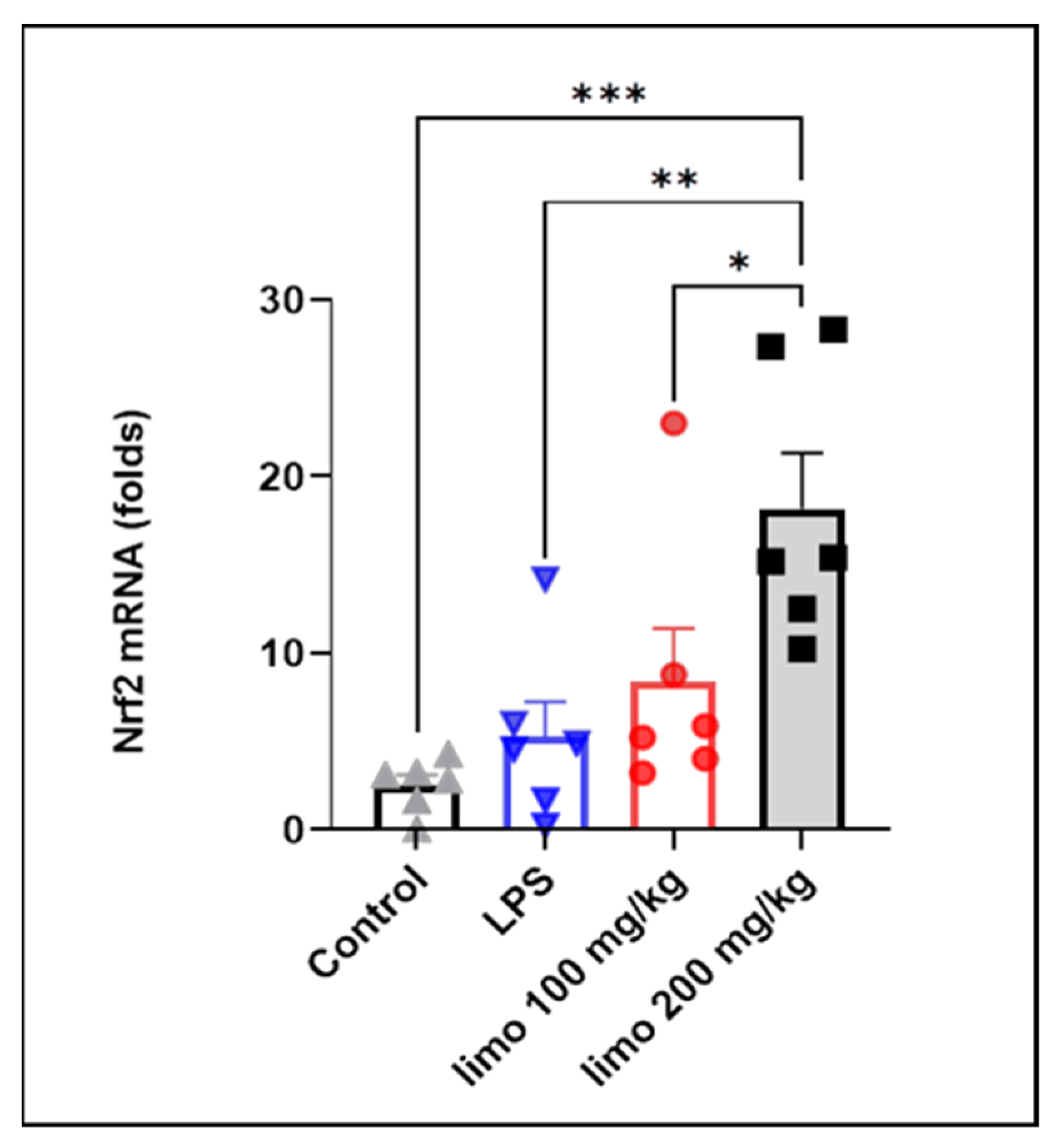 ; LPS: ; limonene 100 mg/kg: ; limonene 200 mg/kg: .
; LPS: ; limonene 100 mg/kg: ; limonene 200 mg/kg: .
; LPS: ; limonene 100 mg/kg: ; limonene 200 mg/kg: .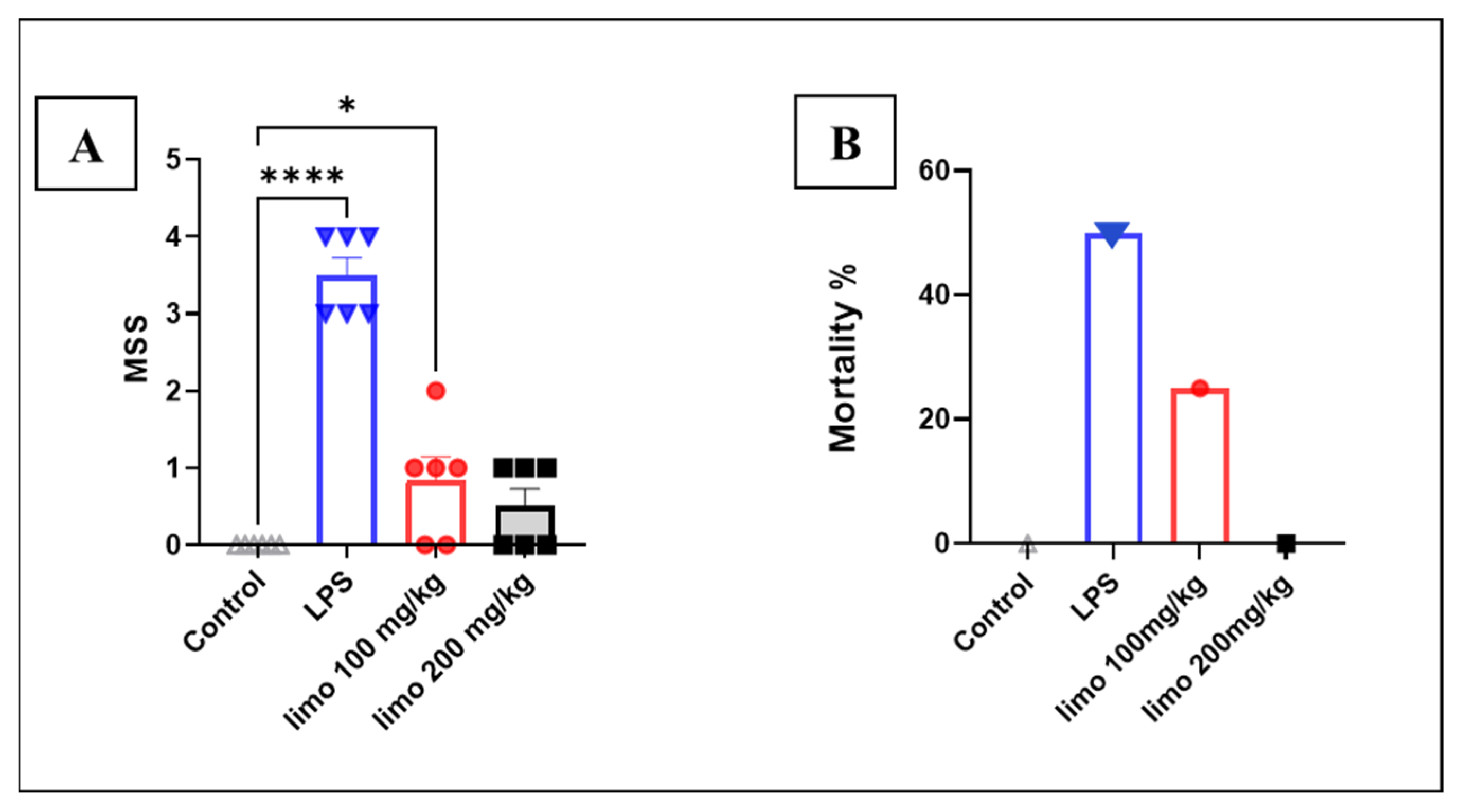 ; LPS: ; limonene 100 mg/kg: ; limonene 200 mg/kg: .
; LPS: ; limonene 100 mg/kg: ; limonene 200 mg/kg: .
; LPS: ; limonene 100 mg/kg: ; limonene 200 mg/kg: .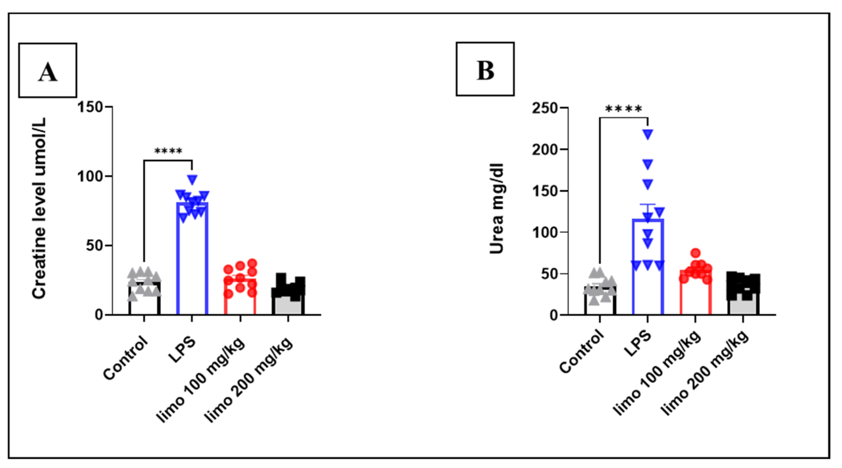

| Primers | Sequences 5′→3′ Direction |
|---|---|
| GAPDH | Forward: CGGGTTCCTATAAATACGGACTG Reverse: CCAATACGGCCAAATCCGTTC |
| TLR4 | Forward: TCCCTGCATAGAGGTAGTTCC Reverse: TCAAGGGGTTGAAGCTCAGA |
| NF-κB | Forward: AAGACAAGGAGCAGGACATG Reverse: AGCAACATCTTCACATCCC |
| IRF3 | Forward: CAATTCCTCCCCTGGCTAGA Reverse: GGGATCCTGAACCTCGTTCG |
| AP-1 | Forward: GCTGCAGGATGATGCGATAG Reverse: TTCTAGCCAGGACGACTTGC |
| iNOS | Forward: GGTGAAGGGACTGAGCTGTT Reverse: ACGTTCTCCGTTCTCTTGCAG |
| IL-1β | Forward: TGCCACCTTTTGACAGTGATG Reverse: TGATGTGCTGCTGCGAGATT |
| TNF-α | Forward: TAGCCCACGTCGTAGCAAAC Reverse: ACAAGGTACAACCCATCGGC |
| COX-2 | Forward: GCTCAGCCAGGCAGCAAATC Reverse: CACCATAGAATCCAGTCCGGG |
Disclaimer/Publisher’s Note: The statements, opinions and data contained in all publications are solely those of the individual author(s) and contributor(s) and not of MDPI and/or the editor(s). MDPI and/or the editor(s) disclaim responsibility for any injury to people or property resulting from any ideas, methods, instructions or products referred to in the content. |
© 2024 by the authors. Licensee MDPI, Basel, Switzerland. This article is an open access article distributed under the terms and conditions of the Creative Commons Attribution (CC BY) license (https://creativecommons.org/licenses/by/4.0/).
Share and Cite
Kathem, S.H.; Nasrawi, Y.S.; Mutlag, S.H.; Nauli, S.M. Limonene Exerts Anti-Inflammatory Effect on LPS-Induced Jejunal Injury in Mice by Inhibiting NF-κB/AP-1 Pathway. Biomolecules 2024, 14, 334. https://doi.org/10.3390/biom14030334
Kathem SH, Nasrawi YS, Mutlag SH, Nauli SM. Limonene Exerts Anti-Inflammatory Effect on LPS-Induced Jejunal Injury in Mice by Inhibiting NF-κB/AP-1 Pathway. Biomolecules. 2024; 14(3):334. https://doi.org/10.3390/biom14030334
Chicago/Turabian StyleKathem, Sarmed H., Yasameen Sh. Nasrawi, Shihab H. Mutlag, and Surya M. Nauli. 2024. "Limonene Exerts Anti-Inflammatory Effect on LPS-Induced Jejunal Injury in Mice by Inhibiting NF-κB/AP-1 Pathway" Biomolecules 14, no. 3: 334. https://doi.org/10.3390/biom14030334
APA StyleKathem, S. H., Nasrawi, Y. S., Mutlag, S. H., & Nauli, S. M. (2024). Limonene Exerts Anti-Inflammatory Effect on LPS-Induced Jejunal Injury in Mice by Inhibiting NF-κB/AP-1 Pathway. Biomolecules, 14(3), 334. https://doi.org/10.3390/biom14030334