Article

Growing Maize Root: Lectins Involved in Consecutive Stages of Cell Development

1
Kazan Institute of Biochemistry and Biophysics, Federal Research Center Kazan Scientific Center of Russian Academy of Sciences, Lobachevsky Str. 2/31, Kazan 420111, Russia
2
Institute of Fundamental Medicine and Biology, Kazan Federal University, Kremlevskaya Str. 18, Kazan 420008, Russia
3
Institute of Physiology, Federal Research Center Komi Science Center of Ural Branch of Russian Academy of Sciences, Kommunisticheskaya Str. 28, Syktyvkar 167982, Russia
*
Author to whom correspondence should be addressed.
Plants 2022, 11(14), 1799; https://doi.org/10.3390/plants11141799
Submission received: 14 June 2022 / Revised: 1 July 2022 / Accepted: 4 July 2022 / Published: 7 July 2022
(This article belongs to the Special Issue Root: From Signaling to Morphogenesis)

Abstract

:
Proteins that carry specific carbohydrate-binding lectin domains have a great variety and are ubiquitous across the plant kingdom. In turn, the plant cell wall has a complex carbohydrate composition, which is subjected to constant changes in the course of plant development. In this regard, proteins with lectin domains are of great interest in the context of studying their contribution to the tuning and monitoring of the cell wall during its modifications in the course of plant organ development. We performed a genome-wide screening of lectin motifs in the Zea mays genome and analyzed the transcriptomic data from five zones of primary maize root with cells at different development stages. This allowed us to obtain 306 gene sequences encoding putative lectins and to relate their expressions to the stages of root cell development and peculiarities of cell wall metabolism. Among the lectins whose expression was high and differentially regulated in growing maize root were the members of the EUL, dirigent–jacalin, malectin, malectin-like, GNA and Nictaba families, many of which are predicted as cell wall proteins or lectin receptor-like kinases that have direct access to the cell wall. Thus, a set of molecular players was identified with high potential to play important roles in the early stages of root morphogenesis.

Graphical Abstract

1. Introduction

Plant morphogenesis is based on individual cell enlargement—a highly dynamic process that involves various aspects such as the increase in cell surface and vacuole. Cell wall extension—the necessary component of cell enlargement—has to be finely tuned in order to coordinate the growth of neighboring cells and to shape the entire organ. Several proteins with lectin domains are reported to be the possible elements for monitoring the cell wall state and evolving signals that regulate organ development [1,2].
Lectin domains are a group of different structural units of proteins capable of specifically recognizing carbohydrate structures [3]. They are found in combination with a wide variety of protein domains and are involved in various physiological processes [4]. Although some lectin motifs are very similar in plants, animals, and bacteria, the combinations of lectin domains with other domains in proteins are most often different in different kingdoms of life and, as a result, the functional role of proteins may vary. In plants, lectins are quite numerous: in total, several hundred genes for proteins with lectin domains have been identified in full-genome studies on higher plant species, namely Arabidopsis thaliana, rice, cucumber, soybean, mulberry and flax [5,6,7,8,9,10].
Plant lectins are quite divergent and can be localized in various cell compartments, suggesting differences in their function. The earliest described and best-known lectins, such as Concanavalin A from jack bean (Canavalia ensiformis) or phytohemagglutinin from the common bean (Phaseolus vulgaris), are those accumulating in large quantities in vacuoles, especially in the storage tissues (cotyledons), to the level of 1% to 8% of the total protein [11]. Another group of lectins has a nucleocytoplasmic location; they are synthesized on the free ribosomes in the cytoplasm, do not possess a signal peptide, remain in the cytoplasm or can be translocated into the nucleus [12]. Such lectins have low abundance in normal conditions of plant development but are induced in the course of stress reactions [13]. A large proportion of lectins are transmembrane proteins; their lectin domains are oriented outside of the plasma membrane, and together with lectins fully located in the apoplast, they have direct access to the cell wall polysaccharides. Such lectins are present in small amounts and can be considered as components of signaling pathways, especially as many of them have kinase domains and are named lectin receptor-like kinases (LecRLK). Some members of the latter group are involved in plant–pathogen interactions [14]. However, proteins with lectin domains located within the cell wall are assumed to participate in the process of cell growth as demonstrated in A. thaliana for lectin receptor-like kinases Hercules receptor kinase 1 (HERK1), THESEUS1 (THE1), and FERONIA (FER) [1]. The plant cell wall has a remarkably varied carbohydrate composition, providing a variety of possible ligands that might be the cause of the emergence of a wide variety of plant lectins themselves. The involvement of proteins in lectin domains in the process of cell elongation has not yet received adequate and systemic attention.
The renowned model to study plant growth is the primary root, which is characterized by the presence of consecutive zones that differ in cell sizes and relative rates of their elongation [15]. In proteomic studies, numerous lectins have been identified in roots [16,17]; however, information on their presence can only be found in the supplementary files of the mentioned papers and was not analyzed and discussed. Correspondingly, the dynamics of proteins with lectin domains has never been related to the process of root elongation, especially as the sampling for proteomic studies followed different logics.
Maize (Zea mays) root tip contains cell zones at different stages of cell development that can be easily distinguished. Previously, transcriptomic data for five zones of maize root apex were published, including a comprehensive analysis of expressed genes for glycosyltransferases and glycoside hydrolases involved in cell wall metabolism [18,19]. The samples that were also extensively analyzed by immunocytochemistry for cell-wall-related epitopes included root cap, meristem zone, early elongation, active elongation and late elongation zones, making it possible to relate gene expression to specific early stages of root morphogenesis. In the current article, we present the results of genome screening for maize genes that encode proteins with lectin domains and relate their expression to the stage of root cell development and peculiarities of cell wall metabolism. For the first time, we revealed the genes for proteins with lectin domains that are specifically up-regulated at the initiation of cell elongation and are important in the course of active elongation or late elongation, opening the door for further studies on the molecular mechanisms of lectin involvement in plant morphogenesis.

2. Results

2.1. Genes for Proteins with Lectin Domains in the Maize Genome

We analyzed the presence of proteins with domains characteristic of various lectin families in the maize genome. In addition to the conventional plant lectin families—amaranthins, calreticulins, C-type lectins, the Euonymus europaeus lectin (EUL) family, the Galanthus nivalis agglutinin (GNA) family, the hevein family, the jacalin-related lectin family, the legume lectin family, the lysin motif (LysM) family, the Nicotiana tabacum agglutinin (Nictaba) family and RicinB lectin family [13]—our list was expanded by several other protein families. As was the case for Jiang et al., we added galactose-binding- and galectin-like-domain-containing proteins to the list, since in many non-plant organisms, these domains fully fit the definition of lectins as proteins carrying at least one carbohydrate-binding domain with no catalytic activity toward their own ligands [6,14]. In plant proteins, these domains occur in combination with additional domains characteristic of β-galactosidases and galactosyltransferases, respectively. We also included the malectin and malectin-like families. The malectin domain is classified as CBM (CBM57) in the CAZy database [20], indicating it as a part of a carbohydrate-active enzyme; however, in plants, as distinct from some non-plant organisms, it is not combined with the domains that are enzymatically active on carbohydrates [10,21].
A search for lectin domains in the maize genome yielded 306 putative genes for proteins with lectin domains (Table 1). The identified maize genes encoded members of 15 different families of proteins with lectin domains. No genes were found for the Agaricus bisporus agglutinin (ABA) and cyanovirin-N (CV-N) families. These lectins were previously detected among plants only in mosses or algae, respectively [22]. The search for the class V chitinases domain that is characteristic of the CRA family in the maize genome yielded no results.
Four lectin families were the most abundant in the maize genome: GNA, legume, malectin-like and LysM (Table 1). They include numerous LecRLKs, which together with the corresponding lectin domain possess a kinase domain (PF00069) and a transmembrane domain. LecRLKs are also common in the malectin family, whose members are structurally similar to malectin-like proteins but less numerous. Another LecRLK family is C-type lectins, which are common in animals and almost absent in plants [13]. This is the smallest maize lectin family, with only one representative identified (Zm00001d040192). A kinase domain was also found in three members of the jacalin-related lectin family (Table 1), although they are probably not receptor-like kinases because they lack a signal peptide and a transmembrane domain.
Among 306 maize genes encoding proteins with lectin domains, 253 were predicted to be localized in the cell wall (termed extracellular in the prediction services) or in the plasma membrane, where they have direct access to cell wall components (Table S1). Proteins with predicted localization in the plasma membrane were members of the GNA, legume, LysM, malectin-like, malectin, and C-type families, whereas proteins with potential localization in the cell wall belonged to the EUL, galactose-binding, hevein, RicinB, and jacalin-related lectin families. Most of the proteins with direct access to cell wall polymers were represented by transmembrane LecRLKs; they have a lectin domain outside the plasma membrane, whereas a kinase domain faces the cytoplasm.

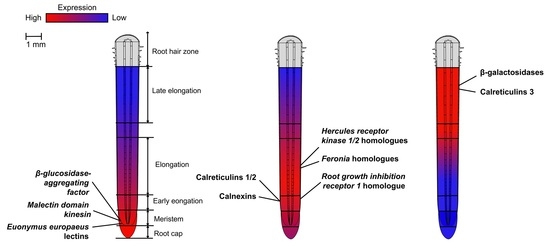
2.2. Expression of Genes of Proteins with Lectin Domains in Various Zones of Growing Maize Root

Over 70% of the genes of proteins with lectin domains (223 of 306) were expressed in growing maize root (TGR over 16 in at least one of the samples) (Table 1). Expression values for all 306 maize genes are presented in the Supplementary Material (Table S1). A hierarchical cluster analysis was performed to better understand the expression patterns of the lectin genes in the samples studied. As a result, 223 expressed genes formed five clusters (Figure 1).
The first cluster (Figure 1) included the genes with the highest mRNA levels at the very tip of the primary root, in the root cap and meristem. The genes with the highest TGR values in this cluster were the genes of the jacalin-related lectin family (Zm00001d019312) and two EUL genes (Zm00001d022421 and Zm00001d028998) (Table 2). An interesting malectin gene (Zm00001d027337) also belonged to this cluster—its ortholog (At2g22610) from A. thaliana encodes the protein, which in addition to the lectin domain, contains a kinesin motor domain (Table 2). The most significant fold drop in mRNA abundance in the following zone of early elongation was detected for one of the LysM genes (Zm00001d021447) (Table 2).
The second cluster consisted of genes with significantly increased mRNA levels in the early elongation zone of maize root (Figure 1). Cluster II included genes encoding calreticulins (Zm00001d019283, Zm00001d003857, Zm00001d025305, and Zm00001d005460), which were extremely highly expressed in most of the zones examined but peaked at elongation initiation (Table 2). Some genes encoding β-galactosidases (Zm00001d022621, Zm00001d044290, and Zm00001d048440) and glycosyltransferases (Zm00001d013052, Zm00001d037233, Zm00001d041504, and Zm00001d049617) with lectin domains were also in this cluster (Table 2 and Table S1). The largest fold in expression at the beginning of cell elongation was observed for the genes of the malectin-like (Zm00001d046838) and legume (Zm00001d049741) families (Table 2).
The genes that were up-regulated during the transition from cell division to cell elongation belonged to several clusters (Figure 1). To identify the genes, which more than doubled their mRNA abundance in the early elongation zone compared to the meristem zone and had a significant level of expression, we used the following criteria: Log2(eElong/Mer) > 1 and TGR (eElong) > 100 (Table S1). Among 38 lectin genes fitting these criteria, 26 (68%) were protein kinases from various families: 12 from GNA, 6 belonged to malectin or malectin-like, 3 from the legume family, and 2 were from LysM family. Moreover, all three maize jacalins with a protein kinase domain (Zm00001d010169, Zm00001d012208, and Zm00001d048876) fell into this group. Lectins without a kinase domain up-regulated at the beginning of elongation included three genes encoding proteins with LysM domains, three with malectin or malectin-like domains, and two with Nictaba domains; Zm00001d040190 for the protein containing the EUL domain was most highly expressed in this group (Table S1). Virtually all of the genes activated in the early elongation zone were additionally up-regulated in the active elongation zone. The number of genes meeting the Log2(Elong/Mer) > 1 and TGR (Elong) > 100 criteria reached 72 (Table S1).
The third gene cluster was characterized by a high expression during early and active elongation and some decrease in mRNA abundance in the late elongation zone (Figure 1). Similar mRNA level dynamics in growing maize root was observed for the genes of cellulose synthases responsible for primary cell wall formation [18]. This cluster contained some LecRLK genes from the malectin-like family (e.g., Zm00001d029047, Zm00001d047533, Zm00001d018789 homologous to FER and HERK), Nictaba family genes (e.g., Zm00001d029673) and jacalin-related lectins (e.g., Zm00001d010169). The only EUL gene (Zm00001d040190) belonging to this cluster was highly expressed (Table 2). Some of the above mentioned genes (FER Zm00001d029047, Nictaba Zm00001d029673, EUL Zm00001d040190) and several others (e.g., GNA Zm00001d043252) were co-expressed with the genes of cellulose synthases associated with the primary cell wall (Table S1).
The fourth cluster was characterized by the expression opposite to that of cluster I (Figure 1). It contained the largest number of expressed genes: 59. For these, mRNA levels increased from the root tip to the late elongation zone. This cluster contained two other members of the calreticulin family (Zm00001d038648 and Zm00001d012170); however, these had a much weaker expression than the members of this family in clusters I and II. Some genes encoding members of the malectin (e.g., Zm00001d002313 and Zm00001d026306) and malectin-like (e.g., Zm00001d027439) families also showed a similar expression pattern (Table 2).
The fifth cluster included genes whose expression was the highest in the zone of late elongation of maize root (Table 1). Although the fifth cluster included quite a few genes, their expression was relatively low. The highest value was shown by β-galactosidase with lectin domain (Zm00001d042654) (Table 2 and Table S1). Genes that were co-expressed with the genes of secondary cell wall cellulose synthases belonged to the fourth and fifth clusters. These included members of calreticulins (maize homolog of CRT3, Zm00001d038648), GNA (Zm00001d007789), LysM (Zm00001d027533) and some other families (Table S1).

2.3. Phylogenetic Analysis of Gene Families Encoding Proteins with Lectin Domains

We performed a phylogenetic analysis to further characterize maize families of proteins with lectin domains and to identify their members homologous to the described representatives of corresponding families from other plant species. The major focus was given to the families with a high proportion of the genes that were actively and differentially expressed in the analyzed samples. As such, the remarkable one is the plant-specific Euonymus europaeus lectin (EUL) family that contains eight genes in maize. All identified members were expressed in growing maize root, and six of them belonged to the top 10 groups from various clusters (Table 2). In maize, EUL domains (in protein databases the EUL domain, PF14200, is still designated as Ricin_B-like_lectin, which should not be confused with the lectin domain of RicinB family, PF00652) are not combined with any other domains within one protein (Table S1), similar to other plant species [1,22]. Double EUL domains were detected in three maize EULs (Table S1). In dicots, EULs are represented by 1–2 genes carrying one lectin domain; the monocot EUL family is more complex [23]. In the phylogenetic tree, genes of monocots and dicots were organized into separate clades, and the same was true for the genes containing single (EULS) and double (EULD) domains (Figure 2).
An exceptionally high expression level was detected for the calreticulins (Table 2), which are evolutionarily conserved lectins mainly localized in the endoplasmic reticulum. Maize calreticulins are divided into several groups on the phylogenetic dendrogram (Figure 3). Calreticulins 1/2, also known as CRT1a/CRT1b, form one group, while calreticulins 3 form two other groups. Additionally, there is a group of calnexins—membrane-bound versions of calreticulins. Calreticulins 1 and 2 are paralogs of each other and correspond to the two types of animal calreticulins, while type 3 calreticulins are specific for plants [24]. Although calreticulins are considered to be constitutively expressed genes, only about half of all maize genes were expressed in the primary root (Table S1). Five calreti-culin genes with no expression in maize roots had a length of amino acid residues that was much shorter than the estimated average length of plant calreticulins (420 aa) [25], so their functional integrity can be questioned. Interestingly, maize calnexins (Zm00001d003857 and Zm00001d025305) and calreticulins 1/2 (Zm00001d019283 and Zm00001d005460) had a similar expression pattern, while two expressed type 3 calreticulins (Zm00001d012170 and Zm00001d038648) had a completely different expression pattern and were considerably activated at the onset of cell elongation (Figure 3, Table S1).
A pronounced differential expression was demonstrated by many members of structurally related malectin and malectin-like families (Table 2). The malectin-like domain consists of two tandem malectin domains [26]. Malectins are widely distributed in nature (in particular, in animals), while the malectin-like domain is found exclusively in plants (Archaeplastida) [22]. In the phylogenetic tree, proteins containing malectin or malectin-like domains were separated into different groups (Figure 4), similar to A. thaliana genes [26]. Malectin-like proteins themselves were subdivided into two groups according to domain organization. The first group, usually called CrRLKs (Catharanthus roseus RLK1-like kinases), included only members with MLD (malectin-like domain) and protein kinase (PK) domain. Another group included mostly LRR-containing proteins with different domain organization. In the extended phylogenetic tree for malectin-like proteins from several plant species, the distinct clades containing only monocots’ or dicots’ genes were also quite pronounced (Figure S6).
In maize, 9 malectins and 32 malectin-like proteins were identified (Table 1). They are mostly receptor-like kinases that sometimes carry leucine-rich repeats involved in protein–protein interactions (Table S1). All but one member were predicted to be localized in the plasma membrane. The malectin domain of this exceptional member (Zm00001d027337) is attached to kinesin, and in the phylogenetic tree, it was allocated to a separate clade together with two genes of A. thaliana, MDKIN1 (At1g72250) and MDKIN2 (At2g22610) (Figure 4). Maize MDKIN showed more similarity to MDKIN2 of A. thaliana that is angiosperm-specific, while MDKIN1 was present in a wider range of plants [26]. One more small, separated group in the phylogenetic tree (Figure 4) was formed by two maize genes (Zm00001d039911 and Zm00001d021566) and their A. thaliana homologs (At1g28340 and At1g25570). Both maize genes belonged to the second cluster, shared the same MLD LRR organization and lacked the kinase domain (Table S1).
Three maize genes (Zm00001d029047, Zm00001d047533, and Zm00001d002175) showed a high homology to FERONIA (FER, At3g51550). FER is well-known for its involvement in cell growth and elongation, as are some other malectin-like kinases, like HERCULES 1/2 (HERK1/2, At3g46290/At1g30570) or THESEUS (THE1, At5g54380) (Shinya et al., 2012). Three maize homologs (Zm00001d037544, Zm00001d046838 and Zm00001d028686) of HERK1 (At3g46290) and two homologs (Zm00001d002282 and Zm00001d018789) of HERK2 (At1g30570) genes were also highly expressed in the studied samples. However, no close homolog of THE1 (At5g54380) was found in the maize genome (Figure 4), and the same was true for rice [27].
Jacalin-related lectins, which are mostly mannose-specific proteins with a localization in the cytoplasm or nucleus [13], have a monocot-specific combination of the lectin domain with dirigent or kinase domain [4,13]. In maize, a combination of lectin domain with the dirigent domain (PF03018) or kinase domain (PF00069) was also detected (Table S1). The number of genes in this family varies greatly from species to species. In maize, 20 genes of the jacalin-related lectin family were found (Table S1), while 50 genes were described in A. thaliana, 4 in flax, 8 in cucumber and 28 in rice [5,6,7,10]. Jacalins of dicots and monocots were mostly well-separated in the phylogenetic tree (Figure 5). Two maize genes of dirigent jacalin-related lectins (Zm00001d005472 and Zm00001d019312) showed a close homology to the wheat gene TaJA1 (TaJRL4) (Figure 5). One of them (Zm00001d019312) encodes β-glucosidase-aggregating factor (BGAF); it showed an extremely high expression in root cap and meristem (Table S1). Dirigent proteins are ubiquitous in plants and are known for their role in the regio- and stereoselective coupling of phenoxy radicals (e.g., during lignan biosynthesis) [28]. However, dirigent-like TaJA1 is functionally distinct from wheat dirigent protein TaDIR13 and does not have any effect on lignan content in plants [29].
Some previously identified jacalin-related proteins were experimentally determined to have intracellular localization [30]. However, most members of the maize jacalin-related lectin family were predicted to be extracellular (Table S1). Apoplast-located jacalin-related proteins were experimentally identified earlier in several other species and described as unconventionally secreted lectins [31,32]. Though several genes of the jacalin-related lectin family encode proteins carrying the protein kinase domain, they do not appear to have a transmembrane domain (Table S1); therefore, they are considered soluble proteins.
Dendrograms for the rest of the families, members of which were rather poorly expressed in growing maize root, are presented in Supplementary Figures S1–S7. Among them, one of the largest families of maize lectins is the legume family, with 51 identified members. Mostly, legume lectin-domain-containing proteins are abundant in seeds and are present in low concentrations in other plant organs [14]. Indeed, more than half of the legume genes were not expressed in the studied zones of maize root, and the other half were had a relatively low expression in comparison to members of other lectin families (Table S1). A similar situation occurred with the Nictaba proteins; they are known as nucleocytoplasmic stress-inducible lectins and are hardly detectable in plants under unstressed conditions [14]. In maize root, all 18 identified genes were expressed (TGR > 16 in at least one sample), although several had an extremely low level (TGR about 20) (Table S1). According to phylogenetic analyses, some genes of maize and A. thaliana are grouped into distinct clades, both in legume and Nictaba families (Figures S3, S4 and S7).
The largest lectin family in maize, GNA family with 81 identified members, included mostly chimeric proteins containing the mannose-specific lectin_B domain, EGF-like domain (epidermal growth factor-like), S-locus glycoprotein domain, PAN/Apple domain and protein kinase domain (Table S1). The EGF-like domain contributes to disulfide bond formation, the S-locus glycoprotein domain is known to be involved in plant self-incompatibility, and the PAN/Apple domain provides interactions with specific carbohydrates or proteins [4,33,34]. Highly expressed maize GNA genes were in clusters III and IV, showing the highest levels of mRNA in the active or late elongation zones of the primary root (Table 2). According to the phylogenetic analysis, the maize GNA genes were divided into three major clades (Figure S1). The first clade included 24 maize genes with only 2 genes of A. thaliana. The second clade (32 maize genes) included genes with different domain organization, mostly lacking the S-locus glycoprotein domain. The third clade (25 maize genes) was subdivided into three subclades: one containing only A. thaliana genes and two in which A. thaliana and maize genes were mixed. Two of the three genes encoding GNA proteins that had significant mRNA levels were co-expressed with the cellulose synthase genes of the primary and secondary cell wall (Table S1), which may indicate their possible involvement in the synthesis or maintenance of this compartment.
As distinct from members of the described above families, clades of the LysM family contained both genes of dicots and monocots (Figure S5). This family mainly contains lectin receptor-like kinases or proteins with a role in the defense against pathogens. They contain one or a few tandem lectin domains that have a carbohydrate-binding activity toward chitin, chitin oligomers or peptidoglycan [14]. Among maize genes, one gene was found to have an F-box domain that is mostly involved in protein ubiquitination (Table S1) [35]. In maize roots, LysM genes were mostly expressed in the late cell elongation zones, with a few exceptions. Zm00001d021447, which was identified as an ortholog for LYM2 (LysM domain-containing GPI-anchored protein 2, At2g17120) gene of A. thaliana, showed a high expression in the root cap zone (Table 2). Another maize homolog of LYM2Zm00001d027533 was actively expressed in the elongation and late elongation zones. Several homologs of the experimentally characterized receptor-like kinases of A. thaliana that belong to the LysM family were also identified in maize (Figure S5). Among them was Zm00001d031676—the homolog of CERK1 (At3g21630) that is a part of the plant’s innate immune system [14].

3. Discussion

3.1. Numerous Maize Lectins Have Access to Cell Wall Polysaccharides

Generally, the genome of a higher plant contains a large number of genes for proteins with lectin domains distributed over numerous different families. The screening of the maize genome revealed over 300 genes carrying lectin domains; half of these genes encode LecRLKs (Table 1). All maize members of GNA, legume, malectin-like, C-type families and most of LysM and malectin proteins are predicted to be membrane-bound with a lectin domain facing the extracellular space. Therefore, they are potentially able to directly interact with cell wall polysaccharides. In addition to transmembrane proteins, whose lectin domains have access to cell wall carbohydrates, there are members of the jacalin-related lectin, galactose-binding, hevein, RicinB and EUL families that are predicted as cell wall proteins (Table S1).
The cell wall is the main carbohydrate-containing structure of plants, and it is logical to assume that lectin domains, as carbohydrate-binding elements, interact with cell wall polysaccharides. The hierarchical clustering of data on the expression level of all the genes encoding proteins, which have lectin domains in various tissues of flax plants, grouped the analyzed genes according to the cell-wall-type characteristic of the studied samples [10]. This indicates the possibility of interactions between cell wall polymers and proteins with lectin domains. In maize, the co-expression of several genes for lectin proteins from various families with the genes for cellulose synthases of the primary and secondary cell wall was well-pronounced.
However, the direct data on such interactions is scarce. Using an in-gel affinity electrophoresis assay, electrophoresis shift assay, dot-blot analysis and ELISA, several proteins from the malectin-like family and their lectin domains were shown to interact with homogalacturonan; those included FER, as well as its individual malectin domains, ANX1/2, and BUPS1 ectodomains [36]. Further studies reported that the extracellular domain of FER (particularly MALA) specifically binds to de-methylesterified pectin both in vitro and in vivo [37]. In contrast to the above works, the results of the crystallographic resolution of ANX1/2 structures and their comparison with other CBMs suggest that these proteins lack the conserved binding surfaces for carbohydrates and can be designed for other ligands [38,39]. Attempts to assess the binding of these proteins to various carbohydrates (including low methyl-esterified pectin) using isothermal titration calorimetry revealed no such interactions [38]. Thus, the question of the carbohydrate ligands of CrRLK1s, as well as those of other proteins with lectin domains, in plant cell walls remains unanswered.
The effective approach to characterize the presence and specificity of a protein binding to various carbohydrates is the application of glycan microarrays [40,41]. Such arrays contain hundreds of spots with individual glycans for the simultaneous analysis of their interactions with the protein under study. The developed glycan microarrays are mainly focused on the importance of protein glycosylation and mostly contain the oligosaccharidic chains of N-glycosylated proteins. The accumulated data in this field already enable machine-learning to be performed for the annotation of lectin specificity toward various glycans present in N- and O-glycosylated proteins [42]. However, such microarrays barely contain any fragments of plant cell wall polysaccharides and, up to now, have not been used to check the ability of plant lectins to bind cell wall glycans.
In addition, the provided predictions based on the approaches of bioinformatics often differ in various services. For example, most maize jacalin-related proteins are predicted as extracellular by the LocTree3 service; however, they are mostly regarded as cytoplasmic by the DeepLoc service (Table S1). Members of the EUL family are also predicted to be localized in the cytoplasm or cell wall according to the two distinct prediction services (Table S1).
Altogether, plant proteins carrying lectin domains have an obvious potential to interact with cell wall carbohydrate molecules. However, our knowledge on such interactions is currently rather poor, and there is a high demand for research in this direction.

3.2. Comparison of Lectin Sets in Monocots and Dicots

The genome-wide identification of genes encoding proteins with lectin domains was performed for several dicotyledonous plants: cucumber, soybean, mulberry, A. thaliana, and flax; and two monocotyledonous species: rice and maize (current paper), allowing for some generalizations [5,6,7,8,9,10]. Evidently, the number of proteins with a lectin domain in various plant families is variable and species-specific, but it is not dictated by plant membership in one class or another. The only noticeable exception is the EUL family, which is represented by 1–3 genes in dicotyledons and by 5–10 genes in monocotyledons [43].
Nevertheless, significant differences in the domain organization of proteins with lectin domains in dicotyledons and monocotyledons are observed. In particular, the combination of the jacalin domain with the dirigent and kinase domains is the specificity of monocot proteins [44]. Within the EUL family protein, the EUL domain is not combined with any other domains, but it may be single (designated as EULS) or double (designated as EULD) [43]. Tandem EUL domains are found only in monocotyledonous plants and form a separate clade on the phylogenetic tree (Figure 2). Moreover, maize and rice EULS are also separated from the EULS of dicots into a distinct group (Figure 2).
In addition, in the phylogenetic trees of many families, there are several clades containing only monocot genes or only dicot genes, although they may have similar domain organization, as was shown for the malectin-like, legume, GNA and Nictaba families (Figures S1 and S3–S7). A feature of the CrRLK set of genes in cereals is that they have no orthologs of the THESEUS1 gene in their genomes (Figure S6) [27]. THE1 is required for hypocotyl cell elongation, responses to cell wall damage induced by the inhibition of cellulose biosynthesis, and during plant–pathogen interactions [45]. Perhaps, in cereals, some other protein fulfills functions of THE1.
Some of the differences mentioned, at least in those proteins whose lectin domains have access to cell wall polysaccharides, may be related to peculiarities of cell wall composition. Maize, like rice, has primary cell walls of type II enriched with mixed-linkage glucans and arabinoxylans, at least at the stage of cell elongation, in contrast to dicotyledonous species whose cell walls are poor in these components [46].

3.3. Lectin Genes with Zone-Specific Up-Regulation in Growing Maize Root

In total, 70% of all identified genes for proteins with lectin domains were expressed in the analyzed zones of maize root. In most cases, genes from the same family have different expression patterns. Each zone has its own set of specifically up-regulated genes of proteins with lectin domains, both related and unrelated to the cell wall.

3.3.1. Meristematic Zone

The zone with active cell divisions is characterized by the up-regulated transcription of several EUL genes, namely Zm00001d028998, Zm00001d022421, and Zm00001d024982 (Table 2). The most up-regulated in maize meristematic zone genes for proteins with EUL domains are the homologs of OsEULD1a (Zm00001d028998), and OsEULD1b (Zm00001d022421) (Table 2). In rice, OsEULD1a and OsEULD1b are also highly expressed in the tips of roots, both primary and lateral [23]. A glycoarray analysis revealed that the EUL domain of the sole A. thaliana lectin of this family, ArathEULS3, interacts with N-glycan structures containing Lewis Y, Lewis X and lactosamine; its specificity differs from that of Euonymus europaeus lectin, despite the high sequence identity of these domains [47]. The rice lectin OsEULD1a preferentially binds to galactose-related sugars [48]. Though the EUL family members are often considered as the nucleocytoplasmic proteins involved in plant defense, in A. thaliana, the only member of the EUL family is involved in ABA-induced stomata closure in leaves [14,49]. It could also be important for the inhibition of cell division or cell elongation controlled by ABA signaling in the root cap under osmotic stress conditions [50]. In rice, all five OsEULs are located in the nucleus, although they lack a classical nuclear localization site, while two are also present in the cytoplasm [23]. In summary, the maize root meristem expresses two genes encoding EUL proteins whose specificity, function and localization are not clear yet and require further studies.
Active expression in the zone of cell division was also detected for the maize gene (Zm00001d027337) encoding a kinesin carrying malectin domain protein. This unwanted combination of two distinct domains is specific to plants and can be found in dicots, monocots, and early vascular plants [51]. Two malectin domain kinesins (At1g72250, At2g22610) were reported in A. thaliana and one was found in the genome of Z. mays (Table S1). Malectin domain kinesin 2 (MDKIN2, At2g22610) and its close homologs are actively expressed in the zones of cell proliferation in different organs in A. thaliana and flax [10,51]. Between cell divisions, it is predominantly localized in the nucleus, while during cell division, it appears close to the tubulin spindle or at the phragmoplast midzone [51]. The co-expression of genes for malectin with a kinesin domain and those encoding several cell-cycle-dependent proteins were revealed in flax [52]. MDKIN2 knockdown mutants showed abnormalities in the development of ~60% of seeds with pleiotropic and stochastic defects. However, no evident effect on root growth was reported [51]. Although it is now believed that MDKIN2 protein is important but non-essential in organs, except for seeding and producing pollen, it is still possible that its detailed study may reveal some unknown features. One more gene encoding the malectin-domain-containing protein was also up-regulated in the maize root meristem (Table 2).
The gene for the jacalin-related protein BGAF1 (β-glucosidase-aggregating factor, Zm00001d019312) was found to be actively expressed in the tip of maize root (Table 2). It encodes dirigent domain-containing jacalin-related lectin that specifically interacts with maize β-glucosidase isozymes ZmBGLU1 (Zm00001d023994) and ZmBGLU2 (Zm00001d024037), forming large insoluble complexes [53]. ZmBGLU1 was characterized by exceptionally high expression in growing maize root; however, in contrast to BGAF1, the highest levels of ZmBGLU1 mRNA were observed in the active and late elongation zones [19]. BGAF1 was identified as a polyspecific lectin with a marked preference for Gal. The physiological role of both BGAF1 and its complex with β-glucosidases is not clear [53]. Interestingly, the ability to form such protein–protein interactions may be unique to maize BGAF1 since the homolog protein from sorghum did not show any specificity toward glucosidases [54]. The close homolog of BGAF1 is wheat jacalin-related lectin TaJA1 (TaJRL4), which is known to confer a basal but broad-spectrum resistance to pathogens [55]. Another homolog is the rice lectin OsJAC1 (LOC_Os12g14440). The expression of its gene was found in young roots and coleoptiles, sheaths, leaves, and nodes of rice stems [56]; injury or pathogen attack led to its significant activation [57]. The overexpression of OsJAC1 resulted, on the one hand, in the suppressed elongation of cells in coleoptiles and internodes [56]. However, on the other hand, the overexpression of OsJAC1 in rice led to a quantitative broad-spectrum resistance against different pathogens, including bacteria, oomycetes and fungi [57]. Afterwards, the possible role of OsJAC1 in resistance to gamma radiation was revealed, and OsJAC1 was suggested to be a key player in DNA damage response in plants [58].
Altogether, the genes up-regulated in the meristematic zone mainly encode intracellularly localized proteins. The proportion of lectin receptor kinases in cluster I, at least those with TGR > 100, was significantly lower than the average for all lectins. In the meri-stematic zone, they constituted 32% (7 of 22) compared to 51% across the whole genome (156 of 306). In the top 10 genes from cluster I (Table 2), only Zm00001d026303 of the malectin family encodes a protein sequence that has the domain structure of a lectin receptor-like kinase (Table S1).

3.3.2. The Early Elongation and Active Elongation

The onset of the cell elongation process in maize primary root is coupled to pronounced changes in the expression profiles of several genes for proteins with lectin domains. The highest expression among all genes for such proteins was detected for calreticulins (Table 2), which are known as highly conserved and ubiquitously expressed proteins located in the endoplasmic reticulum and acting in glycoprotein folding quality control and Ca2+ homeostasis [25,59]. The phylogenetic dendrogram divides maize calreticulins into several groups (Figure 3). Calreticulins 1/2, also known as CRT1a/CRT1b, form one group, while calreticulins 3 form two others. Calnexins, membrane-bound versions of calreticulins, join the fourth group. Two distinct groups of calreticulins (CRT1/2 versus CRT3) were also found by phylogenetic analyses and expression studies in A. thaliana, rice, wheat, Polish canola, and pine (Pinus taeda) [25,60]. The set of genes activated at the beginning of elongation of maize root cells included two calnexins (Zm00001d025305 and Zm00001d003857) and two calreticulins 1/2 (Zm00001d019283 and Zm00001d005460) (Table 2). Calreticulins 1 and 2 of A. thaliana were previously reported to be actively expressed in root tips and other expanding parts of plants [61]. The abundance of calreticulins 1/2 in young tissues can be connected to increased protein synthesis during active growth. Such proteins may include those necessary for cytoskeleton and cell wall reorganization, aquaporins and other transporters, transcription factors, hormone-related proteins, etc. [17].
Among the 38 genes that are more than twofold up-regulated at the transition from cell division to cell elongation (Log2(eElong/Mer) > 1, TGR (eElong) > 100) (Table S1), the predominant group was represented by protein kinases (68%). Out of them, nine genes contained malectin or malectin-like domains. They belong to different clades on the phylogenetic tree (Figure 2); all of them further increase their expression during the active elongation stage and are predicted to be localized in the plasma membrane (Table S1).
The homolog of the RGIR1 (root growth inhibition receptor 1) gene of A. thaliana, Zm00001d052306, showed a high expression in all zones of the maize root with an up-regulation in the early elongation zone (Table 2). RGIR1 is known as a positive regulator of root growth that preferentially acts in the root elongation zone [62]. The maize homolog of RGIR1 contains a malectin domain together with kinase, transmembrane and leucine-rich repeat domains (Table S1), similar to the protein of A. thaliana that was isolated from plasma membrane. Knockdown mutants rgir1-1 have shortened main roots; this phenotype is associated with a lower elongation rate and decreased cortex cell number in the transition zone and elongation zone of the root tip [62]. The maize homolog of RGIR1 may also be involved in root system architecture maintenance.
Two maize homologs of HERK2 and two of FER were significantly up-regulated in the course of active elongation and belonged to cluster III (Table 2 and Table S1). These receptor-like proteins show amazing versatility in terms of control of plant development. FER is known for its role in the tip growth of root hairs and trichomes, cell expansion in roots and petioles, definition of cell shape in the leaf epidermis, maternal control of fertilization and responses to abscisic acid (ABA), among other functions [1,36,63,64,65]. FER is expressed in almost all tissues of A. thaliana, but its expression is increased in the zones of cell expansion. The same observations were made for HERK1 and 2 [1]. The small secreted peptide rapid alkalization factor (RALF) was identified as its protein ligand [65]. Presumably, FER, HERK, and their related proteins function as mechanosensors, regulating cell wall extensibility in multiple ways [66]. The maize homologs of FER and HERK2 gradually increased their expression toward the zone of active elongation and decreased in the zone of late elongation (Table 2 and Table S1). The same expression pattern was demonstrated by the primary cell wall cellulose synthase genes [18]. One homolog of FER was even co-expressed with these cellulose synthases (Table S1). An increase in the area of the primary cell wall requires not only new portions of structural polysaccharides but also the provision of an increasingly larger surface area with the necessary sensors.
Maize has three homologs of FER; one of them, Zm00001d002175, had a different expression character and in phylogeny was grouped with five rice genes (LOC_Os05g25430, LOC_Os04g49690, LOC_Os05g25450, LOC_Os05g25350, LOC_Os05g25370), while the other two maize genes (Zm00001d047533 and Zm00001d029047) were grouped with two remaining rice genes (LOC_Os01g56330 and LOC_Os03g21540) (Figure S7). It is not clear whether the proteins encoded by the genes from this separate subclade have the same function as FER, or whether they have features unique to monocots.
Zm00001d040190 (homolog of rice OsEULS3) from the EUL family was considerably up-regulated and had a high expression level (Table 2 and Table S1). It was also co-expressed with primary cell wall cellulose synthase genes. This correspondence between the expression of the EUL lectin gene and the cellulose synthase genes awaits an explanation.
Another family of genes whose differential expression in the growing root of maize was unexpected is GNA. The GNA family in maize is huge, but its members mostly do not have significant TGR values in the growing root. Nevertheless, three genes were in the top 10 of clusters III and IV (Table 2). Two of these genes (Zm00001d021729 and Zm00001d043252) encode LecRLKs containing five domains (Table S1). The highest mRNA content of both of these genes was found in the zone of active elongation growth, with a further decrease in the zone of late elongation. The A. thaliana homolog of Zm00001d021729 (At4g21390) encodes LecRLK B120. Its function remains unclear; however, it shows a 200-fold up-regulation in response to apoplastic reactive oxygen species [67]. The latter are well-known mediators of cell wall loosening and stiffening during maize root growth [68]. The second of the genes for GNA family proteins, which was up-regulated in the zone of active elongation, was co-expressed with cellulose synthases of the primary cell wall.

3.3.3. The Late Elongation

Many maize genes encoding proteins with lectin domains were up-regulated in the active elongation zone and further increased their mRNA levels in the late elongation zone. These genes were assigned to clusters IV or V (Table 1). In the zone of late elongation, cells of maize root gradually slowed down their growth due to strengthening of the cell wall, cells of vascular tissues deposit a secondary cell wall, and root hairs appear soon after elongation is terminated [18,69].
Maize genes belonging to cluster V and/or co-expressing with the secondary cell wall cellulose synthases have the highest level of mRNA abundance in the zone of late elongation (Table 2 and Table S1). Representatives of Gal-binding, calreticulins 3, and several GNA encoding genes belonged to this group.
β-Galactosidases (EC 3.2.1.23) mediate the hydrolysis of terminal non-reducing β-D-galactose residues in β-D-galactosides. All plant β-galactosidases belong to the glycoside hydrolase (GH) family 35. Thirteen out of sixteen maize β-galactosidases were predicted to have a galactose-binding lectin domain (Table S1). Thus, GH35 proteins and proteins containing the galactose-binding lectin domain are roughly the same. The expression of genes encoding GH35 members in the growing maize root was previously described [19]. It is important to note here that at least one of these genes belonged to the top 10 of each expression cluster (Table 2). This means that not only late elongation but any stage of maize root cell development was characterized by a high level of transcripts for one or another β-galactosidase isoform.
Two calreticulins 3 (Zm00001d012170 and Zm00001d038648) were up-regulated in the active and late elongation zones of maize roots. Calreticulins 3 are specific for land plants and differ from the more common calreticulins 1/2 in the structure of the C-domain [25]. This domain provides the interaction of calreticulin 3 with the misfolded form of brassinosteroid receptor BRI1 and its retention in the endoplasmic reticulum [25,70]. BRI1 homologs, BRL1 and BRL3, are involved in vascular tissue differentiation [71]. It is possible that the expression of calreticulins 3 in maize root reflects the development of the phloem and xylem cells. This is also indicated by the co-expression of one of the maize CRT3 with the cellulose synthase genes of the secondary cell wall (Table S1).
Genes of the extensive but poorly characterized GNA lectin family mostly had a low expression in the growing maize root. However, at least one of them (Zm00001d007789) demonstrated a remarkable up-regulation in the late elongation zone. It has no characterized homologs and, in contrast to GNA members that are up-regulated in the active elongation zone, encodes a sequence containing only three domains. This gene was co-expressed with cellulose synthases of the secondary cell wall. This co-expression may indicate that GNA LecRLKs encoded are required for the construction of a particular type of cell wall.

4. Materials and Methods

4.1. Identification of Genes for Proteins with Lectin Domains in the Maize Genome

The maize genes for proteins containing lectin domains were identified by name search of characteristic pfam domains (Pfam 34.0 database) in the Phytozome v13 database (Zea mays genome assembly RefGen_V4) [72].
The pfam domain names that were used to identify genes are listed in Table 1. Since there is no pfam number for the CRA lectin family, members of this family were searched for the presence of cd02879 (GH18_plant_chitinase_class_V) domain using the CD-search tool [73]. Nevertheless, not a single gene was found. Domain searches for PF14200 (EUL) and PF00652 (RicinB) yielded similar results, so an additional search using PTHR ID was performed for the EUL family.
Protein sequences of maize genes were obtained from the Phytozome database. The presence of lectin domains in the proteins encoded by identified genes was confirmed using the web CD-search tool [73]. The domain organization of maize lectins was resolved using the web tool InterProScan [74].
Amino acid sequences encoded by some genes were re-predicted in the web service Augustus in cases of the presence of non-characteristic domains in the sequence [75]. In cases where Augustus did not give a satisfactory result, the amino acid sequence was re-predicted using the FGENESH web service [76]. If, for any reason, a reliable sequence was not obtained, the sequence taken from the Phytozome database was used. In cases where amino acid sequences were found to not carry lectin domain after analysis with the CD-search and InterProScan services, they were excluded from further study.
All protein sequences were checked both for the presence of a signal peptide with the SignalP-5.0 and TargetP services and for the presence of a transmembrane domain in the TMHMM v2.0 service [77,78,79]. The putative subcellular localization of proteins with lectin domains was predicted using the web services LocTree3 and DeepLoc-1.0 [80,81].

4.2. Phylogenetic Analysis

In addition to maize genes, the genes of A. thaliana and Oryza sativa were taken from the Phytozome v13 database for phylogenetic analysis. The search was carried out by the pfam domain name and characteristics of a certain lectin family (Table 1). Protein sequences of wheat, soybean and flax genes were taken from previous studies [6,10,44].
The multiple alignment of protein sequences was performed using the ClustalW web service for further phylogenetic analysis [82]. The sequence alignments were used to build phylogenetic trees in the IQTREE1.6.12 software using maximum-likelihood phylogenetic analysis [83]. The best-fit model was automatically selected by ModelFinder, and the best-fit model of sequence evolution was selected using the Bayesian Information Criterion (BIC) [84]. The ultrafast bootstrap branch support with 10,000 replicates was used to construct each dendrogram [85]. Only trees with a Robinson–Foulds distance equal to or less than 4 were accepted as reliable. In the case of an additional tree (Figure S4), a distance value equal to 6 was considered as appropriate since it included a large number of sequences.
The trees were visualized using the iTOL 6.3.2 web service and edited in Adobe Illustrator CC 2017 [86]. Their homology was defined using built trees and protein BLAST [87]. The A. thaliana and O. sativa gene descriptions were taken from The Arabidopsis Information Resource (https://www.arabidopsis.org/index.jsp, accessed on 15 April 2022) and the funRiceGenes dataset [88].

4.3. Analysis of Transcriptomic Data

The previously obtained transcriptomic data for 5 zones of the apical part of the primary maize (Zea mays L.) root (before the root hairs initiation) (Figure 1) were used for the analysis of gene expression [18]. Zones that contained cells at different stages of development were distinguished depending on the distance from the root apex: the root cap, the meristem zone (the zone of cell division), the zone of early elongation, the active elongation zone and the late elongation zone (Figure 1A). Plant material, RNA extraction, sequencing procedures and initial steps of transcriptomic analysis were previously described [18,19]. Raw data as fastq files were uploaded to the Sequence Read Archive under BioProject accession number PRJNA639682. Briefly, after pre-processing with the BBDuk utility (https://jgi.doe.gov/data-and-tools/bbtools/bb-tools-user-guide/bbduk-guide/, accessed on 5 May 2022), the resulting clean reads were mapped to (against) the genome using the StringTie v2.0 software [89]. DESeq2′s statistical model was used for normalization and differential expression analysis by the R package DEseq2 v1.30.1 [90].
In total, 26,936 out of 44,146 genes were considered to be expressed according to the cut-off total gene reads count (TGR) that was more or equal to 16 at least in one zone [91]. For a better understanding of the main expression patterns of genes encoding proteins with lectin domains, hierarchical clustering, along with a heatmap analysis, was carried out using R packages (cluster, ggplot2, heatmap.2 et al.) in RStudio v.1.4 [92].
To correlate the expression of genes of proteins with the lectin domain and the synthesis of primary and secondary cell walls in maize root, co-expression networks with cellulose synthases were constructed using the CoExpNetViz (Comparative Co-Expression Network Construction and Visualization) tool. Pairwise similarity between gene expression profiles was calculated using the Pearson correlation measure (0.995) and a set of “bait” genes. Two groups of bait genes were used: six genes encoding putative cellulose synthases of the primary cell wall (Zm00001d019317, Zm00001d037636, Zm00001d005250, Zm00001d019149, Zm00001d009795, and Zm00001d005478) and four genes encoding putative cellulose synthases of the secondary cell wall (Zm00001d043477, Zm00001d032776, Zm00001d020531, and Zm00001d005775) [18]. The resulting co-expression networks were designated as primary and secondary cell wall modules (PCW and SCW), respectively.

5. Conclusions

The early stages of maize root morphogenesis employ hundreds of proteins with lectin domains. Such proteins are highly diverse and belong to all families found in higher plants; they can be localized in different cellular compartments and perform various functions. The onset of cell elongation is coupled with an up-regulation of genes for a subset of lectins, which is strongly enriched in proteins with kinase domains and predicted to be localized in the plasma membrane. Lectin domains of such kinases have direct access to cell wall glycans and can modulate and sense their inevitable rearrangements during cell surface expansion.
An analysis of the expression of genes encoding proteins with lectin domains in the growing maize root revealed some genes whose high and regulated expression during root growth was expected. For example, these include the maize homologs of FERONIA and HERCULES, which are involved in maintaining the integrity of cell walls, and calreticulins, essential for the intensive synthesis of glycosylated proteins that naturally accompany cell growth. Some families of lectins, such as amaranthins and the legume family, showed neither significant levels of mRNA nor a substantial regulation of it. Among the lectins whose expression was surprisingly high and differentially regulated in growing maize root were EULs, dirigent–jacalins, malectins, GNA and Nictaba. The functions of these proteins and their close homologs have either not been established or have not yet been linked to growth processes. Thus, a comprehensive transcriptome analysis of the expression of all lectin genes allowed us to identify a set of molecular players with a high potential to play important roles in the early stages of root morphogenesis. Further studies on the specificity of individual lectin domains in plants, especially toward cell wall glycans and those metabolic and signaling pathways in which the corresponding proteins are involved, are necessary for progress to be made in plant developmental biology.

Supplementary Materials

The following supporting information can be downloaded at: https://www.mdpi.com/article/10.3390/plants11141799/s1, Figure S1: The phylogenetic dendrogram of Galanthus nivalis agglutinin (PF01453) family members of A. thaliana and Z. mays; Figure S2: The phylogenetic dendrogram of hevein (PF00187) lectin family members of A. thaliana and Z. mays; Figure S3: The phylogenetic dendrogram of legume (PF00139) lectin family members of A. thaliana and Z. mays; Figure S4: The phylogenetic dendrogram of legume (PF00139) lectin family members of A. thaliana, Z. mays, G. max and O. sativa; Figure S5: The phylogenetic dendrogram of LysM (PF01476) lectin family members of A. thaliana, Z. mays and G. max; Figure S6: The phylogenetic dendrogram of malectin-like (PF12819) family members of A. thaliana, Z. mays, L. usitatissimum and O. sativa; Figure S7: The phylogenetic dendrogram of Nictaba (PF14299) lectin family members of A. thaliana, Z. mays, L. usitatissimum and O. sativa; Table S1: List of genes encoding proteins with lectin domains in Z. mays.

Author Contributions

Conceptualization, T.G. and A.A.; formal analysis, O.G.; investigation, A.A. and N.P.; data curation, O.G.; writing—original draft preparation, A.A. and T.G.; writing—review and editing, A.A., N.P., O.G., L.K. and T.G.; visualization, A.A.; supervision, T.G.; project administration, T.G.; funding acquisition, L.K. and T.G. All authors have read and agreed to the published version of the manuscript.

Funding

The work was partially supported by the Russian Science Foundation (18-14-00168, transcriptome data; 20-64-47036 bioinformatic analyses). The part of the work was performed with financial support from the Government assignment for the FRC Kazan Scientific Center of RAS (A.A., N.P.). This paper has been supported by the Kazan Federal University Strategic Academic Leadership Program (PRIORITY-2030).

Institutional Review Board Statement

Not applicable.

Informed Consent Statement

Not applicable.

Data Availability Statement

The data presented in this study are available within the article.

Conflicts of Interest

The authors declare no conflict of interest.

References

  1. Hongqing, G.; Lei, L.; Huaxun, Y.; Xiaofei, Y.; Alexandria, A.; Yanhai, Y. Three related receptor-like kinases are required for optimal cell elongation in Arabidopsis thaliana. Proc. Natl. Acad. Sci. USA 2009, 106, 7648–7653. [Google Scholar] [CrossRef] [Green Version]
  2. Li, C.; Wu, H.-M.; Cheung, A.Y. FERONIA and her pals: Functions and mechanisms. Plant Physiol. 2016, 171, 2379–2392. [Google Scholar] [CrossRef] [PubMed] [Green Version]
  3. Van Damme, E.J.M.; Peumans, W.J.; Barre, A.; Rougé, P. Plant lectins: A composite of several distinct families of structurally and evolutionary related proteins with diverse biological roles. CRC Crit. Rev. Plant Sci. 1998, 17, 575–692. [Google Scholar] [CrossRef]
  4. Van Holle, S.; De Schutter, K.; Eggermont, L.; Tsaneva, M.; Dang, L.; Van Damme, E.J.M. Comparative study of lectin domains in model species: New insights into evolutionary dynamics. Int. J. Mol. Sci. 2017, 18, 1136. [Google Scholar] [CrossRef] [PubMed] [Green Version]
  5. Eggermont, L.; Verstraeten, B.; Van Damme, E.J.M. Genome-wide screening for lectin motifs in Arabidopsis thaliana. Plant Genome 2017, 10, plantgenome2017.02.0010. [Google Scholar] [CrossRef]
  6. Jiang, S.-Y.; Ma, Z.; Ramachandran, S. Evolutionary history and stress regulation of the lectin superfamily in higher plants. BMC Evol. Biol. 2010, 10, 79. [Google Scholar] [CrossRef] [Green Version]
  7. Dang, L.; Van Damme, E.J.M. Genome-wide identification and domain organization of lectin domains in cucumber. Plant Physiol. Biochem. 2016, 108, 165–176. [Google Scholar] [CrossRef]
  8. Van Holle, S.; Van Damme, E.J.M. Distribution and evolution of the lectin family in soybean (Glycine max). Molecules 2015, 20, 2868–2891. [Google Scholar] [CrossRef] [Green Version]
  9. Saeed, B.; Baranwal, V.K.; Khurana, P. Identification and expression profiling of the lectin gene superfamily in mulberry. Plant Genome 2016, 9, plantgenome2015.10.0107. [Google Scholar] [CrossRef] [Green Version]
  10. Petrova, N.; Nazipova, A.; Gorshkov, O.; Mokshina, N.; Patova, O.; Gorshkova, T. Gene expression patterns for proteins with lectin domains in flax stem tissues are related to deposition of distinct cell wall types. Front. Plant Sci. 2021, 12, 634594. [Google Scholar] [CrossRef]
  11. Chrispeels, M.J.; Raikhel, N.V. Lectins, lectin genes, and their role in plant defense. Plant Cell 1991, 3, 1–9. [Google Scholar] [CrossRef] [PubMed] [Green Version]
  12. Lannoo, N.; Van Damme, E.J.M. Nucleocytoplasmic plant lectins. Biochim. Biophys. Acta Gen. Subj. 2010, 1800, 190–201. [Google Scholar] [CrossRef] [PubMed]
  13. Tsaneva, M.; Van Damme, E.J.M. 130 years of plant lectin research. Glycoconj. J. 2020, 37, 533–551. [Google Scholar] [CrossRef] [PubMed]
  14. De Coninck, T.; Van Damme, E.J.M. Review: The multiple roles of plant lectins. Plant Sci. 2021, 313, 111096. [Google Scholar] [CrossRef]
  15. Ivanov, V.B.; Dubrovsky, J.G. Longitudinal zonation pattern in plant roots: Conflicts and solutions. Trends Plant Sci. 2013, 18, 237–243. [Google Scholar] [CrossRef] [PubMed]
  16. Huang, T.-K.; Han, C.-L.; Lin, S.-I.; Chen, Y.-J.; Tsai, Y.-C.; Chen, Y.-R.; Chen, J.-W.; Lin, W.-Y.; Chen, P.-M.; Liu, T.-Y.; et al. Identification of downstream components of ubiquitin-conjugating enzyme PHOSPHATE2 by quantitative membrane proteomics in Arabidopsis roots. Plant Cell 2013, 25, 4044–4060. [Google Scholar] [CrossRef] [PubMed] [Green Version]
  17. Marcon, C.; Malik, W.A.; Walley, J.W.; Shen, Z.; Paschold, A.; Smith, L.G.; Piepho, H.-P.; Briggs, S.P.; Hochholdinger, F. A high-resolution tissue-specific proteome and phosphoproteome atlas of maize primary roots reveals functional gradients along the root axes. Plant Physiol. 2015, 168, 233–246. [Google Scholar] [CrossRef]
  18. Kozlova, L.V.; Nazipova, A.R.; Gorshkov, O.V.; Petrova, A.A.; Gorshkova, T.A. Elongating maize root: Zone-specific combinations of polysaccharides from type I and type II primary cell walls. Sci. Rep. 2020, 10, 10956. [Google Scholar] [CrossRef]
  19. Nazipova, A.; Gorshkov, O.; Eneyskaya, E.; Petrova, N.; Kulminskaya, A.; Gorshkova, T.; Kozlova, L. Forgotten actors: Glycoside hydrolases during elongation growth of maize primary root. Front. Plant Sci. 2022, 12, 802424. [Google Scholar] [CrossRef]
  20. Drula, E.; Garron, M.-L.; Dogan, S.; Lombard, V.; Henrissat, B.; Terrapon, N. The carbohydrate-active enzyme database: Functions and literature. Nucleic Acids Res. 2022, 50, D571–D577. [Google Scholar] [CrossRef]
  21. Kumar, V.; Donev, E.N.; Barbut, F.R.; Kushwah, S.; Mannapperuma, C.; Urbancsok, J.; Mellerowicz, E.J. Genome-wide identification of Populus malectin/malectin-like domain-containing proteins and expression analyses reveal novel candidates for signaling and regulation of wood development. Front. Plant Sci. 2020, 11, 588846. [Google Scholar] [CrossRef]
  22. Van Holle, S.; Van Damme, E.J.M. Messages from the past: New insights in plant lectin evolution. Front. Plant Sci. 2019, 10, 36. [Google Scholar] [CrossRef] [PubMed] [Green Version]
  23. Lambin, J.; Demirel Asci, S.; Dubiel, M.; Tsaneva, M.; Verbeke, I.; Wytynck, P.; De Zaeytijd, J.; Smagghe, G.; Subramanyam, K.; Van Damme, E.J.M. OsEUL lectin gene expression in rice: Stress regulation, subcellular localization and tissue specificity. Front. Plant Sci. 2020, 11, 185. [Google Scholar] [CrossRef] [PubMed]
  24. Wasąg, P.; Grajkowski, T.; Suwińska, A.; Lenartowska, M.; Lenartowski, R. Phylogenetic analysis of plant calreticulin homologs. Mol. Phylogenet. Evol. 2019, 134, 99–110. [Google Scholar] [CrossRef] [PubMed]
  25. Jia, X.-Y.; He, L.-H.; Jing, R.-L.; Li, R.-Z. Calreticulin: Conserved protein and diverse functions in plants. Physiol. Plant. 2009, 136, 127–138. [Google Scholar] [CrossRef] [PubMed]
  26. Yang, H.; Wang, D.; Guo, L.; Pan, H.; Yvon, R.; Garman, S.; Wu, H.-M.; Cheung, A.Y. Malectin/malectin-like domain-containing proteins: A repertoire of cell surface molecules with broad functional potential. Cell Surf. 2021, 7, 100056. [Google Scholar] [CrossRef]
  27. Nguyen, Q.-N.; Lee, Y.-S.; Cho, L.-H.; Jeong, H.-J.; An, G.; Jung, K.-H. Genome-wide identification and analysis of Catharanthus roseus RLK1-like kinases in rice. Planta 2015, 241, 603–613. [Google Scholar] [CrossRef]
  28. Davin, L.B.; Jourdes, M.; Patten, A.M.; Kim, K.-W.; Vassao, D.G.; Lewis, N.G. Dissection of lignin macromolecular configuration and assembly: Comparison to related biochemical processes in allyl/propenyl phenol and lignan biosynthesis. Nat. Prod. Rep. 2008, 25, 1015–1090. [Google Scholar] [CrossRef]
  29. Ma, Q.-H.; Liu, Y.-C. TaDIR13, a dirigent protein from wheat, promotes lignan biosynthesis and enhances pathogen resistance. Plant Mol. Biol. Rep. 2015, 33, 143–152. [Google Scholar] [CrossRef]
  30. Peumans, W.J.; Hause, B.; Van Damme, E.J.M. The galactose-binding and mannose-binding jacalin-related lectins are located in different sub-cellular compartments. FEBS Lett. 2000, 477, 186–192. [Google Scholar] [CrossRef]
  31. Grunwald, I.; Heinig, I.; Thole, H.H.; Neumann, D.; Kahmann, U.; Kloppstech, K.; Gau, A.E. Purification and characterisation of a jacalin-related, coleoptile specific lectin from Hordeum vulgare. Planta 2007, 226, 225. [Google Scholar] [CrossRef] [PubMed]
  32. Pinedo, M.; Orts, F.; de Oliveira Carvalho, A.; Regente, M.; Soares, J.R.; Gomes, V.M.; de la Canal, L. Molecular characterization of Helja, an extracellular jacalin-related protein from Helianthus annuus: Insights into the relationship of this protein with unconventionally secreted lectins. J. Plant Physiol. 2015, 183, 144–153. [Google Scholar] [CrossRef] [PubMed]
  33. Teixeira, M.A.; Rajewski, A.; He, J.; Castaneda, O.G.; Litt, A.; Kaloshian, I. Classification and phylogenetic analyses of the arabidopsis and tomato G-type lectin receptor kinases. BMC Genom. 2018, 19, 239. [Google Scholar] [CrossRef] [PubMed] [Green Version]
  34. Larroque, M.; Barriot, R.; Bottin, A.; Barre, A.; Rougé, P.; Dumas, B.; Gaulin, E. The unique architecture and function of cellulose-interacting proteins in oomycetes revealed by genomic and structural analyses. BMC Genom. 2012, 13, 605. [Google Scholar] [CrossRef] [Green Version]
  35. Esch, L.; Schaffrath, U. An update on jacalin-like lectins and their role in plant defense. Int. J. Mol. Sci. 2017, 18, 1592. [Google Scholar] [CrossRef] [Green Version]
  36. Feng, W.; Kita, D.; Peaucelle, A.; Cartwright, H.N.; Doan, V.; Duan, Q.; Liu, M.-C.; Maman, J.; Steinhorst, L.; Schmitz-Thom, I.; et al. The FERONIA receptor kinase maintains cell-wall integrity during salt stress through Ca2+ signaling. Curr. Biol. 2018, 28, 666–675.e5. [Google Scholar] [CrossRef] [Green Version]
  37. Lin, W.; Tang, W.; Pan, X.; Huang, A.; Gao, X.; Anderson, C.T.; Yang, Z. Arabidopsis pavement cell morphogenesis requires FERONIA binding to pectin for activation of ROP GTPase signaling. Curr. Biol. 2022, 32, 497–507.e4. [Google Scholar] [CrossRef]
  38. Moussu, S.; Augustin, S.; Roman, A.-O.; Broyart, C.; Santiago, J. Crystal structures of two tandem malectin-like receptor kinases involved in plant reproduction. Acta Crystallogr. Sect. D 2018, 74, 671–680. [Google Scholar] [CrossRef]
  39. Du, S.; Qu, L.-J.; Xiao, J. Crystal structures of the extracellular domains of the CrRLK1L receptor-like kinases ANXUR1 and ANXUR. Protein Sci. 2018, 27, 886–892. [Google Scholar] [CrossRef]
  40. Blixt, O.; Head, S.; Mondala, T.; Scanlan, C.; Huflejt, M.; Alvarez, R.; Bryan, M.; Fazio, F.; Calarese, D.; Stevens, D.; et al. Printed Covalent Glycan Array for Ligand Profiling of Diverse Glycan Binding Proteins. Proc. Natl. Acad. Sci. USA 2004, 101, 17033–17038. [Google Scholar] [CrossRef] [Green Version]
  41. Van Damme, E.J.M.; Smith, D.F.; Cummings, R.; Peumans, W.J. Glycan Arrays to Decipher the Specificity of Plant Lectins. Adv. Exp. Med. Biol. 2011, 705, 757–767. [Google Scholar] [CrossRef] [PubMed]
  42. Bojar, D.; Meche, L.; Meng, G.; Eng, W.; Smith, D.F.; Cummings, R.D.; Mahal, L.K. A Useful Guide to Lectin Binding: Machine-Learning Directed Annotation of 57 Unique Lectin Specificities. ACS Chem. Biol. 2022. [Google Scholar] [CrossRef] [PubMed]
  43. Fouquaert, E.; Peumans, W.J.; Vandekerckhove, T.T.M.; Ongenaert, M.; Van Damme, E.J.M. Proteins with an euonymus lectin-like domain are ubiquitous in embryophyta. BMC Plant Biol. 2009, 9, 136. [Google Scholar] [CrossRef] [Green Version]
  44. Song, M.; Xu, W.; Xiang, Y.; Jia, H.; Zhang, L.; Ma, Z. Association of jacalin-related lectins with wheat responses to stresses revealed by transcriptional profiling. Plant Mol. Biol. 2014, 84, 95–110. [Google Scholar] [CrossRef]
  45. Laura, B.; Julia, S.; Timo, E.; Zdenka, B.; Lauri, V.; Guqi, Y.; Matthias, G.J.; Tereza, T.; Camilla, Ø.; Nora, G.-B.; et al. THESEUS1 modulates cell wall stiffness and abscisic acid production in Arabidopsis thaliana. Proc. Natl. Acad. Sci. USA 2022, 119, e2119258119. [Google Scholar] [CrossRef]
  46. Carpita, N.C. Structure and biogenesis of the cell walls of grasses. Annu. Rev. Plant Biol. 1996, 47, 445–476. [Google Scholar] [CrossRef]
  47. Van Hove, J.; Fouquaert, E.; Smith, D.F.; Proost, P.; Van Damme, E.J.M. Lectin activity of the nucleocytoplasmic EUL protein from Arabidopsis thaliana. Biochem. Biophys. Res. Commun. 2011, 414, 101–105. [Google Scholar] [CrossRef] [Green Version]
  48. Al Atalah, B.; De Vleesschauwer, D.; Xu, J.; Fouquaert, E.; Höfte, M.; Van Damme, E.J.M. Transcriptional behavior of EUL-related rice lectins toward important abiotic and biotic stresses. J. Plant Physiol. 2014, 171, 986–992. [Google Scholar] [CrossRef]
  49. Van Hove, J.; De Jaeger, G.; De Winne, N.; Guisez, Y.; Van Damme, E.J.M. The Arabidopsis lectin EULS3 is involved in stomatal closure. Plant Sci. 2015, 238, 312–322. [Google Scholar] [CrossRef]
  50. Dubiel, M.; Beeckman, T.; Smagghe, G.; Van Damme, E.J.M. Arabidopsis lectin EULS3 is involved in ABA signaling in roots. Front. Plant Sci. 2020, 11, 437. [Google Scholar] [CrossRef]
  51. Galindo-Trigo, S.; Grand, T.M.; Voigt, C.A.; Smith, L.M. A malectin domain kinesin functions in pollen and seed development in Arabidopsis. J. Exp. Bot. 2020, 71, 1828–1841. [Google Scholar] [CrossRef] [PubMed]
  52. Petrova, N.; Mokshina, N. Using FIBexDB for in-depth analysis of flax lectin gene expression in response to Fusarium oxysporum infection. Plants 2022, 11, 163. [Google Scholar] [CrossRef] [PubMed]
  53. Kittur, F.S.; Lalgondar, M.; Yu, H.Y.; Bevan, D.R.; Esen, A. Maize β-glucosidase-aggregating factor is a polyspecific jacalin-related chimeric lectin, and its lectin domain is responsible for β-glucosidase aggregation. J. Biol. Chem. 2007, 282, 7299–7311. [Google Scholar] [CrossRef] [PubMed] [Green Version]
  54. Kittur, F.S.; Yu, H.Y.; Bevan, D.R.; Esen, A. Homolog of the maize β-glucosidase aggregating factor from sorghum is a jacalin-related GalNAc-specific lectin but lacks protein aggregating activity. Glycobiology 2009, 19, 277–287. [Google Scholar] [CrossRef] [Green Version]
  55. Ma, Q.-H.; Tian, B.; Li, Y.-L. Overexpression of a wheat jasmonate-regulated lectin increases pathogen resistance. Biochimie 2010, 92, 187–193. [Google Scholar] [CrossRef]
  56. Jiang, J.-F.; Xu, Y.-Y.; Chong, K. Overexpression of OsJAC1, a lectin gene, suppresses the coleoptile and stem elongation in rice. J. Integr. Plant Biol. 2007, 49, 230–237. [Google Scholar] [CrossRef]
  57. Weidenbach, D.; Esch, L.; Möller, C.; Hensel, G.; Kumlehn, J.; Höfle, C.; Hückelhoven, R.; Schaffrath, U. Polarized defense against fungal pathogens is mediated by the jacalin-related lectin domain of modular Poaceae-specific proteins. Mol. Plant 2016, 9, 514–527. [Google Scholar] [CrossRef] [Green Version]
  58. Jung, I.J.; Ahn, J.-W.; Jung, S.; Hwang, J.E.; Hong, M.J.; Choi, H.-I.; Kim, J.-B. Overexpression of rice jacalin-related mannose-binding lectin (OsJAC1) enhances resistance to ionizing radiation in Arabidopsis. BMC Plant Biol. 2019, 19, 561. [Google Scholar] [CrossRef] [Green Version]
  59. Del Bem, L.E.V. The evolutionary history of calreticulin and calnexin genes in green plants. Genetica 2011, 139, 255–259. [Google Scholar] [CrossRef]
  60. Persson, S.; Rosenquist, M.; Svensson, K.; Galvão, R.; Boss, W.F.; Sommarin, M. Phylogenetic analyses and expression studies reveal two distinct groups of calreticulin isoforms in higher plants. Plant Physiol. 2003, 133, 1385–1396. [Google Scholar] [CrossRef] [Green Version]
  61. Christensen, A.; Svensson, K.; Thelin, L.; Zhang, W.; Tintor, N.; Prins, D.; Funke, N.; Michalak, M.; Schulze-Lefert, P.; Saijo, Y.; et al. Higher plant calreticulins have acquired specialized functions in arabidopsis. PLoS ONE 2010, 5, e11342. [Google Scholar] [CrossRef] [PubMed] [Green Version]
  62. Yu, N. Regulation of Arabidopsis Root Development by Receptor-Like Kinase RGIR1 and Abiotic Stress. Ph.D. Thesis, University of Groningen, Groningen, The Netherlands, 2017. [Google Scholar]
  63. Juan-Miguel, E.-R.; Norbert, H.; Sharon, K.; Valeria, G.; Jacqueline, G.; Wei-Cai, Y.; Ueli, G. The FERONIA receptor-like kinase mediates male-female interactions during pollen tube reception. Science 2007, 317, 656–660. [Google Scholar] [CrossRef]
  64. Duan, Q.; Liu, M.-C.J.; Kita, D.; Jordan, S.S.; Yeh, F.-L.J.; Yvon, R.; Carpenter, H.; Federico, A.N.; Garcia-Valencia, L.E.; Eyles, S.J.; et al. FERONIA controls pectin- and nitric oxide-mediated male–female interaction. Nature 2020, 579, 561–566. [Google Scholar] [CrossRef] [PubMed]
  65. Haruta, M.; Sabat, G.; Stecker, K.; Minkoff, B.B.; Sussman, M.R. A peptide hormone and its receptor protein kinase regulate plant cell expansion. Science 2014, 343, 408–411. [Google Scholar] [CrossRef] [Green Version]
  66. Nissen, K.S.; Willats, W.G.T.; Malinovsky, F.G. Understanding CrRLK1L function: Cell walls and growth control. Trends Plant Sci. 2016, 21, 516–527. [Google Scholar] [CrossRef]
  67. Xu, E.; Vaahtera, L.; Brosché, M. Roles of defense hormones in the regulation of ozone-induced changes in gene expression and cell death. Mol. Plant 2015, 8, 1776–1794. [Google Scholar] [CrossRef] [Green Version]
  68. Liszkay, A.; van der Zalm, E.; Schopfer, P. Production of reactive oxygen intermediates (O2˙, H2O2, and ˙OH) by maize roots and their role in wall loosening and elongation growth. Plant Physiol. 2004, 136, 3114–3123. [Google Scholar] [CrossRef] [Green Version]
  69. Petrova, A.; Gorshkova, T.; Kozlova, L. Gradients of cell wall nano-mechanical properties along and across elongating primary roots of maize. J. Exp. Bot. 2021, 72, 1764–1781. [Google Scholar] [CrossRef]
  70. Saijo, Y. ER quality control of immune receptors and regulators in plants. Cell. Microbiol. 2010, 12, 716–724. [Google Scholar] [CrossRef]
  71. Caño-Delgado, A.; Yin, Y.; Yu, C.; Vafeados, D.; Mora-García, S.; Cheng, J.-C.; Nam, K.H.; Li, J.; Chory, J. BRL1 and BRL3 are novel brassinosteroid receptors that function in vascular differentiation in Arabidopsis. Development 2004, 131, 5341–5351. [Google Scholar] [CrossRef] [Green Version]
  72. Goodstein, D.M.; Shu, S.; Howson, R.; Neupane, R.; Hayes, R.D.; Fazo, J.; Mitros, T.; Dirks, W.; Hellsten, U.; Putnam, N.; et al. Phytozome: A comparative platform for green plant genomics. Nucleic Acids Res. 2012, 40, D1178–D1186. [Google Scholar] [CrossRef] [PubMed]
  73. Lu, S.; Wang, J.; Chitsaz, F.; Derbyshire, M.K.; Geer, R.C.; Gonzales, N.R.; Gwadz, M.; Hurwitz, D.I.; Marchler, G.H.; Song, J.S.; et al. CDD/SPARCLE: The Conserved Domain Database in 2020. Nucleic Acids Res. 2020, 48, D265–D268. [Google Scholar] [CrossRef] [PubMed] [Green Version]
  74. Blum, M.; Chang, H.-Y.; Chuguransky, S.; Grego, T.; Kandasaamy, S.; Mitchell, A.; Nuka, G.; Paysan-Lafosse, T.; Qureshi, M.; Raj, S.; et al. The InterPro protein families and domains database: 20 years on. Nucleic Acids Res. 2021, 49, D344–D354. [Google Scholar] [CrossRef] [PubMed]
  75. Keller, O.; Kollmar, M.; Stanke, M.; Waack, S. A novel hybrid gene prediction method employing protein multiple sequence alignments. Bioinformatics 2011, 27, 757–763. [Google Scholar] [CrossRef] [Green Version]
  76. Solovyev, V.; Kosarev, P.; Seledsov, I.; Vorobyev, D. Automatic annotation of eukaryotic genes, pseudogenes and promoters. Genome Biol. 2006, 7, S10. [Google Scholar] [CrossRef] [Green Version]
  77. Almagro Armenteros, J.J.; Salvatore, M.; Emanuelsson, O.; Winther, O.; von Heijne, G.; Elofsson, A.; Nielsen, H. Detecting sequence signals in targeting peptides using deep learning. Life Sci. Alliance 2019, 2, e201900429. [Google Scholar] [CrossRef] [Green Version]
  78. Almagro Armenteros, J.J.; Tsirigos, K.D.; Sønderby, C.K.; Petersen, T.N.; Winther, O.; Brunak, S.; von Heijne, G.; Nielsen, H. SignalP 5.0 improves signal peptide predictions using deep neural networks. Nat. Biotechnol. 2019, 37, 420–423. [Google Scholar] [CrossRef]
  79. Krogh, A.; Larsson, B.; von Heijne, G.; Sonnhammer, E.L.L. Predicting transmembrane protein topology with a hidden markov model: Application to complete genomes. J. Mol. Biol. 2001, 305, 567–580. [Google Scholar] [CrossRef] [Green Version]
  80. Goldberg, T.; Hecht, M.; Hamp, T.; Karl, T.; Yachdav, G.; Ahmed, N.; Altermann, U.; Angerer, P.; Ansorge, S.; Balasz, K.; et al. LocTree3 prediction of localization. Nucleic Acids Res. 2014, 42, W350–W355. [Google Scholar] [CrossRef] [Green Version]
  81. Almagro Armenteros, J.J.; Sønderby, C.K.; Sønderby, S.K.; Nielsen, H.; Winther, O. DeepLoc: Prediction of protein subcellular localization using deep learning. Bioinformatics 2017, 33, 3387–3395. [Google Scholar] [CrossRef]
  82. Madeira, F.; Park, Y.M.; Lee, J.; Buso, N.; Gur, T.; Madhusoodanan, N.; Basutkar, P.; Tivey, A.R.N.; Potter, S.C.; Finn, R.D.; et al. The EMBL-EBI search and sequence analysis tools APIs in 2019. Nucleic Acids Res. 2019, 47, W636–W641. [Google Scholar] [CrossRef] [PubMed] [Green Version]
  83. Nguyen, L.-T.; Schmidt, H.A.; von Haeseler, A.; Minh, B.Q. IQ-TREE: A fast and effective stochastic algorithm for estimating maximum-likelihood phylogenies. Mol. Biol. Evol. 2015, 32, 268–274. [Google Scholar] [CrossRef]
  84. Kalyaanamoorthy, S.; Minh, B.Q.; Wong, T.K.F.; von Haeseler, A.; Jermiin, L.S. ModelFinder: Fast model selection for accurate phylogenetic estimates. Nat. Methods 2017, 14, 587–589. [Google Scholar] [CrossRef] [PubMed] [Green Version]
  85. Minh, B.Q.; Nguyen, M.A.T.; von Haeseler, A. Ultrafast approximation for phylogenetic bootstrap. Mol. Biol. Evol. 2013, 30, 1188–1195. [Google Scholar] [CrossRef]
  86. Letunic, I.; Bork, P. Interactive tree of life (ITOL) v5: An online tool for phylogenetic tree display and annotation. Nucleic Acids Res. 2021, 49, W293–W296. [Google Scholar] [CrossRef]
  87. Altschul, S.F.; Madden, T.L.; Schäffer, A.A.; Zhang, J.; Zhang, Z.; Miller, W.; Lipman, D.J. Gapped BLAST and PSI-BLAST: A new generation of protein database search programs. Nucleic Acids Res. 1997, 25, 3389–3402. [Google Scholar] [CrossRef] [PubMed] [Green Version]
  88. Yao, W.; Li, G.; Yu, Y.; Ouyang, Y. FunRiceGenes dataset for comprehensive understanding and application of rice functional genes. Gigascience 2018, 7, gix119. [Google Scholar] [CrossRef] [PubMed] [Green Version]
  89. Pertea, M.; Kim, D.; Pertea, G.M.; Leek, J.T.; Salzberg, S.L. Transcript-level expression analysis of RNA-seq experiments with HISAT, StringTie and Ballgown. Nat. Protoc. 2016, 11, 1650–1667. [Google Scholar] [CrossRef]
  90. Love, M.I.; Huber, W.; Anders, S. Moderated estimation of fold change and dispersion for RNA-seq data with DESeq2. Genome Biol. 2014, 15, 550. [Google Scholar] [CrossRef] [PubMed] [Green Version]
  91. Consortium, S.-I. A comprehensive assessment of RNA-seq accuracy, reproducibility and information content by the sequencing quality control consortium. Nat. Biotechnol. 2014, 32, 903–914. [Google Scholar] [CrossRef] [PubMed]
  92. Team, R.C. R: A Language and Environment for Statistical Computing; R Foundation for Statistical Computing: Vienna, Austria, 2018. [Google Scholar]
Figure 1. Schematic representation of the maize root zones under study (A) and the averaged gene expression profile of the different clusters by zone (B).
Plants 11 01799 g001
Figure 2. Phylogenetic dendrogram of Euonymus europaeus lectin (PF14200) family members of A. thaliana (gene names are given in black font), Z. mays (blue font), G. max (red font), O. sativa (green font), L. usitatissimum (cyan font) and C. sativum (orange font). The colored dots next to Z. mays genes denote the character of expression: white—genes with no expression (TGR value less than 16 in all samples); gray—genes with a TGR value less than 100 in all samples; red—genes from cluster I, expression decreased from meristem and root cap zones to late elongation; yellow—genes from cluster II, expression peaks in the early elongation zone; blue—genes from cluster III, high expression from early to late elongation; purple—genes from cluster IV, expression increased in the root cap and also increased from meristem zone to late elongation; green—genes from cluster V, highest expression in the late elongation zone. Only branches with ultrafast bootstrap support values greater than 90 are shown.
Plants 11 01799 g002
Figure 3. The phylogenetic dendrogram of calreticulin (PF00262) family members of A. thaliana (gene names are given in black font) and Z. mays (blue font). The colored dots next to Z. mays genes denote the character of expression: white—genes with no expression (TGR value less than 16 in all samples); gray—genes with a TGR value less than 100 in all samples; red—genes from cluster I, expression decreased from meristem and root cap zones to late elongation; yellow—genes from cluster II, expression peaks in the early elongation zone; blue—genes from cluster III, high expression from early to late elongation; purple—genes from cluster IV, expression increased in the root cap and also increased from meristem zone to late elongation; green—genes from cluster V, highest expression in the late elongation zone. Only branches with ultrafast bootstrap support values greater than 90 are shown.
Plants 11 01799 g003
Figure 4. Phylogenetic dendrogram of malectin (PF11721) and malectin-like (PF12819) family members of A. thaliana (gene names are given in black font) and Z. mays (blue font). The colored dots next to Z. mays genes denote the character of expression: white—genes with no expression (TGR value less than 16 in all samples); gray—genes with a TGR value less than 100 in all samples; red—genes from cluster I, expression decreased from meristem and root cap zones to late elongation; yellow—genes from cluster II, expression peaks in the early elongation zone; blue—genes from cluster III, high expression from early to late elongation; purple—genes from cluster IV, expression increased in the root cap and also increased from meristem zone to late elongation; green—genes from cluster V, highest expression in the late elongation zone. Only branches with ultrafast bootstrap support values greater than 90 are shown.
Plants 11 01799 g004
Figure 5. Phylogenetic dendrogram of jacalin-related (PF01419) lectin family members of A. thaliana (gene names are given in black font), Z. mays (blue font), G. max (red font) and T. aestivum (purple font). Sequences of two characterized jacalin-related lectins were added for comparison: Helja (from Helianthus annuus) and Horcolin (from Hordeum vulgare) (UniProtKB P82953) [32]. The colored dots next to Z. mays genes denote the character of expression: white—genes with no expression (TGR value less than 16 in all samples); gray—genes with a TGR value less than 100 in all samples; red—genes from cluster I, expression decreased from meristem and root cap zones to late elongation; yellow—genes from cluster II, expression peaks in the early elongation zone; blue—genes from cluster III, high expression from early to late elongation; purple—genes from cluster IV, expression increased in the root cap and also increased from meristem zone to late elongation; green—genes from cluster V, highest expression in the late elongation zone. Only branches with ultrafast bootstrap support values greater than 80 are shown.
Plants 11 01799 g005
Table 1. Maize lectin families and their expression in the primary root.
Lectin FamiliesID (Pfam/cd/
PTHR) 1
Number of GenesExpressed (TGR > 16) 2Kinase DomainNumber of Expressed Genes in Each
Cluster (Cutoff 100 TGR/16 TGR)
IIIIIIIVV
GNAPF014538156685/134/76/713/136/16
LegumePF001395136463/62/23/37/136/12
Malectin-likePF128193221222/23/36/66/62/4
LysMPF014762922103/32/24/48/84/5
Jacalin-relatedPF01419201331/2-6/73/31/1
NictabaPF1429918180-1/25/76/82/2
Galectin-likePF00337171202/24/41/12/21/3
Galactose-bindingPF02140131001/23/31/11/12/3
CalreticulinPF002621170-4/5-2/2-
HeveinPF0018710702/21/2--3/3
MalectinPF117219962/31/2-2/21/2
EULPF14200, PTHR312578803/4-1/1-3/3
AmaranthinPF07468400-----
RicinBPF006523301/1--1/11/1
C-typePF000591111/1----
ABAPF07367000-----
CRAcd02879000-----
CV-NPF08881000-----
Total 30722315626/4125/3233/3651/5932/55
1 Domain IDs are given according to the Pfam (Protein family) and CD (conserved domains) databases and the PANTHER Classification System. 2 The number of expressed genes in each gene cluster shown in Figure 1 is given with 100 and 16 TGR (total gene reads) cutoffs.
Table 2. Top 10 maize genes encoding proteins with lectin domains for each of the gene expression clusters found in growing maize root.
ClusterGene IDFamilyCapMereElongElonglateElongHomologName
IZm00001d019312Jacalin24,78726,38411,92323095341TaJRL4, LOC_Os12g14440TaJA1, OsJAC1
IZm00001d028998EUL14,6367917205625816146LOC_Os03g21040OsEULD1b
IZm00001d022421EUL13,26815,2004837527115,761LOC_Os07g48490OsEULD1a
IZm00001d026303Malectin31991841830343378At1g56120, At1g56130…
IZm00001d021447LysM28331071101207At2g17120LYM2
IZm00001d027803Gal-binding2718232419551144947At4g36360, At1g45130AtBGAL3/5
IZm00001d027337Malectin1724162643360150At2g22610MDKIN2
IZm00001d024982EUL1648704178211At2g39050ArathEULS3
IZm00001d040724Galectin-like124111941022666547At3g06440GALT3
IZm00001d036370Hevein1216706447174178At3g12500CHI-B
IIZm00001d019283Calreticulin19,30530,60641,66021,7826699At1g56340, At1g09210CRT1/2
IIZm00001d003857Calreticulin12,62520,59930,99719,0975488At5g61790, At5g07340CNX1, CNX2
IIZm00001d025305Calreticulin954317,95425,63910,4883021At5g61790, At5g07340CNX1, CNX2
IIZm00001d005460Calreticulin3190652188473301944At1g56340, At1g09210CRT1/2
IIZm00001d046838Malectin-like12171881531496481813At3g46290, At2g39360HERK1, CVY1
IIZm00001d003190Hevein39353508402938231784At3g54420EP3
IIZm00001d044290Gal-binding1334189027601566738At3g13750AtBGAL1
IIZm00001d048440Gal-binding169522832677857496At4g36360, At1g45130AtBGAL3/5
IIZm00001d049741Legume37254020192006176
IIZm00001d030759Legume131175114161657952At3g55550LECRK-S.4
IIIZm00001d040190EUL1653401810,05013,79012,100LOC_Os01g01450OsEULS3
IIIZm00001d028474Gal-binding9053794666528064568At2g28470AtBGAL8
IIIZm00001d029047Malectin-like10841740627911,3985394At3g51550FER
IIIZm00001d047533Malectin-like674959333652792055At3g51550FER
IIIZm00001d043252GNA63437328267394529
IIIZm00001d020691LysM13184129422065386At1g21880, At1g77630LYM1, LYM3
IIIZm00001d018789Malectin-like6281183250029431197At1g30570HERK2
IIIZm00001d029673Nictaba694888192931151876LOC_Os10g37830
IIIZm00001d010169Jacalin226416672354208
IIIZm00001d052306Malectin-like10521127162113661248At2g37050RGIR1/SIMP1
IVZm00001d041880Gal-binding47262893226168397At2g32810AtBGAL9
IVZm00001d038648Calreticulin19861912303745227938At1g08450CRT3
IVZm00001d002313Malectin11971244300593366326At1g56120, At1g56130…
IVZm00001d021729GNA834900230352314371At4g21390, At1g91910B120, -
IVZm00001d007789GNA14814841310774219
IVZm00001d021224Nictaba848666143035163846At3g53000PP2-A15
IVZm00001d012170Calreticulin16361723268434133109At1g08450CRT3
IVZm00001d026306Malectin540378104344213015At1g56120, At1g56130…
IVZm00001d018452Nictaba889954149422572650LOC_Os02g56840
IVZm00001d027439Malectin-like336431131032972614At4g00300
VZm00001d042654Gal-binding12751364131919773103At5g63810AtBGAL10
VZm00001d009936Hevein4472902336201533At3g12500CHI-B
VZm00001d010971Legume4864006027181413At5g03140, At3g53380LECRK-VIII.2/VIII.1
VZm00001d007187EUL385160682201124LOC_Os07g48460OsEULD2
VZm00001d025921Legume527411404640867-
VZm00001d036366Hevein108965150198853At3g12500CHI-B
VZm00001d053695LysM1608685247744At2g33580LYK5
VZm00001d017152Hevein682227150468658At3g54420EP3
VZm00001d038651Jacalin212141112297537LOC_Os05g43240
VZm00001d022420EUL21221467520LOC_Os07g48500OsEULS2
The clusters are presented in Figure 1. Each top was formed in the basis of the TGR values in the key zone of an individual cluster. TGR values are presented as a heat map, where the minimum is indicated by blue and the maximum by red. Color coding was applied separately for each cluster. The IDs and names of the homolog genes in A. thaliana (At), wheat (Ta) and rice (LOC_Os) are given based on the phylogenetic analysis. Blank cells denote genes for which no close homologs were found.
Publisher’s Note: MDPI stays neutral with regard to jurisdictional claims in published maps and institutional affiliations.

