Article

Cuscuta australis Parasitism-Induced Changes in the Proteome and Photosynthetic Parameters of Arabidopsis thaliana

1
Zhejiang Provincial Key Laboratory of Plant Evolutionary Ecology and Conservation, Taizhou University, Taizhou 318000, China
2
Department of Biochemistry, Faculty of Biology, Sofia University “St. Kliment Ohridski”, 8 Dragan Tsankov blvd., 1164 Sofia, Bulgaria
*
Author to whom correspondence should be addressed.
Plants 2022, 11(21), 2904; https://doi.org/10.3390/plants11212904
Submission received: 15 August 2022 / Revised: 28 September 2022 / Accepted: 26 October 2022 / Published: 28 October 2022
(This article belongs to the Special Issue 10th Anniversary of Plants—Recent Advances and Perspectives)

Abstract

:
Cuscuta australis is a widely distributed stem parasitic plant, infecting a variety of host plants. Its parasitism has a negative effect on the hosts, mainly due to the exhaustion of nutrients, thus negatively affecting the growth and development. However, recent studies indicated that the effect of parasitism may extend beyond the simple extraction of organic compounds, water, and minerals. In the present study, the model plant Arabidopsis thaliana was used as a host for Cuscuta australis, to study the effect of the parasite on the photosynthetic parameters and the proteome after short-term infection. To test this, a highly sensitive portable photosynthesis system and gel-based MS/MS proteomics were employed. It was found that the parasite has a dramatic negative effect on the photosynthetic ability of the host, as well as causing the up-regulation of stress-related proteins. Simultaneously, proteins involved in both decreased permeability and loosening of the cell wall of the host were found to be up-regulated.

1. Introduction

Dodders (Cuscuta spp. L., Convolvulaceae, Solanales) are prominent parasitic angiosperms infecting a wide range of predominantly eudicotyledonous host plants [1]. Dodders are stem parasites, e.g., infecting the aerial organs, either stems or leaves, of the hosts. They lack or have limited photosynthesis [2] and lose soil contact after the first successful infection. Instead of autotrophic carbon fixation, they acquire organic as well as mineral nutrients and water from their hosts by establishing a physiological xylem/phloem link, known as haustoria [3]. By doing this, they largely exhaust their host plants and exhibit a negative impact on their growth and development [4].
Most of the current research on dodders is focused on prevention and management strategies due to their potential to be agricultural pests. Several species, indeed, are considered a serious threat to contemporary agriculture and cause significant yield reduction in over 50 economically important crop plants [5]. Although generally regarded as harmful, more and more reports suggest the important role of dodders as biodiversity regulators and an important part of healthy plant societies. They are known to selectively forage on certain species under changing environmental conditions, thus benefiting the growth of other species [6]. Recently it was shown that native Cuscuta spp. might be key players in reducing the spreading of invasive species [7]. In China, both C. australis R. Br. and C. chinensis Lam. were proven as efficient regulators of invasions of several introduced weeds, most notably Mikania micrantha Kunth. [8,9].
Although dodders were extensively studied for their agricultural and ecological impact, knowledge of the host–parasite interactions is still fragmented. A typical dodder lifecycle includes germination, host localization, coiling (also twining), and haustoria formation through penetration to vascular elements of the host, formation of secondary stem and infectious sites, ending with flowering, seed formation, and dispersal [1]. Host localization is believed to involve both light [10] and chemical [11] signal perception. Formation of haustoria was recently reviewed by Shimizu and Aoki [12] and divided into three distinct phases. First, a tight adhesion is achieved through the secretion of adhesive substances and elongation of Cuscuta stem cells toward the host tissue. There are reports, that both pectic substances [13] and arabinogalactan proteins (AGPs) are responsible for the adhesion [14]. The intrusive phase involves differentiation of various cell types, forming searching hyphae and penetrating the host tissue by pushing the host’s cells aside [13]. Various cell wall hydrolyzing and modifying enzymes are also involved in this process in order to loosen host cells’ walls and facilitate the process [15,16]. Finally, during the conductive phase, the searching hyphae differentiate into xylem and establish a xylem (and probably phloem) bridge with the host [12].
Further growth of the parasite results in continuous host exhaustion of both assimilated carbon and nitrogen, and thus, significant host growth restriction [4]. While Jeschke and Hilpert [17] reported increased photosynthetic activity in the host as a compensatory mechanism, more recent literature reported a decrease in photosynthetic parameters [18,19]. We also recently demonstrated that at least the light phase of photosynthesis may be inhibited even in Cuscuta association with poor hosts, where the growth of the parasite is not abundant [20]. This is an important question as it seems that, even at a slow growth rate, the parasite could inhibit the overall performance of the affected hosts.
Accumulation of new knowledge about the molecular mechanisms of dodder–host plants interaction is crucial from a fundamental point of view, but also important for the development of new strategies for control and eradication in agriculture. Such efforts are impeded by both the long seed longevity and continuous germination over the years [10], but most importantly by the difficulties during post-attachment control. By establishing a physiological link with the host plant, dodders are difficult to remove without harm to their hosts and there is a need for highly selective herbicides, affecting only the parasite and no other plants [21]. Elucidation of the host’s response to dodder infection could also help the selection of resistant cultivars [22,23].
Recent reports demonstrated the dodder-specific response of at least some host species, accomplished by specific receptors and response proteins [24], as well as Jasmonic acid and Salicylic acid response pathways [25]. Research on these aspects of dodder–host interactions is still scarce, inconclusive, and further obstructed by the apparent interspecific and intraspecific variations. It was not until recently that dodder research was focused on the use of Arabidopsis thaliana (L.) Heynh. as a model host plant [26]. In the present study, we performed combined measurement of photosynthetic parameters with gel-based proteomics study on the effect of C. australis parasitism on A. thaliana. The aim was to assess both the photosynthetic performance, which was proposed to be significantly affected by parasitism, and differentially abundant proteins in the host, separately, in the affected stem and the relatively distant leaves.

2. Results

2.1. Effect of Parasitism on Photosynthetic Parameters

Attachment and haustoria formation were macroscopically visible at the 24th hour in most cases (Figure 1a) and were further confirmed microscopically (Figure 1b). Despite the fact that there were slight differences in terms of time to attachment, further analyses were performed on infected Arabidopsis plants with the most uniform pattern of infection. Cuscuta australis parasitism decreased most of the photosynthetic parameters in the host, A. thaliana, within 72 h of infection (Table 1). A notable drop in relative chlorophyll content—almost two-fold from 17.5 to 8.8—as well as a significant decrease in photosynthetic rate (Pn), stomatal conductance (gs), and transpiration rate (Ts) was observed. The change in intercellular CO2 (Ci) was not significant but followed the pattern of decrease.

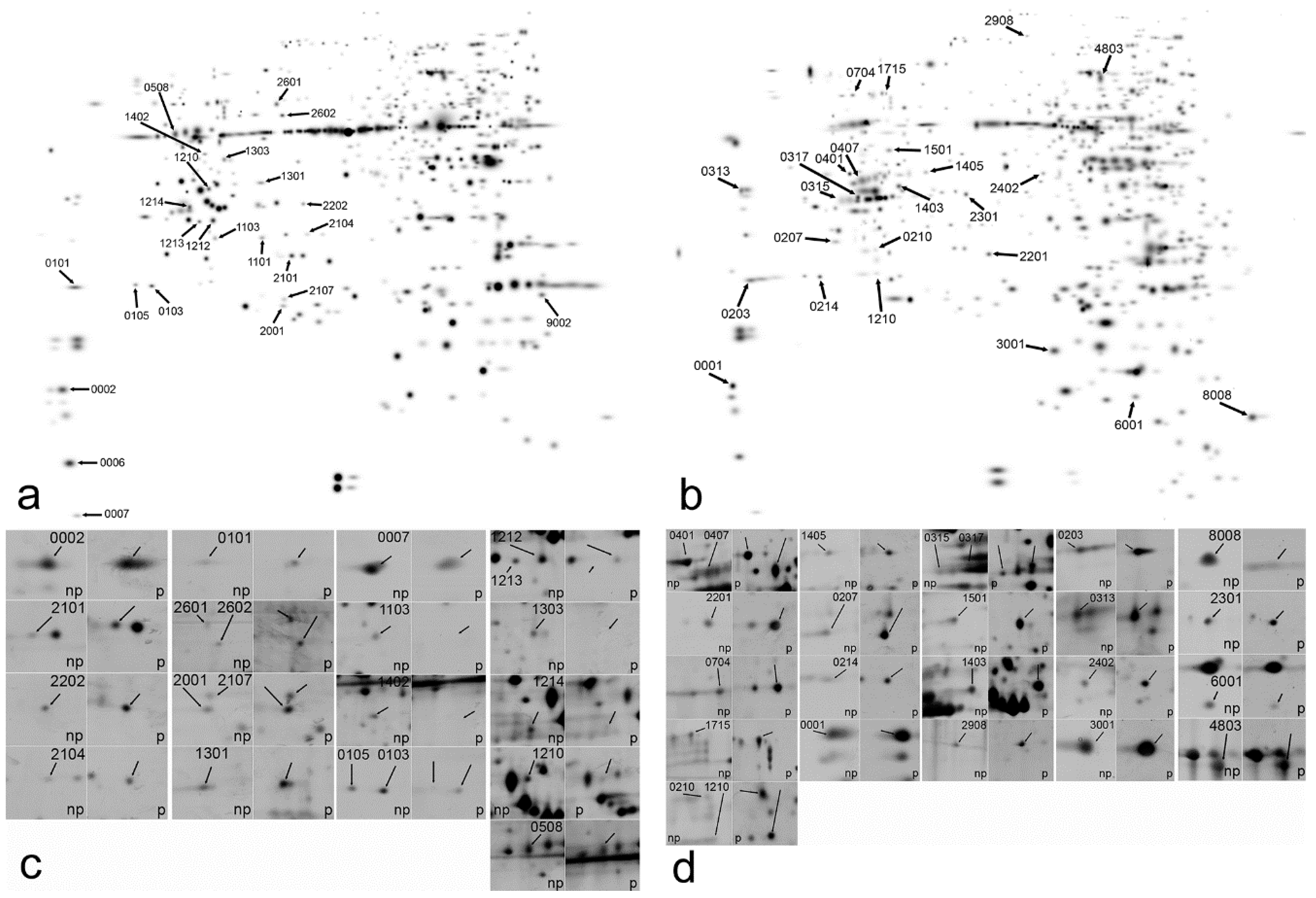
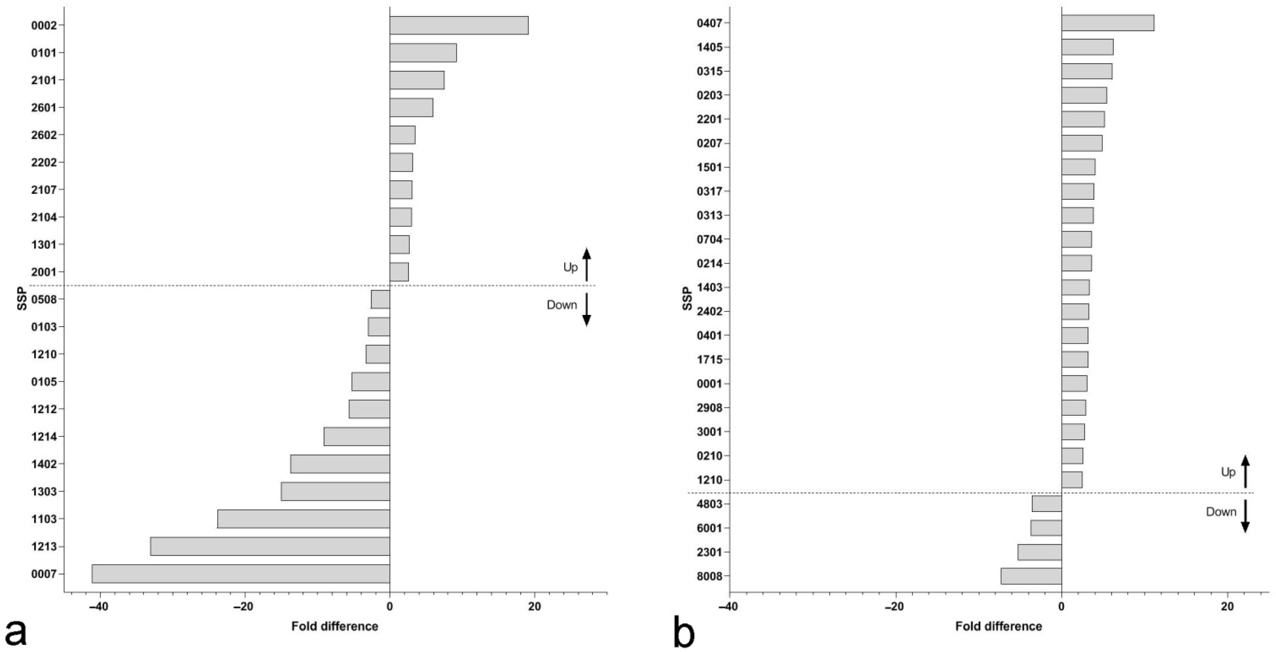
2.2. Quantitative Analysis of Protein Spots on 2D Gels

Differential protein abundance was assessed by two-dimensional polyacrylamide gel electrophoresis, separately in stem and leaves of the host plants. Images were analyzed using PDQuest 8.0 (Bio-Rad, Hercules, CA, USA) Software by deriving a master gel image (Figure 2a,b). Original images are provided as (Supplementary Figures S1–S4). Only protein spots with 2.5-fold difference between non-parasitized and parasitized samples, statistically significant at p < 0.05, Student’s t-test (Figure 2c,d) were considered for further analysis. A total of 21 proteins spots in leaves and 24 protein spots in the stem met the above criteria (Figure 3). Of them, the proportion of up-regulated to down-regulated protein spots were almost equal in leaves (Figure 3a), while in the stem (Figure 3b) the number of up-regulated protein spots was substantially higher than down-regulated (only four). The fold difference in leaves was also higher, reaching 40-fold down-regulation for SSP 0007 and above 20-fold for SSP 1213 and 1103 (Figure 2c and Figure 3a). The fold difference in stem was much lower for all of the protein spots (Figure 3b). All protein spots are indicated by their SSP number, automatically assigned by PDQuest 8.0 Software.

2.3. Protein MS/MS Identification

A total of five protein spots in the leaves and two protein spots in the stem failed to be identified (Figure 3)—protein score, lower than the identity or extensive homology score at p < 0.05. The additional broad database search (Viridiplantae) aimed to find any possible proteins of Cuscuta origin, as such are known to be transferred into hosts [27]. However, no such were found. Out of the identified protein spots (Table 2 and Table 3), one protein was found in a total of four down-regulated spots in leaves glyceraldehyde-3-phosphate dehydrogenase (SSP 1210, 1212, 1213, and 1303). One protein was found in two up-regulated spots in stem Atmp 24.1 glutathione S transferase (SSP 2201 and 3001), but also two isoforms of aspartate aminotransferase were found in two up-regulated spots (SSP 0315 and 1403). Chloroplastic transketolase 1 was found in up-regulated spots in both leaves and stem (SSP 2601 and 1715, respectively) and chloroplastic phosphoribulokinase was found in down-regulated spots in both leaves and stem (SSP 0508 and 8008, respectively). Finally, a member of the glycine cleavage T-protein family was found in a down-regulated spot in leaves (SSP 0105) and up-regulated spot in the stem (SSP 0407).
According to the annotation to gene ontology (GO) biological process, proteins, associated with a variety of stress, including biotic (response to bacterial pathogens) and abiotic (salinity, cold, heavy metals) were equally distributed between both up-regulated and down-regulated protein spots (Table 2 and Table 3). Except for down-regulated protein spots in the stem, the stress-related proteins were not the predominant fraction in either of the identified proteins. A substantial amount, however, is associated with either photosynthesis or carbohydrate metabolism.

3. Discussion

The pronounced effect of C. australis parasitism on the host’s photosynthetic parameters is consistent with previously reported results for the C. campestris—M. micrantha association [18], who reported a significant decrease in Pn after 18 days of infection and C. australis—Bidens pilosa [28,29]. Chen [30] suggested the involvement of abscisic acid (ABA) in the suppression of stomatal conductance and transpiration rate, hence the net photosynthetic rate. Despite differences in the host response in terms of time needed for the adverse effect of Cuscuta infection on photosynthesis to occur, which might be host-specific and dependent on the parasite size, an overall decrease in photosynthetic parameters seems like a common response. Considering the light reactions, we also recently demonstrated a certain negative effect of C. campestris parasitism on the semi-compatible host Ipomoea tricolor [20], suggesting a dramatic effect on the host plant even when the parasite is not developing successfully. Overall, it seems that suppression of photosynthesis in the early stages of Cuscuta parasitism is a global stress response, similar or sharing common signal mechanisms to other abiotic, mainly drought [31] and biotic [32] stresses.
The decrease in CO2 assimilation could be further explained by the down-regulation of at least the large RuBisCO subunit (Table 3), but also the down-regulation of the chloroplastic Glyceraldehyde-3-phosphate dehydrogenase and Phosphoribulokinase (Table 3), both of which are involved in the regeneration of ribulose-1,5-bisphosphate. Accordingly, down-regulation leads to a reduction in CO2 assimilation, otherwise designated as “Calvin-Benson Cycle slow down” [33,34]. In all cases, such down-regulation of CO2 assimilation would largely contribute to the growth inhibition of parasitized hosts, reported previously [18,35] in addition to the reduction in organic nutrients [4]. Furthermore, many of the differentially regulated proteins in the present study are related to the modulation of the carbohydrate or amino acid metabolism and according to GO biological processes are related to the response to different kind of stresses. In contrast, we also found up-regulation of the small RuBisCO subunit, several thylakoid complex proteins—Oxygen-evolving enhancer proteins (PSBO2), and photosystem II subunit P-1 (Table 2). The small RuBisCO subunit is encoded in nuclear DNA and synthesized in the cytoplasm, then it is transferred into the chloroplast [36]. The up-regulation of the small RuBisCO subunit indicated that the compensatory reaction of the nuclear gene to decreased accumulation of photosynthates would be quicker than genes encoded in chloroplast DNA, such as the large RuBisCO subunit gene. PsbO proteins in Arabidopsis are encoded by two genes, psbO1 and psbO2 [37]. A low level of PSBO2 would limit the photosynthetic activity [37] and the function of PBSO2 showed under high light stress. In this study, Arabidopsis plants were grown under natural sunlight conditions in the greenhouse for successful infection of Cuscuta. The up-regulation of PSBO2 might be due to the high light stress response.
Overall, the host’s response to Cuscuta infection is believed to trigger Jasmonic acid and Salicylic acid defense pathways [25] and up-regulation of numerous pathogen-response related genes and proteins was previously reported. Borsics and Lados [38] reported up-regulation of PPRG2 in dodder-infected alfalfa, encoding a homologous to pathogenesis-related (PR-10) protein family product. In Mikania micrantha, infected by C. campestris, up-regulation of a homologous to chitinase gene, Mmchi1, was reported [39]. In our study glutathione S-Transferase Atpm24.1, commonly involved in response to pathogenic fungi, bacteria, and viruses, mainly governing the antioxidant response [40] was found to be up-regulated in the stem of infected plants. More interestingly, it was found that S-adenosylmethionine synthase 4 was up-regulated in leaves and Pectin methyl esterase 3 in the stem. The former is involved in the synthesis of S-adenosylmethionine, an important methyl donor for methylation of lignin precursors for lignin synthesis [41]. As such, it is expected to be involved in the lignification and reinforcement of the cell wall, a common defense mechanism and systemic response against different pathogens [42]. Lignin-based resistance to C. campestris in the tomato plant was established in a particular cultivar, governed by up-regulation of numerous related genes [22]. The up-regulation of Pectin methyl esterase, especially in the stem of the host plants, where infection with the dodder mainly occur is probably involved in the de-esterification of pectic polysaccharides, which in turn facilitates pectin hydrolyses and penetration of the parasite [43]. It is yet to be concluded whether this is a mechanism by which the parasite manipulates the metabolism of the host to increase susceptibility, but it was reported that pectinolytic enzymes are essential in host tissue penetration during haustoria formation [43].
The possibility that some of the up-regulated proteins in the parasitized group are of Cuscuta origin was taken into consideration because of several studies reporting extensive protein exchange between the parasite and the host [27,44], including translation of transferred mRNAs in the host [45]. Liu et al. [27] reported more than 1000 dodder proteins, translocated to the infected Arabidopsis stems. Interestingly, most of the up-regulated and none of the down-regulated proteins in our study were also found to be dodder-to-Arabidopsis mobile proteins [27].

4. Materials and Methods

4.1. Plant Material and Growing Conditions

Seeds of Arabidopsis thaliana ecotype Columbia-0 (Col-0) were kindly provided by Dr Zhongnan Yang (Shanghai Normal University, Shanghai, China). Arabidopsis seeds were surface sterilized using 70% (v/v) ethanol for 10 min, then washed three times with sterile water and stratified for 48 h at 4 °C in the dark. The treated seeds were sown evenly in vermiculite: peat substrate: perlite (16:3:1) mixture and germinated in a growth chamber under 16 h light/8 h dark photoperiod, 150 μmol m−2 s−1 white light intensity, 22 °C, 70% relative humidity. Two-week-old healthy seedlings were transferred to a plastic tray (one seedling per tray), containing a controlled-release fertilizer (Osmocote®, Scotts International B.V., Harderwijk, The Netherlands), and grown under the same conditions. Twenty plants were chosen when the height reached approximately 5 cm in a growth chamber, then separated into the control group and parasitized group (each group had 10 plants) and grown for further 5 days.
Fifteen cm long stems of C. australis were collected from a field population at Taizhou University, Linhai City, Zhejiang Province, China. The parasite’s stem was coiled around the inflorescence stem (the upper one-third, approximately 5 cm from the rosette leaves) of A. thaliana (one parasite per host) in a counter-clockwise direction and plants were grown under natural sunlight conditions in the greenhouse for infection—usually, haustoria are formed after 24 h. After 72 h, a total of six plants from the parasitized group with the most uniform Cuscuta infection and six plants from the control group, respectively, were chosen for photosynthetic measurements and protein analyses.

4.2. Photosynthesis Measurements

Relative chlorophyll content was measured using CCM-200 chlorophyll content meter (OPTI-SCIENCES, Hudson, NH, USA) on fully expanded 5th rosette leaves of Arabidopsis, one leaf per plant. Photosynthetic rate (Pn), stomatal conductance (gs), the concentration of intercellular CO2 (Ci), and transpiration rate (Ts) were measured using a portable photosynthesis system Li-6400 (LI-COR, Lincoln, NE, USA) according to the manufacturer’s instructions on fully expanded rosette leaves. Gas exchange measurements were determined under 25 °C, 1200 μmol m−2 s−1 photons, 400 μmol mol−1 CO2 concentration and 70% relative humidity.

4.3. Protein Extraction and Quantification

For proteomics analyses, Arabidopsis inflorescence stem (2 cm up and down the site of dodder parasitism) and leaves (5th rosette, two leaves per plant) were studied separately after 72 h of infection. Total protein was extracted from the control and parasitized tissues of Arabidopsis as previously described [46]. Two grams of fresh plant material were powder-grounded in liquid nitrogen, dissolved in 20 mL cold acetone, containing 10% (w/v) trichloroacetic acid (TCA), and proteins were precipitated for 1 h at −20 °C. The samples were centrifuged at 15,000 g for 15 min at 4 °C. The procedure was repeated with TCA-free cold acetone, then the pellets were dried by vacuum freeze dryer and dissolved in protein lysis buffer, containing 9 M Urea, 4% (w/v) CHAPS, 1% (w/v) DTT, 1% (v/v) IPG buffer (pH 4–7, GE Healthcare, Danderyd, Sweden) and vortexed at 30 °C for 1 h. The protein concentrations were determined by Bradford’s method [47]. If not immediately used for isoelectric focusing separation, proteins were stored at −80 °C.

4.4. Two-Dimensional Gel Electrophoresis

Equal amounts of protein (100 µg) were separated on Immobiline Dry Strip Gels (pH = 4–7, 24 cm, GE Healthcare, Danderyd, Sweden). Isoelectric focusing (IEF) was performed according to the producer’s recommendations at 20 °C as follows: 250 V for 1 h, 1000 V for 2 h, on a linear gradient from 1000 V to 10,000 V for 1 h, 10,000 V for 1 h. The strips were then incubated under constant gentle shaking in equilibration buffer I (6M urea, 30% v/v glycerol, 2% w/v SDS, 50 mM Tris-HCl pH 8.8, 1% w/v DTT) and buffer II, in which DTT was replaced with 2.5% w/v Iodoacetamide, for 15 min each. Twelve percent T SDS polyacrylamide gels were used for the second-dimension electrophoresis and the gels were stained with Coomassie Brilliant Blue (CBB) G-250 according to the procedure described by Neuhoff [48].

4.5. Image Analysis and Mass Spectrometric Identification

The stained gels (in triplicates, each representing an individual plant) were scanned on Image Scanner (GE Healthcare, Waukesha, WI, USA) at a resolution of 300 dots per inch and the images were analyzed using PDQuest 2-D analysis software 8.0 (Bio-Rad, Hercules, CA, USA), following the steps of spot detection, volumetric quantification and matching. Protein spots, identified to show 2.5-fold higher or lower difference (n = 3, p < 0.05, Student’s t-test) in intensity between control and parasitized Arabidopsis plants were selected as the differentially abundant proteins, designated up-regulated and down-regulated, respectively, and extracted for MS analysis. Selected protein spots were excised from 2-DE gels, de-stained, dehydrated, and digested with trypsin according to the procedure of Shevchenko [49]. Peptide MS and MS/MS were performed on an ABI 5800 MALDI-TOF-TOF plus mass spectrometer (Applied Biosystems, Foster City, CA, USA).
Both the MS and MS/MS data were integrated and processed using the GPS Explorer V3.6 software (Applied Biosystems, Foster City, CA, USA) with default parameters. Based on combined MS and MS/MS spectra, the protein identification was performed using the MASCOT V2.1 search engine (Matrix Science, London, UK), with the following search parameters: NCBIprot (also performed against SwissProt database) thale cress (Arabidopsis thaliana) database (repeated against green plants, Viridiplantae), trypsin as the digestion enzyme, one missed cleavage site; variable modifications: Oxidation (M), Acetyl (Protein N-term), Deamidated (N and Q) and Dioxidation (W); fixed modifications: Carbamidomethyl (C), and a mass deviation of less than 100 ppm. Statistically significant search scores (>95% confidence, equivalent to MASCOT expect value p < 0.05) were chosen [50]. All confirmed proteins—MASCOT p < 0.05 were searched in UniProt database and GO annotated for localization and biological process.

5. Conclusions

In conclusion, it was shown that Cuscuta australis parasitism negatively affected most of the photosynthetic parameters of the host plants. The differential abundance of proteins, however, could not entirely explain such effect. Nevertheless, multiple proteins related to the carbohydrate metabolism were differentially regulated, as well as proteins with known function in abiotic and biotic stress response and defense mechanisms. Notably, S-adenosylmethionine synthase 4 and Pectin methyl esterase 3, involved in cell wall modifications and the parasite–host interaction, are up-regulated in stem and leaves, respectively. Accumulation of data on proteins involved in plant-to-plant interaction could further elucidate the mechanisms of haustoria formation in Cuscuta spp. and allow us to identify individual players in the hosts’ defense with a putative role in at least partial resistance to infection.

Supplementary Materials

The following supporting information can be downloaded at: https://www.mdpi.com/article/10.3390/plants11212904/s1, Figure S1: Representative 2D gel of proteins from leaves of control Arabidopsis thaliana plant; Figure S2: Representative 2D gel of proteins from leaves of Cuscuta australis-parasitized Arabidopsis thaliana plant; Figure S3: Representative 2D gel of proteins from stem of control Arabidopsis thaliana plant; Figure S4: Representative 2D gel of proteins from stem of Cuscuta australis-parasitized Arabidopsis thaliana plant.

Author Contributions

J.L. designed the experiment. J.L., Z.D. and Y.S. performed the experiments. L.Z. and D.T. analyzed the data. L.Z. and J.L. wrote the manuscript. All authors have read and agreed to the published version of the manuscript.

Funding

This work was financially supported by the Talented Young Scientist Program of the Ministry of Science and Technology of People’s Republic of China (given to Lyuben Zagorchev), the Ten Thousand Talent Program of Zhejiang Province (given to Junmin Li), and the National Natural Science Foundation of China (No. 30800133) (given to Junmin Li) and grant KP-06-H31/10 of the National Science Fund of Ministry of Education and Science, Bulgaria.

Institutional Review Board Statement

Not applicable.

Informed Consent Statement

Not applicable.

Data Availability Statement

The data presented in this study are available on request from the corresponding author.

Acknowledgments

We thank Zhongnan Yang from Shanghai Normal University, China, for his kind grant of seeds of Arabidopsis thaliana ecotype Columbia-0 (Col-0).

Conflicts of Interest

The authors declare no conflict of interest.

References

  1. Kaiser, B.; Vogg, G.; Fürst, U.B.; Albert, M. Parasitic plants of the genus Cuscuta and their interaction with susceptible and resistant host plants. Front. Plant Sci. 2015, 6, 45. [Google Scholar] [CrossRef] [PubMed] [Green Version]
  2. Revill, M.J.; Stanley, S.; Hibberd, J.M. Plastid genome structure and loss of photosynthetic ability in the parasitic genus Cuscuta. J. Exp. Bot. 2005, 56, 2477–2486. [Google Scholar] [CrossRef] [PubMed]
  3. Birschwilks, M.; Haupt, S.; Hofius, D.; Neumann, S. Transfer of phloem-mobile substances from the host plants to the holoparasite Cuscuta sp. J. Exp. Bot. 2006, 57, 911–921. [Google Scholar] [CrossRef] [PubMed] [Green Version]
  4. Hibberd, J.M.; Dieter Jeschke, W. Solute flux into parasitic plants. J. Exp. Bot. 2001, 52, 2043–2049. [Google Scholar] [CrossRef] [Green Version]
  5. Parker, C. Parasitic weeds: A world challenge. Weed Sci. 2012, 60, 269–276. [Google Scholar] [CrossRef]
  6. Koch, A.M.; Binder, C.; Sanders, I.R. Does the generalist parasitic plant Cuscuta campestris selectively forage in heterogeneous plant communities? New Phytol. 2004, 162, 147–155. [Google Scholar] [CrossRef] [Green Version]
  7. Yu, H.; Liu, J.; He, W.-M.; Miao, S.-L.; Dong, M. Cuscuta australis restrains three exotic invasive plants and benefits native species. Biol. Invasions 2011, 13, 747–756. [Google Scholar]
  8. Yu, H.; He, W.-M.; Liu, J.; Miao, S.-L.; Dong, M. Native Cuscuta campestris restrains exotic Mikania micrantha and enhances soil resources beneficial to natives in the invaded communities. Biol. Invasions 2009, 11, 835. [Google Scholar] [CrossRef]
  9. Yu, H.; Liu, J.; He, W.-M.; Miao, S.-L.; Dong, M. Restraints on Mikania micrantha by Cuscuta campestris facilitates restoration of the disturbed ecosystems. Biodiversity 2009, 10, 72–78. [Google Scholar] [CrossRef]
  10. Benvenuti, S.; Dinelli, G.; Bonetti, A.; Catizone, P. Germination ecology, emergence and host detection in Cuscuta campestris. Weed Res. 2005, 45, 270–278. [Google Scholar] [CrossRef]
  11. Runyon, J.B.; Mescher, M.C.; De Moraes, C.M. Volatile chemical cues guide host location and host selection by parasitic plants. Science 2006, 313, 1964–1967. [Google Scholar] [CrossRef] [PubMed]
  12. Shimizu, K.; Aoki, K. Development of parasitic organs of a stem holoparasitic plant in genus Cuscuta. Front. Plant Sci. 2019, 10, 1435. [Google Scholar] [PubMed] [Green Version]
  13. Vaughn, K. Attachment of the parasitic weed dodder to the host. Protoplasma 2002, 219, 227–237. [Google Scholar] [CrossRef] [PubMed]
  14. Striberny, B.; Krause, K. Cell wall glycoproteins at interaction sites between parasitic giant dodder (Cuscuta reflexa) and its host Pelargonium zonale. Plant Signal. Behav. 2015, 10, e1086858. [Google Scholar] [CrossRef] [PubMed] [Green Version]
  15. Bleischwitz, M.; Albert, M.; Fuchsbauer, H.-L.; Kaldenhoff, R. Significance of Cuscutain, a cysteine protease from Cuscuta reflexa, in host-parasite interactions. BMC Plant Biol. 2010, 10, 227. [Google Scholar] [CrossRef] [Green Version]
  16. Olsen, S.; Striberny, B.; Hollmann, J.; Schwacke, R.; Popper, Z.; Krause, K. Getting ready for host invasion: Elevated expression and action of xyloglucan endotransglucosylases/hydrolases in developing haustoria of the holoparasitic angiosperm Cuscuta. J. Exp. Bot. 2016, 67, 695–708. [Google Scholar] [CrossRef] [Green Version]
  17. Jeschke, W.; Hilpert, A. Sink-stimulated photosynthesis and sink-dependent increase in nitrate uptake: Nitrogen and carbon relations of the parasitic association Cuscuta reflexa–Ricinus communis. Plant Cell Environ. 1997, 20, 47–56. [Google Scholar] [CrossRef]
  18. Shen, H.; Hong, L.; Ye, W.; Cao, H.; Wang, Z. The influence of the holoparasitic plant Cuscuta campestris on the growth and photosynthesis of its host Mikania micrantha. J. Exp. Bot. 2007, 58, 2929–2937. [Google Scholar] [CrossRef] [Green Version]
  19. Le, Q.V.; Tennakoon, K.U.; Metali, F.; Lim, L.B.; Bolin, J.F. Impact of Cuscuta australis infection on the photosynthesis of the invasive host, Mikania micrantha, under drought condition. Weed Biol. Manag. 2015, 15, 138–146. [Google Scholar]
  20. Zagorchev, L.; Traianova, A.; Teofanova, D.; Li, J.; Kouzmanova, M.; Goltsev, V. Influence of Cuscuta campestris Yunck. on the photosynthetic activity of Ipomoea tricolor Cav.—In Vivo chlorophyll a fluorescence assessment. Photosynthetica 2020, 58, 237–247. [Google Scholar] [CrossRef] [Green Version]
  21. Goldwasser, Y.; Rabinovitz, O.; Hayut, E.; Kuzikaro, H.; Sibony, M.; Rubin, B. Selective and effective control of field dodder (Cuscuta campestris) in chickpea with granular pendimethalin. Weed Technol. 2019, 33, 586–594. [Google Scholar] [CrossRef]
  22. Jhu, M.-Y.; Farhi, M.; Wang, L.; Philbrook, R.N.; Belcher, M.S.; Nakayama, H.; Zumstein, K.S.; Rowland, S.D.; Ron, M.; Shih, P.M. Heinz-resistant tomato cultivars exhibit a lignin-based resistance to field dodder (Cuscuta campestris) parasitism. Plant Physiol. 2022, 189, 129–151. [Google Scholar] [CrossRef] [PubMed]
  23. Córdoba, E.M.; Fernández-Aparicio, M.; González-Verdejo, C.I.; López-Grau, C.; del Valle Muñoz-Muñoz, M.; Nadal, S. Search for Resistant Genotypes to Cuscuta campestris Infection in two legume species, Vicia sativa and Vicia ervilia. Plants 2021, 10, 738. [Google Scholar] [CrossRef] [PubMed]
  24. Hegenauer, V.; Fürst, U.; Kaiser, B.; Smoker, M.; Zipfel, C.; Felix, G.; Stahl, M.; Albert, M. Detection of the plant parasite Cuscuta reflexa by a tomato cell surface receptor. Science 2016, 353, 478–481. [Google Scholar] [CrossRef]
  25. Runyon, J.B.; Mescher, M.C.; Felton, G.W.; De Moraes, C.M. Parasitism by Cuscuta pentagona sequentially induces JA and SA defence pathways in tomato. Plant Cell Environ. 2010, 33, 290–303. [Google Scholar] [CrossRef]
  26. Birschwilks, M.; Sauer, N.; Scheel, D.; Neumann, S. Arabidopsis thaliana is a susceptible host plant for the holoparasite Cuscuta spec. Planta 2007, 226, 1231–1241. [Google Scholar] [CrossRef]
  27. Liu, N.; Shen, G.; Xu, Y.; Liu, H.; Zhang, J.; Li, S.; Li, J.; Zhang, C.; Qi, J.; Wang, L. Extensive inter-plant protein transfer between Cuscuta parasites and their host plants. Mol. Plant 2020, 13, 573–585. [Google Scholar] [CrossRef]
  28. Yang, B.; Li, J.; Zhang, J.; Yan, M. Effects of nutrients on interaction between the invasive Bidens pilosa and the parasitic Cuscuta australis. Pak. J. Bot. 2015, 47, 1693–1699. [Google Scholar]
  29. Li, J.; Hettenhausen, C.; Sun, G.; Zhuang, H.; Li, J.-H.; Wu, J. The parasitic plant Cuscuta australis is highly insensitive to abscisic acid-induced suppression of hypocotyl elongation and seed germination. PLoS ONE 2015, 10, e0135197. [Google Scholar] [CrossRef]
  30. Chen, H.; Shen, H.; Ye, W.; Cao, H.; Wang, Z. Involvement of ABA in reduced photosynthesis and stomatal conductance in Cuscuta campestris—Mikania micrantha association. Biol. Plant. 2011, 55, 545–548. [Google Scholar] [CrossRef]
  31. Flexas, J.; Medrano, H. Drought-inhibition of photosynthesis in C3 plants: Stomatal and non-stomatal limitations revisited. Ann. Bot. 2002, 89, 183–189. [Google Scholar] [CrossRef] [PubMed] [Green Version]
  32. Bonfig, K.B.; Schreiber, U.; Gabler, A.; Roitsch, T.; Berger, S. Infection with virulent and avirulent P. syringae strains differentially affects photosynthesis and sink metabolism in Arabidopsis leaves. Planta 2006, 225, 1–12. [Google Scholar] [CrossRef] [PubMed]
  33. Price, G.D.; Evans, J.R.; von Caemmerer, S.; Yu, J.-W.; Badger, M.R. Specific reduction of chloroplast glyceraldehyde-3-phosphate dehydrogenase activity by antisense RNA reduces CO2 assimilation via a reduction in ribulose bisphosphate regeneration in transgenic tobacco plants. Planta 1995, 195, 369–378. [Google Scholar] [CrossRef]
  34. Elena López-Calcagno, P.; Omar Abuzaid, A.; Lawson, T.; Anne Raines, C. Arabidopsis CP12 mutants have reduced levels of phosphoribulokinase and impaired function of the Calvin–Benson cycle. J. Exp. Bot. 2017, 68, 2285–2298. [Google Scholar] [CrossRef] [PubMed] [Green Version]
  35. Zagorchev, L.; Albanova, I.; Tosheva, A.; Li, J.; Teofanova, D. Salinity effect on Cuscuta campestris Yunck. Parasitism on Arabidopsis thaliana L. Plant Physiol. Biochem. 2018, 132, 408–414. [Google Scholar] [CrossRef]
  36. Portis, A.R.; Parry, M.A. Discoveries in Rubisco (Ribulose 1,5-bisphosphate carboxylase/oxygenase): A historical perspective. Photosynth. Res. 2007, 94, 121–143. [Google Scholar] [CrossRef]
  37. Murakami, R.; Ifuku, K.; Takabayashi, A.; Shikanai, T.; Endo, T.; Sato, F. Functional dissection of two Arabidopsis PsbO proteins: PsbO1 and PsbO2. FEBS J. 2005, 272, 2165–2175. [Google Scholar]
  38. Borsics, T.; Lados, M. Dodder infection induces the expression of a pathogenesis-related gene of the family PR-10 in alfalfa. J. Exp. Bot. 2002, 53, 1831–1832. [Google Scholar] [CrossRef] [Green Version]
  39. Li, D.-M.; Staehelin, C.; Wang, W.-T.; Peng, S.-L. Molecular cloning and characterization of a chitinase-homologous gene from Mikania micrantha infected by Cuscuta campestris. Plant Mol. Biol. Rep. 2010, 28, 90–101. [Google Scholar] [CrossRef]
  40. Gullner, G.; Komives, T.; Király, L.; Schröder, P. Glutathione S-transferase enzymes in plant-pathogen interactions. Front. Plant Sci. 2018, 9, 1836. [Google Scholar] [CrossRef] [PubMed] [Green Version]
  41. Boerjan, W.; Ralph, J.; Baucher, M. Lignin biosynthesis. Annu. Rev. Plant Biol. 2003, 54, 519–546. [Google Scholar] [PubMed]
  42. Bhuiyan, N.H.; Selvaraj, G.; Wei, Y.; King, J. Role of lignification in plant defense. Plant Signal. Behav. 2009, 4, 158–159. [Google Scholar] [CrossRef] [PubMed] [Green Version]
  43. Johnsen, H.R.; Striberny, B.; Olsen, S.; Vidal-Melgosa, S.; Fangel, J.U.; Willats, W.G.; Rose, J.K.; Krause, K. Cell wall composition profiling of parasitic giant dodder (Cuscuta reflexa) and its hosts: A priori differences and induced changes. New Phytol. 2015, 207, 805–816. [Google Scholar] [CrossRef] [PubMed]
  44. Kim, G.; Westwood, J.H. Macromolecule exchange in Cuscuta–host plant interactions. Curr. Opin. Plant Biol. 2015, 26, 20–25. [Google Scholar] [CrossRef]
  45. Park, S.-Y.; Shimizu, K.; Brown, J.; Aoki, K.; Westwood, J.H. Mobile Host mRNAs Are Translated to Protein in the Associated Parasitic Plant Cuscuta campestris. Plants 2021, 11, 93. [Google Scholar] [CrossRef]
  46. Wu, X.; Xiong, E.; Wang, W.; Scali, M.; Cresti, M. Universal sample preparation method integrating trichloroacetic acid/acetone precipitation with phenol extraction for crop proteomic analysis. Nat. Protoc. 2014, 9, 362. [Google Scholar] [CrossRef]
  47. Bradford, M.M. A rapid and sensitive method for the quantitation of microgram quantities of protein utilizing the principle of protein-dye binding. Anal. Biochem. 1976, 72, 248–254. [Google Scholar] [CrossRef]
  48. Neuhoff, V.; Arold, N.; Taube, D.; Ehrhardt, W. Improved staining of proteins in polyacrylamide gels including isoelectric focusing gels with clear background at nanogram sensitivity using Coomassie Brilliant Blue G-250 and R-250. Electrophoresis 1988, 9, 255–262. [Google Scholar]
  49. Shevchenko, A.; Tomas, H.; Havli, J.; Olsen, J.V.; Mann, M. In-gel digestion for mass spectrometric characterization of proteins and proteomes. Nat. Protoc. 2006, 1, 2856. [Google Scholar] [CrossRef]
  50. Sharmin, S.A.; Alam, I.; Rahman, M.A.; Kim, K.-H.; Kim, Y.-G.; Lee, B.-H. Mapping the leaf proteome of Miscanthus sinensis and its application to the identification of heat-responsive proteins. Planta 2013, 238, 459–474. [Google Scholar] [CrossRef]
Figure 1. Point of infection of Arabidopsis thaliana by Cuscuta australis (a) with arrows, showing the site of haustoria formation after 24 h and microscopic cross-section of haustoria (h) after 72 h (b).
Plants 11 02904 g001
Figure 2. PDQuest master gel images of 2D-electrophoregrams of Arabidopsis leaves (a) and stem (b) and the respective differentially regulated protein spots (c,d). Arabidopsis was either parasitized (p) or non-parasitized (np) by Cuscuta australis. Protein spot numbers correspond to the assigned by PDQuest.
Plants 11 02904 g002
Figure 3. Differentially regulated protein spots, shown as SSP numbers, assigned by PDQuest with 2.5-fold difference in leaves (a) and stem (b) of Cuscuta australis—parasitized Arabidopsis thaliana.
Plants 11 02904 g003
Table 1. Effect of parasitism on the chlorophyll content and photosynthetic characteristics of the host. Data were shown as mean ± standard errors (n = 6). Different small letters indicate significant (Student’s t-test) differences between parasitized and non-parasitized treatments.
TraitsNon-ParasitizedParasitized
Photosynthetic rate (Pn)/(μmol CO2 m−2 s−1)3.61 ± 0.73a2.71 ± 0.33b
Stomatal conductance (gs)/(μmol H2O m−2 s−1)0.19 ± 0.05a0.12 ± 0.01b
Concentration of intercellular CO2 (Ci) /(μmol CO2 m−2 s−1)305.66 ± 1.34a301.58 ± 6.23a
Transpiration rate (Tr)/(μmol CO2 m−2 s−1)3.30 ± 0.74a2.31 ± 0.097b
Relative chlorophyll content17.51 ± 2.06a8.79 ± 1.96b
Table 2. MS/MS identified protein spots, up-regulated in leaves and stem of Cuscuta australis—parasitized Arabidopsis thaliana. SSP is the standard spot number.
SSPMascot ScoreUniProt AccessionProtein IdentityGO LocalizationGO Biological Process
Leaves
000298Q41932Oxygen-evolving enhancer protein 3-2, chloroplasticchloroplast thylakoid membranephotosynthetic electron transport chain
010169Q9LUT2S-adenosylmethionine synthase 4cytoplasmlignin biosynthetic process response to cold stress
210154Q9SAJ4Phosphoglycerate kinase 3, cytosolicCytoplasmgluconeogenesis
2601175Q8RWV0Transketolase-1, chloroplasticChloroplastPentose-phosphate shunt
Response to cadmium ions/salt stress
260253P10795Ribulose bisphosphate carboxylase small chain 1A, chloroplasticChloroplastchloroplast ribulose bisphosphate carboxylase complex assembly
response to cold
2107130O49344Putative oxygen-evolving enhancer protein 2-2Chloroplastphotosynthesis
1301212Q42560Aconitate hydratase 1Mitochondrion Cytoplasmcitrate metabolic process
response to salt stress
Stem
0407269O65396Glycine cleavage T-protein familyMitochondrionglycine decarboxylation via glycine cleavage system
response to cadmium ion
1405229B9DHX4Malate dehydrogenase carbohydrate metabolic process
0315194Q56YR4aspartate aminotransferaseMultipleBiosynthetic process
0203229A0A178UUR9VDAC2Mitochondrial outer membranevoltage-gated anion channel
2201169Q8LC43Atpm24.1 glutathione S transferaseCytosol/ERresponse to bacteria
response to abiotic stress
150149A0A178VU56Succinate-CoA ligase [ADP-forming] subunit betaMitochondriontricarboxylic acid cycle
0317352Q9FWA36-phosphogluconate dehydrogenase family proteinCytosol/PeroxisomeD-gluconate metabolic process
response to salt stress
031349A0A178VDL9Pectin methyl esterase 3Cell WallCell wall modification
0704550O50008methionine synthaseCytoplasmresponse to cadmium ion
response to salt stress
1403551P46645aspartate aminotransferase 2Cytoplasm2-oxoglutarate metabolic process
2402191Q944G9Fructose-bisphosphate aldolase 2Chloroplast stromagluconeogenesis
response to abscisic acid/response to cadmium ion
0401170Q96533glutathione-dependent formaldehyde dehydrogenaseCytoplasmethanol oxidation
1715197Q8RWV0Transketolase-1chloroplast stromapentose-phosphate shunt
response to cadmium ion/response to salt stress
0001111A0A178VBH5PSBO2Chloroplast thylakoidphotosystem II assembly
2908334Q93ZF2putative 2,3-bisphosphoglycerate-independent phosphoglycerate mutaseCytoplasmglucose catabolic process
3001168Q8LC43Atpm24.1 glutathione S transferaseCytosol/ERresponse to bacteria
response to abiotic stress
021076O24616Proteasome subunit alpha type-7-BNucleus/Cytoplasmproteasomal protein catabolic process
1210127Q42029 photosystem II subunit P-1chloroplast thylakoidPhotosynthesis
defense response to bacterium
Table 3. MS/MS identified protein spots, down-regulated in leaves and stem of Cuscuta australis—parasitized Arabidopsis thaliana. SSP is the standard spot number.
SSPMascot Score UniProt AccessionProtein IdentityGO LocalizationGO Biological Process
Leaves
1213259A0A178VKK2Glyceraldehyde-3-phosphate dehydrogenase glucose metabolic process
1103138A0A0K1CVP8Ribulose bisphosphate carboxylase large chainChloroplast Photorespiration
1303232A0A178VKK2Glyceraldehyde 3-phosphate dehydrogenase A subunit glucose metabolic process
1402138P22954dnaK-type molecular chaperone hsc70.1—like, partialNucleus Cytoplasmcellular response to heat
response to multiple stresses
1212213A0A178VKK2Glyceraldehyde-3-phosphate dehydrogenase glucose metabolic process
0105200O65396Glycine cleavage T-protein familyMitochondrionglycine decarboxylation via glycine cleavage system
response to cadmium ion
1210137P25856Glyceraldehyde 3-phosphate dehydrogenase A subunitChloroplastglucose metabolic process
response to cold
010382F4KDZ4Peroxisomal NAD-malate dehydrogenase 2Peroxisomecarbohydrate metabolic process
050899P25697Phosphoribulokinase, chloroplasticChloroplastdefense response to bacterium
response to cold
Stem
800840P25697Phosphoribulokinase, chloroplasticChloroplastDefense response to bacteria
Response to cold
2301115P06525Alcohol dehydrogenase class-PCytoplasmResponse to multiple abiotic stresses
600147P31265translationally controlled tumor protein-like proteinCytosolAuxin homeostasis
Response to multiple stresses
4803481O23654vacuolar ATP synthase subunit AVacuoleATP hydrolysis coupled proton transport
Response to salt stress
Publisher’s Note: MDPI stays neutral with regard to jurisdictional claims in published maps and institutional affiliations.

