Article

Arbuscular Mycorrhiza Support Plant Sulfur Supply through Organosulfur Mobilizing Bacteria in the Hypho- and Rhizosphere

1
Department of Biological Sciences, School of Natural Sciences, University of Limerick, V94 T9PX Limerick, Ireland
2
Teagasc Food Research Centre, Moorepark, Fermoy, and APC Microbiome Ireland, P61 C996 Cork, Ireland
*
Author to whom correspondence should be addressed.
Plants 2022, 11(22), 3050; https://doi.org/10.3390/plants11223050
Submission received: 27 October 2022 / Revised: 7 November 2022 / Accepted: 8 November 2022 / Published: 11 November 2022
(This article belongs to the Special Issue Advances in Plant-Soil-Microbe Interactions)

Abstract

:
This study aimed to elucidate the role of bacteria colonising mycorrhizal hyphae in organically bound sulfur mobilisation, the dominant soil sulfur source that is not directly plant available. The effect of an intact mycorrhizal symbiosis with access to stable isotope organo-34S enriched soils encased in 35 µm mesh cores was tested in microcosms with Agrostis stolonifera and Plantago lanceolata. Hyphae and associated soil were sampled from static mesh cores with mycorrhizal ingrowth and rotating mesh cores that exclude mycorrhizal ingrowth as well as corresponding rhizosphere soil, while plant shoots were analysed for 34S uptake. Static cores increased uptake of 34S at early stages of plant growth when sulfur demand appeared to be high and harboured significantly larger populations of sulfonate mobilising bacteria. Bacterial and fungal communities were significantly different in the hyphospheres of static cores when compared to rotating cores, not associated with plant hosts. Shifts in bacterial and fungal communities occurred not only in rotated cores but also in the rhizosphere. Arylsulfatase activity was significantly higher in the rhizosphere when cores stayed static, while atsA and asfA gene diversity was distinct in the microcosms with static and rotating cores. This study demonstrated that AM symbioses can promote organo-S mobilization and plant uptake through interactions with hyphospheric bacteria, enabling AM fungal ingrowth into static cores creating a positive feedback-loop, detectable in the microbial rhizosphere communities.

1. Introduction

The essential macro-nutrient sulfur (S) is increasingly limiting crop yield and quality as a result of reduced atmospheric deposition while high yielding crop varieties rapidly deplete soil S stocks [1]. These factors are exacerbated by the fact that S present in soil is approximately 95% organically bound as sulfate-esters and sulfonates [2,3] and not directly available to plants which rely upon soil microbial populations for organo-S mineralisation [4].
The ability to mineralise sulfate-esters is common for many bacteria and saprotrophic fungi in soil [5,6]. Genes encoding for arylsulfatases that cleave the O-S bond appear to share common conserved regions. One of these arylsulfatase genes was described in Pseudomonas aeroginosa as atsA [5,6] and has been most comprehensively studied for various Pseudomonas species [7,8]. More recently atsA has been established as a molecular marker for arylsulfatase gene diversity studies [9]. The ability of soil bacteria to desulfurize a large variety of sulfonates is based on a multicomponent mono-oxygenase enzyme complex that includes the ssu and asf gene clusters [10]. For aliphatic sulfonate desulfurization the ssuD gene has been developed as a molecular marker [9], while for aromatic sulfonate desulfurization, asfA is an established marker [11,12,13,14,15]. The asfA gene involved in aromatic sulfonate mobilization has been identified in the Beta-Proteobacteria Variovorax, Polaromonas, Hydrogenophaga, Cupriavidus, Burkholderia (Paraburkholderia) and Acidovorax, the Actinobacteria Rhodococcus and Williamsia, and the Gamma-Proteobacteria; Pseudomonas and Stenotrophomonas [11,12,13,14,15].
Soil organosulfur transformation (S cycling) involves complex interactions between free living and symbiotic rhizospheric microbial populations. Arbuscular mycorrhizal (AM) fungi are part of these rhizospheric microbial populations and form an endosymbiosis with 77% of angiosperms, 45% of gymnosperms and 52% of fern and lycopod [16]. Their characteristic structure, the arbuscule, is extensively branched and acts as an efficient site for metabolite exchange with the host plant [17]. Furthermore, their extraradical hyphae (ERH) play an important role in nutrient acquisition via extensive networks of microscopic hyphae that access a volume of soil orders of magnitude greater than plant roots [18]. Studies have established for some time now that the presence of AM fungi enhances S uptake for maize, clover [19] and tomato [20] but to date little is known about the actors or the exact processes that facilitate this increased uptake. In vitro studies with transformed carrot roots demonstrated the uptake of reduced forms of S (e.g., cysteine, methionine) in the presence of the AM fungus Rhizophagus irregularis (formerly known as Glomus intraradices) [21]. Rates of uptake and transfer of reduced S were comparable to that of SO42− when the latter was limited. However, it is currently not clear whether this form of uptake is important in vivo. Uptake of SO42− in the rhizosphere leads to a zone of depletion analogous to that observed for phosphorus [22]. The AM fungal ERH could extend past this zone of SO42− depletion and play an important role in provision of S under conditions of limitation [4]. Additionally, more recent investigations have revealed that AM fungi influence the expression of plant sulfate transporters and as a consequence improve S uptake [23].
AM symbiosis alters the rhizospheric microbial community composition by modifying the biochemical composition of root exudates [24,25]. Additionally, translocation of energy rich C compounds to the extended soil environment via their extensive ERH networks provides an important niche for functional interactions [26]. Diverse soil microbial communities are essential for soil fertility and plant vitality [27,28]. Likewise, AM hyphae in a native grassland ecosystem have been shown to host a large community of sulfonate mobilising bacteria with potential to improve plant S supply [15]. Moreover, it has been shown that the addition of morpholine ethane sulfonic acid to soil stimulated sulfonate mobilising bacteria and their metabolites enhanced ERH growth of R. irregularis (then G. intraradices [29]). This is important for maximising S uptake as enhanced hyphal growth stemming from sulfonate mobilising bacterial metabolites may further stimulate the proliferation of this community in a potential positive feedback loop. Despite the above reported findings, a direct link between the bacterial organo-S mobilizing activity in the hyphosphere of AM fungi and the supply of S of organo-S origin to plants remains elusive.
The hypothesis of this study was that (i) ingrowth of symbiotic AM hyphae into organo-34S enriched mesh cores would increase plant S uptake and that (ii) the hinderance of AM hyphae ingrowth would affect the organo-S mobilising microbes. Since the latter facilitate the organo-S mineralization, rotating the mesh cores containing organo-34S will affect the uptake of newly mineralised S through AM hyphae. The hypothesized associated activity of organo-S mineralization and transport is important to overcome plant S limitations and to reduce over-dependencies on synthetic fertilizer use.

2. Results

2.1. Plant Uptake of 34S from Organo-34S Enriched Soil Microcosms

For P. lanceolata, total sulfur uptake was significantly increased (p < 0.05) for the static cores over the rotating cores at 3 months (0.23 ± 0.006, vs. 0.20 ± 0.02% dry matter (DM)), but not at 6 months. Likewise, A. stolonifera, sulfur uptake increased significantly for the static cores for 3 and 6 months but not for month 9 (0.14 ± 0.01, 0.23 ± 0.03 and 0.23 ± 0.04% for rotating cores and 0.16 ± 0.01, 0.31 ± 0.03 and 0.23 ± 0.04% DM for the static cores, respectively).
Very similar results were obtained for the 34S uptake. The δ34SV-CDT (‰) values were significantly higher for the static cores for Agrostis and Plantago after 3 months of incubation when compared to the rotating core experiment (Figure 1). After 6 months this was only the case for Agrostis and after 9 and 12 months, no significant difference in δ34SV-CDT was detected in the Agrostis plants (Figure 1 and Figure S1). The δ34SV-CDT (‰) values were relatively small at this time point (30 compared to 750 δ34SV-CDT upon original analysis) which may explain the absence of a significant effect.

2.2. S K-Edge X-ray Absorption near Edge Spectroscopy

XANES analysis was undertaken to identify S oxidation states and revealed highly similar spectra for the static and rotating treatments with A. stolonifera with reduced (sulfide, thiols), intermediate (sulfoxides and sulfonates) and oxidised (sulfate esters) S species peaks. The only observed difference between the static and rotating cores was a slightly more pronounced sulfide/thiol peak for the static treatment (Supplementary Figure S2). The photon energy of reduced thiols (2474.4 eV), intermediate sulfonates (2480.2–2480.4 eV) and oxidised sulfate esters (2481.6 eV) were identified in a previous study [30] and correspond to the three main peaks in the present study. The photon energy for the corresponding S species in this study appear to be slightly larger due to a larger step size increment utilised in this study (0.5 eV) in comparison to the way the reference values were obtained (0.2 eV).

2.3. AM Colonisation

The extent of root intracellular colonisation with characteristic AM structures was assessed for both A. stolonifera and P. lanceolata (Table 1). Significantly higher arbuscular, vesicular and hyphal colonization (AC, VC and HC) values were observed for static over rotating core treatments (p < 0.05) that represented a 59–62% increase. Likewise, rates of AC were higher for Agrostis (55%) and Plantago (58–61%) when compared to VC rates.

2.4. Quantification of Cultivable Bacteria

For both plants, cultivable heterotrophic (R2A) and sulfonate mobilising (MM2TS) bacterial communities were more abundant in the microcosms with the static treatment. This was the case for the static cores as well as the rhizosphere (roots outside the cores) when compared to the respective rotating treatment (Table 1). Heterotrophs had a six-fold higher abundance in static A. stolonifera microcosms (cores and rhizosphere, p < 0.05), while for P. lanceolata the increase in abundance was six- (cores) and twelve-fold (rhizosphere) for static over rotating treatments (p < 0.05). Additionally, for A. stolonifera the polymeric sulfonate mobilising (MM2LS) bacterial community was more abundant in the static treatment (rhizosphere and cores) over the respective rotating treatment (twenty-fold; p < 0.05). For P. lanceolata, the polymeric sulfonate mobilising bacterial abundance was not significantly different among the cores but was ten-fold higher in the static rhizosphere when compared to the rotating treatment (p < 0.05; Table 1).
Arylsulfonate mobiliser abundances varied the greatest (104–107 MPN g−1), while the polymeric sulfonate mobilising bacteria were lower in abundance than the arylsulfonate mobilizers (103–105 MPN g−1; Table 1). The heterotrophic bacterial abundance was similar in the cores and rhizospheres for both plants (p > 0.05) for the rotating and the static treatment, respectively. In contrast, aromatic sulfonate mobilisers were highest in the static cores for both plants (Table 1).

2.5. Fingerprinting Based Community Analysis

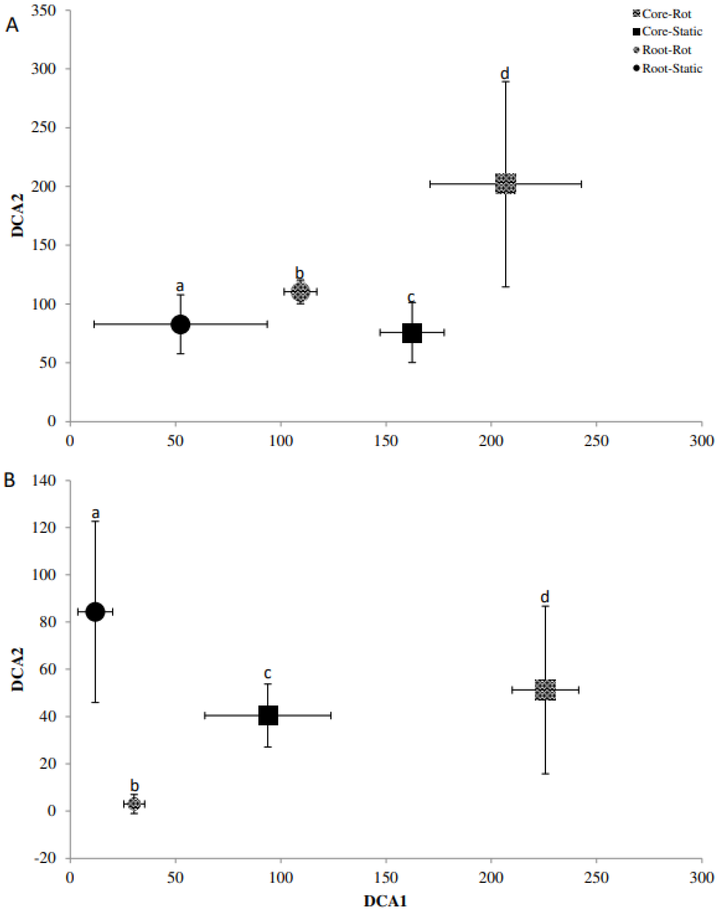
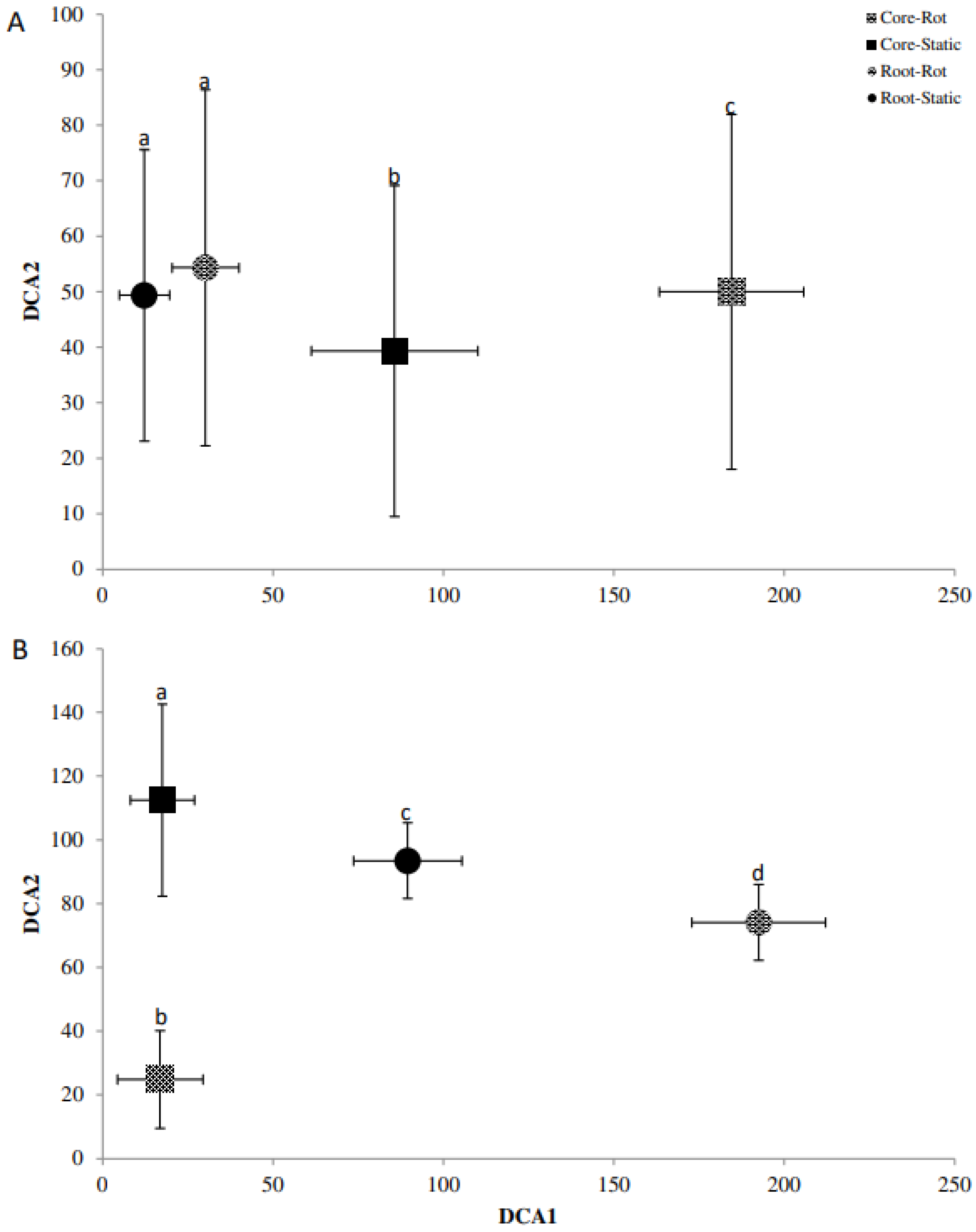
Detrended correspondence analysis (DCA) of the Denaturing Gradient Gel Electrophoresis (DGGE) fingerprints was used in conjunction with Monte Carlo permutation testing to identify differences in the beta diversity of bacterial and fungal communities for A. stolonifera and P. lanceolata in the cores and the rhizosphere (Figure 2, Figure 3 and Figure 4). For the bacterial communities, the rotating and static cores and their respective rhizospheres contained significantly different bacterial communities (p < 0.05; Figure 2). While the core samples of Agrostis, were separated well on the first axis, the rhizosphere samples were separated on the second axis (Figure 2A). This was the other way round for the bacterial communities in the Plantago microcosms (Figure 2B). The AM fungal communities differed also significantly for the cores and rhizospheres for both plants (p < 0.05; Figure 3). In the Agrostis microcosms, the core samples were separated on the second axis while the rhizosphere samples were separated on the first axis (Figure 3A). For Plantago, the separation of cores and rhizospheres was the other way round (Figure 3B). The saprophytic fungal communities (Figure 4, based on ITS fingerprints) differed significantly between the cores and rhizospheres for Plantago where the rhizosphere samples were separated on the first axis, while the core samples were separated on the second axis (p < 0.05; Figure 4B). For Agrostis the rotating and static cores were significantly different and also differed significantly to the rhizospheres, but no significant difference was detected between the rhizosphere samples of static and rotating core microcosms (Figure 4A).

2.6. Arylsulfatase Activity

Sulfate ester mobilising activity was measured in the hyphosphere (cores) and rhizosphere of the static (mycorrhizal) and rotating (severed hyphae) treatments. For both plants, arylsulfatase activity was higher for the rhizosphere than the cores and, additionally, for the rhizospheres from the static over the rotating treatment (p < 0.05). For A. stolonifera, 19.1 (±1.09) and 11.8 (±1.90) µg of p-nitrophenol was released in the rhizosphere from the static and rotating treatments, respectively. For the static and rotating cores this was lower at 5.5 (±1.22) and 8 (±2.23) µg, respectively and was not significantly different (p > 0.05). For P. lanceolata, 23.4 (±2.25) and 17 (±2.30) µg of p-nitrophenol was released from rhizospheres of the static and rotating treatments, respectively. This was significantly higher when compared to the static and rotating cores at 2.5 (±0.50) and 2.5 (±0.30) µg which were identical (p > 0.05) (Supplementary Figure S3).

2.7. Diversity of asfA and atsA Gene

Screening of 200 asfA clones (50 each per treatment in A. stolonifera microcosms) revealed 43 unique OTUs in total; 12 in static cores, 10 in rotating cores, 12 in the rhizosphere of the static and 15 in rhizosphere of the rotating treatments. Only 2 OTUs occurred in all treatments. Both OTUs were the most abundant composing 23% and 12% of the sulfonate mobilising population analysed. Library coverage was calculated to be between 92 and 96%. 18 OTUs had more than three representatives and were subjected to Sanger sequencing. Initial identification of the phylogenetic affiliation was determined with NCBI’s BLAST [31]. All sulfonate mobilising bacterial marker gene fragments were found to belong to the phylum Proteobacteria (Table 2). Sequences (asfA) were translated and incorporated into a phylogenetic tree ([32] (Supplementary Figures S4 and S5). The two OTUs present in all clone libraries (CS16 and CS37) clustered in a clade that contains the genus Cupriavidus (Supplementary Figure S5).
Five OTUs associated with Variovorax and one OTU associated at the family level of the Commamonadaceae (Table 2) clustered within the Variovorax clade of the phylogenetic tree (supplementary Figure S4). Together, they represented 16% of the asfA clones. None of the Variovorax associated clones were found in the static mesh cores, while eight were found within the rotating cores and 23 clones were found in the plant rhizosphere. The Burkholderia clade harboured one OTU (CS12), while the Polaromonas clades contained 2 OTUs (RS4, RS6). OTU CS12 was found to be associated with the genus now identified as Paraburkholderia. Only recently, the genus Burkholderia has been split into two genera, with most of the environmental strains moved into the newly established genus of Paraburkholderia [33]. The clade that was associated with Cupriavidus contained 40% of the asfA clones that included CS37 and CS16. Notably, 29 of the 46 clones of CS16 were found on fungal hyphae in static mesh cores (presumptive mycorrhizal). One OTU was found to be closely associated with Stenotrophomonas (CR38; BLAST and phylogenetic tree) that was only found in the rotating mesh core.
Clone libraries of the sulfate-ester mobilising atsA gene amplicons (160 clones, 40 per treatment) contained 38 OTUs in total; 9 for static cores (5 overlapping), 14 for rotating cores (6 overlapping), 15 for static rhizosphere (5 overlapping), and 13 for rotating rhizosphere (5 overlapping). Library coverage [34] was at or above 85%. OTUs with more than three representatives (15) were sequenced and phylogenetic affiliation established via BLAST (Table 3).
The dominating sulfate-ester mobilising bacteria in possession of the atsA marker gene were found to be associated with Acidobacteria (27.5%), Verrucomicrobia (14%), Bacteroidetes (12.5%) and Rhodospirillales (order) (10%) (Table 3). One OTU, representing 4% of atsA sequences was associated with the eukaryotic phylum Rotifera. Differences in abundance were observed across the four treatments. Acidobacteria were more abundant in the rhizosphere, particularly in the rotating treatment. Rhodospirillales like clones were predominantly isolated from the hyphosphere (cores) and were present in both static and rotating treatments (Table 3).

2.8. NGS Based Bacterial Community Sequences

Using the Chao 1 index [35], the species diversity in the rotating and static cores (A. stolonifera as host plant) was not determined to be different at 4006 (±1624) and 4774 (±1242), respectively. The number of OTUs obtained from the rotating and static core samples were determined to be 3955 (±1644) and 4740 (±1246), respectively. The rotating core samples had a higher Simpson and Shannon index (0.97 and 7.4) than the static core samples (0.87 and 6.6, respectively) [36,37].
PCoA (un-weighted, Unifrac distance matrix) separated the rhizosphere and hyphosphere very clearly (Figure 5). While the rhizosphere treatments clustered together, rhizosphere and core samples were clearly separated on the first axis. A separation between the static and rotating cores (hyphosphere) was visible on the second and third axis (Figure 5).
Bacterial phyla that were most abundant in the rhizosphere treatments (with static and rotating cores) included the Acidobacteria (16–20%), Actinobacteria (7–9%), Planctomycetes (4%), Proteobacteria, (28–31%) and Verrucomicrobia (9–10%). Bacterial phyla that were most abundant in the hyphosphere treatments (within the cores, static or rotating) included the Acidobacteria (6–13%), Actinobacteria (2–10%), Bacteroidetes (2–4%), Firmicutes (3–5%), Planctomycetes (22–34%) and Proteobacteria (24–29%) (Supplementary Figure S6). Of the 18 most abundant phyla, only the Planctomycetes and Gemmatimonadetes were significantly more abundant in the static than the rotating cores, while only the Acidobacteria were significantly more abundant in the rotating over the static cores (p < 0.05). Differences in relative phyla abundance was more widespread between the cores and the rhizospheres (rotating and static) with 11 of the 18 most abundant phyla showing significant differences in abundance (data not shown).
Among the 35 most abundant bacterial families, significant differences (p < 0.05) between the cores (hyphosphere, static, rotating) and the rhizospheres were detected (Table 4, marked in bold). Nine of the 35 most abundant families were significantly more abundant in the cores, most notably the Holophagaceae and Xanthomonadaceae. In contrast, 17 of the 35 most abundant families were significantly more abundant in the rhizospheres than in the cores and included the Acidobactertiaceae, Xanthobacteraceae and the DA101 group (Table 4). Significant differences between the static and rotating cores were rare. Only two of the 35 most abundant families were more abundant in the static (Gemmatimonadaceae, Sphingomonadaceae) and the rotating (DA111 group, Sorangiinae) cores, respectively (Table 4). Significant differences between the rhizospheres of static and rotating core microcosms could not be determined as several replicates of the static rhizosphere samples failed the quality controls of the sequencing run and were subsequently removed from the analysis.

3. Discussion

AM fungi are highly beneficial to plant hosts as they are involved in nutrient mobilisation as determined with organically bound labelled isotopes P and N [38,39,40]. Additionally, AM fungi have been shown to promote plant growth [41] and harbour abundant sulfonate mobilising bacterial communities [15]. While little evidence exists to suggest that AM fungi directly mobilise organically bound S, improved S uptake has been observed in their presence [21]. Indeed, ERH growth has been reported to be enhanced in the presence of organo-S where bacterial activity was suspected to be the driving force behind the growth rates of ERH [29]. The present study aimed to illustrate that the presence of an intact AM symbiosis increases uptake of organo-S via organo-S mobilising microbes and identified increased 34S uptake from organo-S in the first three months of growth of Agrostis and Plantago.
Plant S demand is dependent both on plant species and stage of development with increased demand observed during periods of vegetative growth and seed development [42]. When S demand is high, plant SO42− transporters are up-regulated for rapid uptake in the rhizosphere leading to a SO42− depletion zone [22]. In this zone, bacterial desulfurization of organo-S is induced via AM influenced rhizodeposition [3]. When S demand is lessened at later growth stages, it is no longer viable to expend carbon to stimulate proliferation of organo-S mobilisers and the AM role may be minimised. Findings from the present study appear to support this scenario, as 34S plant uptake from organo-34S enriched soil was increased in the microcosms with static cores after 3 months for A. stolonifera and P. lanceolata and for A. stolonifera also after 6 months. XANES analysis undertaken in the present study uncovered that sulfate-esters and sulfonates were both present in the soil. XANES analysis also revealed that the static cores may have contained more of the reduced S species, which suggests a higher S uptake and incorporation into fungal biomass in the static cores. Indeed, more abundant reduced S has been associated previously with fungal biomass [43]. Other studies have used a conventional soil fractionation approach to estimate ester and sulfonate S pools where the exact S species cannot be identified via operationally defined fractionation methods [1]. However, these fractionation based studies have highlighted that mobilisation of both organo-S species is important for optimisation of plant S supply [44,45].
Excluding the formation of intact AM symbioses inside the rotating microcosm cores resulted in significantly lower root colonisation for both plants analysed when compared to the static cores. While it is plausible that the AM hyphal ingrowth into organo-S enriched soil is beneficial for the overall mycorrhization levels of the host plant, it is important to note that the total amount of soil in the cores represents less than 10% of the soil available for fungal colonization. Consequently, it is possible that the rotating of the cores to prevent fungal ingrowth may not only limit the access to nutrients but could also disturb the overall mycorrhization of the plant host. While the present study has also utilized 1 µm mesh cores that do not need to be rotated, their use was only to compare 34S uptake (data not shown) and not mycorrhization.
Treseder [46] demonstrated that increased AM root colonisation improved both plant yield and nutrient content. The mechanism for which may include increased efficiency of nutrient mobilisation, uptake and transfer via characteristic mycorrhizal structures [47,48]. In the present study, the promoted AMF symbiosis via static cores has not only resulted in an increased percentage root colonisation but has also stimulated bacterial proliferation. Indeed, more abundant cultivable heterotrophs, sulfonate and polymeric sulfonate mobilisers were not only found in the static cores when compared to the rotated ones, but they were also more abundant in the rhizosphere of microcosms with static cores. It appeared that there is a putative positive knock-on effect from the enhanced mycorrhization through access to staic cores that had manifested itself in the bacterial community. In turn, the exclusion of fungal ingrowth into rotating cores had a more profound limiting effect. A previous study has shown that sulfonate mobilisers are abundant in the hyphosphere, putatively attaching to the hyphal surface via a type III secretion system [15]. Likewise, AMF inoculation was found to alter bacterial community dynamics that included sulfonate utilizing bacteria [49]. These recent findings, appear to support the here hypothesised positive knock-on effect.
Previous studies demonstrated that AM fungal symbiosis alters microbial community composition in the hyphosphere and rhizosphere [15,50]. Thus, it was not surprising that community fingerprint analysis in the present study of the organo-34S microcosms identified significant bacterial, fungal and AM fungal community shifts (presented as DCA biplots) for the static (mycorrhizal) and rotating (severed hyphae) treatments. The above hypothesized positive knock-on effect of the hyphal ingrowth in the cores may explain why bacterial rhizosphere community structures were clearly and significantly differentiated as well. Studies on bacterial diversity and abundance associated with AM hyphae have presented equivocal results in the past and the findings in this study were deemed to be a result of AM induced physico-chemical modification of rhizodeposition stimulating dominance of certain bacterial groups [51,52,53].
The amplicon-based sequencing (NGS) approach in the present study showed that Planctomycetes were stimulated greatly in both the static and rotating cores with highest numbers exceeding 34% relative abundance in the mycorrhizal (static) cores. The Proteobacteria were the second dominating phylum in the present study but unlike the Planctomycetes were equally abundant in the cores and rhizospheres. Proteobacteria groups have been identified to be S mobilisers in the past [11] and the current study is no exception when looking at the asfA affiliations. Within the Proteobacteria, the Xanthomonadaceae were most abundant in the static cores with the presence of AM hyphae and this family includes Stenotrophomonas which was recently isolated directly from the hyphosphere and putatively has the ability to attach to and co-migrate with AM hyphae [15]. However, while the abundance of Xanthomonadaceae was significantly lower in the rhizosphere, the abundance of Xanthomonadaceae was not significantly lower in the rotating cores over the static cores. It is possible that clades of this family can be found on various types of hyphe and not only AM fungal hyphae. Likewise, the Comamonadaceae appeared to be stimulated in the mycorrhizal static cores but this observation did not reach significance (p > 0.05). Comamonadaceae repeatedly associated with aromatic sulfonate desulfurization include Variovorax and Polaromonas [12,14]. While both genera were present in the cores and rhizospheres of the present study, no significant difference in relative abundance was identified.
Sulfate-ester (arylsulfatase enzyme assay) and sulfonate mobilising potential (MPN of sulfonate utilizing bacteria) was significantly increased in the static cores presumably by the stimulated AM symbiotic partnership. However, arylsulfatase activity was higher in the rhizosphere of the microcosms with static cores than within the static cores themselves. Since the soils in the cores were treated with glucose and 34SO4 to establish organo-34S, one could speculate that this treatment had an effect not dissimilar to a fertilizer treatment. S-fertilization has been demonstrated to decrease arylsulfatase activities [9], thus it is possible that the core soil treatment had a prolonged negative effect on the arylsulfatase activity. This is in direct contrast to the abundances of sulfonate utilizing bacteria that was highest in the static cores. Previously, the abundance of sulfonate utilizing bacteria was unaffected by fertilization [9], though the amendment of soil with biochar did increase the abundance of these bacteria [14]. Following these observations, one may speculate that within the static cores recently formed sulfonates are predominantly mobilized, while outside the cores in the rhizosphere more of the soil sulfate esters are utilized. However, the XANES spectra of the cores do not support the hypothesis of a selective utilization of sulfonates in static cores. Likewise, in an additional experiment the authors of the present study have established static and rotating cores with soil enriched with 34S that underwent an additional sulfatase treatment [54]. There, no significant increase in 34S plant uptake was identified. Therefore, it is unlikely, that a preferential mobilization of carbon bonded S over ester bonded S took place in the static cores over the duration of the experiment. While the plant 34S uptake was monitored over time, the remainder of the analyses were only possible after destruction of the microcosms, hence 34S uptake at month 3 and 6 cannot be directly compared to the remainder of the results. It is therefore possible that a shift in organo-S mobilization and utilization took place inside and outside the cores. The highlighted sulfatase activity and abundance of sulfonate utilizing bacteria is therefore only a snapshot at month 9 when most of the 34S from the enriched core soils was already mobilized by month 6. Further studies would be beneficial that would carry out enzymatic and microbial analyses within the first 3 months of incubation. Nevertheless, previous studies have shown improved S uptake in the presence of AMF which may be a result of the carbon rich hyphosphere selecting for organo-S mobilising bacteria with the potential to improve nutrient uptake [21,55,56]. Thus, the current study is confirming these previous observations.
Analysis of the diversity of the sulfate-ester mobilising atsA gene revealed differences in presence and relative abundance of taxa in the cores and rhizospheres. While atsA can be considered to be best studied in the Proteobacteria Pseudomonas and Klebsiella [5,6], there was no evidence of a substantial presence of atsA from Proteobacteria in the present study. The atsA clone libraries were dominated by the Acidobacteria and Verrucomicrobia. The former was the most relative abundant phylum in the rhizosphere. In contrast, the Rhodospirillales OTUs were almost exclusively found in the cores. The Planctomycetes atsA clones were exclusively found in the static cores. Based on NGS sequencing data, Planctomycetes were among the most abundant phyla in the microcosms overall. Interestingly, the majority of Planctomycetes 16S sequences were found in the static cores, followed by the rotating cores, while the rhizosphere harboured only a small fraction of the Planctomycetes. This phylum shows some rather unusual cell features when compared to most other bacterial phyla and is commonly detected in marine environments. Some marine isolates were identified for industrial use of their sulfatase enzymes [57]. More recently, it has also been regularly identified in soils [58] and in the present study it’s sulfatase activity may be of importance in connection to the hyphosphere and ultimately to plant-S uptake. However, up to date very limited information is available on microbial sulfatase genes in rhizo- and hyphosphere, thus further research is needed in this area.
Clear differences in presence and relative abundance of genera were observed for the aromatic sulfonate mobilising community (asfA). While taxonomic affiliation via BLAST resulted in most sequences to be associated with Rhizobiales and Comamonadaceae, integration into the phylogenetic tree of AsfA [11,12,13,14,15] associated 41% of all OTUs with the genus Cupriavidus. Most of the OTUs came from the cores and of them again the majority was found in the static cores, hence presumptively associated with AM fungal hyphae. Previous studies that assessed the diversity of asfA in soil, rhizosphere [11,12,13,14,15] and hyphosphere [11,12,13,14,15] did not pick up sequences related to Cupriavidus with the exception of one Cupriavidus like OTU identified in the rhizosphere of Lolium perenne [11,12,13,14,15]. One may speculate that Cupriavidus may play a particular role in sulfonate-S mineralization in the hyphosphere of mycorrhiza that are in particular associated with Agrostis. The genus Cupriavidus has been linked with improved P and N mobilisation in the past [59,60] and may play an important role in overall plant nutrition that now includes also S. Cultivation dependent data from the present study are in line with the cultivation independent results as the MPN approach for sulfonate utilizing bacteria cultivation identified highest abundances in the static cores. Other commonly retrieved sequences of AsfA clustered with Variovorax that dominated the rhizosphere but was also found in the rotating cores. Variovorax was identified as a common sulfonate mobiliser in various rhizospheres including that of Agrostis rupestris of the Damma glacier forefield in Switzerland [61] and the potato rhizosphere from the Netherlands [62]. Particular dominance of Variovorax asfA was detected in the rhizosphere of wheat in the long-term experiment Broadbalk, managed by Rothamsted Research (UK) [12].
In conclusion, this study demonstrates that an intact AM symbiosis increases plant uptake of 34S from organo-34S enriched soil at early stages of growth when S requirement is high. This may be a result of AM induced microbial community shifts leading to improved sulfate-ester and sulfonate mobilising activity. Likewise, allowing fungal ingrowth in the static cores has positively impacted mycorrhization in the microcosms and had a positive feedback onto other microbial putative activities. Therefore, fostering AM symbiosis has the potential to improve plant S uptake and reduce the requirement for unsustainable inorganic S fertilisation practices, while in turn limiting AM access to resources has a potentially negative impact that extends to the plant host and to the microbes inhabiting the hyphosphere and rhizosphere.

4. Materials and Methods

4.1. Site Description

The soil used to create the experiments was obtained from a long-term field trial (“Cowlands”) at Teagasc, Johnstown Castle, Wexford, Ireland (52°16′ N, 6°30′ W, 30 m above sea level). The soil type is a poorly drained gley soil classified as Mollic Histic Stagnosol (WRB 2006) with 11% of organic matter, a pH of 6 and a loamy topsoil with 18% of clay content. The soil has not received P or S fertiliser since 1968 and has not been ploughed since 1970 (P0-0, site A5). Swards are mixtures of Lolium perenne, Dactylis glomerata and various meadow grass species [63].

4.2. Stable Isotope Enriched Soil

The most common form of elemental S in nature is 32S (95.02%) and stable isotope 34S represents 4.21% of S existing naturally in soil. For this study, S uptake and incorporation into plant biomass was undertaken using the ratio of 34S to 32S (δ34S values) [64].
To enrich the soil with organo-34S, 20 g soil aliquots were treated with 34S-SO4 (20 mg/kg soil) and glucose (6 g/kg) to stimulate S immobilisation at 80% water holding capacity (WHC) and optimise the amount of S in the C-bonded fraction [65]. The soil was incubated for 6 weeks at 25 °C and aerated fortnightly. After 6 weeks, the 20 g soil was thoroughly mixed with 20 mL of 0.01 M CaCl2 for 30 min on a Intelli-Mixer RM-2 (Elmi Tech Ltd., Brea, Latvia) and subjected to centrifugation (Allegra X-22R, Beckman Coulter, Brea, CA, USA) at 3900 rcf for 20 min and to remove inorganic S [65]. This process was repeated two additional times before the supernatant was discarded and the residual material was dried, ground and brought to 60% WHC. To counteract the loss of essential nutrients, the enriched fraction was supplemented with Hoagland’s solution (S free, 50%) [66]. The 34S enriched soil samples were transferred into nylon mesh cores (up to 6 g per core) with windows of either 35 µm or 1 µm adapting an experimental setup described previously [40].

4.3. Core Design and Constructing the Soil Microcosms

Cores were constructed adapted from a design published by Johnson and colleagues [40] using acrylonitrile butadiene styrene (ABS) water pipe (150 mm height, 18 mm diameter). Two windows were cut into each core, the size of which constituted 90% of the surface area of the core (Supplementary Figure S7). Nylon mesh (Plastok Associates Ltd., Birkenhead, UK) with 35 µm pores was used to cover the windows of the core and 1 µm nylon mesh was used to cover the base. The mesh was attached using Tensol No. 12 adhesive (Evode Speciality Adhesives Ltd., Leicester, UK) and the completed cores were heated (24 h, 80 °C) to harden the adhesive (cured). The 35 µm mesh is sufficiently large to allow mycorrhizal hyphae to colonise the cores while preventing root access. A permeable base of 1 µm allows free drainage of water through the cores and minimises hyphal access. This set of cores was used to analyse the role of AM in uptake of organo-34S where the plants are cultivated outside the cores and AM hyphae are able to grow inside the cores enriched with organo-34S.

4.4. Organo-34S Enriched Soil Microcosms for Cultivation of Agrostis stolonifera and Plantago lanceolata

Microcosm systems were constructed from cuttings of Plexiglas (12 × 11 × 2.5 cm, chemically welded with Ethylene Dichloride) and filled with 230 g of a sand (Glenview Natural Stone, Ireland) and soil (Teagasc, Johnstown Castle) mixture (one part sand, one part soil w/w). These were planted with A. stolonifera and P. lanceolata, respectively and inoculated with 2 g of Rhizophagus irregularis (purchased as Glomus intraradices; Symbivit, Symbion, Czech Republic). For this experiment, two mesh cores, containing 6 g of organo-34S stable isotope enriched soil, were inserted into each soil microcosm (two cores per microcosm were chosen to minimise the distance of plant roots to the cores, see also Supplementary Figure S7). The cores were kept plant-free and served as the organo-34S source in this experiment. The organo-34S enriched cores were either rotated twice weekly to sever AM hyphae (rotating) or undisturbed (static) to allow AM colonisation. Each treatment was carried out in replicates of six (12 Agrostis microcosms, 6 with rotating cores and 6 with static cores; 12 Plantago microcosms, 6 with rotating cores and 6 with static cores). Plan shoots were repeatedly cut for 34S analysis, while cores and rhizospheres were sampled destructively at the end of the experiments. The microcosms were grown in an A1000 Adaptis plant growth chamber (Conviron, Germany) with day-night temperature of 25–15 °C, respectively, 12 h day, 70% RH, and 320 µmoles m−2 s−2 photosynthetic active radiation (PAR). The systems were watered with dH2O three times a week and once fortnightly supplemented with half times S free Hoagland’s solution [66].

4.5. Determination of 34S Uptake into Shoots of A. stolonifera and P. lanceolata

Determination of 34S uptake was achieved using Elemental Analysis—Isotope Ratio Mass Spectrometry (EA-IRMS) undertaken by Iso-Analytical (Cheshire, UK) as outlined in the Supplementary Information S1. The aboveground biomass was destructively harvested by cutting off shoots at 5 cm above the ground to analyse the S flow from labelled organo-34S in the mesh cores to its incorporation into plant tissue. P. lanceolata was measured at 3 and 6 months (3 replicates per treatment). A. stolonifera was measured at 3, 6 and 9 months (6 replicates) (see also analysis flow chart, Supplementary Figure S8). The plant biomass was dried at 80 °C for 5 d, ground to a fine powder and a 0.5 g aliquot was subjected for EA-IRMS. Additionally, at 9 months one of the two cores in each A. stolonifera microcosm was re-planted into a new microcosm instead of being destructively harvested (second round of microcosms with one core). The second generation of microcosms were established to ascertain long term organo-34S uptake after 12 months of incubation in total (6 replicates).

4.6. S K-Edge X-ray Absorption near Edge Spectroscopy

XANES analysis was used to identify the chemical oxidation states of S, reduced (sulfides, thiols), intermediate (sulfoxides, sulfonates) and oxidised (sulfate esters, sulfates) as described previously [30]. In the present case, soil subsamples from two static and two rotating core experiments of A. stolonifera were compared. Soil samples were dried at 105 °C for 12 h and were ground to a fine powder using a pestle and mortar. SO42− was extracted from each of the samples with 0.01 M CaCl2 using the same approach highlighted above in Section 4.2. The soil was freeze dried in a Labconco FreeZone™ 4.5 L Freeze dry system (Labconco, Kansas City, MO, USA). XANES was carried out on the KMC-1 beam line at BESSY II (Helmholtz-Centre Berlin for Materials and Energy, Berlin, Germany, for further details see Supplementary Information S2). The KMC-1 is a soft X-ray double crystal monochromator beamline for the energy range 2–12 keV. The photon energy of sulfate is the same as for sulfate esters. Since free sulfate was removed from the samples prior to testing, the corresponding peaks are therefore exclusively from sulfate esters.

4.7. Percentage Root Colonisation

At the time of harvest, each plant type and treatment was examined for root colonisation by AM fungi using a modified version of the grid line intersect method [67] as described in the Supplementary Information S3 (analysis flow chart, Supplementary Figure S8). Arbuscular colonization (AC) and vesicular colonization (VC), respectively, were calculated by dividing their respective counts by the total number of intersections examined. Hyphal colonization (HC) was calculated as a proportion of the positive intersections. For each organo-34S stable isotope soil microcosm, 3 replicates of 100 fields of view per treatment, per plant were examined to calculate AM fungal colonisation.

4.8. Extraction and Quantification of Bacteria from AM Hyphae

To harvest the microcosm cores and plant roots of the static and rotating treatments of both plant types (six cores and plant roots per treatment), up to 1 g of hyphosphere material (hyphae with adhering soil, handpicked with fine forceps under a stereomicroscope) was collected from each core and 1 g of rhizosphere (roots with adhering soil) was analysed in parallel. The hyphosphere and rhizosphere bacteria were extracted into 10 mL of sterile saline solution (0.85%) and rotated at 75 rpm on an Elmi Intelli-Mixer RM-2 (Elmi Tech Ltd., Latvia) (30 min, 4 °C). A 0.1 mL aliquot of the resulting suspension was used for bacterial community quantification (cultivation dependent). The remainder of the suspensions were centrifuged at 4500 rpm (4 °C, 20 min) and the obtained pellets were immediately frozen (−18 °C) for subsequent cultivation independent analysis (see Section 4.9, Section 4.11 and Section 4.12 below).
The aliquot of bacterial suspension from above was used for tenfold serial dilutions in saline (0.85% w/v). Most probable number (MPN) analysis was undertaken in agar-free R2A [68], MM2TS and MM2LS ([14]; Supplementary Information S4)) to enumerate the cultivable heterotrophic, sulfonate and polymeric sulfonate mobilising communities, respectively. A 20 µL aliquot from each dilution (101–107) was added to 200 µL of either R2A MM2TS or MM2LS in 96 well microtiter plates. Following 2 weeks of growth in an Innova Incubator Shaker Series (New Brunswick Scientific, Edison, NJ; 75 rpm, 25 °C), the OD590 was recorded using an ELX808IU spectrophotometer (3 min shaking at level 3; Bio Tek Instruments Inc., Winooski, VT) in order to determine the MPN number which in turn was used to obtain an MPN g−1 value [69].

4.9. Community Fingerprinting

The frozen pellets from the organo-34S microcosms of the bacterial extractions were used for community DNA extractions using the UltraClean Soil DNA extraction kit from MoBio as described by the manufacturer (Carlsbad, CA, USA). Subsequent Denaturing Gradient Gel Electrophoresis (DGGE) was carried out for the bacterial 16S rRNA, 18S AM fungal and ITS saprotrophic fungal communities as described previously [70]. PCR and DGGE conditions as well as a list of the selected primers for PCR-DGGE are available in the Supplementary Information S5 and Supplementary Table S1.

4.10. Arylsulfatase Activity

A colorimetric method of assaying soil arylsulfatase activity was used to quantify arylsulfate ester cleaving enzymes in 1 g of soil with p-nitrophenyl sulfate as the substrate [71]. Arylsulfatase activity was analysed for hyphosphere (inside cores) and rhizosphere, static and rotating treatments. Further details are provided in Supplementary Information S6.

4.11. Diversity of asfA and atsA Gene

Diversity of the bacteria with the capacity to mobilize sulfur from sulfonates and sulfate esters from the A. stolonifera microcosms were analysed via their marker genes asfA and atsA, respectively. Amplicons of the corresponding genes were used to generate respective clone libraries of the hyphosphere and rhizosphere from the static microcosms as well as the hyphosphere and rhizosphere from the rotating microcoms.
The asfA gene was amplified with primers asfAF1all [15] and asfBtoA [11] (Supplementary Table S1). A touchdown PCR was carried out under the following conditions: initial denaturation at 98 °C for 3 min, 10 cycles of 98 °C denaturation (10 s), 65–55 °C touchdown (15 s), 68 °C extension (40 s), plus 25 further cycles at 58 °C annealing. Final extension was carried out at 68 °C for 3 min. PCR was undertaken in 25 µL reactions containing; 1 X Terra PCR Direct Buffer (2 mM MgCl2), 0.2 mM dNTP mix, 0.4 µmol of each primer, and 0.7 U of Terra PCR Direct Polymerase (ClonTech Europe, Saint-Germain-en-Laye, France).
The atsA gene was amplified using the AtsA-F1 AtsA-R1 primer pair [9] (Supplementary Table S1). PCR conditions were as follows: initial denaturation at 95 °C for 3 min, 40 cycles of 95 °C denaturation (60 s), 48 °C annealing (60 s), and 72 °C extension (60 s). Final extension was carried out at 72 °C for 10 min. The PCR was undertaken using a Kapa 2G Robust PCR kit (Kapa Biosystems, Woburn, MA, USA) in 25 µL reactions containing; 1× Buffer A, 1× Enhancer, 5% DMSO (Sigma-Aldrich, St. Louis, MO, USA), 2 mM MgCl2, 0.2 mM dNTP, 0.5 µM of each primer and 0.5 U of the Kapa Robust polymerase.
The asfA and atsA PCR products were purified, quantified, and ligated into the cloning vector pJET1.2/blunt (CloneJet, Thermo Scientific, Waltham, MA, USA) and pGEM®-T vector kit (Promega, Madison, WI, USA), respectively. The ligation and transformation steps were undertaken as per manual instructions. The ligation reactions were transformed into E. coli DH5α. In order to ascertain taxonomic diversity of recombinant plasmids containing an insert of the correct size, Restriction Fragment Length Polymorphism (RFLP) analysis was carried out on PCR amplicons using the restriction enzymes AluI and RsaI (5 U per reaction; Thermo Scientific) for 4.5 h at 37 °C. The digested DNA was run on a 2% agarose gel at 100 V for 40 min. Clones with a similar restriction pattern were classified as a genotype (OTU) using Phoretix 1D (Nonlinear Dynamics, Newcastle upon Tyne, UK). Library coverage was calculated as one, subtracted by the ratio of the total number of OTUs with single clones to the total number of clones multiplied by hundred [34]. Unique genotypes with more than one representative were re-amplified and the purified PCR product was used for sequence identification (GATC Biotech, Konstanz, Germany). The sequences obtained were subjected to gene comparison using BLAST [31]. Sequences of asfA were imported into arb (Version 5.2) [72], translated into proteins and integrated into an established asfA phylogenetic trees [32]. Nucleic acids were deposited in EBI’s nucleic acid archive under the accession numbers MT309545-MT309559 & MT380723 (atsA) and MT309560-MT309575 & MT372478-MT372479 (asfA).

4.12. Amplicon Sequencing of 16S sRNA Gene Fragments (NGS)

For Illumina MiSeq 16S library preparation, community DNA from A. stolonifera (after 9 months of incubation) was amplified (cores and rhizosphere, static and rotating microcosms, replicates of six) using the primer pair 16SF and 16SR to target the V3 and V4 region yielding a 460 bp amplicon (Illumina Metagenomic Sequencing Library Preparation Guide; Supplementary Table S1). PCRs were undertaken in 25 µL reactions with 0.5 U of Kapa HiFi Taq, 1× PCR buffer with 1.5 mM MgCl2, 0.2 mM dNTPs each (all Kapa Enzymes, Woburn, MA, USA), and 0.4 µM of each primer. A touchdown PCR protocol was used with the following cycling conditions: initial denaturation at 98 °C for 5 min, 20 cycles of 98 °C denaturation (45 s), 68–58 °C touchdown (60 s), 72 °C extension (60 s), plus 20 further cycles with an annealing temperature at 58 °C. The PCR product was purified using the GenElute PCR purification kit (Sigma-Aldrich, St. Louis, MO, USA). The indexing PCR was carried out to attach the dual indices and Illumina sequencing adapters using the Nextera XT Index Kit (Illumina, San Diego, CA, USA) in accordance with the manufacturer’s instructions with a modified annealing temperature of 63 °C. The indexing PCR product was purified (as before) and quantified using the Qubit dsDNA HS Assay Kit (Life Technologies, Carlsbad, CA, USA) on a Qubit 2.0 Fluorometer (Life Technologies, Carlsbad, CA, USA). The DNA concentrations of each sample were adjusted to 4 nM in 10 mM Tris pH 8.5 and 5 µL was used to mix aliquots for pooling libraries with unique indices. Amplicons were directly sequenced on an Illumina MiSeq NGS platform (Illumina, San Diego, CA, USA) in line with protocols at the Teagasc, Moorepark sequencing centre.

4.13. Data Analysis

Analysis of variance (ANOVA) of EA-IRMS data, percentage root colonisation, and MPN data was carried out using IBM SPSS statistics (Version 22.0; IBM, Armonk, NY, USA). Data were tested for normality and homogeneity of variance (Shapiro–Wilk’s and Levene’s tests). Data that were not normally distributed were transformed logarithmically, analysed by ANOVA and then back transformed. Where normality was confirmed, a Tukey’s HSD post hoc test was applied to assess any significant differences (p < 0.05). Where homogeneity of variance was not achieved, the Games-Howell test was used instead. Pairwise comparison of static versus rotating core as well as core versus rhizosphere sequencing data was carried out using the t-test function. DGGE fingerprinting gels were digitalised and band patterns analysed with the software package Phoretix 1D (Nonlinear Dynamics). Cluster analysis using UPGMA was carried out and obtained band pattern matrixes were exported for Detrended Correspondence Analysis (DCA) and permutation tests (Monte-Carlo with 9999 replicates) as described previously [32]. Error ranges are reported as standard deviation throughout the manuscript.
The following steps were undertaken to analyse the Illumina NGS data; raw Illumina paired-end sequence reads were merged using Flash [73] and quality checked using the split libraries script in QIIME [74]. Quality checking is two phase; for joining reads a min overlap of 10 bp was expected; then for joined reads anything less than 150 bp and quality of 19 was removed. Reads were clustered into OTUs with a similarity cut off at 97% and chimeras were removed with the 64-bit version of USEARCH [75]. Subsequently, OTUs were aligned and a phylogenetic tree was generated within QIIME [74]. Alpha and beta diversity analysis was also implemented within QIIME [74]. Alpha diversity was determined using the Shannon and Simpson diversity indices and the Chao1 richness estimator. Beta diversity was analysed using PCoA (weighted Unifrac) undertaken with EMPeror [76]. Taxonomical assignments were reached using the 16S-specific SILVA database (Version 106). Sequences were deposited in the EBI’s nucleic acid archive with the accession numbers (ERS4414312-ERS4414324).

Supplementary Materials

The following supporting information can be downloaded at: https://www.mdpi.com/article/10.3390/plants11223050/s1, Figure S1: AM uptake of 34S from organo-34S after 12 months (3 months) post transfer into new Agrostis stolonifera microcosms. Rotating = severed hyphae, Static = mycorrhizal. No significant differences (a). Figure S2: Normalised and stacked S K-edge spectra from static (black) and rotating (red) treatments with Agrostis stolonifera as host plant. Peak A represents reduced thiols, peak B represents intermediate sulfonates and peak C represents oxidised sulfate esters. Figure S3: Arylsulfatase activity for Agrostis stolonifera (A) and Plantago lanceolata (B) in organo-34S enriched soil microcosm systems. Rotating = severed hyphae, static = mycorrhizal, roots = rhizosphere, and cores = hyphosphere. Letters (a–c) represent significant differences. Figure S4: Randomised axelerated maximum likelihood tree of truncated AsfA sequences of representative desulfonating operational taxonomic units (OTUs) derived from clonal asfA DNA sequence analysis from static (CS) and rotating (CR) hyphosphere cores and rhizosphere roots from static = mycorrhizal (RS) and rotating = severed mycorrhizal hyphae (RR) treatments of A. stolonifera microcosms. Cultivated (italics) and molecular isolates from this study (bold) are highlighted. Molecular isolates from spring barley rhizospheres (sb), Agrostis grassland rhizospheres (CA), wheat rhizospheres from Broadbalk (W), rhizospheres and soils from the Damma glacier forefield (D; DA) and hyphosphere (H) were isolated previously. Clades expanded include Variovorax, Burkholderia, and Polaromonas. Clades to be expanded in the figure below (Figure S5) are highlighted in red. Figure S5: Randomised axelerated maximum likelihood tree of truncated AsfA sequences of representative desulfonating operational taxonomic units (OTUs) derived from clonal asfA DNA sequence analysis from static (CS) and rotating (CR) hyphosphere cores and rhizosphere roots from static = mycorrhizal (RS) and rotating = severed mycorrhizal hyphae (RR) treatments of A. stolonifera microcosms. Cultivated (italics) and molecular isolates from this study (bold) are highlighted. Molecular isolates from spring barley rhizospheres (sb; [3]), Agrostis grassland rhizospheres (CA), wheat rhizospheres from Broadbalk (W), rhizospheres and soils from the Damma glacier forefield (D; DA) and hyphosphere (H) were isolated previously. Clades expanded include Acidovorax, Arthrobacter and Stenotrophomonas. Clades expanded in the preceding figure (Figure S4) are highlighted in red. Figure S6: Abundance of sequences allocated to major bacterial phyla (cut-off 0.1%) after taxonomic analysis of 16S rRNA amplicons from hyphosphere cores (C) (grey) and rhizosphere roots (R) (black) of Agrostis stolonifera and static (S) = mycorrhizal (solid) and rotating (R) = severed mycorrhizal hyphae (pattern) treatments. Figure S7: TOP—Construction of cores used to contain the 34S stable isotope enriched soil. BOTTOM -The soil microcosm systems with the organo-34S enriched cores (A) and the actively growing system (B). Figure S8: Flow diagram of microcosm sample analysis. Table S1: Primers used in this study. Supplementary Sections S1—Determination of 34S uptake; S2—S K-edge X-ray absorption near edge spectroscopy; S3—Percentage root colonization; S4—Minimal Media 2 with Toluenesulfonate or Lignosulfonate; S5—PCR-DGGE; S6—Arylsulfatase activity. References [6,9,11,12,14,15,30,43,61,67,77,78,79,80,81,82,83,84,85,86,87] are cited in the supplementary materials.

Author Contributions

J.G. carried out the majority of the experiments, contributed to method development and wrote a draft manuscript; O.O. and P.D.C. conducted the 16S amplicon sequencing and analysis; A.S. acquired the research funding, developed the research project ideas and established/developed most methods, contributed to the conduction of experiments, networked with colleagues to carry out additional analyses and brought the manuscript to completion. All authors have read and agreed to the published version of the manuscript.

Funding

This research was funded by the European Commission, FP7 People (CIG no. 293429).

Data Availability Statement

Nucleic acids were deposited in EBI’s nucleic acid archive under the accession numbers MT309545-MT309559 & MT380723 (atsA) and MT309560-MT309575 & MT372478-MT372479 (asfA) and ERS4414312-ERS4414324 (16S amplicon sequences).

Acknowledgments

The authors would like to thank the staff of Teagasc, Johnstown Castle, for providing the soil used in this experiment, Jonathan Leake (University of Sheffield) to provide support for constructing the mesh cores, Christian Vogel and Wolfgang Pritzkow (BESSY II) for conducting the XANES analysis, Aaron Fox (Teagasc) for critically reading the manuscript.

Conflicts of Interest

The authors declare no conflict of interest.

References

  1. Hao, F.; Lehmann, J.; Solomon, D.; Fox, M.; McGrath, S. Sulphur speciation and turnover in soils: Evidence from sulphur K-edge XANES spectroscopy and isotope dilution studies. Soil Biol. Biochem. 2006, 38, 1000–1007. [Google Scholar] [CrossRef]
  2. Autry, A.R.; Fitzgerald, J.W. Sulfonate S: A major form of forest soil organic sulfur. Biol. Fertil. Soils 1990, 10, 50–56. [Google Scholar] [CrossRef]
  3. Kertesz, M.A.; Mirleau, P. The role of microbes in plant sulphur supply. J. Exp. Bot. 2004, 55, 1939–1945. [Google Scholar] [CrossRef] [PubMed] [Green Version]
  4. Kertesz, M.A.; Fellows, E.; Schmalenberger, A. Rhizobacteria and Plant Sulfur Supply. Adv. Appl. Microbiol. 2007, 62, 235–268. [Google Scholar] [CrossRef] [PubMed]
  5. Klose, S.; Moore, J.M.; Tabatabai, M.A. Arylsulfatase activity of microbial biomass in soils as affected by cropping systems. Biol. Fertil. Soils 1999, 29, 46–54. [Google Scholar] [CrossRef]
  6. Beil, S.; Kenhril, H.; James, P.; Staudenmann, W.; Cook, A.M.; Leisinger, T.; Kertesz, M.A. Purification and characterization of the arylsulfatase synthesized by Pseudomonas aeruginosa PAO during growth in sulfate-free medium and cloning of the arylsulfatase gene (atsA). Eur. J. Biochem. 1995, 229, 385–394. [Google Scholar] [CrossRef]
  7. Kahnert, A.; Vermeij, P.; Wietek, C.; James, P.; Leisinger, T.; Kertesz, M.A. The ssu Locus Plays a Key Role in Organosulfur Metabolism in Pseudomonas putida S-313. J. Bacteriol. 2000, 182, 2869–2878. [Google Scholar] [CrossRef] [Green Version]
  8. Kahnert, A.; Mirleau, P.; Wait, R.; Kertesz, M.A. The LysR type regulator SftR is involved in soil survival and sulphate ester metabolism in Pseudomonas putida. Environ. Microbiol. 2002, 4, 225–237. [Google Scholar] [CrossRef]
  9. Ikoyi, I.; Fowler, A.; Storey, S.; Doyle, E.; Schmalenberger, A. Sulfate fertilization supports growth of ryegrass in soil columns but changes microbial community structures and reduces abundances of nematodes and arbuscular mycorrhiza. Sci. Total Environ. 2020, 704, 135315. [Google Scholar] [CrossRef]
  10. Kertesz, M.A. Riding the sulfur cycle–metabolism of sulfonates and sulfate esters in Gram-negative bacteria. FEMS Microbiol. Rev. 2000, 24, 135–175. [Google Scholar] [CrossRef]
  11. Schmalenberger, A.; Kertesz, M.A. Desulfurization of aromatic sulfonates by rhizosphere bacteria: High diversity of the asfA gene. Environ. Microbiol. 2006, 9, 535–545. [Google Scholar] [CrossRef] [PubMed]
  12. Schmalenberger, A.; Hodge, S.; Bryant, A.; Hawkesford, M.; Singh, B.; Kertesz, M.A. The role of Variovorax and other Comamonadaceae in sulfur transformations by microbial wheat rhizosphere communities exposed to different sulfur fertilization regimes. Environ. Microbiol. 2008, 10, 1486–1500. [Google Scholar] [CrossRef] [PubMed]
  13. Schmalenberger, A.; Hodge, S.; Hawkesford, M.; Kertesz, M.A. Sulfonate desulfurization in Rhodococcus from wheat rhizosphere communities. FEMS Microbiol. Ecol. 2009, 67, 140–150. [Google Scholar] [CrossRef] [PubMed]
  14. Fox, A.; Kwapinski, W.; Griffiths, B.S.; Schmalenberger, A. The role of sulfur- and phosphorus-mobilizing bacteria in biochar-induced growth promotion ofLolium perenne. FEMS Microbiol. Ecol. 2014, 90, 78–91. [Google Scholar] [CrossRef] [Green Version]
  15. Gahan, J.; Schmalenberger, A. Arbuscular mycorrhizal hyphae in grassland select for a diverse and abundant hyphospheric bacterial community involved in sulfonate desulfurization. Appl. Soil Ecol. 2015, 89, 113–121. [Google Scholar] [CrossRef] [Green Version]
  16. Wang, B.; Qiu, Y.-L. Phylogenetic distribution and evolution of mycorrhizas in land plants. Mycorrhiza 2006, 16, 299–363. [Google Scholar] [CrossRef]
  17. Smith, S.E.; Read, D.J. Mycorrhizal Symbiosis; Academic Press: Oxford, UK, 1997. [Google Scholar]
  18. Nagahashi, G.; Douds, D.D. Partial separation of root exudate components and their effects upon the growth of germinated spores of AM fungi. Mycol. Res. 2000, 104, 1453–1464. [Google Scholar] [CrossRef]
  19. Gray, L.E.; Gerdemann, J.W. Uptake of sulphur-35 by vesicular-arbuscular mycorrhizae. Plant Soil 1973, 39, 687–689. [Google Scholar] [CrossRef]
  20. Cavagnaro, T.R.; Jackson, L.E.; Six, J.; Ferris, H.; Goyal, S.; Asami, D.; Scow, K.M. Arbuscular Mycorrhizas, Microbial Communities, Nutrient Availability, and Soil Aggregates in Organic Tomato Production. Plant Soil 2006, 282, 209–225. [Google Scholar] [CrossRef]
  21. Allen, J.W.; Shachar-Hill, Y. Sulfur Transfer through an Arbuscular Mycorrhiza. Plant Physiol. 2008, 149, 549–560. [Google Scholar] [CrossRef]
  22. Buchner, P.; Takahashi, H.; Hawkesford, M.J. Plant sulphate transporters: Co-ordination of uptake, intracellular and long-distance transport. J. Exp. Bot. 2004, 55, 1765–1773. [Google Scholar] [CrossRef] [PubMed] [Green Version]
  23. Giovannetti, M.; Tolosano, M.; Volpe, V.; Kopriva, S.; Bonfante, P. Identification and functional characterization of a sulfate transporter induced by both sulfur starvation and mycorrhiza formation in Lotus japonicus. New Phytol. 2014, 204, 609–619. [Google Scholar] [CrossRef] [PubMed]
  24. Barea, J.-M.; Azcón, R.; Azcón-Aguilar, C. Mycorrhizosphere interactions to improve plant fitness and soil quality. Antonie Van Leeuwenhoek 2002, 81, 343–351. [Google Scholar] [CrossRef]
  25. Boer, W.d.; Folman, L.B.; Summerbell, R.C.; Boddy, L. Living in a fungal world: Impact of fungi on soil bacterial niche development. FEMS Microbiol. Rev. 2005, 29, 795–811. [Google Scholar] [CrossRef] [Green Version]
  26. Gryndler, M.; Hršelová, H.; Stříteská, D. Effect of soil bacteria on hyphal growth of the arbuscular mycorrhizal fungus Glomus claroideum. Folia Microbiol. 2000, 45, 545–551. [Google Scholar] [CrossRef]
  27. Gianinazzi, S.; Schüepp, H. Impact of Arbuscular Mycorrhizas on Sustainable Agriculture and Natural Ecosystems; Springer: Berlin/Heidelberg, Germany, 1994. [Google Scholar]
  28. Siciliano, S.D.; Palmer, A.S.; Winsley, T.; Lamb, E.; Bissett, A.; Brown, M.V.; van Dorst, J.; Ji, M.; Ferrari, B.C.; Grogan, P.; et al. Soil fertility is associated with fungal and bacterial richness, whereas pH is associated with community composition in polar soil microbial communities. Soil Biol. Biochem. 2014, 78, 10–20. [Google Scholar] [CrossRef]
  29. Vilarinõ, A.; Frey, B.; Shüepp, H. MES [2-(N-morpholine)-ethane sulphonic acid] buffer promotes the growth of external hyphae of the arbuscular mycorrhizal fungus Glomus intraradices in an alkaline sand. Biol. Fertil. Soils 1997, 25, 79–81. [Google Scholar] [CrossRef]
  30. Schmalenberger, A.; Pritzkow, W.; Ojeda, J.J.; Noll, M. Characterization of main sulfur source of wood-degrading basidiomycetes by S K-edge X-ray absorption near edge spectroscopy (XANES). Int. Biodeterior. Biodegrad. 2011, 65, 1215–1223. [Google Scholar] [CrossRef] [Green Version]
  31. Altschul, S.F.; Gish, W.; Miller, W.; Myers, E.W.; Lipman, D.J. Basic local alignment search tool. J. Mol. Biol. 1990, 215, 403–410. [Google Scholar] [CrossRef]
  32. Schmalenberger, A.; Telford, A.; Kertesz, M. Sulfate treatment affects desulfonating bacterial community structures in Agrostis rhizospheres as revealed by functional gene analysis based on asfA. Eur. J. Soil Biol. 2010, 46, 248–254. [Google Scholar] [CrossRef]
  33. Dobritsa, A.P.; Samadpour, M. Transfer of eleven species of the genus Burkholderia to the genus Paraburkholderia and proposal of Caballeronia gen. nov. to accommodate twelve species of the genera Burkholderia and Paraburkholderia. Int. J. Syst. Evol. Microbiol. 2016, 66, 2836–2846. [Google Scholar] [CrossRef] [PubMed]
  34. Schmalenberger, A.; Drake, H.L.; Küsel, K. High unique diversity of sulfate-reducing prokaryotes characterized in a depth gradient in an acidic fen. Environ. Microbiol. 2007, 9, 1317–1328. [Google Scholar] [CrossRef] [PubMed]
  35. Chao, A. Nonparametric estimation of the number of classes in a population. Scand. J. Stat. 1984, 11, 265–270. [Google Scholar]
  36. Weaver, W. Recent contributions to the mathematical theory of communication. ETC Rev. Gen. Semant. 1949, 10, 261–268. [Google Scholar]
  37. Simpson, E.H. Measurement of diversity. Nature 1949, 163, 688. [Google Scholar] [CrossRef]
  38. Hodge, A.; Fitter, A.H. Substantial nitrogen acquisition by arbuscular mycorrhizal fungi from organic material has implications for N cycling. Proc. Natl. Acad. Sci. USA 2010, 107, 13754–13759. [Google Scholar] [CrossRef] [PubMed] [Green Version]
  39. Hodge, A.; Campbell, C.D.; Fitter, A.H. An arbuscular mycorrhizal fungus accelerates decomposition and acquires nitrogen directly from organic material. Nature 2001, 413, 297–299. [Google Scholar] [CrossRef] [PubMed] [Green Version]
  40. Johnson, D.; Leake, J.; Read, D.J. Novel in-growth core system enables functional studies of grassland mycorrhizal mycelial networks. New Phytol. 2001, 152, 555–562. [Google Scholar] [CrossRef]
  41. Rowe, H.I.; Brown, C.S.; Claassen, V.P. Comparisons of mycorrhizal responsiveness with field soil and commercial inoculum for six native montane species and Bromus tectorum. Restor. Ecol. 2007, 15, 44–52. [Google Scholar] [CrossRef]
  42. Leustek, T.; Saito, K. Sulfate Transport and Assimilation in Plants1. Plant Physiol. 1999, 120, 637–644. [Google Scholar] [CrossRef] [Green Version]
  43. Schmalenberger, A.; Noll, M. Bacterial communities in grassland turfs respond to sulphonate addition while fungal communities remain largely unchanged. Eur. J. Soil Biol. 2014, 61, 12–19. [Google Scholar] [CrossRef]
  44. Freney, J.; Melville, G.; Williams, C. Soil organic matter fractions as sources of plant-available sulphur. Soil Biol. Biochem. 1975, 7, 217–221. [Google Scholar] [CrossRef]
  45. Freney, J.R. Forms and reactions of organic sulfur compounds in soils. In Sulfur in Agriculture; Tabatabai, M.A., Ed.; American Society of Agronomy: Madison, WI, USA, 1986; pp. 207–232. [Google Scholar]
  46. Treseder, K.K. The extent of mycorrhizal colonization of roots and its influence on plant growth and phosphorus content. Plant Soil 2013, 371, 1–13. [Google Scholar] [CrossRef] [Green Version]
  47. Van der Heijden, M.G.A.; Klironomos, J.N.; Ursic, M.; Moutoglis, P.; Streitwolf-Engel, R.; Boller, T.; Wiemken, A.; Sanders, I.R. Mycorrhizal fungal diversity determines plant biodiversity, ecosystem variability and productivity. Nature 1998, 396, 69–72. [Google Scholar] [CrossRef]
  48. Hart, M.M.; Klironomos, J.N. Colonization of roots by arbuscular mycorrhizal fungi using different sources of inoculum. Mycorrhiza 2002, 12, 181–184. [Google Scholar] [CrossRef]
  49. Gahan, J.; Schmalenberger, A. Linking plant growth promoting arbuscular mycorrhizal colonization with bacterial plant sulfur supply. bioRxiv 2021. [Google Scholar] [CrossRef]
  50. Johansson, J.F.; Paul, L.R.; Finlay, R.D. Microbial interactions in the mycorrhizosphere and their significance for sustainable agriculture. FEMS Microbiol. Ecol. 2004, 48, 1–13. [Google Scholar] [CrossRef]
  51. Andrade, G.; Linderman, R.; Bethlenfalvay, G. Bacterial associations with the mycorrhizosphere and hyphosphere of the arbuscular mycorrhizal fungus Glomus mosseae. Plant Soil 1998, 202, 79–87. [Google Scholar] [CrossRef]
  52. Andrade, G.; Mihara, K.; Linderman, R.; Bethlenfalvay, G. Bacteria from rhizosphere and hyphosphere soils of different arbuscular-mycorrhizal fungi. Plant Soil 1997, 192, 71–79. [Google Scholar] [CrossRef]
  53. Scheublin, T.R.; Sanders, I.; Keel, C.; van der Meer, J.R. Characterisation of microbial communities colonising the hyphal surfaces of arbuscular mycorrhizal fungi. ISME J. 2010, 4, 752–763. [Google Scholar] [CrossRef] [Green Version]
  54. Gahan, J.; O’Sullivan, O.; Cotter, P.; Schmalenberger, A. The role of arbuscular mycorrhiza and organosulfur mobilizing bacteria in plant sulphur supply. bioRxiv 2021. [Google Scholar] [CrossRef]
  55. Banerjee, M.R.; Chapman, S.J.; Killham, K. Uptake of fertilizer sulfur by maize from soils of low sulfur status as affected by vesicular-arbuscular mycorrhizae. Can. J. Soil Sci. 1999, 79, 557–559. [Google Scholar] [CrossRef]
  56. Knauff, U.; Schulz, M.; Scherer, H.W. Arylsufatase activity in the rhizosphere and roots of different crop species. Eur. J. Agron. 2003, 19, 215–223. [Google Scholar] [CrossRef]
  57. Wiegand, S.; Jogler, M.; Jogler, C. On the maverick Planctomycetes. FEMS Microbiol. Rev. 2018, 42, 739–760. [Google Scholar] [CrossRef] [PubMed] [Green Version]
  58. Delgado-Baquerizo, M.; Oliverio, A.M.; Brewer, T.E.; Benavent-González, A.; Eldridge, D.J.; Bardgett, R.D.; Maestre, F.T.; Singh, B.K.; Fierer, N. A global atlas of the dominant bacteria found in soil. Science 2018, 359, 320–325. [Google Scholar] [CrossRef] [PubMed] [Green Version]
  59. Van Houdt, R.; Monchy, S.; Leys, N.; Mergeay, M. New mobile genetic elements in Cupriavidus metallidurans CH34, their possible roles and occurrence in other bacteria. Antonie Van Leeuwenhoek 2009, 96, 205–226. [Google Scholar] [CrossRef]
  60. Yu, X.; Liu, X.; Zhu, T.H.; Liu, G.H.; Mao, C. Isolation and characterization of phosphate-solubilizing bacteria from walnut and their effect on growth and phosphorus mobilization. Biol. Fertil. Soils 2011, 47, 437–446. [Google Scholar] [CrossRef]
  61. Schmalenberger, A.; Noll, M. Shifts in desulfonating bacterial communities along a soil chronosequence in the forefield of a receding glacier. FEMS Microbiol. Ecol. 2009, 71, 208–217. [Google Scholar] [CrossRef]
  62. Inceoğlu, Ö.; Sablayrollesb, C.; Elsasa, J.D.; Sallesa, J.F. Shifts in soil bacterial communities associated with the potato rhizosphere in response to aromatic sulfonate amendments. Appl. Soil Ecol. 2013, 63, 78–87. [Google Scholar] [CrossRef]
  63. Tunney, H.; Kirwan, L.; Fu, W.; Culleton, N.; Black, A.D. Long-term phosphorus grassland experiment for beef production—Impacts on soil phosphorus levels and liveweight gains. Soil Use Manag. 2010, 26, 237–244. [Google Scholar] [CrossRef]
  64. Krouse, H.R.; Mayer, B.; Schoenau, J.J. Applications of stable isotope technique to soil sulfur cycling. In Mass Spectrometry of Soils; Boutton, T.W., Yamasaki, S., Eds.; Marcel Dekker: New York, NY, USA, 1996; pp. 247–284. [Google Scholar]
  65. Dedourge, O.; Vong, P.-C.; Lasserre-Joulin, F.; Benizri, E.; Guckert, A. Effects of glucose and rhizodeposits (with or without cysteine-S) on immobilized-35S, microbial bio-mass-35S and arylsulphatase activity in a calcareous and an acid brown soil. Eur. J. Soil Sci. 2004, 55, 649–656. [Google Scholar] [CrossRef]
  66. Hoagland, D.R.; Snyder, W.C. Nutrition of strawberry plants under controlled conditions. Proc. Am. Soc. Hortic. Sci. 1933, 30, 288–294. [Google Scholar]
  67. McGonigle, T.P.; Miller, M.H.; Evans, D.G.; Fairchild, G.L.; Swan, J.A. A new method which gives an objective measure of colonization of roots by vesicular—Arbuscular mycorrhizal fungi. New Phytol. 1990, 115, 495–501. [Google Scholar] [CrossRef] [PubMed]
  68. Reasoner, D.J.; Geldreich, E.E. A new medium for the enumeration and subculture of bacteria from potable water. Appl. Environ. Microbiol. 1985, 49, 1–7. [Google Scholar] [CrossRef] [Green Version]
  69. FDA, BAM Appendix 2: Most Probable Number from Serial Dilutions. 2011. Available online: https://www.fda.gov/food/laboratory-methods-food/bam-appendix-2-most-probable-number-serial-dilutions (accessed on 1 September 2020).
  70. Graça, J.; Daly, K.; Bondi, G.; Ikoyi, I.; Crispie, F.; Cabrera-Rubio, R.; Cotter, P.D.; Schmalenberger, A. Drainage class and soil phosphorus availability shape microbial communities in Irish grasslands. Eur. J. Soil Biol. 2021, 104, 103297. [Google Scholar] [CrossRef]
  71. Tabatabai, M.A.; Bremner, J.M. Factors affecting soil arylsulfatase activity. Soil Sci. Soc. Am. J. 1970, 34, 427–429. [Google Scholar] [CrossRef]
  72. Ludwig, W.; Strunk, O.; Westram, R.; Richter, L.; Meier, H.; Yadhukumar; Buchner, A.; Lai, T.; Steppi, S.; Jobb, G.; et al. ARB: A software environment for sequence data. Nucleic Acids Res. 2004, 32, 1363–1371. [Google Scholar] [CrossRef] [Green Version]
  73. Magoč, T.; Salzberg, S.L. FLASH: Fast length adjustment of short reads to improve genome assemblies. Bioinformatics 2011, 27, 2957–2963. [Google Scholar] [CrossRef] [Green Version]
  74. Caporaso, J.G.; Kuczynski, J.; Stombaugh, J.; Bittinger, K.; Bushman, F.D.; Costello, E.K.; Fierer, N.; Gonzalez Peña, A.; Goodrich, J.K.; Gordon, J.I.; et al. QIIME allows analysis of high-throughput community sequencing data. Nat. Methods 2010, 7, 335–336. [Google Scholar] [CrossRef] [Green Version]
  75. Edgar, R.C. Search and clustering orders of magnitude faster than BLAST. Bioinformatics 2010, 26, 2460–2461. [Google Scholar] [CrossRef] [Green Version]
  76. Vázquez-Baeza, Y.; Pirrung, M.; Gonzalez, A.; Knight, R. EMPeror: A tool for visualizing high-throughput microbial community data. GigaScience 2013, 2, 16. [Google Scholar] [CrossRef] [PubMed] [Green Version]
  77. Solomon, D.; Lehmann, J.; Martínez, C.E. Sulfur K-edge XANES spectroscopy as a tool for understanding sulfur dynamics in soil organic matter. Soil Sci. Soc. Am. J. 2003, 67, 1721–1731. [Google Scholar] [CrossRef]
  78. Pfennig, N.; Lippert, K.D. Über das Vitamin B12-Bedürfnis phototropher Schwefelbakterien. Arch. Für Mikrobiol. 1966, 55, 245–256. [Google Scholar] [CrossRef]
  79. Muyzer, G.; De Waal, E.; Uitterlindin, A.G. Profiling of complex microbial populations by denaturing gradient gel electrophoresis analysis of polymerase chain reaction-amplified genes coding for 16S rRNA. Appl. Environ. Microbiol. 1993, 59, 695–700. [Google Scholar] [CrossRef] [Green Version]
  80. Helgason, T.; Daniell, T.J.; Husband, R.; Fitter, A.H.; Young, J.P.W. Ploughing up the wood-wide web? Nature 1998, 394, 431. [Google Scholar] [CrossRef]
  81. Simon, L.; Lalonde, M.; Bruns, T.D. Specific amplification of 18S fungal ribosomal genes from vesicular-arbuscular endomycorrhizal fungi colonizing roots. Appl. Environ. Microbiol. 1992, 58, 291–295. [Google Scholar] [CrossRef] [Green Version]
  82. Kowalchuk, G.A.; De Souza, F.A.; Van Veen, J.A. Community analysis of arbuscular mycorrhizal fungi associated with Ammophila arenaria in Dutch coastal sand dunes. Mol. Ecol. 2002, 11, 571–581. [Google Scholar] [CrossRef]
  83. Cornejo, P.; Azcón-Aguilar, C.; Miguel Barea, J.; Ferrol, N. Temporal temperature gradient gel electrophoresis (TTGE) as a tool for the characterization of arbuscular mycorrhizal fungi. FEMS Microbiol. Lett. 2004, 241, 265–270. [Google Scholar] [CrossRef]
  84. Gardes, M.; Bruns, T.D. ITS primers with enhanced specificity for Basidiomycetes: Application to the identification of mycorrhizae and rusts. Mol. Ecol. 1993, 2, 113–118. [Google Scholar] [CrossRef]
  85. White, T.J.; Bruns, T.; Lee, S.; Taylor, J. Amplification and Direct Sequencing of Fungal Ribosomal RNA Genes for Phylogenetics. PCR Protocols: A Guide to Methods and Applications; Academic Press Inc.: San Diego, CA, USA, 1990; pp. 315–322. [Google Scholar]
  86. Bougoure, D.S.; Cairney, J.W.G. Cairney, Fungi associated with hair roots of Rhododendron lochiae (Ericaceae) in an Australian tropical cloud forest revealed by culturing and culture independent molecular methods. Environ. Microbiol. 2005, 7, 1743–1754. [Google Scholar] [CrossRef]
  87. Lane, D.J. 16S/23S rRNA Sequencing, in Nucleic Acid Techniques in Bacterial Systematics; Stackebrandt, E., Goodfellow, M., Eds.; Wiley: New York, NY, USA, 1991; pp. 115–147. [Google Scholar]
Figure 1. AM uptake of 34S from organo-34S following 3, 6 and 9 months of growth. (A) = Plantago lanceolata, (B) = Agrostis stolonifera, rotating (Rot) = severed hyphae, and static (St) = mycorrhizal. P. lanceolata was measured at 3 and 6 months, only (3 replicates per treatment). A. stolonifera was measured at 3, 6 and 9 months (all 6 replicates were analysed). Letters (A,B) indicate significant differences. The level of background (leaching) 34S uptake was measured at 3 months for A. stolonifera and is highlighted (dashed horizontal line).
Plants 11 03050 g001
Figure 2. Detrended correspondence analysis (DCA) bi-plot of the 16S rRNA community fingerprint for Agrostis stolonifera (A) and Plantago lanceolata (B). DCA1 = Axis 1, DCA2 = Axis 2. Treatments; roots = circle, cores = square, static (St) = solid, rotating (Rot) = pattern. Letters (a–d) indicate significant differences; error bars represent standard deviation among replicates.
Plants 11 03050 g002
Figure 3. Detrended correspondence analysis (DCA) bi-plot of the 18S AM fungal community fingerprint for Agrostis stolonifera (A) and Plantago lanceolata (B). DCA1 = Axis 1, DCA2 = Axis 2. Treatments; roots = circle, cores = square, static (Static) = solid, rotating (Rot) = pattern. Letters (a–d) indicate significant differences; error bars represent standard deviation among replicates.
Plants 11 03050 g003
Figure 4. Detrended correspondence analysis (DCA) bi-plot of the fungal ITS community fingerprint for Agrostis stolonifera (A) and Plantago lanceolata (B). DCA1 = Axis 1, DCA2 = Axis 2. Treatments; roots = circle, cores = square, static (Static) = solid, rotating (Rot) = pattern. Letters (a–d) indicate significant differences; error bars represent standard deviation among replicates.
Plants 11 03050 g004
Figure 5. Principal Ccoordinates Analysis (PCoA) of bacterial community sequences based on 16S rRNA amplicons from hyphosphere cores (red (static, S) and blue (rotating, R)) and rhizosphere roots (green (static, S) and orange (rotating, R)) of Agrostis stolonifera. Static = mycorrhizal and rotating = severed mycorrhizal hyphae treatments. PCoA was calculated using an un-weighted Unifrac distance matrix and visualised with EMPeror.
Plants 11 03050 g005
Table 1. Arbuscular mycorrhizal colonisation and abundance of cultivable bacteria (most probable number, MPN) from static and rotating cores as well as rhizospheres from microcosms with static and rotating cores (n = 6). Letters (a–d) indicate significant differences in mycorrhization and bacterial abundances across cores and rhizospheres (static and rotating).
Agrostis stoloniferaPlantago lanceolata
CoresRhizosphereCoresRhizosphere
RotatingStaticRotatingStaticRotatingStaticRotatingStatic
Mycorrhization [%]
Arbuscular Colonisation 43.9 a73.3 b 48.1 a82.1 b
Vesicular Colonisation 24.2 a40.3 b 29.5 a47.7 b
Hyphal Colonisation 38.9 a66.4 b 44.7 a75.4 b
Bacterial Abundance
Heterotrophs 1 [MPN/g 107]2.0 a11.6 b1.8 a11.5 b2.6 a16.9 b1.2 a15.8 b
Arylsulfonate utilizers 2 [MPN/g 104]152.0 a1930.0 b2.9 c14.1 d107.0 a1630.0 b3.9 c1200.0 b
Alcylsulfonate utilizers 3 [MPN/g 103]6.13 a124 b5.7 a136 b97.4 a159.0 a14.1 b 149.0 a
1 Reasoner 2 medium without agar. 2 Minimal medium with toluenesulfonate as sole sulfur source. 3 Minimal medium with lignosulfonate as sole sulfur source.
Table 2. Taxonomic assignment of representative desulfonating operational taxonomic units (OTUs) derived from clonal asfA DNA sequence analysis from static hyphosphere cores (CS), rotating hyphosphere cores (CR), rhizosphere roots from microcosms with static cores (RS) and rhizosphere roots from microcosms with rotating cores (RR).
OTU IDAffiliationAbundance per TreatmentSimilarity [%] DNA/Protein
CSCRRSRR
RR2α-Proteobacteria 870/84
RR8α-Proteobacteria 471/68
RS34α-Proteobacteria 3 70/68
CS37Rhizobiales5112672/66
CR11Rhizobiales 6 72/66
CS16Rhizobiales29105272/66
RR6Rhizobiales 771/74
RS6Comamonadaceae 13 72/76
RR34Comamonadaceae 371/71
RS4Comamonadaceae 4 78/92
RS40Comamonadaceae 6 82/94
CR38Stenotrophomonas 8 65/88
CS12Paraburkholderia3 99/98
RR29Variovorax 581/95
CR8Variovorax 4 88/99
CR13Variovorax 4 83/92
RR22Variovorax 482/86
RS20Variovorax 82/86
Table 3. Taxonomic assignment of representative desulfonating Operational Taxonomic Units (OTUs) derived from clonal atsA nucleic acid sequence analysis from static hyphosphere cores (CS), rotating hyphosphere cores (CR), rhizosphere roots from microcosms with static cores (RS) and rhizosphere roots from microcosms with rotating cores (RR).
OTU IDAffiliationAbundance per TreatmentOverall Abundance [%]Similarity [%] Protein
CSCRRSRR
1Bacteroidetes 261212.564
7Acidobacteria13 77.568
16Acidobacteria3 272
19Acidobacteria1 56877
20Acidobacteria 6497
36Acidobacteria 3 279
33Acidobacteria 6 457
2Chthoniobacter 212373
14Verrucomicrobia 4 368
3Verrucomicrobia32215.565
10Verrucomicrobia 2 2368
23Verrucomicrobia3 12.557
18Planctomycetes6 459
4Rhodospirillales (order) 6 451
5Rhodospirillales (order)72 1654
30Eukaryotes, Rotifera 6 473
Table 4. Relative abundance (%) of 35 most abundant bacterial families in Agrostis stolonifera microcosms (16S rRNA gene fragment amplicon libraries) from static hyphosphere cores (CS), rotating hyphosphere cores (CR), rhizosphere roots from microcosms with static cores (RS) and rhizosphere roots from microcosms with rotating cores (RR); bold numbers indicate significant differences between cores and rhizosphere (C, R). Letters A–B indicate significant difference between static and rotating cores (CS, and CR, n ≥ 3).
PhyumFamilyCRCSRRRS
AcidobacteriaAcidobactertiaceae0.240.184.344.38
Holophagaceae7.482.590.060.02
Bryobacter0.400.221.100.84
ActinobacteriaTM2140.060.080.790.89
Mycobacteriaceae0.000.000.890.94
Acidothermaceae0.000.001.311.90
Micromonosporaceae9.251.210.190.20
BacteroidetesPorphyromonadaceae1.600.090.010.00
Cytophagaceae1.050.220.400.37
Chitinophagaceae0.310.660.671.16
FirmicutesBacillaceae1.901.920.490.69
GemmatimonadetesGemmatimonadaceae0.68 A1.55 B2.322.19
PlanctomycetesPlanctomycetaceae4.132.952.001.98
ProteobacteriaCaulobacteraceae0.370.350.190.26
Bradyrhizobiaceae0.620.492.452.97
Hyphomicrobiaceae0.370.340.911.14
Xanthobacteraceae0.250.263.273.61
Acetobacteraceae0.350.060.380.42
DA1110.04 A0.01 B0.470.53
Rhodospirillaceae1.020.380.360.37
Sphingomonadaceae0.12 A0.46 B0.400.62
Alcaligenaceae0.050.070.761.15
Burkholderiaceae1.261.100.090.04
Comamonadaceae1.603.031.211.03
Oxalobacteraceae0.560.270.100.10
Nitrosomonadaceae0.270.121.641.51
Rhodocyclaceae2.040.950.290.35
Cystobacterineae0.340.070.500.47
Nannocystineae0.430.241.791.62
Sorangiineae0.21 A0.03 B0.840.66
Sinobacteraceae0.070.051.101.02
Xanthomonadaceae9.5716.050.960.86
VerrucomicrobiaChthoniobacteraceae0.180.310.210.28
DA1010.020.016.016.02
Xiphinematobacteraceae0.010.010.650.87
Publisher’s Note: MDPI stays neutral with regard to jurisdictional claims in published maps and institutional affiliations.