Article

Antidiabetic and Toxicological Effects of the Tea Infusion of Summer Collection from Annona cherimola Miller Leaves

by
Jesús Martínez-Solís
1,2,*,
Fernando Calzada
2,*,
Elizabeth Barbosa
1 and
Juan Manuel Gutiérrez-Meza
1
1
Sección de Estudios de Posgrado e Investigación, Escuela Superior de Medicina (ESM), Instituto Politécnico Nacional, Plan de San Luis y Salvador Díaz Mirón S/N, Col. Casco de Santo Tomás, Mexico City CP 11340, Mexico
2
Unidad de Investigación Médica en Farmacología, UMAE Hospital de Especialidades 2° Piso CORSE Centro Médico Nacional Siglo XXI, Instituto Mexicano del Seguro Social, Av. Cuauhtémoc 330, Col. Doctores, Mexico City CP 06720, Mexico
*
Authors to whom correspondence should be addressed.
Plants 2022, 11(23), 3224; https://doi.org/10.3390/plants11233224
Submission received: 9 September 2022 / Revised: 15 November 2022 / Accepted: 16 November 2022 / Published: 24 November 2022
(This article belongs to the Special Issue Biological and Chemical Activity of Metabolites of Medicinal Plants)

Abstract

:
Annona cherimola Miller (Ac) is a plant used in Mexican traditional medicine for the treatment of diabetes. In this work, the tea infusion extracts obtained from 1.5 g of leaf powder from Ac collected in May (AcMa), June (AcJun), July (AcJul), and August (AcAu) were evaluated on streptozocin-induced diabetic (STID) mice and for subchronic toxicity in STID and non-diabetic (ND) mice. In addition, extracts were subjected to high-performance liquid chromatography with diode array detection (HPLC-DAD). Results showed that the tea infusion extract of the sample collected in August (AcAu) exhibited the most significant antihyperglycemic activity during all acute assays. The analysis of the extracts (AcMa, AcJu, AcJul, and AcAu) by HPLC-DAD revealed that flavonoid glycosides, rutin, narcissin, and nicotiflorin were the major components. In addition, the sample AcAu contained the best concentration of flavonoids. In the case of subchronic oral toxicity, the AcAu sample did not cause mortality in STID mice, and histopathological analysis revealed significant improvement in the changes associated with diabetes in the liver and kidneys. These findings suggest that the Ac leaves collected in August may be a source of flavonoids such as rutin, with antidiabetic potential. In addition, these findings support the use of Ac to treat diabetes in traditional medicine.

1. Introduction

Diabetes mellitus (DM) is a multifactorial disease characterized by a hyperglycemic chronic state that results from resistance to or lack of insulin secretion from the pancreas [1]. Particularly, DM type 2 is the most frequent, representing the largest proportion of all diabetes and affecting over 537 million people worldwide in 2021 [2]. This disease is commonly related to pathological changes in the structure and function of several organs such as the liver, as well as microvascular progressive deterioration, which is characteristic of diabetic nephropathy [3,4]. These changes are consequences at least in part to disturbances in carbohydrates and lipid metabolism derived from chronic hyperglycemia [5] that leads to the synthesis of advanced glycation end products (AGEP) and are related to oxidative stress due to the increase in releasing free radicals and lipid peroxidation, as well as attenuating the antioxidant protection as a result of non-enzymatic glycosylation of circulating and intracellular proteins [6], resulting in harmful damage mainly to the liver, kidney, and other tissues [7,8].
Currently, there are different kinds of drugs with anti-diabetic effects that improve glucose uptake or avoid blood glucose rise [9,10]. However, all these drugs induce side effects as a result of their continuous use, generating the need to search for other complementary treatments to prevent the early appearance of microvascular complications and damage in organs or to stop its progression [11]. One of the main sources of novel antidiabetic complementary treatments are medicinal plants that have been used by traditional medicinal practitioners around the world [12]. In industrialized regions, over 50% of the population look up to traditional medicine and herbal remedies to attend to health care and also have used complementary or alternative medicine at least once [11]. Moreover, the addition of herbal teas to oral hypoglycemic therapy has shown a potent synergistic effect in hyperlipidemia and DM management [4]. Unfortunately, although studies have shown the positive effects of plant extracts [13], it is still necessary to know how the environmental conditions affect the particular components and activities of the extracts [14] in order to validate the effect of these products. Therefore, more studies are necessary to evaluate the association between phytochemical variations and biological and toxicological effects in preclinical models in order to propose their possible use in humans.
Annona cherimola Miller (Ac) belongs to the Annonaceae family [15]. It is an evergreen tree of edible fruit known as Annona or cherimoya by local populations [16] and is distributed in subtropical areas in America and also around the world [17]. It is from 3 to 10 m tall, often low-branched, and somewhat shrubby or spreading. The semi-deciduous leaves are ovate-lanceolate, sometimes obovate or elliptical, 12–20 cm × 8 cm, and persistently have brownish-velvety tomentose underneath. They are alternate and two-ranked, with minutely hairy petioles, slightly hairy on the upper surface and velvety on the underside. Flowers are fragrant and extra-axillary, often opposite a leaf at the base of a branchlet and usually solitary with three outer oblong-linear tepals, up to 3 cm long, greenish to pale yellow, marked with a purple spot at the base inside; with three very small inner tepals, reddish to purplish; androecium consisting of numerous free fleshy stamens, spirally arranged on the basal part of a conical receptacle; gynoecium comprising numerous free pistils on the upper part of the receptacle. Ac is known to be eaten as a food and used in folkloric medicine [18]. Local populations use this plant for the treatment of diseases such as gastrointestinal disorders, worms, and diarrhea, as well as diabetes [19]. Phenolic and aromatic compounds such as rutin as well as other flavonoid glycosides are highlighted due to having been identified to be present in the most amount in the leaves of Ac [20] and to be responsible for a wide number of biological effects [21], including having antidepressant [22], pro-apoptotic [23], antilipidemic [24], and antihyperglycemic properties [25]. Although its leaves are present almost all year round, the changes in its antihyperglycemic and toxicological effects as a consequence of its appearance, as well as the environmental conditions and the number of active compounds in the aqueous extract of this species, are unknown.
Therefore, our study aimed to evaluate the effect of the tea infusion extract of Ac (IELAc) leaves from serial collections and the histopathological analysis of liver and kidney samples in streptozocin-induced diabetic (STID) and non-diabetic (ND) mice to obtain information on the best time to use it as a possible new adjuvant safety treatment for diabetes mellitus.

2. Results

2.1. Effect of the Tea Infusion Extract from A. cherimola Miller in STID Mice

To evaluate the effect of IELAc obtained from the different collections, we administrated it in streptozocin-induced diabetic (STID) mice. The blood glucose level of the treated groups was measured after treatment at 0, 1, 3, 5, and 7 h post-administration. As observed in Table 1, the single administration of the tea infusion extract made from the collections in May (AcMa), June (AcJun), and July (AcJul) caused a significant reduction of the hyperglycemic values in STID mice from the first to the seventh hour after the administration compared with the STID control groups. Nevertheless, the antihyperglycemic effect was higher with the IELAc made from the collection of the August (AcAu) control groups. Moreover, in the case of the acarbose group, the antihyperglycemic effect was observed only from the first to the fifth hour post-administration, rising to hyperglycemic levels at the end of the assay and being significantly lower than the effect observed with the IELAc-treated groups. Finally, the non-diabetic (ND) control group showed normoglycemic values throughout the evaluation and no mortality was observed in any case

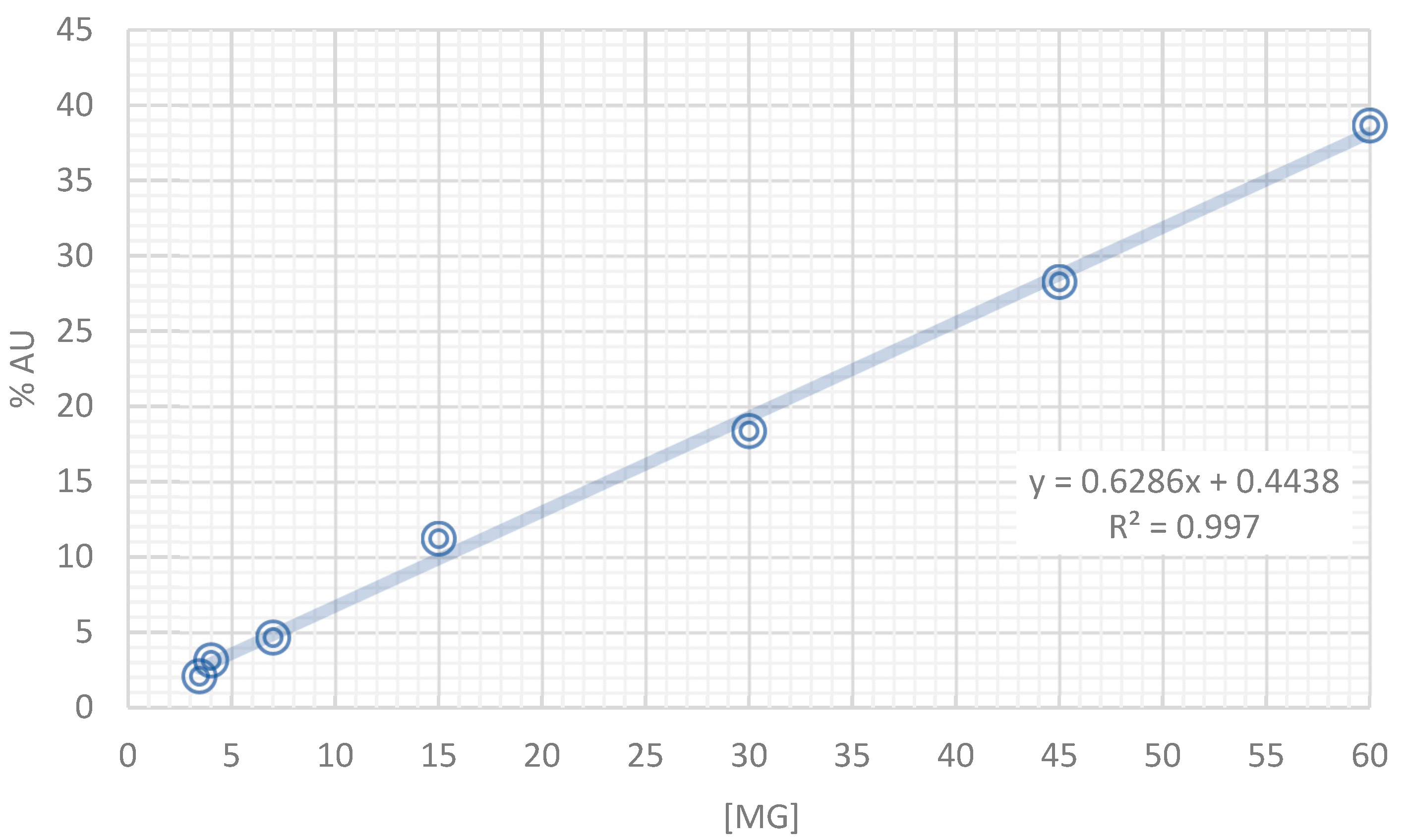
2.2. HPLC-DAD Analysis of the Tea Infusion Extract from Annona cherimola (IELAc)

The analysis by HPLC-DAD of three samples of each tea infusion extract from Ac was performed to evaluate the tea infusion composition. As can be observed in Figure 1, the presence of constituents with retention times ranging between 4.0 and 8.0 min was confirmed. These peaks were compared using the standards of the flavonol glycosides corresponding with rutin, narcissin, and nicotiflorin. Due to rutin being the highest compound found, it was used as a marker and measured through the collection period to evaluate the difference in the collections.
The calibration curve of rutin—the major flavonoid glycoside—into the extract at 254 nm is shown in Figure 2. In addition, the concentration of rutin, narcissin, and nicotiflorin throughout the collection period (May (AcMa), Jun (AcJun), Jul (AcJul), and August (AcAu) obtained quantitatively through the %AU is shown in Table 2. The tea infusion extract was made with 1.5 g of fresh-harvested leaf powder from Ac from different months. Three samples of each tea infusion extract were analyzed by HPLC-DAD to determine the changes in the concentration of these flavonoids. As can be observed, the yield and concentration of these flavonoids were increasing, being significantly greater with the infusion made with the leaf powder collected in August (AcAu). Therefore, this data reflects that collecting the plant material in August is the best moment compared with the previous months. Moreover, an infusion extract from the plant material in this month could exert the best effect due to the high concentration of its active compounds.

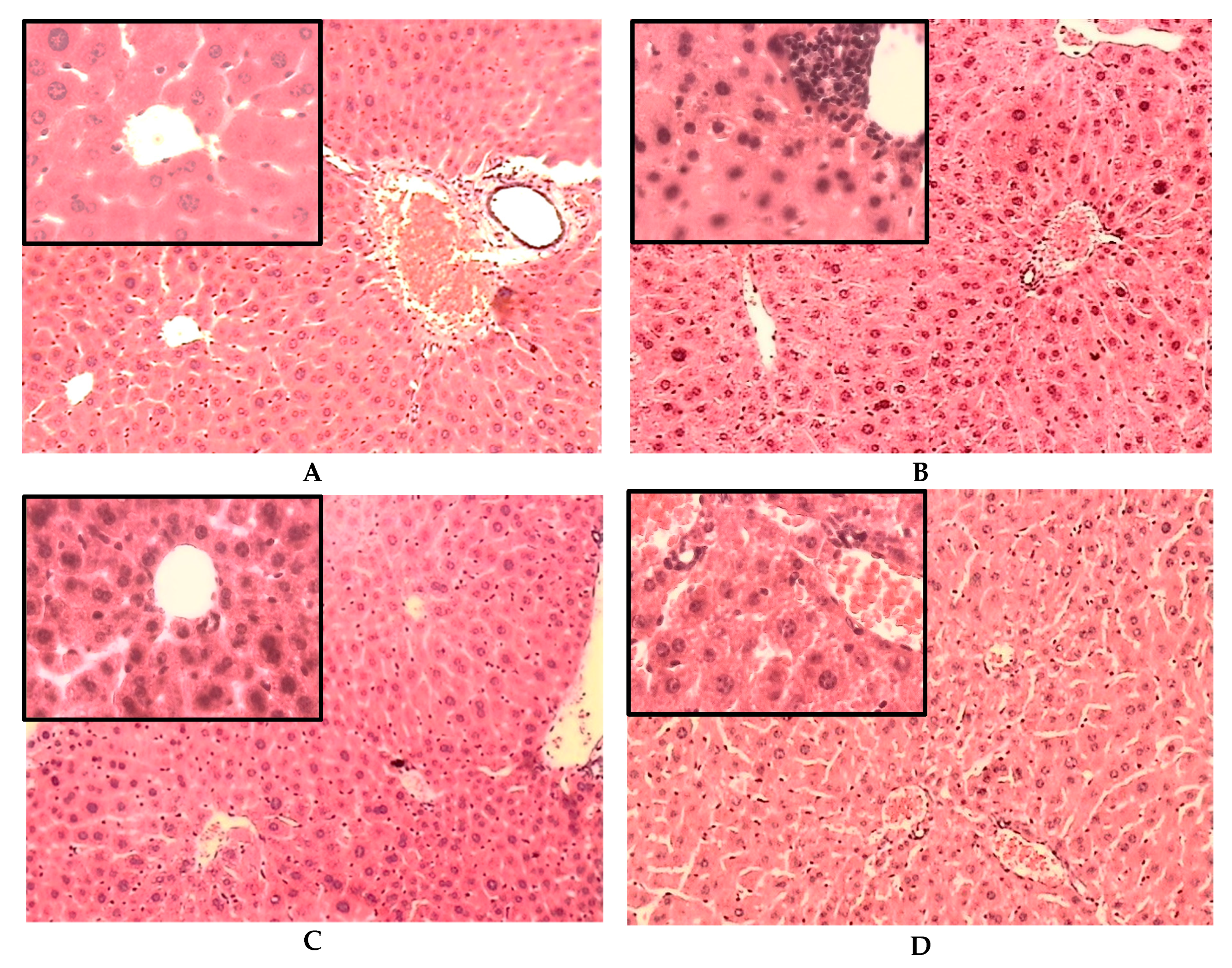
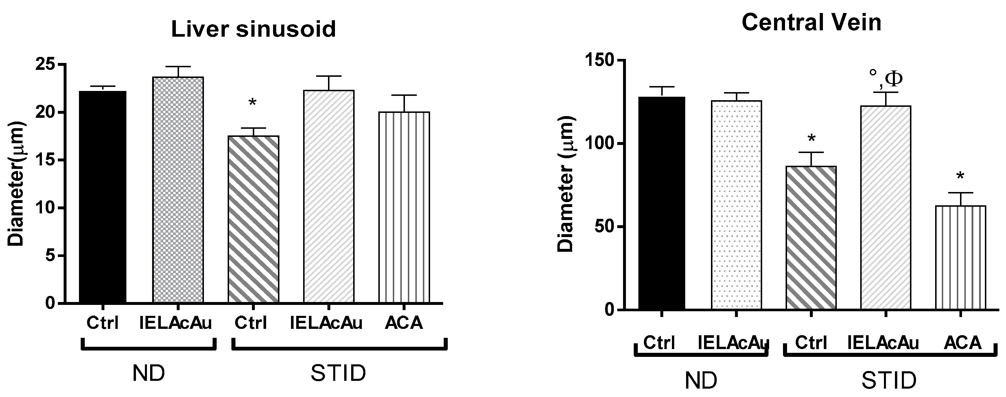
2.3. Histological Results

2.3.1. Histological Changes on the Liver of Non-Diabetic (ND) and STID Mice Treated with the Tea Infusion Extract of Ac

The analysis of the photomicrographs of liver samples stained with hematoxylin and eosin (H&E) of all groups is shown in Figure 3. The architecture of hepatic tissue of ND control mice (Figure 3A) was observed with polygonal hepatocytes with central euchromatic nuclei and eosinophilic granular cytoplasm. Some hepatocytes were binucleated. Sinusoids were found converging towards the central vein. Portal areas contained connective tissue and portal triad. The architecture of the liver sinusoids and the diameter of the central vein were normal. In addition, some Kupffer cells were in the sinusoidal space, as well as some endothelial cells lining the central vein. After the treatment with IELAc from August in ND mice, no changes were found in the liver histology, which appeared more or less like the control group and preserved the overall structure of the hepatic lobe.
The representative photomicrograph of the liver sections of the STID control group (Figure 3B) revealed a reduction in the diameter of the central veins and blood sinusoids, which also showed as less arrayed. Moreover, cellular damage characterized by ballooned hepatocytes with vacuolated cytoplasm was found. Signs of apoptosis as karyorrhexis, fragmented nuclei, altered cytoplasmic regions, as well as accentuated perivascular inflammatory congestion were also observed. Some hepatocytes appeared with a small cytoplasm and big dark nuclei. These changes observed in the liver samples of the STID control mice were a probable consequence of fat metabolism disorders associated with diabetes induced by streptozocin administration.
The representative image of the liver sections of treatment with IELAc from August in STID mice (Figure 3C) appeared somewhat more like the normal histological architecture. The hepatocyte appearance was observed as more arrayed. Less vacuolization was also observed in the cytoplasm of the hepatocytes. Sinusoid cordons were observed as more linear, with discrete inflammatory processes. Finally, the vein and sinusoid diameter were like the ND control group.
In the case of the liver sections of the diabetic mice treated with acarbose (Figure 3D), a diffuse inflammatory process was observed compared with the STID control group. Moreover, the cordons of the hepatocytes were found disarrayed and ballooned, with a great number of intracytoplasmic vacuoles; this is maybe associated with fatty liver conditions in diabetes. The diameter of the veins, but not of the liver sinusoids, was reduced.

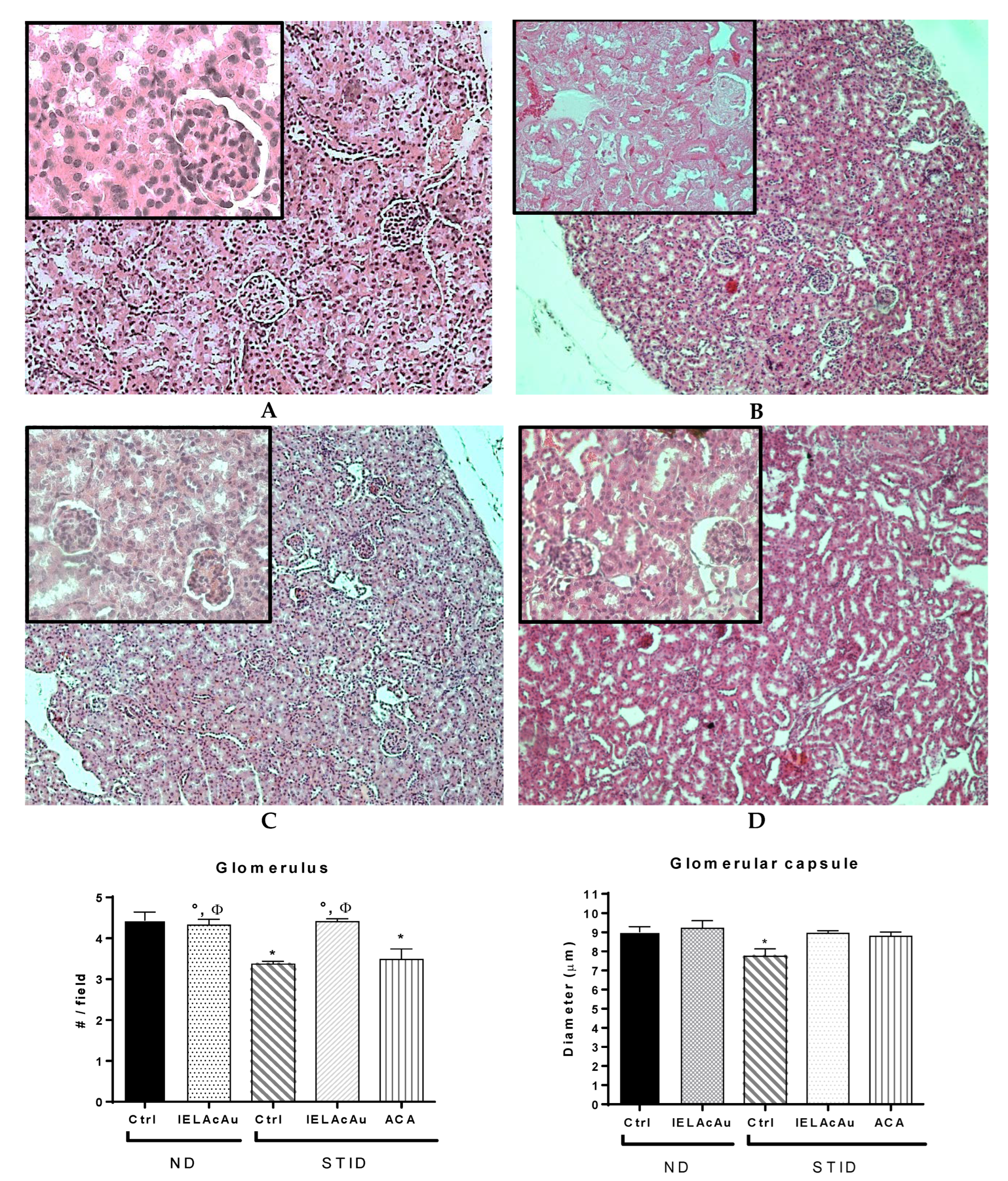
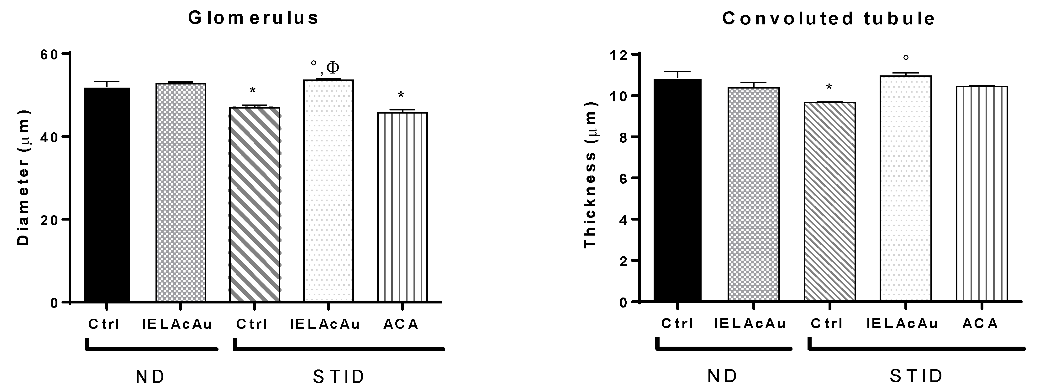
2.3.2. Histological Changes on the Kidneys of Non-Diabetic (ND) and STID Mice Treated with the Tea Infusion Extract of Ac (IELAc)

The photomicrographs of the histological kidney samples stained with hematoxylin and eosin (H&E) of all experimental groups (Figure 4) were analyzed to explore the effects of the administration of the tea infusion extract from August (IELAcAu) on the renal architecture in ND and STID mice. In the case of the ND control group (Figure 4A), the glomerular structure was intact, and the macula cells were observed with adequate size and disposition, with a normal proportion of glomerular sacs per field. Moreover, the renal tubules were arranged homogeneously with cubical epithelial cells with central homogeneous nuclei and their brush border, without signs of atrophy or cytoplasmic vacuolar lesion. Additionally, there was no inflammatory infiltration in the interstitium and no signs of pathology or hemorrhagic areas in the cortex or medulla. The treatment with IELAcAu in the ND mice did not cause observable or significant changes in the renal architecture compared with the ND control mice.
In the case of the representative photomicrographs of the STID control group (Figure 4B), a significant diminution in the number of the glomerulus was observed. In addition, the renal tubules were arranged disorderly and inflammatory cells as well as interstitial infiltration and extracellular matrix deposits were observed, suggesting a fibrosis process. Moreover, atrophy signs such as glomerular contraction and reduced diameter as well as loss of continuity of the convoluted tubules were also observed.
The representative photomicrographs of the renal samples of the STID mice treated with IELAcAu (Figure 4C) were found with fewer glomerular lesions compared with the STID control group. In addition, the glomeruli were less contracted, and the glomerular diameter was recovered. Tubular distribution was more or less like the ND control groups, without signs of atrophy, inflammatory infiltration, or hemorrhagic areas.
However, in the STID group treated with acarbose (Figure 4D), kidney sections showed glomeruli significantly contracted with reduced diameter, suggesting signs of renal dysfunction. In addition, convoluted tubules were disordered, with loss of continuity in some regions; however, less inflammatory infiltration was found compared with the STID control group.

3. Discussion

Diabetes mellitus management is an important scope in public health [26], but, although diabetes cannot be cured, diabetic complications can be managed successfully by efficiently normalizing the glucose and lipid blood levels [27]. Many attempts have been used to treat DM, but objectives have not yet been achieved due to several limitations of the current therapies [3]. This leads to the need to find complementary treatments that must be effective without undesirable side effects [26]. In this sense, people who usually drink herbal teas are less likely to develop diabetic disorders [28]. In addition, medicinal plants are known for their antidiabetic properties [29] and are used as supplements to help achieve glycemic control in people with diabetes. Furthermore, the consumption of natural antioxidants derived from herbal extracts has increased recently due to their specific biological activity and low toxicity, making them potential compounds for the prevention of oxidative stress disorders that occur in metabolic syndrome and diabetes [30]. Some antioxidant flavonoids are considered dietary supplements in the prevention or treatment of diabetic complications [27], with no reported toxicity or side effects compared with current hypoglycemic therapies that have inevitable side effects [31].
Medicinal plants have been used in folkloric medicine for different purposes [32]. In this regard, the genus Annona includes several species that have been studied due to their properties to treat illnesses [33]; therefore, it is a good source to search for compounds with medicinal properties. The aqueous extracts of Annona muricata, A. stenophylla, and A. macroprophyllata have shown antidiabetic, antioxidant, antilipidemic, and antinociceptive effects that are attributed to the presence of phenolic compounds in their leaves [34,35,36,37]. The antidiabetic properties of extracts from Ac have also been studied in previous publications [25] and it has been established that making a tea infusion from the leaf powder is a good way to produce the antilipidemic effect [38]; therefore, this extract was used in the present study. Nevertheless, no reports showing the histological changes induced by the treatment with this extract or changes in the effect related to the harvest period of the leaves have been studied.
First, the analysis by HPLC-DAD of the samples of each tea infusion extract made with 1.5 g of leaf powder from the seriated harvest showed the presence of compounds in which the glycoside flavonoid rutin was highest, followed by narcissin and nicotiflorin. The quantification of rutin was considered a marker compound of the tea infusion extracts due to the major compound with demonstrated activity [39]. Flavonoids are a class of naturally occurring benzo-γ-pyrone derivatives of plant origin [27], widely distributed and found in many vegetal species, and have been used as a restorative in preparations of herbal medicine with a non-toxic and protective behavior [40]. Furthermore, flavonoids have been studied for the redox effect associated with their phenolic groups and thus are proposed as effective supplements against oxidative stress-related secondary complications [41]. Moreover, flavonoids exert antidiabetic and antilipidemic effects, suppressing the intestinal absorption of glucose, cholesterol, and triglycerides by the inhibition of α-glucosidase activity [42], stimulating the glucose transporter 4 (GLUT4)-mediated glucose uptake, increasing the insulin sensibility [43], and suppressing the pancreatic phospholipase-A2 (PA2) essential for lipid hydrolysis [44]. These concentration-related activities are considered important for preventing a wide variety of diseases, including diabetes and other related illnesses such as inflammation and hepatic damage [27]. Particularly, it has already been demonstrated that the extract from Ac reduces cholesterol absorption in intestinal cells [24], has an inhibiting α-glucosidase activity that is responsible for its antihyperglycemic effect [25], and its infusions reduce the blood level of cholesterol and triglycerides in mice [38]; therefore, this shows that the aqueous extract could have beneficial effects over other diabetic related complications.
The evaluation of the seriated collections shows that the growth state of the leaves changes the flavonoid concentration; this can modify the antidiabetic effect exerted, being higher in the months when the growth is accentuated. In this sense, phytochemical analysis in other studies has demonstrated that the therapeutic effect and composition of extracts vary greatly depending on the site or season of the collection, even in the same country or in consecutive months [11,45]. Although Ac is considered an evergreen species due to the presence of leaves during almost the whole year, the season could modify the number of metabolites in its extracts [46]. Therefore, in this work, we consider it important to evaluate the effect of the samples collected at serial times to determine whether these environmental changes are capable of inducing modifications in the reported beneficial effects. We analyzed the growth conditions of the specimen, the appearance of the leaf, and how this characteristic can modify the number of active compounds, resulting in a variation in efficacy. Our results, based on the measuring of flavonoids in the tea infusion extract from Ac by HPLC-DAD, showed that the content of the major marker flavonoid rutin, as well other flavonoids that are implied at least partially in the therapeutic effect, changes throughout the annual course, being higher during the rainy season months, leading to modifications in the potency of the antihyperglycemic effect. This makes sense, since the active compounds are highly polar, and maybe the metabolic pathway could be influenced by fluvial conditions in that season. Although this species sheds its leaves in the last of winter and the beginning of spring, the growth occurs during the annual rainfall season [45] and is related to the pollination and flowering period [47,48,49], which, in subtropical places in America, takes place mainly from the second half of May to August. Moreover, it is known that the best growth of this species occurs under high relative humidity and vapor pressure [50,51], not doing so well at high temperatures or high drought or CO2 amounts [52,53]. This information indicates that environmental conditions could influence not only the leaf appearance of this species but could have an impact on the concentration of bioactive compounds and, later, on the biological activity. Therefore, collecting fresh and mature leaves during the rainy season in August is the best time to obtain a great proportion of active compounds and good activity.
On the other hand, chronic hyperglycemia in DM is related not only to vascular damage that increases the cardiovascular risk but also to changes in the architecture of several organs, resulting in their dysfunction. The impaired function of the liver and the kidneys is the most common complication of diabetes [4]. In this work, we wanted to analyze whether the treatment with IELAcAu would be able to induce beneficial changes in both these organs in STID mice.
The liver is the major organ involved in the detoxifying process [54] and plays an important role in the metabolism of carbohydrates, lipids, and other substances [27]. For this reason, it is one of the organs most susceptible to oxidative stress in diabetes and is affected by insulin resistance [14], which stimulates the release of free fatty acids from the adipose tissue and its accumulation inside the hepatocytes [55]. This lipid imbalance related to hyperglycemia has been associated with hepatic damage [56,57] and results in a pathological condition known as non-alcoholic fatty liver disease (NAFLD) or steatohepatitis [4]. Moreover, the excess lipid infiltration and triglyceride accumulation in the liver cells release pro-inflammatory cytokines and generate reactive oxygen species (ROS) [58]; this plays a pivotal role in the pathophysiology of diabetic hepatotoxicity [27]. Thus, the search for alternative treatments must be oriented not only toward improving hyperglycemia but also to reducing the pathological changes associated with diabetes in the liver architecture that is responsible, at least in part, for the chronic failure of the treatment. Previously, it has been demonstrated that the extract of Ac can improve the lipid profile in mice [38] by reducing the blood levels of cholesterol and triglycerides and by increasing the HDL level. On the other hand, it is already known that rutin inhibits cholesterol absorption from the intestine, thereby lowering the serum and hepatic lipid content [56]. This effect protects the liver from pathological changes associated with hyperlipidemia caused by insulin resistance [59]. Moreover, flavonoids reduce the synthesis of LDL, blocking the inflammation in the liver [56]. Therefore, the presence of rutin as the major compound in the infusion extract explains its antihyperlipidemic effect, which is reflected in the reduced vacuolization of the liver sinusoids and less inflammatory proliferation near the central veins of the liver sections samples in the STID mice group treated with IELAcAu in this work, keeping the architecture more like the ND mice. This is in accordance with other studies that have shown that dietary intervention with flavonoids reduces lipid peroxidation and histopathological changes in the liver of diabetic mice [27]. Moreover, the administration of IELAcAu in the ND mice did not cause appreciable or quantitative changes in the liver, which reflects that this treatment does not lead to pathological modifications, and it is safe in this model.
Diabetic nephropathy (DN) is another of the main microvascular long-term complications related to chronic hyperglycemia and causes end-stage renal failure [4,60], leading these patients to the need for peritoneal dialysis and hemodialysis [11]. This disease involves factors such as the overproduction of reactive oxygen species (ROS) and inflammatory infiltration that changes the renal parenchyma [61]. In diabetes, the accumulation of high-glucose amounts leads to an improper renal function [62] due to the overproduction of AGEP, which plays an important role in the fast progression of DN [63,64]. AGEP induces ROS production and promotes inflammatory infiltration [63], generating glomerular and tubule-interstitial damage [65]. In this work, the analysis of kidney section samples in the STID control group revelated changes in the renal glomerulus and tubules associated with DN. Nevertheless, the treatment with the IELAc significantly reduced the changes associated with DN. Previous studies have reported that flavonoids such as rutin have strong inhibitory activities against AGEP formation [66] and ameliorate renal function by decreasing the expression of inflammatory cytokines involved in the progression of DN [11]. Similarly, herbal teas have shown the potential to ameliorate diabetic renal dysfunction thorough inhibiting AGEP formation and cutting off inflammatory pathways via trapping metabolite methylglyoxal [4]. In this sense, it has been shown that the IELAc is also able to reduce the HbA1c levels in STID mice [38]. HbA1c is the main AGEP used as a marker to evaluate the progression in people with diabetes [67]. In the present work, the reduction of the changes associated with DN observed in the STID mice group treated with IELAc could be explained at least in part by the inhibitory α-glucosidase activity of this infusion, which reduces the rise of the glucose level in the bloodstream, thus preventing its linkage with circulant or cellular proteins, and therefore the synthesis of AGEPs, resulting in the reduction of the renal glucotoxicity and DN.
Polar flavonoids in IELAc may be responsible for its protective effect observed against the damage of diabetic complications related to oxidative stress on the liver and kidney. Polyphenolic compounds as flavonoids have great potential in reducing oxidative stress [27] by lowering the malondialdehyde (MDA) [40], increasing glutathione and superoxide dismutase levels [68], and preventing lipid peroxidation [4], which is related to lipid synthesis. In this sense, the flavonol glycoside rutin is one of the major flavonoid compounds that occurs in food [56]. It has great antioxidant activity, realized through known mechanisms such as activating the intercellular antioxidant enzymes, scavenging oxygen radicals, protecting against lipid peroxidation, and chelating metal ions [27]. These antioxidant mechanisms seem to be primarily associated with their structural features as the presence of functional hydroxyl groups on the A-ring and B chain [14]. Moreover, it has already been observed that the intensity of this beneficial effect could be associated with the first-pass effect, where some supplements with antioxidant properties undergo biotransformation [27]. In the same line, it has been shown that flavonoids effectively avert morphological changes in the liver [56]. Other studies have demonstrated that the administration of the aqueous extracts from Annona muricata protects against damage induced by drugs or infections and can revert liver failure [69,70]. Our current results show that the tea infusion extract of Ac is also able to induce a reduction in the pathological changes of the hepatic and renal parenchyma; this may be influenced by the therapeutic antioxidant potential of the polyphenolic compounds present in the infusion extract and follows other studies that report that aqueous extracts rich in flavonoids exert an antidiabetic effect [71]. Therefore, this could explain other benefic effects over damage associated with diabetes and could provide evidence for future clinical testing.
Finally, taking into consideration the first results of the histological analysis of the treatment with the tea infusion extract of Ac in streptozocin-induced diabetic (STID) and non-diabetic (ND) mice, the findings suggest that the presence of polar flavonoids has a positive effect on the liver and kidney, while having no pathological effect on the normal architectures in ND mice. Moreover, the polyphenolic compounds present in the infusion extract allow it to be considered a dietary supplement that could contribute to the treatment of diabetic complications and other diseases related to oxidative stress in humans and could also lead to partially show the beneficial role of the infusion extract for other states of metabolic syndrome, such as dyslipidemia and non-alcoholic fatty liver disease (NAFLD), or for chronic complications such as diabetic nephropathy (DN). However, further research to evaluate the security of other possible effects of IELAc in other regulations of metabolic processes and pregnancy is currently underway. It is relevant to note that these data support the use of an infusion of Ac as it occurs in folkloric medicine that could have a protective effect on reducing the risk factors that contribute to the death of people with diabetes and could give further information on understanding the antihyperglycemic effect of this herbal formulation as a good agent to treat not only hyperglycemia but also several other complications related to diabetes mellitus.

4. Materials and Methods

4.1. Plant Material

For this study, Annona cherimola Miller (Ac) leaves were collected monthly from a specimen under organic conditions from July to August 2020 in the town of San Gregorio Atlapulco, Xochimilco, Mexico (19°15′08.3″ N 99°03′14.3″ W). Then, samples of every collection were authenticated by the botanist MSc Santiago Xolapa Molina of the Medicinal Plant Herbarium (IMSSM) in the Mexican Institute of Social Security (IMSS) and stored for further reference. The name of the plant was also verified at http://www.theplantlist.org (http://www.theplantlist.org/tpl1.1/record/kew-2640812 accessed on 17 November 2022). Finally, leaves were washed twice with purified fresh water and left to dry in the dark at room temperature. For use after that, they were finely powdered using a commercial blender and stored in air tight Ziplock bags for extract obtention and further analysis.

4.2. Reagents

Streptozocin (≥75% α-anomer basis, PN: S0130-5G), nicotinamide (≥99.5%, PN: 47865-U), acarbose (PN: PHR1253-500MG), (3-Aminopropyl) trimethoxysilane, buffer solution (citric acid/sodium hydroxide/hydrogen chloride, pH 4.00, CC: 109445), Harris hematoxylin (HH280) and eosin (I.C. 768) were purchased from Sigma-Aldrich® (Sigma®, Saint Louis, MO, USA); saline solution 0.9% (solution 1000 mL) and DX-5 glucose solution 5% were purchased from PISA® Pharmaceutics (PISA®, Mexico City, México); histological paraffin (fusion point 56–58 °C) was acquired from a commercial source. Ethanol, butanol, xylene, and solvents of analytical grade were used.

4.3. Plant Extract Procedure

For the elaboration of all the tea infusion extracts of Ac (IELAc) used in this work, 1.5 g of the powder obtained from the leaves of different collections was placed in a tea bag and soaked in 125 mL of water that was pre-boiled and distilled for 20 min to match traditional common usage. The resulting extract was then filtered through grade 1 filter paper and concentrated on a rotary evaporator (Büchi Labortechnik AG, Flawil, Switzerland) at 40 °C under reduced pressure. This process was carried out monthly with all the extracts. Finally, the sticky precipitate obtained was stored at 4 °C for further reference and an aliquot of the extract residue was weighed and resuspended in water for administration. In all cases, the extraction yield per gram of vegetable powder was evaluated by the difference in the weight of the container before and after pouring the extract.

4.4. Characterization of the Tea Infusion Extract of Leaves from Ac by High-Performance Liquid Chromatography

Once obtained, the infusion extracts of the leaves from Ac (IELAc) of three samples from each monthly extract were analyzed by an HPLC-diode array detection (DAD) to identify compounds in the extracts; the concentration of the main one was also measured throughout the entire collection period. For this, an HPLC-DAD Waters 2795 liquid chromatography system was used, coupled with a Waters 996 photodiode array detector and an analytical Millennium 3.1 workstation equipped with a C18 column (200 × 15 mm), the particle size of 5 μm (Spherisorb S50D52, Waters Corporation, Milford, MA, USA). For the analysis, 10 mg of IELAc was solved on 10 mL of ethanol and a sample volume of 20 μL from that solution was injected. For elution, a system comprising a binary mobile phase of acetonitrile solvent/acetic acid 2% in water (A) and acetonitrile 100% (B) was used. The chromatograph operating conditions were programmed to give the following gradients: 1st stage, linear gradient of 80 (A)/20 (B) for 8 min; 2nd stage, linear gradient of 40/60 for 5 min; 3rd stage, linear gradient of 30/70 for 6 min; 4th stage, linear gradient of 90/10 for 6 min with a flow rate of 1 mL/min of the mobile phase. The detection was carried out at room temperature and at a total elution time of 25 min at a wavelength (λ) of 254 nm, because the highest absorption peak of these flavonoids is found at this wavelength [45]. Then, the collected data were plotted. Reference standards of flavonoid rutin, narcissin, and nicotiflorin, as well as acetonitrile, ethanol, and acetic acid of HPLC grade, were acquired from Sigma (St. Louis, MO, USA) and were prepared and analyzed separately under the same conditions described above. In all cases, the water used was of HPLC quality and purified in a Milli-Q system (Millipore, Bedford, MA, USA). The presence of substances in the extract of the leaves from Ac was confirmed by comparing the retention times and the UV spectra of each of the standards. The concentration of flavonoid glycosides in each infusion extract was obtained by measuring the area under the curve (AU) of each compound identified in its obtained chromatogram.

4.5. Experimental Animals

For this study, healthy albino Balb/c mice of both sexes aged 8–10 weeks (25 ± 5 g) were obtained from the Animal House of the National Medical Center “Siglo XXI” from Instituto Mexicano del Seguro Social (IMSS), housed under standard laboratory conditions at a temperature of 22 °C ± 2 °C, 50% of humidity with a 12-h light/dark cycle, and fed with a standard diet and purified water ad libitum. This study was approved by the Specialty Hospital Ethical Committee of the National Medical Center “Siglo XXI” from IMSS, with the registered numbers: R-2020-3601-038 and R-2019-3601-004. Furthermore, all the experimental animal models were performed following the Mexican Official Regulations on animal care and experimental management [72].

4.5.1. Grouping

Animals were assigned randomly to different groups of six mice each, as follows: Groups 1 and 2 were assigned as non-diabetic (ND) and Group 2 was treated with IELAc at 300 mg/kg body weight (bw). Groups 3 to 5 were streptozocin-induced diabetic (STID) mice. IELAc at 300 mg/kg bw was given to Group 4 of STID mice. Group 5 received acarbose at 50 mg/kg bw, which was used as a reference drug. Groups 1 and 3 performed as a control and non-received treatment. All treatments were administrated by the oral route through a gavage and solved in distilled water; the volume was calculated according to the OECD guidelines [73] as 2 mL/100 g bw.

4.5.2. Induction of Experimental Type 2 Diabetes

In the case of the STID model, the animals fasted previously overnight and experimental type 2 diabetes was induced by an intraperitoneal injection of freshly prepared STZ (100 mg/kg bw) solved in 0.1 M citrate buffer (pH 4.5). After 30 min, a single intraperitoneal dose of nicotinamide (240 mg/kg) in a saline solution of 0.9% was given. Then, the animals were allowed to drink a 5% glucose solution overnight to stabilize the drug-induced hyperglycemia [74]. To confirm an experimental type 2 diabetes model, a single dose of 5 mg/kg bw glibenclamide—a secretagogue drug—was given orally to the STID mice to confirm that the endocrine function of the pancreas was preserved and not completely obliterated due to the action of the STZ [75]. Glycemia was determined after the last administration, using the glucose oxidase method (Accu-Chek® Performa Glucometer, Boehringer Mannheim, Germany) [76], using blood samples collected during the assay by puncture from the tail vein. Those animals with blood glucose values above 290 mg/dL and below 350 mg/dL on the third day after the STZ injection were considered diabetic and used in this study, while animals with capillary glycemia under these values were considered non-diabetic (ND).

4.6. Study of Variation of the Antihyperglycemic Effect of the Infusion Extract of Ac

To evaluate the possible changes in the antihyperglycemic effect induced by the IELAc made from seriated collections, the extract was administrated to streptozocin-induced diabetic (STID) and non-diabetic (ND) mice. Both control groups received water, while the experimental group received the IELAc from May (AcMa), June (AcJun), July (AcJul), or August (AcAu) at 300 mg/kg bw, given as described above. In this case, the blood glucose levels were measured at 0, 1, 3, 5, and 7 h after treatment, according to the method mentioned above.

4.7. Evaluation of the Sub-Chronic Effect of the IELAc

Once determined which extract exerts the greatest effect, we evaluated the sub-chronic assay at 300 mg/kg or acarbose 50 mg/Kg bw in STID and ND mice daily for 28 days. The animals were weighed weekly and kept in conditioned cages under standard conditions with free availability of food and water; changes in their behavior or physical appearance were recorded throughout the treatment.

4.8. Effect of the Treatment on Internal Organs and Histology

After the sub-chronic assay, the animals were weighed and euthanized according to animal management standards [72] by abdominal incision. Then, the abdominal visceral organs were excised, washed with physiological saline solution, and weighed to obtain the absolute weights. Later, they were fixed in 4% PFH and washed with bidistilled water. Before inclusion, different transversal portions of the liver and kidney were sectioned and dehydrated in a battery of alcohols of increasing concentrations and included in paraffin for further analysis. The paraffin blocks were sectioned with a microtome (Leica RM 2125RT) to obtain serial sections of 7 µm thickness. In total, 3 sections were obtained from each sample, with a separation of 100 µm from each other. Finally, the samples were stained with H&E [77]. For each histological sample, at least 9 fields were observed. Histological analysis and photographic recording were performed using an optical microscope Nikon Eclipse E600 (Nikon Instruments Inc., Melville, NY, USA) with a color video camera digital camera SONY Exwave HAD (Sony, Electronics Inc., Paramus, NJ, USA).

Quantitative Morphometric Measurement

To obtain morphometric data in this study, Image Pro Plus 5.1.0 Image Analysis and Processing Software was used (Media cybernetics, Rockville, MD, USA, EU). Nine non-overlapping fields in stained slides of each animal in each group were examined. Measurements were performed on 7 µm thick H&E-stained sections at a magnification of 10× and 40× to estimate:
  • Diameter of liver sinusoids in the control and the treated groups.
  • Diameter of central veins in the liver of the control and the treated groups.
  • Number of renal glomeruli per field observed in the control and the treated groups.
  • Diameter of the glomerular capsule in the control and the treated groups.
  • Diameter of the renal glomerulus in the control and the treated groups.
  • Diameter of convoluted tubules in the control and the treated groups.

4.9. Statistical Analysis

Data are expressed as the mean ± standard error mean (SEM). Statistical significance was determined by a one-way analysis of variance (ANOVA) followed by a Tukey test for multiple comparisons (GraphPad Prism Version 6.01; GraphPad Software Inc., La Jolla, CA, USA), considering the p-value < 0.05 as indicative of the significant difference between group means.

5. Conclusions

In this study, the therapeutic effect attributed to polar flavonoids in the tea infusion extract made with 1.5 g of Annona cherimola Miller leaf powder changes throughout the month of harvest in the summer season of the year. The sub-chronic administration of the extract made from leaves collected in August (IELAcAu) with the highest amount of flavonoids reduces the pathological changes in the liver and the kidney in STZ-induced diabetic (STID) mice, with no pathological changes observed in non-diabetic (ND) mice. These results support the use of an infusion extract of the leaves from Ac as a safe supplement useful for the treatment of diabetes mellitus complications.

Author Contributions

Conceptualization, J.M.-S., F.C., E.B. and J.M.G.-M.; funding acquisition, F.C. and E.B.; investigation, J.M.-S.; methodology, J.M.-S., F.C., E.B. and J.M.G.-M.; project administration, F.C. and E.B.; resources, F.C. and E.B.; supervision, F.C., E.B. and J.M.G.-M.; validation, F.C. and E.B.; writing—original draft, J.M.-S.; writing—review editing, F.C., E.B. and J.M.G.-M. All authors have read and agreed to the published version of the manuscript.

Funding

This research was supported by grants from Instituto Mexicano del Seguro Social (IMSS): FIS/IMSS/PROT/PRIO/19/110 and FIS/IMSS/PROT/G17-2/1722. Awarded to F.C.

Institutional Review Board Statement

The study was conducted according to the guidelines of the Declaration of Helsinki and the Official Mexican Regulations on animal care and experimental management NOM-062-ZOO-1999. This study was approved by the Local Ethics Committee of CMN SXXI in the Instituto Mexicano del Seguro Social under protocol code R-2020-3601-038 (9 June 2021) and R-2019-3601-004 (30 March 2022).

Data Availability Statement

The data presented in this article are available on request from the corresponding authors.

Acknowledgments

This work is part of the thesis of Jesus Martínez-Solís. We would like to thank Consejo Nacional de Ciencia y Tecnología (CONACYT) for the scholarship 663929, also IPN for the scholarship BEIFI ID: 1963 through SIP project 20211098. J.M-S would like to thank Gregorio Rodriguez for the facilities to obtain the specimen samples and also the academic staff from Morphology Laboratory N° 301 of ESM1.

Conflicts of Interest

The authors declare no conflict of interest.

Sample Availability

Samples used are available from the authors.

References

  1. Guthrie, R.A.; Guthrie, D.W. Pathophysiology of Diabetes Mellitus. Crit. Care Nurs. Q. 2004, 27, 113–125. [Google Scholar] [CrossRef] [PubMed]
  2. IDF Diabetes Atlas 10th Edition. 2021. Available online: https://diabetesatlas.org/idfawp/resource-files/2021/07/IDF_Atlas_10th_Edition_2021.pdf (accessed on 6 May 2022).
  3. Khursheed, R.; Singh, S.K.; Wadhwa, S.; Kapoor, B.; Gulati, M.; Kumar, R.; Ramanunny, A.K.; Awasthi, A.; Dua, K. Treatment strategies against diabetes: Success so far and challenges ahead. Eur. J. Pharmacol. 2019, 862, 172625. [Google Scholar] [CrossRef]
  4. Atia, T.; Sakr, H.I.; Damanhory, A.A.; Moawad, K.; Alsawy, M. The protective effect of green tea on diabetes-induced hepato-renal pathological changes: A histological and biochemical study. Arch. Physiol. Biochem. 2020, 20, 1–12. [Google Scholar] [CrossRef]
  5. Giri, B.; Dey, S.; Das, T.; Sarkar, M.; Banerjee, J.; Dash, S.K. Chronic hyperglycemia mediated physiological alteration and metabolic distortion leads to organ dysfunction, infection, cancer progression and other pathophysiological consequences: An update on glucose toxicity. Biomed. Pharmacother. 2018, 107, 306–328. [Google Scholar] [CrossRef]
  6. Oršolić, N.; Bašić, I. Honey bee products and their polyphenolic compounds in the treatment of diabetes. Phytopharm. Ther. Values 2008, 22, 455–471. [Google Scholar]
  7. Yue, K.K.; Leung, S.-N.; Man, P.-M.; Yeung, W.-F.; Chung, W.-S.; Lee, K.-W.; Leung, A.W.; Cheng, C.H. Alterations in antioxidant enzyme activities in the eyes, aorta and kidneys of diabetic rats relevant to the onset of oxidative stress. Life Sci. 2005, 77, 721–734. [Google Scholar] [CrossRef]
  8. Obrosova, I.G.; Minchenko, A.G.; Vasupuram, R.; White, L.; Abatan, O.I.; Kumagai, A.K.; Frank, R.N.; Stevens, M.J. Aldose Reductase Inhibitor Fidarestat Prevents Retinal Oxidative Stress and Vascular Endothelial Growth Factor Overexpression in Streptozotocin-Diabetic Rats. Diabetes 2003, 52, 864–871. [Google Scholar] [CrossRef] [PubMed] [Green Version]
  9. American Diabetes Association. Pharmacologic Approaches to Glycemic Treatment: Standards of Medical Care in Diabetes—2021. Diabetes Care 2021, 44 (Suppl. 1), S111–S124. [Google Scholar] [CrossRef]
  10. Tan, S.Y.; Wong, J.L.M.; Sim, Y.J.; Wong, S.S.; Elhassan, S.A.M.; Tan, S.H.; Lim, G.P.L.; Tay, N.W.R.; Annan, N.C.; Bhattamisra, S.K.; et al. Type 1 and 2 diabetes mellitus: A review on current treatment approach and gene therapy as potential intervention. Diabetes Metab. Syndr. Clin. Res. Rev. 2019, 13, 364–372. [Google Scholar] [CrossRef]
  11. Granados-Pineda, J.; Uribe-Uribe, N.; García-López, P.; Ramos-Godinez, M.D.P.; Rivero-Cruz, J.F.; Pérez-Rojas, J.M. Effect of Pinocembrin Isolated from Mexican Brown Propolis on Diabetic Nephropathy. Molecules 2018, 23, 852. [Google Scholar] [CrossRef] [Green Version]
  12. Yaseen, G.; Ahmad, M.; Zafar, M.; Sultana, S.; Kayani, S.; Cetto, A.A.; Shaheen, S. Traditional management of diabetes in Pakistan: Ethnobotanical investigation from Traditional Health Practitioners. J. Ethnopharmacol. 2015, 174, 91–117. [Google Scholar] [CrossRef] [PubMed]
  13. Khalili, M.; Ebrahimzadeh, M.A.; Safdari, Y. Antihaemolytic activity of thirty herbal extracts in mouse red blood cells. Arch. Ind. Hyg. Toxicol. 2014, 65, 399–406. [Google Scholar] [CrossRef] [PubMed] [Green Version]
  14. Sirovina, D.; Oršolić, N.; Gregorović, G.; Končić, M.Z. Naringenin ameliorates pathological changes in liver and kidney of diabetic mice: A preliminary study/Naringenin reducira histopatološke promjene u jetri i bubregu miševa s dijabetesom. Arch. Ind. Hyg. Toxicol. 2016, 67, 19–24. [Google Scholar] [CrossRef] [PubMed] [Green Version]
  15. Andrade-Cetto, A.; Heinrich, M. Mexican plants with hypoglycaemic effect used in the treatment of diabetes. J. Ethnopharmacol. 2005, 99, 325–348. [Google Scholar] [CrossRef] [PubMed]
  16. Quílez, A.; Fernández-Arche, M.; García-Giménez, M.; De la Puerta, R. Potential therapeutic applications of the genus Annona: Local and traditional uses and pharmacology. J. Ethnopharmacol. 2018, 225, 244–270. [Google Scholar] [CrossRef]
  17. Larranaga, N.; Albertazzi, F.J.; Fontecha, G.; Palmieri, M.; Rainer, H.; van Zonneveld, M.; Hormaza, J.I. A Mesoamerican origin of cherimoya (Annona cherimola Mill.): Implications for the conservation of plant genetic resources. Mol. Ecol. 2017, 26, 4116–4130. [Google Scholar] [CrossRef]
  18. CABI, Annona cherimola (Cherimoya). Invasive Species Compendium; CAB International: Wallingford, UK, 2014; Available online: https://www.cabi.org/isc/datasheet/5806 (accessed on 8 November 2022).
  19. Calzada, F.; Correa-Basurto, J.; Barbosa, E.; Mendez-Luna, D.; Yépez-Mulia, L. Antiprotozoal Constituents from Annona cherimola Miller, a Plant Used in Mexican Traditional Medicine for the Treatment of Diarrhea and Dysentery. Pharmacogn. Mag. 2017, 13, 148–152. [Google Scholar] [CrossRef]
  20. Díaz-De-Cerio, E.; Aguilera-Saez, L.M.; Gómez-Caravaca, A.M.; Verardo, V.; Fernández-Gutiérrez, A.; Fernández, I.; Arráez-Román, D. Characterization of bioactive compounds of Annona cherimola L. leaves using a combined approach based on HPLC-ESI-TOF-MS and NMR. Anal. Bioanal. Chem. 2018, 410, 3607–3619. [Google Scholar] [CrossRef]
  21. Albuquerque, T.G.; Santos, F.; Sanches-Silva, A.; Oliveira, M.B.; Bento, A.C.; Costa, H.S. Nutritional and phytochemical composition of Annona cherimola Mill. fruits and by-products: Potential health benefits. Food Chem. 2014, 193, 187–195. [Google Scholar] [CrossRef]
  22. Martínez-Vázquez, M.; Estrada-Reyes, R.; Escalona, A.A.; Velázquez, I.L.; Martínez-Mota, L.; Moreno, J.; Heinze, G. Antidepressant-like effects of an alkaloid extract of the aerial parts of Annona cherimolia in mice. J. Ethnopharmacol. 2012, 139, 164–170. [Google Scholar] [CrossRef]
  23. Ammoury, C.; Younes, M.; El Khoury, M.; Hodroj, M.H.; Haykal, T.; Nasr, P.; Sily, M.; Taleb, R.I.; Sarkis, R.; Khalife, R.; et al. The pro-apoptotic effect of a Terpene-rich Annona cherimola leaf extract on leukemic cell lines. BMC Complement. Altern. Med. 2019, 19, 365. [Google Scholar] [CrossRef]
  24. Falé, P.L.; Ferreira, C.; Maruzzella, F.; Florêncio, M.H.; Frazão, F.N.; Serralheiro, M.L. Evaluation of cholesterol absorption and biosynthesis by decoctions of Annona cherimola leaves. J. Ethnopharmacol. 2013, 150, 718–723. [Google Scholar] [CrossRef] [PubMed]
  25. Calzada, F.; Solares-Pascasio, J.I.; Ordoñez-Razo, R.; Velazquez, C.; Barbosa, E.; García-Hernández, N.; Mendez-Luna, D.; Correa-Basurto, J. Antihyperglycemic activity of the leaves from Annona cherimola miller and rutin on alloxan-induced diabetic rats. Pharmacogn. Res. 2017, 9, 1–6. [Google Scholar] [CrossRef] [PubMed] [Green Version]
  26. El-Tantawy, W.H.; Temraz, A. Management of diabetes using herbal extracts: Review. Arch. Physiol. Biochem. 2018, 124, 383–389. [Google Scholar] [CrossRef]
  27. Sirovina, D.; Oršolić, N.; Končić, M.Z.; Kovačević, G.; Benkovic, V.; Gregorović, G. Quercetin vs chrysin: Effect on liver his-topathology in diabetic mice. Hum. Exp. Toxicol. 2013, 32, 1058–1066. [Google Scholar] [CrossRef]
  28. Abolfathi, A.A.; Mohajeri, D.; Rezaie, A.; Nazeri, M. Protective Effects of Green Tea Extract against Hepatic Tissue Injury in Streptozotocin-Induced Diabetic Rats. Evid.-Based Complement. Altern. Med. 2012, 2012, 740671. [Google Scholar] [CrossRef] [PubMed] [Green Version]
  29. Choudhury, H.; Pandey, M.; Hua, C.K.; Mun, C.S.; Jing, J.K.; Kong, L.; Ern, L.Y.; Ashraf, N.A.; Kit, S.W.; Yee, T.S.; et al. An update on natural compounds in the remedy of diabetes mellitus: A systematic review. J. Tradit. Complement. Med. 2018, 8, 361–376. [Google Scholar] [CrossRef]
  30. Meng, J.-M.; Cao, S.-Y.; Wei, X.-L.; Gan, R.-Y.; Wang, Y.-F.; Cai, S.-X.; Xu, X.-Y.; Zhang, P.-Z.; Li, H.-B. Effects and Mechanisms of Tea for the Prevention and Management of Diabetes Mellitus and Diabetic Complications: An Updated Review. Antioxidants 2019, 8, 170. [Google Scholar] [CrossRef] [Green Version]
  31. Moradi, B.; Abbaszadeh, S.; Shahsavari, S.; Alizadeh, M.; Beyranvand, F. The most useful medicinal herbs to treat diabetes. Biomed. Res. Ther. 2018, 5, 2538–2551. [Google Scholar] [CrossRef]
  32. Giovannini, P.; Howes, M.-J.R.; Edwards, S.E. Medicinal plants used in the traditional management of diabetes and its sequelae in Central America: A review. J. Ethnopharmacol. 2016, 184, 58–71. [Google Scholar] [CrossRef] [Green Version]
  33. Leite, D.O.D.; Nonato, C.D.F.A.; Camilo, C.J.; De Carvalho, N.K.G.; Da Nobrega, M.G.L.A.; Pereira, R.C.; Da Costa, J.G.M. Annona Genus: Traditional Uses, Phytochemistry and Biological Activities. Curr. Pharm. Des. 2020, 26, 4056–4091. [Google Scholar] [CrossRef] [PubMed]
  34. Florence, N.T.; Benoit, M.Z.; Jonas, K.; Alexandra, T.; Désiré, D.D.P.; Pierre, K.; Théophile, D. Antidiabetic and antioxidant effects of Annona muricata (Annonaceae), aqueous extract on streptozotocin-induced diabetic rats. J. Ethnopharmacol. 2014, 151, 784–790. [Google Scholar] [CrossRef] [PubMed]
  35. Adewole, S.; Ojewole, J. Protective effects of Annona muricata linn. (Annonaceae) leaf aqueous extract on serum lipid profiles and oxidative stress in hepatocytes of streptozotocin-treated diabetic rats. Afr. J. Tradit. Complement. Altern. Med. 2008, 6, 30–41. [Google Scholar] [CrossRef] [PubMed] [Green Version]
  36. Taderera, T.; Chagonda, L.S.; Gomo, E.; Katerere, D.; Shai, L.J. Annona stenophylla aqueous extract stimulate glucose uptake in established C2Cl2 muscle cell lines. Afr. Health Sci. 2019, 19, 2219–2229. [Google Scholar] [CrossRef] [Green Version]
  37. Brindis, F.; González-Trujano, M.E.; González-Andrade, M.; Aguirre-Hernández, E.; Villalobos-Molina, R. Aqueous Extract of Annona macroprophyllata: A Potentialα-Glucosidase Inhibitor. BioMed Res. Int. 2013, 2013, 591313. [Google Scholar] [CrossRef] [Green Version]
  38. Martínez-Solís, J.; Calzada, F.; Barbosa, E.; Valdés, M. Antihyperglycemic and Antilipidemic Properties of a Tea Infusion of the Leaves from Annona cherimola Miller on Streptozocin-Induced Type 2 Diabetic Mice. Molecules 2021, 26, 2408. [Google Scholar] [CrossRef]
  39. Ghorbani, A. Mechanisms of antidiabetic effects of flavonoid rutin. Biomed. Pharmacother. 2017, 96, 305–312. [Google Scholar] [CrossRef]
  40. Singh, S.; Singh, D.K.; Meena, A.; Dubey, V.; Masood, N.; Luqman, S. Rutin protects t-butyl hydroperoxide-induced oxidative impairment via modulating the Nrf2 and iNOS activity. Phytomedicine 2019, 55, 92–104. [Google Scholar] [CrossRef]
  41. Kamalakkannan, N.; Prince, P.S.M. Antihyperglycaemic and Antioxidant Effect of Rutin, a Polyphenolic Flavonoid, in Streptozotocin-Induced Diabetic Wistar Rats. Basic Clin. Pharmacol. Toxicol. 2006, 98, 97–103. [Google Scholar] [CrossRef]
  42. Tadera, K.; Minami, Y.; Takamatsu, K.; Matsuoka, T. Inhibition of alpha-Glucosidase and alpha-Amylase by Flavonoids. J. Nutr. Sci. Vitaminol. 2006, 52, 149–153. [Google Scholar] [CrossRef] [Green Version]
  43. Ueda-Wakagi, M.; Nagayasu, H.; Yamashita, Y.; Ashida, H. Green Tea Ameliorates Hyperglycemia by Promoting the Translocation of Glucose Transporter 4 in the Skeletal Muscle of Diabetic Rodents. Int. J. Mol. Sci. 2019, 20, 2436. [Google Scholar] [CrossRef]
  44. Stepien, M.; Kujawska-Luczak, M.; Szulinska, M.; Kregielska-Narozna, M.; Skrypnik, D.; Suliburska, J.; Skrypnik, K.; Regula, J.; Bogdanski, P. Beneficial dose-independent influence of Camellia sinensis supplementation on lipid profile, glycemia, and insulin resistance in an NaCl-induced hypertensive rat model. J. Physiol. Pharmacol. 2018, 69, 278–282. [Google Scholar] [CrossRef]
  45. Merlín-Lucas, V.; Ordoñez-Razo, R.M.; Calzada, F.; Solís, A.; García-Hernández, N.; Barbosa, E.; Valdés, M. Antitumor Potential of Annona muricata Linn. An Edible and Medicinal Plant in Mexico: In Vitro, In Vivo, and Toxicological Studies. Molecules 2021, 26, 7675. [Google Scholar] [CrossRef]
  46. Amador, M.D.C.V.; Rodríguez, F.M.; Rodríguez, Z.M.; Guerra, M.J.M.; Barreiro, M.L. Tamizaje Fitoquímico, Actividad Antiinflamatoria y Toxicidad Aguda de Extractos de Hojas de Annona Squamosa L. Rev. Cuba. Plantas Med. 2006, 11, 1–12. [Google Scholar]
  47. Auger, J.; Pérez, I.; Esterio, M. Occurrence of Root Rot Disease of Cherimoya (Annona cherimola Mill.) Caused by Dactylonectria macrodidyma in Chile. Plant Dis. 2015, 99, 1282. [Google Scholar] [CrossRef]
  48. Mwatawala, M.; De Meyer, M.; Makundi, R.; Maerere, A. Host range and distribution of fruit-infesting pestiferous fruit flies (Diptera, Tephritidae) in selected areas of Central Tanzania. Bull. Entomol. Res. 2009, 99, 629–641. [Google Scholar] [CrossRef] [PubMed]
  49. Saul, W.-C.; Roy, H.E.; Booy, O.; Carnevali, L.; Chen, H.-J.; Genovesi, P.; Harrower, C.A.; Hulme, P.E.; Pagad, S.; Pergl, J.; et al. Assessing patterns in introduction pathways of alien species by linking major invasion data bases. J. Appl. Ecol. 2017, 54, 657–669. [Google Scholar] [CrossRef] [Green Version]
  50. George, A.; Nissen, R. The effects of temperature, vapour pressure deficit and soil moisture stress on growth, flowering and fruit set of custard apple (Annona cherimola × Annona squamosa) ‘African Pride’. Sci. Hortic. 1988, 34, 183–191. [Google Scholar] [CrossRef]
  51. Lora, J.; Herrero, M.; Hormaza, J.I. Stigmatic receptivity in a dichogamous early-divergent angiosperm species, Annona cherimola (Annonaceae): Influence of temperature and humidity. Am. J. Bot. 2011, 98, 265–274. [Google Scholar] [CrossRef] [Green Version]
  52. Maldonado, R.; Molina-Garcia, A.D.; Sanchez-Ballesta, M.T.; Escribano, M.I.; Merodio, C. High CO2 Atmosphere Modulating the Phenolic Response Associated with Cell Adhesion and Hardening of Annona cherimola Fruit Stored at Chilling Temperature. J. Agric. Food Chem. 2002, 50, 7564–7569. [Google Scholar] [CrossRef] [PubMed]
  53. González-Agüero, M.; Cifuentes-Esquivel, N.; Ibañez-Carrasco, F.; Gudenschwager, O.; Campos-Vargas, R.; Defilippi, B.G. Identification and Characterization of Genes Differentially Expressed in Cherimoya (Annona cherimola Mill) after Exposure to Chilling Injury Conditions. J. Agric. Food Chem. 2011, 59, 13295–13299. [Google Scholar] [CrossRef] [PubMed]
  54. Sikder, K.; Kesh, S.B.; Das, N.; Manna, K.; Dey, S. The high antioxidative power of quercetin (aglycone flavonoid) and its glycone (rutin) avert high cholesterol diet induced hepatotoxicity and inflammation in Swiss albino mice. Food Funct. 2014, 5, 1294–1303. [Google Scholar] [CrossRef] [PubMed]
  55. Duwaerts, C.C.; Maher, J.J. Macronutrients and the Adipose-Liver Axis in Obesity and Fatty Liver. Cell. Mol. Gastroenterol. Hepatol. 2019, 7, 749–761. [Google Scholar] [CrossRef] [Green Version]
  56. Del Moral, M.L.; Esteban, F.J.; Torres, M.I.; Camacho, M.V.; Hernandez, R.; Jimenez, A.; Aránega, A.; Pedrosa, J.A.; Peinado, M.A. High-Fat Sunflower and Olive Oil Diets Affect Serum Lipid Levels in Steatotic Rat Liver Differently. J. Nutr. Sci. Vitaminol. 1997, 43, 155–160. [Google Scholar] [CrossRef] [PubMed]
  57. Wanless, I.R.; Belgiorno, J.; Huet, P. Hepatic sinusoidal fibrosis induced by cholesterol and stilbestrol in the rabbit: 1. Morphology and inhibition of fibrogenesis by dipyridamole. Hepatology 1996, 24, 855–864. [Google Scholar] [CrossRef]
  58. Pessayre, D.; Mansouri, A.; Fromenty, B. Nonalcoholic steatosis, and steatohepatitis. V. Mitochondrial dysfunction in steato-hepatitis. Am. J. Physiol. Gastrointest Liver Physiol. 2002, 282, G193–G199. [Google Scholar] [CrossRef]
  59. Rivera, L.; Morón, R.; Sánchez, M.; Zarzuelo, A.; Galisteo, M. Quercetin Ameliorates Metabolic Syndrome and Improves the Inflammatory Status in Obese Zucker Rats. Obesity 2008, 16, 2081–2087. [Google Scholar] [CrossRef]
  60. Li, F.; Chen, Y.; Li, Y.; Huang, M.; Zhao, W. Geniposide alleviates diabetic nephropathy of mice through AMPK/SIRT1/NF-κB pathway. Eur. J. Pharmacol. 2020, 886, 173449. [Google Scholar] [CrossRef]
  61. Kim, Y.; Park, C.W. Adenosine monophosphate–activated protein kinase in diabetic nephropathy. Kidney Res. Clin. Pract. 2016, 35, 69–77. [Google Scholar] [CrossRef] [Green Version]
  62. Papadopoulou-Marketou, N.; Chrousos, G.P.; Kanaka-Gantenbein, C. Diabetic nephropathy in type 1 diabetes: A review of early natural history, pathogenesis, and diagnosis. Diabetes/Metabolism Res. Rev. 2017, 33, e2841. [Google Scholar] [CrossRef]
  63. Beisswenger, P.J.; Howell, S.K.; Russell, G.B.; Miller, M.E.; Rich, S.S.; Mauer, M. Early Progression of Diabetic Nephropathy Correlates with Methylglyoxal-Derived Advanced Glycation End Products. Diabetes Care 2013, 36, 3234–3239. [Google Scholar] [CrossRef] [Green Version]
  64. Do, M.H.; Hur, J.; Choi, J.; Kim, M.; Kim, M.J.; Kim, Y.; Ha, S.K. Eucommia ulmoides Ameliorates Glucotoxicity by Suppressing Advanced Glycation End-Products in Diabetic Mice Kidney. Nutrients 2018, 10, 265. [Google Scholar] [CrossRef]
  65. Busch, M.; Franke, S.; Rüster, C.; Wolf, G. Advanced glycation end-products and the kidney. Eur. J. Clin. Investig. 2010, 40, 742–755. [Google Scholar] [CrossRef]
  66. Matsuda, H.; Wang, T.; Managi, H.; Yoshikawa, M. Structural requirements of flavonoids for inhibition of protein glycation and radical scavenging activities. Bioorg. Med. Chem. 2003, 11, 5317–5323. [Google Scholar] [CrossRef] [PubMed]
  67. Kovatchev, B.P. Metrics for glycaemic control—From HbA1c to continuous glucose monitoring. Nat. Rev. Endocrinol. 2017, 13, 425–436. [Google Scholar] [CrossRef] [PubMed]
  68. Caro-Ordieres, T.; Marín-Royo, G.; Opazo-Ríos, L.; Jiménez-Castilla, L.; Moreno, J.A.; Gómez-Guerrero, C.; Egido, J. The Coming Age of Flavonoids in the Treatment of Diabetic Complications. J. Clin. Med. 2020, 9, 346. [Google Scholar] [CrossRef] [Green Version]
  69. Larbie, C.; Arthur, F.N.; Woode, E.; Terlabi, E. Evaluation of hepatoprotective effect of aqueous extract of Annona muricata (Linn.) leaf against carbon tetrachloride and acetaminophen-induced liver damage. J. Nat. Pharm. 2012, 3, 25–30. [Google Scholar] [CrossRef]
  70. Bitar, R.; Fakhoury, R.; Fahmi, R.; Borjac, J. Histopathological Effects of the Annona muricata Aqueous Leaves Extract on the Liver and Kidneys of Albino Mice. Transl. Med. 2017, 7, 2161-1025.100019 . [Google Scholar] [CrossRef]
  71. Jessica, G.G.; Mario, G.L.; Alejandro, Z.; Cesar, A.P.J.; Ivan, J.V.E.; Ruben, R.R.; Javier, A.-A.F. Chemical Characterization of a Hypoglycemic Extract from Cucurbita Ficifolia Bouche That Induces Liver Glycogen Accumulation in Diabetic Mice. Afr. J. Tradit. Complement. Altern. Med. 2017, 14, 218–230. [Google Scholar] [CrossRef] [Green Version]
  72. Diario Oficial de la Federación. NOM-062-ZOO-1999, 2001. Norma Oficial Mexicana. Especificaciones Técnicas Para La Producción, Cuidado y Uso de Los Animales de Laboratorio; Diario Oficial de La Federación: México City, México, 1999; p. 58. [Google Scholar]
  73. OECD Test Guidelines for Chemicals—OECD. Available online: http://www.oecd.org/chemicalsafety/testing/oecdguidelinesforthetestingofchemicals.htm (accessed on 6 May 2022).
  74. Kaleem, M.; Asif, M.; Ahmed, Q.U.; Bano, B. Antidiabetic and antioxidant activity of Annona squamosa extract in strepto-zotocin-induced diabetic rats. Singapore Med. J. 2006, 47, 670–675. [Google Scholar]
  75. Fernando, C.; Valdes, M.; Garcia, H.N.; Velázquez, C.; Barbosa, E.; Bustos, B.C.; Quijano, L.; Pina, J.E.; Mendieta, W.J.E. Antihyperglycemic activity of the leaves from Annona diversifolia Safford. and farnesol on normal and alloxan-induced diabetic mice. Pharmacogn. Mag. 2019, 15 (Suppl. 1), 5. [Google Scholar] [CrossRef]
  76. Ngueguim, F.; Théophile, D.; Dzeufiet, P.; Bertin, V.; Etienne, D.; Beauwens, R.; Acha, A.; Louis, Z.; Kamtchouing, P. Anti-diabetic Activities of Methanol-Derived Extract of Dorstenia picta Twigs in Normal and Streptozotocin-Induced Diabetic Rats. Asian J. Tradit. Med. 2007, 2, 140–148. [Google Scholar]
  77. Luna, L.G. Manual of Histologic Staining Methods of the Armed Forces Institute of Pathology, 3rd ed.; McGraw-Hill: New York, NY, USA, 1968. [Google Scholar]
Figure 1. Representative high-performance liquid chromatography with diode array detection (HPLC-DAD) analysis at 254 nm of a sample of tea infusion extract of the leaves from Ac (IELAc) (a) and the flavonoid standards used rutin (b), narcissin (c), and nicotiflorin (d).
Plants 11 03224 g001
Figure 2. Calibration curve of the standard rutin (the major compound in the tea infusion extracts of Ac) of high-performance liquid chromatography with diode array detection (HPLC-DAD) analysis at 254 nm.
Plants 11 03224 g002
Figure 3. (Top) Representative photomicrographs with H&E staining of transversal liver sections after the administration for 28 days of IELAcAu in non-diabetic (ND) and streptozocin-induced diabetic (STID) mice. (A) ND control. (B) STID control. (C) STID with IELAcAu (300 mg/kg bw). (D) STID with acarbose (ACA) (50 mg/kg bw) (magnification 10× and box 40×). (Bottom) Quantitative analysis of vascular diameter and sinusoids of liver sections. Results are expressed as the mean ± SEM, n = at least 9 fields in each group. * p < 0.05 vs. ND control; ° p < 0.05 vs. STID control; Φ p < 0.05 vs. acarbose.
Plants 11 03224 g003aPlants 11 03224 g003b
Figure 4. (Top) Representative photomicrographs with H&E staining of transversal kidney sections after the administration of IELAcAu in non-diabetic (ND) and streptozocin-induced diabetic (STID) mice. (A) ND control. (B) STID control. (C) STID with IELAcAu (300 mg/kg bw). (D) STID with Acarbose (ACA) (50 mg/Kg bw) (magnification 10× and box 40×). (Bottom) Quantitative analysis of glomerulus and convoluted tubules of kidney sections. Results are expressed as the mean ± SEM, n = at least 9 fields in each group. * p < 0.05 vs. ND control; ° p < 0.05 vs. STID control; Φ p < 0.05 vs. acarbose.
Plants 11 03224 g004aPlants 11 03224 g004b
Table 1. Blood glucose levels of streptozocin-induced diabetic (STID) mice and nondiabetic mice (ND) at 0, 1, 3, 5, and 7 h post-administration of IELAc from May (AcMa), June (AcJun), July (AcJul), and August (AcAu).
GroupBlood Glucose Levels (mg/dL)
0 h1 h3 h5 h7 h
ND Control135.3 ± 3.04 b140.3 ± 3.18 b127.4 ± 5.31 b140.6 ± 8.70 b122.0 ± 5.94 b
AcMa335.6 ± 5.95 a314.2 ± 2.44 a,b,c301.6 ± 5.67 a,b308.6 ± 2.95 a,b314.3.6 ± 8.38 a,b,c
AcJun307.5 ± 3.50 a253.0 ± 8.12 a,b269.1 ± 1.84 a,b,c,d295.6 ± 1.14 a,b318.3 ± 4.09 a,b,c
AcJul319.2 ± 6.71 a324.3 ± 7.55 a,b202.5 ± 9.75 a,b,c,d312.1 ± 5.96 a,b326.3 ± 10.45 a,b,c
AcAu325.5 ± 3.90 a283.0 ± 8.35 a,b,d245.8 ± 6.59 a,b,c,d258.8 ± 7.87 a,b210.0 ± 6.68 a,b,c
ACA343.5 ± 1.36 a301.0 ± 3.42 a,b314.5 ± 9.34 a,b295.0 ± 8.37 a,b366.0 ± 2.01 a
STID control343.3 ± 2.50 a391.6 ± 1.65 a,c379.0 ± 9.33 a,c375.5 ± 10.38 a,c386.0 ± 2.15 a
The data are expressed as the mean ± SEM (n = 6) a p < 0.05 vs. ND control group; b p < 0.05 vs. STID control group; c p < 0.05 vs. ACA control group; d p < 0.05 vs. initial value. ND—non-diabetic mice; STID—STZ-induced diabetic mice; Aca—group administered acarbose 50 mg/kg as pharmacological control.
Table 2. The concentration of flavonoids in the tea infusion extracts of Ac throughout of the summer collection.
MONTHYield (mg)(Rutin) (mg/g)(Narcissin) (mg/g)(Nicotiflorin) (mg/g)
AcMa35.67 ± 0.18 c,d3.44 ± 0.17 d0.58 ± 0.02 c,d0.08 ± 0.002 c,d
AcJun49.24 ± 3.94 d4.30 ± 0.21 d1.38 ± 0.42 d0.31 ± 0.01 d
AcJul89.81 ± 8.03 a4.61 ± 0.43 d2.68 ± 0.89 a,d0.34 ± 0.07 a,d
AcAu93.17 ± 17.18 a,b7.96 ± 0.89 a,b,c4.24 ± 0.68 a,b,c0.52 ± 0.02 a,b,c
The data are expressed as the mean ± SEM. n = 3 analyzed samples. a p < 0.05 vs. AcMa; b p < 0.05 vs. AcJun c p < 0.05 vs. AcJul; d p < 0.05 vs. AcAug.
Publisher’s Note: MDPI stays neutral with regard to jurisdictional claims in published maps and institutional affiliations.

