Article

Preliminary Phytochemical Profile and Bioactivity of Inga jinicuil Schltdl & Cham. ex G. Don

by
Ammy Joana Gallegos-García
1,2,
Carlos Ernesto Lobato-García
1,*,
Manasés González-Cortazar
2,*,
Maribel Herrera-Ruiz
2,
Alejandro Zamilpa
2,
Patricia Álvarez-Fitz
3,
Ma Dolores Pérez-García
2,
Ricardo López-Rodríguez
1,
Ever A. Ble-González
1,
Eric Jaziel Medrano-Sánchez
1,
Max R. Feldman
4,
Alejandro Bugarin
4 and
Abraham Gómez-Rivera
1,*
1
División Académica de Ciencias Básicas, Universidad Juárez Autónoma de Tabasco, Carretera Cunduacán-Jalpa Km. 0.5, Cunduacán 86690, Tabasco, Mexico
2
Centro de Investigación Biomédica del Sur, Instituto Mexicano del Seguro Social, Argentina No. 1, Col. Centro, Xochitepec 62790, Morelos, Mexico
3
Laboratorio de Toxicología, Cátedra CONACyT-Universidad Autónoma de Guerrero, Av. Lázaro Cárdenas s/n. Col. La Haciendita, Chilpancingo 39070, Guerrero, Mexico
4
Department of Chemistry and Physics, Florida Gulf Coast University, Fort Myers, FL 33965, USA
*
Authors to whom correspondence should be addressed.
Plants 2022, 11(6), 794; https://doi.org/10.3390/plants11060794
Submission received: 17 February 2022 / Revised: 9 March 2022 / Accepted: 11 March 2022 / Published: 17 March 2022
(This article belongs to the Special Issue Plant Therapeutics)

Abstract

:
Several Mesoamerican cultures have used Inga jinicuil as traditional medicine for the treatment of gastrointestinal, inflammatory, and infectious issues. The aims of this contribution were to elucidate the phytochemical profile of the organic extracts from the bark and leaves of I. jinicuil and to assess the anti-inflammatory and antibacterial properties of these extracts. The preliminary chemical profile was determined by HPLC-PDA and GC-MS; the anti-inflammatory activity was evaluated with a mouse ear edema model, whereas the antibacterial activity was screened against several bacteria. The phytochemical profile of both organs (bark and leaves) of I. jinicuil led to the identification of 42 compounds, such as polyphenolic, flavonoids, triterpenes, prenol-type lipids, and aliphatic and non-aliphatic esters. This molecular diversity gave moderate anti-inflammatory activity (67.3 ± 2.0%, dichloromethane bark extract) and excellent antibacterial activity against Pseudomona aeruginosa and methicillin-resistant Sthaphylococcus aureus (MIC values of ˂3.12 and 50 µg/mL, respectively). These results contribute to the chemotaxonomic characterization and the rational use in traditional medicine of Inga jinicuil Schltdl & Cham. ex G. Don.

1. Introduction

Anti-inflammatory and antimicrobial properties have been attributed to a great diversity of plants used in traditional medicine, from which many commercial drugs have been developed [1]. These properties have been related to the presence of secondary metabolites such as tannins, terpenes, and flavonoids, among many others [2]. Currently, medicinal plants are a valuable alternative and, in agreement with the WHO strategies on complementary and traditional medicine, it is necessary to perform studies aimed at identifying their bioactive compounds and confirming their pharmacological activity in order to guarantee their safe, effective, and rational use [3].
Even though there are several alternatives for the treatment of inflammation, some anti-inflammatory drugs, such as aspirin, ketorolac, naproxen, or piroxicam, have adverse effects (e.g., the risk of developing intestinal bleeding) [2]; therefore, a constant search for new anti-inflammatory treatments is critical in order to achieve an increased pharmacological response with the lowest degree of unwanted side effects [4]. The rise of microbial strains resistant to current antibiotics similarly presses the medical field to find new, effective compounds. These issues have led to the search for alternatives derived from natural sources to help in either the prevention or treatment of infectious problems [5].
Related to the above statements, a promissory prospect is the tropical species Inga jinicuil Schltdl & Cham. ex G. Don, known in Mexico as “cuijinicuil”, “cuajicuil”, or “jinicuil”, and named in the Maya-Chontal language as “bujte”. This plant belongs to the Leguminosae family and is classified as a multi-purpose tree in Mesoamerican indigenous communities, where it is mainly used as a shade tree in agroecosystems for cocoa and coffee plantations [6,7]. It is also an ornamental tree present in many gardens, parks, and roads, and it is highly recommended for repopulating watersheds [6]. The cotton pulp that covers the seeds can be consumed fresh or used in jellies and drinks [8]. Some indigenous communities of the Maya-Chontal region in Mexico and in the Amazon boil the seeds in salt water and use it as an appetizer or complement in traditional stews [6,7]. The aerial parts are used for healing purposes for the treatment of parasitic and infectious problems [6,8]. A mixture of seeds and leaves is also used as both an antidiarrheal and antirheumatic remedy [6]; in rural communities of Veracruz and Tabasco, Mexico, this plant is also utilized for gastrointestinal diseases by taking an infusion made from the pod and bark [7,8].
There are few reports on the phytochemical and biological activity of the Inga genus. For instance, a recent report highlights the antibacterial activity of the organic extracts from the leaves of I. semialata, which had an inhibitory effect on the growth of Staphylococcus aureus, Staphylococcus epidermidis, Micrococcus luteus, and Klebsiella pneumoniae associated with recurrent infections; the analysis of the extracts revealed the presence of gallic acid, epicatechin, and rutin [9]. There is also a series of reports aimed at the phytochemical and pharmacological analysis of I. edulis and I. laurina, where antimicrobial and antioxidant activities have been associated mainly with the presence of phenolic compounds [10,11,12,13]. In the case of I. edulis, the dichloromethane extract from leaves exerts a moderate antibacterial activity (MIC 7.0 mg/mL) against two strains of S. aureus; whereas for I. laurina, its effect against some strains of aerobic and anaerobic micro-organisms has been reported. The chemical composition of the active extract was determined by GC-MS, finding terpenoids, fatty acids, and esters [10,11,12,13]. Despite the extensive use of I. jinicuil in traditional medicine in southeastern Mexico, only the antimicrobial activity of hexanic and chloroform extracts from the seeds has been reported. These extracts proved to be active against S. aureus 25,923 and Listeria monocytogenes 244, with an MIC of 100 µg/mL for each micro-organism [14]; however, to date, no studies have been found about the phytochemical composition of the bark and leaves of this plant, nor on the evaluation of its anti-inflammatory activity. Therefore, the objectives of this work were to analyze the phytochemical profile of organic extracts from the bark and leaves of I. jinicuil via chromatographic methods, to evaluate their anti-inflammatory activity in the phorbol ester (TPA)-induced mouse ear edema test, and to expose its antibacterial activity against strains of clinical importance.

2. Results and Discussion

The yield of the extracts from Inga jinicuil are shown in Table 1. Three types of extracts (in order of increasing polarity) were acquired from bark (Hexane Ij-BH, Dichloromethane Ij-BD, & Hydroalcoholic Ij-BHac) and three more from leaves (Hexane Ij-LH, Dichloromethane Ij-LD, & Hydroalcoholic Ij-LHac). It was found that, in general, the extracts from leaves provided higher yields when compared to bark extracts, with the hydroalcoholic extract from leaves being the one with the highest percentage.

2.1. HPLC and UV-Vis Spectra Analysis of Polar Extracts from Inga jinicuil

HPLC analysis helped to determine the presence of terpenic and flavonoid-type compounds in both the dichloromethane and hydroalcoholic extracts from I. jinicuil. The chromatograms of the four polar extracts (Ij-LD, Ij-LHac, Ij-BD, and Ij-BHac) are presented in Figure 1. A total of 21 peaks related to terpenic and flavonoid-type compounds were observed. Table 2 presents a summary of the following information: retention times, main absorption bands of the UV-Vis spectra, and the presence of each peak in the four extracts analyzed.
As seen in Figure 1, the peaks in the chromatograms can be differentiated into two main groups based on their retention time (tR): the first group consists of 14 peaks with tR between 8 and 12 min, while the second group has 7 peaks with tR from 26 to 29 min.
Based on the retention times and the absorption bands in the UV-Vis spectra (Figures S1–S4) of the peaks shown in Table 2, it was possible to perform a preliminary analysis of each of the metabolites present in the extracts by comparing those parameters with known standards and data from the literature. Accordingly, for Peak 1, the observed absorption bands at 220.4, 261.6, and 294.7 nm were equal with those of the protocatechuic acid standard, which when analyzed in identical chromatographic conditions presented the same tR (8.46 min). Since Peak 2 showed chromatographic behavior similar to 1 along with the analysis of the UV-Vis bands and the literature, it is inferred that it may be a derivative of protocatechuic acid [15,16].
Regarding Peaks 3 and 7, when their tR and UV-Vis absorption bands were compared with the gallic acid standard (tR = 7.46 min, λmax = 220.4, 272.2 nm), a good match was found in the UV-Vis spectrum; however, the differences in retention times suggested the presence of gallic acid derivatives [17,18].
The analysis of the UV-Vis bands for Peaks 4, 6, and 9 indicated that these compounds may be of the flavonoid type; this inference was strengthened when they were compared with an apigenin standard (tR = 16.76 min, λmax = 211, 267.5, 338.6 nm) and an identical match was found in their UV-Vis spectra. The differences in retention times led us to potentially consider these peaks as glycosylated analogs of this flavone [19,20].
For the case of Peak 5, its UV-Vis pattern was comparable to those reported for lignane-type compounds. Similarly, Peak 8 presented characteristic bands associated with derivatives of coumaric acid [23]. On the other hand, for Peaks 12 and 18, their UV-Vis spectra were characteristic of those reported for coumarin-type compounds [25,26,27].
Regarding the analysis of the UV-Vis spectra of Peaks 10, 11, 14, and 16, it was possible to associate them with previous reports for terpene derivatives [24]. Likewise, Peaks 13 and 17 were consistent with bibliographical data of epigallocatechin gallate derivatives [28], and Peak 15 may be associated with vanillic acid derivatives [29,30]. Finally, the absorption bands of Peaks 19, 20, and 21 were associated with salicylate derivatives [31].
Considering the above information, Peaks 10, 11, 16, 19, 20, and 21, attributed to terpenes and salicylates, were detected in the four extracts analyzed, whereas Peak 14, which was also recognized as a terpene, was found in three extracts (absent in Ij-BHac). The two leaf extracts shared the presence of Peaks 6 and 12, which were consistent with apigenin and coumarin derivatives, respectively. Despite this, as can be appreciated in Table 2, around 60% of the metabolites identified in the polar extracts of the bark and leaves of Inga jinicuil were only found in one extract.
It should be noted that, to date, no reports have been found on secondary metabolites present in bark or leaves from I. jinicuil, so this work represents a first approach for the phytochemical study of these organs of the plant. However, there are reports about the phytochemical composition for other species of the Inga genus, where the presence of a high content of polyphenols with an important antioxidant capacity has been demonstrated [31]. For I. semialata and I. edulis, the analysis of leaf extracts allowed the identification of compounds such as: epicatechin, apigenin C-di-hexoside, myricetin-O-hexose-deoxyhexose, myricetin-O-deoxyhexose, and vicenin-2 [9,32]. Likewise, other studies on leaf extracts from I. edulis, reported the presence of four triterpenes (lupeol, α-amirin, olean-18-ene acid, and frideline), three flavonoids, eight phenolic acids, an anthocyanin derived from delphinidin-3-glycoside, and a mixture of five acylated anthocyanins. It is important to highlight the fact that gallic acid, methyl gallate, protocatechuic acid, and quercetin were also identified [33]. For I. laurina, there is a presence of flavonoids 3-O-(2″-O-galloyl)-α-rhamnopyranoside and myricetin-3-rhamnoside in leaf extracts [19], as well as gallic acid, myricetin derivatives, quercetin glycoside, and glycoside myricetin-3-O-rhamnosid from ethanolic extracts of leaves from this plant [20].
In view of the above-mentioned studies, our preliminary assessment of the phytochemical profile of Inga jinicuil allowed the identification of a chemotaxonomic resemblance with other species of the same genus, since a shared presence of phenolic and terpenic compounds, such as gallates, protocatechuic acid, and its derivatives, as well as flavonoids such as apigenin, can be recognized. It should be emphasized that in published work, the phytochemical research reports on I semialata, I. eludis, and I. laurina refer mainly to polar leaf extracts, whereas the phytochemical analysis of bark has been oriented to non-polar extracts (as discussed below). Therefore, this report also contributes to the identification of secondary metabolites in polar extracts from this organ for a species of the Inga genus.

2.2. Chemical Profile of Hexane Extracts from Bark and Leaves of I. jinicuil by GC-MS

The analysis of the GC-MS chromatograms of Ij-BH and Ij-LH [Figure 2A,B] allowed the identification of 21 compounds, where 7 of them were only found in the bark extracts, 11 compounds only appeared in the analysis of the leaf extracts, and 3 werecommon to the extracts of both organs. Table 3 presents a list of the compounds detected arranged according to their elution order. The most abundant compounds detected for Ij-BH were prenol, α-tocopherol (relative abundance: 40.49%), and triterpene 24-methylenecycloartan-3-one (38.61%); these compounds represented approximately 80% of the content of this extract. For the Ij-LH extract, the triterpenes included lup-20 (29)-en-3-one (26.74%) and lupeol (16.44%), as well as the aliphatic compound hentriacontane (16.66%), all of which constituted nearly 60% of its metabolic content. The compounds identified in both extracts were hexadecanoic acid methyl ester, hexadecanoic acid ethyl ester, and octadecanoic acid methyl ester. It is worth mentioning that these three compounds were found in greater abundance in Ij-BH.
It is worth noting that this is the first report of a CG-MS analysis of hexanic extracts from Inga jinicuil. However, similar studies have been documented for other species of the Inga genus; such is the case for I. edulis, where triterpene compounds including lupeol and stigmasterol, as well as aliphatic compounds, have been identified from extracts of the bark and leaves [33,34]. Likewise, extracts from the bark and leaves of I. laurina have shown the presence of terpenes such as phytol, the aliphatic nonacosane, and esterified aliphatic acids [35], whereas in a hexanic fraction obtained from the leaves of I. semialata, the main compounds isolated were triterpenes, such as lupeol, α-amyrin, oleanolic acid, and friedelin [30]. In this report, the presence of esterified aliphatic acids was identified and, as in other species of the Inga genus, the presence of lupeol has been established. However, the following compounds: hentriacontane, α-tocopherol, lup-20 (29)-en-3-one and 24-methylenecycloartan-3-one, are reported for the first time for this genus.

2.3. Anti-Inflammatory Activity of Organic Extracts from Inga jinicuil

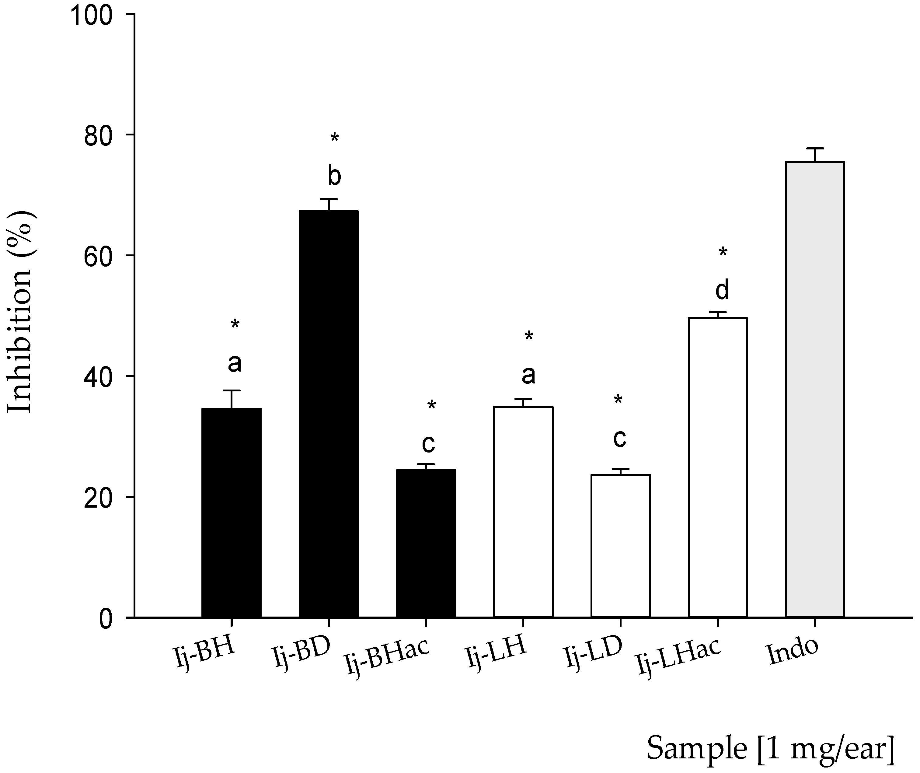
The results corresponding to the anti-inflammatory study of the organic extracts are presented in Figure 3. At the same dose of 1.0 mg/ear, all of the extracts showed anti-inflammatory activity. For the bark extracts, the percentages of inhibition were: Ij-BH 34.6 ± 3.0%, Ij-BD 67.3 ± 2.0%, and Ij-BHac 24.4 ±1.0%, and for leaf extracts, the corresponding percentages were: Ij-LH 34.9 ± 1.3%, Ij-LD 23.0 ± 1.0%, and Ij-LHac 49.6 ± 1.0%. For indomethacin (Indo), which was employed as the reference drug, the inhibition percentage was 75.5 ± 2.2%. As can be seen, the two extracts with the greatest anti-inflammatory activity were Ij-BD followed by Ij-LHac, and the statistical comparison between the anti-inflammatory activities of the extracts and the reference drug revealed significant differences (p < 0.05). No extract reached an effect equal to or greater than that of Indo (Indomethacin). However, the comparison using the Tukey test of the effect of the extracts and the reference drug showed that there were no significant differences (p < 0.05) between some of the extracts, such as Ij-BH compared to Ij-LH and Ij-BHac compared to Ij-LD.
Even when species such as I. laurina, I. edulis, I. marginata, and I. jinicuil are employed to treat stomach and inflammatory disorders in traditional medicine, few studies have been conducted to confirm their attributed pharmacological properties. However, recent reports have shown the presence of flavonoids and other phenolic compounds in several of these species that may be associated with pharmacological effects [36]. The present study represents a preliminary approach in the assessment of the anti-inflammatory activity of I. jinicuil, with the bark extracts exerting a more consistent effect and Ij-BD showing the highest activity. It is noteworthy to mention that the chemical profile of this extract showed the presence of salicylates, terpenoids, and derivatives of epigallocatechin gallate, as well as derivatives of protocatechuic and coumaric acids, which may be associated with its biological effect [24,28,31]. In the case of the extracts from leaves, Ij-LHac showed the best inhibitory effect, and the analysis of its metabolic content revealed the presence of polyphenolic compounds, terpenoids, coumarins, vanillic acid derivatives, and flavonoid-type compounds such as apigenin derivatives, all of which have reported anti-inflammatory effects [19,34,37]. Furthermore, previous reports regarding several of the metabolites present in both extracts have postulated an anti-inflammatory activity that proceeds via the inhibition of cyclooxygenase enzymes (COX) [38,39,40], which happens to be the known mechanism of the reference drug (Indo) [40]. Finally, it is important to mention that the two extracts with the highest activity have the presence of terpenes and salicylates in common; these compounds are recognized for their analgesic and anti-inflammatory effects [24,31].

2.4. Antibacterial Activity of Inga jinicuil Organic Extracts

The antibacterial activity of bark and leaf I. jinicuil extracts were evaluated on clinically important micro-organisms. As showed in Table 4, the three extracts of bark (Ij-BH, Ij-BD, and Ij-BHac) exhibited excellent activity against Pseudomona aeruginosa (Pa) and methicillin-resistant Staphylococcus aureus (Sa1), with MIC values of ˂3.12 and 50 µg/mL, respectively. Only Ij-BH showed activity against Staphylococcus epidermidis (Se1). Similarly, Ij-LD and Ij-LHac had good activity against Pseudomona aeruginosa (Pa; MIC ˂ 3.12 µg/mL), methicillin-resistant Staphylococcus aureus (Sa1; MIC = 50 µg/mL) and Staphylococcus epidermidis (Se1; MIC = 200 µg/mL).
The results obtained are interesting considering that in 2017 the WHO published a list of “priority pathogens” resistant to antibiotics, which include Pseudomonas aeruginosa (resistant to carbapenems) and Staphylococcus aureus (resistant to methicillin), emphasizing the urgent need for the search for new agents against these micro-organisms [5].
The antibacterial activity of bark and leaf extracts against Pseudomona aeruginosa (Pa) and methicillin-resistant Staphylococcus aureus (Sa1) can be attributed to the presence of several secondary metabolites: hentriacontane and α-tocopherol in Ij-BH, and polyphenols, flavonoids, and terpenoids in both Ij-BD and Ij-BHac [41]. Special attention may be paid to the presence of gallate and coumarin derivatives, since their antibacterial mechanism has been described at the cell membrane level by repressing the transport system of proteins and inhibiting the biofilm formation in clinical strains of Sa1 [17,42,43]. Furthermore, coumarin derivatives are considered as potential antibacterial agents that act as inhibitors to several binding proteins of Sa1 and potential competitive inhibitors of the DNA-gyrase [44,45]. It is worth noting that the chemical moiety responsible for the antibacterial activity of coumarins is the basic structure of benzopyrone, which resembles the structure of benzopyridone present in antibacterial drugs derived from quinolone [44,45]. Therefore, the wide range of chemical structures found in these extracts may represent a potential source of molecular templates for new antibacterial drugs.

3. Materials and Methods

3.1. Plant Material and Extraction of Inga jinicuil

Aerial parts of Inga jinicuil Schltdl & Cham. ex G. Don were collected in July 2019, in Libertad, Cunduacán, Tabasco, Mexico (10 m.a.s.l., latitude 18°10′53.06 N, longitude 93°22′28.13 W). A specimen was deposited at the Herbarium of the Academic Division of Biological Sciences of the Universidad Juárez Autónoma de Tabasco for its taxonomic identification (voucher number: 36576).
Plant material was dried at room temperature in the dark for 72 h, with drying and spraying in Pulvex MP300 milled (4–6 mm). The extracts were obtained by maceration with n-hexane, dichloromethane, and a 60:40 ethanol:water mixture 1:4; the maceration procedure was performed three times for each solvent in order to ensure an exhaustive extraction. These extracts were filtered, concentrated in a rotary evaporator (Heidolph G3, Schwabach, Germany), and then lyophilized (Heto Drywinner DW3) to give the bark (n-Hexane Ij-BH, Dichloromethane Ij-BD, Hydroalcoholic Ij-BHac) and leaf extracts (n-Hexane Ij-LH, Dichloromethane Ij-LD, Hydroalcoholic Ij-LHac).

3.2. HPLC Analysis

Chromatographic analysis was carried out in a Waters 2695 separation module system with a Waters 2695 photodiode matrix detector and Empower Pro software (Waters Corporation, Milford, MA, USA). Chemical separation was performed using a Supelcosil LC-F column (4.6 mm × 250 mm i.d., particle size 5 μm) (Sigma-Aldrich, Bellefonte, PA, USA). The mobile acid phase was performed using 0.5% triflouroacetic, aqueous solution (solvent A), and acetonitrile (solvent B) gradient: 0–1 min, 0% of B; 2–3 min, 5% of B; 4–20 min, 30% of B; 21–23 min, 50% of B; 24–25 min, 80% of B; 26–27 min, 100% of B; 28–30 min, 0% of B. The flow rate was 0.9 mL/min with a volume of 10 μL sample. Absorbance was measured at 270 nm [46]. A preliminary identification of the peaks resolved was performed by comparison with tR and UV-Vis characteristic bands of known standards and literature data.

3.3. GC-MS Analysis of Hexane Extracts

The chemical composition of Ij-BH and Ij -LH was analyzed on Gas Chromatography-Mass Spectrometry (GC-MS) equipment, consisting of an Agilent 6890 plus gas chromatograph coupled to a simple quadrupole mass spectrometry detector, model 5972N (Agilent Technology, Santa Clara, CA, USA).
Volatile compounds were separated on an HP 5MS capillary column (25 m long, 0.2 mm i.d., with 0.3-μm film thickness). Oven temperature was set at 40 °C for 2 min, then programmed at 40–260 °C for 10 °C/min, and maintained for 20 min at 260 °C. Mass detector conditions were as follows: interphase temperature, 200 °C, and mass acquisition range, 20–550. Injector and detector temperatures were set at 250 and 280 °C, respectively. Splitless injection mode was carried out with 1 μL of each fraction (3 mg/mL solution). The carrier gas was helium at a flow rate of 1 mL/min. The identification of volatiles was performed, comparing their mass spectra with those of the National Institute of Standards and Technology (NIST) 1.7 Library and comparing these with data from the literature [47].

3.4. Pharmacological Activity

3.4.1. Anti-Inflammatory Activity

Male ICR mice with a weight range of 25–30 g, from Envigo RMS, S.A. de C.V., were used throughout the experiments. These animals were maintained in the Bioterium of Centro de Investigación Biomédica Del Sur (CIBIS-IMSS) under a 12 h light-dark cycle and constant temperature (23–25 °C) with free access to food and water. The animals were treated under the Mexican federal regulations for care and use of laboratory animals, NOM-062-ZOO-1999 Guidelines [48], and international ethical guidelines for the care and use of experimental animals [49]; the number of animals (n = 5) and the intensity of the noxious stimuli utilized were the minimum necessary to demonstrate the consistent effects of the pharmacological treatments. The animal studies were approved by the Ethics Committee of the Mexican Social Security Institute (R-2020-1702-008).
Auricular inflammation was induced following the method previously described [50]. The dose evaluated for the extracts was 1.0 mg/ear. A control group received acetone as vehicle, and Indomethacin (Indo, Sigma-Aldrich, Toluca, Mexico) 1.0 mg/ear was utilized as an anti-inflammatory positive control. All treatments were dissolved in acetone and applied topically on both ears immediately after the solution of 12-O-tetradecanoylphorbol-13-acetate (TPA, Sigma-Aldrich, Toluca, México) as an inflammatory agent. Six hours after the administration of TPA, the animals were euthanized by cervical dislocation.
Circular sections 6 mm in diameter were taken from both the treated (t) and non-treated (nt) ears, which were weighed to determine the inflammation. The percentage of inhibition was obtained employing the expression below:
% Inhibition = [Dw control − Dw treated/Dw control] × [100]
where Dw = wt − wnt; wt is the weight of the section of the treated ear; and wnt the weight of untreated ear section.

3.4.2. Antibacterial Activity

The extracts were evaluated against bacterial strains ATCC: methicillin-resistant Staphylococcus aureus (Sa1; ATCC 43330, Staphylococcus aureus (Sa2; ATCC 29213), Staphylococcus epidermis (Se1; ATCC 12228), Staphylococcus epidermis (Se2; ATCC 35984), Enterococcus fecalis (Ef; ATCC 29212), Escherichia coli (Ec1; ATCC 25922), Enterobacter cloacae (Ec2; ATCC 700323), Klebsiella pneumoniae (Kp; ATCC 700603), Pseudomona aeruginosa (Pa; ATCC 27853), and the clinically isolated Staphylococcus haemolyticus (Sh; 1038). The strains were reseeded in antibiotic agar No. 1 (Bioxon, Mexico) for 24 h at 37 °C. The strain of clinical isolate was provided from the General Hospital of Acapulco, State of Guerrero, Mexico, to the Bacteria Bank of the Autonomous University of Guerrero (UAGro).
For the trials, cultures with 24 h of incubation (37 °C) were used and about 3–4 colonies were taken of each strain and diluted in Müeller–Hinton broth (MHb; Bioxon, Toluca, Mexico). The inoculums were adjusted using the 0.5 MacFarland scale (1.5 × 10⁸ UFC/mL). Subsequently, dilution with distilled water was performed to obtain 1 × 10⁴ UFC/mL.
The MIC of extracts was determined by the microtiter broth dilution method [51]. Briefly, the samples (50 mg/mL) were dissolved in a DMSO–water mixture (20:80), and the tested concentrations were 3.37, 6.75, 12.5, 25, 50, 100, and 200 μg/mL. The samples were added to sterile microplates of 96 wells, along with 200 μL of MHb and 2 μL of inoculum (1 × 10⁴ UFC/mL). The viability controls used were: MHb + DMSO + inoculum and MHb+ inoculum; Gentamicin (C+, 100 μg/mL; Sigma Aldrich, Mexico) was employed as the reference antibiotic. The plates were incubated at 37 °C for 24 h, and after incubation the MIC was determined by adding 30 μL of a solution (0.05%) of 3-(4,5-dimethylthiazol-2-yl)-2,5, diphenyl tetrazolium bromide (MTT, Sigma-Aldrich, Hong Kong, China) in every well, of which purple development was observed if there was viability of bacteria and colorless if there was no feasibility. All assays were performed in triplicate.

3.5. Statistical Analysis

For the analysis of the anti-inflammatory activity, the data were expressed as the mean ± standard error of the mean (SEM), and statistical significance was determined using an analysis of variance (ANOVA) with a confidence level of 95% (* p ≤ 0.05), followed by the one-tailed Dunnet test compared to Indo and the Tukey test. All analyses was performed using IBM SPSS statistics ver. 23.0 statistical program (GraphPad Software, IBM, San Diego, CA, USA).

4. Conclusions

This report presents the biological activity of organic extracts obtained from the bark and leaves of I. jinicuil. The anti-inflammatory activity tests showed moderate to good effects, with the dichloromethane extract from bark showing the highest activity, followed by the hexanic extract from leaves. Based on the findings of anti-inflammatory activity, it is possible to propose the exploration of the potential antinociceptive effect of the tested extracts, using an appropriate pharmacological model. Likewise, it was found that the three extracts from the bark of this plant have excellent antibacterial activity (primarily against methicillin-resistant Staphylococcus aureus and Pseudomonas aeruginosa strains) and this leads us to consider the possibility of extending antibacterial activity tests to a greater number of microbiological strains of clinical interest. On the other hand, it should be mentioned that, to our knowledge, this is the first approach to the phytochemical profiling of bark and leaves of I. jinicuil, which is consistent with the chemotaxonomic profiles reported for other species of Inga and suggest the presence of polyphenolic compounds, flavonoids, triterpenes, and lipid prenols, as well as aliphatic and esterified aliphatic lipids; these natural products may be responsible for both bioactivities assessed in this work. These results allow predicting a wide potential for future studies aimed at the isolation and structural characterization of compounds that might serve as molecular templates with specific biological activities. Finally, it is important to highlight that these results systematically contribute to the use in traditional Mexican medicine of a highly important sociocultural and nutritional species such as Inga jinicuil Schltdl & Cham. ex G. Don.

Supplementary Materials

The following are available online at https://www.mdpi.com/article/10.3390/plants11060794/s1, Figure S1: UV-spectra of the main compounds of (Ij-BD) I. jinicuil; Figure S2: UV-spectra of the main compounds of (Ij-BHac) I. jinicuil; Figure S3: UV-spectra of the main compounds of (Ij-LD) I. jinicuil; Figure S4: UV-spectra of the main compounds of (Ij-LHac) I. jinicuil.

Author Contributions

Conceptualization, M.G.-C., C.E.L.-G. and A.G.-R.; methodology, A.J.G.-G., R.L.-R. and P.Á.-F.; validation, M.H.-R. and M.D.P.-G.; formal analysis, A.Z.; investigation, A.G.-R. and A.B.; resources, A.J.G.-G., R.L.-R. and M.R.F.; data curation, E.J.M.-S. and M.R.F.; writing—original draft preparation, A.J.G.-G., R.L.-R. and E.A.B.-G.; writing—review and editing, A.G.-R., C.E.L.-G. and A.B.; visualization, M.H.-R. and P.Á.-F.; supervision, M.D.P.-G.; project administration, M.G.-C., A.G.-R. and C.E.L.-G.; funding acquisition, A.G.-R., M.G.-C. and A.Z. All authors have read and agreed to the published version of the manuscript.

Funding

This research was funded by PRODEP, grant number UJAT-EXB-242.

Institutional Review Board Statement

The animal studies were approved by the Ethics Committee of the Mexican Social Security Institute (R-2020-1702-008).

Informed Consent Statement

Not applicable.

Data Availability Statement

Data is contained within the article and Supplementary Material.

Acknowledgments

The authors wish to thank Arturo Pérez, Jonathan Orduño, and Ixchel Palacios (CIBIS-IMSS), and Dora Elena Aguilar Dominguez and Wilbert Pérez Fuentes (DACB-UJAT) for technical and analytical assistance; al Consejo Nacional de Ciencia y Tecnologia (CONACYT) por la beca otorgada con No. de CVU: 703106; R.L.-R. (478597) was supported by CONACYT postdoctoral fellowship (866998).

Conflicts of Interest

The authors declare no conflict of interest.

References

  1. Maione, F.; Russo, R.; Khan, H.; Mascolo, N. Medicinal plants with anti-inflammatory activities. Nat. Prod. Res. 2015, 30, 1343–1352. [Google Scholar] [CrossRef] [PubMed]
  2. Heydari, H.; Saltan, I.G.; Eryilmaz, M.; Bahadir, A.Ö.; Yilmaz, S.S.; Tekin, M.; Çoban, T. Antimicrobial and Anti-Inflammatory Activity of Some Lathyrus L. (Fabaceae) Species Growing in Turkey. Turk. J. Pharm. Sci. 2019, 16, 240–245. [Google Scholar] [CrossRef] [PubMed]
  3. WHO. WHO Traditional Medicine Strategy: 2014–2023; WHO Press, World Health Organization: Hong Kong, China, 2013; pp. 15–19. [Google Scholar]
  4. Ghasemian, M.; Owlia, S.; Owlia, M.B. Review of Anti-Inflammatory Herbal Medicines. Adv. Pharmacol. Sci. 2016, 2016, 9130979. [Google Scholar] [CrossRef] [PubMed] [Green Version]
  5. WHO. Global Priority List of Antibiotic-Resistant Bacteria to Guide Research, Discovery, and Development of New Antibiotics. Available online: https://www.who.int/medicines/publications/WHO-PPL-Short_Summary_25Feb-ET_NM_WHO.pdf (accessed on 11 January 2022).
  6. Vargas, S.G.; Pire, R. Inga jinicuil Schtdl; Multiuso, Á., Ed.; Universidad Juárez Autónoma de Tabasco: Villahermosa, Mexico, 2017; pp. 2–4. ISBN 978-607-606-393-4. [Google Scholar]
  7. Alejandro, M.A.M.; Campillo, L.M.G.; Méndez, R.M. El Uso de las Plantas Medicinales en las Comunidades Maya-Chontales de Nacajuca, Tabasco, México. Polibotánica 2010, 29, 213–2062. Available online: http://www.scielo.org.mx/scielo.php?script=sci_arttext&pid=S1405-27682010000100011&lng=es&nrm=iso (accessed on 13 January 2022).
  8. Maldonado, M.F. Flora Medicinal del Estado de Tabasco: Uso, Manejo y Conservación, 2nd ed.; Instituto para el Desarrollo de Sistemas de Producción del Trópico Húmedo de Tabasco: Villahermosa, Tabasco, Mexico, 2005; p. 50. ISBN 968-7991-24-0. [Google Scholar]
  9. Maziero, M.; Lovato, M.O.; Lorenzoni, V.V.; Moraes, G.G.; Dornelles, R.C.; Sagrillo, M.R.; Horner, R.; Manfron, M.P. Phytochemical Study, an Evaluation of the Antioxidant Potential and the Antimicrobial Activity of Inga semialata (Vell.) C. Mart. Hydroalcohol Extract. Nat. Prod. Res. 2019, 34, 192–196. [Google Scholar] [CrossRef]
  10. Pompeu, D.R.; Rogez, H.; Monteiro, K.M.; Tinti, S.V.; Carvalho, J.E. Capacidad antioxidante e triagem farmacológica de extratos brutos de folhas de Byrsonima crassifolia e de Inga edulis. Acta Amaz. 2012, 42, 165–172. [Google Scholar] [CrossRef]
  11. Dib, H.X.; de Oliveira, D.G.L.; de Oliveira, C.F.R.; Taveira, G.B.; de Oliveira Mello, E.; Verbisk, N.V.; Chang, M.R.; Corrêa, D., Jr.; Gomes, V.M.; Macedo, M.L.R. Biochemical Characterization of a Kunitz Inhibitor from Inga edulis Seeds with Antifungal Activity against Candida spp. Arch. Microbiol. 2019, 201, 223–233. [Google Scholar] [CrossRef] [Green Version]
  12. Alves do Nascimento, V.H.; Guimarães Sobrinho, A.C.; De Oliveira Souza, C.; Silva de Souza, J.N.; Sousa, C.L. Determination of Phenolic Compounds with Antimicrobial Activity of Byrsonima crassifolia and Inga edulis Leaves Extracts. Ens. Ciênc. 2021, 25, 21–28. [Google Scholar] [CrossRef]
  13. Lima, N.; Santos, V.; La Porta, F. Chemodiversity, Bioactivity and Chemosystematics of the Genus Inga (FABACEAE): A Brief Review. Rev. Virtual Quim. 2018, 10, 459–473. [Google Scholar] [CrossRef]
  14. Rugerio, M.O.; Hernández, M.H.H.; Suárez, R.A.; Vargas, D.M.E.; Daniel, A.B. Estudio químico y antimicrobiano de Inga jinicuil del estado de Tlaxcala. In Proceedings of the 7th Reunión Internacional de Investigación de Productos Naturales “Dr. Pedro Joseph-Nathan”, Revista Latinoamérica de Química, Morelia, Mexico, 18–20 May 2011; Navarete, A., Ed.; Mixim: Naucalpan de Juárez, Mexico, 2011; C-95, p. 149. Available online: https://www.yumpu.com/es/document/view/27637927/latinoamericana-de-quimica-revista-latinoamericana-de- (accessed on 13 January 2022).
  15. Sun, J.; Liang, F.; Bin, Y.; Li, P.; Duan, C. Screening Non-Colored Phenolics in Red Wines Using Liquid Chromatography/Ultraviolet and Mass Spectrometry/Mass Spectrometry Libraries. Molecules 2007, 12, 679–693. [Google Scholar] [CrossRef] [Green Version]
  16. Robbins, R.J. Phenolic Acids in Foods: An Overview of Analytical Methodology. J. Agric. Food Chem. 2003, 51, 2866–2887. [Google Scholar] [CrossRef] [PubMed]
  17. Gabe, V.; Kacergius, T.; Abu-Lafi, S.; Kalesinskas, P.; Masalha, M.; Falah, M.; Abu-Farich, B.; Melninkaitis, A.; Zeidan, M.; Rayan, A. Inhibitory Effects of Ethyl Gallate on Streptococcus mutans Biofilm Formation by Optical Profilometry and Gene Expression Analysis. Molecules 2019, 24, 529. [Google Scholar] [CrossRef] [PubMed] [Green Version]
  18. Martins, C.M.; de Morais, S.A.L.; Martins, M.M.; Cunha, L.C.S.; da Silva, C.V.; Martins, C.H.G.; Leandro, L.F.; de Oliveira, A.; de Aquino, F.J.T.; do Nascimento, E.A.; et al. Chemical Composition, Antifungal, and Cytotoxicity Activities of Inga laurina (Sw.) Willd Leaves. Sci. World J. 2019, 2019, 9423658. [Google Scholar] [CrossRef] [PubMed] [Green Version]
  19. Falcoski, T.O.R.; Lima, N.M.; Navegante, G.; Serafim, R.B.; Sorbo, J.M.; Valente, V.; Santos, V.N.C.; Santos, R.A.; Silva, D.H.S.; Soares, C.P. Genotoxicity, Cytotoxicity and Chemical Profile from Inga laurina (Fabaceae). Nat. Prod. Res. 2021, 35, 676–680. [Google Scholar] [CrossRef] [PubMed]
  20. Kumar, S.; Pandey, A.K. Chemistry and Biological Activities of Flavonoids: An Overview. Sci. World J. 2013, 2013, 162750. [Google Scholar] [CrossRef] [Green Version]
  21. Sun, Y.; Zeng, Q.-H.; Lu, H.-Q.; Meng, F.-C.; Shen, Y.; Zeng, W.-Y.; Chi, H.; Zhou, Y.-Q.; Chen, M. Two New Lignans from Zanthoxylum armatum. Nat. Prod. Res. 2020, 2020, 1–6. [Google Scholar] [CrossRef] [PubMed]
  22. Guo, K.; Tong, C.; Fu, Q.; Xu, J.; Shi, S.; Xiao, Y. Identification of Minor Lignans, Alkaloids, and Phenylpropanoid Glycosides in Magnolia officinalis by HPLC-DAD-QTOF-MS/MS. J. Pharm. Biomed. Anal. 2019, 170, 153–160. [Google Scholar] [CrossRef]
  23. Holser, R.A. Principal Component Analysis of Phenolic Acid Spectra. ISRN Spectrosc. 2012, 2012, 493203. [Google Scholar] [CrossRef]
  24. Renuka, B.; Sanjeev, B.; Ranganathan, D. Evaluation of Phytoconstituents of Caralluma nilagiriana by FTIR and UV-VIS Spectroscopic Analysis. J. Pharmacogn. Phytochem. 2016, 5, 105–108. Available online: https://www.phytojournal.com/archives?year=2016&vol=5&issue=2&ArticleId=813 (accessed on 18 January 2022).
  25. Fatima, S.; Mansha, A.; Asim, S.; Shahzad, A. Absorption Spectra of Coumarin and Its Derivatives. Chem. Pap. 2022, 76, 627–638. [Google Scholar] [CrossRef]
  26. Hroboňová, K.; Sádecká, J. Coumarins Content in Wine: Application of HPLC, Fluorescence Spectrometry, and Chemometric Approach. J. Food Sci. Technol. 2020, 57, 200–209. [Google Scholar] [CrossRef] [PubMed]
  27. Li, G.J.; Wu, H.J.; Wang, Y.; Hung, W.L.; Rouseff, R.L. Determination of Citrus Juice Coumarins, Furanocoumarins and Methoxylated Flavones Using Solid Phase Extraction and HPLC with Photodiode Array and Fluorescence Detection. Food Chem. 2019, 271, 29–38. [Google Scholar] [CrossRef] [PubMed]
  28. Sun, S.; He, G.; Yu, H.; Yang, J.; Borthakur, D.; Zhang, L.; Shen, S.; Das, U.N. Free Zn2+ Enhances Inhibitory Effects of EGCG on the Growth of PC-3 Cells. Mol. Nutr. Food Res. 2008, 52, 465–471. [Google Scholar] [CrossRef] [PubMed]
  29. Wodnicka, A.; Huzar, E. Synthesis and Photoprotective Properties of New Salicylic and Vanillic Acid Derivatives. Curr. Chem. Lett. 2017, 6, 125–134. [Google Scholar] [CrossRef]
  30. Lima, N.M.; de Marqui, S.R.; Silva, D.H.S. Phytochemical, Metabolic Profiling and Antiparasitic Potential from Inga semialata Leaves (Fabaceae). Nat. Prod. Res. 2020, 2020, 1–6. [Google Scholar] [CrossRef]
  31. Hussain, M.A.; Badshah, M.; Iqbal, M.S.; Tahir, M.N.; Tremel, W.; Bhosale, S.V.; Sher, M.; Haseeb, M.T. HPMC-Salicylate Conjugates as Macromolecular Prodrugs: Design, Characterization, and Nano-Rods Formation: Rapid Communication. J. Polym. Sci. A Polym. Chem. 2009, 47, 4202–4208. [Google Scholar] [CrossRef]
  32. Alves, G.A.D.; Fernandes, S.D.; Venteu, T.T.; de Souza, R.O.; Rogez, H.; Fonseca, M.J.V. Obtainment of an Enriched Fraction of Inga edulis: Identification Using UPLC-DAD-MS/MS and Photochemopreventive Screening. Prep. Biochem. Biotechnol. 2020, 50, 28–36. [Google Scholar] [CrossRef]
  33. Tchuenmogne, A.M.T.; Donfack, E.V.; Kongue, M.D.T.; Lenta, B.N.; Ngouela, S.; Tsamo, E.; Sidhu, N.; Dittrich, B.; Laatsch, H. Ingacamerounol, A New Flavonol and Other Chemical Constituents from Leaves and Stem Bark of Inga edulis Mart. Bull. Korean Chem. Soc. 2013, 34, 3859–3862. [Google Scholar] [CrossRef] [Green Version]
  34. Lima, N.M.; Andrade, T.J.A.S.; Silva, D.H.S. Dereplication of Terpenes and Phenolic Compounds from Inga edulis Extracts Using HPLC-SPE-TT, RP-HPLC-PDA and NMR Spectroscopy. Nat. Prod. Res. 2022, 36, 488–492. [Google Scholar] [CrossRef]
  35. Furtado, F.B.; de Aquino, F.J.T.; Nascimento, E.A.; de Martins, C.M.; de Morais, S.A.L.; Chang, R.; Cunha, L.C.S.; Leandro, L.F.; Martins, C.H.G.; Martins, M.M.; et al. Seasonal Variation of the Chemical Composition and Antimicrobial and Cytotoxic Activities of the Essential Oils from Inga laurina (Sw.) Willd. Molecules 2014, 19, 4560–4577. [Google Scholar] [CrossRef] [Green Version]
  36. Marqui, S.; Santos, L.; Young, M.; Torres, L.; Bolzani, V.; Moraes, M.; Soares, C.; Silva, D. Chemopreventive Potential of Rhamnosyl Depsides from Inga laurina. Planta Med. 2010, 76, 47. [Google Scholar] [CrossRef]
  37. Nogueira, K.M.; de Souza, L.K.M.; Medeiros, J.V.R. Technological Prospection of Anti-Inflammatory Vanilic Acid Activity, with Emphasis on Its Semisynthetic Derivative Isopropyl Vanilate. Res. Soc. Dev. 2021, 10, e35910313451. [Google Scholar] [CrossRef]
  38. Enciso, E.; Arroyo, J. Efecto antiinflamatorio y antioxidante de los flavonoides de las hojas de Jungia rugosa Less (matico de puna) en un modelo experimental en ratas. An. Fac. Med. 2013, 72, 231. [Google Scholar] [CrossRef] [Green Version]
  39. Howes, M.J.R. Phytochemicals as Anti-Inflammatory Nutraceuticals and Phytopharmaceuticals. In Immunity and Inflammation in Health and Disease; Chatterjee, S., Jungraithmayr, W., Bagchi, W., Eds.; Academic Press: Cambridge, UK, 2018; pp. 363–388. [Google Scholar] [CrossRef]
  40. Molehin, O.R.; Adeyanju, A.A.; Adefegha, S.A.; Oyeyemi, A.O.; Idowu, K.A. Protective Mechanisms of Protocatechuic Acid against Doxorubicin-Induced Nephrotoxicity in Rat Model. J. Basic Clin. Physiol. Pharmacol. 2019, 30, 20180191. [Google Scholar] [CrossRef] [PubMed]
  41. Pistelli, L.; Bertoli, A.; Noccioli, C.; Mendez, J.; Musmanno, R.A.; Di Maggio, T.; Coratza, G. Antimicrobial Activity of Inga fendleriana Extracts and Isolated Flavonoids. Nat. Prod. Commun. 2009, 4, 1679–1683. [Google Scholar] [CrossRef] [Green Version]
  42. Favela-Hernández, J.M.J.; Clemente-Soto, A.F.; Balderas-Rentería, I.; Garza-González, E.; del Camacho-Corona, M.R. Potential Mechanism of Action of 3’-Demethoxy-6-O-Demethyl-Isoguaiacin on Methicillin Resistant Staphylococcus aureus. Molecules 2015, 20, 12450–12458. [Google Scholar] [CrossRef]
  43. Wang, D.; Jin, Q.; Xiang, H.; Wang, W.; Guo, N.; Zhang, K.; Tang, X.; Meng, R.; Feng, H.; Liu, L.; et al. Transcriptional and Functional Analysis of the Effects of Magnolol: Inhibition of Autolysis and Biofilms in Staphylococcus aureus. PLoS ONE 2011, 6, e26833. [Google Scholar] [CrossRef]
  44. Pisano, M.B.; Kumar, A.; Medda, R.; Gatto, G.; Pal, R.; Fais, A.; Era, B.; Cosentino, S.; Uriarte, E.; Santana, L.; et al. Antibacterial Activity and Molecular Docking Studies of a Selected Series of Hydroxy-3-Arylcoumarins. Molecules 2019, 24, 2815. [Google Scholar] [CrossRef] [Green Version]
  45. Kampranis, S.C.; Gormley, N.A.; Tranter, R.; Orphanides, G.; Maxwell, A. Probing the Binding of Coumarins and Cyclothialidines to DNA Gyrase. Biochemistry 1999, 38, 1967–1976. [Google Scholar] [CrossRef]
  46. De Araújo, R.S.A.; Barbosa, F.J.M.; Scotti, M.T.; Scotti, L.; da Cruz, R.M.D.; Falcão, S.V.D.S.; de Siqueira, J.J.P.; Mendonça, J.F.J.B. Modulation of Drug Resistance in Staphylococcus aureus with Coumarin Derivatives. Scientifica 2016, 2016, 6894758. [Google Scholar] [CrossRef] [Green Version]
  47. Adam, R.P. Identification of Essential Oils Components by Gas Chromatography/Mass Spectrometry, 4th ed.; Allured Business Media: Carol Stream, IL, USA, 2007; pp. 1–804. ISBN 978-193-263-32-14. [Google Scholar]
  48. SAGARPA. Norma Oficial Mexicana NOM-062-ZOO-1999. Especificaciones Técnicas para la Producción, Cuidado y Uso de los Animales de Laboratorio (Technical Specifications for the Production, Care, and Use of Laboratory Animals). México Diario Oficial de la Federación. 22 August 2001. Available online: https://www.gob.mx/cms/uploads/attachment/file/203498/NOM-062-ZOO-1999_220801.pdf (accessed on 2 February 2022).
  49. Zimmermann, M. Ethical guidelines for investigations of experimental pain in conscious animals. Pain 1983, 16, 109–110. [Google Scholar] [CrossRef]
  50. Payá, M.; Ferrándiz, M.L.; Sanz, M.J.; Bustos, G.; Blasco, R.; Rios, J.L.; Alcaraz, M.J. Study of the Antioedema Activity of Some Seaweed and Sponge Extracts from the Mediterranean Coast in Mice. Phytother. Res. 1993, 7, 159–162. [Google Scholar] [CrossRef]
  51. Salazar, P.D.T.; Castro, A.N.; Moreno, G.M.E.; Nicasio, T.M.P.; Perez, H.J.; Alvarez, F.P. Antibacterial and Anti-Inflammatory Activity of Extracts and Fractions from Agave cupreata. Int. J. Pharmacol. 2017, 13, 1063–1070. [Google Scholar] [CrossRef]
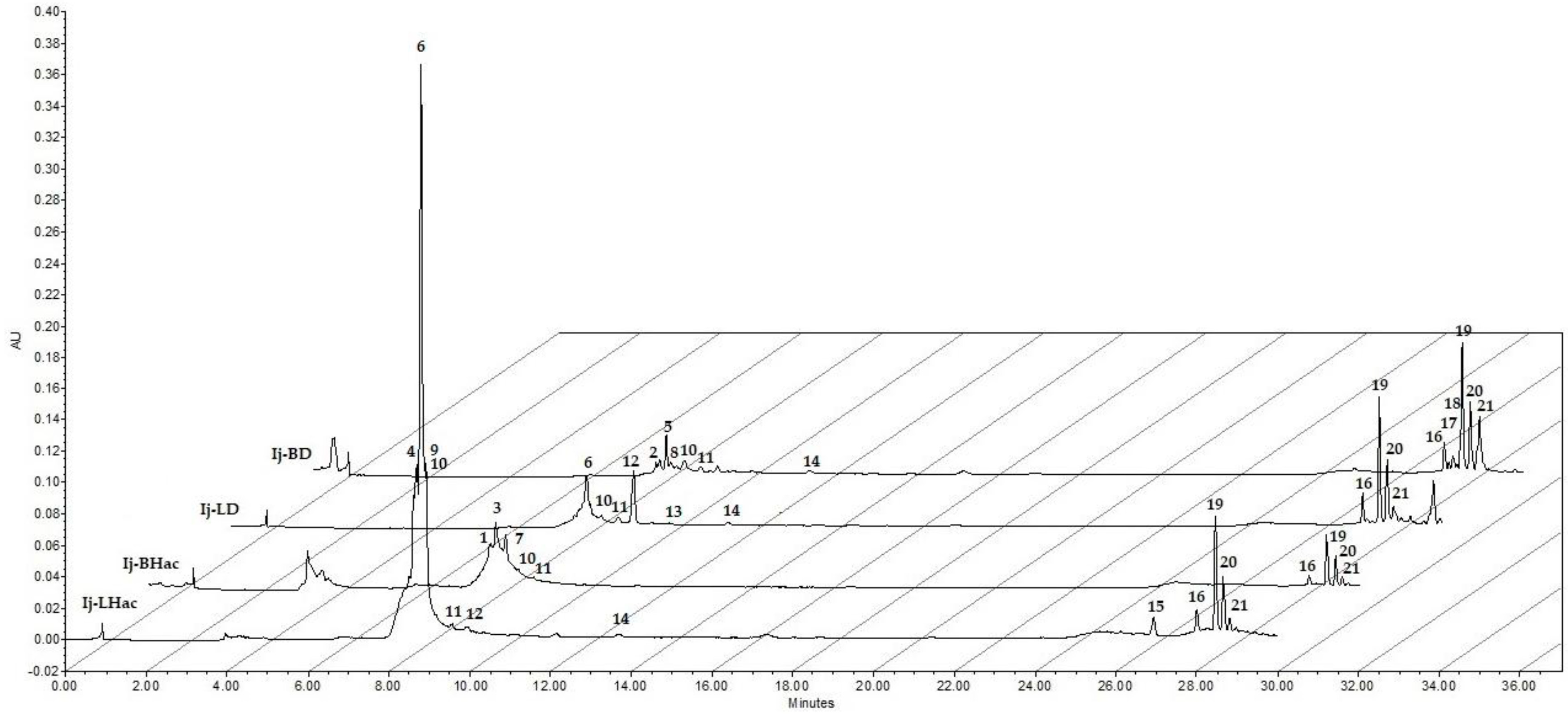
Figure 1. HPLC chromatograms of bark (Ij-BD, Ij-BHac) and leaf (Ij-LD, Ij-LHac) extracts. The peaks are numbered in ascending order according to their retention times (λ = 270 nm).
Plants 11 00794 g001
Figure 2. GC chromatograms of the hexanic extracts: (A) Ij-BH and (B) Ij-LH. The peaks are numbered in ascending order according to their retention times.
Plants 11 00794 g002
Figure 3. Percentage inhibition of inflammation (%) of Ij-BH, Ij-BD, Ij-BHac, Ij-LH, Ij-LD, and Ij-LHac extracts from Inga jinicuil and Indo (Indomethacin) in edema induced by TPA in mouse ear at 1.0 mg/ear. Values are presented as means ± standard error of the means (SEM). n = 5. ANOVA, with post-test Dunnet with * p ≤ 0.05 in comparison with Indo and Tukey test, where different letters indicate significant differences among them.
Plants 11 00794 g003
Table 1. Percentages obtained from Inga jinicuil extracts.
Extractn-Hexane
(% Yield)
Dichloromethane
(% Yield)
Hydroalcoholic
(% Yield)
Bark extract0.0950.820.25
Leaf extract0.951.024.65
Table 2. Preliminary phytochemical profile by HPLC-UV-Vis analysis of polar extracts from bark and leaves of I. jinicuil.
PeakRetention Time (min)Absorption Bands (nm)Extract(s) *Compound Affinity **Ref.
18.46220.4, 261.6, 294.7Protocatechuic acidStandard [15,16]
28.58249.8, 273.6Protocatechuic acid derivativeStandard [15,16]
38.58218.1, 276.9Gallic acid derivativeStandard [17,18]
48.66212.2, 251.5, 352.9Glycosylated Flavone. Apigenin derivativeStandard [19,20]
58.75219.2, 249.8, 273.4LignaneStandard [21,22]
68.81215.7, 269.8, 337.4○□Glycosylated Flavone. Apigenin derivativeStandard [19,20]
78.85219.2, 279.3Gallic acid derivativeStandard [17,18]
88.86215.7, 308.9Coumaric acid derivativeStandard [23]
98.91207.5, 269.8, 335.1Glycosylated Flavone. Apigenin derivative[19,20]
109.18249.8○□●■Terpene[24]
119.58245.1○□●■Terpene[24]
129.96209.9, 294.7, 338.6○□Coumarin derivative[25,26,27]
1310.03276.9Epigallocatechin Gallate derivativeStandard [28]
1412.30235.7, 266.3○□●Terpene[24]
1526.91219.2, 273.4, 293.5Vanillic acid derivative[29,30]
1628.01204, 248.6○□●■Terpene[24]
1728.11278.1Epigallocatechin Gallate derivative[28]
1828.21245.1, 278.1, 327.9Coumarin derivative[25,26,27]
1928.43201.7, 261.6○□●■Salicylate derivativeStandard [31]
2028.65200.5, 263.9○□●■Salicylate derivativeStandard [31]
2128.81263.9○□●■Salicylate derivativeStandard [31]
* Extracts: Bark extracts, ● (Ij-BD), ■ (Ij-BHac); Leaf extracts, ○ (Ij-LD), □ (Ij-LHac). ** Compounds were suggested by a preliminary comparison of retention time (tR) and UV-Vis bands with standards and literature data.
Table 3. Phytochemicals identified in hexanic extracts from the bark (Ij-BH) and leaves (Ij-LH) of Inga jinicuil by GC-MS.
PeakRetention Time (min)Molecular Weight (amu)Extract(s)
(% in the Sample) *
Compound **
2217.80,
17.75
268.5▲ (1.07),
△ (1.18)
2-pentadecanone,6,10,14-trimethyl
2317.80296.5▲ (1.07)3,7,11,15-Tetramethyl-2-hexadecen-1-ol
2418.55270.5△ (1.04)Hexadecanoic acid, methyl ester
2518.61276.3△ (0.88)7,9-Di-tert-butyl-1-oxaspiro(4,5)deca-6,9-diene-2,8-dione
2618.68,
18.61
270.5▲ (3.83),
△ (0.88)
Hexadecanoic acid, ethyl ester
2720.51296.5▲ (11.74)Phytol
2820.60,
20.47
298.5▲ (3.14),
△ (0.44)
Octadecanoic acid, methyl ester
2922.53324.5△ (1.35)4,8,12,16-tetramethylheptadecan-4-olide
3024.63390.6△ (0.80)1,2-benzenedicarboxylic acid diisooctyl ester
3127.76380.6△ (1.62)15-Tetracosenoic acid, methyl ester
3229.19518.7△ (1.02)Tetradecanoic acid, 3,3a,4,6a,7,8,9,10,10a, 10b-decahydro-3a, 10a, dihydroxy-5-(hydroxymethyl)-2, 10-dimethyl-3-oxobenz [e] azulen-8-yl ester
3329.21410.7▲ (5.98)Squalene
3430.05408.8▲ (12.55)Nonacosane
3531.95416.7▲ (3.74)β-Tocopherol
3632.44436.8▲ (16.66)Hentriacontane
3733.07430.7▲ (40.49)α-Tocopherol
3836.91424.7△ (26.74)Lup-20 (29)-en-3-one
3937.39426.7△ (16.43)Lupeol
4038.35438.7△ (38.61)24-Methylenecycloartan-3-one
4138.64412.7△ (2.27)Stigmast-4-en-3-one
4241.85440.7△ (4.99)9,19-Cyclolanostan-3-ol,24-methylene-, (3β)-
* Extracts: Bark extract (▲ (Ij-BH) and leaf extract (△ (Ij-LH). ** Compared with the National Institute of Standards and Technology (NIST) 1.7 Library.
Table 4. Antibacterial activity (MIC µg/mL) of extracts from Inga jinicuil.
BacterialStrains
Gram-Positive Gram-Negative
ExtractSa1Sa2Se1Se2ShEc1Ec2EfKpPa
Ij-LH>200>200>200>200>200>200>200>200>200>200
Ij-LD50>200200>200>200>200>200>200>200<3.12
IjLHac50>200>200>200>200>200>200>200>200<3.12
Ij-BH50>20050>200>200>200>200>200>200<3.12
Ij-BD50>200>200>200>200>200>200>200>200<3.12
IjBHac50>200>200>200>200>200>200>200>200<3.12
C1**********
C2**********
C+--------------------
Sa1: methicillin-resistant S. aureus; Sa2: S. aureus; Se1: S. epidermis; Se2: S. epidermis; Sh: clinically isolated S. haemolyticus; Ec1: E. coliEc2: E. cloacae; Ef: E. fecalis; Kp: K. pneumoniae; Pa: P. aeruginosa; C1 and C2: controls of viability (*: bacterial growth); C+: positive control (Gentamicine 100 μg/mL; --: not bacterial growth).
Publisher’s Note: MDPI stays neutral with regard to jurisdictional claims in published maps and institutional affiliations.