Article

Multi Omics Analysis Revealed a Resistance Mechanism of Tibetan Barley (Hordeum vulgare L., Qingke) Infected by Ustilago hordei

1
Institute of Agro-Products Processing Science and Technology, Sichuan Academy of Agricultural Sciences, Chengdu 610011, China
2
State Key Laboratory of Food Nutrition and Safety, Tianjin University of Science and Technology, Tianjin 300457, China
3
Tibet Academy of Agriculture and Animal Husbandry Sciences, Lhasa 850031, China
*
Author to whom correspondence should be addressed.
Plants 2023, 12(1), 157; https://doi.org/10.3390/plants12010157
Submission received: 24 August 2022 / Revised: 21 November 2022 / Accepted: 25 November 2022 / Published: 29 December 2022
(This article belongs to the Section Plant Molecular Biology)

Abstract

:
Tibetan barley (Hordeum vulgare L., qingke) is the principal cereal cultivated on Tibet. Ustilago hordei causing covered smut is a serious disease that limits the yield of qingke. Here, based on multi omics study including metabolome, proteome and transcriptome, we show that during infection, primary metabolisms such as carbohydrate, amino acid, and lipids were significantly changed. Jasmonic acid, which perform as a biotic stress signaler, was significantly repressed, and related genes or proteins also showed different expression in infected qingke. In addition, other defense-related compounds such as riboflavin, ascorbic acid, and protease inhibitors were also detected in omics data. Our results revealed a preliminary biological profile of qingke infected by U. hordei and provide a resource for further research.

1. Introduction

Barley (Hordeum vulgare L.) is one of the important crops in the world that contributes 7% of global production [1]. Tibetan barley (H. vulgare L., qingke), a six-rowed hulless barely, is the principal cereal cultivated on the Tibetan Plateau and has been used as a traditional staple food for Tibetans. The disease is an important factor affecting the yield of barley. Ustilago hordei, a fungal pathogen causing covered smut, is one of several serious diseases that limit the yield of barley. Infected grain shows black powder, and 100% yield per spike would be lost in qingke. Meanwhile, the annual average loss of 2–5% has been reported due to covered smut [2].
Plants are threatened by different kinds of biotic stresses in natural environment and rely on innate immunity to protect themselves [3]. The physical barrier is the first defense line such as cell wall and the cytoskeleton to deter pathogens [3,4]. The second line is that plant could recognize certain pathogens molecular patterns, which called pathogen-associated molecular patterns (PAMP). Pathogens interact with plant surface by PAMP or translocate effector proteins to the host cell, resulting in signal transduction cascades and activation of resistance genes [5,6]. PAMP-triggered immunity (PTI) and effector-triggered immunity (ETI) composed the plant’s innate immunity system. Cytoskeletal reorganization, cell wall fortification, the generation of reactive oxygen species (ROS), and the synthesis of phytoalexins were induced during the early response, and hypersensitive response (HR) induced in later defense response caused programmed cell death (PCD) to limit pathogen spread [7].
The regulation and execution of both PTI and ETI proceed via the biosynthesis of small metabolic signals such as salicylic acid (SA), jasmonic acid (JA) [8]. High levels of SA and its methyl ester cause the production of pathogen-related (PR) proteins, including chitinases and other hydrolytic enzymes. SA also takes an important role in HR to defense pathogens [9]. JA, which is produced from linolenic acid in chloroplasts and peroxisomes, plays a significant role in inducing plants against necrotrophic and hemibiotrophic pathogens [9]. During pathogen infection, JA and JA-isoleucine can be rapidly induced to active major secondary metabolites and protein expression involved in defense response such as alkaloids, terpenoids, phenylpropane, amino acid derivatives, anti-nutritional proteins, and some pathogen-related proteins [10]. Moreover, the SA- and JA-signaling pathways could negatively influence each other [11]. Some vitamins, such as thiamine (TH), riboflavin (RF), folic acid (FA), and ascorbic acid (AA), also were reported to be linked with defense response [12]. Thiamine is able to activate almost all innate-defense mechanisms, including HR, ROS, callose, PR protein, and phytoalexins, RF could activate the majority defense except HR, while FA induces mainly PR proteins [12].
Energy is critical during plant defense responses due to the expression of genes from multiple defense pathways, and the role of primary metabolism during plant–pathogen interactions is suggested to support cellular energy requirements for plant defense responses [13]. Arabidopsis mutant that constitutively expressed defense responses showed stunted development and decreased fertility [14]. In addition to energy, primary metabolites perform as molecules signaling to trigger defense response by signal transduction and pathogen recognition processes such as carbohydrates, proteins, and lipids [15]. For example, Hypersenescence1 (HYS1) mutant has altered sensitivity to sugars or sugar signaling that is mediated by hexokinaseits [16]. Constitutive expresser of PR genes 5 (CPR5), allelic gene of HYS1, mutant shows constitutive pathogen defense responses such as ROS and elevated levels of salicylic acid [17]. Extensive crosstalk between SA, ET, and JA signaling pathways provides the potential for efficient energy allocation [18].
In order to have a comprehensive understanding of molecular mechanism and further detail of interaction between U. hordei infected and the defense response of qingke that is still unknown, we preform multiple omics analysis for qingke infected by U. hordei in this study and could provide a resource for further research.

2. Results

2.1. Metabolome Profile of Infected Qingke

Untargeted metabolomics was applied to detect metabolic variations in susceptible qingke based on LC-MS. The control and treatment groups each had 12 repetitions. The total ion chromatogram (TIC) and PCA diagram of the samples showed the stability of the device and repeatability of samples (Figure S1). A total of 1856 classes of metabolites were detected. Organic acids and derivatives and lipids and lipid-like molecules were the top two superclasses, accounting for 24.57% and 21.82%, respectively (Figure 1A).
The filter of significantly different metabolites based on OPLS-DA method found a variable importance for the projection (VIP) > 1 and p value < 0.05. A total of 187 significant variations in metabolic were detected, including 82 down-regulation and 105 up-regulation. Acetylcarnitine was the maximum down-regulated metabolites, and 4′–methyl-n-methyl hexanophenone was the maximum up-regulated adduct (Table S1). Moreover, lipids and lipid-like molecules including 57 types of adducts (30.5%) were the largest superclass in significant different metabolites (Figure S2).
KEGG (Kyoto Encyclopedia of Genes and Genomes) was employed to analyze pathways of significant different metabolites. The 187 different metabolites were mapped to 88 KEGG pathways and metabolic pathways had the largest number of adducts (Table S2, Figure 2B). Enrichment analysis showed that 15 pathways were significantly enriched, such as ABC transporters, bacterial chemotaxis, and plant hormone signal transduction (Figure 1C). ABC transporters were the most significant different metabolites, which contain N-acetyl-d-glucosamine and riboflavin that take roles in plant defense. In addition, gibberellin a4 and jasmonic acid, two plant stress response components, were classified into the biosynthesis of secondary metabolites, which was also enriched in the KEGG pathway. Differential abundance score (DA score) was calculated for a total change in ensemble pathways, and six pathways such as glycine, serine, threonine metabolism, and vitamin-related metabolism were more significant than others shown in the results (Figure S3).

2.2. Proteome Profile of Infected Qingke
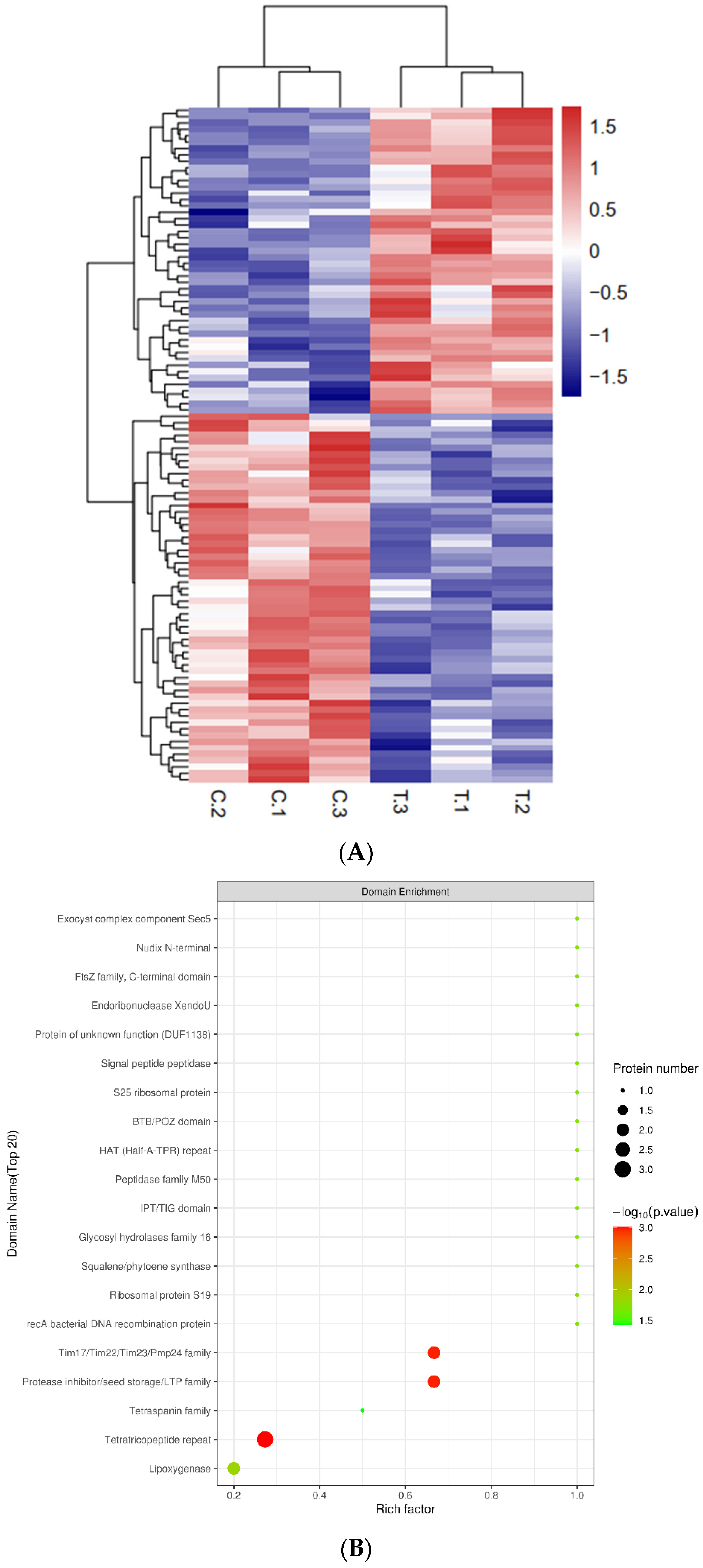
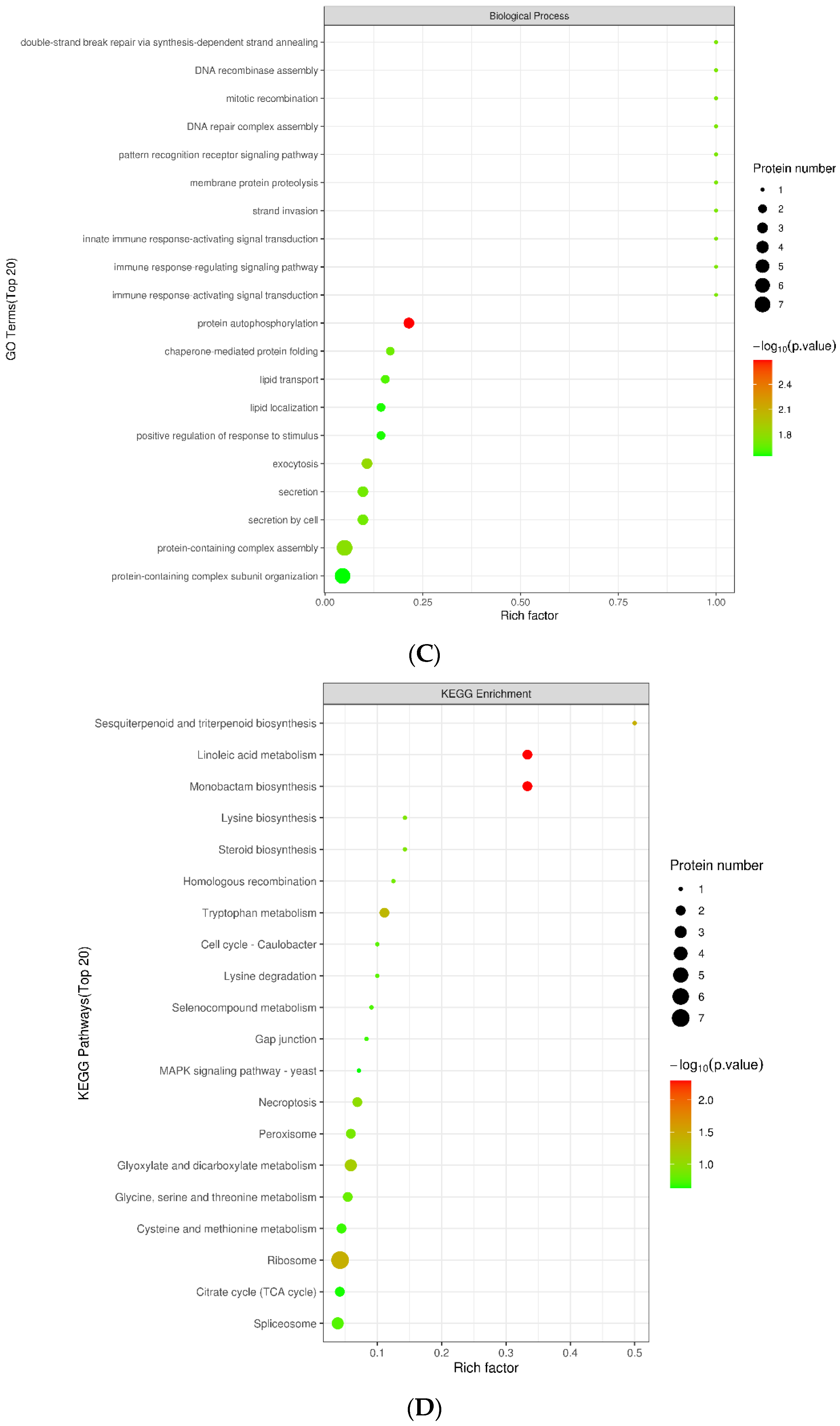
To identify the differently expressed proteins (DEPs) between healthy and infected qingke, we performed a TMT quantitative proteomic analyses. QC results showed that mass deviations of peptides less than 10 ppm, MASCOT score of 72.11% peptides above 20 and abundance ratios between treat and control were close to 1. These indicated the reliability of the pathogen responsive proteome (Figure S4). A total of 5611 proteins were identified, and 5608 of them were quantified, including 58 up-regulated and 48 down-regulated proteins (>1.2 fold change, p value < 0.05) that control and treat could be clearly distinguished (Table S3, Figure 2A). Three reported stress-related proteins had been detected based on BlastGO, including ABC1-LIKE KINASE 3 (ABC1K3), RING DOMAIN LIGASE 4 (RGLG4), and AVRPPHB SUSCEPTIBLE 1 (PBS1). ABC1K3 and RGLG4 are reported to be involved in the jasmonic acid pathway [19], while PBS1 is reported toconfers resistance to potyvirus infection in Arabidopsis and soybean [20].
To further understand the functions of DEPs, multiple function analysis was applied for proteomic research. Subcellular location analysis showed that DEPs were mostly located in cytoplasm (42) and nucleus (30) (Figure S5). Domain enrichment analysis showed that tetratricopeptide repeat (TPR), protease inhibitor/seed storage/LTP family and Tim17/Tim22/Tim23/Pmp24 family were the top three significant enrichment domains (Figure 2B). Among them, TPR proteins of pathogens have been reported to be related to virulence-associated functions [21]. Protease inhibitor also plays important roles in plant–pathogen interactions [22]. GO classification showed that metabolic process is the main biological process (BP)-related GO term of DEPs, and catalytic activity and cell part/cell were the largest molecular function- and cell component-related GO terms, respectively (Figure S6). GO terms such as protein autophosphorylation, exocytosis and protein-containing complex assembly were significantly enriched in BP (Figure 2C). Meanwhile, cell cortex part and calmodulin-dependent protein kinase activity were the most significant GO terms in CCand MF, respectively (Figure S7).
In addition, KEGG analysis showed all DEPs were mainly distributed in ribosome, glyoxylate and dicarboxylate metabolism and spliceosome (Table S4). Monobactam biosynthesis, linoleic acid metabolism, sesquiterpenoid and triterpenoid biosynthesis, tryptophan metabolism, together with ribosome, were significantly enriched pathways (Figure 2D).
Protein-Protein Interaction (PPI) is a useful tool for understanding molecular system metabolism or signaling pathways. PPI result showed that ELF5A-3, AT5G09500 and ATP1 were nodes which had the most connections with others (Figure S8).
Figure 2. Expression profilling and enrichment of U. hordei responsive proteome. (A) expression heatmap of differently expressed proteins. The relative expression level of proteins is indicated by color. C: control, T: treat. Hypergeometric tests were performed based on the domain, GO, and KEGG annotation results of DEPs. (B) Domain enrichment of DEPs, (C) GO enrichment of DEPs in the biological process, (D) KEGG enrichment of DEPs. Abscissa is a rich factor which is defined as the ratio of DEP number, and the number of genes has been annotated in this pathway; point size represents DEP number; p-value, shown by color from green to red.
Plants 12 00157 g002aPlants 12 00157 g002b

2.3. Transcriptome Analysis of Infected Qingke

High-throughput sequencing obtained 70.3 Gb clean data and 531,075,012 clean reads (99.14% of raw reads), average Q30 > 94%. A total of 262,166 transcripts and 153,979 unigenes were assembled. The N50 of transcript and unigenes were 2131 and 1220 bp, respectively. 56.21%, 23.33% and 18.72% of unigenes were annotated successfully in NR, Swiss-Prot and Pfam. A total of 79,565 and 6957 unigenes were annotated in GO and KEGG database. Cellular process and metabolism pathway had the largest number of unigenes in GO and KWGG pathway classification, respectively (Table S5).
After 82.86% of clean reads were mapped to the reference sequence assembled by Trinity, fragments Per Kilobase of exon model per Million mapped fragments (FPKM) were calculated to quantify the expression of genes. Overall, a total of 240 different expression genes (DEGs) were detected, including 230 down-regulated and 10 up-regulated genes in infected qingke (Figure 3A, Table S6). GO enrichment showed that DEGs enriched in various metabolic processes, including small molecule, lipid, and oxoacid metabolic process in BP. And results of enrichment in MF indicated binding function was the most common GO term of DEGs (Figure 3B, Table S7). In the KEGG enrichment results, fatty acid degradation was the most significantly enriched pathway, whereas amino acid metabolism-related pathways were the largest enriched term in results indicating primary metabolic pathway, especially amino acid metabolism, was critical for resistance (Figure 3C, Table S8). Moreover, two bZIP transcript factors which reported to be involved in biotic stress were detected in DEGs.

2.4. Co-Analysis of Multi Omics

We obtained metabolome, proteome, and transcriptome data between healthy and infected qingke and checked whether they had overlap within GO and KEGG pathways. Results showed that there were 13 overlapped in GO enrichment of proteome and transcriptome, including multiple protein related and lipid related GO terms, secretion and exocytosis in BP, protein binding in MF, cell periphery in CC (Table 1). For KEGG pathways, most pathways of amino acid metabolism and carbohydrate metabolism were overlapped; Cysteine and methionine metabolism, glycine, serine and threonine metabolism, valine, leucine and isoleucine degradation and tryptophan metabolism. Glutathione metabolism in amino acid metabolism, alpha-linolenic acid metabolism in lipid metabolism, drug metabolism—other enzymes in Xenobiotics biodegradation and metabolism were all detected in metabolome, proteome and transcriptome (Table 2). Plant hormone signal transduction, cytochrome P450 and others that related with assistance also co-occurred in two omics at least. These overlapped GOs and pathways may take important roles in defense response of qingke during the infection.

3. Discussion

In the process of interaction with pathogens, plants activate their own immunity by responding to the invasion of pathogens and adopt a variety of defense mechanisms to resist the invasion of pests and diseases. In this study, we used multiple omics technology to detected biological changes between health and U. hordei infected qingke.
In metabolome, 1856 classes of metabolites were detected. It was found that 187 were significantly different metabolites and organic acids and derivatives, and lipid and lipid-like molecules were the top two function classes of these metabolites. Lipids have significant influences on pathogenesis and could be used as a resistance mechanism during plant–microbe interactions [23]. Literature reported that plant lipid metabolism is the target of pathogens secreted toxins [24]. Plants could adapt defense mechanisms against pathogens using different compounds such as lipopolysaccharides, sphingolipids, and lipid-binding proteins [25]. Regulatory lipids are lipids that mediate of signaling and regulatory cascades and have an effective function at low concentrations [26]. These classes of lipids include polyunsaturated fatty acid derivatives like oxylipins, eicosanoids and jasmonic acid [27,28]. In this study, it was found that jasmonic acid (JA) was significantly up-regulated by the infection of pathogen. JA could crosstalk with other phytohormone including abscisic acid (ABA), ethylene (ET) and salicylic acid (SA) to regulate plant defense against pathogen [29,30,31,32]. For example, JA-mediated defense responses are raised against fungal pathogens like Botrytis cinerea. They have been demonstrated to play a role in the defense against some hemibiotrophic pathogens, such as Xanthomonas oryzae [33]. Another detected hormone in this study was Gibberellin (GA), which is essential for multiple developmental processes in plants. JA and GA signaling pathways could regulate plant growth and defense response antagonistically that JA defense over growth by interfering with gibberellin signaling cascade [34]. Riboflavin and ascorbic acid, two kinds of vitamin, were also detected in significantly different metabolites identified between heathy and infected qingke. Riboflavin has been reported to protect or induce resistance against various pathogens in different plants such as Arabidopsis, tobacco, rice, and soybean [12]. Riboflavin treatment could up-regulate multiple host–defense responses in several plants. A rapid H2O2 accumulation as a critical step in riboflavin signal transduction during P. syringae pv. Tomato DC3000 infected Arabidopsis [35]. Moreover, riboflavin could induce expression of genes such as NPR1 that control systemic acquired resistance [36]. In addition, riboflavin is implicated callose deposition which callose act as physical barrier against pathogen [37,38]. Ascorbic acid is the most abundant cellular antioxidant considered as a major antioxidant compound among the plant antioxidant-defense system [39]. The de novo biosynthesis of ascorbic acid could be stimulated by treatment with methy-ljasmonate in suspension cells of N. tabacum and A. thaliana [40]. In addition, ascorbic acid is a main precursor of oxalic acid (OA), which also was detected as significantly different metabolites in this study; OA was reported to be involves in the synthesis of H2O2 and take a critical role in plant-pathogen interaction [41]. N-acetyl-d-glucosamine, which is a basic constituent unit of chitin, showed significant increase in infected qingke. Chitin is an important component of fungal pathogenicity that could be recognized by plants and triggers various defense responses [42]. Moreover, chitin could induce ion efflux and ROS, increased levels of phytoalexins and hypersensitivity in infected cells [43,44]. These results indicated that qingke could elicit by chitin and against U. hordei through phytohormones such as JA and vitamins such as riboflavin and ascorbic acid.
For proteome, 106 significantly different proteins were identified. ABC1K3 and RGLG4, homologous genes of TR36261_c0_g2_ORF and TR2862_c2_g1_ORF, were reported to be involved in JA pathway. abc1k3 showed rapid chlorosis upon high light stress, and irreversible, senescence-like phenotype during drought stress and nitrogen limitation, plastid jasmonate biosynthesis enzymes were recruited to the abc1k3 plastoglobules but not wild-type [19]. RGLG4 has ubiquitin ligase activities and expression changes of RGLG4, and its homologous RGLG4 could affect JA-inductive gene expression. Both of them responded to methyl JA, P. syringae pv. tomato DC3000 and wounding in a COI1-dependent manner [45]. PBS1 is defense related gene that is a homologous gene of a significantly different protein, TR584_c2_g1_ORF. PBS1 could form a preactivation complex with RESISTANCE TO PSEUDOMONAS SYRINGAE 5 (RPS5) and triggers RPS5 activation upon AvrPphB-dependent cleavage [20]. In domain enrichment analysis, TPR and protease inhibitors were significantly enriched. TPR-containing proteins have been reported to play virulence-associated functions, such as the translocation of virulence factors into host cells, and the blocking of phagolysosomal maturation in bacterial pathogens [21]. Arabidopsis suppressor of rps4-RLD 1 (SRFR1), which encodes a conserved tetratricopeptide repeat protein, functions as a negative regulator and could enhance the resistance of bacterial effector AvrRps4 in srfr1 mutant [45]. Plant protease inhibitors (PI) were also found to take roles in plant immunity through regulation of endogenous plant proteases and inhibition of pathogen proteases [46,47]. PI from barley could act against proteases from Fusarium culmorum and the A. thaliana unusual serine protease inhibitor could defend against necrotrophic fungi Botrytis cinerea and Alternaria brassicicola [48]. On the other hand, pathogens may inhibit plant PI from reducing deleterious effects, such as U. maydis, which is a closely related species of U. hordei, could induce maize cystatin CC9 when infected to inhibit cysteine proteases [49]. These results favorably proved JA was important component in qingke resistance. It suggested TPR family genes and plant protease inhibitors may contribute for U. hordei defense. ELF5A-3, AT5G09500 and ATP1, the homologous of TR5765_c0_g1_ORF, TR4063_c0_g1_ORF_1 and TR41168_c0_g1_ORF, were the top 3 genes within PPI analysis. EIF5A-3, homologous of ELF5A-3, could regulate programmed cell death caused by the infection of Pseudomonas syringae pv. tomato DC3000 (Pst DC3000) [50]. AT5G09500 is a Ribosomal protein S19 family protein that showed expression change in response to Agrobacterium tumefaciens, suggested TR5765_c0_g1_ORF and TR4063_c0_g1_ORF_1 may play important roles in qingke.
We also processed transcriptome analyses. The 240 DEGs and GO, KEGG enrichment results were shown in Figure 3. For a more profound and more comprehensive understanding biological change within U. hordei infected qingke, we have counted the overlapping parts of GO and KEGG terms in the transcriptome, proteome, and metabolome (Table 1 and Table 2). In the overlapping part of KEGG, primary metabolism was the most abundant pathway, including amino acid metabolism, carbohydrate metabolism and lipid metabolism. Expression of genes involved in carbohydrate metabolism processes such as glycolysis, pentose phosphate pathway and TCA could be induced by pathogens or pathogen-derived elicitors, resulting in downstream defense responses, such as the generation of ROS and the activation of PR genes [7]. For example, AtHXK1, which phosphorylates glucose to glucose-6-phosphate, is a positive regulator of PR-1 and PR-5 [51]. Moreover, Nicotiana benthamiana downregulation of HXK1 expression caused by virus-induced gene silencing could increase the accumulation of H2O2 and expression of transcripts associated with defence responses result in PCD [52]. Overexpression of the Pyruvate decarboxylase (PDC) gene could increase callose deposition and expression of PR genes and impair pathogen spread upon inoculation with P. infestans [53]. Amino acid metabolism is another primary metabolism that has much overlapping terms. Some omics papers also reveal amino acid metabolism changes in result, such as valine, leucine, and tyrosine [54]. For example, lht1 (lysine histidine transporter 1) of Arabidopsis has reduced contents of glutamine, alanine, and proline compared with wild-type, shows enhanced resistance to diverse bacterial, fungal and oomycete pathogens and exhibited increased callose deposition, higher accumulation of salicylic acid (SA) and constitutive expression of PR-1 [55]. Levels of aspartic acid, threonine and valine showed a decrease and the level of tryptophan showed an increase in our data. Oleic acid, which is classed in fatty acid biosynthesis of lipid metabolism, was detected in metabolome and one gene involved in β-oxidation was detected in the transcriptome. Suppressor of SA-insensitivity (SSI2/FAB2), which catalyzes the desaturation of the stearic acid to oleic acid, its Arabidopsis ssi2 mutant showed higher expression of the resistance and exhibited spontaneous lesion formation associated with high levels of SA [56]. In addition, linolenic acid was detected in alpha-linolenic acid metabolism of the metabolome. LA was shown to activate the O2−-generating enzyme NADPH oxidase. O2− is required for HR in fad7/fad8 double mutant which was unable to suppress the P. syringae pv. tomato DC3000 because of insufficient LA [57]. Both proteins and genes involved in LA participated in the JA biosynthesis pathway.
LA is one precursor of JA [34]. TR64995_c0_g3 homologous, ACYL-COA OXIDASE 1 (ACX1), encoded a medium to long-chain acyl-CoA oxidase and was involved in jasmonate biosynthesis; its expression could be induced by abscisic acid, jasmonate and abiotic stress [58,59]. Lipoxygenase 2.1 is blast result of TR3086_c0_g2_ORF, which exhibits linoleate 13-lipoxygenase and arachidonate 15-lipoxygenase activity in barely, could be induced by exogenous application of jasmonic acid methyl [60]. In addition, cytochrome P450 related pathways had overlapped in proteome and transcriptome. Cytochrome P450s (CYPs) are an oxidoreductases class of enzyme and catalyze NADPH/O2-dependent hydroxylation [61]. CYPs could protect plants from harsh environmental conditions, including abiotic and biotic stress, by enhancing antioxidant activity [62]. These suggested that primary metabolism, including carbohydrate, amino acid and lipid, were largely changed in infected qingke and these metabolisms may take important roles against U. hordei.
Overall, the metabolomic results indicated that qingke might elicit resistance to U. hordei through chitin and some phytohormones, such as JA, riboflavin, and ascorbic acid. Proteomic data favorably proved JA was an important component in qingke resistance and suggested TPR family genes and plant protease inhibitors may contribute for U. hordei defense. Expressions of TR5765_c0_g1_ORF, TR4063_c0_g1_ORF_1, and TR41168_c0_g1_ORF were changed and may take important roles against U. hordei. Transcriptome analysis showed that primary metabolism including carbohydrate, amino acid, and lipid were largely changed in infected qingke, and these metabolisms may take important roles in U. hordei defense.

4. Materials and Methods

4.1. Sample Collection

Diseased and healthy tibetan barley (Hordeum vulgare L., qingke) seeds were cultured in the experimental field of Agriculture Research Institute, Tibet Academy of Agriculture and Animal Husbandry Sciences, China. Leaves of health qingke and U. hordei infected qingke were sampled as control and treated group during five-leaf stage, respectively. All samples were frozen immediately in liquid nitrogen and stored at −80 °C. Metabolome analysis had 12 biological repeats, proteome and transcriptome had three biological repeats, respectively.

4.2. RNA Extraction and Sequencing

Total RNA was extracted using TRIzol® reagents (Invitrogen, Carlsbad, CA, USA) and qualified by 1% agarose gel electrophoresis. Then the RNA quaity and purity were assessed using Nanodrop (ThermoFisher, Waltham, MA, USA) and Bioanalyzer (Agilent 2100, Santa Clara, CA, USA). The RNA samples met all the quality standards (A260/A280 = 1.8–2.2, and RNA integrity number (RIN) > 6.5) were used for cDNA library construction. Libraries were synthesized using TruSeq RNA Library Preparation Kit (Illumina, San Diego, CA, USA) and paired-end (PE) sequenced by Hiseq 4000 (Illumina, San Diego, CA, USA).

4.3. Protein Extraction and Digestion

SDT buffer (4% SDS, 100 mM Tris-HCl, 1 mM DTT, H7.6) was used for sample lysis and protein extraction. The BCA Protein Assay Kit (Bio-Rad, Hercules, CA, USA) was used for the quantification of protein content. Protein was digested based on filter-aided sample preparation (FASP) and desalted on C18 Cartridges (Empore™ SPE Cartridges C18 (standard density), then concentrated by vacuum centrifugation and reconstituted in 40 µL of 0.1% (v/v) formic acid. A total of 100 μg peptide mixture of each sample was labeled using iTRAQ reagent according to the manufacturer’s instructions (Applied Biosystems, Waltham, MA, USA).

4.4. LC-MS/MS Analysis

For metabolomics, UHPLC (1290 Infinity LC, Agilent Technologies, Santa Clara, CA, USA) coupled to a quadrupole time-of-flight (AB Sciex TripleTOF 6600) were used. 2.1 mm × 100 mm ACQUIY UPLC BEH 1.7 µm column (waters, Ireland) was used for sample separation. After separation, both electrospray ionization (ESI) positive and negative modes were applied. In order to avoid the influence caused by the fluctuation of the instrument detection signal, a random order was used for continuous analysis of the sample. For ESI source conditions were set as Ion Source Gas1 = 60, Ion Source Gas2 = 60, curtain gas = 30, source temperature: 600 °C, IonSpray Voltage Floating (ISVF) ± 5500 V.
Proteomics was analyzed on a Q Exactive mass spectrometer (Thermo Scientific) that was coupled to Easy nLC (Proxeon Biosystems, now Thermo Fisher Scientific) for 60 min. Reverse phase trap column (Thermo Scientific Acclaim PepMap100, 100 μm × 2 cm, nanoViper C18) that contains peptides connected to the C18-reversed phase analytical column (Thermo Scientific Easy Column, 10 cm long, 75 μm inner diameter, 3 μm resin) in buffer A (0.1% Formic acid) and separated with buffer B (84% acetonitrile and 0.1% formic acid) at a flow rate of 300 nL/min. MS set to positive mode and using data-dependent top10 method to choose the most abundant precursor ions from the survey scan (300–1800 m/z).

4.5. Metabolome Data Processing

The raw MS data (wiff.scan files) were converted to MzXML files by ProteoWizard MSConvert and imported into XCMS software (Scripps Research, La Jolla, CA, USA). CAMERA (Collection of Algorithms of MEtabolite pRofile Annotation) was used for the annotation of isotopes and adducts. Variables having more than 50% of the nonzero measurement values in at least one group were submitted for the following analyses. Compound identification was performed by comparing accuracy m/z value (<25 ppm). Orthogonal partial least-squares discriminant analysis (OPLS-DA) [63] were performed by R package (ropls). The variable importance in the projection (VIP) value of each variable in the OPLS-DA model was calculated, VIP > 1 and p value < 0.05 were used to screen significant changed metabolites.

4.6. Proteome Data Processing

The MS raw data were searched using the MASCOT engine (Matrix Science, London, UK; version 2.2) embedded into Proteome Discoverer 1.4 software (Thermo Fisher Scientific, Waltham, MA, USA) for identification and quantitation analyses. The protein ratios are calculated as the median of only unique peptides of the protein. Protein with Fold Change (FC) > 1.2 or <0.83 and p value < 0.05 were considered as differentially expressed proteins.

4.7. Transcriptome Data Processing

The clean data were assembled by Trinity after quality control. Unigenes were annotated in NR, Swissprot, PFAM, GO, and KO by NCBI blast 2.6.0, Blast2GO v2.5 and KAAS. Clean reads were mapped to transcripts that assembled by Trinity as a reference and expected number of fragments per kilobase of transcript sequence per millions base pairs sequenced (FPKM) calculated by featureCounts were used for analysis. DEseq2 [64] was used for identification of different expression genes (log2Fold Change (FC) > |1|, q value < 0.05).

4.8. Cluster Analysis

Cluster 3.0 http://bonsai.hgc.jp/~mdehoon/software/cluster/software.htm (accessed on 12 November 2021) and Java Treeview 1.1.1 (Oracle, Redwood Shore, CA, USA) [65] were employed for hierarchical clustering analysis based on Euclidean distance algorithm.

4.9. Subcellular Localization and Function Annotation

CELLO [66] was used to predict protein subcellular localization. Protein domain was identified by InterProScan software. For differentially expressed proteins, NCBI BLAST+ client software (ncbi-blast-2.2.28+-win32.exe) and InterProScan were used to find homologue sequences, then gene ontology (GO) terms were annotated by Blast2GO which also used for annotation of DEGs in transcriptome. The studied proteins and transcripts were blasted against the online Kyoto Encyclopedia of Genes and Genomes (KEGG) database http://geneontology.org/ (accessed on 23 November 2021) to retrieve KEGG terms. Enrichment analysis was applied based on Fisher’s exact test, and Benjamini–Hochberg correction for multiple testing was further applied to adjust derived p-values. Moreover, only functional categories and pathways with p-values under a threshold of 0.05 were considered significant in metabolomic and proteomic, but q-value in the transcriptome. The protein–protein interaction (PPI) information was searched from IntAct molecular interaction database http://www.ebi.ac.uk/intact/ (accessed on 6 December 2021) and visualized by Cytoscape [67].

5. Conclusions

In summary, we used multiple omics methods to analyze biological changes in transcript, protein and metabolism levels between healthy gingke and U. hordei infected qingke. A model of defense responses of qingke to U. hordei was shown in Figure 4. During U. hordei infection, qingke may recognize chitin to trigger cascades of activities, including the changes in primary metabolism and JA-related and vitamin-related pathways, then induce downstream defense responses such as ROS and PCD against U. hordei, whereas U. hordei could repress response compounds, such as JA and ascorbic acid that were downregulated in our data, to ensure spread. Our study revealed a comprehensive omics profile of qingke infected by U. hordei and provided a resource for further research of covered smut.

Supplementary Materials

The following supporting information can be downloaded at: https://www.mdpi.com/article/10.3390/plants12010157/s1, Figure S1. (A) and (B) are total ion chromatograms of positive ion model and negative ion model, respectively; (C) and (D) are PCA analysis plots of positive ion model and negative ion model, respectively; Figure S2: Fold change plot of significantly different metabolites. (A) positive ion model; (B) negative ion model; up-regulation and down-regulation are shown by red and green. The different colored words represent different superclasses of metabolites; Figure S3: Differential abundance score. The x-axis is DA score, which represents total changes for all metabolites in a metabolic pathway; plus means up-regulation and minus means down-regulation. The length of the line represents the absolute Da score, and the dot size of the end point of the line represents the number of metabolites in that pathway. Shades of color of point are proportional to DA score; Figure S4: QC of proteome. (A) Mass error distribution. (B) Ion score distribution. (C) Ratio distribution between treat and control; Figure S5: Subcellular localization statistic of DEPs. Number represents number of DEPs; Figure S6: GO statistic of DEPs. Biological process, molecular function and cellular component were distinguished by different colors; Figure S7: GO enrichment analysis of proteome. (A) Molecular function; (B) cell component. Abscissa is rich factor defined as ratio of DEP number, and the number of genes was annotated in this pathway; point size represents DEP number; p value shown by color from green to red; Figure S8: Protein–protein interaction network. Circle nodes indicate differentially expressed proteins, and lines indicate protein–protein interactions. Red indicates up-regulated protein, and blue is down-regulated protein. Circle size indicates the degree of protein connectivity. Table S1: List of metabonomics significant differences; Table S2: Annotation and enrichment list of metabolite kegg; Table S3: List of significant differences in proteomics; Table S4: Annotation and enrichment list of protein kegg; Table S5: Qualitative and quantitative list of transcriptomics; Table S6: List of significant difference gene annotations; Table S7: GO annotation list of significant difference genes; Table S8: KEGG annotation list of significant difference genes.

Author Contributions

Conceptualization, J.L. and T.W.; methodology, J.L. and J.Z.; software, J.L.; validation, J.L., T.W. and P.L. (Pei Liu); formal analysis, J.Z. and P.L. (Pu Li); investigation, J.Z. and Y.C.; resources, J.L. and P.L. (Pu Li); data curation, J.L. and T.W.; writing—original draft preparation, J.L.; writing—review and editing, J.Z. and X.Y.; visualization, H.L.; supervision, X.Y.; project administration, X.Y.; funding acquisition, X.Y. All authors have read and agreed to the published version of the manuscript.

Funding

This work was supported by the Science and Technology Project of Tibet Autonomous Region, China (project No. XZ-2019-NK-NS-001) and Major Science and Technology Project of Tibet Autonomous Region: Research on Key technologies of highland barley Germplasm Creation, quality and efficiency improvement (project No. XZ202101ZD004N).

Data Availability Statement

Not applicable.

Conflicts of Interest

The authors declare no conflict of interest.

References

  1. Singh, J.; Kaur, A.; Sharma, V.K. Evaluation of barley genotypes for resistance against covered smut disease. Indian Phytopathol. 2020, 73, 359–360. [Google Scholar] [CrossRef]
  2. Gangwar, O.P.; Bhardwaj, S.C.; Singh, G.; Prasad, P.; Kumar, S. Barley diseases and their management: An Indian perspective. Wheat Barley Res. 2018, 10, 138–150. [Google Scholar] [CrossRef] [Green Version]
  3. Mysore, K.S.; Ryu, C.-M. Nonhost resistance: How much do we know? Trends Plant Sci. 2004, 9, 97–104. [Google Scholar] [CrossRef] [PubMed]
  4. Muthappa, S.-K.; Mysore, K. Nonhost Resistance Against Bacterial Pathogens: Retrospectives and Prospects. Annu. Rev. Phytopathol. 2013, 51, 10–1146. [Google Scholar] [CrossRef]
  5. Boller, T.; He, S.Y. Innate Immunity in Plants: An Arms Race Between Pattern Recognition Receptors in Plants and Effectors in Microbial Pathogens. Science 2009, 324, 742–744. [Google Scholar] [CrossRef] [Green Version]
  6. Bonardi, V.; Dangl, J. How complex are intracellular immune receptor signaling complexes? Front. Plant Sci. 2012, 3, 237. [Google Scholar] [CrossRef] [PubMed] [Green Version]
  7. Rojas, C.; Muthappa, S.-K.; Tzin, V.; Mysore, K. Regulation of primary metabolism during plant-pathogen interactions and its contribution to plant defense. Front. Plant Sci. 2014, 5, 17. [Google Scholar] [CrossRef] [PubMed] [Green Version]
  8. Wildermuth, M.C.; Dewdney, J.; Wu, G.; Ausubel, F.M. Isochorismate synthase is required to synthesize salicylic acid for plant defence. Nature 2001, 414, 562–565. [Google Scholar] [CrossRef]
  9. Robert-Seilaniantz, A.; Grant, M.; Jones, J.D. Hormone crosstalk in plant disease and defense: More than just jasmonate-salicylate antagonism. Annu. Rev. Phytopathol. 2011, 49, 317–343. [Google Scholar] [CrossRef] [PubMed]
  10. De Vleesschauwer, D.; Gheysen, G.; Höfte, M. Hormone defense networking in rice: Tales from a different world. Trends Plant Sci. 2013, 18, 555–565. [Google Scholar] [CrossRef]
  11. Koornneef, A.; Pieterse, C. Cross Talk in Defense Signaling. Plant Physiol. 2008, 146, 839–844. [Google Scholar] [CrossRef] [PubMed] [Green Version]
  12. Boubakri, H.; Gargouri, M.; Mliki, A.; Brini, F.; Chong, J.; Jbara, M. Vitamins for enhancing plant resistance. Planta 2016, 244, 529–543. [Google Scholar] [CrossRef] [PubMed]
  13. Bolton, M.D. Primary metabolism and plant defense—Fuel for the fire. Mol. Plant-Microbe Interact. 2009, 22, 487–497. [Google Scholar] [CrossRef] [PubMed] [Green Version]
  14. Heil, M.; Baldwin, I.T. Fitness costs of induced resistance: Emerging experimental support for a slippery concept. Trends Plant Sci. 2002, 7, 61–67. [Google Scholar] [CrossRef]
  15. Zaynab, M.; Fatima, M.; Sharif, Y.; Zafar, M.H.; Ali, H.; Khan, K.A. Role of primary metabolites in plant defense against pathogens. Microb. Pathog. 2019, 137, 103728. [Google Scholar] [CrossRef] [PubMed]
  16. Yoshida, S.; Ito, M.; Nishida, I.; Watanabe, A. Identification of a novel gene HYS1/CPR5 that has a repressive role in the induction of leaf senescence and pathogen-defence responses in Arabidopsis thaliana. Plant J. Cell Mol. Biol. 2002, 29, 427–437. [Google Scholar] [CrossRef] [Green Version]
  17. Boch, J.; Verbsky, M.; Robertson, T.; Larkin, J.; Kunkel, B. Analysis of Resistance Gene-Mediated Defense Responses in Arabidopsis thaliana Plants Carrying a Mutation in CPR5. Mol. Plant-Microbe Interact. 1998, 11, 1196–1206. [Google Scholar] [CrossRef] [Green Version]
  18. Mur, L.A.J.; Kenton, P.; Atzorn, R.; Miersch, O.; Wasternack, C. The outcomes of concentration-specific interactions between salicylate and jasmonate signaling include synergy, antagonism and oxidative stress leading to cell death. Plant Physiol. 2006, 140, 249–262. [Google Scholar] [CrossRef] [Green Version]
  19. Lundquist, P.; Poliakov, A.; Giacomelli, L.; Friso, G.; Appel, M.; McQuinn, R.; Krasnoff, S.; Rowland, E.; Ponnala, L.; Sun, Q.; et al. Loss of Plastoglobule Kinases ABC1K1 and ABC1K3 Causes Conditional Degreening, Modified Prenyl-Lipids, and Recruitment of the Jasmonic Acid Pathway. Plant Cell 2013, 25, 1818–1839. [Google Scholar] [CrossRef] [Green Version]
  20. Pottinger, S.; Bak, A.; Margets, A.; Helm, M.; Tang, L.; Casteel, C.; Innes, R. Optimizing the PBS1 Decoy System to Confer Resistance to Potyvirus Infection in Arabidopsis and Soybean. Mol. Plant-Microbe Interact. 2020, 33, 932–944. [Google Scholar] [CrossRef]
  21. Cerveny, L.; Straskova, A.; Dankova, V.; Härtlova, A.; Ceckova, M.; Staud, F.; Stulík, J. Tetratricopeptide Repeat Motifs in the World of Bacterial Pathogens: Role in Virulence Mechanisms. Infect. Immun. 2012, 81, 629–635. [Google Scholar] [CrossRef] [Green Version]
  22. Karimi Jashni, M.; Mehrabi, R.; Collemare, J.; Mesarich, C.; De Wit, P. The battle in the apoplast: Further insights into the roles of proteases and their inhibitors in plant–pathogen interactions. Front. Plant Sci. 2015, 6, 584. [Google Scholar] [CrossRef] [Green Version]
  23. Christensen, S.A.; Kolomiets, M.V. The lipid language of plant–fungal interactions. Fungal Genet. Biol. 2011, 48, 4–14. [Google Scholar] [CrossRef]
  24. Ding, Z. Lipid metabolism disorders contribute to the pathogenesis of Hepatospora eriocheir in the crab Eriocheir sinensis. J. Fish Dis. 2021, 44, 305–313. [Google Scholar] [CrossRef] [PubMed]
  25. Zeier, J. New insights into the regulation of plant immunity by amino acid metabolic pathways. Plant Cell Environ. 2013, 36, 2085–2103. [Google Scholar] [CrossRef] [PubMed]
  26. Zhang, H.; Zhang, X.; Mao, B.; Li, Q.; He, Z. Alpha-picolinic acid, a fungal toxin and mammal apoptosis-inducing agent, elicits hypersensitive-like response and enhances disease resistance in rice. Cell Res. 2004, 14, 27–33. [Google Scholar] [CrossRef] [Green Version]
  27. Ingólfsson, H.; Melo, M.; van Eerden, F.; Arnarez, C.; López, C.; Wassenaar, T.A.; Periole, X.; Vries, A.; Tieleman, D.; Marrink, S. Lipid Organization of the Plasma Membrane. J. Am. Chem. Soc. 2014, 136, 14554–14559. [Google Scholar] [CrossRef]
  28. Wältermann, M.; Steinbüchel, A. Neutral Lipid Bodies in Prokaryotes: Recent Insights into Structure, Formation, and Relationship to Eukaryotic Lipid Depots. J. Bacteriol. 2005, 187, 3607–3619. [Google Scholar] [CrossRef] [Green Version]
  29. Shiji, H.; Kenichi, T. Salicylic acid and jasmonic acid crosstalk in plant immunity. Essays Biochem. 2022, 66, 647–656. [Google Scholar] [CrossRef]
  30. Ku, Y.S.; Sintaha, M.; Cheung, M.Y.; Lam, H.M. Plant hormone signaling crosstalks between biotic and abiotic stress responses. Int. J. Mol. Sci. 2018, 19, 3206. [Google Scholar] [CrossRef]
  31. Li, N.; Han, X.; Feng, D.; Yuan, D.; Huang, L.J. Signaling crosstalk between salicylic acid and ethylene/jasmonate in plant defense: Do we understand what they are whispering? Int. J. Mol. Sci. 2021, 20, 671. [Google Scholar] [CrossRef] [PubMed] [Green Version]
  32. Liechti, R.; Farmer, E. The Jasmonate Biochemical Pathway. Sci. STKE Signal Transduct. Knowl. Environ. 2003, 2003, CM18. [Google Scholar] [CrossRef] [PubMed]
  33. Liu, C.; Chen, L.; Zhao, R.; Rui, L.; Zhang, S.; Yu, W.; Sheng, J.; Shen, L. Melatonin Induces Disease Resistance to Botrytis cinerea in Tomato Fruit by Activating Jasmonic Acid Signaling Pathway. J. Agric. Food Chem. 2019, 67, 6116–6124. [Google Scholar] [CrossRef] [PubMed]
  34. Wasternack, C.; Hause, B. Jasmonates: Biosynthesis, Perception, Signal Transduction and Action in Plant Stress Response, Growth and Development. An Update to the 2007 Review in Annals of Botany. Ann. Bot. 2013, 111, 1021–1058. [Google Scholar] [CrossRef] [PubMed]
  35. Azami-Sardooei, Z.; França, S.C.; De Vleesschauwer, D.; Höfte, M. Riboflavin induces resistance against Botrytis cinerea in bean, but not in tomato, by priming for a hydrogen peroxide-fueled resistance response. Physiol. Mol. Plant Pathol. 2010, 75, 23–29. [Google Scholar] [CrossRef]
  36. Bowling, S.A.; Clarke, J.D.; Liu, Y.; Klessig, D.F.; Dong, X. The cpr5 mutant of Arabidopsis expresses both NPR1-dependent and NPR1-independent resistance. Plant Cell 1997, 9, 1573–1584. [Google Scholar]
  37. Zhang, S.; Yang, X.; Sun, M.; Sun, F.; Deng, S.; Dong, H. Riboflavin-induced Priming for Pathogen Defense in Arabidopsis thaliana. J. Integr. Plant Biol. 2009, 51, 167–174. [Google Scholar] [CrossRef]
  38. Zhou, J.; Sun, A.; Xing, D. Modulation of cellular redox status by thiamine-activated NADPH oxidase confers Arabidopsis resistance to Sclerotinia sclerotiorum. J. Exp. Bot. 2013, 64, 3261–3272. [Google Scholar] [CrossRef]
  39. Turner, J.G.; Ellis, C.; Devoto, A. The jasmonate signal pathway. Plant Cell 2002, 14, S153–S164. [Google Scholar] [CrossRef] [Green Version]
  40. Wolucka, B.; Goossens, A.; Inze, D. Methyl jasmonate stimulates the de novo biosynthesis of vitamin C in plant cell suspensions. J. Exp. Bot. 2005, 56, 2527–2538. [Google Scholar] [CrossRef] [Green Version]
  41. Dias, C.V.; Mendes, J.S.; dos Santos, A.C.; Pirovani, C.P.; da Silva Gesteira, A.; Micheli, F.; Gramacho, K.P.; Hammerstone, J.; Mazzafera, P.; de Mattos Cascardo, J.C. Hydrogen peroxide formation in cacao tissues infected by the hemibiotrophic fungus Moniliophthora perniciosa. Plant Physiol. Biochem. 2011, 49, 917–922. [Google Scholar] [CrossRef] [PubMed]
  42. Yu, J.; Han, J.; Kim, Y.-J.; Song, M.; Yang, Z.; He, Y.; Fu, R.; Luo, Z.; Hu, J.; Liang, W.; et al. Two rice receptor-like kinases maintain male fertility under changing temperatures. Proc. Natl. Acad. Sci. USA 2017, 114, 12327–12332. [Google Scholar] [CrossRef] [PubMed] [Green Version]
  43. Malik, A.; Kumar, I.S.; Nadarajah, K. Elicitor and Receptor Molecules: Orchestrators of Plant Defense and Immunity. Int. J. Mol. Sci. 2020, 21, 963. [Google Scholar] [CrossRef] [PubMed] [Green Version]
  44. Felix, G.; Regenass, M.; Boller, T. Specific perception of subnanomolar concentrations of chitin fragments by tomato cells: Induction of extracellular alkalinization, changes in protein phosphorylation, and establishment of a refractory state. Plant J. 2002, 4, 307–316. [Google Scholar] [CrossRef]
  45. Kwon, S.I.; Kim, S.H.; Bhattacharjee, S.; Noh, J.-J.; Gassmann, W. SRFR1, a suppressor of effector-triggered immunity, encodes a conserved tetratricopeptide repeat protein with similarity to transcriptional repressors. Plant J. Cell Mol. Biol. 2009, 57, 109–119. [Google Scholar] [CrossRef]
  46. Kim, J.-Y.; Park, S.-C.; Hwang, I.; Cheong, H.; Nah, J.-W.; Hahm, K.-S.; Park, Y. Protease Inhibitors from Plants with Antimicrobial Activity. Int. J. Mol. Sci. 2009, 10, 2860–2872. [Google Scholar] [CrossRef] [Green Version]
  47. Valueva, T.; Mosolov, V. Role of inhibitors of proteolytic enzymes in plant defense against phytopathogenic microorganisms. Biochemistry 2004, 69, 1305–1309. [Google Scholar] [CrossRef]
  48. Laluk, K.; Mengiste, T. The Arabidopsis extracellular UNUSUAL SERINE PROTEASE INHIBITOR functions in resistance to necrotrophic fungi and insect herbivory. Plant J. Cell Mol. Biol. 2011, 68, 480–494. [Google Scholar] [CrossRef]
  49. van der Linde, K.; Hemetsberger, C.; Kastner, C.; Kaschani, F.; Hoorn, R.; Kumlehn, J.; Doehlemann, G. A Maize Cystatin Suppresses Host Immunity by Inhibiting Apoplastic Cysteine Proteases. Plant Cell 2012, 24, 1285–1300. [Google Scholar] [CrossRef] [Green Version]
  50. Hopkins, M.; Lampi, Y.; Wang, T.-W.; Liu, Z.; Thompson, J. Eukaryotic Translation Initiation Factor 5A Is Involved in Pathogen-Induced Cell Death and Development of Disease Symptoms in Arabidopsis. Plant Physiol. 2008, 148, 479–489. [Google Scholar] [CrossRef] [Green Version]
  51. Xiao, W.Y.; Sheen, J.; Jang, J. The role of hexokinase in plant sugar signal transduction and growth and development. Plant Mol. Biol. 2000, 44, 451–461. [Google Scholar] [CrossRef] [PubMed]
  52. Kim, M.; Lim, J.-H.; Ahn, C.; Park, K.; Kim, G.; Kim, W.T.; Pai, H.-S. Mitochondria-Associated Hexokinases Play a Role in the Control of Programmed Cell Death in Nicotiana benthamiana. Plant Cell 2006, 18, 2341–2355. [Google Scholar] [CrossRef] [PubMed] [Green Version]
  53. Tadege, M.; Bucher, M.; Stähli, W.; Suter, M.; Dupuis, I.; Kuhlemeier, C. Activation of plant defense responses and sugar efflux by expression of pyruvate decarboxylase in potato leaves. Plant J. 1998, 16, 661–671. [Google Scholar] [CrossRef] [Green Version]
  54. Scheideler, M.; Schlaich, N.L.; Fellenberg, K.; Beissbarth, T.; Hauser, N.C.; Vingron, M.; Slusarenko, A.J.; Hoheisel, J.D. Monitoring the Switch from Housekeeping to Pathogen Defense Metabolism in Arabidopsis thaliana Using cDNA Arrays. J. Biol. Chem. 2002, 277, 10555–10561. [Google Scholar] [CrossRef] [Green Version]
  55. Liu, G.; Ji, Y.; Bhuiyan, N.; Pilot, G.; Selvaraj, G.; Zou, J.; Wei, Y. Amino Acid Homeostasis Modulates Salicylic Acid-Associated Redox Status and Defense Responses in Arabidopsis. Plant Cell 2010, 22, 3845–3863. [Google Scholar] [CrossRef] [Green Version]
  56. Mandal, M.; Chandra-Shekara, A.C.; Jeong, R.-D.; Yu, K.; Zhu, S.; Chanda, B.; Navarre, R.; Kachroo, A.; Kachroo, P. Oleic acid-dependent modulation of NITRIC OXIDE ASSOCIATED1 protein levels regulates nitric oxide-mediated defense signaling in Arabidopsis. Plant Cell 2012, 24, 1654–1674. [Google Scholar] [CrossRef] [Green Version]
  57. Yaeno, T.; Matsuda, O.; Iba, K. Role of chloroplast trienoic fatty acids in plant disease defense responses. Plant J. Cell Mol. Biol. 2005, 40, 931–941. [Google Scholar] [CrossRef]
  58. Ebeed, H.; Stevenson, S.; Cuming, A.; Baker, A. Conserved and differential transcriptional responses of peroxisome associated pathways to drought, dehydration and ABA. J. Exp. Bot. 2018, 69, 4971–4985. [Google Scholar] [CrossRef]
  59. Yan, Y.; Stolz, S.; Chételat, A.; Reymond, P.; Pagni, M.; Dubugnon, L.; Farmer, E. A Downstream Mediator in the Growth Repression Limb of the Jasmonate Pathway. Plant Cell 2007, 19, 2470–2483. [Google Scholar] [CrossRef] [Green Version]
  60. Vörös, K.; Feussner, I.; Kühn, H.; Lee, J.; Graner, A.; Löbler, M.; Parthier, B.; Wasternack, C. Characterization of a methyljasmonate-inducible lipoxygenase from barley (Hordeum vulgare cv. Salome) leaves. Eur. J. Biochem. 2001, 251, 36–44. [Google Scholar] [CrossRef] [Green Version]
  61. Denisov, I.; Makris, T.; Sligar, S.; Schlichting, I. Structure and Chemistry of Cytochrome P450. Chem. Rev. 2005, 105, 2253–2277. [Google Scholar] [CrossRef] [PubMed]
  62. Pandian, B.A.; Sathishraj, R.; Maduraimuthu, D.; Prasad, P.V.V.; Jugulam, M. Role of Cytochrome P450 Enzymes in Plant Stress Response. Antioxidants 2020, 9, 454. [Google Scholar] [CrossRef] [PubMed]
  63. Boccard, J.; Rutledge, D.N. A consensus orthogonal partial least squares discriminant analysis (OPLS-DA) strategy for multiblock Omics data fusion. Anal. Chim. Acta 2013, 769, 30–39. [Google Scholar] [CrossRef] [PubMed]
  64. Love, M.; Huber, W.; Anders, S. Moderated Estimation of Fold Change and Dispersion for RNA-Seq Data with DESeq2. Genome Biol. 2014, 15, 550. [Google Scholar] [CrossRef] [PubMed] [Green Version]
  65. Saldanha, A. Java Treeview—Extensible visualization of microarray data. Bioinformatics 2004, 20, 3246–3248. [Google Scholar] [CrossRef] [Green Version]
  66. Yu, C.-S.; Lin, C.-J.; Hwang, J.-K. Predicting subcellular localization of proteins for Gram-negative bacteria by support vector machines based on n-peptide compositions. Protein Sci. 2004, 13, 1402–1406. [Google Scholar] [CrossRef] [Green Version]
  67. Shannon, P.; Markiel, A.; Ozier, O.; Baliga, N.; Wang, J.; Ramage, D.; Amin, N.; Schwikowski, B.; Ideker, T. Cytoscape: A Software Environment for Integrated Models of Biomolecular Interaction Networks. Genome Res. 2003, 13, 2498–2504. [Google Scholar] [CrossRef]
Figure 1. Annotation and enrichment analyses of U. hordei responsive metabolome. (A) statistic of identified metabolites. Different superclasses show in various colors; (B) statistic of annotated KEGG pathways of metabolites; (C) KEGG enrichment analysis, hypergeometric tests were performed based on the KEGG classification of significant different metabolites. Abscissa is a rich factor which is defined as the ratio of different metabolites number and the number of genes has been annotated in this pathway; point size represents metabolite number; p value, shown by color from green to red.
Plants 12 00157 g001
Figure 3. Expression profilling and enrichment of U. hordei responsive transcriptome. (A) volcano plot of different expression genes. Significantly up–regulated genes are shown by redpoint and down–regulated by green point; (B) GO enrichment of DEGs. Hypergeometric tests were performed based on the GO classification of DEGs. Ordinate is gene number, and p value of various GO terms are shown by color from orange to red. (C) KEGG enrichment of DEGs. Hypergeometric tests were performed based on the KEGG classification of DEGs. Abscissa is rich factor defined as ratio of DEG number and the number of genes has been annotated in this pathway; point size represents DEG number; p value, shown by color form green to red.
Plants 12 00157 g003aPlants 12 00157 g003b
Figure 4. Hypothesis model of qingke interacted with Ustilago hordei. Arrow indicated activation and horizontal line indicated repression, dotted line indicated need to further biological evidence in qingke.
Plants 12 00157 g004
Table 1. Statistics of overlapping GO term in proteomic and transcriptome.
GO IDTermCategory
GO: 0022607cellular component assemblyBP
GO: 0006887exocytosisBP
GO: 0010876lipid localizationBP
GO: 0006869lipid transportBP
GO: 0048584positive regulation of response to stimulusBP
GO: 0006468protein phosphorylationBP
GO: 0065003protein-containing complex assemblyBP
GO: 0043933protein-containing complex subunit organizationBP
GO: 0080134regulation of response to stressBP
GO: 1901700response to oxygen-containing compoundBP
GO: 0046903secretionBP
GO: 0032940secretion by cellBP
GO: 0071944cell peripheryCC
GO: 0005515protein bindingMF
The BP, CC, and MF are the abbreviations of biological process, cellular component, and molecular function, respectively.
Table 2. Statistics of overlapping KEGG pathways.
PathwayKEGG IDMetabolomicProteomicTranscriptome
Amino acid metabolism
Cysteine and methionine metabolismko00270YYY
Glycine, serine and threonine metabolismko00260YYY
Tryptophan metabolismko00380YYY
Valine, leucine and isoleucine degradationko00280YYY
Lysine degradationko00310 YY
Alanine, aspartate and glutamate metabolismko00250Y Y
Arginine biosynthesisko00220Y Y
Histidine metabolismko00340Y Y
Tyrosine metabolismko00350Y Y
Lysine biosynthesisko00300YY
Biosynthesis of other secondary metabolites
Monobactam biosynthesisko00261YY
Phenylpropanoid biosynthesisko00940Y Y
Carbohydrate metabolism
Amino sugar and nucleotide sugar metabolismko00520Y Y
Ascorbate and aldarate metabolismko00053Y Y
Galactose metabolismko00052Y Y
Glycolysis/Gluconeogenesisko00010 YY
Glyoxylate and dicarboxylate metabolismko00630 YY
Pentose and glucuronate interconversionsko00040Y Y
Propanoate metabolismko00640 YY
Pyruvate metabolismko00620 YY
Starch and sucrose metabolismko00500Y Y
Cell growth and death
Ferroptosisko04216 YY
Energy metabolism
Carbon fixation in photosynthetic organismsko00710YY
Methane metabolismko00680 YY
Environmental adaptation
Thermogenesisko04714Y Y
Global and overview maps
Biosynthesis of amino acidsko01230Y Y
Biosynthesis of secondary metabolitesko01110Y Y
Carbon metabolismko01200Y Y
Degradation of aromatic compoundsko01220Y Y
Metabolic pathwaysko01100Y Y
Microbial metabolism in diverse environments Y Y
Lipid metabolism
alpha-Linolenic acid metabolismko01120YYY
Biosynthesis of unsaturated fatty acidsko01040Y Y
Fatty acid biosynthesisko00592Y Y
Steroid biosynthesisko01040YY
Metabolism of cofactors and vitamins
Pantothenate and CoA biosynthesisko00770Y Y
Metabolism of other amino acids
beta-Alanine metabolismko00410Y Y
Glutathione metabolismko00480YYY
Metabolism of terpenoids and polyketides
Limonene and pinene degradationko00903Y Y
Nucleotide metabolism
Purine metabolismko00230YY
Pyrimidine metabolismko00240Y Y
Signal transduction
cAMP signaling pathwayko04024Y Y
cGMP-PKG signaling pathwayko04022YY
HIF-1 signaling pathwayko04066YY
Plant hormone signal transductionko04075Y Y
Transport and catabolism
Peroxisomeko04146 YY
Xenobiotics biodegradation and metabolism
Drug metabolism—cytochrome P450ko00982 YY
Drug metabolism—other enzymesko00983YYY
Metabolism of xenobiotics by cytochrome P450ko00980 YY
The Y is the abbreviation of “Yes”, means the pathways was annotated.
Disclaimer/Publisher’s Note: The statements, opinions and data contained in all publications are solely those of the individual author(s) and contributor(s) and not of MDPI and/or the editor(s). MDPI and/or the editor(s) disclaim responsibility for any injury to people or property resulting from any ideas, methods, instructions or products referred to in the content.