Article

Integrated Characterization of Cassava (Manihot esculenta) Pectin Methylesterase (MePME) Genes to Filter Candidate Gene Responses to Multiple Abiotic Stresses

1
College of Life Sciences, Hainan University, Haikou 570228, China
2
Institute of Tropical Bioscience and Biotechnology, Chinese Academy of Tropical Agricultural Sciences, Haikou 571101, China
3
Max Planck Institute of Molecular Plant Physiology, 14476 Potsdam-Golm, Germany
4
College of Chemical and Materials Engineering, Hainan Vocational University of Science and Technology, Haikou 571126, China
*
Authors to whom correspondence should be addressed.
These authors contributed equally to this work.
Plants 2023, 12(13), 2529; https://doi.org/10.3390/plants12132529
Submission received: 11 May 2023 / Revised: 29 June 2023 / Accepted: 30 June 2023 / Published: 3 July 2023

Abstract

:
Plant pectin methylesterases (PMEs) play crucial roles in regulating cell wall modification and response to various stresses. Members of the PME family have been found in several crops, but there is a lack of research into their presence in cassava (Manihot esculent), which is an important crop for world food security. In this research, 89 MePME genes were identified in cassava that were separated into two types (type-Ⅰ and type-Ⅱ) according to the existence or absence of a pro-region (PMEI domain). The MePME gene members were unevenly located on 17 chromosomes, with 19 gene pairs being identified that most likely arose via duplication events. The MePMEs could be divided into ten sub-groups in type-Ⅰ and five sub-groups in type-Ⅱ. The motif analysis revealed 11 conserved motifs in type-Ⅰ and 8 in type-Ⅱ MePMEs. The number of introns in the CDS region of type-Ⅰ MePMEs ranged between one and two, and the number of introns in type-Ⅱ MePMEs ranged between one and nine. There were 21 type-Ⅰ and 31 type-Ⅱ MePMEs that contained signal peptides. Most of the type-Ⅰ MePMEs had two conserved “RK/RLL” and one “FPSWVS” domain between the pro-region and the PME domain. Multiple stress-, hormone- and tissue-specific-related cis-acting regulatory elements were identified in the promoter regions of MePME genes. A total of five co-expressed genes (MePME1, MePME2, MePME27, MePME65 and MePME82) were filtered from different abiotic stresses via the use of UpSet Venn diagrams. The gene expression pattern analysis revealed that the expression of MePME1 was positively correlated with the degree of cassava postharvest physiological deterioration (PPD). The expression of this gene was also significantly upregulated by 7% PEG and 14 °C low-temperature stress, but slightly downregulated by ABA treatment. The tissue-specific expression analysis revealed that MePME1 and MePME65 generally displayed higher expression levels in most tissues than the other co-expressed genes. In this study, we obtain an in-depth understanding of the cassava PME gene family, suggesting that MePME1 could be a candidate gene associated with multiple abiotic tolerance.

1. Introduction

Pectin methylesterases (PMEs) are one kind of cell-wall-associated enzyme that have the ability to render the pectin demethylesterified [1]. The degree of methylesterification of pectin determines its functional properties, and affects the cell wall firmness, thereby regulating the plant cellular response to different vital activities, such as growth, development, senescence and response to (a)biotic stresses [2,3]. PME activity is, therefore, frequently measured as a proxy in evaluating plant resistance to different stress conditions. For example, PME activity in chilling-sensitive maize was reduced using low temperature treatment with the pectin content in the cell wall being simultaneously decreased [4]. During strawberry fruit ripening, PME activity also gradually decreased [5]. By contrast, during tomato fruit ripening, PME activity gradually increased [6]. On the exogenous application of PME combined with calcium chloride, the fruit softening of raspberry was delayed due to an extended maintenance of cell wall integrity [7]. Moreover, by increasing the PME activity, the phosphorus fixed in the cell wall was remobilized [8]. During lettuce seed germination, the PME activity was lower in the endosperm cap, but higher in the radicle [9]. When suffering aluminum (Al) stress, the PME activity in the root tip of an Al-sensitive rice variety was maintained at a higher level than that in a resistant variety [10]. PME activity is also induced when the plant suffers a pathogen attack [11]. In all cases, PME enzymatic activity is tightly regulated by PME inhibitor proteins (PMEIs) [3,12,13].
In seed plants, PMEs belong to a super-family that is present at different numbers in different species. According to existing reports, the number of PME family members in different plant species ranges from 41 in foxtail millet [14] to 121 in tobacco [15]. In addition, the number of PME genes reported in dicotyledonous plants is generally higher than that reported in monocotyledonous plants. For example, in the monocots rice [16] and foxtail millet [14], the PME numbers are under 50, whilst in the dicot plants potato [17], Arabidopsis [18], Populus euphratica, tobacco [15], flax [19], navel orange [20], tomato [21] and Brassica rapa [22], the numbers of PME are 54, 66, 80, 121, 105, 53, 79 and 110, respectively. The PMEs initially encode pro-proteins with pro-regions or signal peptides, which are crucial for protein orientation in the endoplasmic reticulum. The pro-proteins undergo a series of processing in the Golgi and are finally secreted to the cell wall in a mature state (with pro-region removed). According to the reported PME gene families of different plant species, the number of type-Ⅰ PMEs (with pro-region) is usually larger than that of type-Ⅱ PMEs (without pro-region). For example, there are 28 type-Ⅰ and 26 type-Ⅱ PMEs in potato, 43 type-Ⅰ and 23 type-Ⅱ PMEs in Arabidopsis, 36 type-Ⅰ and 35 type-Ⅱ PMEs in peach and 47 type-Ⅰ and 33 type-Ⅱ PMEs in cotton [17,18,23,24].
Some reports have shown that the expression of PME genes is affected by plant growth and development and stress. For example, AtPME41 in Arabidopsis [25] and nine CsPME genes in navel orange were induced by low temperature treatment [20]. In contrast, AtPME34 was induced by heat stress and ABA; additionally, it was highly expressed in guard cells [26], whilst AtPME2 was associated with lateral root emergence and hypocotyl elongation, stomatal development and guard cell movement [27,28]. Moreover, AtPME35 was involved in regulating stem mechanical strength [29], AtPME6 was increased during embryo development [30] and AtPME31 was induced by salt stress [31]. Similarly, PME genes in rice, rye and tea were induced by aluminum stress [10,32,33,34]. Furthermore, PME gene expression was dynamically regulated during fruit ripening and postharvest storage [23,35,36,37].
Cassava (Manihot esculenta) is a species of the Euphorbiaceae, with considerable agronomic value. The cassava storage roots are full of starch, which can be used not only for food, but also for ethanol production and medicine [38,39,40]. Cassava has several advantages as a crop staple such as its high tolerance to heat, drought and poor soil, but it also has some disadvantages such as a susceptibility to mosaic virus infection, the tendency to easily suffer postharvest deterioration as well as an intolerance to salt and low temperatures [41,42]. In recent years, the function of several genes in cassava have been analyzed [43,44,45,46]. However, the function of PME family gene members in this species remains unclear. To rectify this, in the current study, the PME family genes of cassava were systematically analyzed, and key multiple functional genes were identified. As such, this in-depth study lays a theoretical foundation for further functional validation of the utility of MePME genes.

2. Results

2.1. The Manihot Esculenta Genome Contains 89 PME Genes

We identified 89 MePME genes in the cassava genome (V6.1) using the keyword search for the PFAM domain Pectinesterase (Pfam01095). According to whether there is a pro-region PMEI (Pfam04043) within the amino acid sequence, these PMEs were separated into two sub-types, i.e., type-Ⅰ PMEs with the PMEI domain and type-Ⅱ PMEs without the PMEI domain. In this study, we found 44 type-Ⅰ MePMEs and 45 type-Ⅱ MePMEs in cassava (Table S1).
Various physical and chemical parameters of MePMEs were assessed using the ProtParam algorithm embedded in the Expasy website [47], revealing that the protein length of type-Ⅰ MePMEs ranged from 469 aa (MePME38) to 614 aa (MePME28), with most of them being around 550 aa, while the protein length of type-Ⅱ MePMEs ranged from 170 aa (MePME43) to 639 aa (MePME37), with most of them being around 350 aa (Table S1, Figure 1A). The molecular weights of type-Ⅰ MePMEs ranged from 52.49 KDa (MePME38) to 69.11 KDa (MePME28), while those of type-Ⅱ MePMEs ranged from 18.89 KDa (MePME24) to 71.25 KDa (MePME37) (Table S1, Figure 1B). The isoelectric points (pIs) of type-Ⅰ MePMEs ranged from 4.89 (MePME16) to 9.74 (MePME44), while those of type-Ⅱ MePMEs ranged from 4.94 (MePME12) to 9.69 (MePME7). Most of the MePMEs (30 out of 44 in type-Ⅰ, and 33 out of 45 in type-Ⅱ) are alkaline proteins, as their pI values are greater than 7, whilst the rest of the MePMEs (14 in type-Ⅰ, 12 in type-Ⅱ) are acidic proteins (Table S1, Figure 1C). All type-Ⅰ MePMEs are hydrophilic proteins due to their grand average hydrophobic indices being below zero. Most type-Ⅱ MePMEs (43 out of 45) are also hydrophilic proteins, except for MePME24 and MePME89 (Table S1, Figure 1D). The protein stability analysis indicated that most MePMEs (37 out of 44 in type-Ⅰ, and 42 out of 45 in type-Ⅱ) are instable proteins exhibiting instability indices lower than 40 (Table S1, Figure 1E). The protein aliphatic indices of type-Ⅰ MePMEs ranged from 71.92 (MePME68) to 91.91 (MePME78), while those of type-Ⅱ MePMEs ranged from 58.47 (MePME18) to 91.32 (MePME89) (Table S1, Figure 1F).

2.2. Chromosome Distribution and Collinear Analysis of MePMEs

The chromosome localization analysis indicated that 87 of the MePMEs were randomly located across the 17 chromosomes, whereas the chromosome positions of two MePMEs (MePME88 and MePME89) could not be determined (defined as ChrS) (Table S1, Figure 2). In addition, chromosome 1 (Chr01) contains the largest number of MePME genes, with a total of twelve (three type-Ⅰ, and nine type-Ⅱ). Chr03 has ten MePMEs, including six type-Ⅰ and four type-Ⅱ MePMEs. Chr02 has eight MePMEs, including two type-Ⅰ and six type-Ⅱ MePMEs. Chr07, Chr08 and Chr16 all have six MePMEs, with five type-Ⅰ and one type-Ⅱ MePMEs. Chr05 and Chr10 both have five MePMEs including two type-Ⅰ and three type-Ⅱ MePMEs. Chr04, Chr06, Chr11, Chr14 and Chr15 all have four MePMEs, of which Chr04, Chr11 and Chr15 contain two type-Ⅰ and two type-Ⅱ MePMEs, while Chr06 contains one type-Ⅰ and three type-Ⅱ MePMEs, and Chr14 only contains four type-Ⅱ MePMEs. Chr09 and Chr12 both have three MePMEs, of which Chr09 has one type-Ⅰ and two type-Ⅱ MePMEs, while Chr12 only has three type-Ⅰ MePMEs. Chr17 has two type-Ⅰ MePMEs and ChrS has two type-Ⅱ MePMEs. Chr13 has one gene, MePME71, which is a type-Ⅰ MePME. The collinearity analysis revealed that 19 MePME pairs likely resulted from gene duplication events in cassava (Figure 2, Table S2). There are thirteen type-Ⅰ MePME pairs, six type-Ⅱ MePME pairs and no gene duplication events leading to MePMEs pairs of different types. In addition, MePME duplication occurred only between different chromosomes, but not on the same chromosome. It was noted that there were duplication relationships among the three type-Ⅰ genes, MePME1, MePME2 and MePME80. The type-Ⅱ MePME32 had duplication relationships with three other type-Ⅱ genes, namely, MePME13, MePME59 and MePME76.

2.3. Phylogenetic Analysis of MePMEs

To better understand the relationship between PMEs, a phylogenetic tree was drawn linking 89 cassava PMEs and 66 Arabidopsis PMEs following a multiple sequence alignment via ClustalW, tree construction via the neighbor-joining method using MEGA 11 software [48] and graphic beautification using iTOL [49]. As presented in Figure 3, we were able to divide type-Ⅰ PMEs into ten sub-groups (Ⅰ-1~10), and type-Ⅱ PMEs into five sub-groups (Ⅱ-1~5). Most groups had roughly similar numbers of genes in both cassava and Arabidopsis. However, in group Ⅰ-2, two cassava MePMEs clustered with six Arabidopsis PMEs. In group Ⅱ-5, 21 MePMEs clustered with 5 PMEs from Arabidopsis. It is worth noting that Arabioposis AtPME2 and AtPME3, which have been reported to have stress-resistance functions, clustered with four MePMEs (including MePME1, MePME2, MePME25 and MePME80) in group Ⅰ-7. Arabidopsis AtPME31, which has been characterized to play a role in salt tolerance, clustered with cassava MePME3 and MePME18 in group Ⅱ-4.

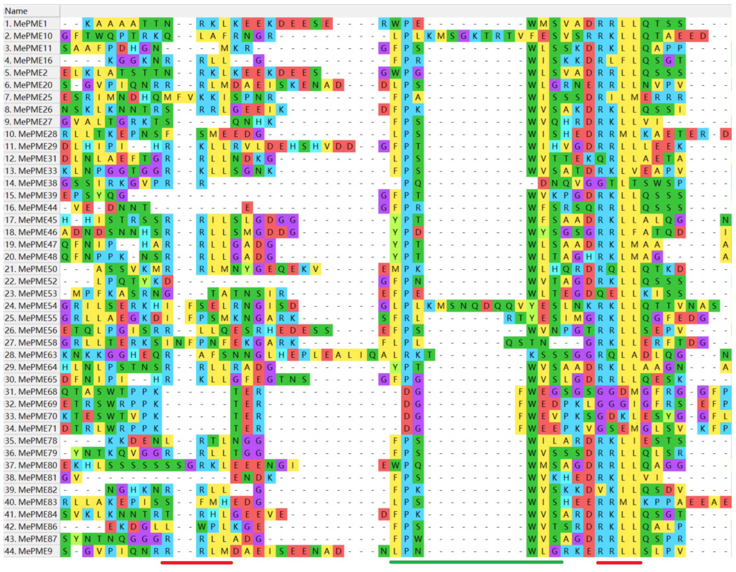
2.4. Gene Structure and Motif Analysis of MePME Genes in Cassava

The amino acid sequences of 89 MePMEs were used to draw a phylogenetic tree, using the MEGA11 software, in which the two types of MePMEs were well separated (Figure 4a). Twelve kinds of motifs were searched for within the MePMEs’ amino acid sequences using MEME analysis (Figure 4b). Each type-Ⅰ MePME contains 11 motifs including motifs 1~7 and motifs 9~12, indicating that these 11 motifs are conserved in type-Ⅰ MePMEs. In type-Ⅱ MePMEs, motifs 1~7 and motif 11 are conserved, as they appear in each type-Ⅱ MePME (Figure 4b). In addition, motif 8 was only apparent in one of the type-Ⅰ MePMEs, namely, MePME33, while it appeared in 13 type-Ⅱ MePMEs (Figure 4b). The gene structure analysis revealed that most type-Ⅰ MePMEs (32 out of 44) have only one intron within their CDS, while 12 of them have two introns. In addition, 25 type-Ⅰ MePMEs have both up- and down-stream UTRs, while four genes have only down-stream UTRs (Figure 4c). The intron numbers in type-Ⅱ MePMEs varied from one to nine. Moreover, eleven type-Ⅱ MePMEs had up- or down-stream UTRs (Figure 4c). By and large, the more analogous the MePME genes, the more similar the motifs and gene structures (Figure 4).

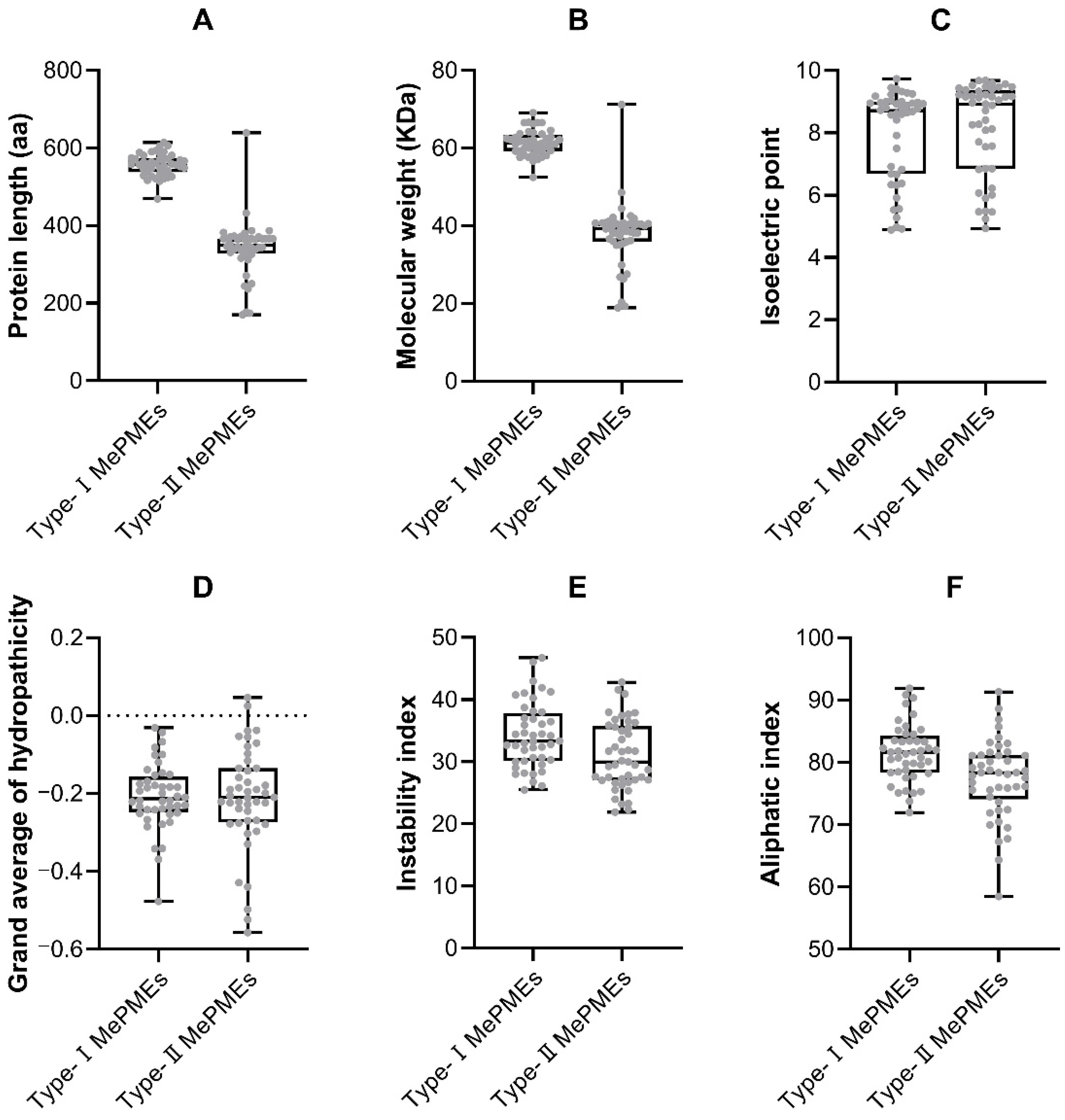
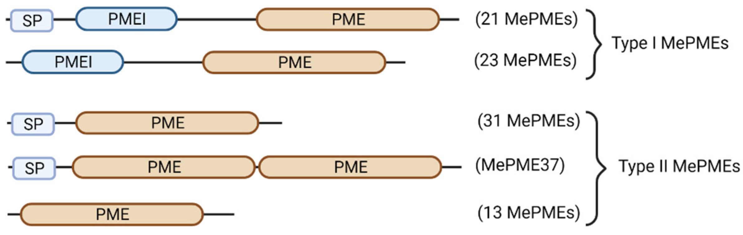
2.5. Domain Analysis of MePMEs

To survey the domains in cassava MePMEs, the 89 MePME amino acid sequences were analyzed. Each of the 44 type-Ⅰ MePMEs contains one PMEI domain and one PME domain. Most (44 out of 45) type-Ⅱ MePMEs contain only one PME domain, except MePME37, which has two PME domains (Figure 5). In 52 out of 89 MePMEs (21 type-Ⅰ and 31 type-Ⅱ), a signal peptide was predicted with high confidence by using the SignalP program (Table S3). Through multiple sequence alignment, we found that most type-Ⅰ MePMEs contain two basic RK/RLL motifs (Figure 6), with the second one being more conserved than the first one. In addition, between the RK/RLL motifs, there is usually an “FPSWVS” motif; here, in type-Ⅰ MePMEs, the amino acid residues “P” and “W” are relatively more conserved. Only in four type-Ⅰ MePMEs (MePME68~71), the “P” was replaced with “D”, whilst the “W” in six type-Ⅰ MePMEs including MePME10, MePME38, MePME54, MePME55, MePME58 and MePME63 was changed into “F”, “-”, “Y”, “Y”, “T” and “K”, respectively (Figure 6). These changes may affect the function of MePMEs.

2.6. Cis-Acting Regulatory Elements Analysis of MePMEs

Cis-acting regulatory elements (CAREs) are important binding sites on the promoter region of genes for regulating gene expression. We predicted stress-, hormone- and tissue-specific-related CAREs on the MePMEs promoters (2000 bp upstream of initiation codon “ATG”). The results display that the types and quantities of CAREs in the promoter regions of MePMEs were diverse (Figure 7). The vast majority of MePMEs members possessed at least one type of stress-related and hormone-related CARE. In detail, 33 MePMEs had at least one low-temperature-related CARE, 57 MePMEs had drought-response-related CAREs, and 64 MePMEs had anaerobic-induction-related CAREs. Only two MePMEs (MePME59 and MePME63) harbored wound-response CAREs. For hormone-related CAREs, ABA-response-related CAREs appeared in 66 MePMEs, MeJA-response-related CAREs appeared in 41 MePMEs, gibberellin-response-related CAREs existed in 30 MePMEs, auxin-response-related CAREs were found in 24 MePMEs and salicylic-acid-response-related CAREs appeared in 36 MePMEs. These results indicate that MePMEs play roles in response to stress and are regulated by hormones in cassava. In addition, some MePMEs harbor tissue-specific expression-related CAREs, such as endosperm, meristem and seed. This may relate to the special function of these tissues.

2.7. Expression Analysis of MePMEs in Cassava under Different Stresses

Postharvest physiological deterioration (PPD) rapidly occurs after cassava is harvested, and severely restricts the economic value of the crop. A previous study indicated that PME activity and expression was associated with fruit postharvest quality. Therefore, we analyzed the expression of MePMEs in cassava transcriptome data in response to PPD. A total of 31 MePME genes with values of transcription per kilobase per million mapping readings (FPKM) in each sample were found in the transcriptomic data of two cassava varieties (PPD-sensitive cassava SC8 and PPD-tolerant cassava RYG1) after 0- and 21-day storage (SC8 severely deteriorated, while RYG1 maintained good quality) [41]. A total of ten MePME genes were found to participate in the response to PPD in the transcriptome data of SC124 cassava during PPD at time points of 0 h (control), 6 h (no symptoms of PPD), 12 h (local symptoms of PPD) and 48 h (global PPD symptoms) after deterioration [50]. A total of twelve MePMEs were reported to be associated with melatonin-delayed PPD in SC124 [51] (Table S3). As a tropical crop, cassava is sensitive to low temperatures. To reveal the response mechanism of cassava to chilling stress, a transcriptomic analysis of a mixture of leaf and root samples was performed for plants that were kept at 24 °C (CK), at 14 °C for 5 days (T1), at 14 °C for 5 days followed by 4 °C for 5 days (T2) and at 24 °C for 5 days followed by 4 °C for 5 days (T3) [52]. We found that a total of 73 MePMEs had FPKM data in each sample (Table S3). Cassava has a high tolerance to drought. ABA acts as a crucial signal in response to drought stress. To reveal whether MePMEs are responsive to drought stress and the ABA signal, the transcriptome data of cassava treated with 7% polyethylene glycol (PEG) and 50 μmol/L abscisic acid (ABA) were analyzed. A total of 43 MePMEs had FPKM data in both the PEG- and ABA-treated cassava (Table S3). To select crucial MePME genes displaying multiple stress responses, UpSet Venn diagrams were used to filter the MePMEs shared by PPD, chilling stress, ABA and PEG treatments. The results show that there are five MePME genes in all of the above transcriptomes—namely, MePME1, MePME2, MePME27, MePME65 and MePME82 (Figure 8, Table S3). Interestingly, all five shared genes are type-Ⅰ MePMEs.
The expression patterns of the five shared MePMEs in response to PPD (in different PPD-tolerant cassava and in early stage of PPD occurrence), chilling, ABA and PEG stresses were analyzed. The expression levels of all the five shared MePMEs were a bit higher at t0 in RYG1 than in SC8, but these differences were not significant. After 21 days of storage, the expression levels of most of the shared MePMEs were inhibited in both SC8 and RYG1. Particularly, the downregulation of MePME1 expression in RYG1 was even greater than that in SC8 (Figure 9a). During the early stages of PPD, when no obvious PPD symptoms appeared in the slices, the expressions of MePME1 and MePME27 were inhibited, whereas the expression of MePME2, MePME65 and MePME82 were increased. When the PPD symptoms appeared in some of the slices 12 h after sectioning, the expressions of MePME1, MePME2, MePME65 and MePME82 were significantly upregulated, whereas the expression of MePME27 was significantly downregulated. When the PPD symptoms appeared in all surfaces of slices at 48 h after sectioning, the expressions of MePME1, MePME2 and MePME65 were still higher than those in the control, but less than those at 12 h, whereas the expressions of MePME27 and MePME82 at 48 h were decreased compared to those in the control (Figure 9b). The expression level of MePME1 was significantly induced by 7% PEG-simulated drought stress, while the expression of four other genes, MePME2, MePME27, MePME65 and MePME82, were inhibited (Figure 9c). The expressions of all five shared MePMEs were decreased to various degrees after 50 μmol/L ABA treatment; in particular, the expressions of MePME2, MePME65 and MePME82 were significantly inhibited (Figure 9c). The expression levels of MePME1, MePME2 and MePME27 were slightly upregulated after T1 and T3 treatments, while the expressions of MePME65 and MePME82 were slightly inhibited. In addition, the expressions of MePME1 and MePME82 were increased after T2 treatment (Figure 9d). In general, MePME1 showed a relatively strong response to the above adversity treatments.

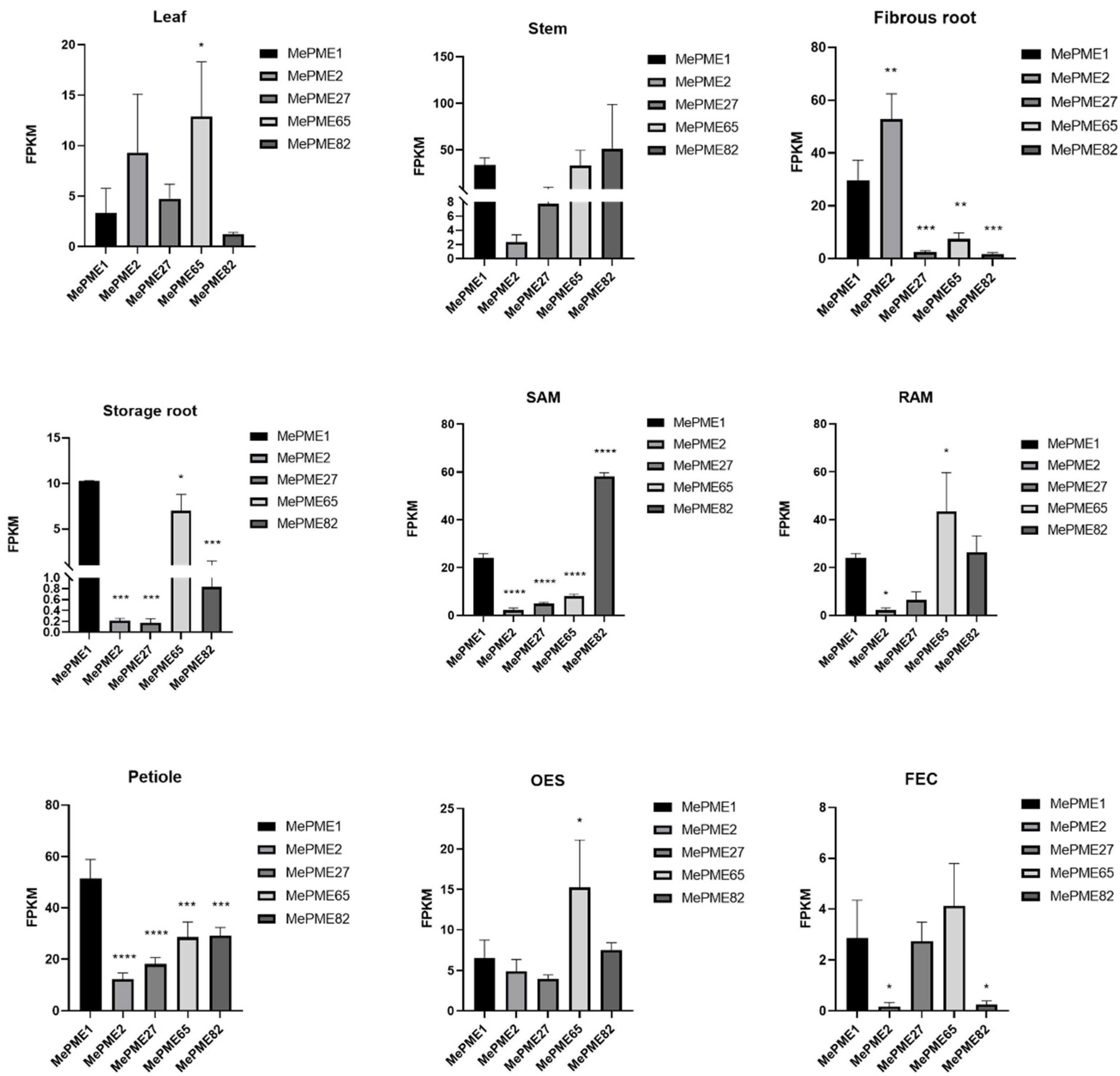
2.8. Expression of Shared MePMEs in Different Tissues of Cassava

The expression of genes in certain tissues may be related to the differentiation and development of these tissues or the special functions performed by these tissues. To know the tissue-specific expression patterns of the five shared MePME genes, we compared their expression levels in nine different tissue types including leaf, stem, fibrous and storage root, stem and root apex meristem (SAM and RAM), petiole, organized embryonic structure (OES) and friable embryogenic callus (FEC) (Figure 10). Overall, the five shared MePMEs were expressed to varying degrees in the nine tissues. An analysis of the top two genes that were highly expressed in each tissue showed that MePME65 and MePME2 are the top two genes in leaves, MePME82 and MePME1 are the most expressed in stems, while MePME2 and MePME1 predominate in fibrous roots, and MePME1 and MePME65 are mostly expressed in storage roots. Meanwhile, MePME82 and MePME1 are the most expressed in SAM, and MePME65 and MePME82 are the most expressed in RAM, whilst MePME1 and MePME65 predominate in petiole, MePME65 and MePME82 predominate in OES and MePME65 and MePME1 predominate in FEC. In addition, the above analysis results also show that both MePME1 and MePME65 were ranked in the top two for a total of six times. These results indicate that MePME1 and MePME65 may play a critical role in these tissues.

3. Discussion

The pectin methylesterase family genes have attracted considerable research attention due to their vital role in the regulation of cell wall modification. The systematic analysis of PME family genes was achieved in Arabidopsis [18] and several other plant species, including rice [16], tomato [21] and foxtail millet [14]. However, the abundance and function of PME genes in other plant species remain unclear. Herein, 89 cassava PME genes were uncovered and subjected to bioinformatic analysis at the whole-genome level.
Compared to the reported PME families in other dicots, the PME gene family in cassava is of mid size. However, compared to the monocots, such as foxtail millet (forty-one members) [14] and rice (forty-four members) [16], there are nearly twice as many PMEs in cassava. The difference in the gene family size among different species is most likely due to genome-wide duplication events. According to our research, 19 paralogous pairs (13 pairs in type-Ⅰ, and 6 pairs in type-Ⅱ) were present among the 89 MePME genes (Figure 2). Duplication events were also present in other species. For example, in tobacco, nine duplication pairs were identified [15]. In citrus, 23 pairs of PME genes were identified as duplicated genes [20]. In Brassica rapa, twelve PME gene pairs were found to have experienced duplication [22].
By comparing the PME sequences between cassava and Arabidopsis, the cassava PME were distinguished and further sub-divided into ten and five sub-groups (Figure 3). It is worth noting that MePME1, MePME2 and MePME80 are duplicates of each other (Figure 2). The phylogenetic relationship between them is closest and they additionally exhibit a higher similarity with AtPME2 and AtPME3 from Arabidopsis in group Ⅰ-7 (Figure 3). This indicates that they may have a similar biological function as AtPME2 and AtPME3.
According to previous studies, type-Ⅰ PMEs contain two conserved “RK/RLL” domains and one “FPSWVS” domain between the pro-region and PME domain. These domains were considered to be important for the cleavage of the pro-region to generate mature PME proteins [53]. In our study, most MePMEs contain two conserved “RK/RLL”, but some do not contain this domain (Figure 6). For the “FPSWVS” domain in cassava, “P” and “W” were more conserved than the other amino acid residues (Figure 6). However, whether the missing cleavage domains or the changed amino acid residues affect the pro-region cleavage remains unclear, and experimental evidence will likely be required in order to address this.
Cis-acting regulatory elements (CAREs) are important DNA sequences in the promoters of genes, which could bind to corresponding transcription factors in order to regulate gene transcription under different conditions. In this study, the CAREs in the promoter region of MePME family members were analyzed. We found a great deal of CAREs that respond to hormones such as abscisic acid, methyl jasmonate, auxin, gibberellin and salicylic acid. We additionally found some CAREs that respond to stresses including low-temperature, drought and anaerobic and wounding stresses. Finally, we found a set of CAREs associated with tissue-specific expression in the endosperm, meristem and seed (Figure 7). Similar findings were reported for the tobacco PME family members, where low-temperature-, wound-, anaerobic-, ABA-, auxin- and MeJA-related CAREs exist [15]. These results indicate that MePMEs may respond to a variety of abiotic stresses and participate in plant growth and development. A few studies confirmed that some transcription factors regulate the expression of PME. For instance, the transcription factor BELLRINGER (BLR) is a key transcription factor that regulates AtPME5-mediated organ initiation and cell extension [54]. The transcription factor CsRVE1 was confirmed to directly bind to the promoter of CsPME3 in navel orange [20]. Two BEL1-like homeodomains (BLHs) BLH2 and BLH4 bind to the promoter of PME58 to activate its expression in seed mucilage [55]. MYB80 was confirmed to target one PME gene in Arabidopsis [56]. In strawberry, the FvMYB79 can activate the expression of FvPME38 and lead to ABA-dependent fruit softening after harvest [57]. However, the transcription factors regulating PME expression in cassava remain to be studied.
Some previous studies proved the functions of several PME genes in regulating abiotic stresses. For instance, AtPME41 was induced by chilling stress in Arabidopsis [25]. Nine CsPME genes were upregulated after low temperature treatment for 6 h [20]. Similarly, AtPME34 was associated with heat stress, with the pme34 mutant being sensitive to heat stress [26]. In addition, AtPME34 was highly expressed in guard cells and was induced by ABA [26]. The expression of AtPME2 was improved during lateral root emergence and hypocotyl elongation [27]. Furthermore, both AtPME2 and AtPME3 participated in stomatal development, and regulated guard cell movement [28]. AtPME35 was specifically expressed in Arabidopsis in the basal part of the inflorescence stem, and was involved in regulating the stem’s mechanical strength [29]. The expression of AtPME6 was increased during mucilage secretion and played an important role in regulating embryo development [30]. Meanwhile, AtPME31 was induced by salt stress, and when it was knocked out, the plants showed hypersensitivity to salt stress [31], whereas in apple, the expression profile of an AtPME2-similar gene MdPME2 was downregulated during apple mealiness development [35]. Similarly, in rice, six OsPME genes were induced by aluminum stress, the overexpression of OsPME14 decreased the resistance of transgenic plants to aluminum stress [10,32] and three peach PME genes were highly expressed in melting flesh during peach fruit ripening [23]. By contrast, one PME gene in rye was inhibited by aluminum stress in tolerant cultivar [33]. In tea plants, 8 PME genes in leaves and 15 genes in roots responded to aluminum stress [34]. These studies thus highlight a common mechanism by which plants respond to different stresses, namely, the regulation of cell wall modifications involving PME, which is often overlooked, because it was previously thought that the cell wall was a relatively stable cell structure that mainly played a role in maintaining cell morphology and plant rigidity. On the further development of research in the area, the cell wall was found to be highly dynamic with the growth of the plant and changes in response to the external environment [58,59,60,61]. As one of the main components of the cell wall, pectin not only plays a crucial role in cell wall remodeling, but also participates in intercellular communication, morphogenesis and environmental sensing [60]. PME is the first enzyme in the process of pectin degradation, which regulates the homeostasis of pectin in the cell wall, and thus, mediates the remodeling of the cell wall in response to changes in the external environment [62].
As an important food and renewable bioenergy raw material that is widely cultivated in tropical and subtropical regions of the world, cassava has many advantages and disadvantages. It is very suitable for arid and barren soil, but it is very sensitive to low temperatures, and its storage roots very readily deteriorate after harvest [41,63]. In this study, we filtered out five MePMEs (MePME1, MePME2, MePME27, MePME65 and MePME82), which were shared by different stress treatments, namely, PPD, chilling, 50 μmol/L ABA and 7% PEG stresses. Although studies on the function of the PME gene are still relatively few, some studies have shown that a single PME gene has multiple functions. AtPME3 was rapidly induced by Pectobacterium carotovorum and Botrytis cinerea, and caused sensitivity to Zn2+ following mis-expression [64,65]. AtPME31 could improve the resistance of transgenic plants to salt stress and various insect pests [31,66]. AtPME34 was induced by ABA and participated in stomatal movement and heat stress response [26]. In tobacco, NtPME043 was induced by both salt and ABA stress [15]. It is speculated that these genes may be involved in cell wall modification under various stresses. Additionally, among the shared MePMEs, we found that the expression of MePME1 was significantly induced during the occurrence of PPD 12 h after the cassava tuberous roots were sliced. In PPD-resistant cassava RYG1, in which no PPD symptoms occurred after 21 days of storage, the expression of MePME1 was significantly downregulated (Figure 9). These results indicate that MePME1 plays a crucial role in regulating cassava resistance to PPD. The expression of MePME1 was also increased following chilling acclimation (14 °C for 5 days), and by using 7% PEG treatment (Figure 9). Notably, MePME1 was also the most highly expressed of the five selected genes in storage root and petiole. Moreover, it was also the second highest expressed of these genes in the stem, fibrous root, shoot apex meristem, and friable embryogenic callus (Figure 10). These results suggest that MePME1 should be subjected to further investigation.
In summary, eighty-nine PME genes were identified in cassava. An integrated characterization of the MePMEs in cassava was performed through the analysis of their physical and chemical properties, chromosome localization and duplication, conserved motifs and gene structures and phylogenetic relationships and cis-acting regulatory elements. Through the co-expression analysis on different transcriptomic data, five MePMEs were selected. A further expression pattern analysis of the shared MePMEs under different stresses and in different tissues suggested that MePME1 may be the member of the PME family that is worthy of the deepest investigation. The findings from this research thus provide valuable information for further functional investigations on cassava PME genes in the near future.

4. Materials and Methods

4.1. Identification of Pectin Methylesterase Gene Family Members in Cassava

The protein family (PFAM) serial number of the PME domain (PF01095) was used as keyword to search the cassava genome database v6.1 (https://Phytozome-next.jgi.doe.gov/info/Mesculenta_v6_1, accessed on 18 April 2022) [67]. This allowed us to identify all cassava MePME members and download their annotation information. Next, we filtered the MePME information to find the genes with a PFAM serial number indicating the PMEI domain (PF04043), and these genes were classified as type-Ⅰ MePMEs. The residual genes without a PMEI domain were classified as type-Ⅱ MePMEs.

4.2. Characterization of MePMEs

The sequences of MePME proteins were next submitted to the ProtParam tool in the ExPASy web page (https://web.expasy.org/protparam/, accessed on 20 April 2022) [47] in order to generate the basic properties, which included the protein length (aa), molecular weight (MW), theoretical isoelectric point (pI), aliphatic index, instability index and grand average of hydropathicity. If the pI value is greater than 7, the MePME is classified as an alkaline protein, if the pI value is less than 7, the MePME is classified as an acidic protein and if the pI value is 7, then the MePME is classified as a neutral protein. The instability index is used to evaluate the stability of a protein. When the instability index is less than 40, the protein is regarded as stable, while when the value is greater than 40, the protein is regarded as unstable [68]. The box and whiskers diagrams were outputted via Graphpad Prism 8. The signal peptide of each MePME was predicted by submitting the amino acid sequences to SignalP 6.0 [69] (https://services.healthtech.dtu.dk/services/SignalP-6.0, accessed on 17 April 2023).

4.3. Chromosomal Location, Duplication, Gene Structure, Conserved Motifs and Cis-Acting Regulatory Element Analysis of MePMEs

The CDS sequences, specific location information of each MePME gene on chromosome and the whole chromosome information of cassava were acquired from the cassava genomic database (https://Phytozome-next.jgi.doe.gov/info/Mesculenta_v6_1, accessed on 7 May 2023). Then, TBtools was used to calculate gene collinearity and export the graph [70]. For conserved motif analysis of all MePMEs, MEME (http://meme-suit.org, accessed on 7 May 2023) was used [71]. Gene structure analysis was generated using Gene Structure Server Display Server online (http://gsds.cbi.pku.edu.cn, accessed on May 2023) [72]. The type-Ⅰ MePMEs conserved motifs between PME domain and PMEI domain, which were analyzed via multiple alignment using ClustalW. The promoter sequence, which is 2000 bp upstream of start codon (ATG) of each MePME, was download from cassava genome, and inputted into PlantCARE [73] (http://bioinformatics.psb.ugen.be/webtools/plantcare/html, accessed on 24 October 2022) to search for the CAREs. The main CARE localizations on the promoter was visualized using TBtools. The number of each kind of CARE was statistic and visualized using Microsoft Excel.

4.4. Multiple Sequence Alignment and Phylogenetic Analysis of MePMEs with PMEs in Arabidopsis

Sixty-six PME protein sequences of Arabidopsis were downloaded from Arabidopsis thaliana genome TAIR10 (https://phytozome-next.jgi.doe.gov/info/Athaliana_TAIR10, accessed on 8 August 2022) [74]. Then, all the amino acid sequences of 89 MePMEs and 66 PMEs from Arabidopsis were aligned using ClustalW in MEGA11 software [48] with the default parameter. After that, the aligned sequences were used to generate a phylogenetic tree using the statistical method of maximum likelihood and Jones–Taylor–Thornton model. At last, the visualization and annotation of the phylogenetic tree graphic was obtained using the online tool iTOL v6 (https://itol.embl.de, accessed on 18 April 2023) [49].

4.5. Multifunctional MePME Gene Screening and Analysis of Their Expression Patterns

For screening MePME genes with multiple functions, cassava transcriptome data under different stresses were used to find the MePME family genes. The intact tuberous roots of PPD-sensitive cassava SC8 and PPD-tolerant cassava RYG1 were stored for 0 and 21 days and sampled to perform transcriptome analysis [41]. Two biological replicates were used. Transcriptome data with values of transcription per kilobase per million mapping readings (FPKM) in each sample were used to screen and count the number and gene IDs of MePME genes. SC124 cassava tuberous roots were crosscut into 5 mm thick slices, and stored in dark condition of 28 °C and 60% humidity for 0, 6, 12 and 48 h. Samples were then collected for transcriptome sequencing. To filter differentially expressed genes (DEGs) the screening condition was set as fold change ≥ 2 and p value ≤ 0.05. Three biological replicates were used [50]. MePME genes that were significantly expressed in at least one comparison group were selected. The intact cassava tuberous roots were soaked with 100 μmol/L melatonin for 2 h, and those treated with water were used as control. After that, the cassavas were cut crosswise into 5 mm thick slices and stored at 28 °C with 60% humidity in the dark for 6, 12 and 48 h to perform the transcriptome analysis. The screening condition for DEGs was fold change ≥ 2 and p value ≤ 0.05. Three biological replicates were used [51]. MePME genes, which were significantly expressed in at least one stage of melatonin-delayed PPD, were selected. SC124 cassava stem cuttings were cultured in pots containing red soil and vermiculite (1:1 in volume ratio) and irrigated using Hoagland’s solution once a week for 2 months. The uniform seedlings were selected for different chilling stress treatments, including 24 °C for 10 days as control, T1 (24 °C for 5 days + 14 °C for 5 days) as chilling acclimation, T2 (14 °C for 5 days + 4 °C for 5 days) as chilling stress after chilling acclimation, and T3 (24 °C for 5 days + 4 °C for 5 days) as chilling shock. The folded leaves, fully expanded leaves and roots after different chilling treatments at 6 h, 24 h and 5 d were collected to extract total RNA. Then, equal amounts of RNA samples for each time point and each organ of each treatment were collected and pooled together to generate mRNA libraries and sequenced via RNA-Seq. Three biological replicates were used [52]. Transcriptome data with FPKM values in each sample were used to screen MePME genes in response to chilling stress. Two-month-old TM60444 cassava seedlings were treated with 7% PEG and 50 μmol/L ABA, separately [75]. After 48 h of treatment, mature leaves were collected to extract total RNA and perform RNA-Seq analysis (PRJNA658570). Three biological replicates were used. Transcriptome data with FPKM values in each sample were used to screen MePME genes in response to PEG or ABA stress. Subsequently, the Omicshare tools, a free online platform for data analysis (https://www.omicshare.com/tools/Home/Soft/seniorvenn, accessed on 13 June 2023), was used to output UpSet Venn diagram and to illustrate the shared MePMEs in different transcriptome data. For expression pattern analysis of shared MePMEs under different stresses, the FPKM values from the above transcriptome were selected to generate histogram.
For tissue-specific expression analysis, TME 204 cassava seedlings cultured in a greenhouse for 3 months were used to harvest different tissue samples, including leaf blade, petiole, stem, shoot apical meristem, fibrous roots, root apical meristem and storage roots. In addition, the 4-week-old OES and 3-week-old FEC tissues of TME204 were also sampled. Total RNA was isolated from the above different samples for RNA-Seq analysis, separately (PRJNA324539). With the exception of two biological replicates set in storage roots, three biological replicates were set in each of the remaining eight samples [76]. The FPKM values of shared MePMEs in nine cassava tissues were selected from the transcriptome data for subsequent analysis. All the histograms were outputted via Graphpad Prism 8. The significant differences were evaluated using ordinary one-way ANOVA analysis and Dunnett’s multiple comparisons test.

Supplementary Materials

The following supporting information can be downloaded at: https://www.mdpi.com/article/10.3390/plants12132529/s1, Table S1: Physical and chemical information of MePMEs in cassava, Table S2: Information of MePMEs duplication pairs, Table S3: signal peptide prediction of MePMEs, Table S4: MePMEs in different transcriptome data.

Author Contributions

Methodology, R.L. and A.R.F.; investigation, S.W., R.L., Y.Z., Z.D., Q.Z. and Y.C.; writing—original draft preparation, S.W. and R.L.; writing—review and editing, A.R.F., Y.Y., J.L., Y.W., X.H. and J.G.; supervision, X.H. and J.G.; funding acquisition, J.G. All authors have read and agreed to the published version of the manuscript.

Funding

This research was funded by the National Natural Science Foundation of China, 31601359; the Provincial Natural Fund of Hainan Province, 323RC537; the Earmarked Fund for China Agriculture Research System, CARS-11-HNGJC; the Major Science and Technology plan of Hainan Province, ZDKJ2021012; the State Scholarship Fund of China Scholarship Council, CSC202003260020.

Data Availability Statement

The datasets generated during and/or analyzed during the current study are available from the corresponding author on reasonable request.

Acknowledgments

We fully appreciate Pingjuan Zhao for helping with the data analysis, and the editors and all anonymous reviewers for their constructive comments on this manuscript.

Conflicts of Interest

The authors declare no conflict of interest.

References

  1. Bosch, M.; Cheung, A.Y.; Hepler, P.K. Pectin methylesterase, a regulator of pollen tube growth. Plant Physiol. 2005, 138, 1334–1346. [Google Scholar] [CrossRef] [Green Version]
  2. Shi, D.C.; Ren, A.Y.; Tang, X.F.; Qi, G.; Xu, Z.C.; Chai, G.H.; Hu, R.B.; Zhou, G.K.; Kong, Y.Z. MYB52 Negatively Regulates Pectin Demethylesterification in Seed Coat Mucilage. Plant Physiol. 2018, 176, 2737–2749. [Google Scholar] [CrossRef] [Green Version]
  3. Zhou, Y.; Li, R.; Wang, S.; Ding, Z.; Zhou, Q.; Liu, J.; Wang, Y.; Yao, Y.; Hu, X.; Guo, J. Overexpression of MePMEI1 in Arabidopsis enhances Pb tolerance. Front. Plant Sci. 2022, 13, 996981. [Google Scholar] [CrossRef]
  4. Bilska-Kos, A.; Solecka, D.; Dziewulska, A.; Ochodzki, P.; Jonczyk, M.; Bilski, H.; Sowinski, P. Low temperature caused modifications in the arrangement of cell wall pectins due to changes of osmotic potential of cells of maize leaves (Zea mays L.). Protoplasma 2017, 254, 713–724. [Google Scholar] [CrossRef] [Green Version]
  5. Draye, M.; Van Cutsern, P. Pectin methylesterases induce an abrupt increase of acidic pectin during strawberry fruit ripening. J. Plant Physiol. 2008, 165, 1152–1160. [Google Scholar] [CrossRef] [PubMed]
  6. Blumer, J.M.; Clay, R.P.; Bergmann, C.W.; Albersheim, P.; Darvill, A. Characterization of changes in pectin methylesterase expression and pectin esterification during tomato fruit ripening. Can. J. Bot.-Rev. Can. Bot. 2000, 78, 607–618. [Google Scholar] [CrossRef]
  7. Yan, R.; Han, C.; Fu, M.R.; Jiao, W.X.; Wang, W.H. Inhibitory Effects of CaCl2 and Pectin Methylesterase on Fruit Softening of Raspberry during Cold Storage. Horticulturae 2022, 8, 1. [Google Scholar] [CrossRef]
  8. Wu, Q.; Tao, Y.; Zhang, X.L.; Dong, X.Y.; Xia, J.X.; Shen, R.F.; Zhu, X.F. Pectin Methylesterases Enhance Root Cell Wall Phosphorus Remobilization in Rice. Rice Sci. 2022, 29, 179–188. [Google Scholar] [CrossRef]
  9. Xu, Z.J.; Yang, M.; Li, Z.Y.; Xiao, J.; Yang, X.Q.; Wang, H.; Wang, X.F. Tissue-specific pectin methylesterification and pectin methylesterase activities play a role in lettuce seed germination. Sci. Hortic. 2022, 301, 111134. [Google Scholar] [CrossRef]
  10. Yang, X.Y.; Zeng, Z.H.; Yan, J.Y.; Fan, W.; Bian, H.W.; Zhu, M.Y.; Yang, J.L.; Zheng, S.J. Association of specific pectin methylesterases with Al-induced root elongation inhibition in rice. Physiol. Plant. 2013, 148, 502–511. [Google Scholar] [CrossRef]
  11. Lionetti, V. PECTOPLATE: The simultaneous phenotyping of pectin methylesterases, pectinases, and oligogalacturonides in plants during biotic stresses. Front. Plant Sci. 2015, 6, 331. [Google Scholar] [CrossRef] [PubMed] [Green Version]
  12. Raiola, A.; Camardella, L.; Giovane, A.; Mattei, B.; De Lorenzo, G.; Cervone, F.; Bellincampi, D. Two Arabidopsis thaliana genes encode functional pectin methylesterase inhibitors. FEBS Lett. 2004, 557, 199–203. [Google Scholar] [CrossRef] [PubMed]
  13. Camardella, L.; Carratore, V.; Ciardiello, M.A.; Servillo, L.; Balestrieri, C.; Giovane, A. Kiwi protein inhibitor of pectin methylesterase—Amino-acid sequence and structural importance of two disulfide bridges. Eur. J. Biochem. 2000, 267, 4561–4565. [Google Scholar] [CrossRef] [PubMed]
  14. Ge, W.N.; Chen, H.L.; Zhang, Y.C.; Feng, S.Y.; Wang, S.L.; Shang, Q.; Wu, M.; Li, Z.Q.; Zhang, L.; Guo, H.; et al. Integrative genomics analysis of the ever-shrinking pectin methylesterase (PME) gene family in foxtail millet (Setaria italica). Funct. Plant Biol. 2022, 49, 874–886. [Google Scholar] [CrossRef] [PubMed]
  15. Sun, J.H.; Tian, Z.; Li, X.X.; Li, S.P.; Li, Z.Y.; Wang, J.L.; Hu, Z.Y.; Chen, H.Q.; Guo, C.; Xie, M.M.; et al. Systematic analysis of the pectin methylesterase gene family in Nicotiana tabacum and reveal their multiple roles in plant development and abiotic stresses. Front. Plant Sci. 2022, 13, 998841. [Google Scholar] [CrossRef]
  16. Jeong, H.Y.; Nguyen, H.P.; Lee, C. Genome-wide identification and expression analysis of rice pectin methylesterases: Implication of functional roles of pectin modification in rice physiology. J. Plant Physiol. 2015, 183, 23–29. [Google Scholar] [CrossRef]
  17. Li, Y.Q.; He, H.Y.; He, L.F. Genome-Wide Analysis of the Pectin Methylesterase Gene Family in Potato. Potato Res. 2021, 64, 1–19. [Google Scholar] [CrossRef]
  18. Louvet, R.; Cavel, E.; Gutierrez, L.; Guenin, S.; Roger, D.; Gillet, F.; Guerineau, F.; Pelloux, J. Comprehensive expression profiling of the pectin methylesterase gene family during silique development in Arabidopsis thaliana. Planta 2006, 224, 782–791. [Google Scholar] [CrossRef]
  19. Pinzon-Latorre, D.; Deyholos, M.K. Characterization and transcript profiling of the pectin methylesterase (PME) and pectin methylesterase inhibitor (PMEI) gene families in flax (Linum usitatissimum). BMC Genom. 2013, 14, 742. [Google Scholar] [CrossRef] [Green Version]
  20. Li, Z.X.; Wu, L.M.; Wang, C.; Wang, Y.; He, L.G.; Wang, Z.J.; Ma, X.F.; Bai, F.X.; Feng, G.Z.; Liu, J.H.; et al. Characterization of pectin methylesterase gene family and its possible role in juice sac granulation in navel orange (Citrus sinensis Osbeck). BMC Genom. 2022, 23, 1–18. [Google Scholar] [CrossRef]
  21. Jeong, H.Y.; Nguyen, H.P.; Eom, S.H.; Lee, C. Integrative analysis of pectin methylesterase (PME) and PME inhibitors in tomato (Solanum lycopersicum): Identification, tissue-specific expression, and biochemical characterization. Plant Physiol. Biochem. 2018, 132, 557–565. [Google Scholar] [CrossRef] [PubMed]
  22. Duan, W.K.; Huang, Z.N.; Song, X.M.; Liu, T.K.; Liu, H.L.; Hou, X.L.; Li, Y. Comprehensive analysis of the polygalacturonase and pectin methylesterase genes in Brassica rapa shed light on their different evolutionary patterns. Sci. Rep. 2016, 6, 25107. [Google Scholar] [CrossRef] [PubMed]
  23. Zhu, Y.; Zeng, W.; Wang, X.; Pan, L.; Niu, L.; Lu, Z.; Cui, G.; Wang, Z. Characterization and Transcript Profiling of PME and PMEI Gene Families during Peach Fruit Maturation %J Journal of the American Society for Horticultural Science. J. Am. Soc. Hortic. Sci. 2017, 142, 246–259. [Google Scholar] [CrossRef] [Green Version]
  24. Li, W.J.; Shang, H.H.; Ge, Q.; Zou, C.S.; Cai, J.; Wang, D.J.; Fan, S.M.; Zhang, Z.; Deng, X.Y.; Tan, Y.N.; et al. Genome-wide identification, phylogeny, and expression analysis of pectin methylesterases reveal their major role in cotton fiber development. BMC Genom. 2016, 17, 1–13. [Google Scholar] [CrossRef] [PubMed] [Green Version]
  25. Qu, T.; Liu, R.; Wang, W.; An, L.; Chen, T.; Liu, G.; Zhao, Z. Brassinosteroids regulate pectin methylesterase activity and AtPME41 expression in Arabidopsis under chilling stress. Cryobiology 2011, 63, 111–117. [Google Scholar] [CrossRef] [PubMed]
  26. Huang, Y.C.; Wu, H.C.; Wang, Y.D.; Liu, C.H.; Lin, C.C.; Luo, D.L.; Jinn, T.L. PECTIN METHYLESTERASE34 Contributes to Heat Tolerance through Its Role in Promoting Stomatal Movement. Plant Physiol. 2017, 174, 748–763. [Google Scholar] [CrossRef]
  27. Hocq, L.; Habrylo, O.; Voxeur, A.; Pau-Roblot, C.; Safran, J.; Sénéchal, F.; Fournet, F.; Bassard, S.; Battu, V.; Demailly, H.J.b. Arabidopsis AtPME2 has a pH-dependent processivity and control cell wall mechanical properties. BioRxiv 2021, 3, 433777. [Google Scholar] [CrossRef]
  28. Tsakali, A.; Asitzoglou, I.-C.; Basdeki, V.; Podia, V.; Adamakis, I.-D.S.; Giannoutsou, E.; Haralampidis, K. The Role of PME2 and PME3 in Arabidopsis Stomatal Development and Morphology &dagger. Biol. Life Sci. Forum 2022, 11, 36. [Google Scholar] [CrossRef]
  29. Hongo, S.; Sato, K.; Yokoyama, R.; Nishitani, K. Demethylesterification of the Primary Wall by PECTIN METHYLESTERASE35 Provides Mechanical Support to the Arabidopsis Stem. Plant Cell 2012, 24, 2624–2634. [Google Scholar] [CrossRef] [Green Version]
  30. Levesque-Tremblay, G.; Müller, K.; Mansfield, S.D.; Haughn, G.W. HIGHLY METHYL ESTERIFIED SEEDS Is a Pectin Methyl Esterase Involved in Embryo Development. Plant Physiol. 2015, 167, 725–737. [Google Scholar] [CrossRef] [Green Version]
  31. Yan, J.; He, H.; Fang, L.; Zhang, A. Pectin methylesterase31 positively regulates salt stress tolerance in Arabidopsis. Biochem. Biophys. Res. Commun. 2018, 496, 497–501. [Google Scholar] [CrossRef]
  32. Yang, J.L.; Li, Y.Y.; Zhang, Y.J.; Zhang, S.S.; Wu, Y.R.; Wu, P.; Zheng, S.J. Cell wall polysaccharides are specifically involved in the exclusion of aluminum from the rice root apex. Plant Physiol. 2008, 146, 602–611. [Google Scholar] [CrossRef]
  33. El-Moneim, D.A.; Contreras, R.; Silva-Navas, J.; Gallego, F.J.; Figueiras, A.M.; Benito, C. Pectin methylesterase gene and aluminum tolerance in Secale cereale. Environ. Exp. Bot. 2014, 107, 125–133. [Google Scholar] [CrossRef]
  34. Huang, D.J.; Mao, Y.X.; Guo, G.Y.; Ni, D.J.; Chen, L. Genome-wide identification of PME gene family and expression of candidate genes associated with aluminum tolerance in tea plant (Camellia sinensis). BMC Plant Biol. 2022, 22, 1–13. [Google Scholar] [CrossRef]
  35. Segonne, S.M.; Bruneau, M.; Celton, J.M.; Le Gall, S.; Francin-Allami, M.; Juchaux, M.; Laurens, F.; Orsel, M.; Renou, J.P. Multiscale investigation of mealiness in apple: An atypical role for a pectin methylesterase during fruit maturation. BMC Plant Biol. 2014, 14, 375. [Google Scholar] [CrossRef] [Green Version]
  36. Li, J.; Wu, Z.; Zhu, Z.; Xu, L.; Wu, B.; Li, J. Botrytis cinerea mediated cell wall degradation accelerates spike stalk browning in Munage grape. J. Food Biochem. 2022, 46, e14271. [Google Scholar] [CrossRef] [PubMed]
  37. Romero, I.; Rosales, R.; Escribano, M.I.; Merodio, C.; Sanchez-Ballesta, M.T. Short-Term Gaseous Treatments Improve Rachis Browning in Red and White Table Grapes Stored at Low Temperature: Molecular Mechanisms Underlying Its Beneficial Effect. Int. J. Mol. Sci. 2022, 23, 13304. [Google Scholar] [CrossRef]
  38. Sonnewald, U.; Fernie, A.R.; Gruissem, W.; Schläpfer, P.; Anjanappa, R.B.; Chang, S.H.; Ludewig, F.; Rascher, U.; Muller, O.; van Doorn, A.M.J.T.P.J. The Cassava Source–Sink project: Opportunities and challenges for crop improvement by metabolic engineering. Plant J. 2020, 103, 1655–1665. [Google Scholar] [CrossRef]
  39. Obata, T.; Klemens, P.A.W.; Rosado-Souza, L.; Schlereth, A.; Gisel, A.; Stavolone, L.; Zierer, W.; Morales, N.; Mueller, L.A.; Zeeman, S.C.; et al. Metabolic profiles of six African cultivars of cassava (Manihot esculenta Crantz) highlight bottlenecks of root yield. Plant J. 2020, 102, 1202–1219. [Google Scholar] [CrossRef] [Green Version]
  40. Rosado-Souza, L.; David, L.C.; Drapal, M.; Fraser, P.D.; Hofmann, J.; Klemens, P.A.W.; Ludewig, F.; Neuhaus, H.E.; Obata, T.; Perez-Fons, L.; et al. Cassava Metabolomics and Starch Quality. Curr. Protoc. Plant Biol. 2019, 4, e20102. [Google Scholar] [CrossRef] [PubMed] [Green Version]
  41. Li, R.M.; Yuan, S.; Zhou, Y.J.; Wang, S.J.; Zhou, Q.; Ding, Z.P.; Wang, Y.J.; Yao, Y.; Liu, J.; Guo, J.C. Comparative Transcriptome Profiling of Cassava Tuberous Roots in Response to Postharvest Physiological Deterioration. Int. J. Mol. Sci. 2023, 24, 246. [Google Scholar] [CrossRef] [PubMed]
  42. Li, R.; Yuan, S.; He, Y.; Fan, J.; Zhou, Y.; Qiu, T.; Lin, X.; Yao, Y.; Liu, J.; Fu, S.; et al. Genome-Wide Identification and Expression Profiling Analysis of the Galactinol Synthase Gene Family in Cassava (Manihot esculenta Crantz). Agronomy 2018, 8, 250. [Google Scholar] [CrossRef] [Green Version]
  43. Lin, X.; Li, R.; Zhou, Y.; Tang, F.; Wang, Y.; Lu, X.; Wang, S.; Yao, Y.; Liu, J.; Hu, X.; et al. Overexpression of Cassava MeAnn2 Enhances the Salt and IAA Tolerance of Transgenic Arabidopsis. Plants 2021, 10, 941. [Google Scholar] [CrossRef]
  44. Chen, X.; Lai, H.; Li, R.; Yao, Y.; Liu, J.; Yuan, S.; Fu, S.; Hu, X.; Guo, J. Character changes and Transcriptomic analysis of a cassava sexual Tetraploid. BMC Plant Biol. 2021, 21, 188. [Google Scholar] [CrossRef] [PubMed]
  45. Tang, F.; Li, R.; Zhou, Y.; Wang, S.; Zhou, Q.; Ding, Z.; Yao, Y.; Liu, J.; Wang, Y.; Hu, X.; et al. Genome-Wide Identification of Cassava Glyoxalase I Genes and the Potential Function of MeGLYⅠ-13 in Iron Toxicity Tolerance. Int. J. Mol. Sci 2022, 23, 5212. [Google Scholar]
  46. Li, R.; Ji, Y.; Fan, J.; Yang, C.; Yao, Y.; Zhou, Y.; Duan, R.; Liu, J.; Fu, S.; Hu, X.; et al. Isolation and characterization of a C-repeat binding factor (CBF)-like gene in cassava (Manihot esculenta Crantz). Acta Physiol. Plant. 2014, 36, 3089–3093. [Google Scholar] [CrossRef]
  47. Gasteiger, E.; Hoogland, C.; Gattiker, A.; Duvaud, S.E.; Wilkins, M.R.; Appel, R.D.; Bairoch, A. Protein Identification and Analysis Tools on the ExPASy Server; Springer: Berlin/Heidelberg, Germany, 2005. [Google Scholar]
  48. Tamura, K.; Stecher, G.; Kumar, S. MEGA11: Molecular Evolutionary Genetics Analysis Version 11. Mol. Biol. Evol. 2021, 38, 3022–3027. [Google Scholar] [CrossRef]
  49. Letunic, I.; Bork, P. Interactive Tree Of Life (iTOL) v5: An online tool for phylogenetic tree display and annotation. Nucleic Acids Res. 2021, 49, W293–W296. [Google Scholar] [CrossRef]
  50. Yan, Y.; Zhao, S.; Ding, Z.; Tie, W.; Hu, W. Comparative Transcriptomic Analysis of Storage Roots in Cassava During Postharvest Physiological Deterioration. Plant Mol. Biol. Report. 2021, 39, 607–616. [Google Scholar] [CrossRef]
  51. Hu, W.; Kong, H.; Guo, Y.L.; Zhang, Y.L.; Ding, Z.H.; Tie, W.W.; Yan, Y.; Huang, Q.X.; Peng, M.; Shi, H.T.; et al. Comparative Physiological and Transcriptomic Analyses Reveal the Actions of Melatonin in the Delay of Postharvest Physiological Deterioration of Cassava. Front. Plant Sci. 2016, 7, 736. [Google Scholar] [CrossRef] [Green Version]
  52. Zeng, C.; Chen, Z.; Xia, J.; Zhang, K.; Chen, X.; Zhou, Y.; Bo, W.; Song, S.; Deng, D.; Guo, X.; et al. Chilling acclimation provides immunity to stress by altering regulatory networks and inducing genes with protective functions in Cassava. BMC Plant Biol. 2014, 14, 207. [Google Scholar] [CrossRef] [Green Version]
  53. Wolf, S.; Rausch, T.; Greiner, S. The N-terminal pro region mediates retention of unprocessed type-I PME in the Golgi apparatus. Plant J. 2009, 58, 361–375. [Google Scholar] [CrossRef]
  54. Peaucelle, A.; Louvet, R.; Johansen, J.N.; Salsac, F.; Morin, H.; Fournet, F.; Belcram, K.; Gillet, F.; Hofte, H.; Laufs, P.; et al. The transcription factor BELLRINGER modulates phyllotaxis by regulating the expression of a pectin methylesterase in Arabidopsis. Development 2011, 138, 4733–4741. [Google Scholar] [CrossRef] [PubMed] [Green Version]
  55. Xu, Y.; Wang, Y.P.; Wang, X.Y.; Pei, S.Q.; Kong, Y.Z.; Hu, R.B.; Zhou, G.K. Transcription Factors BLH2 and BLH4 Regulate Demethylesterification of Homogalacturonan in Seed Mucilage. Plant Physiol. 2020, 183, 96–111. [Google Scholar] [CrossRef] [Green Version]
  56. Phan, H.A.; Iacuone, S.; Li, S.F.; Parish, R.W. The MYB80 Transcription Factor Is Required for Pollen Development and the Regulation of Tapetal Programmed Cell Death in Arabidopsis thaliana. Plant Cell 2011, 23, 2209–2224. [Google Scholar] [CrossRef] [PubMed] [Green Version]
  57. Cai, J.F.; Mo, X.L.; Wen, C.J.; Gao, Z.; Chen, X.; Xue, C. FvMYB79 Positively Regulates Strawberry Fruit Softening via Transcriptional Activation of FvPME38. Int. J. Mol. Sci. 2022, 23, 101. [Google Scholar] [CrossRef] [PubMed]
  58. Municio-Diaz, C.; Muller, E.; Drevensek, S.; Fruleux, A.; Lorenzetti, E.; Boudaoud, A.; Minc, N. Mechanobiology of the cell wall—Insights from tip-growing plant and fungal cells. J. Cell Sci. 2023, 135, 259208. [Google Scholar] [CrossRef]
  59. Dabravolski, S.A.; Isayenkov, S.V. The regulation of plant cell wall organisation under salt stress. Front. Plant Sci. 2023, 14, 1118313. [Google Scholar] [CrossRef]
  60. Shin, Y.; Chane, A.; Jung, M.; Lee, Y. Recent Advances in Understanding the Roles of Pectin as an Active Participant in Plant Signaling Networks. Plants 2021, 10, 1712. [Google Scholar] [CrossRef]
  61. Ganie, S.A.; Ahammed, G.J. Dynamics of cell wall structure and related genomic resources for drought tolerance in rice. Plant Cell Rep. 2021, 40, 437–459. [Google Scholar] [CrossRef]
  62. Sanchez, N.; Gutiérrez-López, G.F.; Cáez-Ramírez, G. Correlation among PME activity, viscoelastic, and structural parameters for Carica papaya edible tissue along ripening. J. Food Sci. 2020, 85, 1805–1814. [Google Scholar] [CrossRef]
  63. Zeng, C.Y.; Ding, Z.H.; Zhou, F.; Zhou, Y.F.; Yang, R.J.; Yang, Z.; Wang, W.Q.; Peng, M. The Discrepant and Similar Responses of Genome-Wide Transcriptional Profiles between Drought and Cold Stresses in Cassava. Int. J. Mol. Sci. 2017, 18, 2668. [Google Scholar] [CrossRef] [Green Version]
  64. Weber, M.; Deinlein, U.; Fischer, S.; Rogowski, M.; Geimer, S.; Tenhaken, R.; Clemens, S. A mutation in the Arabidopsis thaliana cell wall biosynthesis gene pectin methylesterase 3 as well as its aberrant expression cause hypersensitivity specifically to Zn. Plant J. 2013, 76, 151–164. [Google Scholar] [CrossRef]
  65. Raiola, A.; Lionetti, V.; Elmaghraby, I.; Immerzeel, P.; Mellerowicz, E.J.; Salvi, G.; Cervone, F.; Bellincampi, D. Pectin Methylesterase Is Induced in Arabidopsis upon Infection and Is Necessary for a Successful Colonization by Necrotrophic Pathogens. Mol. Plant-Microbe Interact. 2011, 24, 432–440. [Google Scholar] [CrossRef] [PubMed] [Green Version]
  66. Dixit, S.; Upadhyay, S.K.; Singh, H.; Sidhu, O.P.; Verma, P.C.; Chandrashekar, K. Enhanced Methanol Production in Plants Provides Broad Spectrum Insect Resistance. PLoS ONE 2013, 8, e79664. [Google Scholar] [CrossRef] [PubMed]
  67. Bredeson, J.V.; Lyons, J.B.; Prochnik, S.E.; Wu, G.A.; Ha, C.M.; Edsinger-Gonzales, E.; Grimwood, J.; Schmutz, J.; Rabbi, I.Y.; Egesi, C.; et al. Sequencing wild and cultivated cassava and related species reveals extensive interspecific hybridization and genetic diversity. Nat. Biotechnol. 2016, 34, 562–570. [Google Scholar] [CrossRef] [Green Version]
  68. Mohan, R.; Venugopal, S. Computational structural and functional analysis of hypothetical proteins of Staphylococcus aureus. Bioinformation 2012, 8, 722–728. [Google Scholar] [CrossRef] [PubMed]
  69. Teufel, F.; Almagro Armenteros, J.J.; Johansen, A.R.; Gíslason, M.H.; Pihl, S.I.; Tsirigos, K.D.; Winther, O.; Brunak, S.; von Heijne, G.; Nielsen, H. SignalP 6.0 predicts all five types of signal peptides using protein language models. Nat. Biotechnol. 2022, 40, 1023–1025. [Google Scholar] [CrossRef]
  70. Chen, C.; Chen, H.; Zhang, Y.; Thomas, H.R.; Frank, M.H.; He, Y.; Xia, R. TBtools: An Integrative Toolkit Developed for Interactive Analyses of Big Biological Data. Mol. Plant 2020, 13, 1194–1202. [Google Scholar] [CrossRef]
  71. Bailey, T.L.; Johnson, J.; Grant, C.E.; Noble, W.S. The MEME Suite. Nucleic Acids Res. 2015, 43, W39–W49. [Google Scholar] [CrossRef] [Green Version]
  72. Hu, B.; Jin, J.; Guo, A.-Y.; Zhang, H.; Luo, J.; Gao, G. GSDS 2.0: An upgraded gene feature visualization server. Bioinformatics 2015, 31, 1296–1297. [Google Scholar] [CrossRef] [Green Version]
  73. Lescot, M.; Dehais, P.; Thijs, G.; Marchal, K.; Moreau, Y.; Van de Peer, Y.; Rouze, P.; Rombauts, S. PlantCARE, a database of plant cis-acting regulatory elements and a portal to tools for in silico analysis of promoter sequences. Nucleic Acids Res. 2002, 30, 325–327. [Google Scholar] [CrossRef] [PubMed]
  74. Lamesch, P.; Berardini, T.Z.; Li, D.; Swarbreck, D.; Wilks, C.; Sasidharan, R.; Muller, R.; Dreher, K.; Alexander, D.L.; Garcia-Hernandez, M.; et al. The Arabidopsis Information Resource (TAIR): Improved gene annotation and new tools. Nucleic Acids Res. 2011, 40, D1202–D1210. [Google Scholar] [CrossRef]
  75. Zhao, P.; Guo, X.; Wang, B.; Zhang, X.; Sun, J.; Ruan, M.; Peng, M. Overexpression of MeH1.2 gene inhibited plant growth and increased branch root differentiation in transgenic cassava. Crop. Sci. 2021, 61, 2639–2650. [Google Scholar] [CrossRef]
  76. Wilson, M.C.; Mutka, A.M.; Hummel, A.W.; Berry, J.; Chauhan, R.D.; Vijayaraghavan, A.; Taylor, N.J.; Voytas, D.F.; Chitwood, D.H.; Bart, R.S. Gene expression atlas for the food security crop cassava. New Phytol. 2017, 213, 1632–1641. [Google Scholar] [CrossRef] [PubMed] [Green Version]
Figure 1. Physical and chemical parameters analysis of two types of MePMEs in cassava genome v6.1. (A) Protein length analysis of MePMEs; aa: amino acid. (B) Molecular weight analysis of MePMEs; KDa: kilo Dalton. (C) Isoelectric point analysis of MePMEs. (D) Protein hydrophobic analysis of MePMEs. (E) Protein stability analysis of MePMEs. (F) Protein aliphatic index analysis of MePMEs.
Plants 12 02529 g001
Figure 2. Distribution and collinearity analysis of pectin methylesterase family genes on chromosomes in cassava genome v6.1. Chr is the abbreviation of chromosome; the black lines indicate pairwise replication of MePMEs.
Plants 12 02529 g002
Figure 3. Phylogenetic trees of PME family proteins in cassava genome v6.1 and Arabidopsis genome TAIR10. The phylogenetic relationship was calculated based on maximum likelihood method and Jones–Taylor–Thornton model and was visualized using iTOL v6. The yellow background indicates type-Ⅰ PMEs, and the green background indicates type-Ⅱ PMEs.
Plants 12 02529 g003
Figure 4. Integrated analysis of phylogenetic relationship (a), motifs (b) and structure (c) of MePME family members in cassava genome v6.1.
Plants 12 02529 g004
Figure 5. Different domain distribution patterns of MePMEs in cassava genome v6.1. SP, signal peptide. This figure was created using BioRender.com.
Plants 12 02529 g005
Figure 6. Conserved basic motifs in type-Ⅰ MePMEs from cassava genome v6.1. Red line indicates the “RK/RLL” basic motif, and green line indicates the “FPSWVS” motif.
Plants 12 02529 g006
Figure 7. Important Cis-acting regulatory element localization (a) and number analysis (b) in MePME promoters from cassava genome v6.1.
Plants 12 02529 g007
Figure 8. UpSet Venn diagram illustrating shared genes in different transcriptome data of cassava. A: MePME genes appear with FPKM values in each sample in transcriptome data of PPD-tolerant cassava (RYG1) and PPD-sensitive cassava (SC8) following storage for 0 and 21 days. B: MePMEs appear in transcriptome data of SC124 cassava at 0, 6, 12 and 48 h after section, and are significantly differentially expressed in at least one comparison after filter via fold change ≥ 2 and p-value ≤ 0.05. C: MePMEs appear in transcriptome data of melatonin- and water (control)-treated cassava at 6, 12 and 48 h after section, and are significantly differentially expressed in at least one comparison between melatonin and control after filter via fold change ≥ 2 and p-value ≤ 0.05. D: MePMEs appear with FPKM values in each sample in transcriptome data of cassava treated with different chilling stress. E: MePMEs appear with FPKM values in each sample in transcriptome data of cassava treated with 50 μmol/L ABA and 7% PEG for 48 h, separately. The blue horizontal bar chart on the left shows the element statistics for each collection. A single black dot in the middle matrix represents an element specific to a certain set. The lines between points represent the unique intersection of different sets. The red vertical bar chart represents the corresponding intersection element values, respectively.
Plants 12 02529 g008
Figure 9. Expression patterns of five shared MePMEs under different stresses in cassava. (a) Expression patterns of five shared MePMEs after storage in two cassava varieties with different PPD tolerance. A1: 0 days of PPD-sensitive cassava SC8; A2: 21 days of storage of SC8; B1: 0 days of PPD-tolerant cassava RYG1; B2: 21 days of storage of RYG1. (b) Gene expression patterns of five shared MePMEs during SC124 cassava PPD occurrence 0, 6, 12 and 48 h after section. (c) Gene expression patterns of five shared MePMEs in response to 50 μmol/L ABA and 7% PEG stresses. (d) Gene expression patterns of five shared MePMEs in response to different chilling treatments. CK: control (24 °C, 10 days); T1: chilling acclimation (24 °C, 5 days + 14 °C, 5 days); T2: chilling after chilling acclimation (14 °C for 5 days followed by 4 °C for 5 days); T3: chilling shock (24 °C for 5 days followed by 4 °C for 5 days). Values are means and standard deviations. The ordinary one-way ANOVA analysis and Dunnett’s multiple comparisons test were used to analyze the significant difference. * indicates the significant difference p ≤ 0.05, ** indicates the significant difference p ≤ 0.01, *** indicates the significant difference p ≤ 0.001, **** indicates the significant difference p ≤ 0.0001.
Plants 12 02529 g009
Figure 10. Expression pattens of co-expression MePMEs in different tissues of cassava. SAM, stem apex meristem. RAM, root apex meristem. OES, organized embryonic structure. FEC, friable embryogenic callus. Values are means and standard deviations. The ordinary one-way ANOVA analysis and Dunnett’s multiple comparisons test were used to analyze the significant difference. * indicates the significant difference p ≤ 0.05, ** indicates the significant difference p ≤ 0.01, *** indicates the significant difference p ≤ 0.001, **** indicates the significant difference p ≤ 0.0001.
Plants 12 02529 g010
Disclaimer/Publisher’s Note: The statements, opinions and data contained in all publications are solely those of the individual author(s) and contributor(s) and not of MDPI and/or the editor(s). MDPI and/or the editor(s) disclaim responsibility for any injury to people or property resulting from any ideas, methods, instructions or products referred to in the content.

