Article

MBD3 Regulates Male Germ Cell Division and Sperm Fertility in Arabidopsis thaliana

1
College of Life Sciences, Northwest A&F University, Yangling 712100, China
2
School of Advanced Agricultural Sciences, Peking University, Beijing 100871, China
3
Academy for Advanced Interdisciplinary Studies, Peking University, Beijing 100871, China
4
College of Chemistry & Pharmacy, Northwest A&F University, Yangling 712100, China
*
Authors to whom correspondence should be addressed.
Plants 2023, 12(14), 2654; https://doi.org/10.3390/plants12142654
Submission received: 31 May 2023 / Revised: 3 July 2023 / Accepted: 7 July 2023 / Published: 15 July 2023
(This article belongs to the Special Issue Research on Plant Genomics and Breeding 2023)

Abstract

:
DNA methylation plays important roles through the methyl-CpG-binding domain (MBD) to realize epigenetic modifications. Thirteen AtMBD proteins have been identified from the Arabidopsis thaliana genome, but the functions of some members are unclear. AtMBD3 was found to be highly expressed in pollen and seeds and it preferably binds methylated CG, CHG, and unmethylated DNA sequences. Then, two mutant alleles at the AtMBD3 locus were obtained in order to further explore its function using CRISPR/Cas9. When compared with 92.17% mature pollen production in the wild type, significantly lower percentages of 84.31% and 78.91% were observed in the mbd3-1 and mbd3-2 mutants, respectively. About 16–21% of pollen from the mbd3 mutants suffered a collapse in reproductive transmission, whereas the other pollen was found to be normal. After pollination, about 16% and 24% of mbd3-1 and mbd3-2 mutant seeds underwent early or late abortion, respectively. Among all the late abortion seeds in mbd3-2 plants, 25% of the abnormal seeds were at the globular stage, 31.25% were at the transition stage, and 43.75% were at the heart stage. A transcriptome analysis of the seeds found 950 upregulated genes and 1128 downregulated genes between wild type and mbd3-2 mutants. Some transcriptional factors involved in embryo development were selected to be expressed, and we found significant differences between wild type and mbd3 mutants, such as WOXs, CUC1, AIB4, and RGL3. Furthermore, we found a gene that is specifically expressed in pollen, named PBL6. PBL6 was found to directly interact with AtMBD3. Our results provide insights into the function of AtMBD3 in plants, especially in sperm fertility.

1. Introduction

DNA methylation is an older epigenetic modification in eukaryotes that plays many important roles in the regulation of gene expression and embryo development [1]. It is essential for mammalian embryo development, as well as in plants [2]. DNA methylation can occur in all sequence contexts in plants, including CG, CHG (which are symmetrical sites), and CHH (H = A, T, or C), which is considered asymmetrical. Two met1b null mutants of OsMET1b in rice showed abnormal seed phenotypes [3]. The Oscmt3a mutants exhibited a severe decrease in CHG methylation and pleiotropic developmental abnormalities [4]. Double mutants of maize (Zmet2/Zmet5 and Chr101/Chr106) result in seed problems [5].
Plant DNA methylation mainly occurs at transposons and repetitive DNA elements [6,7]. Its process involves writer, reader, and editor proteins. The reader proteins can specifically bind methylated CpG dinucleotides through methyl-CpG-binding domains (MBDs) or SET- and ring-finger-associated domains [8,9,10]. MBDs are the earliest proteins found to specifically read and bind methylated sequences, playing important roles in the regulation of DNA methylation, chromatin remodeling, and histone modification [11,12].
The MBD protein family includes 13 members in Arabidopsis thaliana, 17 in rice, 14 in Populus, 14 in maize, and 6 in wheat [13]. In A. thaliana, only AtMBD5, AtMBD6, and AtMBD7 have been found to specifically bind to methylated CG sites, whereas AtMBD4 binds to unmethylated DNA [14,15]. AtMBD4 negatively regulates the phosphate starvation response, altering root morphology [16]. AtMBD5 performs an important function in maintaining chromatin structure and mitosis [17]. The AtMBD7 gene is associated with active DNA demethylation and transcriptional gene silencing [18,19]. Other AtMBDs, such as AtMBD8, result in a delay in flowering time during both long and short days [20]. The mutation of AtMBD9 results in a significantly earlier flowering than that of wild-type plants [21]. AtMBD11 displays a variety of phenotypic effects, including abnormal flower position, fertility problems, and late flowering [22]. However, to this day, the functions of the other AtMBD members are poorly understood.
In mammals, the MBD3 knockout is embryonically lethal in mice [23], suggesting the decisive role of mbd3 in early embryonic development and late embryonic stages. To further explore the function of AtMBD3 in plants, especially throughout embryo development, CRISPR/Cas9 technology was used to form the mbd3 mutants of A. thaliana and to obtain developmental phenotypes. These mutants will help us understand the regulation of gene expression, particularly in relation to essential and cell cycle genes from the gametophytic to sporophytic phases. Our results provide insights into the function of MBD3 in pollen and embryo development.

2. Results

2.1. AtMBD3 Was Highly Expressed in Pollen and Seeds

A high degree of amino acid sequence conservation was found among MBD1-13 of A. thaliana. The distinct expression profiles of ten MBD proteins were presented from the A. thaliana eFP Browser, and the results showed that these proteins had different expression patterns but similar evolutionary characteristics (Figure S1).
A quantitative RT-PCR analysis was performed to evaluate the gene expression of 13 AtMBDs (Table S1). Except for AtMBD4 and AtMBD11, most of the AtMBDs were highly expressed in seeds. AtMBD3 was also found to be highly expressed in pollen among all the 13 members (Figure 1).

2.2. AtMBD3 Preferred to Bind Symmetric Methylated and Unmethylated Sequences

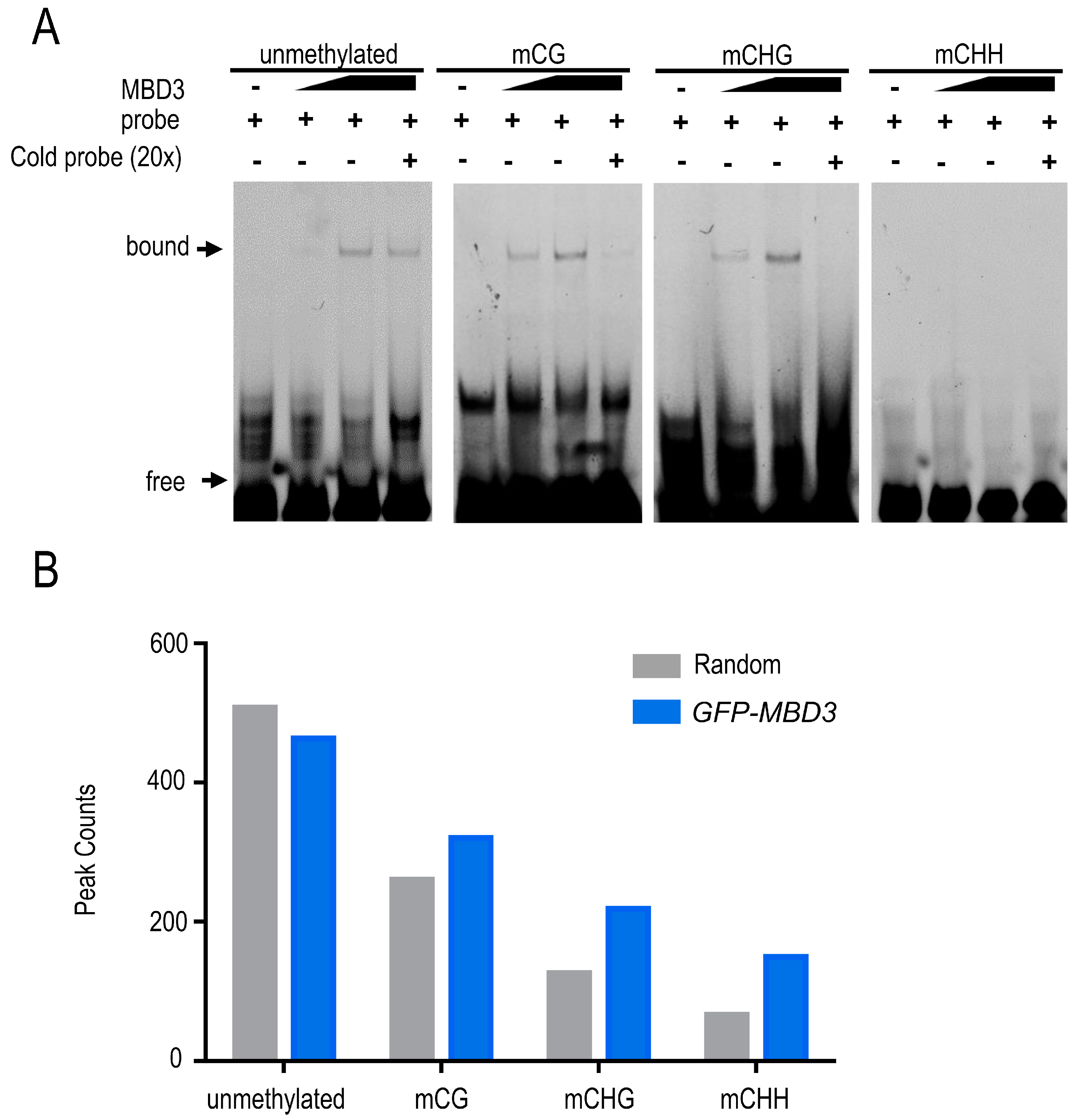
EMSA (electrophoretic mobility shift assays) was used to detect the binding potential of fusion protein GST-MBD3 and DNA sequences in vitro. The result showed that AtMBD3 had a strong binding preference to symmetrically methylated CG and CHG DNA sequences, and to unmethylated DNA sequences, except for methylated CHH (Figure 2). Although AtMBD3 could bind unmethylated DNA sequences, its binding ability was weaker than that of methylated CG and CHG. The EMSA result of fusion protein MBP-SUVH1 was used to test the labeled DNA sequence (Figure S2, Table S2).
To further determine the binding potential of MBD3 in vivo, ChIP assay was performed by expressing pJIM(Bar)-GFP:MBD3/Col-0 in seedlings. Then, a total of 711 peaks were obtained (Table S3), among which 687 had a higher DNA methylation in transgenic plants than in Col-0. A total of 320 peaks had an overlap with mCG methylation regions, 218 peaks overlapped with mCHG regions, and 149 peaks overlapped with mCHH regions. This difference also confirmed the binding preference of the MBD3 protein.

2.3. MBD3 Is Important for Plant Embryo Development

We designed two sgRNAs that target the MBD domain of MBD3 by using CRISPR/Cas9, and obtained two homozygous transgenic plants, named mbd3-1 and mbd3-2 (Figure S3A). Both mutants were found to affect embryo development. In mbd3-1 (n = 1068) and mbd3-2 (n = 1144) siliques, 5.71% and 5.77% of seeds underwent early abortion in plants, respectively. A total of 10.86% and 18.88% of seeds in the mbd3-1 and mbd3-2 plants were wrinkled, respectively (Figure 3).
When the normal embryos in mbd3-2 (n = 96) were at the cotyledon stage, 25% of abnormal seeds were at the globular stages, 31.25% at the transition stage, and 43.75% at the heart stage. This means that the embryogenesis defects in mbd3 plants lead to a delayed embryo development, and that these abnormal seed developments could be divided into three stages.

2.4. MBD3 Mutation Impairs Pollen Development

To understand why these seeds underwent abortion, we first examined the anther development in Col-0 and mbd3 mutant plants by using cryo-SEM. The anther and pollen morphologies were not aberrant obviously (Figure S3B). The pollen viability was evaluated by Alexander’s staining, and it was found to produce no effects (Figure S3C). The in vitro germination of pollen was also examined. The germination time, pollen length or quantity of mbd3 mutant were all found to have no significant difference from Col-0 (Figure S3D).
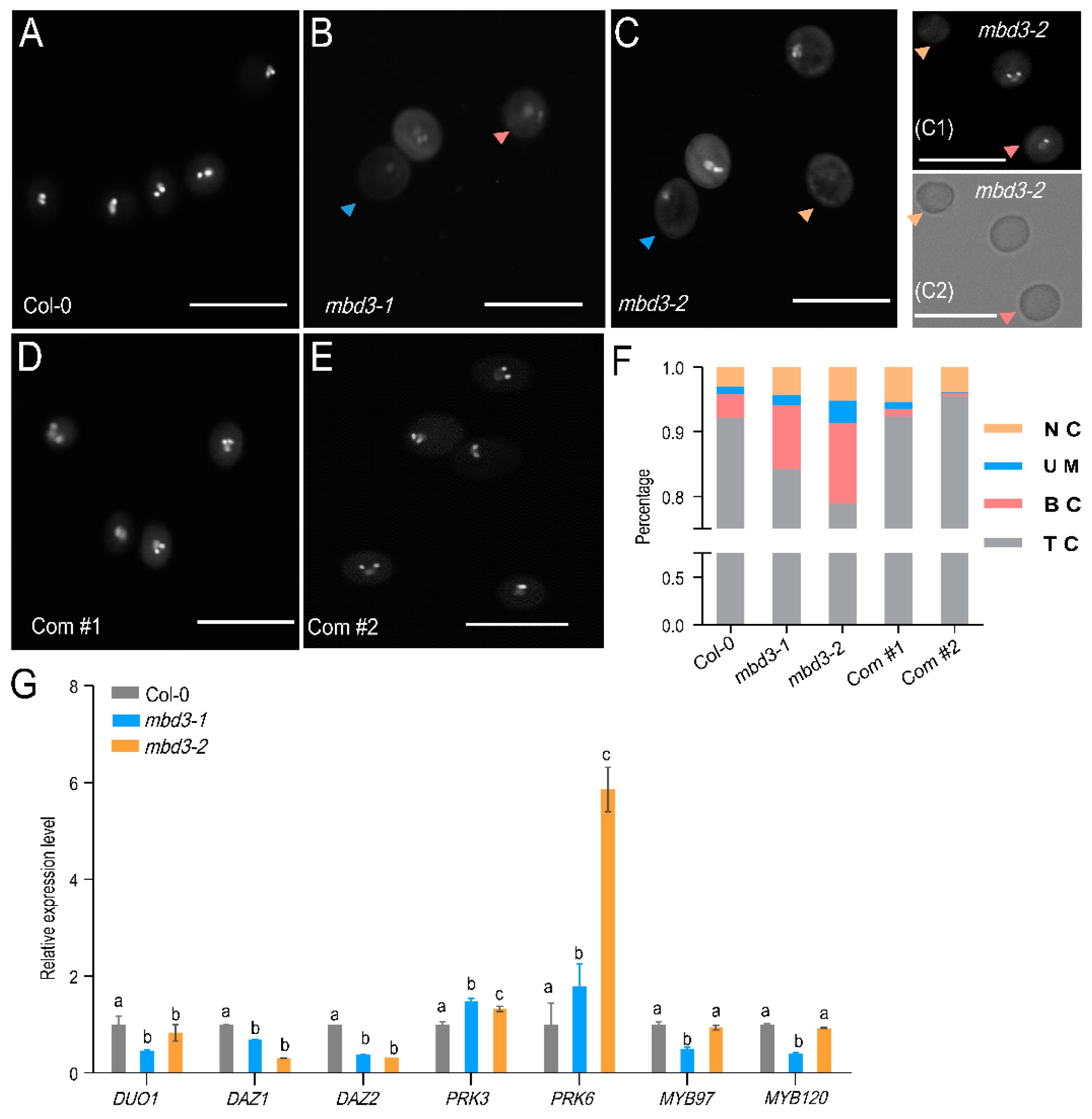
To further evaluate the effect of mbd3 mutation on pollen development. Mature pollens from Col-0 and mbd3 plants were stained with DAPI, and observed with fluorescence microscopy. About 16~21% of pollen from mbd3-1 and mbd3-2 plants all suffered a collapse in reproductive transmission. Compared with 92.17% of mature pollen production in Col-0, significantly lower percentages of 84.31% and 78.91% were found in mbd3-1 and mbd3-2 plants, respectively. Of them, about 1.61% and 3.45% contained one vegetative nucleus, lacking condensed sperm cells. Approximately 9.82% and 12.42% contained one vegetative nucleus and one condensed sperm cells. Approximately 4.25% and 5.17% did not contain any nuclei. The percentage of two complementary lines, Com #1 and Com #2, approached that of Col-0 (Figure 4).
A quantitative RT-PCR was performed to dissect the gene expression involved in the male gametophyte development of mbd3. The expression of seven genes, including male germline-specific R2R3 MYB transcription factor DUO1; two DUO1 target genes DUO1-ACTIVATED ZINC FINGER1/2 (DAZ1/2); POLLEN RECEPTOR-LIKE KINASE3 (PRK3); PRK6; and MYB transcription factors MYB97 and MYB120 showed a distinct pattern in mbd3-1 and mbd3-2 (Figure 4G). Meanwhile, duo1 mutant and daz1/daz2 double-mutant were found to block generative cell division, producing high percentages of binuclear pollen.

2.5. mbd3 Embryo Development Delay Caused by Asymmetric Divisions

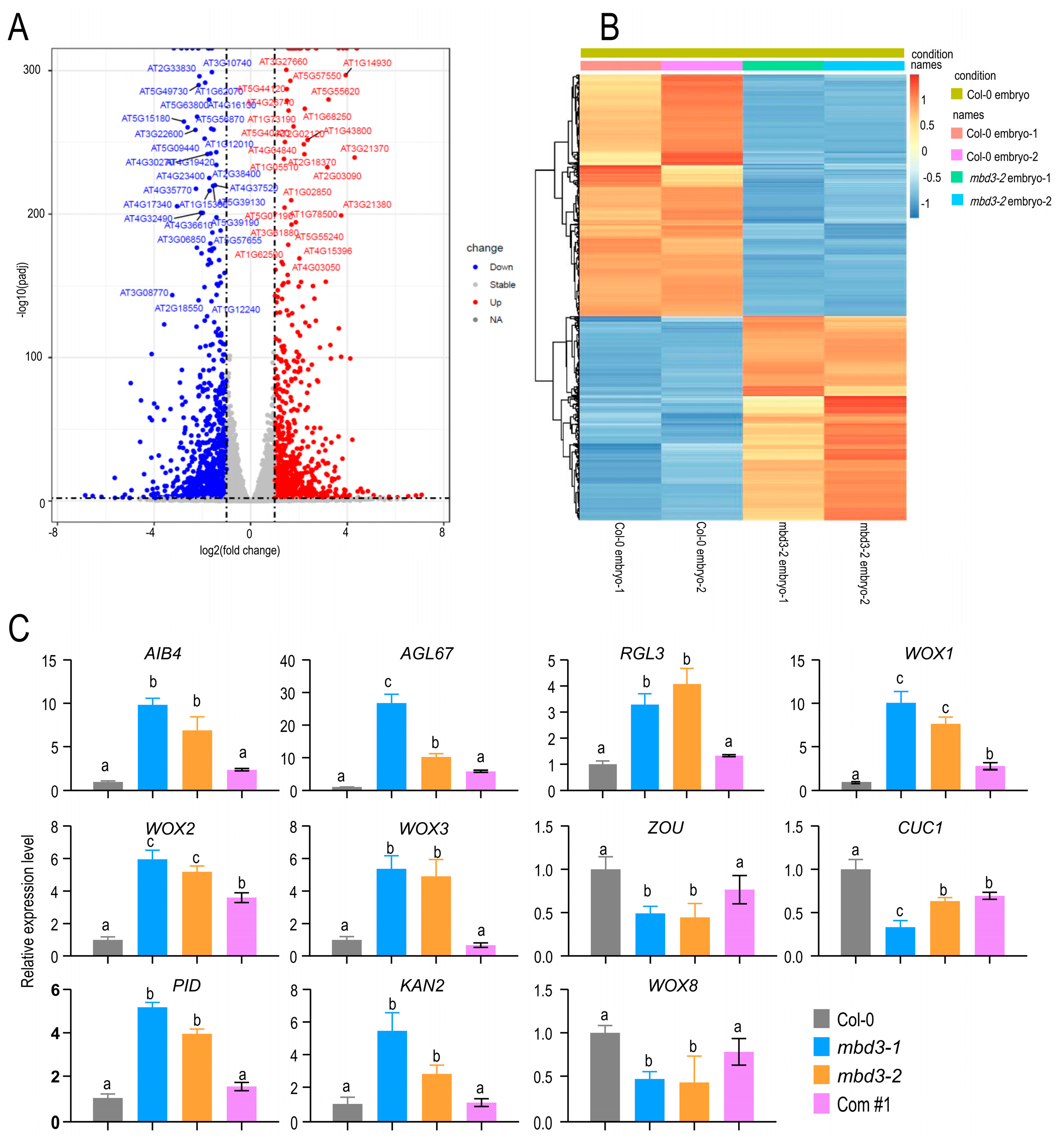
The RNA-seq results analysis of embryos between Col-0 and mbd3-2 mutant was performed, in which 950 upregulated genes and 1128 downregulated genes were found (Figure 5; Table S4). Some transcriptional factors, which were selected, were reported to be involved in embryonic development (Table S5), such as CUC1 (CUP-SHAPED COTYLEDON1) [24], WOX2 (WUSCHEL-related homeobox2), WOX8/STIMPY-LIKE/STPL [25], PID1 (PINOID) [26], ABI4 [27], RGE1/ZOU (RETARDED GROWTH OF EMBRYO1/ZHOUPI) [28], and AGL67 [29]. The expression pattern of these involved genes indicated that MBD3 may play an important role in embryo development.

2.6. Interaction Protein Screening of AtMBD3

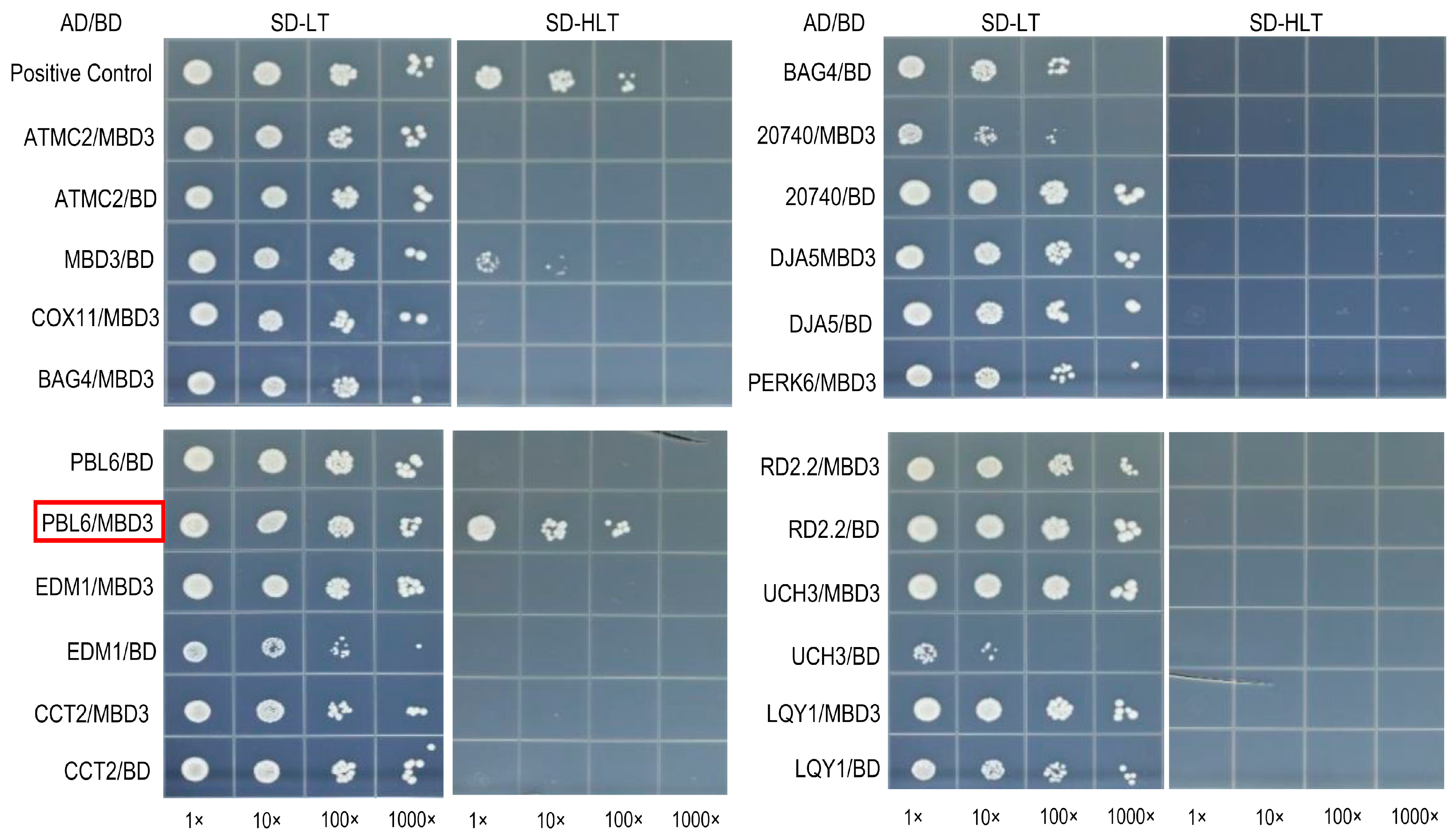
Y2H and IP-MS were used to screen the interaction proteins of MBD3. BD-MBD3 plasmid was used as bait, while the prey library was the A. thalinana cDNA library. The resulting progenies were selected on SD/-Leu/-Trp/-His/-Ade plates by adding β-galactosidase to eliminate false positives. A total of 69 positive progenies were obtained (Table S6). PCR was adopted to verify these proteins. Through sequencing and blasting the 69 verified proteins, 12 repeatedly identified positive proteins were selected and listed in Table 1. Finally, only the PBL6 (PBS1-like 6, At2G28590) was found to interact with MBD3 (Figure 6, Figure S4). PBL6 was specifically expressed in pollen, which was confirmed with the IP-MS of GFP-tagged PBL6 transgenic plants (Table 2).

3. Discussion

The MBD protein family in plants has been known to act as both “readers” and “erasers” of DNA methylation [30], but the detailed functions of its members are poorly understood. Only a few of them have been reported, and most remain to be characterized. A previous study failed to amplify the RT-PCR products of AtMBD3 due to its low expression level [22]. In this study, we successfully obtained mbd3-mutant plants and found that, despite a low expression level of AtMBD3, it played important roles in regulating the plant’s reproductive development. Similarly, mbd3 knockout in mice proved to be embryonically lethal [23]. Furthermore, we verified the binding capability of methylated and unmethylated DNA sequences of MBD3 protein both in vitro and in vivo.

3.1. MBD3 Have the Ability to Bind Sequences More than Methylated DNA

Zemach and Grafi first demonstrated that AtMBD5, AtMBD6, and AtMBD7 can bind the methylated sequences of CG dinucleotides in vitro [14]. Despite its high homology, AtMBD5 can bind both symmetrically methylated CG and asymmetrically methylated CHH sequences, whereas AtMBD6 can only bind methylated CG complexes [31]. Mammalian MBD3 was found not to be able to bind mCG [32]. Our results of AtMBD3, which has a high level of amino acid homology with mammalian MBDs, showed a capability to bind both symmetrically methylated and unmethylated DNA sequences. This result was firstly confirmed both in vitro and in vivo. The capability is more prominent in symmetrically methylated sequences. The ability of MBD5 to bind to mCG may be due to the interaction between conserved 5mCpG residues and residue pairs of R-D and R-E. However, in AtMBD3, residue pairs were changed into R-V and K-E pairs [33]. These substitutions may allow AtMBD3 to not only bind symmetrically methylated DNA sequences, but also guide AtMBD3 to target-specific DNA sequences.

3.2. MBD3 Participates in Embryo Development with Other Genes

DNA methylation affecting plant development has been found in many species. The mutation of OsMET1b leads to abnormal seed phenotypes in rice, which is associated with either viviparous germination or early embryonic lethality [3,34]. In this study, MBD3 was also found to be highly expressed in seeds. At the same time, some transcriptional factor genes involved in embryo development were expressed after transcriptome analysis, including WOX and PID (Figure 5). WOXs have been found to make cell-fate decisions during early embryogenesis. WOX1, WOX2, and WOX3 were highly expressed in mbd3 mutants, whereas WOX8 was not. Both WOX2 and WOX8 are initially co-expressed in egg cells and zygotes, and then specifically expressed in apical and basal cell lineages, respectively, after zygotic division [35]. PID regulates PIN [36], which is responsible for establishing auxin gradients in early embryogenesis [37]. Except for WOXs and PID, the other expressed genes also showed clear differences between Col-0 and mbd3 mutants, such as CUC1, AIB4, AGL67, and ZOU. Although AtMBD3 and these genes are significantly regulated by MBD3 in mbd3-mutant plants, but their relationship has not been confirmed in this paper, it is clear that they coordinately participate in embryo development in some specific manner.

3.3. MBD3 Regulates Pollen and Embryo Development with Other Genes

DNA methylation is also important for the fertility of male plants. Many differentially methylated regions have been identified during tomato fruit development [38]. In Capsella rubella, NRPD1 knockout leads to the interruption of pollen development at the microspore stage [39]. Pollen formation in A. thaliana is associated with the reprogramming of CHH methylation in pollen vegetative cells and a locus-specific restoration in sperm [40]. Our investigation found that AtMBD3 was expressed in pollen, and it obstructed the development of male germ cell division in the pollen of two mbd3 mutants. The selected genes in this paper were confirmed to be involved in germ cell development, such as DUO1, DAZ1, and DAZ2, and also found to be downregulated in the pollen of mbd3 mutants. Moreover, mbd3, duo1, and daz1/daz2 double mutants all have problems undergoing fertilization. Therefore, we suspect that MBD3 and DUO1 may coordinately play roles in the same pathway. Although DUO1 and DAZ2 have no DNA methylation site in genes or promoter regions [41], MBD3 may have the ability to bind them.
PBL6, a specific expressed gene in pollen, was found to directly interact with MBD3 to form a complex. Both PBL6 and MBD3 were also found to interact with MBD5 and MBD6 (Table 2). It has been reported that MBD5 and MBD6 are important in pollen vegetative cell development [42], which indicates that the interaction of MBD3 and PBL6 is important for pollen development, and may interact with MBD5 and MBD6. We hypothesize that through the methylation DNA sequences’ binding ability, MBD3 and PBL6 complex may be recruited into MBD5 and MBD6 binding sites and then impair pollen development. Our study provides direct clues for further research on AtMBD3 and a theoretical basis for a better understanding of AtMBDs family.

4. Materials and Methods

4.1. Plant Materials and Growth Conditions

Arabidopsis thaliana of Columbia-0 (Col-0) ecotype was used as the wild type reference, and all mutant seeds were Col-0 in this study. Seeds were sterilized with 75% ethanol and 5% sodium hypochlorite and then washed three times with 100% ddH2O. Sterilized seeds were sown on a 1/2 Murashige and Skoog (1/2 MS) solid medium containing 1% sucrose and 0.7% plant agar powder (w/v). After being wrapped with aluminum foil at 4 °C for 2 d, the plates were grown at 20–22 °C under cycles of 16 h of light and 8 h of dark in growth chambers. The 2-week-old seedlings were harvested for further experiments or transferred into soil.

4.2. Phylogenetic Analysis of MBD Genes

The search for an HMM profile of the MBD domain (PS50982) in 40 representative species, which are in 11 phyla, including Rhodophyta, Glaucophyta, Chlorophyta, Streptophyta, Phragmoplastophyta, Marchantiophyta, Bryophyta, Lycopodiophyta, Polypodiophyta, Gymnospermae, and Angiospermae (Table S5), was performed by using the InterProScan software. Phylogenetic trees were constructed in IQTREE software by using maximum likelihood and WAG+R5 with 1000 replicates.

4.3. Generation of the CRISPR Allele of mbd3 Mutants

Given the absence of available T-DNA alleles of mbd3, the CRISPR/Cas9 system was used to generate the mbd3 (AT4G00416) mutant, named as mbd3-1 and mbd3-2. The guide for a 20 bp targeting sequence of sgRNAs was selected from the CRISPR-PLANT platform (https://www.genome.arizona.edu/crispr/CRISPRsearch.html) (accessed on 11 March 2018), and cloned into a YAO promoter-driven CRISPR/Cas9 system in A. thaliana [43]. The CRISPR/Cas9 construction was transformed into the Agrobacterium tumefaciens strain GV3101, and the floral dip method was used to transform A. thaliana Col-0 plants [44]. The T1 transformants were selected on hygromycin plates. After sequencing the AT4G00416 gene from these T1 plants, three of them revealed that they had homozygous deletion, which occurred at the tail of the MBD domain.

4.4. ChIP-seq

ChIP-seq experiments were performed by using 2 g of 11-day-old seedlings with biological duplicates on Col-0 and pJIM(Bar)-GFP-linker::MBD3/Col-0 transgenic lines. The samples were fixed in 1% formaldehyde. Then, chromatin was extracted from fixed tissue and fragmented using a Bioruptor® Pico (Diagenode, Belgium) of 200–500 bp fragments. The sheared chromatin was immunoprecipitated overnight by using the following antibodies: anti-GFP (ab290, abcam, Cambridge, dilution 1:100) and antibody IgG (ab6730). Protein A Sepharose beads CL-4B (GE Healthcare, California) were used to capture immunocomplexes. Protein-A beads were washed before use. Chromatin was eluted and de-crosslinked at 65 °C overnight. DNA from immunoprecipitated chromatin was treated with RNase and proteinase K, and then purified by phenol–chloroform extraction and ethanol precipitation. For ChIP-seq, two independent biological replicates of immunoprecipitations were treated to prepare the next-generation sequencing libraries. The Ovation® Ultralow Library Systems (NuGEN, Shanghai) was used for end repair, A-tailing, and the ligation of Illumina-compatible adapters. The data analysis was performed as previously described [45]. The clean reads were mapped to TAIR10 with Bowtie2 and default parameters after using fastqc, multiqc, and trimmomatic to obtain clean reads. Bigwig files were generated by bamCoverage from deeptools, and RPKM was normalized to remove PCR duplication by PICARD. Lastly, the data quality was checked by an integrative genomics viewer (IGV).

4.5. Electrophoretic Mobility Shift Assay (EMSA)

MBD3 protein was cloned behind GST (glutathione S-transferase tag). MBD3 was amplified and cloned into bacterial expression vector pGEX-4T-1. The control protein in Figure S2 was cloned behind MBP tag. The construction for expression was transformed into Escherichia coli strain BL21 and purified with glutathione Sepharose 4B. A 98 °C water bath was performed on single-stranded nucleotide primers labeled with FAM at the 3′ end, which were used for EMSA, for 10 min and then cooled to room temperature away from light to form labeled double-stranded nucleotides primers. The labeled double-stranded nucleotides primers (160 nM) were incubated with purified AtMBD3 protein or with cold competitors (32 μM) in a total volume of 20 μL reactive system, including 4 μL binding buffer (100 mM Tris-HCl (pH 7.6), 50 mM MgCl2, 1% NP40); 5 mM DTT was added before each reaction. Binding reactions were carried out away from light for 20 min, in room temperature, and 1 μL of loading buffer (Beyotime, GS006, Shanghai) was added to each reaction. The protein–substrate complexes were resolved on 6% nondenaturing polyacrylamide gels at 80 V for at least 90 min on ice in a pre-cold 0.5× TBE buffer (44.6 mM Tris, 44.5 mM boric acid, and 1 mM disodium EDTA). After electrophoresis, the gels were detected by using a Typhoon FLA 9500.

4.6. Affinity Purification and Mass Spectrometry

The GST-MBD3 fusion protein was used for the binding assays. A total of 2.5 g WT seedlings was ground into fine powder, suspended in 10 mL lysis buffer (50 mM Tris-HCl (pH = 8.0), 230 mM NaCl, 5 mM MgCl2, 10% glycerin, 0.2% NP-40, 0.5 mM dithiothreitol (DTT), 1 mM phennylmethysulfonyl fluoride (PMSF) and proteinase inhibitor cocktail tablets (Roche, 14696200)) at 4 °C for 30 min with rotation. Then, 150 μL GST-MBD3 fusion protein was added. After incubation at 4 °C for 3 h, 13,500 g centrifugation for 15 min at 4 °C was performed. The supernatant was incubated with the GST fusion protein beads at 4 °C for 3 h. The bound proteins were boiled in SDS loading buffer after elution with lysis buffer three times, and resolved on 15% denaturing poly-acrylamide gels.
Mass spectrometric identification of the affinity-purified proteins was performed as described previously (Table 2) [46]. Protein bands on the SDS-PAGE were de-stained and in-gel digested with sequencing grade trypsin (0.5 ng/μL). Peptides were extracted by HPLC and sprayed into an LTQ Orbitrap Elite System mass spectrometer (Thermo Scientific, Massachusetts). Database search was performed on an in-house Mascot server (Matrix Science Ltd., London, UK) against the IPI (International Protein Index) A. thaliana protein database.

4.7. Microscopy Analysis

The morphologies of anther and pollen were observed using cryogenic scanning electron microscopy (cryo-SEM). The dehiscent anthers and pollens were placed on glass slides with double-sided tape, and transferred to the cold stage of chamber under vacuum for sublimation (90 °C, 5 min) after quickly freezing in liquid nitrogen and coated with platinum sputter (10 mA, 30 s) with three repetitions. Finally, the samples were transferred to another cold stage in the scanning electron microscope and imaged. The morphology of anther and pollen was observed under a spinning disc confocal microscope (Zeiss Cell Observer SD; Zeiss, Oberkochen, Germany).

4.8. Alexander Dye and 4′,6-Diamidino-2-Phenylindole Staining

An appropriate amount of Alexander dye or 4′,6-diamidino-2-phenylindole (DAPI) was dropped onto glass slides, and the dehiscent anthers were placed on glass slides with dissecting needles. The pollen were gently and quickly suspended in Alexander dye or DAPI, and then the glass slides were covered. After staining, the pollen were observed under the fluorescence microscope (Olympus BX53, Miyazaki). DAPI staining was carried out in the dark.

4.9. In Vitro Pollen Germination Analysis

The pollen of newly blooming flowers was collected and placed on the surface of a solid pollen germination medium (SPGM) (18% sucrose, 0.01% boric acid, 2 mM CaCl2, 1 mM Ca(NO3)2, 1 mM KCl, 1 mM MgSO4, adjusted to pH = 7, 1.5% agar). The medium was placed in a humid box at 22 °C for at least 6 h of incubation, and then the pollen germination was observed. The ImageJ software was used to measure and calculate the length of the pollens’ tubes and their germination.

4.10. RNA Isolation and Quantitative Real-Time RT-PCR (qPCR)

Total RNA was extracted by using Quick RNA isolation kit (Huayueyang, Beijing) and treated with in-column DNase. About 2 μg mRNA was used to amplify the first-strand cDNA by using the superScriptIII First-Strand Synthesis SuperMix Kit. The RT-qPCR was performed using SYBR Green Mastermix (Bio-Rad). An amount of 1 μL cDNA reaction mixture was used as template after dilution in a 20 μL reaction system. The reaction conditions were as follows: firstly, 95 °C for 10 min; secondly, 95 °C for 15 s and 60 °C for 1 min, this step is repeated 40 times; and thirdly, 95 °C for 15 s and 60 °C for 1 min, 95 °C for dissociation. The calculation of the expression level was used as 2−ΔΔCt. Two biological replicates from WT and mbd3 mutants were performed. All analyses of variance were performed in SPSS software by using t-test of the ANOVA program.
Mature pollen were collected from 2 to 3 bunches of unbloomed floral buds from each plant in the same tray. Nine DAP seeds were collected from about 15~20 mature siliques. The fruit pod was removed by using a dissecting needle, after placing siliques on a clean glass slide. The seeds were collected by using tweezers and put in liquid nitrogen for quick freezing.

4.11. Yeast Two-Hybrid Screening

Both pGADT7 vector containing the GLA4 AD and pGBKT7 vector containing the GLA4 BD for the yeast (Saccharomyces cerevisiae) were used for two-hybrid assay. BD-MBD3 was constructed by cloning the full-length sequence of MBD3 into the pGBKT7 vector at the NdeI and EcoR1 sites to be fused in-frame with the sequence encoding the GAL4 DNA-binding domain (BD). The A. thaliana cDNA library cloned into the prey vector pGADT7-RecAB was constructed by Clontech. The AtMBD3 interaction proteins were screened by using the yeast two-hybrid system, according to the manufacturer’s instructions (Clontech, Matchmaker GAL4 Two-Hybrid System 3 and Libraries User Manual, PT3247-1). The bait plasmid with BD-MBD3 and the prey library DNA were co-transformed into the yeast strain AH109. The resulting progenies were first selected on SD/-Leu/-Trp/-His/-Ade plates, then, β-galactosidase were added for the activity detection to eliminate false positives. Plasmids harboring positive-prey cDNAs were isolated for RT-PCR and sequenced to verify the proteins that interact with AtMBD3.

Supplementary Materials

The following supporting information can be downloaded at: https://www.mdpi.com/article/10.3390/plants12142654/s1, Figure S1: Comparison analysis of MBDs in plantae.; Figure S2: The binding potential of SUVH1 as a positive control.; Figure S3: Deletion of mbd3 has no influence on pollen viability.; Figure S4: Self-activation of MBD3.; Table S1: Primers and sgRNAs used in this study.; Table S2: The oligonucleotide sequences used in this study.; Table S3: ChIP-seq results of MBD3.; Table S4: Genes involved in embryo development. Related to Figure 5; Table S5: Selected genes from RNA-seq results of mbd3 embryo development. Related to Figure 5; Table S6: Y2H screening results of BD-MBD3.; Table S7: Identification of co-purified proteins of MBD3 by mass spectrometry. GST-MBD3 protein was used for IP/MS.; Table S8: The species involved in phylogenic tree.

Author Contributions

J.S. performed the experiments, analyzed the data, and wrote the manuscript. X.Y. performed the ChIP experiment, Y.L. and Y.M. performed bioinformatics analysis. B.Z. and A.Z. performed some material collections. J.D. and H.G. designed the project and revised the manuscript. All authors have read and agreed to the published version of the manuscript.

Funding

This research was financially supported by the Natural Science Foundation of Shaanxi Province (2022JM-099) and National College Students’ Innovation and Entrepreneurship Training Program (202210712227).

Data Availability Statement

RNA-seq and the ChIP-seq data of mbd3 have been submitted to the NCBI Sequencing Read Archive under accession no. PRJNA936625. The DNA methylome of Col-0 was previously deposited in SRR005412 [47] and PRJNA686693 [41].

Acknowledgments

We thank Qi-Jun Chen for providing the CRISPR/Cas9 system. We thank JiaJun Wang for providing pJIM(Bar): GFP-linker vector.

Conflicts of Interest

The authors declare no conflict of interest.

References

  1. Suzuki, M.M.; Bird, A. DNA methylation landscapes: Provocative insights from epigenomics. Nat. Rev. Genet. 2008, 9, 465–476. [Google Scholar] [CrossRef]
  2. Xiao, W.; Custard, K.D.; Brown, R.C.; Lemmon, B.E.; Harada, J.J.; Goldberg, R.B.; Fischer, R.L. DNA methylation is critical for Arabidopsis embryogenesis and seed viability. Plant Cell 2006, 18, 805–814. [Google Scholar] [CrossRef] [PubMed] [Green Version]
  3. Yamauchi, T.; Johzuka-Hisatomi, Y.; Terada, R.; Nakamura, I.; Iida, S. The MET1b gene encoding a maintenance DNA methyltransferase is indispensable for normal development in rice. Plant. Mol. Biol. 2014, 85, 219–232. [Google Scholar] [CrossRef] [PubMed]
  4. Cheng, C.; Tarutani, Y.; Miyao, A.; Ito, T.; Yamazaki, M.; Sakai, H.; Fukai, E.; Hirochika, H. Loss of function mutations in the rice chromomethylase OsCMT3a cause a burst of transposition. Plant J. 2015, 83, 1069–1081. [Google Scholar] [CrossRef] [Green Version]
  5. Li, Q.; Eichten, S.R.; Hermanson, P.J.; Zaunbrecher, V.M.; Song, J.; Wendt, J.; Rosenbaum, H.; Madzima, T.F.; Sloan, A.E.; Huang, J.; et al. Genetic perturbation of the maize methylome. Plant Cell 2014, 26, 4602–4616. [Google Scholar] [CrossRef] [PubMed] [Green Version]
  6. Zhang, X.; Yazaki, J.; Sundaresan, A.; Cokus, S.; Chan, S.W.L.; Chen, H.; Henderson, I.R.; Shinn, P.; Pellegrini, M.; Jacobsen, S.E.; et al. Genome-wide High-Resolution Mapping and Functional Analysis of DNA Methylation in Arabidopsis. Cell 2006, 126, 1189–1201. [Google Scholar] [CrossRef] [Green Version]
  7. Cokus, S.J.; Feng, S.; Zhang, X.; Chen, Z.; Merriman, B.; Haudenschild, C.D.; Pradhan, S.; Nelson, S.F.; Pellegrini, M.; Jacobsen, S.E. Shotgun bisulphite sequencing of the Arabidopsis genome reveals DNA methylation patterning. Nature 2008, 452, 215–219. [Google Scholar] [CrossRef] [PubMed] [Green Version]
  8. Bogdanović, O.; Veenstra, G.J.C. DNA methylation and methyl-CpG binding proteins: Developmental requirements and function. Chromosoma 2009, 118, 549–565. [Google Scholar] [CrossRef] [Green Version]
  9. Unoki, M.; Nishidate, T.; Nakamura, Y. ICBP90, an E2F-1 target, recruits HDAC1 and binds to methyl-CpG through its SRA domain. Oncogene 2004, 23, 7601–7610. [Google Scholar] [CrossRef] [Green Version]
  10. Filion, G.J.P.; Zhenilo, S.; Salozhin, S.; Yamada, D.; Prokhortchouk, E.; Defossez, P.-A. A Family of Human Zinc Finger Proteins That Bind Methylated DNA and Repress Transcription. Mol. Cell. Biol. 2006, 26, 169–181. [Google Scholar] [CrossRef] [Green Version]
  11. Bird, A.; Nan, X.; Ng, H.-H.; Johnson, C.A.; Laherty, C.D.; Turner, B.M.; Eisenman, R.N. Transcriptional repression by the methyl-CpG-binding protein MeCP2 involves a histone deacetylase complex. Nature 1998, 393, 386–389. [Google Scholar] [CrossRef]
  12. Wolffe, A.P.; Jones, P.L.; Jan Veenstra, G.C.; Wade, P.A.; Vermaak, D.; Kass, S.U.; Landsberger, N.; Strouboulis, J. Methylated DNA and MeCP2 recruit histone deacetylase to repress transcription. Nat. Genet. 1998, 19, 187–191. [Google Scholar] [CrossRef]
  13. Springer, N.M.; Kaeppler, S.M. Evolutionary Divergence of Monocot and Dicot Methyl-CpG-Binding Domain Proteins. Plant Physiol. 2005, 138, 92–104. [Google Scholar] [CrossRef] [Green Version]
  14. Zemach, A.; Gideon, G. Characterization of Arabidopsis thaliana methyl-CpG-binding domain (MBD) proteins. Plant J. 2003, 34, 565–572. [Google Scholar] [CrossRef] [PubMed]
  15. Zemach, A.; Grafi, G. Methyl-CpG-binding domain proteins in plants: Interpreters of DNA methylation. Trends Plant Sci. 2007, 12, 80–85. [Google Scholar] [CrossRef]
  16. Parida, A.P.; Sharma, A.; Sharma, A.K. AtMBD4: A methylated DNA binding protein negatively regulates a subset of phosphate starvation genes. J. Biosci. 2019, 44, 14. [Google Scholar] [CrossRef]
  17. Yano, A.; Kodama, Y.; Koike, A.; Shinya, T.; Kim, H.-J.; Matsumoto, M.; Ogita, S.; Wada, Y.; Ohad, N.I.R.; Sano, H. Interaction Between Methyl CpG-Binding Protein and Ran GTPase during Cell Division in Tobacco Cultured Cells. Ann. Bot. 2006, 98, 1179–1187. [Google Scholar] [CrossRef] [PubMed] [Green Version]
  18. Wang, C.; Dong, X.; Jin, D.; Zhao, Y.; Xie, S.; Li, X.; He, X.; Lang, Z.; Lai, J.; Zhu, J.-K.; et al. Methyl-CpG-Binding Domain Protein MBD7 Is Required for Active DNA Demethylation in Arabidopsis. Plant Physiol. 2015, 167, 905–914. [Google Scholar] [CrossRef] [PubMed] [Green Version]
  19. Lang, Z.; Lei, M.; Wang, X.; Tang, K.; Miki, D.; Zhang, H.; Mangrauthia, S.K.; Liu, W.; Nie, W.; Ma, G.; et al. The Methyl-CpG-Binding Protein MBD7 Facilitates Active DNA Demethylation to Limit DNA Hyper-Methylation and Transcriptional Gene Silencing. Mol. Cell 2015, 57, 971–983. [Google Scholar] [CrossRef] [Green Version]
  20. Stangeland, B.; Rosenhave, E.M.; Winge, P.; Berg, A.; Amundsen, S.S.; Karabeg, M.; Mandal, A.; Bones, A.M.; Grini, P.E.; Aalen, R.B. AtMBD8 is involved in control of flowering time in the C24 ecotype of Arabidopsis thaliana. Physiol. Plant 2009, 136, 110–126. [Google Scholar] [CrossRef]
  21. Peng, M.; Cui, Y.; Bi, Y.M.; Rothstein, S.J. AtMBD9: A protein with a methyl-CpG-binding domain regulates flowering time and shoot branching in Arabidopsis. Plant J. 2006, 46, 282–296. [Google Scholar] [CrossRef]
  22. Berg, A.; Meza, T.J.; Mahic, M.; Thorstensen, T.; Kristiansen, K.; Aalen, R.B. Ten members of the Arabidopsis gene family encoding methyl-CpG-binding domain proteins are transcriptionally active and at least one, AtMBD11, is crucial for normal development. Nucleic Acids Res. 2003, 31, 5291–5304. [Google Scholar] [CrossRef] [Green Version]
  23. Hendrich, B.; Guy, J.; Ramsahoye, B.; Wilson, V.A.; Bird, A. Closely related proteins MBD2 and MBD3 play distinctive but interacting roles in mouse development. Genes Dev. 2001, 15, 710–723. [Google Scholar] [CrossRef] [PubMed] [Green Version]
  24. Agarwal, P.; Kapoor, S.; Tyagi, A.K. Transcription factors regulating the progression of monocot and dicot seed development. Bioessays 2011, 33, 189–202. [Google Scholar] [CrossRef] [PubMed]
  25. Abrash, E.B.; Bergmann, D.C. Asymmetric cell divisions: A view from plant development. Dev. Cell 2009, 16, 783–796. [Google Scholar] [CrossRef] [Green Version]
  26. Benjamins, R.; Quint, A.; Weijers, D.; Hooykaas, P.; Offringa, R. The PINOID protein kinase regulates organ development in Arabidopsis by enhancing polar auxin transport. Development 2001, 128, 4057–4067. [Google Scholar] [CrossRef]
  27. Penfield, S.; Li, Y.; Gilday, A.D.; Graham, S.; Graham, I.A. Arabidopsis ABA INSENSITIVE4 Regulates Lipid Mobilization in the Embryo and Reveals Repression of Seed Germination by the Endosperm. Plant Cell 2006, 18, 1887–1899. [Google Scholar] [CrossRef] [Green Version]
  28. Yang, S.; Johnston, N.; Talideh, E.; Mitchell, S.; Jeffree, C.; Goodrich, J.; Ingram, G. The endosperm-specific ZHOUPI gene of Arabidopsis thaliana regulates endosperm breakdown and embryonic epidermal development. Development 2008, 135, 3501–3509. [Google Scholar] [CrossRef] [Green Version]
  29. Lehti-Shiu, M.D.; Adamczyk, B.J.; Fernandez, D.E. Expression of MADS-box genes during the embryonic phase in Arabidopsis. Plant Mol. Biol. 2005, 58, 89–107. [Google Scholar] [CrossRef] [PubMed]
  30. Zhang, H.; Lang, Z.; Zhu, J.-K. Dynamics and function of DNA methylation in plants. Nat. Rev. 2018, 19, 489–506. [Google Scholar] [CrossRef]
  31. Scebba, F.; Bernacchia, G.; De Bastiani, M.; Evangelista, M.; Cantoni, R.M.; Cella, R.; Locci, M.T.; Pitto, L. Arabidopsis MBD proteins show different binding specificities and nuclear localization. Plant Mol. Biol. 2003, 53, 755–771. [Google Scholar] [CrossRef] [PubMed]
  32. Ho, H.L.; McNae, L.W.; Schmiedeberg, L.; Klose, R.J.; Bird, A.; Walkinshaw, M.D. MeCP2 binding to DNA depends upon hydration at methyl-CpG. Mol. Cell 2008, 29, 525–531. [Google Scholar] [CrossRef] [PubMed] [Green Version]
  33. Ichino, L.; Boone, B.A.; Strauskulage, L.; Harris, C.J.; Kaur, G.; Gladstone, M.A.; Tan, M.; Feng, S.; Jami-Alahmadi, Y.; Duttke, S.H.; et al. MBD5 and MBD6 couple DNA methylation to gene silencing through the J-domain protein SILENZIO. Science 2021, 372, 1434–1439. [Google Scholar] [CrossRef]
  34. Hu, L.; Li, N.; Xu, C.; Zhong, S.; Lin, X.; Yang, J.; Zhou, T.; Yuliang, A.; Wu, Y.; Chen, Y.-R.; et al. Mutation of a major CG methylase in rice causes genome-wide hypomethylation, dysregulated genome expression, and seedling lethality. Proc. Natl. Acad. Sci. USA 2014, 111, 10642–10647. [Google Scholar] [CrossRef] [PubMed]
  35. Wu, X.; Chory, J.; Weigel, D. Combinations of WOX activities regulate tissue proliferation during Arabidopsis embryonic development. Dev. Biol. 2007, 309, 306–316. [Google Scholar] [CrossRef] [Green Version]
  36. Christensen, S.K.; Dagenais, N.; Chory, J.; Weigel, D. Regulation of Auxin Response by the Protein Kinase PINOID. Cell 2000, 100, 469–478. [Google Scholar] [CrossRef] [Green Version]
  37. Friml, J.Y.X.; Michniewicz, M.; Weijers, D.; Quint, A.; Tietz, O.; Benjamins, R.; Ouwerkerk, P.B.; Ljung, K.; Sandberg, G.; Hooykaas, P.J.; et al. A PINOID-dependent binary switch in apical-basal PIN polar targeting directs auxin efflux. Science 2004, 306, 862–865. [Google Scholar] [CrossRef] [Green Version]
  38. Zhong, S.; Fei, Z.; Chen, Y.R.; Zheng, Y.; Huang, M.; Vrebalov, J.; McQuinn, R.; Gapper, N.; Liu, B.; Xiang, J.; et al. Single-base resolution methylomes of tomato fruit development reveal epigenome modifications associated with ripening. Nat. Biotechnol. 2013, 31, 154–159. [Google Scholar] [CrossRef]
  39. Wang, Z.; Butel, N.; Santos-Gonzalez, J.; Borges, F.; Yi, J.; Martienssen, R.A.; Martinez, G.; Kohler, C. Polymerase IV Plays a Crucial Role in Pollen Development in Capsella. Plant Cell 2020, 32, 950–966. [Google Scholar] [CrossRef] [Green Version]
  40. Walker, J.; Gao, H.; Zhang, J.; Aldridge, B.; Vickers, M.; Higgins, J.D.; Feng, X. Sexual-lineage-specific DNA methylation regulates meiosis in Arabidopsis. Nat. Genet. 2018, 50, 130–137. [Google Scholar] [CrossRef]
  41. Liang, W.; Li, J.; Sun, L.; Liu, Y.; Lan, Z.; Qian, W. Deciphering the synergistic and redundant roles of CG and non-CG DNA methylation in plant development and transposable element silencing. New Phytol. 2022, 233, 722–737. [Google Scholar] [CrossRef] [PubMed]
  42. Ichino, L.; Picard, C.L.; Yun, J.; Chotai, M.; Wang, S.; Lin, E.K.; Papareddy, R.K.; Xue, Y.; Jacobsen, S.E. Single-nucleus RNA-seq reveals that MBD5, MBD6, and SILENZIO maintain silencing in the vegetative cell of developing pollen. Cell Rep. 2022, 41, 111699. [Google Scholar] [CrossRef]
  43. Yan, L.; Wei, S.; Wu, Y.; Hu, R.; Li, H.; Yang, W.; Xie, Q. High-efficiency genome editing in Arabidopsis using YAO promoter-driven CRISPR/Cas9 system. Mol. Plant 2015, 8, 1820–1823. [Google Scholar] [CrossRef] [PubMed] [Green Version]
  44. Clough, S.J.; Bent, A.F. Floral dip: A simplified method forAgrobacterium-mediated transformation of Arabidopsis thaliana: Floral dip transformation of Arabidopsis. Plant. J. Cell Mol. Biol. 1998, 16, 735–743. [Google Scholar] [CrossRef] [PubMed] [Green Version]
  45. Yang, T.; Wang, D.; Tian, G.; Sun, L.; Yang, M.; Yin, X.; Xiao, J.; Sheng, Y.; Zhu, D.; He, H.; et al. Chromatin remodeling complexes regulate genome architecture in Arabidopsis. Plant Cell 2022, 34, 2638–2651. [Google Scholar] [CrossRef]
  46. Li, Q.; Wang, X.; Sun, H.; Zeng, J.; Cao, Z.; Li, Y.; Qian, W. Regulation of Active DNA Demethylation by a Methyl-CpG-Binding Domain Protein in Arabidopsis thaliana. PLoS Genet. 2015, 11, e1005210. [Google Scholar] [CrossRef] [Green Version]
  47. Stroud, H.; Do, T.; Du, J.; Zhong, X.; Feng, S.; Johnson, L.; Patel, D.J.; Jacobsen, S.E. Non-CG methylation patterns shape the epigenetic landscape in Arabidopsis. Nat. Struct. Mol. Biol. 2014, 21, 64–72. [Google Scholar] [CrossRef] [Green Version]
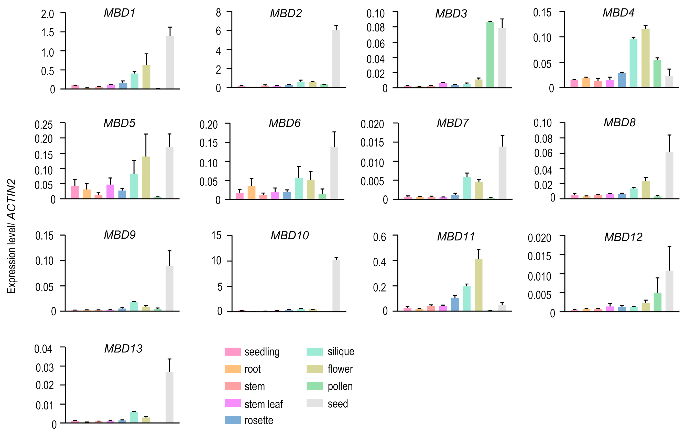
Figure 1. Gene expression of MBDs in Arabidopsis thaliana. The expression of 13 MBDs in 9 tissues of wild-type Col-0 were determined using qPCR. ACTIN2 was used as an internal control. The error bars represent standard deviations.
Plants 12 02654 g001
Figure 2. The binding capacity of MBD3 with different mCG, mCHG, mCHH, and unmethylated sequences. (A) EMSA results showed that MBD3 protein had the priority to bind mCG, mCHG, and unmethylated sequences. 3′-Fam-labeled substrates (160 nM) were incubated with concentrations of MBD3. The protein concentrations in lanes 1, 2, 3, and 4 were 0, 0.8, 1.6, and 1.6 μM, respectively. For competition analysis, 32 μM of substrates with or without methylation labeling were used as cold probes and incubated with GST-MBD3; and of 3′-Fam-labeled substrates in every fourth lane. For EMSA, at least three independent biological replicates were performed. “Bound” represents the binding band, and “free” represents the free probe. (B) ChIP-seq assay was carried out with transgenic pJIM(Bar)-GFP-linker::MBD3/Col-0 seedlings, showing the binding potential of AtMBD3 with different methylated regions. The gray column “random” was the control, representing the binding regions in Col-0. The blue columns “GFP-MBD3”represent the binding regions in MBD3.
Plants 12 02654 g002
Figure 3. Phenotype of mbd3 mutant 9 days after pollination. (A) Seeds set of Col-0, mbd3-1, mbd3-2, and complementation lines Com #1 and Com #2. Com #1 and Com #2 represent two individual proMBD3::MBD3/mbd3-2 transgenic plants. Ovules with development problems are indicated by arrows. “Blue “ and “yellow “ arrows represent early- and late-aborted seeds, respectively. Bars = 1 mm. (B) Seed abortion rates of Col-0 (n = 701), mbd3-1 (n = 1068), mbd3-2 (n = 1144), Com #1 (n = 1213), and Com #2 (n = 1156) plants. (C) Differential interference contrast (DIC) microscopy of whole-mount-cleared ovules showed abnormal cell division. (C1C3) Embryo patterns and percentages of normal and aborted embryos in mbd3-2 siliques (n = 96) are shown. (C3-1C3-3) Three stages of late-aborted embryos in mbd3-2. Bars = 100 µm.
Plants 12 02654 g003
Figure 4. Mutation of MBD3 impairs pollen development. (AE) DAPI staining of mature pollen from Col-0, mbd3-1, mbd3-2, Com #1, and Com #2 plants. Different arrows represent abnormal pollen. Red arrows represent binuclear pollen; blue arrows represent mononuclear pollen; yellow arrows represent nonnuclear pollen. Bright area of (C1) was zoomed in (C2). Bars = 50 µm. (F) Phenotypical percentages in uninucleate microspores (UM), binucleate (BC), trinucleate (TC), and nonnucleate (NC) pollen of Col-0, mbd3-1, mbd3-2, Com #1, and Com #2 plants (n > 300, for each genotype). (G) Relative gene expression levels with significant differences in the mature pollen of Col-0 and mbd3 plants. ACTIN2 was used as an internal control. Two independent experiments were conducted with similar results. Data from one experiment with four technical replicates. Error bars indicate the mean ± SD; a, b, and c are significant differences (p < 0.05, one-way ANOVA).
Plants 12 02654 g004
Figure 5. MBD3 is important for embryogenesis in A. thaliana. (A) The upregulated and downregulated genes in seeds of Col-0 and mbd3-2 mutant plants. (B) Heat map showing the different expression patterns between Col-0 and mbd3-2 mutant plants. (C) The relative expression level of different transcription factors regulated embryo development by using RT-qPCR analysis. Tublin8 was used as internal control, given that the expression of Actins changed in mbd3-2. Two independent experiments were conducted and obtained similar results. Data from one experiment with four technical replicates. Error bars indicate the mean ± SD; a, b, and c are significant differences (p < 0.05, one-way ANOVA).
Plants 12 02654 g005
Figure 6. Positive proteins selected to screen interaction with MBD3 by using Y2H. Detailed information on proteins listed in Table 1. Proteins that interacted with AD or BD were negative controls to detect self-activated. The red rectangle is the positive result. The empty wells on the images signify no direct interaction.
Plants 12 02654 g006
Table 1. Positive proteins selected to screen the interaction with MBD3.
FrequencyIDNameDescription
13AT4G25110ATMC2Encodes a type I metacaspase.
11AT2G21620RD2Encodes a gene that is induced in response to desiccation
4AT5G20740 Plant invertase/pectin methylesterase inhibitor superfamily protein
4AT3G51780BAG4Plant homologs of mammalian regulators of apoptosis
3AT2G28590PBL6Protein kinase superfamily protein
2AT3G18810PERK6Encodes a member of the PERK family
2AT4G39960DJA5Molecular chaperone Hsp40/DnaJ family protein
2AT1G75690LQY1DnaJ/Hsp40 cysteine-rich domain superfamily protein
2AT1G02410COX11Encodes a member of the cytochrome c oxidase 11 protein family
2AT4G17510UCH3Ubiquitin C-terminal hydrolase 3
2AT5G20890CCT2TCP-1/cpn60 chaperonin family protein
2AT4G11260EDM1Functions in plant-disease-resistance signaling
‘Frequency’ indicates the number of positive reactions that the protein was identified through RT-PCR.
Table 2. Identification of co-purified proteins of GFP-tagged PBL6.
IDGeneDescriptionUnique Peptides
AT2G28590.1PBL6Protein kinase superfamily protein40
AT4G00416.1MBD3Methyl-CPG-binding domain 37
AT5G59380.1MBD6Methyl-CPG-binding domain 65
AT3G46580.1MBD5Methyl-CPG-binding domain 52
AT5G20890.1CCT2TCP-1/cpn60 chaperonin family protein1
Disclaimer/Publisher’s Note: The statements, opinions and data contained in all publications are solely those of the individual author(s) and contributor(s) and not of MDPI and/or the editor(s). MDPI and/or the editor(s) disclaim responsibility for any injury to people or property resulting from any ideas, methods, instructions or products referred to in the content.