
Xanthomonas euvesicatoria-Specific Bacteriophage BsXeu269p/3 Reduces the Spread of Bacterial Spot Disease in Pepper Plants
 , , , and
Abstract
:1. Introduction
2. Results
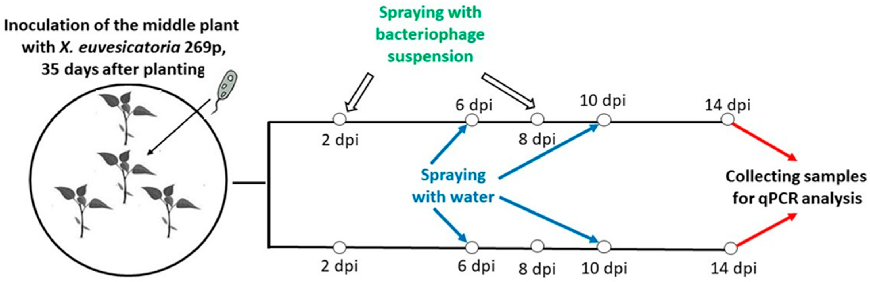
2.1. Aerosol-Mediated Transmission (Model System I)
2.1.1. X. euvesicatoria Strain 269p Was Detected in the Aerosol
2.1.2. BsXeu269p/3 Treatment Reduced Aerosol-Mediated Transmission (AMT) of X. euvesicatoria Strain 269p
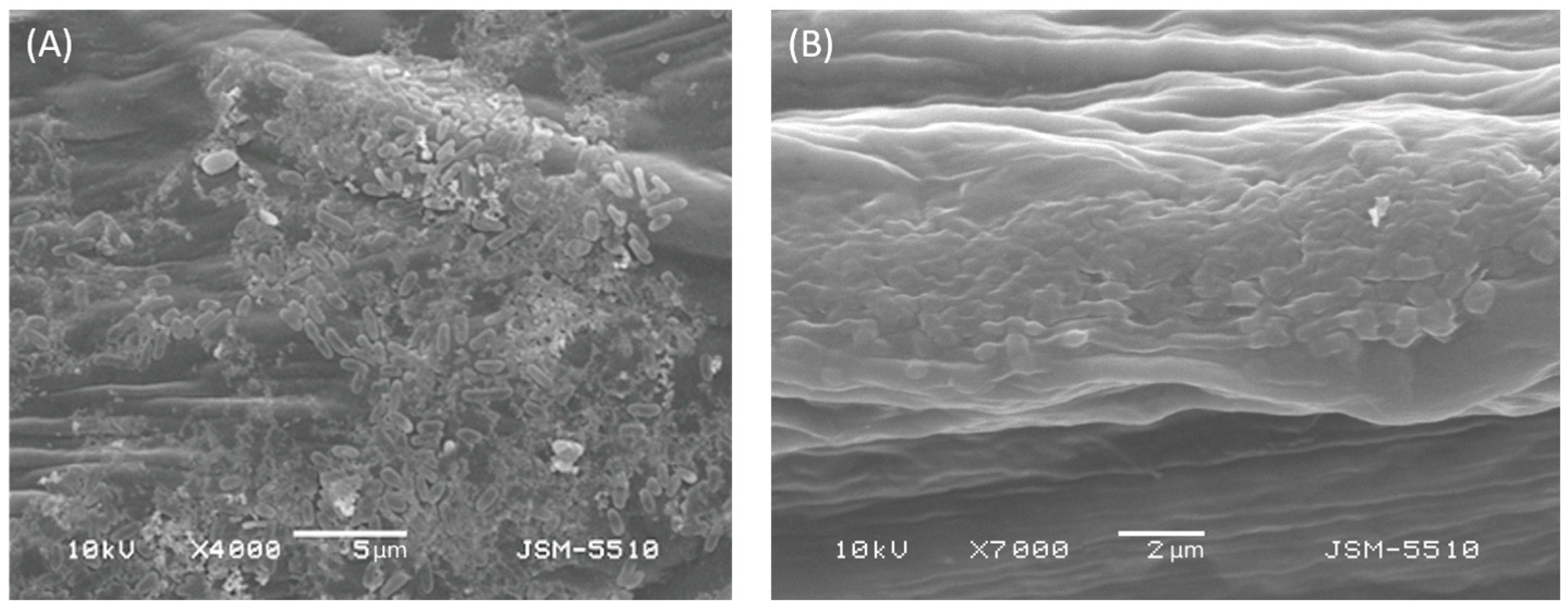
2.2. Visual Monitoring of Leaf Surface by SEM
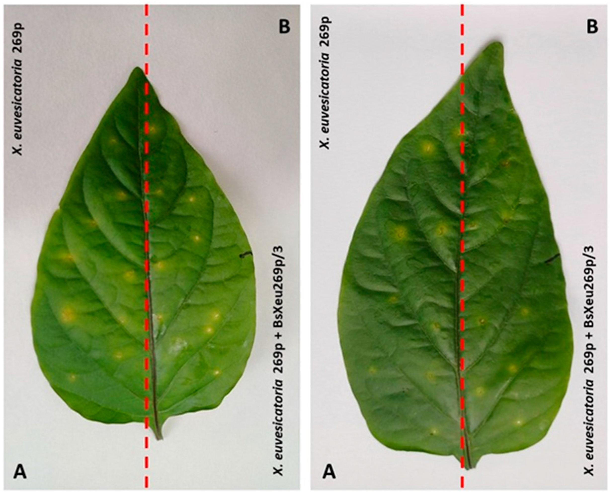
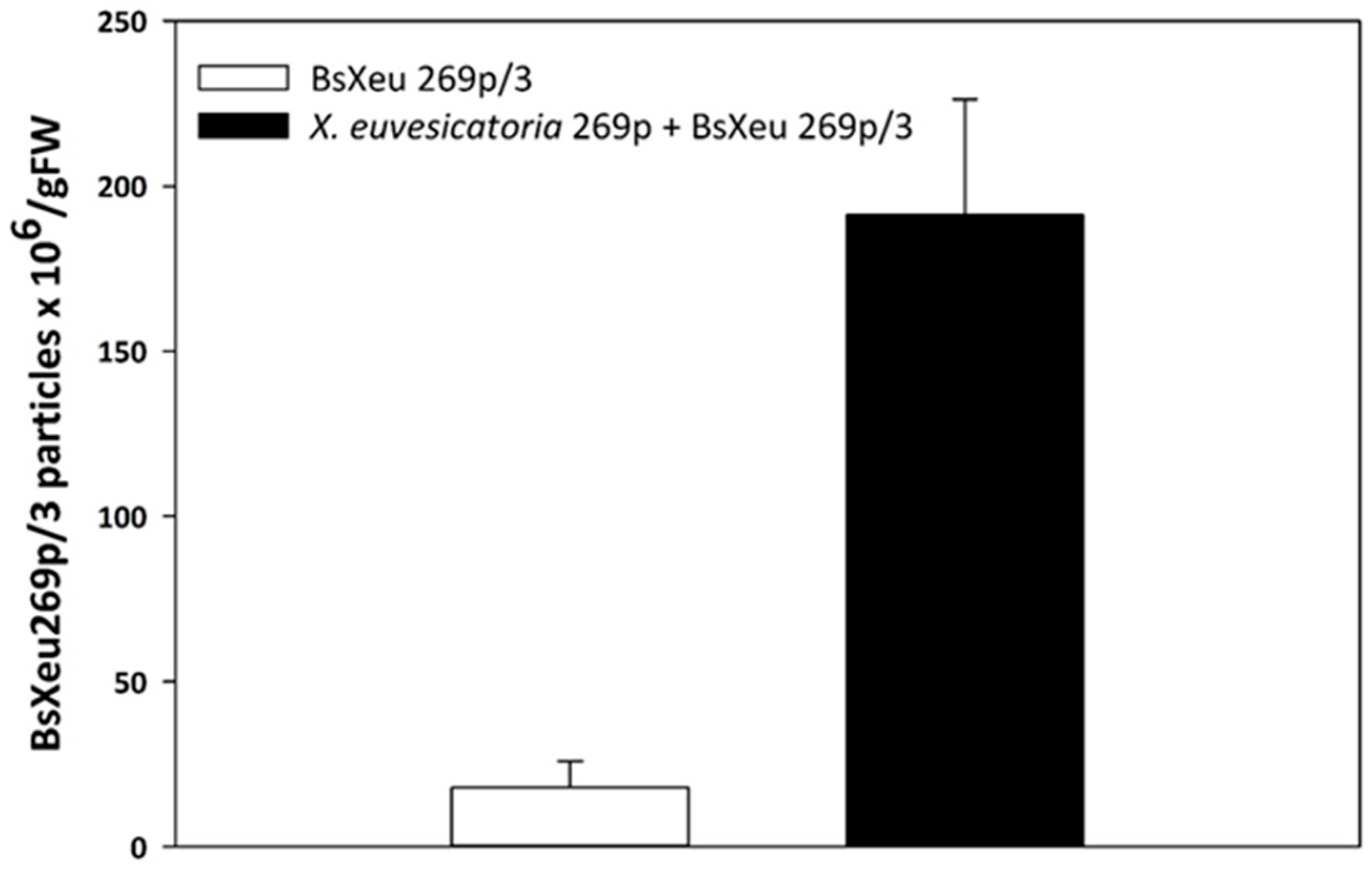
2.3. Needle Pricking (Model System II)
3. Discussion
3.1. Bacteriophages as Biocontrol Agents
3.2. X. euvesicatoria and Its Dissemination
3.3. Bacteriolytic Effect of BsXeu269p/3 on X. euvesicatoria 269p
3.4. Model System I
3.5. Model System II
4. Materials and Methods
4.1. Microorganisms
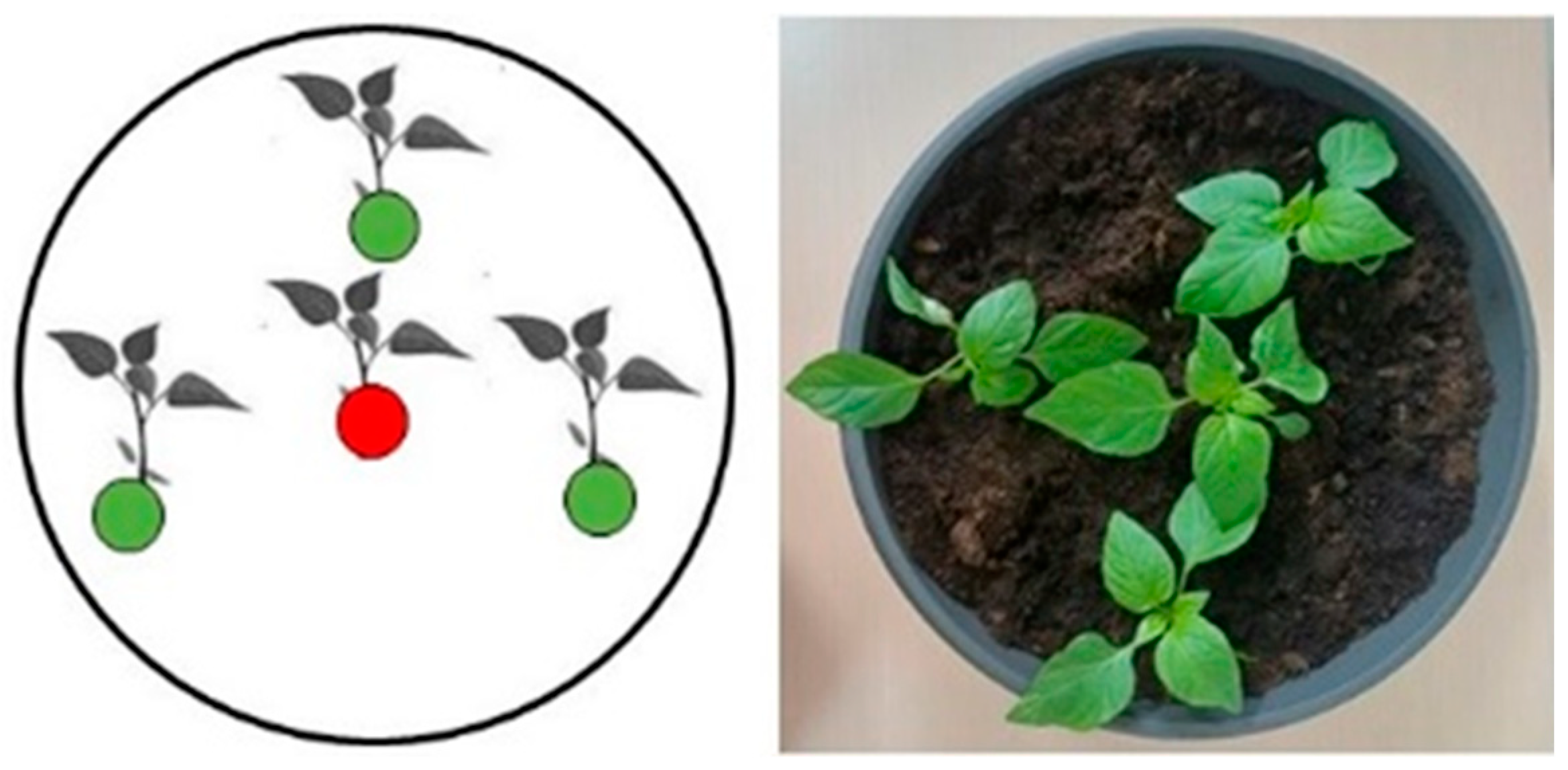
4.2. Plant Material
4.3. Model System I (Aerosol-Mediated Transmission)
4.4. Detection of X. euvesicatoria in the Aerosol
4.5. Model System II (Needle Pricking)
4.6. Scanning Electron Microscopy (SEM)
4.7. DNA Extraction
4.7.1. From Plants
4.7.2. From Swabs
4.8. Real Time PCR
4.9. TaqMan qPCR Assay
5. Conclusions
Author Contributions
Funding
Data Availability Statement
Acknowledgments
Conflicts of Interest
References
- Osdaghi, E.; Jones, J.B.; Sharma, A.; Goss, E.M.; Abrahamian, P.; Newberry, E.A.; Potnis, N.; Carvalho, R.; Choudhary, M.; Paret, M.; et al. A centenary for bacterial spot of tomato and pepper. Mol. Plant Pathol. 2021, 22, 1500–1519. [Google Scholar] [CrossRef]
- European and Mediterranean Plant Protection Organization. PM 7/110 (1) Xanthomonas spp. (Xanthomonas euvesicatoria, Xanthomonas gardneri, Xanthomonas perforans, Xanthomonas vesicatoria) causing bacterial spot of tomato and sweet pepper. Bull. OEPP/EPPO 2013, 43, 7–20. [Google Scholar] [CrossRef]
- EFSA PLH Panel (EFSA Panel on Plant Health). Scientific opinionon the pest categorisation of Xanthomonas campestris pv. Vesicatoria (Doidge) dye. EFSA J. 2014, 12, 3720. [Google Scholar]
- Bogatzevska, N.; Vancheva-Ebben, T.; Vasileva, K.; Kizheva, Y.; Moncheva, P. An overview of the diversity of pathogens causing bacterial spot on tomato and pepper in Bulgaria. Bulg. J. Agri. Sci. 2021, 27, 137–146. [Google Scholar]
- Bogatzevska, N.; Stoimenova, E.; Mitrev, S. Bacterial and virus diseases spread in Bulgaria and Macedonia on field and greenhouse pepper. Plant Prot. Skopje 2007, 18, 17–21. [Google Scholar]
- Kizheva, Y.; Vancheva, T.; Hristova, P.; Stoyanova, M.; Bogatzevska, N.; Moncheva, P. Diversity of Xanthomonas spp. causal agents of bacterial spot on pepper and tomato plants in Bulgaria. Biotechnol. Biotechnol. Equip. 2011, 25, 98–104. [Google Scholar] [CrossRef]
- Kizheva, Y.; Vancheva, T.; Stoyanova, M.; Bogatzevska, N.; Moncheva, P.; Hristova, P. 16S-23S ITS rDNA PCR-RFLP Approach as a tool for identification and differentiation of bacterial spot-causing Xanthomonas. J. Plant Pathol. 2016, 98, 645–649. [Google Scholar] [CrossRef]
- Kizheva, Y.; Urshev, Z.; Rasheva, I.; Vancheva, T.; Hristova, P.; Bogatzevska, N.; Moncheva, P. PFGE: A tool for examination of heterogeneity between the bacterial spot-causing xanthomonads of tomato plant in Bulgaria. Z. Naturforsch. C 2018, 73, 257–264. [Google Scholar] [CrossRef]
- Kizheva, Y.; Vancheva-Ebben, T.; Hristova, P.; Bogatzevska, N.; Moncheva, P. First report of Xanthomonas euvesicatoria on tomato in Bulgaria. C. R. Acad. Bulg. Sci. 2020, 73, 140–146. [Google Scholar]
- Vancheva, T.; Stoyanova, M.; Tatyozova, M.; Bogatsevska, N.; Moncheva, P. Sub-species diversity of Xanthomonas euvesicatoria Bulgarian and Macedonian strains from pepper. Biotechnol. Biotechnol. Equip. 2014, 28, 592–601. [Google Scholar] [CrossRef]
- Vancheva, T.; Stoyanova, M.; Tasheva—Terzieva, E.; Bogatzevska, N.; Moncheva, P. Molecular methods for diversity assessment among xanthomonads of Bulgarian and Macedonian pepper. Braz. J. Microbiol. 2018, 49, 246–259. [Google Scholar] [CrossRef] [PubMed]
- Aysan, Y.; Sahin, F. Occurrence of bacterial spot disease, caused by Xanthomonas axonopodis pv. vesicatoria, on pepper in the Eastern Mediterranean region of Turkey. Plant Pathol. 2003, 52, 781. [Google Scholar] [CrossRef]
- Mitrev, S.; Kovacevic, B. Characterisation of Xanthomonas axonopodis pv. vesicatoria isolated from peppers in Macedonia. J. Plant Pathol. 2006, 88, 321–324. [Google Scholar]
- Bashan, Y.; Azaizeh, M.; Diab, S.; Yunis, H.; Okon, Y. Crop loss of pepper plants artificially infected with Xanthomonas campestris pv. vesicatoria in relation to symptom expression. Crop Prot. 1985, 4, 77–84. [Google Scholar] [CrossRef]
- La Torre, A.; Iovino, V.; Caradonia, F. Copper in plant protection. Phytopathol. Mediterr. 2018, 57, 201–236. [Google Scholar]
- Lugo, A.J.; Elibox, W.; Jones, J.B.; Ramsubhag, A. Copper resistance in Xanthomonas campestris pv. campestris affecting crucifers in Trinidad. Eur. J. Plant Pathol. 2013, 136, 61–70. [Google Scholar] [CrossRef]
- Bondarczuk, K.; Piotrowska-Seget, Z. Molecular basis of active copper resistance mechanisms in Gram-negative bacteria. Cell Biol. Toxicol. 2013, 29, 397–405. [Google Scholar] [CrossRef] [PubMed]
- Bosmans, L.; Van Calenberge, B.; Paeleman, A.; Moerkens, R.; Wittemans, L.; Van Kerckhove, S.; De Mot, R.; Lievens, B.; Rediers, H. Efficacy of hydrogen peroxide treatment for control of hairy root disease caused by rhizogenic agrobacteria. J. Appl. Microbiol. 2016, 121, 519–527. [Google Scholar] [CrossRef]
- Mahlein, A.-K.; Kuska, M.T.; Behmann, J.; Polder, G.; Walter, A. Hyperspectral sensors and imaging technologies in phytopathology: State of the art. Annu. Rev. Phytopathol. 2018, 56, 535–558. [Google Scholar] [CrossRef]
- van Lenteren, J.C.; Bolckmans, K.; Köhl, J.; Ravensberg, W.J.; Urbaneja, A. Biological control using invertebrates and microorganisms: Plenty of new opportunities. BioControl 2018, 63, 39–59. [Google Scholar] [CrossRef]
- Holtappels, D.; Lavigne, R.; Huys, I.; Wagemans, J.; Holtappels, D.; Lavigne, R.; Huys, I.; Wagemans, J. Protection of phage applications in crop production: A patent landscape. Viruses 2019, 11, 277. [Google Scholar] [CrossRef] [PubMed]
- Holtappels, D.; Fortuna, K.; Lavigne, R.; Wagemans, J. The future of phage biocontrol in integrated plant protection for sustainable crop production. Curr. Opin. Biotechnol. 2021, 68, 60–71. [Google Scholar] [CrossRef] [PubMed]
- Petrzik, K.; Vacek, J.; Kmoch, M.; Binderová, D.; Brázdová, S.; Lenz, O.; Ševcík, R. Field Use of Protective Bacteriophages against Pectinolytic Bacteria of Potato. Microorganisms 2023, 11, 620. [Google Scholar] [CrossRef] [PubMed]
- Bertolini, E.; Figàs-Segura, À.; Álvarez, B.; Biosca, E.G. Development of TaqMan Real-TimePCR Protocols for Simultaneous Detection and Quantification of the Bacterial Pathogen Ralstonia solanacearum and Their Specific Lytic Bacteriophages. Viruses 2023, 15, 841. [Google Scholar] [CrossRef] [PubMed]
- Zaczek-Moczydłowska, M.A.; Young, G.K.; Trudgett, J.; Fleming, C.C.; Campbell, K.; O’Hanlon, R. Genomic characterization, formulation and efficacy in planta of a siphoviridae and podoviridae protection cocktail against the bacterial plant pathogens Pectobacterium spp. Viruses 2020, 12, 150. [Google Scholar] [CrossRef] [PubMed]
- Buttimer, C.; McAuliffe, O.; Ross, R.P.; Hill, C.; O’Mahony, J.; Coffey, A. Bacteriophages and bacterial plant diseases. Front. Microbiol. 2017, 8, 34. [Google Scholar] [CrossRef] [PubMed]
- Sevic, M.; Gasic, K.; Ignjatov, M.; Mijatovic, M.; Prokic, A.; Obradovic, A. Integration of biological and conventional treatments in control of pepper bacterial spot. Crop Prot. 2019, 119, 46–51. [Google Scholar] [CrossRef]
- Ogunyemi, S.O.; Chen, J.; Zhang, M.; Wang, L.; Masum, M.M.I.; Yan, C.; An, Q.; Li, B.; Chen, J. Identification and characterization of five new OP2-related Myoviridae bacteriophages infecting different strains of Xanthomonas oryzae pv. oryzae. J. Plant Pathol. 2019, 101, 263–273. [Google Scholar] [CrossRef]
- Balogh, B.; Nga, N.T.T.; Jones, J.B. Relative Level of Bacteriophage Multiplication in vitro or in Phyllosphere May Not Predict in planta Efficacy for Controlling Bacterial Leaf Spot on Tomato Caused by Xanthomonas perforans. Front. Microbiol. 2018, 9, 2176. [Google Scholar] [CrossRef]
- Gašić, K.; Kuzmanović, N.; Ivanović, M.; Prokić, A.; Šević, M.; Obradović, A. Complete genome of the Xanthomonas euvesicatoria specific bacteriophage KΦ1, its survival and potential in control of pepper bacterial spot. Front. Microbiol. 2018, 9, 2021. [Google Scholar] [CrossRef]
- Balogh, B.; Jones, J.B.; Momol, M.T.; Olson, S.M.; Obradovic, A.; King, P.; Jackson, L. Improved Efficacy of Newly Formulated Bacteriophages for Management of Bacterial Spot on Tomato. Plant Dis. 2003, 87, 949–954. [Google Scholar] [CrossRef] [PubMed]
- Iriarte, F.B.; Balogh, B.; Momol, M.T.; Smith, L.M.; Wilson, M.; Jones, J.B. Factors affecting survival of bacteriophage on tomato leaf surfaces. Appl. Environ. Microbiol. 2007, 73, 1704–1711. [Google Scholar] [CrossRef]
- Iriarte, F.A.; Wernsing, M.H.; Jackson, L.E.; Balogh, B.; Hong, J.A.; Momol, M.; Jones, J.; Vallad, G. Soil-based systemic delivery and phyllosphere in vivo propagation of bacteriophages. Bacteriophage 2012, 4, e23530. [Google Scholar] [CrossRef] [PubMed]
- Kolozsváriné Nagy, J.; Schwarczinger, I.; Künstler, A.; Pogány, M.; Király, L. Penetration and translocation of Erwinia amylovoraspecific bacteriophages in apple—a possibility of enhanced control of fire blight. Eur. J. Plant Pathol. 2015, 142, 815–827. [Google Scholar] [CrossRef]
- Kizheva, Y.; Urshev, Z.; Dimitrova, M.; Bogatzevska, N.; Moncheva, P.; Hristova, P. Phenotypic and Genotypic Characterization of Newly Isolated Xanthomonas euvesicatoria-Specific Bacteriophages and Evaluation of Their Biocontrol Potential. Plants 2023, 12, 947. [Google Scholar] [CrossRef] [PubMed]
- Brankova, L.; Shopova, E.; Ivanov, S.; Kizheva, Y.; Urshev, Z.; Rasheva, I.; Aleksandrov, V.; Dimitrova, L.; Dimitrova, M.; Hristova, P. The Involvement of Oxidative Stress in the Localization of Bacterial Spot Infection in Pepper Plants. J. Biosci. 2023; accepted. [Google Scholar]
- Ignjatov, M.; Gasic, K.; Ivanovic, M.; Sevic, M.; Obradovic, A.; Milosevic, M. Characterization of Xanthomonas euvesicatoria strains pathogens of pepper in Serbia. Pestic. Fitomed. 2010, 25, 139–149. [Google Scholar] [CrossRef]
- Potnis, N.; Timilsina, S.; Strayer, A.; Shantharaj, D.; Barak, J.D.; Paret, M.L.; Vallad, G.E.; Jones, J.B. Bacterial spot of tomato and pepper: Diverse Xanthomonas species with a wide variety of virulence factors posing a worldwide challenge. Mol. Plant Pathol. 2015, 16, 907–920. [Google Scholar] [CrossRef]
- An, S.Q.; Potnis, N.; Dow, M.; Vorhölter, F.J.; He, Y.Q.; Becker, A.; Teper, D.; Li, Y.; Wang, N.; Bleris, L.; et al. Mechanistic insights into host adaptation, virulence and epidemiology of the phytopathogen Xanthomonas. FEMS Microbiol. Rev. 2020, 44, 1–32. [Google Scholar] [CrossRef]
- Wang, F.; Xiao, Y.; Lu, Y.; Deng, Z.; Deng, X.; Lin, L. Bacteriophage Lytic Enzyme P9ly as an Alternative Antibacterial Agent Against Antibiotic-Resistant Shigella dysenteriae and Staphylococcus aureus. Front. Microbiol. 2022, 13, 821989. [Google Scholar] [CrossRef]
- Bock, C.H.; Cook, A.Z.; Parker, P.E.; Gracham, J.H. Short-distance dispersal of splashed bacteria of Xanthomonas citri subsp. citri from canker-infected grapefruit tree canopies in turbulent wind. Plant. Pathol. 2012, 61, 829–836. [Google Scholar] [CrossRef]
- Pinheiro, L.A.M.; Pereira, C.; Esther Barreal, M.; Pablo Gallego, P.; Balcao, V.M.; Almeida, A. Use of phage phi 6 to inactivate Pseudomonas syringae pv. actinidiae in kiwifruit plants: In Vitro and ex vivo experiments. Appl. Microbiol. Biotechnol. 2020, 104, 1319–1330. [Google Scholar] [CrossRef] [PubMed]
- Skliros, D.; Papazoglou, P.; Gkizi, D.; Paraskevopoulou, E.; Katharios, P.; Goumas, D.E.; Tjamos, S.; Flemetakis, E. In planta interactions of a novel bacteriophage against Pseudomonas syringae pv. tomato. Appl. Microbiol. Biotechnol. 2023, 107, 3801–3815. [Google Scholar] [CrossRef] [PubMed]
- Mazzocco, A.; Waddell, T.E.; Lingohr, E.; Johnson, R.P. Enumeration of bacteriphages using the small drop plaque assay system. In Bacteriophages: Methods and Protocols; Clokie, M.R., Kropinski, A.M., Eds.; Humana Press: New York, NY, USA, 2009; Volume 501, pp. 81–85. [Google Scholar] [CrossRef]
- Moretti, C.; Amatulli, M.T.; Buonaurio, R. PCR-based assay for the detection of Xanthomonas euvesicatoria causing pepper and tomato bacterial spot. Lett. Appl. Microbiol. 2009, 49, 466–471. [Google Scholar] [CrossRef] [PubMed]
- Ganeva, T.; Stefanova, M.; Koleva, D.; Ruiz, S.R. Isolation and recrystallization of epicuticular waxes from Sorbus and Cotoneaster leaves. Open Life Sci. 2015, 10, 497–504. [Google Scholar] [CrossRef]
- Untergasser, A.; Cutcutache, I.; Koressaar, T.; Ye, J.; Faircloth, B.C.; Remm, M.; Rozen, S.G. Primer3-new capabilities and interfaces. Nucleic Acids Res. 2012, 40, e115. [Google Scholar] [CrossRef]


| Percentage of Positive Identification on qPCR | Total Number of Probes | Bacterial Cell ×104 per mL | |
|---|---|---|---|
| X. euvesicatoria 269p | 53 | 46 | 20.4 ± 5.1 |
| X. euvesicatoria 269p + BsXeu269p/3 | 25 | 57 | 4.3 ± 0.8 |
| X. euvesicatoria 269p | X. euvesicatoria 269p + BsXeu269p/3 | ||
|---|---|---|---|
| 1 | Percentage of positive identification on qPCR | 61.1 | 54.2 |
| Total number of probes | 27 | 32 | |
| Bacterial cell × 106/gFW | 13.2 ± 1.6 | 10.3 ± 1.3 | |
| 2 | Percentage of positive identification on qPCR | 42.9 | 31.6 |
| Total number of probes | 35 | 38 | |
| Bacterial cell × 106/gFW | 24.7 ± 2 | 8.2 ± 1.5 | |
| 3 | Percentage of positive identification on qPCR | 35.1 | 30 |
| Total number of probes | 37 | 50 | |
| Bacterial cell × 106/gFW | 20.1 ± 1.6 | 6.6 ± 0.9 | |
| Av | Percentage of positive identification on qPCR | 46.4 | 38.6 |
| Total number of probes | 99 | 120 | |
| Bacterial cell × 106/gFW | 19.4 ± 1.6 | 8.4 ± 1.6 |
| Bacterial Cell × 107/gFW | Percentage of the Reduction of Bacterial Cells after BsXeu269p/3 Treatment | ||
|---|---|---|---|
| X. euvesicatoria 269p | X. euvesicatoria 269p + BsXeu269p/3 | ||
| 1 | 1581.3 ± 233.2 | 765.2 ± 54.5 | 51.6 |
| 2 | 255 ± 63.6 | 96.8 ± 28 | 62 |
| 3 | 986.7 ± 210.6 | 325.2 ± 90.6 | 66.9 |
| Av | 982.5 ± 155.9 | 395.7 ± 92.7 | 59.7 |
| Primers | 5′-Oligo Seq-3′ | Target Microorganisms | References |
|---|---|---|---|
| Xeu 2.4 | CTGGGAAACTCATTCGCAGT | X. euvesicatoria 269p | [45] |
| Xeu 2.4 mod | GTTTATTGCCGGCTATCTAATCC | X. euvesicatoria 269p | This study |
| Xeu 2.5 | TTGTGGCGCTCTTATTTCCT | X. euvesicatoria 269p | [45] |
| Xeu 2.4 probe | FAM-TCGGTGTTCCCTGCGACAC-TAMRA | X. euvesicatoria 269p | This study |
| YK-MjCPF1 | CCAGTACGCCGTGTTCATCA | BsXeu269p/3 | This study |
| YK-MjCPR1 | GTCGTTCATCAGGCCGTAGT | BsXeu269p/3 | This study |
| YK-MjCPF1/R1 | HEX-CGGCATCGACTGGGCTGCGCGCC-BHQ1 | BsXeu269p/3 | This study |
Disclaimer/Publisher’s Note: The statements, opinions and data contained in all publications are solely those of the individual author(s) and contributor(s) and not of MDPI and/or the editor(s). MDPI and/or the editor(s) disclaim responsibility for any injury to people or property resulting from any ideas, methods, instructions or products referred to in the content. |
© 2023 by the authors. Licensee MDPI, Basel, Switzerland. This article is an open access article distributed under the terms and conditions of the Creative Commons Attribution (CC BY) license (https://creativecommons.org/licenses/by/4.0/).
Share and Cite
Shopova, E.; Brankova, L.; Ivanov, S.; Urshev, Z.; Dimitrova, L.; Dimitrova, M.; Hristova, P.; Kizheva, Y. Xanthomonas euvesicatoria-Specific Bacteriophage BsXeu269p/3 Reduces the Spread of Bacterial Spot Disease in Pepper Plants. Plants 2023, 12, 3348. https://doi.org/10.3390/plants12193348
Shopova E, Brankova L, Ivanov S, Urshev Z, Dimitrova L, Dimitrova M, Hristova P, Kizheva Y. Xanthomonas euvesicatoria-Specific Bacteriophage BsXeu269p/3 Reduces the Spread of Bacterial Spot Disease in Pepper Plants. Plants. 2023; 12(19):3348. https://doi.org/10.3390/plants12193348
Chicago/Turabian StyleShopova, Elena, Liliana Brankova, Sergei Ivanov, Zoltan Urshev, Lyudmila Dimitrova, Melani Dimitrova, Petya Hristova, and Yoana Kizheva. 2023. "Xanthomonas euvesicatoria-Specific Bacteriophage BsXeu269p/3 Reduces the Spread of Bacterial Spot Disease in Pepper Plants" Plants 12, no. 19: 3348. https://doi.org/10.3390/plants12193348