Article

Phenolic Compounds from Wild Plant and In Vitro Cultures of Ageratina pichichensis and Evaluation of Their Antioxidant Activity

by
Elizabeth Alejandra Motolinia-Alcántara
1,
Adrián Marcelo Franco-Vásquez
2,
Antonio Nieto-Camacho
3,
Roberto Arreguín-Espinosa
2,
Mario Rodríguez-Monroy
4,
Francisco Cruz-Sosa
1,* and
Angelica Román-Guerrero
1,*
1
Departamento de Biotecnología, Universidad Autónoma Metropolitana-Iztapalapa, Av. Ferrocarril de San Rafael Atlixco 186, Col. Leyes de Reforma 1a. Sección, Alcaldía Iztapalapa, Ciudad de Mexico 09310, Mexico
2
Departamento de Química de Biomacromoléculas, Instituto de Química, Universidad Nacional Autónoma de México, Coyoacán, Ciudad de Mexico 04510, Mexico
3
Laboratorio de Pruebas Biológicas, Instituto de Química, Universidad Nacional Autónoma de México, Coyoacán, Ciudad de Mexico 04510, Mexico
4
Centro de Desarrollo de Productos Bióticos (CEPROBI), Departamento de Biotecnología, Instituto Politécnico Nacional (IPN), Yautepec 62731, Mexico
*
Authors to whom correspondence should be addressed.
Plants 2023, 12(5), 1107; https://doi.org/10.3390/plants12051107
Submission received: 19 January 2023 / Revised: 24 February 2023 / Accepted: 25 February 2023 / Published: 1 March 2023
(This article belongs to the Special Issue Antioxidant Activity of Medicinal and Aromatic Plants)

Abstract

:
Ageratina pichichensis, is commonly used in traditional Mexican medicine. In vitro cultures were established from wild plant (WP) seeds, obtaining in vitro plant (IP), callus culture (CC), and cell suspension culture (CSC) with the objective to determine total phenol content (TPC) and flavonoids (TFC), as well as their antioxidant activity by DPPH, ABTS and TBARS assays, added to the compound’s identification and quantification by HPLC, from methanol extracts obtained by sonication. CC showed significantly higher TPC and TFC than WP and IP, while CSC produced 2.0–2.7 times more TFC than WP, and IP produced only 14.16% TPC and 38.8% TFC compared with WP. There were identified compounds such as epicatechin (EPI), caffeic acid (CfA), and p-coumaric acid (pCA) in in vitro cultures that were not found in WP. The quantitative analysis shows gallic acid (GA) as the least abundant compound in samples, whereas CSC produced significantly more EPI and CfA than CC. Despite these results, in vitro cultures show lower antioxidant activity than WP, for DPPH and TBARS WP > CSC > CC > IP and ABTS WP > CSC = CC > IP. Overall, A. pichichensis WP and in vitro cultures produce phenolic compounds with antioxidant activity, especially CC and CSC, which are shown to be a biotechnological alternative for obtaining bioactive compounds.

1. Introduction

Ageratina pichichensis, commonly known as “axihuitl”, is a herb belonging to the Asteraceae family, endemic to the Americas and native to Mexico. This plant has been used especially in Mexican folk medicine for treating skin infections, wounds, indigestion and gastritis, and is supported by several scientific studies where the extracts from its aerial parts possess antimicrobial, antifungal, anti-inflammatory, anti-ulcer and healing properties [1,2,3,4,5]. Due to these pharmacological activities, clinical trials have been conducted and demonstrated its efficiency in the treatment of onychomycosis, chronic leg ulcers, diabetic foot ulcers, and vulvovaginal candidiasis [6,7,8,9,10,11]. Because the pharmacological potential of any medicinal plants depends on the secondary metabolites they produce, the extracts from the aerial parts of A. pichichensis have stated the presence of a wide variety of bioactive compounds including benzofurans, benzochromenes, terpenes and phenolic compounds [12].
One of the most abundant phytochemical groups in plants are the phenolic compounds, produced trough the secondary metabolism in plants with remarkable physiological and morphological importance. Phenolic compounds range from simple molecules, such as phenolic acids, to large molecules such as tannins, and include different classes such as flavonoids, lignans, xanthones, anthraquinones and hydroxycinnamic acids [13,14]. Among the functional activities displayed by the phenolic compounds, the antioxidant properties are the most sought after and highlighted, due to their ability to stabilize and deactivate free radicals, before they attack targets in biological cells, preventing the action of these radical molecules, involved in the development of multiple acute and chronic disorders such as diabetes, atherosclerosis, aging, immunosuppression, and neurodegeneration [15,16].
Obtaining bioactive compounds from wild plants (WP) generally represents less than 1% wt., which leads to an overexploitation, threat or extinction of plant species [17,18]. In vitro plant cell culture is an environmentally friendly alternative to increase the production of bioactive compounds as systems that ensure the continuous production of bioactive compounds [19,20]. In vitro plant cell cultures include callus culture (CC) and cell suspension culture (CSC), the latter being one of the most widely used, as it has excellent scaling attributes for the production of bioactive compounds [21,22]. In vitro cultures of A. pichichensis have been obtained which produce compounds with anti-inflammatory activity [23]; however, there are no reports of the production of phenolic compounds and their possible antioxidant activity.
Therefore, the aim of this work is to establish in vitro cultures of A. pichichensis, such as germinated plant in vitro (IP), CC and CSC, initiated and derived from WP, to enhance the production of bioactive compounds, identifying the major compounds extracted from each culture, and evaluating the antioxidant activity by 2,2-diphenylpicrylhydrazyl (DPPH), 2,2′-azino-bis(3-ethylbenzothiazoline-6-sulfonic acid) (ABTS), and thiobarbituric acid reactive substances (TBARS) assays.

2. Results and Discussion

2.1. Establishment of the In Vitro Cultures of A. pichichensis

The in vitro cultures of A. pichichensis were obtained from the germination of seeds excised from WP (Figure 1a). The resultant IP (Figure 1b) were 2 months old and used to obtain the callus cultures (CC) from explants of nodal segments (Figure 1c). In turn, they were cultured for two months and those with a friable appearance were disaggregated and incorporated into a liquid culture medium for obtaining the A. pichichensis cell suspension culture (CSC; Figure 1d).
For callus induction, the effect of the concentration of plant growth regulators (PGR): auxin (NAA; α-naphthaleneacetic acid) and cytokinin (KIN; kinetin) in the explants from nodal segments on the morphogenetic response is shown in Table 1. The morphogenetic response was evident at day 20 of culture. Of all the treatments tested, only eight displayed a positive response regarding the callus formation, where T3, T4, and T7 showed percentages higher than 50%. From these data, as preliminary information for establishing the better PGR conditions for callus induction, the higher percentage of callus formation was obtained when NAA and KIN were 4.52–6.79 μM and 0.46 μM, respectively, in treatments T3 and T4. In both cases, the callus formed developed friable appearance and light-yellow to beige color (Figure 2). Treatment T7 also displayed the simultaneous production of callus and roots while T3 and T4 showed the production of callus and plants. Several studies have reported that the combination of auxin and cytokinin favors the production of callus [24,25]. Similar results were obtained by Sánchez-Ramos et al. [26] for the establishment CC from leaf explants of A. pichichensis.
The treatments T3 and T4 were transferred to Murashige and Skoog (MS) liquid medium using the same concentration of PGR as in the CC (Figure 2A). The establishment of CSC was determined based on the performance of better callus disaggregation. Treatment T4 displayed greater disaggregation and dispersion of the cells that T3 at day seven of culture. Cell disaggregation when CSC processes are looked for is a parameter of great relevance for ensuring the mass transfer of nutrients to each individual cell, ensuring the cell viability, and improving the production of bioactive compounds. In this work, due to the best disaggregation of callus in the liquid medium, treatment T4 was selected for the establishment of CSC, growth kinetics were maintained for 21 days (Figure 2B), reaching a maximum biomass of 8.32 ± 0.22 g/L at 16 days, a growth rate (µ) of 0.1576 days−1 and a doubling time (dt) of 4.39 days−1. The following was performed, and the phytochemical characterization was performed at 12 and 16 days of culture. Similar results were obtained for Sánchez-Ramos et. al. [23] on CSC of A. pichichensis obtained from leaf explants at concentrations of, differing in this study, in which CSCs were obtained from nodal segment explants.

2.2. Quantification of Phenolic Compounds

Phenolics and flavonoids are a type of natural compounds present in plants considered of great relevance due to their important medicinal attributes and benefits for humans [27]. Quantification of phenolic compounds (phenols and flavonoids) was conducted on methanolic extracts from different plant tissues and cultures of A. pichichensis: aerial parts from WP, IP (2-month-old), CC (20-days-old), and CSC cultures (12-days-old). Regarding the total phenolic content (TPC), the different extracts of A. pichichensis yielded TPC values from 14.24 to 122.12 mg gallic acid equivalent/g dried biomass weight (mgGAE/gDW), where the CC from treatments T3 and T4 showed significantly higher TPC (p < 0.05), followed by WP and CSC at 12 days of culture, and the CSC at 16 days of culture, being IP the extract with the lowest content, as shown in Table 2. Differences between the treatments indicate that probably the culture of CC for 20 days may induce higher stress to the culture due to the consumption of substrate compounds, eliciting the major synthesis of secondary metabolites, approaching to that found in the extracts from the wild plant. For the CSC, the time of culture influenced the yielding of TPC, suffering a reduced content when culture went from 12 to 16 days. In the case of IP cultured for 2 months, the extract exhibited the lowest TPC yield; this could be associated to the requirement of the plant to reach its adult state (around 41–100 days for some plants of the Asteraceae family) [28,29], and being grown in adequate and aseptic conditions; the plant is growing in a process directed towards its primary metabolism for the preferential production of biomass, over the production of secondary metabolites. Total flavonoid content (TFC) ranged between 29.72 to 209.93 mg quercetin equivalent/gDW (mgQCT/gDW), as described in Table 2, and following the same behavior found for TPC. The CC from T3 and CSC at 12 days of culture showed significantly higher TFC yields (p < 0.05), followed by CC obtained from T4 > CSC at 16 days of culture and WP > IP. Once again, IP showed the lowest yield for TFC, agreeing to the lowest content of TPC. Even though WP and PI are closely related to the plant of origin, the differences observed in the production of phenolic compounds and flavonoids are associated with the differences in the biotic and abiotic stress conditions to which plants are exposed. These differences in the environment promote the activation of secondary metabolism as an adaptive response to ensure plant survival under stress conditions [30,31]. Similar results have been obtained on the production of phenolic compounds in plants growing under natural conditions versus in vitro plants of Baccharis antioquensis (Asteraceae) [32], Tanacetum parthenium (Asteraceae) [30], Thalictrum foliolosum (Ranunculaceae) [33], Argylia radiata (Bignoniaceae) [31], and Passiflora alata Curtis (Passifloraceae) [34]. On the other hand, in vitro plant cell cultures have been employed as a strategy to increase the production of bioactive compounds, ensuring their continuous production [19,20]. In particular, CSC is considered a simple and cost-effective method that offers the possibility for obtaining bioactive compounds on a large scale [22,35]. Moreover, in vitro CC extract exhibited significantly higher TPC and TFC (p < 0.05) than WP and IP treatments, agreeing with the reported by Castro et al. [19] for in Byrsonima verbascifolia (Malpighiaceae), Coimbra et al. [36] for Pyrostegia venustaa (Bignoniaceae), and Arciniega-Carreón et al. [37] for Ibervillea sonorae (Cucurbitaceae), who achieved higher production of phenolic compounds in the in vitro CC.
Based on these results, CSC produced 2.0–2.7 times more TFC than WP, whereas IP (2-months-old) produced only 38.8% TFC of that obtained in WP. Increased production of phenolic compounds in CSC has also been reported by Arciniega-Carreón et al. [37] in cultivation of Ibervillea sonorae (Cucurbitaceae), where TPC production was 10–20% higher than in extracts obtained from plant growing under natural conditions. Dary Mendoza et al. [20] reported that TPC and TFC in suspension cell cultures of Thevetia peruviana (Apocynaceae) were higher compared to explants obtained from the plant growing under natural conditions and in vitro callus culture. The high content of phenolic compounds in in vitro cultures may be due to the presence of growth regulators, especially the combination of auxins and cytokinin, which favor the production of bioactive compounds [38,39]. These results indicate that in vitro techniques promote and increase the production of phenolic compounds in A. pichichensis.

2.3. HPLC Analysis

The extracts of A. pichichensis and its cultures were analyzed by HPLC. Previous reports about the phytochemical analysis of species belonging to the Asteraceae family inform the presence of phenolic acids, flavonoids, and terpenoids compounds. Therefore, the elucidation of major bioactive compounds in A. pichichensis and its cultures was based on the use of different standard compounds where only six were identify: gallic acid (GA), caffeic acid (CfA), p-coumaric acid (pCA), catechin (CAT), rutin (RUT), and epicatechin (EPI) as shown in Figure 3.
Among the identified compounds, the phenolic acid GA (retention time, rt = 4.908 min), and flavonoids CAT (rt = 12.375 min) and RUT (rt = 17.33 min) were found in WP extracts, whereas only the flavonoid EPI (rt = 14.255 min) was identified for IP extracts. On the other hand, extracts obtained from CC-T3 and CC-T4 were similar in the elution profiles, differing on the height of some signals, being higher for CC-T4. In these extracts, three phenolic acids were identified: GA, CfA (rt = 13.858 min), and pCA (rt = 17.741 min), and the flavonoid EPI. The use of CSC as source of bioactive compounds when culture during 12 o 16 days show chromatographic patterns such as those exhibited by CC extracts, differing in the absence of CfA in CSC-T4 12d. These differences could be related to the different metabolic routes and growth stages of the in vitro culture, that lead to enhancing or limiting the production of specific metabolites in response to different stress factors. The production of phenolic acids and flavonoids in vitro cultures is frequently reported [36,40,41,42,43,44,45]. In this study, GA was found in all extracts, except IP, while the flavonoids EPI and pCA were produced in all the in vitro cultures of A. pichichensis. According to the quantitative analysis of the phenolic compounds identified in the extracts (Table 3), there are significant differences between the concentration of each compound in the different treatments, excepting EPI in CC-T3 and CC-T4. The compound with the lowest abundance is GA (CC-T4 > CC-T3 > WP > CSC-T4 16d > CSC-12d) and EPI the most abundant (CSC-T4 16d > CSC-12d > CC-T4 = CC-T3 > IP). According to these results, in vitro cultures of A. pichichensis were not only able to produce compounds that the WP did not, but also CSCs produced more of them (GA and CAT). Finally, it is highlighted that CSCs produce more compounds such as: CAT, CfA, EPI and pCA in contrast to CC. Similar results were obtained by Ali et al. [46] in CC and CSC of Artemisia absinthium L. (Asteraceae) and Modarres et al. [42] in Salvia leriifolia Benth (Lamiaceae) cultures. The identified compounds in this study have been investigated for their medicinal and biological properties as antioxidants [47,48], anticancer [49], antimicrobial [50] and anti-inflammatory agents [51]. Despite other compounds were found in the chromatographic characterization, further characterization should be done for elucidating the complete outlook of the bioactive compounds synthesized in the cell cultures treatments, identifying the major compounds and their contribution to the functional activities.

2.4. Antioxidant Activity

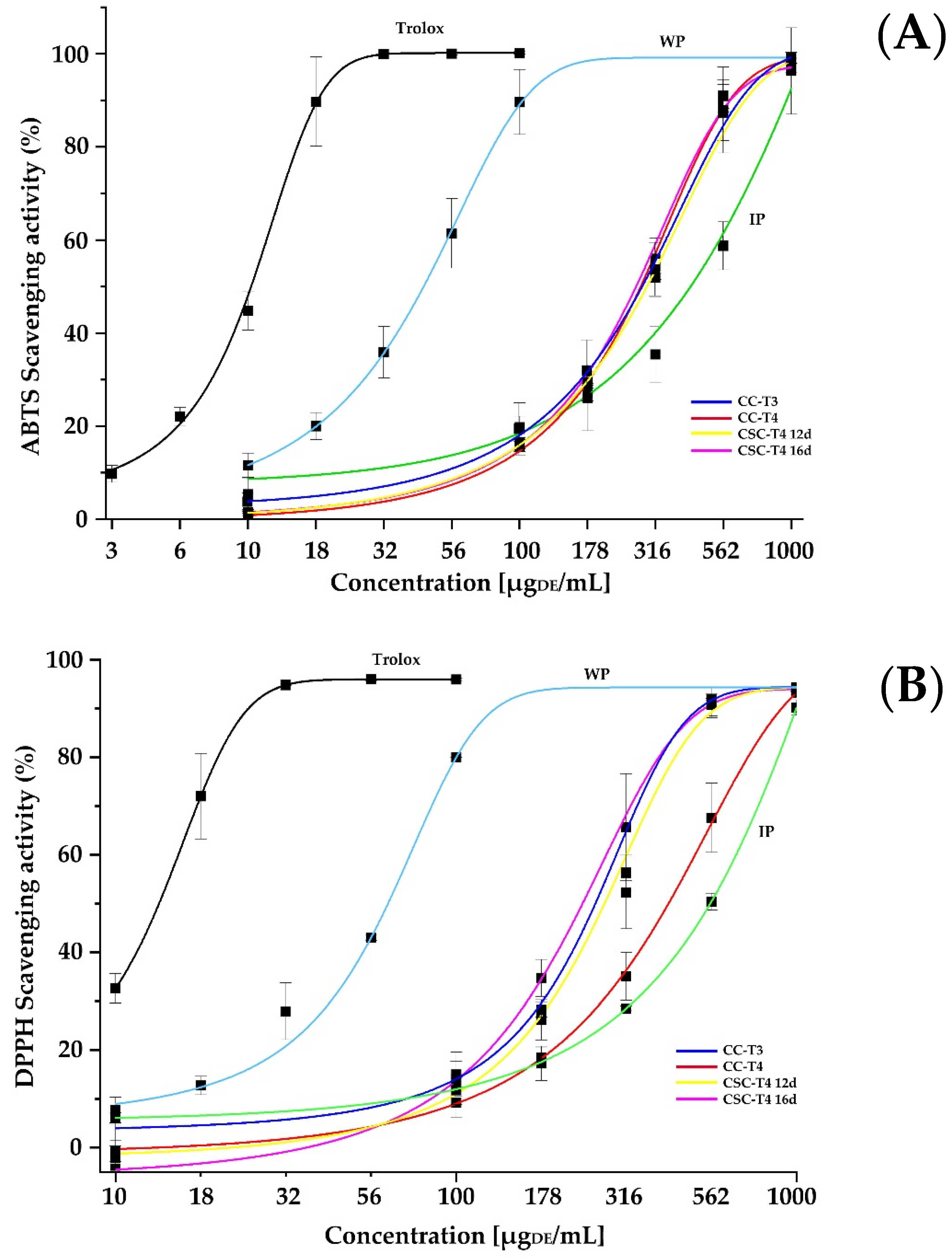
The advantage of using in vitro cell culture is mainly justified by the improvement of bioactive molecules production in shorter times, where the antioxidant activity is one of the most important biological functions, the extracts of A. pichichensis were characterized by the capacity of their extracted compounds to scavenge or inhibit free radical molecules through ABTS, DPPH, and TBARS assays. Figure 4 shows the antioxidant capacity of the different extracts of A. pichichensis against different free radicals when exposed to concentrations between 10 to 1000 μg dried extract (DE)/mL. Trolox was used as control.
For ABTS inhibition, the extract from WP required lower concentration for inhibiting this radical reaching a plateau around 178 µgDE/mL, but higher than Trolox. For the rest of treatments, when the antioxidant concentration was below 100 µgDE/mL, the better antioxidant capacity was performed by IP, followed by CC-T3, and the rest of treatments, but above this concentration, the ihibition of ABTS was modified substantially by the CC and CSC treatments, with a significant reduction of antioxidant capacity in the IP extracts. Regarding the scavenging of the DPPH radical, Trolox and WP extract showed the same trend observed for ABTS, but for this radical CSC-T4 12d, CSC-T4 16d, and CC-T3 were grouped with better antioxidant capacity than CC-T4 and IP extracts. In the case of TBARS, Trolox and WP extract required lower concentrations for the inhibition of lipid peroxidation, followed by CSC-T4 12d, CC-T3, CSC-T4 16d, CC-T4, and IP extracts as those with the lesser antioxidant capacity. For all the treatments, the percentage of scavenging effect on the DPPH and ABTS radicals was increased with increasing the concentration of A. pichichensis extracts, where the highest percentage of DPPH radical inhibition was obtained in a concentration of 1000 μgDE/mL, with percentages of 94.32 ± 0.66, 90.14 ± 1.58, 93.14 ± 0.39 and 94.16 ± 0.19% for WP, IP, CC and CSC extracts, respectively. In the case of Trolox, the highest percentage inhibition was achieved at a concentration of 56.25 μgDE/mL. Table 2 shows the values for the IC50 from each extract required for inhibiting the 50% of the initial concentration of free radical.
Because free radicals are known to play a definite role in a wide variety of pathological manifestations. Antioxidants fight against free radicals and protect us from various diseases. They perform their action either by scavenging the reactive oxygen species or protecting the antioxidant defense mechanisms [15]. The different extracts from A. pichichensis showed statistical differences (p < 0.05) in antioxidant activity, their DPPH radical scavenging potential was as follows: WP > CSC > CC > IP, corresponding to IC50 values in Table 2. Similar results were obtained in the ABTS assay, the highest percent inhibition was obtained at a concentration of 1000 μgDE/mL with 99.16 ± 1.19, 96.29 ± 9.27, 98.47 ± 0.56 and 94.47 ± 0.56% for WP, IP, CC and CSC extracts, respectively. The ABTS radical scavenging potential was as follows: WP > CS = CC > IP, agreeing to IC50 (Table 2). Similar results were reported by Esmaeili et al. [52], when evaluating the antioxidant activity of plant extract growing in natural conditions and in vitro cultures of Trifolium pratense L. (plant and callus), the antioxidant potential reported was as follows: WP > CC > PI.
Differences observed between the two assays (DPPH and ABTS) could be related to the chemical nature of the bioactive substances contained in each extract, and to the nature of the radical used to measure this property. In the case of the ABTS radical cation (ABTS+), it is reactive against most antioxidants, and is soluble in both aqueous and organic solvents, allowing it to be applied in a wide range of pH and/or ionic strength [53], whereas the DPPH assay is based mainly on the electron transfer reaction, and the interactions between antioxidants-DPPH· radicals are determined by the structural conformation of the antioxidants. Thus, some compounds react very rapidly with DPPH·, reducing the number of DPPH· molecules in correspondence to the number of available hydroxyl groups in the antioxidant compound. Nevertheless, this mechanism seems to be more complex and the observed reactions are slower in most antioxidants [54]. Hence, the extracts methanolic of A. pichichensis contain diverse types of phenolic compounds with different polarities and solvent affinities, affecting the scavenging capacity of the crude extracts.
The antioxidant activity of A. pichichensis extracts, evaluated by TBARS assay (Figure 4C), also showed that the highest percentage inhibition was obtained at a concentration of 1000 μgDE/mL, reaching 94.48 ± 2.17, 62.25 ± 3.08, 90.65 ± 4.17 and 94.04 ± 2.45% for WP, IP, CC, and CSC extracts, respectively. According to the IC50 (Table 2), the activity of the extracts was as follows WP >CSC>CC>CC>IP. In general, the best results of antioxidant activity were in WP extracts followed by in vitro cultures of A. pichichensis (CC and CSC), in the three assays evaluated (DPPH, ABTS and TBARS), especially in the inhibition of lipid peroxidation, evaluated by the production of TBARS. It is noteworthy that even when CC-T3 extract displayed better antioxidant activity, its capacity to disaggregate for leading to CSC cultures was significantly lower than CC-T4, therefore, this culture can be considered as an alternative for good antioxidant compounds production when no CSC establishment is required. Regarding the CSC when cultured during 12 or 16 days, no significant differences were observed for DPPH and ABTS, being significantly better for CSC-T4 12d for TBARS assay. Similar results were reported by Costa et al. [55] on water/ethanol extracts of Lavandula viridis L’Hér growing under natural conditions and callus culture, which were efficient in inhibiting lipid peroxidation. The results obtained in this study suggest that A. pichichensis extracts contain bioactive compounds that are capable of donating hydrogen to a free radical to eliminate potential damage.

2.5. Correlation between Phenolic Content and Antioxidant Activity

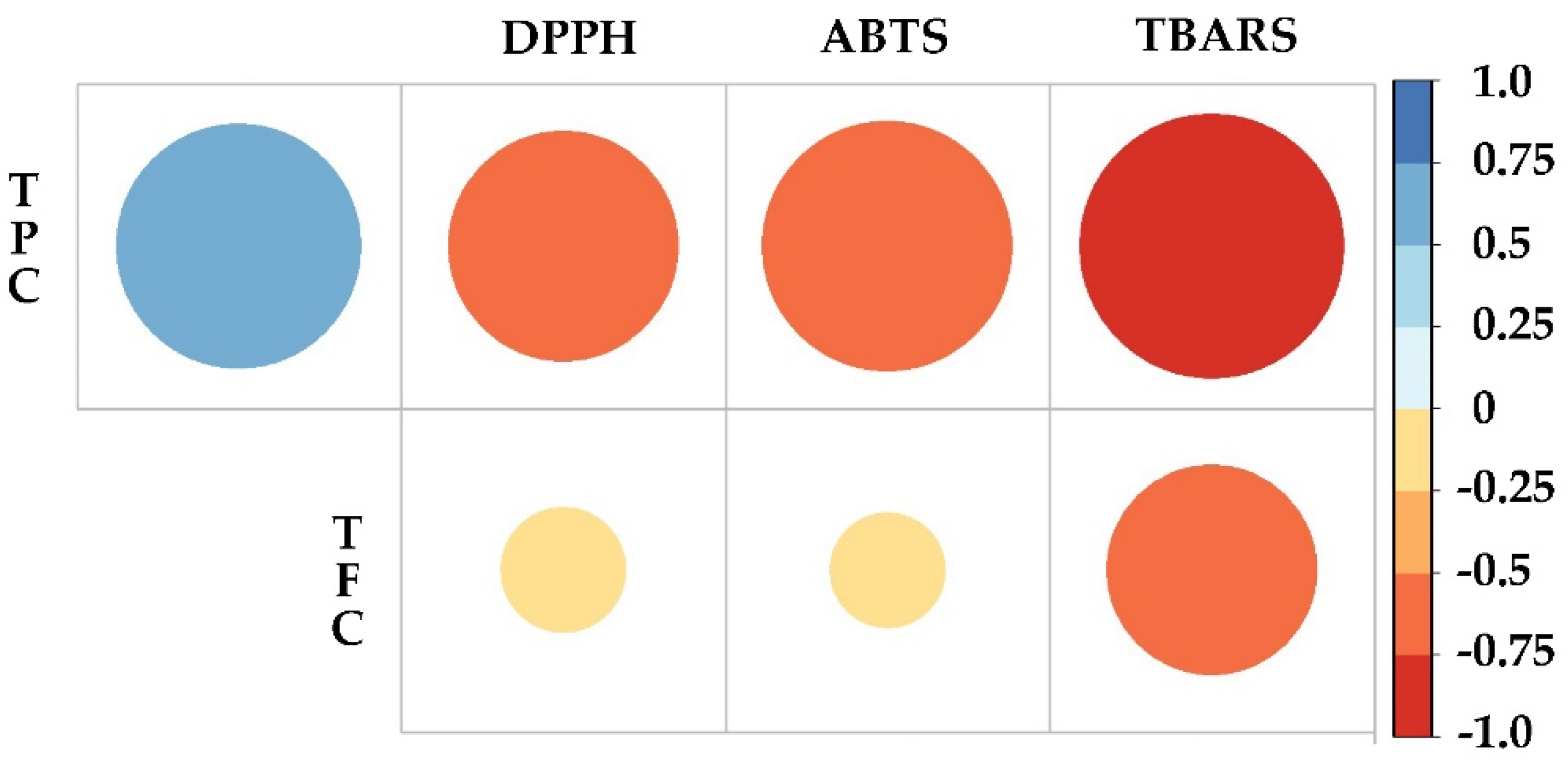
To elucidate the relationship between phenol and flavonoid content and antioxidant activity, Pearson’s correlation analysis was applied considering the TPC, TFC and IC50 obtained in the DPPH, ABTS and TBARS assays (Figure 5). Based on the results obtained, TPC showed a positive correlation (p < 0.05) with TFC, which demonstrates a direct correlation, i.e., the higher the phenol content, the higher the flavonoid content. In the case of TPC, the correlation was negative with DPPH (p < 0.05), ABTS (p < 0.01) and TBARS (p < 0.01). As for TFC, the correlation was also negative, however, with lower significance, for DPPH (p = 0.05), ABTS (p = 0.05) and TBARS (p < 0.05). Therefore, it could be concluded that the relationship of phenol and flavonoid content is inversely proportional to the IC50, i.e., the higher the content of bioactive compounds, the higher the antioxidant activity, and the lower IC50 value.

3. Materials and Methods

3.1. Plant Material

Wild plant (WP) and seeds of Ageratina pichinchensis were collected in Tepoztlán Morelos, Mexico, identified by Biol. Gabriel Flores Franco and deposited at the HUMO Herbarium of the Universidad Autónoma del Estado de Morelos (UAEM), with the voucher number 33913.

3.2. In Vitro Cultures of A. pichichensis

3.2.1. Plant

The seeds of A. pichichinses were surface disinfected in a solution of commercial detergent for 10 min; passed through a sodium hypochlorite solution at 10% (v/v) for 15 min and through an ethanol solution at 10% (v/v) for 30 s; the material was washed three times with sterile water for 5 min and finally inoculated in Murashige and Skoog medium (MS) [56], supplemented with 30 g/L of sucrose, 2 g/L of phytagel. Plants were kept in a growth chamber at 25 ± 2 °C and in photoperiods of 16 h light/8 h darkness; at the end of 2 months of incubation, a proportion was used for extraction (Section 3.3) and the other for callus induction. The resultant plants were coded as IP.

3.2.2. Callus Induction

Calluses (CC) were obtained following the methodology described by Sánchez-Ramos et al. [26], with some modifications in the concentrations of growth regulators and the type of explant used. The nodal segments of IP were cut into pieces and four explants were transferred into jars containing 25 mL of Murashige and Skoog medium (MS) [56], and plant growth regulators (PGRs): auxin (NAA; 2,4-dichlorophenoxyacetic acid) and cytokinin (kinetin, KIN). Different concentrations of NAA (0.0, 0.45, 2.26, 4.52 or 6.79 μM) were combined with KIN (0.0, 0.46, 1.39, or 2.32 μM) and were kept in a growth chamber at 25 ± 2 °C and in photoperiods of 16 h light/8 h darkness. Callus subcultures were carried out every 20 days and the percentage of callus induction was determined.

3.2.3. Cell Suspension Culture

The 20-day-old calluses were used to establish the cell suspension cultures (CSC). Fresh calluses (5.71 ± 0.71 g) were transferred to 250 mL Erlenmeyer flasks containing 50 mL of MS liquid culture medium and PGRs as in the callus cultures. The CSC were kept under stirring (110 rpm) and incubated at 25 ± 2 °C and in photoperiods of 16 h light/8 h darkness. CSC were sub-cultured every 12 days using 200 µm nylon filters (Whatman No. 1) to obtain a homogeneous cell culture, an inoculum of 40 g/L were transferred to 500 mL Erlenmeyer flasks with 100 mL of liquid culture medium [23].

3.3. Preparations of Extracts

Fresh biomass of the aerial parts of WP and IP of A. pichichensis (Figure 1) was dried in a drying oven at 40 °C. Samples (1 g) were used to obtain the extracts by sonication (BRANSONIC, CPX1800H, Danbury, CT, USA) at 40 kHz frequency with 10 mL of methanol at room temperature (25 °C) during 15 min. Then, the extracts were centrifuged at 6000× g for 15 min. The supernatant phase was recovered, and the pellet was used for a second and third extraction under the same conditions. Finally, the supernatants were mixed, and the solvent was evaporated under reduced pressure, the resultant dried extracts were stored at −70 °C in amber vials until further analysis.

3.4. Characterization of Extracts

3.4.1. Total Phenolic Content (TPC)

The total phenolic content (TPC) was determined according to the colorimetric method described by Giordano et al. [31] and Singleton et al. [57], with some modifications. Briefly, an aliquot of 200 µL of each extract was mixed with 1 mL of Folin–Ciocalteu reagent 1:10 (v/v), after 1 min, 0.8 mL of Na2CO3 7.5% (w/v) were mixed for 30 s and kept for 60 min in the dark at room temperature, after the reaction time the absorbance was measured at 765 nm in a spectrophotometer (Genesys 2, Spectronics, Waltham, MA, USA), gallic acid was used to construct a standard curve (0–100 µg/mL), the results were expressed in mg of gallic acid equivalents (GAE)/g of dried biomass weight (DW) from each treatment of A. pichichensis evaluated.

3.4.2. Total Flavonoid Content

The total flavonoid content (TFC) was determined by the colorimetric method described by Barreira et al. [58], an aliquot of 250 µL of each extract was mixed with 1250 µL of distilled water and 75 µL of NaNO2 5% (w/v), after 5 min 150 µL of AlCl3-6H2O 5% (w/v) was added, incubated for 6 min at room temperature, then, 500 μL of NaOH (1 M) and 275 μL of distilled water were added, the solution was mixed and the absorbance was measured at 510 nm. Quercetin was used to construct a standard curve (0–400 µg/mL), the results were expressed as mg quercetin equivalents (QCT)/gDW from each treatment of A. pichichensis evaluated.

3.5. High Performance Liquid Chromatography (HPLC)

The identification of compounds was carried out on solutions prepared from plant extracts and in vitro cultures at 1000 ppm and standards compounds at 20 ppm. HPLC analysis was performed following the methodology proposed by Ramirez-Lopez et al. [59] with the following modifications, an HPLC equipment (Shimadzu SPD-10A, SpectraLab Scientific Inc., Markham, ON, Canada) with a Zorbax Eclipse XDB C 18 column (4.6 mm × 250 mm, 5μm) was used. The mobile phases A and B were employed as follows: mobile phase A contained 0.1% formic acid in MilliQ water and phase B contained 0.1% formic acid in acetonitrile (HPLC grade). Data acquisition was applied for 45 min with a total run of 60 min. Gradient elution was as follows: 92% A/8% B, t 0 min; 85% A/15% B at 5 min; 40% A/60% B at 45 min; 40% A/60% B at 55 min; and back to initial conditions 92% A/8% B at 60 min a flow rate of 1 mL/min. The extracts were monitored at a wavelength of 280 and 370 nm. Gallic acid (GA), caffeic acid (CfA), p-coumaric acid (pCA), catechin (CAT), rutin (RUT), and epicatechin (EPI), were used as standard compounds.

3.6. Antioxidant Capacity Assays

3.6.1. DPPH Radical Scavenging Activity

The radical scavenging capacity by DPPH was determined according to Dominguez et al. [60]. Fifty µL of extract in methanol at different concentrations were added to an ethanolic solution of DPPH (100 µM, 150 µL) in 96 wells microplates. Mixtures were incubated for 30 min at 37 °C in the dark and their absorbances were measured at 515 nm in a microplate reader Synergy HT™ (BioTek Instruments, Winooski, VT, USA); Trolox was used as standard The DPPH radical scavenging activity (%) was calculated as follows:
DPPH Scavenging   activity   ( % ) = A 0 A 1 A 0 × 100
where A0 is the absorbance of the blank sample (without antioxidant) and A1 is the absorbance of the sample containing the extract. Median inhibition concentration (IC50) values were calculated from plotted graph of percentage scavenging activity against the concentration of the extracts and denote the concentration of antioxidant required to inhibit 50% of radical and expressed as μg dried extract (DE)/mL.

3.6.2. ABTS Radical Scavenging Activity

The radical scavenging capacity by ABTS was determined according to Re et al. [61]. The ABTS stock solution was prepared by adding 90.3 mg of ABTS salt and 16.1 mg of K2S2O8 in 25 mL of distilled water. Stock solution was stored in the dark for 16 h at room temperature before use. The ABTS+ radical solution was diluted with distilled water until reach an absorbance value of 0.70 ± 0.05 at 734 nm. Afterwards, 1000 μL of diluted ABTS+ radical solution was mixed with 10 μL of different concentrations of extracts. The mixture was allowed to react for 10 min, at 30 °C in the dark and their absorbances were measured at 734 nm in a microplate reader Synergy HT™ (BioTek Instruments, Winooski, USA); Trolox was used as standard. The ABTS+ radical scavenging activity (%) was calculated as follows:
ABTS Scavenging   activity +   ( % ) = A 0 A 1 A 0 × 100
where A0 is the absorbance of the blank sample (without antioxidant) and A1 is the absorbance of the sample containing the extract. Median inhibition (IC50) values were calculated from the plotted graph of percentage scavenging activity against the concentration of the extracts and the concentration denoted of the antioxidant required to inhibit 50% of radical and expressed as μgDE/mL.

3.6.3. Determination of TBARS

Animals

Adult male Wistar rat (200–250 g) was provide by Instituto de Fisiología Celular, Universidad Nacional Autónoma de México (UNAM). Procedures and care of animals were conducted in conformity with Mexican Official Norm for Animal Care and Handling (NOM-062-ZOO-1999). They were maintained at 23 ± 2 °C on a 12/12 h light–dark cycle with free access to food and water.

Rat Brain Homogenate Preparation

Animal euthanasia was carried out avoiding unnecessary pain with CO2. The cerebral tissue (whole brain) was rapidly dissected and homogenized in phosphate-buffered saline (PBS) solution (0.2 g of KCl, 0.2 g of KH2PO4, 8 g of NaCl, and 2.16 g of NaHPO4 ·7 H2O/l, pH adjusted to 7.4) as reported elsewhere [60,62] to produce a 1/10 (w/v) homogenate. Homogenate was centrifuged for 10 min at 800× g. The supernatant protein content was measured using the Folin–Ciocalteu’s phenol reagent Lowry. [63] and adjusted with PBS at 2.66 mg of protein/mL.

Induction of Lipid Peroxidation and Thiobarbituric Acid Reactive Substances (TBARS) Quantification

As an index of lipid peroxidation, TBARS levels were measured using rat brain homogenates according to the method described by Ng et al. [64], with some modifications. Supernatant (375 µL) was added with 50 µL of 20 µM EDTA and 25 µL of each extract concentration solved in methanol (25 µL of methanol for control group) and incubated at 37 °C for 30 min. Lipid peroxidation was started adding 50 µL of freshly solution FeSO4 100 µM and incubated at 37 °C for 1 h. The TBARS content was determined as described by Ohkawa et al. [65], with 500 µL of TBA reagent (0.5% 2-thiobarbituric acid in 0.05 N NaOH and 30% trichloroacetic acid, in 1:1 proportion) added at each tube and cooled on ice for 10 min, centrifugated at 13,400× g for 5 min and heated at 80 °C in a water bath for 30 min. After cooling at room temperature, the absorbance of 200 µL of supernatant was measured at 540 nm in a Bio-Tek Microplate Reader Synergy HT. Trolox was used as standard. Concentration of TBARS was calculated by interpolation in a standard curve of tetra-methoxypropane (TMP) as a precursor of malondialdehyde (MDA) [66]. Results were expressed as nmol of TBARS per mg of protein. The percentage of inhibition of lipid peroxidation (IR%) was calculated as follows:
IR   ( % ) = A 0 A 1 A 0 × 100
where A0 is the absorbance of the blank sample (without antioxidant) and A1 is the absorbance of the sample containing the extract. Median inhibition (IC50) values were calculated from a plotted graph of percentage scavenging activity against the concentration of the extracts and denoted the concentration of antioxidant required to inhibit 50% of radical and expressed as μgDE/mL.

3.7. Statistical Analysis

All the experiments were carried out in triplicate and the results were expressed as means ± SD. TPC, TFC, and antioxidant activity (DPPH, ABTS and TBARS) were compared using the Tukey and ANOVA test. A Pearson correlation test was performed to establish significant effects among the variables: TPC, TFC, antioxidant activity by DPPH, ABTS and TBARS assays. Significant levels were defined at p < 0.05 and p < 0.01. Statistical analysis was tested with SigmaPlot 12.5 software (Systat Software Inc, 2011, Palo Alto, CA, USA). A Pearson correlation test was performed in R Core Team (2022): R: a language and environment for statistical computing.–R Foundation for Statistical Computing, Vienn.

4. Conclusions

In vitro cultures (CC and CSC) were obtained from the IP nodal segment of A. pichichensis in the presence of a combination of NAA and KIN in MS medium. The disaggregation of the CC was a decisive factor for the establishment of CSC, because only one treatment exhibited disaggregation in liquid medium. On the other hand, in vitro CC and CSC cultures were able to produce compounds that WP did not, and CSC produced more phenolic compounds than CC. Based on these results, in vitro CC and CSC cultures are an alternative for obtaining phenolic compounds continuously and in a shorter time than plants growing under natural conditions of A. pichichensis. However, although in vitro cultures were able to produce a higher number of phenolic compounds, the antioxidant activity obtained in the different assays evaluated were significantly lower than in WP. These differences could be related to the affinity of the extracted compounds for the different radicals evaluated, so, a more robust characterization of A. pichichensis extracts is necessary for the identification and quantification of the majority compounds and their contribution in the antioxidant activity. Finally, TPC and TFC showed a correlation inversely proportional to the IC50 found in the DPPH, ABTS and TBARS assays. With this information we can say that WP and in vitro cultures of A. pichichensis can produce phenolics compounds with antioxidant activity and that in vitro cultures such as CC and CSC are a powerful biotechnological alternative for obtaining bioactive compounds.

Author Contributions

Conceptualization, E.A.M.-A., A.R.-G. and F.C.-S.; methodology, E.A.M.-A., A.M.F.-V. and A.N.-C.; investigation, E.A.M.-A.; resources, F.C.-S. and M.R.-M.; writing—original draft preparation, E.A.M.-A.; writing—review and editing, A.R.-G., A.M.F.-V., R.A.-E. and F.C.-S.; project administration, A.R.-G. and F.C.-S. All authors have read and agreed to the published version of the manuscript.

Funding

This research was partially funded by the Consejo Nacional de Ciencia y Tecnología (CONACyT), scholarship grant of E.A.M.-A. (616138).

Data Availability Statement

No new data were created or analyzed in this study. Data sharing is not applicable to this article.

Acknowledgments

The authors give thanks to the Consejo Nacional de Ciencia y Tecnología (CONACyT) for the scholarship grant of E.A.M.-A. (616138). M.V.Z. Claudia V. Rivera Cerecedo and Héctor Malagon Rivero from the Bioterio del Instituto de Fisiología Celular for providing biological material.

Conflicts of Interest

The authors declare no conflict of interest.

References

  1. Navarro García, V.M.; Gonzalez, A.; Fuentes, M.; Aviles, M.; Rios, M.Y.; Zepeda, G.; Rojas, M.G. Antifungal activities of nine traditional Mexican medicinal plants. J. Ethnopharmacol. 2003, 87, 85–88. [Google Scholar] [CrossRef]
  2. Sánchez-Mendoza, M.E.; Reyes-Trejo, B.; Sánchez-Gómez, P.; Rodríguez-Silverio, J.; Castillo-Henkel, C.; Cervantes-Cuevas, H.; Arrieta, J. Bioassay-guided isolation of an anti-ulcer chromene from Eupatorium aschenbornianum: Role of nitric oxide, prostaglandins and sulfydryls. Fitoterapia 2010, 81, 66–71. [Google Scholar] [CrossRef]
  3. Torres-Barajas, L.; Rojas-Vera, J.; Morales-Méndez, A.; Rojas-Fermín, L.; Lucena, M.; Buitrago, A. Chemical composition and evaluation of antibacterial activity of essential oils of Ageratina jahnii and Ageratina pichinchensis collected in Mérida, Venezuela. Boletín Latinoam. Caribe Plantas Med. Aromáticas 2013, 12, 92–98. [Google Scholar]
  4. Romero-Cerecero, O.; Zamilpa, A.; González-Cortazar, M.; Alonso-Cortés, D.; Jiménez-Ferrer, E.; Nicasio-Torres, P.; Aguilar-Santamaría, L.; Tortoriello, J. Pharmacological and chemical study to identify wound-healing active compounds in Ageratina pichinchensis. Planta Med. 2013, 79, 622–627. [Google Scholar] [CrossRef]
  5. Romero-Cerecero, O.; Zamilpa, A.; Díaz-García, E.R.; Tortoriello, J. Pharmacological effect of Ageratina pichinchensis on wound healing in diabetic rats and genotoxicity evaluation. J. Ethnopharmacol. 2014, 156, 222–227. [Google Scholar] [CrossRef]
  6. Romero-Cerecero, O.; Rojas, G.; Navarro, V.; Herrera-Arellano, A.; Zamilpa-Álvarez, A.; Tortoriello, J. Effectiveness and tolerability of a standardized extract from Ageratina pichinchensis on patients with tinea pedis: An explorative pilot study controlled with ketoconazole. Planta Med. 2006, 72, 1257–1261. [Google Scholar] [CrossRef]
  7. Romero-Cerecero, O.; Zamilpa, A.; Jiménez-Ferrer, J.E.; Rojas-Bribiesca, G.; Román-Ramos, R.; Tortoriello, J. Double-blind clinical trial for evaluating the effectiveness and tolerability of Ageratina pichinchensis extract on patients with mild to moderate onychomycosis. A comparative study with ciclopirox. Planta Med. 2008, 74, 1430–1435. [Google Scholar] [CrossRef]
  8. Romero-Cerecero, O.; Zamilpa-Álvarez, A.; Jiménez-Ferrer, E.; Tortoriello, J. Exploratory study on the effectiveness of a standardized extract from Ageratina pichinchensis in patients with chronic venous leg ulcers. Planta Med. 2012, 78, 304–310. [Google Scholar] [CrossRef]
  9. Romero-Cerecero, O.; Zamilpa, A.; Tortoriello, J. Effectiveness and tolerability of a standardized extract from Ageratina pichinchensis in patients with diabetic foot ulcer: A randomized, controlled pilot study. Planta Med. 2015, 81, 272–278. [Google Scholar] [CrossRef]
  10. Romero-Cerecero, O.; Islas-Garduño, A.L.; Zamilpa, A.; Tortoriello, J. Effectiveness of Ageratina pichinchensis Extract in Patients with Vulvovaginal Candidiasis. A Randomized, Double-Blind, and Controlled Pilot Study. Phyther. Res. 2017, 31, 885–890. [Google Scholar] [CrossRef]
  11. Romero-Cerecero, O.; Islas-Garduño, A.L.; Zamilpa, A.; Tortoriello, J. Effectiveness of an encecalin standardized extract of Ageratina pichinchensis on the treatment of onychomycosis in patients with diabetes mellitus. Phyther. Res. 2020, 34, 1678–1686. [Google Scholar] [CrossRef] [PubMed]
  12. Sánchez-Ramos, M.; Marquina-Bahena, S.; Alvarez, L.; Román-Guerrero, A.; Bernabé-Antonio, A.; Cruz-Sosa, F. Phytochemical, pharmacological, and biotechnological study of Ageratina pichinchensis: A native species of Mexico. Plants 2021, 10, 2225. [Google Scholar] [CrossRef] [PubMed]
  13. Balasundram, N.; Sundram, K.; Samman, S. Phenolic compounds in plants and agri-industrial by-products: Antioxidant activity, occurrence, and potential uses. Food Chem. 2006, 99, 191–203. [Google Scholar] [CrossRef]
  14. Talukder, P.; Talapatra, S.; Ghoshal, N.; Sen Raychaudhuri, S. Antioxidant activity and high-performance liquid chromatographic analysis of phenolic compounds during in vitro callus culture of Plantago ovata Forsk. and effect of exogenous additives on accumulation of phenolic compounds. J. Sci. Food Agric. 2016, 96, 232–244. [Google Scholar] [CrossRef]
  15. Saeed, N.; Khan, M.R.; Shabbir, M. Antioxidant activity, total phenolic and total flavonoid contents of whole plant extracts Torilis leptophylla L. BMC Complement. Altern. Med. 2012, 12, 221. [Google Scholar] [CrossRef] [Green Version]
  16. Pereira Nunes, X.; Souza Silva, F.; Guedes da Almeida, J.R.S.; Tolentino de Lima, J.; de Araújo Ribeiro, L.A.; Quintans Júnior, L.J.; Barbosa-Filho, J.M. Biological Oxidations and Antioxidant Activity of Natural Products. In Phytochemicals as Nutraceuticals; IntechOpen: London, UK, 2012; pp. 1–22. ISBN 978-953-51-0203-8. [Google Scholar]
  17. Georgiev, M.I.; Eibl, R.; Zhong, J.J. Hosting the plant cells in vitro: Recent trends in bioreactors. Appl. Microbiol. Biotechnol. 2013, 97, 3787–3800. [Google Scholar] [CrossRef]
  18. Bhaskar, R.; Xavier, L.S.E.; Udayakumaran, G.; Kumar, D.S.; Venkatesh, R.; Nagella, P. Biotic elicitors: A boon for the in-vitro production of plant secondary metabolites. Plant Cell Tissue Organ Cult. 2021, 149, 7–24. [Google Scholar] [CrossRef]
  19. Castro, A.H.F.; Braga, K.D.Q.; de Sousa, F.M.; Coimbra, M.C.; Chagas, R.C.R. Callus induction and bioactive phenolic compounds production from Byrsonima verbascifolia (L.) DC. (Malpighiaceae). Rev. Cienc. Agron. 2016, 47, 143–151. [Google Scholar] [CrossRef] [Green Version]
  20. Mendoza, D.; Arias, J.P.; Cuaspud, O.; Arias, M. Phytochemical screening of callus and cell suspensions cultures of Thevetia peruviana. Braz. Arch. Biol. Technol. 2020, 63, 1–14. [Google Scholar] [CrossRef]
  21. Ruffoni, B.; Pistelli, L.; Bertoli, A.; Pistelli, L. Plant cell cultures: Bioreactors for industrial production. Adv. Exp. Med. Biol. 2010, 698, 203–221. [Google Scholar] [CrossRef]
  22. Motolinia-Alcántara, A.E.; Castillo-Araiza, C.O.; Rodríguez-Monroy, M.; Román-Guerrero, A.; Cruz-Sosa, F. Engineering Considerations to Produce Bioactive Compounds from Plant Cell Suspension Culture in Bioreactors. Plants 2022, 10, 2762. [Google Scholar] [CrossRef]
  23. Sánchez-Ramos, M.; Alvarez, L.; Romero-Estrada, A.; Bernabé-Antonio, A.; Marquina-Bahena, S.; Cruz-Sosa, F. Establishment of a cell suspension culture of Ageratina pichinchensis (Kunth) for the improved production of anti-inflammatory compounds. Plants 2020, 9, 1398. [Google Scholar] [CrossRef]
  24. Pandey, H.; Pandey, P.; Singh, S.; Gupta, R.; Banerjee, S. Production of anti-cancer triterpene (betulinic acid) from callus cultures of different Ocimum species and its elicitation. Protoplasma 2015, 252, 647–655. [Google Scholar] [CrossRef]
  25. Patel, S.R.; Joshi, A.G.; Pathak, A.R.; Shrivastava, N.; Sharma, S. Somatic embryogenesis in Leptadenia reticulata (Retz.) Wight and Arn along with assessment of shoot and callus cultures for HPTLC fingerprint and quantification of p-coumaric acid. Plant Cell Tissue Organ Cult. 2021, 145, 173–189. [Google Scholar] [CrossRef]
  26. Mariana, S.R.; Bahena, S.M.; Antonio, R.E.; Antonio, B.A.; Francisco, C.S.; Judith, G.C.; Juan, J.A.F.; Irene, P.A.; Alvarez, L. Establishment and phytochemical analysis of a callus culture from Ageratina pichinchensis (Asteraceae) and its anti-inflammatory activity. Molecules 2018, 23, 1258. [Google Scholar] [CrossRef] [Green Version]
  27. Patle, T.K.; Shrivas, K.; Kurrey, R.; Upadhyay, S.; Jangde, R.; Chauhan, R. Phytochemical screening and determination of phenolics and flavonoids in Dillenia pentagyna using UV–vis and FTIR spectroscopy. Spectrochim. Acta Part A Mol. Biomol. Spectrosc. 2020, 242, 118717. [Google Scholar] [CrossRef]
  28. Verma, N.; Shukla, S. Impact of various factors responsible for fluctuation in plant secondary metabolites. J. Appl. Res. Med. Aromat. Plants 2015, 2, 105–113. [Google Scholar] [CrossRef]
  29. Cicevan, R.; Sestras, A.F.; Plazas, M.; Boscaiu, M.; Vilanova, S.; Gramazio, P.; Vicente, O.; Prohens, J.; Sestras, R.E. Biological Traits and Genetic Relationships Amongst Cultivars of Three Species of Tagetes (Asteraceae). Plants 2022, 11, 760. [Google Scholar] [CrossRef]
  30. Nieto-Trujillo, A.; Buendía-González, L.; García-Morales, C.; Román-Guerrero, A.; Cruz-Sosa, F.; Estrada-Zúñiga, M.E. PHENOLIC COMPOUNDS AND PARTHENOLIDE PRODUCTION FROM in vitro CULTURES OF Tanacetum parthenium. Rev. Mex. Ing. Química 2017, 16, 371–383. [Google Scholar] [CrossRef]
  31. Giordano, A.; Morales-Tapia, P.; Moncada-Basualto, M.; Pozo-Martínez, J.; Olea-Azar, C.; Nesic, A.; Cabrera-Barjas, G. Polyphenolic Composition and Antioxidant Activity (ORAC, EPR and Cellular) of Different Extracts of Argylia radiata Vitroplants and Natural Roots. Molecules 2022, 27, 610. [Google Scholar] [CrossRef]
  32. Naranjo-Gómez, E.J.; Puertas-Mejía, M.A.; Mejía-Giraldo, J.C.; Amaya-Nieto, A.Z.; Atehortúa, L. Micropropagation of Baccharis antioquensis (Asteraceae) and photoinduction of polyphenols by UV radiation. Rev. Biol. Trop. 2018, 66, 754–764. [Google Scholar] [CrossRef] [Green Version]
  33. Mishra, M.K.; Pandey, S.; Niranjan, A.; Misra, P. Comparative analysis of phenolic compounds from wild and in vitro propagated plant Thalictrum foliolosum and antioxidant activity of various crude extracts. Chem. Pap. 2021, 75, 4873–4885. [Google Scholar] [CrossRef]
  34. Lugato, D.; Simão, M.J.; Garcia, R.; Mansur, E.; Pacheco, G. Determination of antioxidant activity and phenolic content of extracts from in vivo plants and in vitro materials of Passiflora alata Curtis. Plant Cell Tissue Organ Cult. 2014, 118, 339–346. [Google Scholar] [CrossRef]
  35. Gauchan, D.P.; Bhuju, S.; Lamichhane, J.; Shakya, R.; García-Gil, M.R. Establishment of Regenerative Callus, Cell Suspension System, and Molecular Characterization of Taxus wallichiana Zucc. For the In Vitro Production of Taxol. J. Appl. Pharm. Sci. 2021, 11, 022–034. [Google Scholar] [CrossRef]
  36. Coimbra, M.C.; Chagas, R.C.R.; Vilela, M.S.P.; Castro, A.H.F. Growth, morphology and bioactive phenolic compounds production in Pyrostegia venusta calli. Biocatal. Agric. Biotechnol. 2019, 18, 101036. [Google Scholar] [CrossRef]
  37. Arciniega-Carreón, I.Y.; Ramírez-Sotelo, M.G.; Ramos-Valdivia, A.C.; Salas, C.E.; Ortega, A.; Oliver-Salvador, C. Metabolites in cultured cells of Ibervillea sonorae (S. Watson) Greene display increased hypoglycemic activity compared to that seen in plant roots. Hortic. Environ. Biotechnol. 2020, 61, 1039–1049. [Google Scholar] [CrossRef]
  38. Coimbra, M.C.; Chagas, R.C.R.; Duarte-Almeida, J.M.; Castro, A.H.F. Influence of plant growth regulators and light on callus induction and bioactive phenolic compounds production in Pyrostegia venusta (Bignoniaceae). Indian J. Exp. Biol. 2017, 55, 584–590. [Google Scholar]
  39. Isah, T.; Umar, S.; Mujib, A.; Sharma, M.P.; Rajasekharan, P.E.; Zafar, N.; Frukh, A. Secondary metabolism of pharmaceuticals in the plant in vitro cultures: Strategies, approaches, and limitations to achieving higher yield. Plant Cell Tissue Organ Cult. 2018, 132, 239–265. [Google Scholar] [CrossRef]
  40. Ramezannezhad, R.; Aghdasi, M.; Fatemi, M. Enhanced production of cichoric acid in cell suspension culture of Echinacea purpurea by silver nanoparticle elicitation. Plant Cell Tissue Organ Cult. 2019, 139, 261–273. [Google Scholar] [CrossRef]
  41. Mendoza, D.; Cuaspud, O.; Arias, J.P.; Ruiz, O.; Arias, M. Effect of salicylic acid and methyl jasmonate in the production of phenolic compounds in plant cell suspension cultures of Thevetia peruviana. Biotechnol. Rep. 2018, 19, e00273. [Google Scholar] [CrossRef]
  42. Modarres, M.; Esmaeilzadeh Bahabadi, S.; Taghavizadeh Yazdi, M.E. Enhanced production of phenolic acids in cell suspension culture of Salvia leriifolia Benth. using growth regulators and sucrose. Cytotechnology 2018, 70, 741–750. [Google Scholar] [CrossRef] [PubMed]
  43. Szopa, A.; Kubica, P.; Snoch, A.; Ekiert, H. High production of bioactive depsides in shoot and callus cultures of Aronia arbutifolia and Aronia × prunifolia. Acta Physiol. Plant. 2018, 40, 48. [Google Scholar] [CrossRef] [Green Version]
  44. Taghizadeh, M.; Nasibi, F.; Kalantari, K.M.; Benakashani, F. Callogenesis optimization and cell suspension culture establishment of Dracocephalum polychaetum Bornm. and Dracocephalum kotschyi Boiss.: An in vitro approach for secondary metabolite production. South Afr. J. Bot. 2020, 132, 79–86. [Google Scholar] [CrossRef]
  45. Kikowska, M.; Thiem, B.; Szopa, A.; Ekiert, H. Accumulation of valuable secondary metabolites: Phenolic acids and flavonoids in different in vitro systems of shoot cultures of the endangered plant species—Eryngium alpinum L. Plant Cell Tissue Organ Cult. 2020, 141, 381–391. [Google Scholar] [CrossRef] [Green Version]
  46. Ali, M.; Abbasi, B.H. Ihsan-ul-haq. Production of commercially important secondary metabolites and antioxidant activity in cell suspension cultures of Artemisia absinthium L. Ind. Crop. Prod. 2013, 49, 400–406. [Google Scholar] [CrossRef]
  47. Mlcek, J.; Jurikova, T.; Skrovankova, S.; Sochor, J. Quercetin and its anti-allergic immune response. Molecules 2016, 21, 623. [Google Scholar] [CrossRef] [Green Version]
  48. Urbaniak, A.; Kujawski, J.; Czaja, K.; Szelag, M. Antioxidant properties of several caffeic acid derivatives: A theoretical study. Comptes Rendus Chim. 2017, 20, 1072–1082. [Google Scholar] [CrossRef]
  49. Zeng, N.; Hongbo, T.; Xu, Y.; Wu, M.; Wu, Y. Anticancer activity of caffeic acid n-butyl ester against A431 skin carcinoma cell line occurs via induction of apoptosis and inhibition of the mTOR/PI3K/AKT signaling pathway. Mol. Med. Rep. 2018, 17, 5652–5657. [Google Scholar] [CrossRef] [Green Version]
  50. Lima, V.N.; Oliveira-Tintino, C.D.M.; Santos, E.S.; Morais, L.P.; Tintino, S.R.; Freitas, T.S.; Geraldo, Y.S.; Pereira, R.L.S.; Cruz, R.P.; Menezes, I.R.A.; et al. Antimicrobial and enhancement of the antibiotic activity by phenolic compounds: Gallic acid, caffeic acid and pyrogallol. Microb. Pathog. 2016, 99, 56–61. [Google Scholar] [CrossRef]
  51. Liu, Y.L.; Hsu, C.C.; Huang, H.J.; Chang, C.J.; Sun, S.H.; Lin, A.M.Y. Gallic Acid Attenuated LPS-Induced Neuroinflammation: Protein Aggregation and Necroptosis. Mol. Neurobiol. 2020, 57, 96–104. [Google Scholar] [CrossRef]
  52. Esmaeili, A.K.; Taha, R.M.; Mohajer, S.; Banisalam, B. Antioxidant Activity and Total Phenolic and Flavonoid Content of Various Solvent Extracts from In Vivo and In Vitro Grown Trifolium pratense L. (Red Clover). Biomed Res. Int. 2015, 2015, 643285. [Google Scholar] [CrossRef] [Green Version]
  53. Martysiak-Żurowska, D.; Wenta, W. A comparison of ABTS and DPPH methods for assessing th total antioxidant capacity of human milk. Acta Sci. Pol. Technol. Aliment. 2012, 1, 83–89. [Google Scholar]
  54. Brand-Williams, W.; Cuvelier, M.E.; Berset, C. Use of a free radical method to evaluate antioxidant activity. LWT Food Sci. Technol. 1995, 28, 25–30. [Google Scholar] [CrossRef]
  55. Costa, P.; Gonçalves, S.; Valentão, P.; Andrade, P.B.; Romano, A. Accumulation of phenolic compounds in in vitro cultures and wild plants of Lavandula viridis L’Hér and their antioxidant and anti-cholinesterase potential. Food Chem. Toxicol. 2013, 57, 69–74. [Google Scholar] [CrossRef] [PubMed]
  56. Murashige, T.; Skoog, F. A Revised Medium for Rapid Growth and Bio Assays with Tobacco Tissue Cultures. Physiol. Plant. 1962, 15, 474–497. [Google Scholar] [CrossRef]
  57. Singleton, V.L.; Orthofer, R.; Lamuela-Raventós, R.M. Analysis of Total Phenols and Other Oxidation Substrates and Antioxidants by Means of Folin-Ciocalteu. Methods Enzymol. 1999, 299, 152–178. [Google Scholar] [CrossRef]
  58. Barreira, J.C.M.; Ferreira, I.C.F.R.; Oliveira, M.B.P.P.; Pereira, J.A. Antioxidant activities of the extracts from chestnut flower, leaf, skins and fruit. Food Chem. 2008, 107, 1106–1113. [Google Scholar] [CrossRef]
  59. Ramirez-Lopez, L.M.; DeWitt, C.A.M. Analysis of phenolic compounds in commercial dried grape pomace by high-performance liquid chromatography electrospray ionization mass spectrometry. Food Sci. Nutr. 2014, 2, 470–477. [Google Scholar] [CrossRef] [PubMed]
  60. Domínguez, M.; Nieto, A.; Marin, J.C.; Keck, A.S.; Jeffery, E.; Céspedes, C.L. Antioxidant activities of extracts from Barkleyanthus salicifolius (Asteraceae) and Penstemon gentianoides (Scrophulariaceae). J. Agric. Food Chem. 2005, 53, 5889–5895. [Google Scholar] [CrossRef]
  61. Re, R.; Pellegrini, N.; Proteggente, A.; Pannala, A.; Yang, M.; Rice-Evans, C. Development and characterisation of carbon nanotube-reinforced polyurethane foams. Free Radic. Biol. Med. 1999, 26, 1231–1237. [Google Scholar] [CrossRef]
  62. Rossato, J.I.; Ketzer, L.A.; Centurião, F.B.; Silva, S.J.N.; Lüdtke, D.S.; Zeni, G.; Braga, A.L.; Rubin, M.A.; Da Rocha, J.B.T. Antioxidant properties of new chalcogenides against lipid peroxidation in rat brain. Neurochem. Res. 2002, 27, 297–303. [Google Scholar] [CrossRef] [PubMed]
  63. LOWRY, O.H.; ROSEBROUGH, N.J.; FARR, A.L.; RANDALL, R.J. Protein measurement with the Folin phenol reagent. J. Biol. Chem. 1951, 193, 265–275. [Google Scholar] [CrossRef] [PubMed]
  64. Ng, T.B.; Liu, F.; Wang, Z.T. Antioxidative activity of natural products from plants. Life Sci. 2000, 66, 709–723. [Google Scholar] [CrossRef]
  65. Ohkawa, H.; Ohishi, N.; Yagi, K. Assay for lipid peroxides in animal tissues by thiobarbituric acid reaction. Anal. Biochem. 1979, 95, 351–358. [Google Scholar] [CrossRef] [PubMed]
  66. Esterbauer, H.; Cheeseman, K.H. Determination of aldehydic lipid peroxidation products: Malonaldehyde and 4-hydroxynonenal. Methods Enzymol. 1990, 186, 407–421. [Google Scholar] [CrossRef] [PubMed]
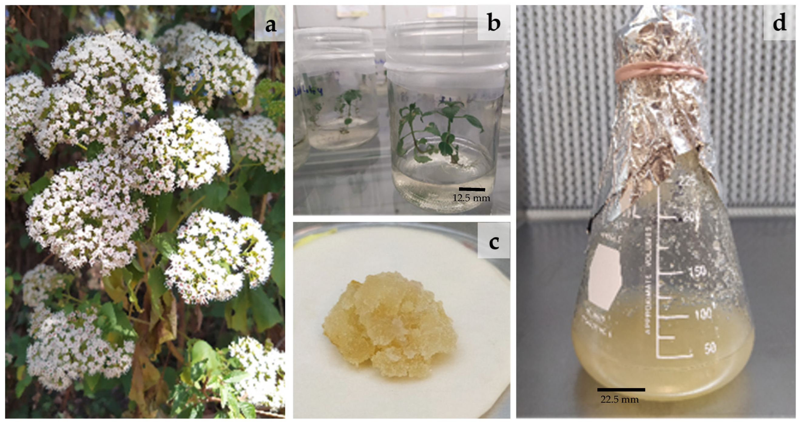
Figure 1. Ageratina pichichensis: (a) Wild plants (WP) collected at the natural site, (b) germinated plant in vitro (IP), (c) callus cell culture (CC), and (d) cell suspension culture (CSC).
Plants 12 01107 g001
Figure 2. (A) Establishment of A. pichichensis cell suspension culture (CSC) in liquid Murashige and Skoog medium (MS) from callus culture (CC) treatments T3 and T4, maximum disaggregation in CSC was achieved at seven-day culture. (B) Kinetic growth of T4 suspension cell culture (CSC) during 21 days of culture.
Plants 12 01107 g002
Figure 3. HPLC chromatograms for methanol extracts of A. pichichensis from wild plant (WP), in vitro plant (IP), callus culture (CC-T3 and CC-T4), suspension cell culture (CSC-T4 12d and CSC-T4-16d). Gallic acid (GA), catechin (CAT), caffeic acid (CfA), epicatechin (EPI), p-coumaric acid (pCA) and rutin (RUT).
Plants 12 01107 g003
Figure 4. Antioxidant capacity of extracts from A. pichichensis and its cell cultures against (A) ABTS, (B) DPPH radicals and (C) TBARS lipid peroxidation.
Plants 12 01107 g004aPlants 12 01107 g004b
Figure 5. Pearson correlation coefficients for the antioxidant activity characterization. TPC, total phenolic compounds; TFC, total flavonoid compounds; ABTS and DPPH radical scavenging activity, TBARS inhibition of lipid peroxidation.
Plants 12 01107 g005
Table 1. Effect of plant growth regulators on the morphogenetic response of A. pichichensis nodal segment explants at 20 days of culture.
TreatmentPGRCallus Induction ResponseCallus and Roots Induction ResponseCallus and Plants Induction Response
(μM)(%)Fresh Weight (g) Dried Weight (g)(%)Fresh Weight (g) Dried Weight (g)(%)Fresh Weight (g) Dried Weight (g)
NAAKIN
Control 00NDNDNDNDNDNDNDNDND
T10.450.46NDNDNDNDNDNDNDNDND
T22.260.46NDNDNDNDNDNDNDNDND
T34.520.4693.75 ± 12.50 a5.25 ± 1.69 a0.19 ± 0.06 aNDNDND37.50 ± 25.00 a6.72 ± 1.63 a0.23 ± 0.08 a
T46.790.4681.25 ± 12.51 a6.42 ± 2.00 a0.24 ± 0.09 aNDNDND16.66 ± 14.43 b16.75 ± 3.17 b1.85 ± 0.73 b
T50.451.3937.50 ± 14.43 b3.29 ± 0.76 b2.31 ± 0.3 bNDNDNDNDNDND
T62.261.3918.75 ± 12.50 c1.46 ± 0.24 c0.97 ± 0.15 cNDNDNDNDNDND
T74.521.3968.75 ± 12.50 b1.53 ± 0.18 c0.70 ± 0.08 d18.75 ± 12.523.83 ± 0.841.75 ± 0.83NDNDND
T86.791.39NDNDNDNDNDNDNDNDND
T90.452.32NDNDNDNDNDNDNDNDND
T102.262.3245.83 ± 29.46 b4.70 ± 1.43 b3.00 ± 0.48 bNDNDNDNDNDND
T114.522.32NDNDNDNDNDNDNDNDND
T126.792.32NDNDNDNDNDNDNDNDND
ND means Not detected. Different superscripts in the same column mean significant differences at p < 0.05.
Table 2. Phytochemical characterization and antioxidant activity of extracts from A. pichichensis and its cell cultures.
TreatmentTPCTFCABTSDPPHTBARS
(mgGAE/gDW)(mgQCT/gDW)IC50 (µg DE/mL)
WP100.56 ± 6.83 bc77.26 ± 4.16 c45.09 ± 6.29 a60.24 ± 2.66 a27.85 ± 0.29 a
IP14.24 ± 1.71 e29.72 ± 3.78 d459.68 ± 59.32 c557.29 ± 16.49 d783.99 ± 28.06 e
CC-T3 122.12 ± 3.44 a209.93 ± 0.57 a284.77 ± 24.52 b242.58 ± 23.24 b260.49 ± 9.48 c
CC-T4 113.33 ± 4.46 ab155.60 ± 12.49 b282.81 ± 9.01 b422.67 ± 49.48 c362.72 ± 12.22 d
CSC-T4 12d91.89 ± 4.83 c196.93 ± 2.08 a282.2 ± 3.28 b229.96 ±30.81b157.01 ± 5.27 b
CSC-T4 16d 61.47 ± 6.37 d 88.5 ± 9.19 c285.04 ± 45.12 b276 ± 15.53 b234.31 ± 6.52 c
TPC, total phenolic compounds; TFC, total flavonoid compounds; ABTS and DPPH radical scavenging activity, TBARS inhibition of lipid peroxidation. Different letters in the same column mean significant differences between treatments (p < 0.05).
Table 3. Quantitative analysis of identified phenolics compounds of extracts from A. pichichensis wild plant (WP), in vitro plant (IP), callus culture (CC-T3 and CC-T4), suspension cell culture (CSC-T4 12d and CSC-T4-16d).
TreatmentIdentified Phenolic Compounds
µg GA/gDWµg CAT/gDWµg CfA/gDWµg EPI/gDWµg pCA/gDWµg RUT/gDW
WP1.75 a ± 0.062.58 a ± 0.08NiNiNi3.36 ± 2.20
IPNiNiNi34.24 a ± 6.18NiNi
CC-T32.46 b ± 0.04Ni7.14 a ± 0.2897.91 b ± 8.733.53 a ± 0.02Ni
CC-T43.92 c ± 0.09Ni8.11 b ± 0.0391.17 b ± 4.991.53 b ± 0.14Ni
CSC-T4 12d0.57 d ± 0.049.97 b ± 1.33Ni145.10 c ± 3.362.45 c ± 0.07Ni
CSC-T4 16d1.17 e ± 0.046.56 c ± 1.1614.73 c ± 0.51195.87 d ± 3.775.06 d ± 0.31Ni
Gallic acid (GA), catechin (CAT), caffeic acid (CfA), epicatechin (EPI), p-coumaric acid (pCA), rutin (RUT). Ni = Not identified. Different letters in the same column mean significant differences between treatments (p < 0.05).
Disclaimer/Publisher’s Note: The statements, opinions and data contained in all publications are solely those of the individual author(s) and contributor(s) and not of MDPI and/or the editor(s). MDPI and/or the editor(s) disclaim responsibility for any injury to people or property resulting from any ideas, methods, instructions or products referred to in the content.