Communication

Antibacterial Potential of Microwave-Assisted Extraction Prepared Hydrolates from Different Salvia Species

Faculty of Natural Sciences, University of SS. Cyril and Methodius, J. Herdu 2, SK-917 01 Trnava, Slovakia
*
Author to whom correspondence should be addressed.
Plants 2023, 12(6), 1325; https://doi.org/10.3390/plants12061325
Submission received: 14 February 2023 / Revised: 8 March 2023 / Accepted: 13 March 2023 / Published: 15 March 2023
(This article belongs to the Special Issue Structural and Functional Analysis of Extracts in Plants III)

Abstract

:
Salvia is a widely used herb that also contains essential oils and other valuable compounds. In this work, the hydrolates of five Salvia sp. were evaluated for their potential antimicrobial and antioxidant activity against four bacterial strains. The hydrolates were obtained from fresh leaves by microwave-assisted extraction. Chemical composition analysis by gas chromatography and mass spectrometry revealed that their major constituents were isopulegol (38.2–57.1%), 1,8-cineole (4.7–19.6%), and thujone (5.6–14.1%). The minimum inhibitory concentration (MIC) of the plant hydrolates was tested by the microdilution method at concentrations ranging from 1.0 to 512 μg/mL. The hydrolates prepared from Salvia officinalis and S. sclarea showed inhibitory activity on the tested Gram-positive and Gram-negative bacteria, taxon Salvia nemorosa showed inhibitory activity only partially. The hydrolate of S. divinorum had practically no antibacterial effect. Enterobacter asburiae was the only bacterium for which we found sensitivity to the hydrolate of S. aethiopis, with a MIC50 value of 216.59 µL/mL. The antioxidant activity of the hydrolates was low, ranging from 6.4 to 23.3%. Therefore, salvia hydrolates could be used as antimicrobial agents in medicine, cosmetics, and food preservation.

1. Introduction

In natural medicine, plants and their products are used to solve many health problems. Nowadays, few people know about natural medicine, and even fewer actively use it to help with health problems. It is important to choose appropriate herbal products for a particular health problem, and the choice depends on the presence and amount of biologically active components responsible for antimicrobial, antiviral, antiparasitic, anticarcinogenic, and antioxidant activity [1]. We are encountering great interest in these natural products, mainly due to the resistance of microorganisms to antibiotics. In addition, there is a growing interest in producing safer food crops and developing new antibacterial agents that can be used in food production.
The genus Salvia, which belongs to the Lamiaceae family, includes over 1000 species distributed worldwide [2,3,4]. Some members of the genus are used in cosmetics, as flavourings, or as medicines. The members of this genus produce many useful secondary metabolites—the essential oils. The composition of essential oils varies depending on the species and variety [5]. They may contain about 20–60 constituents in completely different concentrations, with usually 2–3 major constituents in relatively high concentrations. Essential oils contain constituents such as thujone, camphor, 1,8-cineole, and borneol in Salvia officinalis [6,7]; β-caryophyllene, germacrene D, and caryophylene oxide in S. nemorosa [8]; α-copaene and β-cubenene δ-cadinene in S. aethiopis [9]; linalool, linalyl acetate, α-terpineol, and sclareol in S. sclarea [10]; psychoactive diterpenes salvinorin A and salvinorin B in S. divinorum [11], but also carnosic acid, nicotinic acid, rosmarinic and oleanic acids, carnazole, tannins, saponins, bitter compounds, and polyphenols [12].
Salvia essential oils and extracts are effective against a variety of organisms, including bacteria, fungi, viruses, and insects. Salvia is characterized by antibacterial, antifungal, antiviral, and anti-inflammatory properties [13,14,15,16]. In recent decades, scientific research has focused its interest on sage essential oils as natural sources of antimicrobial compounds. Many studies have described the inhibitory effect of sage essential oils on Gram-positive bacteria (Microccous luteus, Staphylococcus aureus, Bacilus cereus, Bacilus subtilis, Clostridium perfingens, Streptococcus pneumoniae, and Mycobacterium smegmatis) [2,17,18,19,20,21] and Gram-negative bacteria (Klepsiella oxytoca and Aeromonas hydrophyla) [2]. Much less attention is paid to the byproduct of essential oil production—hydrolates, which contain many bioactive hydrophilic substances [22]. A hydrolate (H) is internationally defined as the distilled aromatic water that remains after hydro- or steam distillation and separation of the essential oil (EO) (ISO 9235:2013). Hydrolates form a heterogeneous suspension of essential oil and water-soluble substances obtained via distillation and exhibit interesting antimicrobial activities against microorganisms [23,24,25]. Hydrolates have an intense aroma, and it is known that the difference in the composition of a hydrolate from that of essential oils is mainly quantitative [26,27]. Considering their aroma and hydrophilicity, hydrolates offer promising prospects for food processing applications and are capable of controlling Listeria biofilms and, in particular, preventing their formation [28]. Hydrolates are easy and inexpensive to produce and appear to be less toxic to human health compared to essential oils [25]. This suggests that they can be used in medicine as potential antimicrobial agents. In this study, we aimed to determine the antimicrobial activity of hydrolates prepared by microwave-assisted extraction from different Salvia species against selected strains of Gram-positive bacteria (Micrococcus luteus DSM 1790 and Bacillus subtilis DSM 5552) and strains of Gram-negative bacteria (Escherichia coli CCM 3954 and Enterobacter asburiae CCM 8546).

2. Results

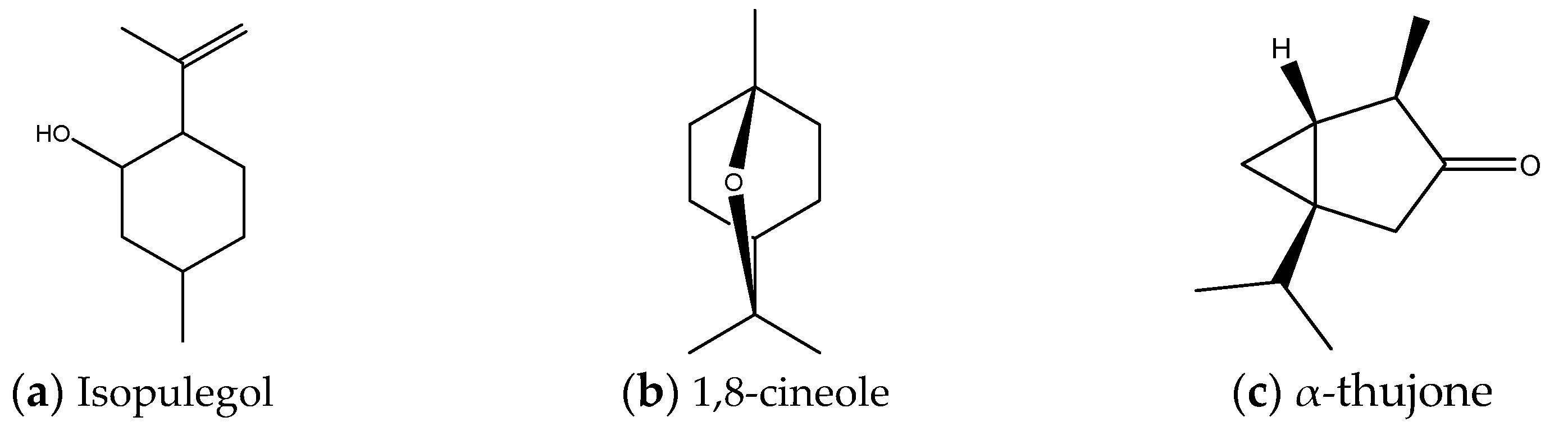
2.1. Chemical Composition

The chemical composition of hydrolates was determined by gas chromatography followed by mass spectrometry (GC/MS). Table 1 shows the results of the relative composition of hydrolates obtained by microwave-assisted extraction of fresh plant material. In total, from 20 different possible compounds, 13 were identified. Most compounds (eleven) were identified in the hydrolates of S. divinorum and S. sclarea, and at least six were identified in the hydrolate of S. officinalis. All hydrolates primarily contain the isoprenoid isopulegol (38.22–57.11%) (Figure 1a), followed by 1,8-cineole (Figure 1b) in S. officinalis (19. 57%) and S. nemorosa (15.67%) hydrolates, and thujone (Figure 1c), the main component present in hydrolates of S. nemorosa, S. aethiopis, and S. divinorum in amounts greater than 10%.

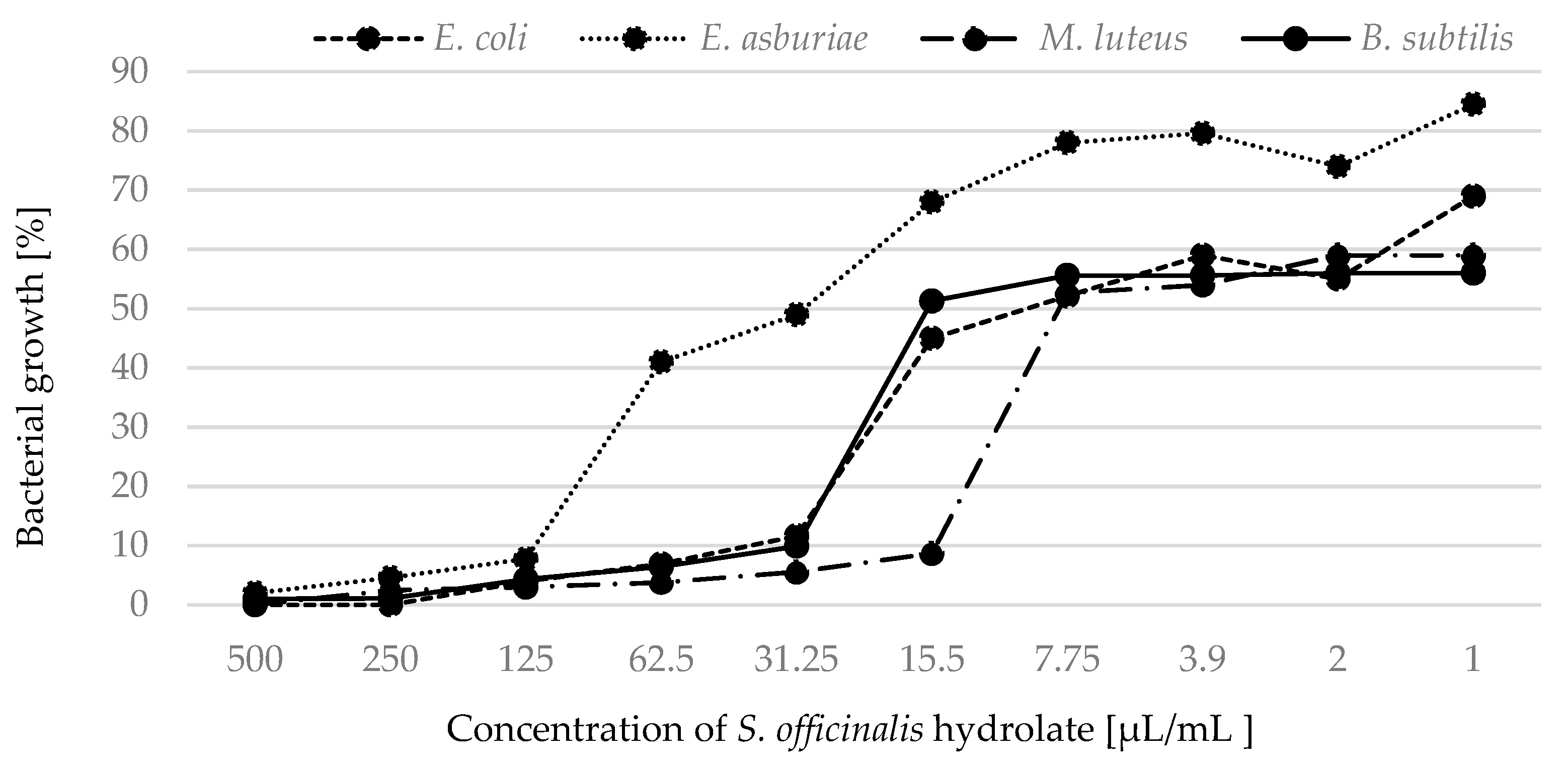
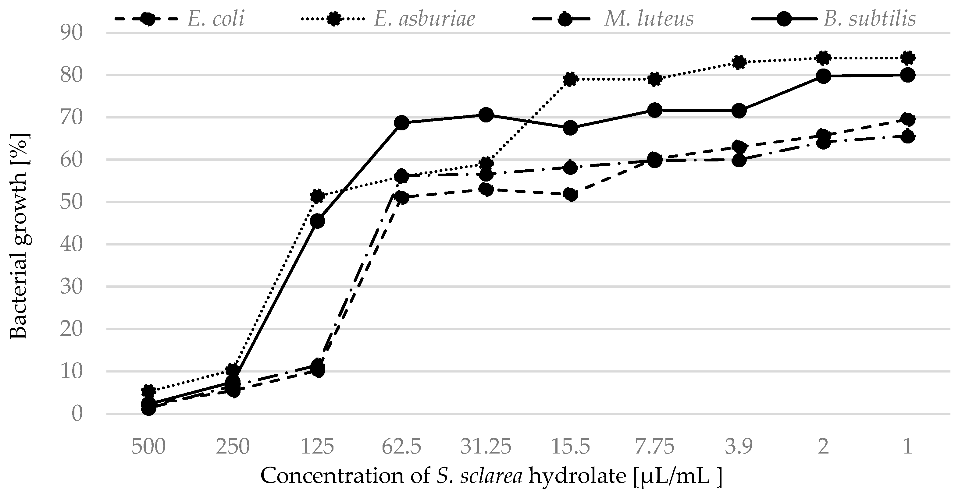
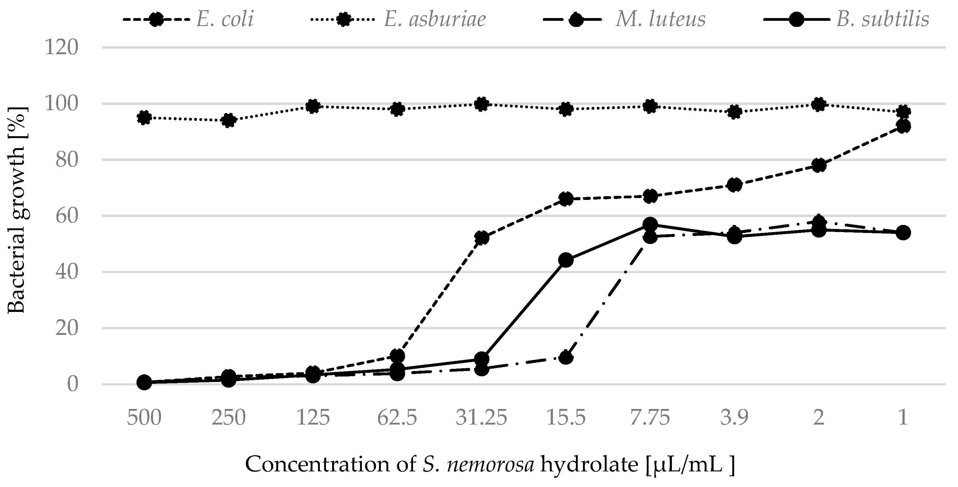
2.2. Antimicrobial Activity

The minimum inhibitory concentrations (MIC) MIC50 and MIC90 (expressed in μL/mL) against four bacterial strains are summarized in Table 2. The MIC tests showed the strongest antibacterial activity of S. officinalis hydrolate on M. luteus with MIC50 (5.69 µL/mL) and MIC90 (7.81 µL/mL) values.
Based on the measured absorption, we focused on the percentage expression of bacterial growth in the presence of hydrolates. We determined 100% bacterial growth in pure MHB and statistically compared this value with bacterial growth when treated with antimicrobials (the hydrolates). Growth of all strains was inhibited by hydrolates of S. officinalis (Figure 2) and S. sclarea (Figure 3).
The results showed that hydrolates of S. nemorosa inhibited more Gram-positive than Gram-negative bacterial strains (Figure 4).
The least inhibitory effect was observed for the strain E. asburiae when treated with S. nemorosa hydrolate. The growth of M. luteus was completely inhibited by S. officinalis and by S. nemorosa hydrolates at a concentration of 125 µL/mL.

2.3. Antioxidant Activity

In the analysis of Salvia hydrolates, the DPPH method was used. The hydrolates showed varying antioxidant activity (Table 3).
The results showed the strongest antioxidant activity of S. officinalis hydrolate at 23.25 ± 0.26% of inhibition.

3. Discussion

Salvia, an aromatic and medicinal herb, is known for many purposes [29]. The preparations of sage are known for their antibacterial activity against various bacteria [30,31]. Hydrolates have recently attracted attention for their antimicrobial activity, especially against pathogenic and spoilage microorganisms, including bacteria and fungi [32]. The chemotype of a hydrolate is important to understand the mechanisms of its biological activity. For example, phenolic compounds are considered potent antibacterial agents that tend to attack the outer membrane of bacteria. They also have effects on the structural and functional properties of the cytoplasmic membrane [25]. Other hydrolates have shown antibacterial effects as they accumulate in the cell membrane, affecting its integrity and causing the release of intracellular material [33]. It is known that the volatile profiles of hydrolates depend on the origin of the plant material [32].
By evaluating the chemical composition analysis via GC/MS, the relative proportion of the different compounds with potential antimicrobial activity was determined. All hydrolates primarily contain isopulegol and the presence of other isoprenoids. More than 10% of 1,8-cineole contains hydrolates of S. officinalis (19.57%), S. nemorosa (15.67%), and S. aethiopis (11.75%). Thujone, the main component of Salvia essential oils, was present in hydrolates of S. nemorosa, S. aethiopis, S. sclarea, and S. divinorum in amounts greater than 10%. Other scientists also studied different salvia hydrolates. The results of [4] showed that the main constituents of S. sclarea and S. officinalis are 1,8-cineole, α-thujone, linalool, and borneol. The aromatic water obtained from a sage sample collected in Turkey showed camphor, 1,8-cineole, thujone, and borneol as the main constituents [34]. The results of 1,8-cineole and thujone were in agreement with our results. In contrast with [25], our results did not confirm the presence of carvacrol in hydrolates.
The ability of hydrolates to inhibit bacterial growth depends on their chemical composition and the bacterial strain. The antimicrobial activity of hydrolates is explained by secondary major and minor components, which, in some cases, coincide with those of essential oils (although in different amounts), while in others they are completely different [25]. According to the results of phytochemical analysis of five Salvia sp. hydrolates, the main components are terpenes and their derivatives—isopulegol, 1,8-cineole, thujone, borneol, and linalool. Antimicrobial activities have been reported for borneol and 1,8-cineole. The minor constituents such as 1,8-cineole might be involved in the antibacterial activity of hydrolates [25]. The antimicrobial activity of thujone has also been confirmed [35,36,37]. Some researchers suggested that the hydroxyl groups of eugenol could bind to proteins by preventing the action of microbial enzymes [38]. In this study, we tested hydrolates of five Salvia sp. We found no antimicrobial effect of the hydrolate of S. divinorum in this study where MIC50 and MIC90 were more than 500 µL/mL for three bacteria—E. coli, M. luteus, and B. subtilis, and a very low antimicrobial effect on E. asburiae with a MIC50 value of 325.61 µL/mL. Similarly, this strain was sensitive to S. aethiopis hydrolate, with a MIC50 value of 216.59 µL/mL. The Gram-positive strains were resistant to S. aethiopis hydrolates. All strains proved sensitive to hydrolates of S. officinalis with MIC50 value of 5.69–56.65 µL/mL and S. sclarea with MIC50 value of 74.99–141.41 µL/mL. M. luteus proved to be the most sensitive to salvia hydrolate with MIC50 value (5.69 µL/mL), followed by B. subtilis (18.43 µL/mL), E. coli (27.50 µL/mL) and E. asburiae (56.65 µL/mL). The Gram-positive strains, with the exception of the hydrolate of S. officinalis, were also more sensitive to hydrolates of S. nemorosa than Gram-negative strains. E. asburiae was the only one in which we observed sensitivity to S. aethiopis and S. divinorum hydrolates.
When we focused on monitoring the percentage growth of bacteria treated with hydrolates, we confirmed and supplemented the results obtained by MIC. The hydrolates of S. officinalis and S. nemorosa were able, at the lowest concentrations, to partially inhibit the growth of Gram-positive strains throughout the cultivation. Similarly, the hydrolate of S. sclarea was marginally effective against bacterial strains at low concentrations. Ovidi et al. [4] tested essential oils of S. sclarea and defined them as bactericidal against the susceptible bacterial strains. Significant activity of these essential oils against S. aureus and S. epidermidis strains (MIC values of 10.0 and 5.0 mg/mL, respectively) was found [39]. Salvia species is among the plants effective against bacterial strains. Sagdiç and Ozcan [40] investigated the ability of S. fruticosa Mill. and S. aucheri L. to control common bacteria. Other authors tested the antibacterial activity of hydrolates of S. officinalis L. [41,42,43,44]. Tornuk et al. [41] indicated that the application of hydrolates showed a remarkable inhibitory effect on S. aureus. Sage was used to control the growth of S. typhimurium, E. coli O157:H7, and L. monocytogenes. In general, approximately three log reductions were obtained in the pathogens populations by using hydrosols [25]. Tornuk et al. [41] investigated the inhibitory effect of sage against S. typhimurium and E. coli O157:H7. The treatment procedures resulted in a significant reduction in the tested pathogenic bacteria. Ozturk et al. [42] studied the effect of plant hydrolates obtained from sage. Sage hydrolates eliminated L. monocytogenes. In contrast, according to [45], hydrolates from S. officinalis were ineffective against E. coli and Pseudomonas aeruginosa. In other studies [40,44], sage hydrolate was reported to be ineffective against fifteen bacteria in the in vitro test.
The antioxidant properties of plants are of interest because of their potential use as natural additives. There are few studies addressing the antioxidant properties of hydrolates. Their results show moderate to very low antioxidant capacity [37,46]. These results are consistent with the results of our study (see Table 3). In previous studies, the authors of [47] investigated the antioxidant activity of 55 Turkish Salvia species. In their study, the DPPH radical scavenging activity of S. nemorosa was reported to be 90.75% at 100 µg/mL. In contrast, Jeshvaghani et al. [48] reported 53% at 500 µg/mL. In other studies, the IC50 value of leaf extract of S. sclarea was 58.20 µg/mL [49] and 25 µg/mL [50]. Tepe et al. [51] reported the antioxidant activity of S. sclarea to be 23.4% at 50 µg/mL. These values of Turkish sage were higher than those of Slovak sage. According to [51], extracts of S. aethiopis did not show radical scavenging activity. Our results could not confirm this.

4. Materials and Methods

4.1. Isolation of Hydrolates

We used the leaves of S. officinalis, S. nemorosa, S. aethiopis, S. sclarea, and S. divinorum obtained from the Gene Bank of the Slovak Republic at the Research Institute of Plant Production in Piešťany. We added about 200 g of fresh sage leaves to the extraction container. The extraction took place in a microwave oven Bosch FFL023MS2 (Gerlingen, Germany) at 800 W for 8 min in EssenEx® 100A Essential Oil Extraction Kit (Corvallis, OR, USA). We used ice to condense the steam and oil. After extraction, we separated essential oils and hydrolates. We stored the sage hydrolates (25 mL from each batch) in dark-coloured bottles at 4 °C until we tested their antimicrobial activity.

4.2. GC- MS Analysis of Salvia Hydrolates

The constituents were identified and the relative composition of the salvia hydrolates was determined by gas chromatography followed by mass spectrometry (GC/MS), as described by [52]. Prior to injection, hydrolates were extracted in hexane in a ratio of 1.5:1 and dried over anhydrous sodium sulphate according to [37]. Analyses were carried out using an Agilent 7890A GC coupled to an Agilent MSD5975C MS detector (Agilent Technologies, Palo Alto, CA, USA) with a HP-5MS column (30 m × 0.25 mm, 0.25 mm film thickness). The analytical conditions were as follows: injector temperature 250 °C and the oven temperature from 60 °C to 231 °C at 3 °C/min. Components were identified by matching their mass spectra and retention indices with those of authentic samples and/or NIST/Wiley spectra libraries and available literature data [53]. Relative proportions were calculated by dividing the individual peak area by the total area of all peaks. Only compounds with more than 1% were considered.

4.3. Free Radical Scavenging Assays

The antioxidant activity of the studied hydrolates was determined using the 2,2-diphenyl-1-picrylhydrazyl radical (DPPH, Sigma-Aldrich, Germany) according to [54], with a modification for the microplate form. Briefly, 50 μL of the tested samples were added to 100 μL of a 1.2% DPPH-ethanol solution. The solution was shaken and incubated for 30 min in the dark at room temperature. Absorbance was measured at 517 nm using the Opsys MRTM microplate reader from Dynex (Chantilly, VA, USA). Antioxidant activity was expressed as a percentage (%) of scavenging activity: DPPH scavenging activity (%) = [(Ac − As)/(Ac − Ab)] × 100, where Ac is the absorbance of the control (absorbance of the DPPH solution with distilled water), As is the absorbance of the sample, and Ab is the absorbance of the blank (ethanol). The analyses were performed in triplicate.

4.4. Bacterial Strains

Gram-negative strains Escherichia coli CCM 3954 and Enterobacter asburiae CCM 8546, and Gram-positive strains Micrococcus luteus DSM 1790 and Bacillus subtilis DSM 5552 obtained from the Czech Collection of Microorganisms (CCM; Brno, Czech Republic) and the German Collection of Microorganisms and Cell Cultures (DSMZ, Braunschweig, Germany) were used in this study to determine the antibacterial activity of the hydrolates. Broth cultures of the test strains were grown overnight in Mueller–Hinton broth medium (Hi-medium, India) (MHB) at 37 °C for 16 h.

4.5. Preparation of the Inoculum

The overnight cultures were diluted in culture medium and adjusted to a final concentration of 5 × 10⁵ CFU/mL. This was confirmed by colony counting according to CLSI guidelines [55]. The same procedure for preparing the inoculum was used for all experiments performed in this study.

4.6. Determination of Minimum Inhibitory Concentration

The determination of the minimal inhibitory concentration (MIC) was performed via serial microdilution in 96-well microtiter plates using Mueller–Hinton broth. Briefly, the hydrolates were diluted in MHB to obtain an initial concentration of 512 μg/mL. The twofold serially diluted concentrations of all hydrolates ranged from 512 to 0.25 μg/mL. The final bacterial concentration was adjusted to 5 × 10⁵ CFU/mL. The wells in the last column of all plates served as positive controls for measuring the optical density of all strains tested, and the wells in the first column were used as sterility controls for MHB. Microplates were incubated at 37 °C for 18–20 h. The lowest concentration of antimicrobial agent that prevented bacterial growth was defined as the minimum inhibitory concentration (MIC). Optical density was measured at 405 nm using an Opsys MRTM microplate reader from Dynex (Chantilly, VA, USA). The 96-well microplates were measured before and after the experiment (approximately 18 h). Results were expressed as the mean of three replicates in three independent experiments.

4.7. Statistical Analysis

Statistical analysis was performed by probit analysis using the Statgraphic program (Statpoint technologies, Warrenton, VA, USA) according to [56], with some modifications.

5. Conclusions

This study focused on the chemical analysis of hydrolates obtained via microwave-assisted extraction of five Salvia species and the evaluation of their antimicrobial and antioxidant potential. Salvia hydrolates are a promising source of various compounds with potential antimicrobial activity and plant antioxidants such as isopulegol, 1,8-cineole, thujone, borneol, and linalool. The results of this study indicate that the hydrolates have promising antimicrobial activity. The highest antimicrobial activity was observed for S. officinalis hydrolate. The best minimum inhibitory concentration (MIC) was found against Gram-positive bacteria M. luteus DSM 1790 by S. officinalis hydrolate with MIC90 7.81 μg/mL. The interesting antimicrobial activity of S. sclarea and S. nemorosa hydrolates was also described. Our results showed a low antioxidant capacity of the hydrolates. Our postulated aims of the research in this study were achieved: We wanted to remark on the differences between various Salvia varieties from the chemical composition and antibacterial and antioxidant activity points of view. These were proved and described. This study suggests that hydrolates could be used, for example, as natural antimicrobial agents or food preservatives, but further testing is needed.

Author Contributions

Conceptualization, E.Ü. and T.M.; methodology, E.Ü. and T.M.; data curation, E.Ü., Ľ.U., M.V. and T.M.; writing—original draft preparation, E.Ü., Ľ.U. and T.M.; supervision, E.Ü. All authors have read and agreed to the published version of the manuscript.

Funding

This research was funded by SRDA-20-413, and by the Operational Program—Integrated Infrastructure, the project Long-term strategic research of prevention, intervention and mechanisms of obesity and its comorbidities, NFP313011V344, co-financed by the European Regional Development Fund.

Institutional Review Board Statement

Not applicable.

Informed Consent Statement

Not applicable.

Data Availability Statement

Data are contained within the article.

Conflicts of Interest

The authors declare no conflict of interest.

References

  1. Živković, J.; Ristić, M.; Kschonsek, J.; Westphal, A.; Mihailović, M.; Filipović, V.; Böhm, V. Comparison of chemical profile and antioxidant capacity of seeds and oils from Salvia sclarea and Salvia officinalis. Chem. Biodivers. 2017, 14, e1700344. [Google Scholar] [CrossRef]
  2. Delamare, L.A.P.; Moschen-Pistorello, I.T.; Artico, L.; Atti-Serafini, L.; Echeverrigaray, S. Antibacterial activity of the esential oil of Salvia officinalis L. cultivated in South Brazil. Food Chem. 2007, 100, 603–608. [Google Scholar] [CrossRef]
  3. Darwish, M.S.A.; Cabral, C.; Fereirra, I.V.; Gonçalves, M.J.; Cavaleiro, C.; Cruz, M.T.; Al-Bdour, T.H.; Salgueiro, L. Essential Oil of Common Sage (Salvia officinalis L.) from Jordan: Assessment of Safety in Mammalian Cells and Its Antifungal and Anti-Inflammatory Potential. BioMed Res. Int. 2013, 2013, 538940. [Google Scholar] [CrossRef] [Green Version]
  4. Ovidi, E.; Laghezza Masci, V.; Zambelli, M.; Tiezzi, A.; Vitalini, S.; Garzoli, S. Laurus nobilis, Salvia sclarea and Salvia officinalis Essential Oils and Hydrolates: Evaluation of Liquid and Vapor Phase Chemical Composition and Biological Activities. Plants 2021, 10, 707. [Google Scholar] [CrossRef]
  5. Sefidkon, F.; Khajavi, M.S. Chemical composition of the essential oils of two Salvia species from Iran: Salvia verticillata L. and Salvia santolinifolia Boiss. Flavour Frag. J. 1999, 14, 77–78. [Google Scholar] [CrossRef]
  6. Craft, J.D.; Satyal, P.; Setzer, W.N. The Chemotaxonomy of Common Sage (Salvia officinalis) Based on the Volatile Constituents. Medicines 2017, 4, 47. [Google Scholar] [CrossRef] [Green Version]
  7. Boutebouhart, H.; Didaoui, L.; Tata, S.; Sabaou, N. Effect of Extraction and Drying Method on Chemical Composition, and Evaluation of Antioxidant and Antimicrobial Activities of Essential Oils from Salvia officinalis L. J. Essent. Oil Bear. Plants 2019, 3, 717–727. [Google Scholar] [CrossRef]
  8. Chizzola, R. Composition and Variability of the Essential Oil of Salvia nemorosa (Lamiaceae) from the Vienna Area of Austria. Nat. Prod. Commun. 2012, 17, 1671–1672. [Google Scholar] [CrossRef] [Green Version]
  9. Güllüce, M.; Özer, H.; Bar, I.Ş.Ö.; Daferera, D.; Şahín, F.; Polissiou, M. Chemical Composition of the Essential Oil of Salvia aethiopis L. Turk. J. Biol. 2006, 30, 231–233. [Google Scholar]
  10. Carrubba, A.; La Torre, R.; Picaglia, R.; Marotti, M. Characterization of an Italian biotype of clary sage (Salvia sclarea L.) grown in a semi-arid Mediterranean environment. Flavour Frag. J. 2002, 17, 91–194. [Google Scholar] [CrossRef]
  11. Siebert, D.J. Salvia divinorum and salvinorin A: New pharmacologic findings. J. Ethnopharmacol. 1994, 43, 53–56. [Google Scholar] [CrossRef] [PubMed]
  12. El Euch, S.K.; Hassine, D.B.; Cazaux, S.; Bouzouita, N.; Bouajila, J. Salvia officinalis essential oil: Chemical analysis and evaluation of anti-enzymatic and antioxidant bioactivities. S. Afr. J. Bot. 2019, 120, 253–260. [Google Scholar] [CrossRef]
  13. Loizzo, M.R.; Saab, A.M.; Tundis, R.; Statti, G.A.; Menichini, F.; Lampronti, I.; Gambari, R.; Cinatl, J.; Doerr, H.W. Phytochemical analysis and in vitro antiviral activities of the essential oils of seven Lebanon species. Chem. Biodivers. 2008, 5, 461–470. [Google Scholar] [CrossRef]
  14. Kontogianni, V.G.; Tomic, G.; Nikolic, I.; Nerantzaki, A.A.; Sayyad, N.; Stosic-Grujicic, S.; Stojanovic, I.; Gerothanassis, I.P.; Tzakos, A.G. Phytochemical profile of Rosmarinus officinalis and Salvia officinalis extracts and correlation to their antioxidant and anti-proliferative activity. Food Chem. 2013, 136, 120–129. [Google Scholar] [CrossRef]
  15. Farimani, M.M.; Bahadori, M.B.; Koulaei, S.A.; Salehi, P.; Ebrahimi, S.N.; Khavasi, H.R.; Hamburger, M. New ursane triterpenoids from Salvia urmiensis Bunge: Absolute configuration and anti-proliferative activity. Fitoterapia 2015, 106, 1–6. [Google Scholar] [CrossRef]
  16. Ghorbani, A.; Esmaeilizadeh, M. Pharmacological properties of Salvia officinalis and its components. J. Tradit. Complement. Med. 2017, 7, 433–440. [Google Scholar] [CrossRef]
  17. Farah, A.; Mattazi, N.; Fadil, M.; Chraibi, M.; Benbrahim, K.F. Esential oils analysis and antibacterial activity of the leaves of Rosmarinus officinalis, Salvia officinalis and Menta piperita cultivated in Agadir (Morocco). Int. J. Pharm. Pharm. Sci. 2015, 7, 73–79. [Google Scholar]
  18. Balouiri, M.; Sadiki, M.; Ouedrhiri, W.; Farah, A.; El Abed, E.; Koraichi, S.I. Antibacterial activity of extractfrom Salvia officinalis and Rosmarinus officinalis obtained by sonication and maceration methods. Int. J. Pharm. Sci. 2014, 6, 167–170. [Google Scholar]
  19. Tepe, B.; Donmez, F.; Unlu, M.; Candan, F.; Daferera, D.; Unlu, V.G.; Polisiou, M.; Sokmen, A. Antimicrobial and antioxidative activities of the essential oil and methanol extracts of Salvia crypthanta (Montbret et Aucher ex Bent) and Salvia multicaulis. Food Chem. 2004, 84, 519–525. [Google Scholar] [CrossRef]
  20. Tepe, B.; Daferera, D.; Sokmen, A.; Sokmen, M.; Polisiou, M. Antimicrobial and antioxidant activities of the essential oil and various extracts of Salvia tomentosa Miller (Lamiaceae). Food Chem. 2005, 90, 333–340. [Google Scholar] [CrossRef]
  21. Ćulafić, D.M.; Vuković-Gačić, B.; Knežević-Vukčević, J.; Stanković, S.; Simić, D. Comparative study on the antibacterial activity of volatiles from sage (Salvia officinalis L.). Arch. Biol. Sci. 2005, 57, 173–178. [Google Scholar] [CrossRef]
  22. Smigielski, K.; Prusinowska, R.; Stobiecka, A.; Kunicka-Styczynska, A.; Gruska, R. Biological properties and chemical composition of essential oils from flowers and aerial parts of lavender (Lavandula angustifolia). J. Essent. Oil Bear. Plants 2018, 21, 1303–1314. [Google Scholar] [CrossRef]
  23. Hay, Y.O.; Abril-Sierra, M.A.; Sequeda-Castaneda, L.G.; Bonnafous, C.; Raynaud, C. Evaluation of combinations of essential oils and essential oils with hydrosols on antimicrobial and antioxidant activities. J. Pharm. Pharmacogn. Res. 2018, 6, 216–230. [Google Scholar]
  24. Lira, P.D.; Reeta, D.; Tkacik, E.; Ringuelet, J.; Coussio, J.D.; van Baren, C.; Bandoni, A.L. Essential oil and by-products of distillation of bay leaves (Laurus nobilis L.) from Argentina. Ind. Crops Prod. 2009, 30, 259–264. [Google Scholar] [CrossRef]
  25. D’Amato, S.; Serio, A.; Lopez, C.C.; Paparella, A. Hydrosols: Biological activity and potential as antimicrobials for food applications. Food Control 2018, 86, 126–137. [Google Scholar] [CrossRef]
  26. Hamedi, A.; Moheimani, S.M.; Sakhteman, A.; Etemadfard, H.; Moein, M. An overview on indications and chemical composition of aromatic waters (hydrosols) as functional beverages in Persian nutrition culture and folk medicine for hyperlipidemia and cardiovascular conditions. J. Evid.-Based Integr. Med. 2017, 22, 544–561. [Google Scholar] [CrossRef]
  27. Šilha, D.; Švarcová, K.; Bajer, T.; Královec, K.; Tesarová, E.; Moucková, K.; Pejchalová, M.; Bajerová, P. Chemical composition of natural hydrolates and their antimicrobial activity. Molecules 2020, 25, 5654. [Google Scholar] [CrossRef]
  28. Rossi, C.; Maggio, F.; Chaves-López, C.; Valbonetti, L.; Berrettoni, M.; Paparella, A.; Serio, A. Effectiveness of selected essential oils and one hydrolate to prevent and remove Listeria monocytogenes biofilms on polystyrene and stainless steel food-contact surfaces. J. Appl. Microbiol. 2021, 132, 1866–1876. [Google Scholar] [CrossRef]
  29. Vosoughi, N.; Gomarian, M.; Pirbalouti, A.G.; Khaghani, S.; Malekpoor, F. Essential oil composition and total phenolic, flavonoid contents, and antioxidant activity of sage (Salvia officinalis L.) extract under chitosan application and irrigation frequencies. Ind. Crops Prod. 2018, 117, 366–374. [Google Scholar] [CrossRef]
  30. Mandras, N.; Nostro, A.; Roana, J.; Scalas, D.; Banche, G.; Ghisetti, V.; Del Re, S.; Fucale, G.; Cuffini, A.M.; Tullio, V. Liquid and vapourphase antifungal activities of essential oils against Candida albicans and non-albicans Candida. BMC Complement. Altern. Med. 2016, 16, 330. [Google Scholar] [CrossRef] [Green Version]
  31. Selim, S.; Almuhayawi, M.S.; Alqhtani, H.; Al Jaouni, S.K.; Saleh, F.M.; Warrad, M.; Hagagy, N. Anti-Salmonella and antibiofilm potency of Salvia officinalis L. essential oil against antibiotic-resistant Salmonella enterica. Antibiotics 2022, 11, 489. [Google Scholar] [CrossRef]
  32. Politi, M.; Ferrante, C.; Menghini, L.; Angelini, P.; Flores, G.A.; Muscatello, B.; Braca, A.; De Leo, M. Hydrosols from Rosmarinus officinalis, Salvia officinalis, and Cupressus sempervirens: Phytochemical analysis and bioactivity evaluation. Plants 2022, 11, 349. [Google Scholar] [CrossRef]
  33. Jabra-Rizk, M.A.; Meiller, T.F.; James, C.E.; Shirtliff, M.E. Effect of farnesol on Staphylococcus aureus biofilm formation and antimicrobial susceptibility. Antimicrob. Agents Chemother. 2006, 50, 1463–1469. [Google Scholar] [CrossRef] [Green Version]
  34. Baydar, H.; Sangun, M.K.; Erbas, S.; Kara, N. Comparison of arome compounds indistilled end extracted products of sage (Salvia officinalis L.). J. Essent. Oil-Bear. Plants 2013, 16, 39–44. [Google Scholar] [CrossRef]
  35. Rashid, S.; Rather, M.A.; Shah, W.A.; Bhat, B.A. Chemical composition, antimicrobial, cytotoxic and antioxidant activities of the essential oil of Artemisia indica Willd. Food Chem. 2013, 138, 693–700. [Google Scholar] [CrossRef]
  36. Tavares, C.S.; Gameiro, J.A.; Roseiro, L.B.; Figueiredo, A.C. Hydrolates: A review on their volatiles composition, biological properties and potential uses. Phytochem. Rev. 2022, 21, 1661–1737. [Google Scholar] [CrossRef]
  37. Stan (Tudora), C.; Muscalu, A.; Burnichi, F.; Popescu, C.; Gatea, F.; Sicuia, O.-A.; Vladut, N.V.; Israel-Roming, F. Evaluation of essential oils and hydrolate from a new hyssops variety (Hyssopus officinalis L.). Not. Bot. Horti Agrobot. Cluj-Napoca 2022, 50, 12639. [Google Scholar] [CrossRef]
  38. Wedakoon, C.N.; Sakaguchi, M. Combined effect of sodium cholride and clove on growth and biogenic amine formation of Enterobacter aerogenes in mackerel muscle extract. J. Food Prot. 1993, 56, 410–413. [Google Scholar] [CrossRef] [PubMed]
  39. Kuzma, L.; Kalemba, D.; Rozalski, M.; Rozalska, B.; Więckowska-Szakiel, M.; Krajewska, U.; Wysokinska, H. Chemical composition and biological activities of essential oil from Salvia sclarea plants regenerated in vitro. Molecules 2009, 14, 1438–1447. [Google Scholar] [CrossRef] [PubMed] [Green Version]
  40. Saǧdıç, O.; Özcan, M. Antibacterial activity of Turkish spice hydrosols. Food Control 2003, 14, 141–143. [Google Scholar] [CrossRef]
  41. Tornuk, F.; Cankurt, H.; Ozturk, I.; Sagdiç, Ö.; Bayram, O.; Yetim, H. Efficacy of various plant hydrosols as natural food sanitizers in reducing Escherichia coli O157:H7 and Salmonella typhimurium on fresh cut carrots and apples. Int. J. Food Microbiol. 2011, 148, 30–35. [Google Scholar] [CrossRef]
  42. Ozturk, I.; Tornuk, F.; Saǧdıç, O.; Kisi, O. Application of non-linear models to predict inhibition effects of various plant hydrosols on Listeria monocytogenes inoculated on fresh-cut apples. Foodborne Pathog. Dis. 2012, 9, 607–616. [Google Scholar] [CrossRef]
  43. Tornuk, F.; Ozturk, I.; Sagdiç, Ö.; Yilmaz, A.; Erkmen, O. Application of predictive inactivation models to evaluate survival of Staphylococcus aureus in fresh-cut apples treated with different plant hydrosols. Int. J. Food Prop. 2014, 17, 587–598. [Google Scholar] [CrossRef] [Green Version]
  44. Ozturk, L.; Tornuk, F.; Caliskan-Aydigan, O.; Zeki Durak, M.; Sagdiç, Ö. Decontamination of iceberg lettuce by some plant hydrosols. LWT-Food Sci. Technol. 2016, 74, 48–54. [Google Scholar] [CrossRef]
  45. Oral, N.; Vatansever, L.; Guven, A.; Gulmez, M. Antibacterial activity of some Turkish plant hydrosols. Kafkas Univ. Vet. Fak. Gerg. 2008, 14, 205–209. [Google Scholar]
  46. Jakubczyk, K.; Tuchowska, A.; Janda-Milczarek, K. Plant hydrolates—Antioxidant properties, chemical composition and potential applications. Biomed. Pharmacother. 2021, 142, 112033. [Google Scholar] [CrossRef] [PubMed]
  47. Senol, S.; Orhan, I.; Celep, F.; Kahraman, A.; Dogan, M.; Yilmaz, G.; Şeneraet, B. Survey of 55 Turkish Salvia taxa for their acetylcholinesterase inhibitory and antioxidant activities. Food Chem. 2010, 120, 34–43. [Google Scholar] [CrossRef]
  48. Jeshvaghani, Z.A.; Rahimmalek, M.; Talebi, M.; Hossein Goli, S.A. Comparison of total phenolic content and antioxidant activity in different Salvia species using three model systems. Ind. Crops Prod. 2015, 77, 409–414. [Google Scholar] [CrossRef]
  49. Ben Taari, M.; Msaada, K.; Hosni, K.; Marzouk, B. Fatty acids, phenolic changes and antioxidant activity of clary sage (Salvia sclarea L.) rosette leaves grown under saline conditions. Ind. Crops Prod. 2012, 38, 58–63. [Google Scholar] [CrossRef]
  50. Stagos, D.; Portesis, N.; Spanou, C.; Mossialos, D.; Aligiannis, N.; Chaita, E.; Panagoulis, C.; Reri, E.; Skaltsounis, L.; Tsatsakis, A.M.; et al. Correlation of total polyphenolic content with antioxidant and antibacterial activity of 24 extracts from Greek domestic Lamiaceae species. Food Chem.Toxicol. 2012, 50, 4115–4124. [Google Scholar] [CrossRef]
  51. Tepe, B.; Sokmen, M.; Akpulat, H.A.; Sokmen, A. Screening of the antioxidant potentials of six Salvia species from Turkey. Food Chem. 2006, 95, 200–204. [Google Scholar] [CrossRef]
  52. Císarová, M.; Hleba, L.; Tančinová, D.; Florková, M.; Foltinová, D.; Charousová, I.; Vrbová, K.; Božik, M.; Klouček, P. Inhibitory effect of essential oils from some lamiaceae species on growth of Eurotium spp. isolated from bread. J. Microbiol. Biotechnol. Food Sci. 2018, 8, 857–862. [Google Scholar] [CrossRef]
  53. Adams, R.P. Identification of Essential Oil Components by Gas Chromatography/Mass Spectrometry, 4th ed.; Allured Publishing Corporation: Carol Stream, IL, USA, 2007. [Google Scholar]
  54. Hatano, T.; Kagawa, H.; Yasuhara, T.; Okuda, T. Two new flavonoids and other constituents in licorice root: Their relative astringency and radical scavenging effects. Chem. Pharm. Bull. 1988, 36, 2090–2097. [Google Scholar] [CrossRef] [PubMed] [Green Version]
  55. Clinical And Laboratory Standards Institute (CLSI). Methods for Dilution Antimicrobial Susceptibility Tests for Bacteria That Grow Aerobically, Approved Standard, 8th ed.; CLSI Document M7-A8; CLSI: Wayne, PA, USA, 2009. [Google Scholar]
  56. Hleba, L.; Vuković, N.; Horská, E.; Petrová, J.; Sukdolak, S.; Kačániová, M. Phenolic profile and antimicrobial activities to selected microorganisms of some wild medical plant from Slovakia. Asian Pac. J. Trop. Dis. 2014, 4, 269–274. [Google Scholar] [CrossRef]
Figure 1. The main compounds in hydrolates.
Plants 12 01325 g001
Figure 2. The growth of bacterial strains in the presence of S. officinalis hydrolate.
Plants 12 01325 g002
Figure 3. The growth of bacterial strains in the presence of S. sclarea hydrolate.
Plants 12 01325 g003
Figure 4. The growth of bacterial strains in the presence of S. nemorosa hydrolate.
Plants 12 01325 g004
Table 1. Qualitative and quantitative analysis (%) of salvia hydrolates by GC/MS.
RI bCompoundS. officinaliscS. nemorosacS. aethiopiscS. sclareacS. divinorumc
982(-)-β-Pinene a-1.31%-1.20%3.15%
10341,8-Cineole a19.57%15.67%11.75%4.71%6.29%
1100(-)-Linalool a4.01%2.68%3.70%3.44%3.40%
1117α- and β-Thujone5.57%14.10%13.78%11.50%10.95%
1137Unidentified--1.03%1.13%1.21%
1146(-)-Isopulegol57.11%47.20%56.54%40.26%38.22%
1167(-)-Borneol a5.41%4.74%5.06%5.28%6.55%
1178Menthol (+/−) a-2.02%--2.71%
1197Carvomenthenol a1.12%----
1403Unidentified----1.38%
1413Unidentified-1.50%1.15%--
1475α-Cubebene---3.90%4.10%
1517Caryophyllene---3.28%2.71%
1552α-Caryophyllene---3.34%3.04%
1579Germacrene D---4.59%4.21%
1585Unidentified----1.29%
1623Unidentified----1.10%
1625Unidentified----1.20%
1680Unidentified---1.28%-
1689Naphthalene-1.75%1.26%6.41%-
-Total92.79%90.95%94.27%90.31%91.52%
Legend: a Identification confirmed by co-injection of the authentic standard; b RI: identification based on Kovat’s retention indices (HP-5MS capillary column) and mass spectra; c relative proportion was calculated in percentage by dividing the area of each peak by the total area of all peaks.
Table 2. The minimum inhibitory concentration (MIC) of the tested hydrolates expressed in μL/mL.
Tested HydrolatesE. coliE. asburiaeM. luteusB. subtilis
MIC50MIC90MIC50MIC90MIC50MIC90MIC50MIC90
S. officinalis27.5042.1256.65123.165.697.8118.4331.25
S. nemorosa38.8262.58>500>5008.5115.6311.2628.14
S. aethiopis>500>500216.59407.13>500>500>500>500
S. sclarea74.99125.41141.41253.0181.68136.38106.58201.11
S. divinorum>500>500325.61472.61>500>500>500>500
Table 3. Antioxidant activity expressed as percentage inhibition of DPPH radical at a concentration of 100 μg/mL of the tested hydrolates.
Tested HydrolatesAntioxidant Activity (%)TEAC (mg/mL)
S. officinalis23.25 ± 0.2652.16 ± 2.85
S. nemorosa14.61 ± 0.0931.08 ± 1.09
S. aethiopis10.62 ± 0.9624.12 ± 0.96
S. sclarea18.99 ± 0.5842.12 ± 1.58
S. divinorum6.42 ± 0.5714.41 ± 0.57
Legend: Values represent the average (standard deviations) for triplicate analyses. Each value is given as mean ± standard deviation.
Disclaimer/Publisher’s Note: The statements, opinions and data contained in all publications are solely those of the individual author(s) and contributor(s) and not of MDPI and/or the editor(s). MDPI and/or the editor(s) disclaim responsibility for any injury to people or property resulting from any ideas, methods, instructions or products referred to in the content.

