Communication

Unique Morphology of Sarcobatus baileyi Male Inflorescence and Its Botanical Implications

1
Key Laboratory of Resource Biology and Biotechnology in Western China (Ministry of Education), School of Life Sciences, Northwest University, Xi’an 710069, China
2
State Key Laboratory of Systematic and Evolutionary Botany, Institute of Botany, Chinese Academy of Sciences, Beijing 100093, China
3
State Key Laboratory of Palaeobiology and Stratigraphy, Nanjing Institute of Geology and Palaeontology and CAS Center for Excellence in Life and Paleoenvironment, Chinese Academy of Sciences, Nanjing 210008, China
*
Author to whom correspondence should be addressed.
Plants 2023, 12(9), 1917; https://doi.org/10.3390/plants12091917
Submission received: 20 February 2023 / Revised: 28 April 2023 / Accepted: 2 May 2023 / Published: 8 May 2023
(This article belongs to the Special Issue Floral Biology 2.0)

Abstract

:
A typical angiosperm flower is usually bisexual, with entomophilous plants having four whorls of organs: the calyx, corolla, stamens, and gynoecium. The flower is usually colorful, and thus, distinct from the dull-colored reproductive organs of gymnosperms; however, this formula is not applicable to all flowers. For example, the male flower of Sarcobatus baileyi is reduced into only a single stamen. Such unusual flowers are largely poorly documented and underappreciated. To fill such a lacuna in our knowledge of the male reproductive organ of S. baileyi, we collected and studied materials of the male inflorescence of S. baileyi (Sarcobataceae). The outcomes of our Micro-CT (micro computed tomography), SEM (scanning electron microscopy), and paraffin sectioning indicate that a male inflorescence of S. baileyi is more comparable with the cone of conifers; its male flowers lack the perianth, are directly attached to a central axis and sheltered by peltate indusium-like shields. To understand the evolutionary logic underlying such a rarely seen male inflorescence, we also studied and compared it with a female cone of Cupressus sempervirens. Although the genera Sarcobatus and Cupressus belong to two distinct major plant groups (angiosperms and gymnosperms), they apply the same propagule-protecting strategy.

1. Introduction

Angiosperms are characterized by their flowers, which are frequently a focus of studies in botanical research (including morphological [1,2,3,4,5,6,7,8,9,10,11,12,13,14,15,16,17,18,19,20,21,22] and molecular research [23,24,25,26,27,28]). Most of the flowers in the assumed basal angiosperms (either Magnoliales in former system [29] or ANITA clade in the APG (angiosperm phylogeny group) system [30]) are bisexual. This bisexuality of the reproductive organs in angiosperms constitutes a drastic contrast against the unisexuality of the reproductive organs in gymnosperms (the only sister group of angiosperms in seed plants). In light of this contrast, male flowers in angiosperms seem more likely to bridge the morphological gap between angiosperms and gymnosperms, as female reproductive organs in these two groups are distinct and very hard to homologize. Many male flowers of angiosperms are not purely unisexual; they still have residual gynoecium [31,32]. This makes the pure male flowers in S. baileyi especially meaningful in addressing the evolution of male reproductive organs in angiosperms. This prompts us to choose S. baileyi as a study material.
Sarcobatus (Sarcobataceae) is a genus established by Nees in 1839 based on materials collected by Wied in July, 1833 [33]. It used to be called Fremontia, Sarcacanthus, Sarcobatis, pulpy-leaved thorn, salt cedar, sage, greasewood, and black greasewood by various authors and local peoples [33]. It was placed in Urticeae and Chenopodiaceae by various authors before it was placed in its own monotypic family Sarcobataceae [33]. APG clusters Sarcobataceae, Gieskiaceae, Phytolaccaceae, and Nyctaginaceae together as a clade in Caryophyllales [30]. Different from the other three families in the clade, Sarcobataceae have unisexual flowers and P3cf-form sieve-element plastids, distinguishing them from the other three families in the clade and justifying a separate family, which is also supported by cpDNA sequencing [33]. There are two species, S. vermiculatus and S. baileyi (the latter is sometimes regarded as a variation of the former by some authors) in the family. The plants of Sarcobatus are branching stiff spiny shrubs endemic to the alkali plains and deserts in the Great Basin and the southwest deserts in western North America [33,34,35,36]. They are widely used for household equipment, fuel, construction, weapons, food, forage, and music instrument. Furthermore, probably due to its unusual habitat, the leaves of S. baileyi are pulpy, and, more importantly, its male inflorescence is cone-like [37]. Although the female flowers of Sarcobataceae were studied in the 19th century [38], little is known of their male inflorescences other than that their male flowers are arranged beneath peltate scaly bracts, have a very short filament, and have long anthers [30,37]. This poorly understood male inflorescence/flower is the focus of the current study.
If we restricted our study to male flowers in angiosperms and without any control, our study would have limited significance with regard to plant evolution. Male cones in conifers protect their pollen sacs under their lateral appendages, and ovulate cones in Pinaceae may protect their seeds after pollination [39]. What happened to the female cones in Cupressaceae remains poorly documented. Therefore, we chose an ovulate cone of Cupressus sempervirens (Cupressaceae) for the purpose of comparison.

2. Materials and Methods

The S. baileyi material studied was collected from a bush on the south shore of Mono Lake, CA, USA, in June 2019 during a field trip organized by UCRS (https://napc2019.ucr.edu/field-trips-workshops#postmeeting accessed on 3 January 2023). The Cupressus material was collected from Mallorca, Spain in June 2016 during a field trip organized by the University of Vigo. Both taxa are listed as Least Concern (LC), which means that no conservation actions are needed as per IUCN. All field collections of plant materials complied with relevant institutional, national, and international guidelines and legislation. Field observations indicated that male inflorescences of S. baileyi and ovulate cones of C. sempervirens were uniform in dimensions; therefore, a minimal collections (two organs) was made. Observations and photographs were made with a Nikon SMZ1500 stereoscopic microscope at the Nanjing Institute of Geology and Palaeontology, Nanjing, China. Micro-CT (micro computed tomography) of S. baileyi was performed using a Zeiss Xradia 520 at the Nanjing Institute of Geology and Palaeontology, Nanjing, China, while Micro-CT of Cupressus was performed using a Skyscan 1172 at the Institute of Botany, Beijing, China. The 3D reconstruction and virtual sections were generated using VG Studio MAX 3.0. The specimen of S. baileyi was fixed with FAA (formalin-aceto-alcohol), dehydrated, embedded in paraffin, sliced into 16 μm thick sections using a Leica RM2135 rotary microtome, followed by dewaxing twice in xylene for 20 min each time and then dehydrating through a graded ethanol series (in 100% twice, once each in 90%, 85%, and 70%) for 30 min each time. Then, the sections were stained with Safranin O and Fast Green. Sections were examined and digitally photographed on a Leica microscope (DMLB) equipped with a video camera (Leica, DC 300F; Leica Microscopy and Systems GmbH, Wetzlar, Germany). The materials were dried in open air before observation. SEM observation on pollen grains of S. baileyi was performed using a TESCAN MAIA3 SEM (scanning electron microscope) housed at the Nanjing Institute of Geology and Palaeontology, CAS. Our measurements were performed based on images of pictures made during Micro-CT, SEM, stereomicrography, and light microscopy. The paraffin sections were deposited in the School of Life Sciences, Northwest University, Xi’an, China. All images were recorded in TIFF or JPEG format and organized together using Photoshop 7.0 for publication.

3. Results

Sarcobatus baileyi are bushes that are about 1~2 m tall and intricately branched, and their proximal branches are in contact with ground. Their leaves are clustered on cushion-like bases on older wood, and their blade is dull green to grayish green, usually terete, arcuate, 5–16 mm, and pubescent (n = 12) (Figure 1a). The male inflorescence is cylindrical in form, approximately 9 mm long (from the bottom to the top), and 3 mm in diameter (n = 2) with a central axis (Figure 1a,e). The central axis is approximately 0.4 mm in diameter, tapering distally (n = 2) (Figure 1e). Peltate shields are helically arranged around the central axis, each with a stalk (approximately 0.1 mm wide basally and 1 mm long), as well as a terminal rhomboidal expanded shield (approximately 1.4 mm wide) with a central apophysis (n = 5) (Figure 1a,e). Male flowers are reduced and equivalent to a single stamen, without any trace of perianth and gynoecium. A total of five to six caducous stamens are centered around the stalk of each peltate shield, attached to the central axis by 0.1 mm long filaments (n = 8) (Figure 1a,d–f). Each stamen is tetrasporangiate, approximately 0.8 mm long, 0.5 mm wide, and 0.4 mm thick (n = 7), and have 2 lateral pollen sacs that are fused when mature, dehiscing by valves (Figure 1b–f). Pollen grains are spherical, pantoporate, and 24 μm in diameter (n > 40) (Figure 1g).
The C. sempervirens plant we sampled is a tall tree growing in a farm on Mallorca Island in the Mediterranean Sea (Spain) (Figure 2a). The tree is tall, evergreen, and columnar in shape with blue-green needles (Figure 2a). The ovulate cones are spheroidal in form and 2.1 to 2.3 cm in diameter (n = 2) (Figure 2b–e). There are 7–8 peltate bracts attached to the central axis (Figure 2b–e). Each bract is about 10 mm long, 1 mm wide basally, and 16 mm wide apically with cavities (possibly containing resin) of variable sizes near the tip (Figure 2b–e). Numerous ovules are clustered in the space around the pedicels of the bracts and are sheltered by the bracts completely (Figure 2b–e). Ovules are slightly flattened, with two ridges, about 4 mm long, 3 mm wide, and 1~1.6 mm thick (n = 10).

4. Discussion

Sarcobataceae, Gieskiaceae, Phytolaccaceae, and Nyctaginaceae are clustered in a clade of Caryophyllales in APG IV [30]. One of the features unique to Sarcobataceae is that its flowers/inflorescences are unisexual, a feature unusual enough to distinguish Sarcobataceae from other families in the same clade [30]. This makes its male reproductive organs (inflorescences/flowers) incomparable to other Caryophyllales and even to other angiosperms. If the details of the fruit and pollen grains are ignored, S. baileyi appears more like a gymnosperm; its inflorescence is unisexual, its leaves are fleshy as seen in conifers, and, most interestingly, its male inflorescences are cone-like catkins (Figure 1a). This comparison is intriguing, and it may shed light on the homology and origin of the male inflorescence of S. baileyi (angiosperms). It is well known that pollen sacs are in naked clusters in Ginkgoales [40], Gnetales [41], and Caytoniales [42,43], while the pollen sacs are present and, thus, protected on the abaxial/adaxial surface of “microsporophylls” in Cycadales [43] and Coniferales [43,44]/Bennetttiales [43,45]. The cover and protection for the pollen sacs are provided by the scale-form “microsporophylls”, reducing the possibility of harmful attacks on the pollen sacs. It is curious that such a protection for pollen sacs appears lacking in angiosperms, which usually protect their pollen sacs using petals or tepals. The male florescence of Sarcobataceae documented here provides a rare exception and supplementation for the above generalization: pollen sacs/stamens/male flowers are sheltered by peltate shields in Sarcobataceae. With this supplementation, the pollen organs in gymnosperms and angiosperms seem to implement the same logic in their evolution: offspring development conditioning (ODC). ODC has recently been found to be an almost ubiquitous trend in the evolution of reproduction in sexually reproducing organisms (including all higher plants) [46]. Our new observation seems to bridge and narrow the gap between gymnosperms and angiosperms, which otherwise are thought to be widely separated.
Coverage and protection of pollen sacs are important insurance for the successful reproduction of plants; however, this insurance as an implementation of ODC is not restricted to pollen sacs alone. This rule applies to the female organs equally. Ovules enclosed before pollination are restricted to angiosperms [3,39]. Such an enclosure is a more apparent coverage and protection for the ovules, and besides protection, this enclosure supplies secured nutrition for the healthy development of ovules/seeds [3,39]. Therefore, angiospermy/angio-ovuly, just as the above protection for pollen sacs, is another way to implement ODC.
If ODC is such a ubiquitous trend in the reproduction of plants, one cannot help but wonder whether it is applicable to the female parts in gymnosperms. If we can find an example in gymnosperms, it will strengthen the ODC hypothesis. Relative to angiosperms, gymnosperms are famous for their naked ovules/seeds; however, this does not mean that gymnosperms do not care for their ovules/seeds. Instead, gymnosperms implement protection equally, although in diversified ways. At least some conifers tend to seclude their seeds from the exterior after pollination through cone closure, which is implemented by enlargement and intercalary growth of bract bases [39], while the ovules/seeds of Bennettitales are protected by peltate interseminal scales with only their micropylar tubes exposed [47,48,49] and sometimes by contractile seeds [48]. It is noteworthy that the way Bennettitales protect their ovules is comparable to the method that Sarcobataceae apply to protect their male flowers/stamens/pollen sacs (Figure 1a,e), although they differ in the number of pollen sacs/ovules per peltate shield/interseminal scale. The ovules/seeds of C. sempervirens (Coniferales) (Figure 2b–e) are also sheltered by peltate shields in a way similar to those of Sarcobataceae (Figure 2b–e); the seed scales (former secondary fertile branches) are reduced to only clusters of ovules/seeds that are independent of the bracts, as seen in Juniperus [50,51]. Such a resemblance spanning remotely related major groups of seed plants is intriguing, and it may reflect a shared pattern underlying the evolution of reproductive organs across all seed plants. The occurrence of such a phenomenon in widely separated plant groups apparently cannot be due to phylogeny; it may well represent a convergence in evolution leading to protection of reproductive parts before pollination and anthesis, as well as parallel implementations of the same evolution trend, namely ODC.
Although various molecular studies [23,24,25,26,27,28] have been carried out to elucidate the gene network underlying flower development, male flowers in angiosperms (especially those of Sarcobataceae) appear understudied in molecular studies [52]. The organization of Amborella flowers has attracted much attention, and some conclusions have been published [53]; however, male flowers, such as those of S. baileyi, are left molecularly intact and poorly understood. Such a situation may be related to the huge gap between female parts in angiosperms and gymnosperms, which constitutes an obvious challenge in botany. We wish to call for more attention and studies to elucidate the molecular mechanisms underlying the development and evolution of the unique male reproductive organs of Sarcobataceae.

5. Conclusions

We documented the unique morphology and organization of male inflorescence of Sarcobataceae. Unlike in other typical angiosperm flowers, the male flowers of Sarcobataceae are sheltered under peltate shields until anthesis. Similar protection for reproductive parts occurs in various seed plants in different ways, either through sheltering using peltate-formed shields (as documented here), physical enclosing (angiospermy, angio-ovuly), or timed opening and closing of parts. These diverse methods seem to be connected by a common rule: offspring development conditioning (ODC).

Author Contributions

X.W. collected and identified the specimens; W.L. carried out the paraffin sectioning; X.X. carried out the Micro-CT observation; X.W. and W.L. drafted, modified, and finalized the manuscript. All authors have read and agreed to the published version of the manuscript.

Funding

This research is supported by the National Natural Science Foundation of China (42288201, 41688103, 91514302) and the Strategic Priority Research Program (B) of the Chinese Academy of Sciences (XDB26000000).

Institutional Review Board Statement

Not applicable.

Informed Consent Statement

Not applicable.

Data Availability Statement

All data generated or analyzed during this study are included in this published article..

Acknowledgments

We thank Xintang Ma for helping with accessing the specimens of Sarcobatus female flowers in Peking Herbarium, Institute of Botany, CAS. We also thank Suping Wu for helping with part of the Micro-CT observation and Yan Fang for helping with SEM observation. We appreciate the kind help and constructive suggestions from the reviewers.

Conflicts of Interest

The authors declare no conflict of interest. The funders had no role in the design of the study; in the collection, analyses, or interpretation of data; in the writing of the manuscript; or in the decision to publish the results.

References

  1. Wang, X.; Duan, S.; Geng, B.; Cui, J.; Yang, Y. Schmeissneria: A missing link to angiosperms? BMC Evol. Biol. 2007, 7, 14. [Google Scholar] [CrossRef] [PubMed]
  2. Wang, X.; Shih, C.; Liu, Z.-J.; Lin, L.; Singh, K.J. Reconstructing the Callianthus plant–An early aquatic angiosperm from the Lower Cretaceous of China. Cretac. Res. 2021, 128, 104983. [Google Scholar] [CrossRef]
  3. Wang, X. The Dawn Angiosperms, 2nd ed.; Springer: Cham, Switzerland, 2018; p. 407. [Google Scholar]
  4. Han, G.; Liu, Z.; Wang, X. A Dichocarpum-like angiosperm from the Early Cretaceous of China. Acta Geol. Sin. (Engl. Ed.) 2017, 90, 1–8. [Google Scholar]
  5. Han, G.; Liu, Z.-J.; Liu, X.; Mao, L.; Jacques, F.M.B.; Wang, X. A whole plant herbaceous angiosperm from the Middle Jurassic of China. Acta Geol. Sin. (Engl. Ed.) 2016, 90, 19–29. [Google Scholar]
  6. Liu, Z.-J.; Wang, X. A perfect flower from the Jurassic of China. Hist. Biol. 2016, 28, 707–719. [Google Scholar] [CrossRef] [PubMed]
  7. Liu, Z.-J.; Wang, X. Yuhania: A unique angiosperm from the Middle Jurassic of Inner Mongolia, China. Hist. Biol. 2017, 29, 431–441. [Google Scholar] [CrossRef] [PubMed]
  8. Liu, Z.-J.; Wang, X. A novel angiosperm from the Early Cretaceous and its implications on carpel-deriving. Acta Geol. Sin. (Engl. Ed.) 2018, 92, 1293–1298. [Google Scholar] [CrossRef]
  9. Santos, A.A.; Wang, X. Pre-Carpels from the Middle Triassic of Spain. Plants 2022, 11, 2833. [Google Scholar] [CrossRef]
  10. Fu, Q.; Diez, J.B.; Pole, M.; Garcia-Avila, M.; Liu, Z.-J.; Chu, H.; Hou, Y.; Yin, P.; Zhang, G.-Q.; Du, K.; et al. An unexpected noncarpellate epigynous flower from the Jurassic of China. eLife 2018, 7, e38827. [Google Scholar] [CrossRef]
  11. Fu, Q.; Hou, Y.; Yin, P.; Diez, J.B.; Pole, M.; García-Ávila, M.; Wang, X. Micro-CT results exhibit ovules enclosed in the ovaries of Nanjinganthus. Sci. Rep. 2023, 13, 426. [Google Scholar] [CrossRef] [PubMed]
  12. Friis, E.M.; Crane, P.R.; Pedersen, K.R. The Early Flowers and Angiosperm Evolution; Cambridge University Press: Cambridge, UK, 2011; p. 596. [Google Scholar]
  13. Canright, J.E. The comparative morphology and relationships of the Magnoliaceae. III. Carpels. Am. J. Bot. 1960, 47, 145–155. [Google Scholar] [CrossRef]
  14. Sattler, R.; Lacroix, C. Development and evolution of basal cauline placentation: Basella rubra. Am. J. Bot. 1988, 75, 918–927. [Google Scholar] [CrossRef]
  15. Retallack, G.; Dilcher, D.L. Early angiosperm reproduction: Prisca reynoldsii, gen. et sp. nov. from the mid-Cretaceous coastal deposits in Kansas, U.S.A. Paläontogr. B 1981, 179, 103–107. [Google Scholar]
  16. Retallack, G.; Dilcher, D.L. A coastal hypothesis for the dispersal and rise to dominance of flowering plants. In Paleobotany, Paleoecology and Evolution; Niklas, K.J., Ed.; Praeger Publishers: New York, NY, USA, 1981; pp. 27–77. [Google Scholar]
  17. Retallack, G.; Dilcher, D.L. Arguments for a glossopterid ancestry of angiosperms. Paleobiology 1981, 7, 54–67. [Google Scholar] [CrossRef]
  18. Melville, R. A new theory of the angiosperm flower: I. The gynoecium. Kew Bull. 1962, 16, 1–50. [Google Scholar] [CrossRef]
  19. Melville, R. A new theory of the angiosperm flower: II. The androecium. Kew Bull. 1963, 17, 1–63. [Google Scholar] [CrossRef]
  20. Melville, R. The origin of flowers. New Sci. 1964, 22, 494–496. [Google Scholar]
  21. Meeuse, A.D.J. From ovule to ovary: A contribution to the phylogeny of the megasporangium. Acta Biotheor. 1963, 16, 127–182. [Google Scholar] [CrossRef]
  22. Meeuse, A.D.J. Angiosperm phylogeny, floral morphology and pollination ecology. Acta Biotheor. 1972, 21, 145–166. [Google Scholar] [CrossRef]
  23. Ma, Q.; Zhang, W.; Xiang, Q.-Y. Evolution and developmental genetics of floral display-A review of progress. J. Syst. Evol. 2017, 55, 487–515. [Google Scholar] [CrossRef]
  24. Dreni, L.; Zhang, D. Flower development: The evolutionary history and functions of the AGL6 subfamily MADS-box genes. J. Exp. Bot. 2016, 67, 1625–1638. [Google Scholar] [CrossRef]
  25. Skinner, D.J.; Hill, T.A.; Gasser, C.S. Regulation of ovule development. Plant Cell 2004, 16, S32–S45. [Google Scholar] [CrossRef] [PubMed]
  26. Mathews, S.; Kramer, E.M. The evolution of reproductive structures in seed plants: A re-examination based on insights from developmental genetics. New Phytol. 2012, 194, 910–923. [Google Scholar] [CrossRef]
  27. Roe, J.L.; Nemhauser, J.L.; Zambryski, P.C. TOUSLED participates in apical tissue formation during gynoecium development in Arabidopsis. Plant Cell 1997, 9, 335–353. [Google Scholar] [PubMed]
  28. Rounsley, S.D.; Ditta, G.S.; Yanofsky, M.F. Diverse roles for MADS box genes in Arabidopsis development. Plant Cell 1995, 7, 1259–1269. [Google Scholar] [PubMed]
  29. Cronquist, A. The Evolution and Classification of Flowering Plants, 2nd ed.; New York Botanical Garden: New York, NY, USA, 1988; p. 555. [Google Scholar]
  30. APG. APG IV: An update of the Angiosperm Phylogeny Group classification for the orders and families of flowering plants. Bot. J. Linn. Soc. 2016, 181, 1–20. [Google Scholar] [CrossRef]
  31. Bai, S.N.; Xu, Z.H. Unisexual cucumber flowers, sex and sex differentiation. In International Review of Cell and Molecular Biology; Jeon, K., Ed.; Academic Press: Amsterdam, The Netherlands, 2013; Volume 304, pp. 1–56. [Google Scholar]
  32. Posluszny, U.; Tomlinson, P.B. Aspects of inflorescence and floral development in the putative basal angiosperm Amborella trichopoda (Amborellaceae). Can. J. Bot. 2003, 81, 28–39. [Google Scholar] [CrossRef]
  33. Behnke, H.-D. Sarcobataceae—A new family of Caryophyllales. Taxon 1997, 46, 495–507. [Google Scholar] [CrossRef]
  34. Jepson, W.L. A Manual of the Flowering Plants of California; University of California Press: Berkeley, CA, USA, 1951; p. 1238. [Google Scholar]
  35. Munz, P.A.; Keck, D.D. A California Flora; University of California Press: Berkeley, CA, USA, 1959. [Google Scholar]
  36. Khan, M.A.; Gul, B.; Weber, D.J. Seed germination in relation to salinity and temperature in Sarcobatus vermiculatus. Biol. Plant. 2001, 45, 133–135. [Google Scholar] [CrossRef]
  37. Sanderson, S.C.; Stutz, H.C.; Stutz, M.; Roos, R.C. Chromosome races in Sarcobatus (Sarcobataceae, Caryophyllales). Great Basin Nat. 1999, 59, 301–314. [Google Scholar]
  38. Baillon, H.E. Développement de la fleur femelle du Sarcobatus. Bull. Soc. Linn. Paris 1887, 1, 649. [Google Scholar]
  39. Tomlinson, P.B.; Takaso, T. Seed cone structure in conifers in relation to development and pollination: A biological approach. Can. J. Bot. 2002, 80, 1250–1273. [Google Scholar] [CrossRef]
  40. Zhou, Z.; Yang, X.; Wu, X. Ginkgophytes; Science Press: Beijing, China, 2020; p. 458. [Google Scholar]
  41. Martens, P. Les Gnetophytes; Gebrueder Borntraeger: Berlin, Germany, 1971. [Google Scholar]
  42. Zavada, M.S.; Crepet, W.L. Pollen grain wall structure of Caytonanthus arberi (Caytoniales). Plant Syst. Evol. 1986, 153, 259–264. [Google Scholar] [CrossRef]
  43. Taylor, T.N.; Taylor, E.L.; Krings, M. Paleobotany: The Biology and Evolution of Fossil Plants, 2nd ed.; Elsevier: Amsterdam, The Netherlands, 2009; p. 1230. [Google Scholar]
  44. Farjon, A. A Monograph of Cupressaceae and Sciadopitys; Royal Botanic Gardens, Kew: Richmond, UK, 2005. [Google Scholar]
  45. Crane, P.R. The morphology and relationships of the Bennettitales. In Systematic and Taxonomic Approaches in Palaeobotany; Spicer, R.A., Thomas, B.A., Eds.; The Systematics Association Special Volume; Clarendon Press: Oxford, UK, 1986; Volume 31, pp. 163–175. [Google Scholar]
  46. Fu, Q.; Liu, J.; Wang, X. Offspring development conditioning (ODC): A universal evolutionary trend in sexual reproduction of organisms. J. Northwest Univ. (Nat. Sci. Ed.) 2021, 51, 163–172. [Google Scholar] [CrossRef]
  47. Rothwell, G.W.; Stockey, R.A. Independent evolution of seed enclosure in the bennettitales: Evidence from the anatomically preserved cone Foxeoidea connatum gen. et sp. nov. In Plants in the Mesozoic Time: Innovations, Phylogeny, Ecosystems; Gee, C.T., Ed.; Indiana University Press: Bloomington, IN, USA, 2010; pp. 51–64. [Google Scholar]
  48. Rothwell, G.W.; Stockey, R.A. Anatomically preserved Cycadeoidea (Cycadeoidaceae), with a reevaluation of systematic characters for the seed cones of Bennettitales. Am. J. Bot. 2002, 89, 1447–1458. [Google Scholar] [CrossRef] [PubMed]
  49. Rothwell, G.W.; Crepet, W.L.; Stockey, R.A. Is the anthophyte hypothesis alive and well? New evidence from the reproductive structures of Bennettitales. Am. J. Bot. 2009, 96, 296–322. [Google Scholar] [CrossRef] [PubMed]
  50. Jagel, A.; Dörken, V.M. Morphology and morphogenesis of the seed cones of the Cupressaceae—Part II: Cupressoideae. Bull. Cupressus Conserv. Proj. 2015, 4, 51–78. [Google Scholar]
  51. Schulz, C.; Jagel, A.; Stützel, T. Cone morphology in Juniperus in the light of cone evolution in Cupressaceae s.l. Flora 2003, 198, 161–177. [Google Scholar] [CrossRef]
  52. Bai, S.N. Are unisexual flowers an appropriate model to study plant sex determination? J. Exp. Bot. 2020, 71, 4625–4628. [Google Scholar] [CrossRef]
  53. Sauquet, H.; von Balthazar, M.; Magallón, S.; Doyle, J.A.; Endress, P.K.; Bailes, E.J.; Barroso de Morais, E.; Bull-Hereñu, K.; Carrive, L.; Chartier, M.; et al. The ancestral flower of angiosperms and its early diversification. Nat. Commun. 2017, 8, 16047. [Google Scholar] [CrossRef]
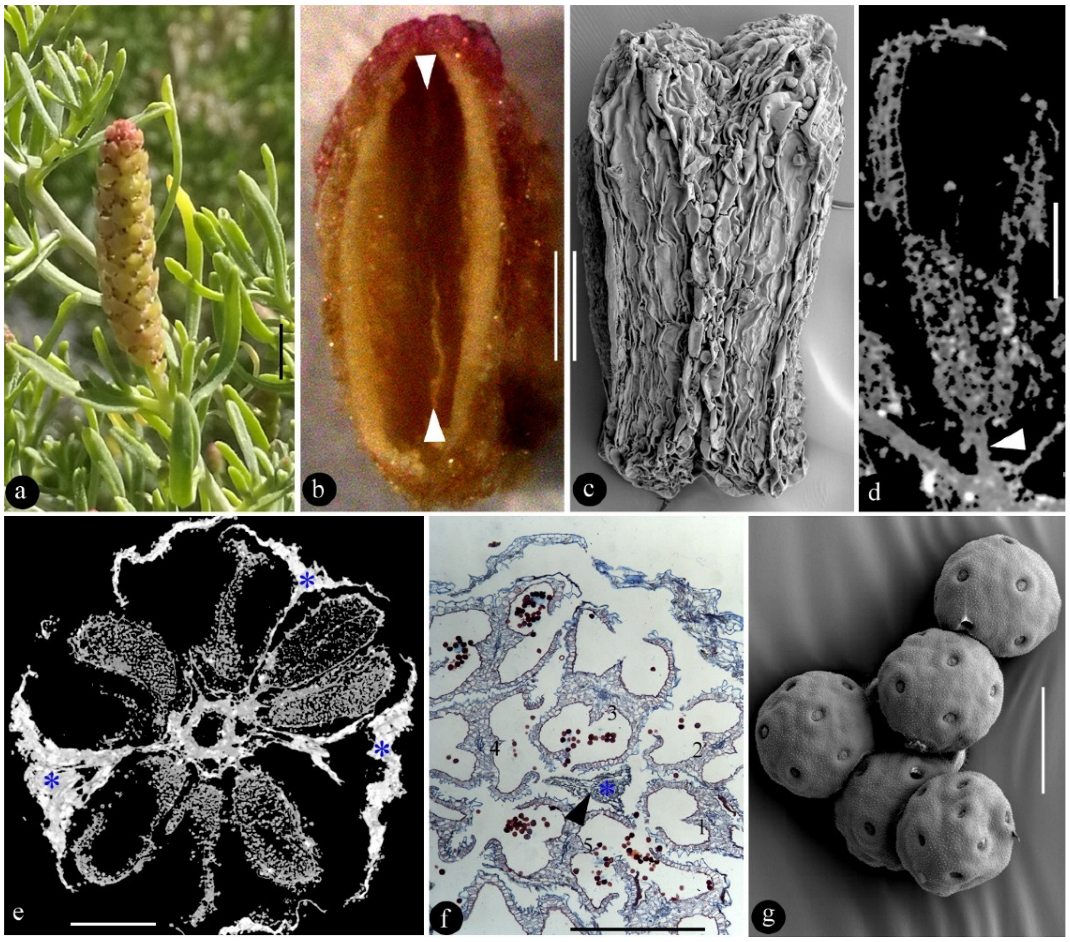
Figure 1. A male inflorescence of S. baileyi and its details. (a), a terminal upright male inflorescence. Scale bar = 2 mm. (b), lateral view of an opened anther. Note the residual ridge (triangles) between the former paired pollen sacs. Scale bar = 0.2 mm. (c), abaxial view of an anther. Scale bar = 0.2 mm. (d), Micro-CT virtual longitudinal section of a male flower attached to the axis by a short filament (triangle). Scale bar = 0.2 mm. (e), Micro-CT virtual cross section of the inflorescence, with male flowers (gray) and peltate shields (asterisks, white) arranged around the central axis. Scale bar = 0.5 mm. (f), tangential paraffin section of the male inflorescence showing five male flowers (1–5) centered around a shield stalk (triangle). Scale Bar = 0.5 mm. (g), pantoporate pollen grains. SEM. Scale bar = 20 μm.
Plants 12 01917 g001
Figure 2. A female cone of Cupressus sempervirens (Cupressaceae). (a), a row of tall trees in a farm on Mallorca Island, Spain. (b), top view of the cone. Scale bar = 1 cm. (c), transverse section of the cone showing the top bract (asterisk) flanked by two groups of seeds (arrowheads), as orientated in (b). Scale bar = 1 cm. (d), side view of the cone. Scale bar = 1 cm. (e), tangential section cutting through the base (asterisk) of the bract, which is surrounded by many seeds, as orientated in (c). Scale bar = 1 cm.
Plants 12 01917 g002
Disclaimer/Publisher’s Note: The statements, opinions and data contained in all publications are solely those of the individual author(s) and contributor(s) and not of MDPI and/or the editor(s). MDPI and/or the editor(s) disclaim responsibility for any injury to people or property resulting from any ideas, methods, instructions or products referred to in the content.