Understanding Macroalgae: A Comprehensive Exploration of Nutraceutical, Pharmaceutical, and Omics Dimensions
Abstract
:1. Introduction
2. Macroalgae—A Brief Overview
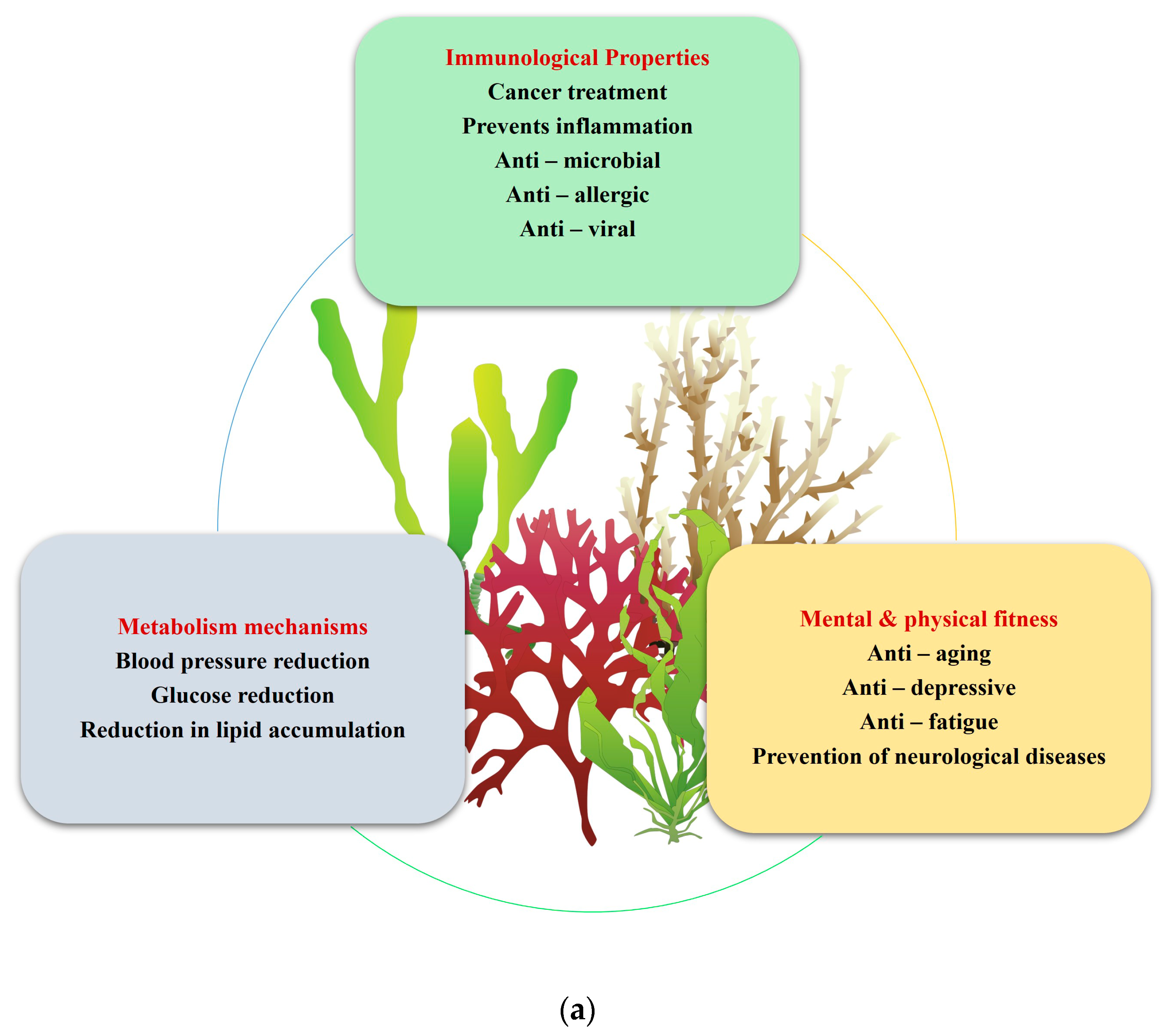
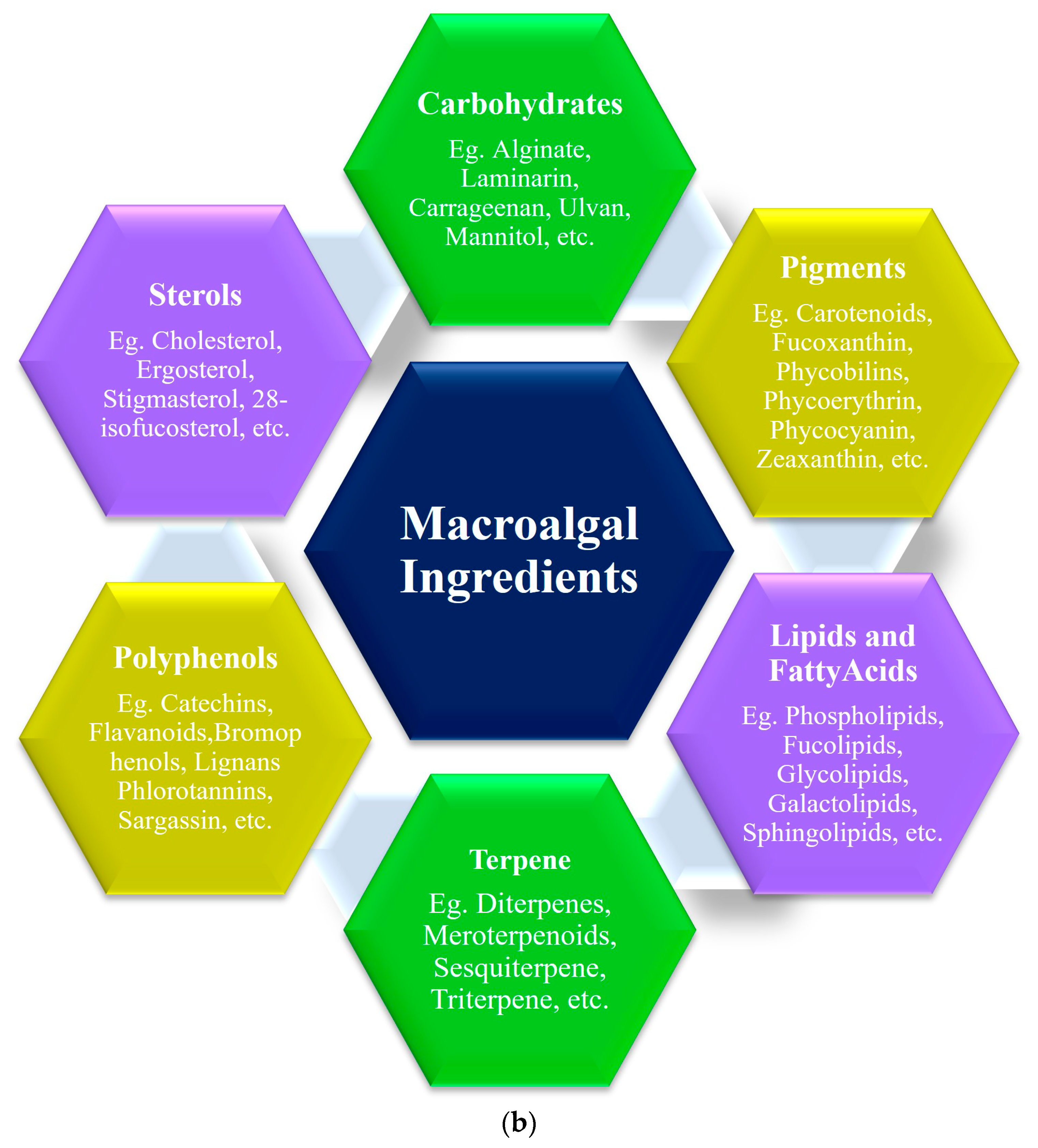
2.1. Nutraceutical Potentials of Macroalgae
2.2. Pharmaceutical Potentials of Macroalgae
2.2.1. Antioxidant Activity
2.2.2. Anticancer and Antiproliferative Activities
2.2.3. Anti-Inflammatory Activity
2.2.4. Anticoagulant
2.2.5. Macroalgae in Skincare
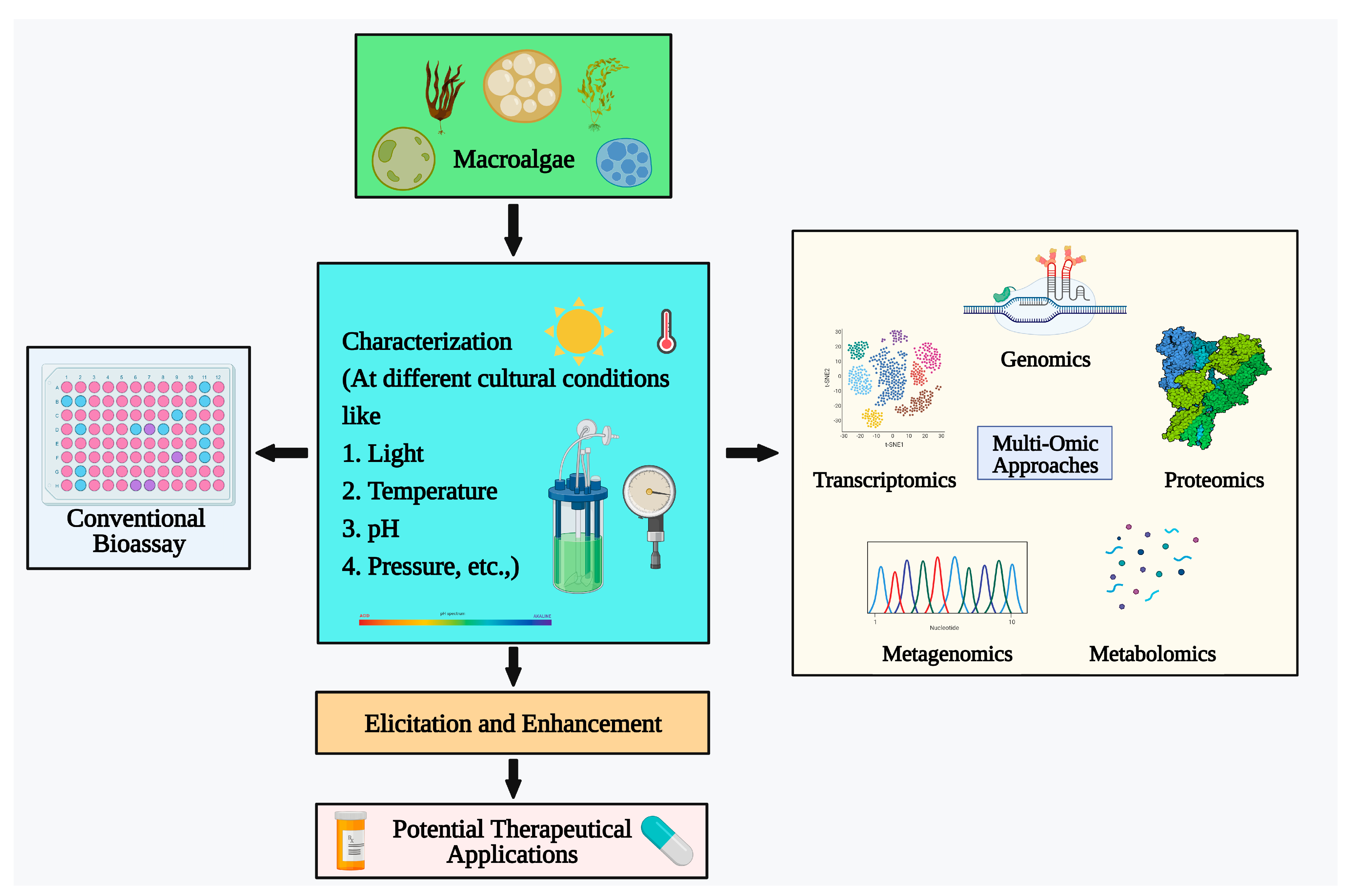
3. Advancements in Algal Research
3.1. Genomics
3.1.1. Genome Sequencing and Gene Mining
3.1.2. Genomic Analysis of Metabolic Pathways
3.1.3. Web Resources for Genomic Analysis
- AlgaePath: A platform for predicting metabolic pathways in algae [146].
- pico-PLAZA: A database of genome information for algae [147].
- Joint Genome Institute (JGI) Portal: A portal that provides access to genome data and analysis tools from the JGI [148].
- Symbiodiniaceae and Algal Genomic Resource Database (SAGER): A database of genome data for Symbiodiniaceae algae [149].
- Organelle Genome Database for Algae (OGDA): A database of organelle genome data for algae [150].
- BioSyc: A database of metabolic pathways for algae [151].
- GOLD database: A database of genome data for over 8000 organisms (https://gold.jgi.doe.gov/ (accessed on 23 July 2023)).
3.1.4. Genetic Engineering and DNA Barcoding
- Phylogenetic analyses: To study the evolutionary relationships between distinct species of algae;
- DNA barcoding: To identify and classify algae species using short DNA sequences which are important for commercial and industrial applications. Genetic information can also be used for genetic engineering. Techniques such as RAPD, RFLP, hybridization, and AFLP are commonly used for barcoding [153]. Genetic engineering technologies such as TALENs, ZFNs, and CRISPR can also be used to alter the genetic material of macroalgae, enhancing the expression of genes with medicinal applications [137]. For example, CRISPR has been used to enhance the production of fucoxanthin in Saccharina japonica [154].
3.2. Transcriptomics
3.3. Metagenomics
- Identify novel algal strains with potential for bioproduction of valuable compounds.
- Activate silent cryptic gene clusters in known algal strains to produce new bioactive compounds.
- Understand the interactions between macroalgae and their associated microbiome.
- Develop new bioprospection strategies for the discovery of novel enzymes and other biotechnologically important molecules.
3.4. Proteomics
- One-dimensional gel electrophoresis;
- Peptide fingerprinting;
- Sequencing;
- Two-dimensional electrophoresis;
- Mass spectrometry.
- Identify novel proteins with potential for bioproduction of valuable compounds;
- Understand the molecular mechanisms of stress responses and other important metabolic pathways in macroalgae;
- Develop new bioprospection strategies for the discovery of new drugs, enzymes, and other biotechnologically important molecules.
3.5. Metabolomics
- Mass spectrometry (MS);
- Gas chromatography (GC);
- Nuclear magnetic resonance (NMR);
- Thin layer chromatography (TLC).
- Identify novel bioactive compounds with potential for therapeutic or industrial applications;
- Develop new bioproduction methods for valuable compounds;
- Understand various metabolic pathways in macroalgae.
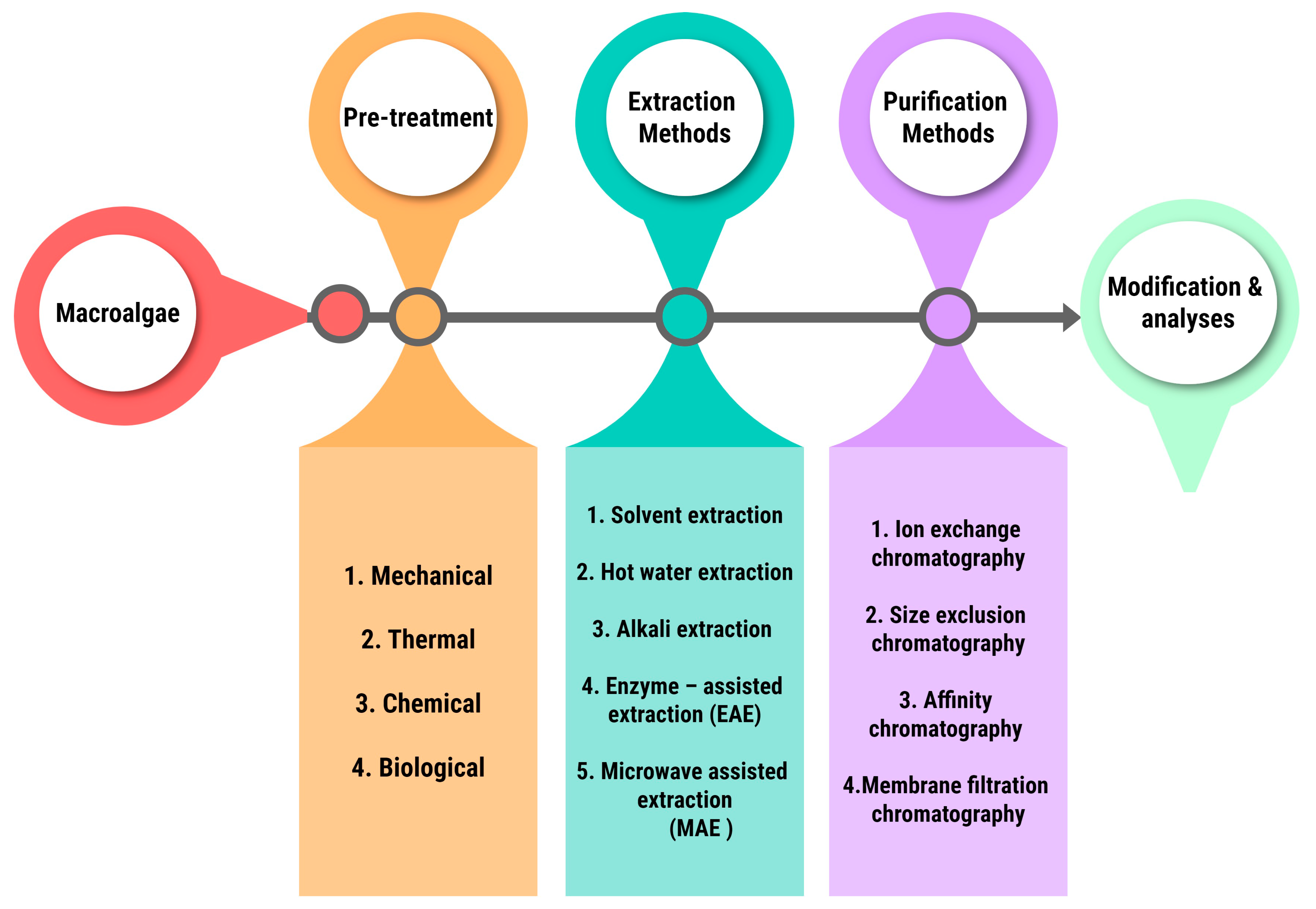
4. Macroalgal Industrial Extraction and Purification Methods
5. Future Prospectives
6. Conclusions
Author Contributions
Funding
Institutional Review Board Statement
Informed Consent Statement
Data Availability Statement
Conflicts of Interest
References
- Newman, D.J.; Cragg, G.M. Natural Products as Sources of New Drugs over the 30 Years from 1981 to 2010. J. Nat. Prod. 2012, 75, 3113–3137. [Google Scholar] [CrossRef] [PubMed]
- Afonso, N.C.; Catarino, M.D.; Silva, A.M.S.; Cardoso, S.M. Brown Macroalgae as Valuable Food Ingredients. Antioxidants 2019, 8, 365. [Google Scholar] [CrossRef] [PubMed]
- Leandro, A.; Pereira, L.; Gonçalves, A.M.M.; Gonçalves, A.M.M. Diverse Applications of Marine Macroalgae. Mar. Drugs 2019, 18, 17. [Google Scholar] [CrossRef] [PubMed]
- Pimentel, F.B.; Alves, R.C.; Rodrigues, F.; Oliveira, M.B.P.P. Macroalgae-derived Ingredients for Cosmetic Industry—An Update. Cosmetics 2017, 5, 2. [Google Scholar] [CrossRef]
- Shibasaki, S.; Ueda, M. Utilization of Macroalgae for the Production of Bioactive Compounds and Bioprocesses Using Microbial Biotechnology. Microorganisms 2023, 11, 1499. [Google Scholar] [CrossRef]
- Pérez, M.J.; Falqué, E.; Domínguez, H. Antimicrobial Action of Compounds from Marine Seaweed. Mar. Drugs 2016, 14, 52. [Google Scholar] [CrossRef] [PubMed]
- Bleakley, S.; Hayes, M. Algal Proteins: Extraction, Application, and Challenges Concerning Production. Foods 2017, 6, 33. [Google Scholar] [CrossRef]
- Guo, J.; Qi, M.; Chen, H.; Zhou, C.; Ruan, R.; Yan, X.; Cheng, P. Macroalgae-Derived Multifunctional Bioactive Substances: The Potential Applications for Food and Pharmaceuticals. Foods 2022, 11, 3455. [Google Scholar] [CrossRef]
- Kumar, Y.; Tarafdar, A.; Badgujar, P.C. Seaweed as a source of natural antioxidants: Therapeutic activity and food applications. J. Food Qual. 2021, 2021, 5753391. [Google Scholar] [CrossRef]
- Mota, C.S.C.; Pinto, O.; Sá, T.; Ferreira, M.; Delerue-Matos, C.; Cabrita, A.R.; Almeida, A.; Abreu, H.; Silva, J.; Fonseca, A.J.; et al. A Commercial Blend of Macroalgae and Microalgae Promotes Digestibility, Growth Performance, and Muscle Nutritional Value of European Seabass (Dicentrarchus labrax L.) Juveniles. Front. Nutr. 2023, 10, 1165343. [Google Scholar] [CrossRef]
- Ugwu, C.U.; Aoyagi, H.; Uchiyama, H. Photobioreactors for Mass Cultivation of Algae. Bioresour. Technol. 2008, 99, 4021–4023. [Google Scholar] [CrossRef] [PubMed]
- Shannon, E.; Abu-Ghannam, N. Seaweeds as nutraceuticals for health and nutrition. Phycologia 2019, 58, 563–577. [Google Scholar] [CrossRef]
- Peñalver, R.; Lorenzo, J.M.; Ros, G.; Amarowicz, R.; Pateiro, M.; Nieto, G. Seaweeds as a Functional Ingredient for a Healthy Diet. Mar. Drugs 2020, 18, 301. [Google Scholar] [CrossRef] [PubMed]
- Ismail, M.M.; Alotaibi, B.S.; El-Sheekh, M.M. Therapeutic Uses of Red Macroalgae. Molecules 2020, 25, 4411. [Google Scholar] [CrossRef] [PubMed]
- Agregán, R.; Barba, F.J.; Gavahian, M.; Franco, D.; Khaneghah, A.M.; Carballo, J.; Ferreira, I.C.F.; Barretto, A.C.S.; Lorenzo, J.M. Fucus vesiculosus extracts as natural antioxidants for improvement of physicochemical properties and shelf life of pork patties formulated with oleogels. J. Sci. Food Agric. 2019, 99, 4561–4570. [Google Scholar] [CrossRef] [PubMed]
- Dai, N.; Wang, Q.; Xu, B.; Chen, H. Remarkable Natural Biological Resource of Algae for Medical Applications. Front. Mar. Sci. 2022, 9, 1060. [Google Scholar] [CrossRef]
- Saha, D.; Bhattacharya, S. Hydrocolloids as Thickening and Gelling Agents in Food: A Critical Review. Food Biophys. 2010, 47, 587–597. [Google Scholar] [CrossRef]
- Rebours, C.; Marinho-Soriano, E.; Zertuche-González, J.A.; Hayashi, L.; Vásquez, J.A.; Kradolfer, P.; Soriano, G.; Ugarte, R.; Abreu, M.H.; Bay-Larsen, I. Seaweeds: An Opportunity for Wealth and Sustainable Livelihood for Coastal Communities. J. Appl. Phycol. 2014, 26, 1939–1951. [Google Scholar] [CrossRef]
- Buschmann, A.H.; Camus, C.; Infante, J.; Neori, A.; Israel, A.; Hernández-González, M.C.; Pereda, S.V.; Gómez-Pinchetti, J.L.; Golberg, A.; Tadmor-Shalev, N. Seaweed Production: Overview of the Global State of Exploitation, Farming and Emerging Research Activity. Hydrobiologia 2017, 52, 391–406. [Google Scholar] [CrossRef]
- Pangestuti, R.; Kim, S.-K. Biological Activities and Health Benefit Effects of Natural Pigments Derived from Marine Algae. J. Funct. Foods 2011, 3, 255–266. [Google Scholar] [CrossRef]
- Torres, M.D.; Flórez-Fernández, N.; Domínguez, H. Integral Utilization of Red Seaweed for Bioactive Production. Mar. Drugs 2019, 17, 314. [Google Scholar] [CrossRef] [PubMed]
- Gómez-Guzmán, M.; Rodríguez-Nogales, A.; Algieri, F.; Gálvez, J. Potential Role of Seaweed Polyphenols in Cardiovascular-Associated Disorders. Mar. Drugs 2018, 16, 250. [Google Scholar] [CrossRef] [PubMed]
- Percival, E.; McDowell, R.H. Algal Walls—Composition and Biosynthesis. In Algae and Environment; Lobban, C.S., Wynne, M.J., Eds.; Blackwell Scientific Publications: Oxford, UK, 1981; pp. 277–316. [Google Scholar] [CrossRef]
- Hunt, D.; Dewar, A.; Dal Molin, F.; Willey, N. Enhancing Radiological Monitoring of 137Cs in Coastal Environments Using Taxonomic Signals in Brown Seaweeds. Sci. Total Environ. 2023, 268–269, 107261. [Google Scholar] [CrossRef] [PubMed]
- Rasmussen, R.S.; Morrissey, M.T. Marine Biotechnology for Production of Food Ingredients. Adv. Food Nutr. Res. 2007, 52, 237–292. [Google Scholar]
- Abirami, R.G.; Kowsalya, S. Phytochemical Screening, Microbial Load, and Antimicrobial Activity of Underexploited Seaweeds. Int. Res. J. Microbiol. 2012, 3, 328–332. [Google Scholar]
- Ganesan, A.R.; Tiwari, U.; Rajauria, G. Seaweed Nutraceuticals and Their Therapeutic Role in Disease Prevention. Food Sci. Hum. Wellness. 2019, 8, 252–263. [Google Scholar] [CrossRef]
- Ganesan, A.R.; Shanmugam, M.; Bhat, R. Producing Novel Edible Films from Semi Refined Carrageenan (SRC) and Ulvan Polysaccharides for Potential Food Applications. Int. J. Biol. Macromol. 2018, 112, 1164–1170. [Google Scholar] [CrossRef]
- Seedevi, P.; Moovendhan, M.; Viramani, S.; Shanmugam, A. Bioactive Potential and Structural Chracterization of Sulfated Polysaccharide from Seaweed (Gracilaria corticata). Carbohydr. Polym. 2017, 155, 516–524. [Google Scholar] [CrossRef]
- Freitas, A.C.; Rodrigues, D.; Rocha-Santos, T.; Rocha-Santos, T.; Gomes, A.M.P.; Duarte, A.C. Marine Biotechnology Advances Towards Applications in New Functional Foods. Biotechnol. Adv. 2012, 30, 1506–1515. [Google Scholar] [CrossRef]
- Healy, L.E.; Zhu, X.; Pojić, M.; Sullivan, C.; Tiwari, U.; Curtin, J.; Tiwari, B.K. Biomolecules from Macroalgae-Nutritional Profile and Bioactives for Novel Food Product Development. Biomolecules 2023, 13, 386. [Google Scholar] [CrossRef]
- Urbano, M.G.; Goñi, I. Bioavailability of Nutrients in Rats Fed on Edible Seaweeds, Nori (Porphyra tenera) and Wakame (Undaria pinnatifida), as a Source of Dietary Fibre. Food Chem. 2002, 76, 477–483. [Google Scholar] [CrossRef]
- Domínguez-González, R.; Romarís-Hortas, V.; García-Sartal, C.; Moreda-Piñeiro, A.; Barciela-Alonso, M.C.; Bermejo-Barrera, P. Evaluation of an in Vitro Method to Estimate Trace Elements Bioavailability in Edible Seaweeds. Talanta 2010, 82, 1092–1098. [Google Scholar] [CrossRef] [PubMed]
- Hamann, M.T. Technology Evaluation: Kahalalide F, Pharmamar. Curr. Opin. Investig. Drugs 2004, 6, 1088–1093. [Google Scholar]
- Rocha, C.P.; Pacheco, D.; Cotas, J.; Marques, J.C.; Pereira, L.; Gonçalves, A.M. Seaweeds as valuable sources of essential fatty acids for human nutrition. Int. J. Environ. Res. Public Health 2021, 18, 4968. [Google Scholar] [CrossRef] [PubMed]
- Biancarosa, I.; Belghit, I.; Bruckner, C.G.; Liland, N.S.; Waagbø, R.; Amlund, H.; Heesch, S.; Lock, E.J. Chemical Characterization of 21 Species of Marine Macroalgae Common in Norwegian Waters: Benefits of and Limitations to Their Potential Use in Food and Feed. J. Sci. Food Agric. 2018, 98, 2035–2042. [Google Scholar] [CrossRef] [PubMed]
- Peng, Y.; Hu, J.; Yang, B.; Lin, X.-P.; Zhou, X.-F.; Yang, X.-W.; Liu, Y. Chemical Composition of Seaweeds. In Seaweeds: Diversity, Environment, Applications; Elsevier: Amsterdam, The Netherlands, 2015; pp. 79–124. [Google Scholar]
- Kellogg, J.; Esposito, D.; Grace, M.H.; Komarnytsky, S.; Lila, M.A. Alaskan Seaweeds Lower Inflammation in RAW 264.7 Macrophages and Decrease Lipid Accumulation in 3T3-L1 Adipocytes. J. Funct. Foods 2015, 15, 396–407. [Google Scholar] [CrossRef]
- Hernández-Carmona, G.; Carrillo-Domínguez, S.; Arvizu-Higuera, D.L.; Rodríguez-Montesinos, Y.E.; Murillo-Álvarez, J.I.; Muñoz-Ochoa, M.; Castillo-Domínguez, R.M. Monthly Variation in the Chemical Composition of Eisenia arborea J.E. Areschoug. J. Appl. Phycol. 2009, 21, 141–148. [Google Scholar] [CrossRef]
- Norziah, M.H.; Ching, C.Y. Nutritional Composition of Edible Seaweed Gracilaria Changgi. Food Chem. 2000, 68, 69–76. [Google Scholar] [CrossRef]
- Sarojini, Y.; Sarma, N. Vitamin c Content of Some Macroalgae of Visakhapatnam, East Coast of India. Indian J. Mar. Sci. 1999, 28, 408–412. [Google Scholar]
- Galasso, C.; Corinaldesi, C.; Sansone, C. Carotenoids from Marine Organisms: Biological Functions and Industrial Applications. Antioxidants 2017, 6, 96. [Google Scholar] [CrossRef]
- Viera, I.; Pérez-Gálvez, A.; Roca, M. Bioaccessibility of Marine Carotenoids. Mar. Drugs 2018, 16, 397. [Google Scholar] [CrossRef] [PubMed]
- Skrovankova, S. Seaweed Vitamins as Nutraceuticals. Food Chem. 2011, 125, 357–369. [Google Scholar] [CrossRef]
- Zhong, R.; Wan, X.; Wang, D.; Zhao, C.; Liu, D.; Gao, L.; Wang, M.; Wu, C.; Nabavid, S.M.; Daglia, M. Polysaccharides from Marine Enteromorpha: Structure and Function. Mar. Drugs 2020, 18, 535. [Google Scholar] [CrossRef]
- Plaza, M.; Cifuentes, A.; Ibáñez, E. In the search of new functional food ingredients from algae. Trends Food Sci Technol. 2008, 19, 31–39. [Google Scholar] [CrossRef]
- Mahadevan, K. Seaweeds: A Sustainable Food Source. Biotechnol. Adv. 2015, 33, 347–364. [Google Scholar] [CrossRef]
- Bito, T.; Teng, F.; Watanabe, F. Bioactive compounds of edible purple laver Porphyra sp.(Nori). J. Agri. Food Chem. 2017, 65, 10685–10692. [Google Scholar] [CrossRef] [PubMed]
- Lafeuille, B.; Tamigneaux, É.; Berger, K.; Provencher, V.; Beaulieu, L. Impact of Harvest Month and Drying Process on the Nutritional and Bioactive Properties of Wild Palmaria palmata from Atlantic Canada. Mar. Drugs 2023, 21, 392. [Google Scholar] [CrossRef]
- Pina, A.L.; Costa, A.R.; Lage-Yusty, M.A.; López-Hernández, J. An Evaluation of Edible Red Seaweed (Chondrus crispus) Components and Their Modification during the Cooking Process. LWT-Food Sci. Technol. 2014, 56, 175–180. [Google Scholar] [CrossRef]
- Raimundo, S.C.; Pattathil, S.; Eberhard, S.; Hahn, M.G.; Popper, Z.A. β-1,3-Glucans are components of brown seaweed (Phaeophyceae) cell walls. Protoplasma 2016, 254, 997–1016. [Google Scholar] [CrossRef]
- Kadam, S.U.; O’Donnell, C.P.; Rai, D.K.; Hossain, M.B.; Burgess, C.M.; Walsh, D.; Tiwari, B.K. Laminarin from Irish Brown Seaweeds Ascophyllum nodosum and Laminaria hyperborea: Ultrasound Assisted Extraction, Characterization and Bioactivity. Mar. Drugs 2015, 13, 4270–4280. [Google Scholar] [CrossRef]
- Blunden, G.; Campbell, S.A.; Smith, J.R.; Guiry, M.D.; Hession, C.C.; Griffin, R.L. Chemical and Physical Characterization of Calcified Red Algal Deposits Known as Maërl. J. Appl. Phycol. 1997, 9, 11–17. [Google Scholar] [CrossRef]
- Pirazzoli, P.A. Geomorphological Indicators. In Sea Level Studies; Elsevier: Amsterdam, The Netherlands, 2007; pp. 2974–2983. [Google Scholar] [CrossRef]
- García-Vaquero, M.; Rajauria, G.; O’Doherty, J.V.; Sweeney, T. Polysaccharides from Macroalgae: Recent Advances, Innovative Technologies and Challenges in Extraction and Purification. Food Res Intl. 2017, 99, 1011–1020. [Google Scholar] [CrossRef] [PubMed]
- Gao, G.; Burgess, J.G.; Wu, M.; Wang, S.; Gao, K. Using Macroalgae as Biofuel: Current Opportunities and Challenges. Bot. Mar. 2020, 63, 355–370. [Google Scholar] [CrossRef]
- Rabanal, M.; Ponce, N.A.; Navarro, D.A.; Gómez, R.M.; Stortz, C.A. The System of Fucoidans from the Brown Seaweed Dictyota dichotoma: Chemical Analysis and Antiviral Activity. Carbohydr. Polym. 2014, 101, 804–811. [Google Scholar] [CrossRef] [PubMed]
- D’Archino, R.; Piazzi, L. Macroalgal assemblages as indicators of the ecological status of marine coastal systems: A review. Ecol. Indic. 2021, 129, 107835. [Google Scholar] [CrossRef]
- Basak, R.; Wahid, K.A.; Dinh, A. Estimation of the Chlorophyll-a Concentration of Algae Species Using Electrical Impedance Spectroscopy. Water 2021, 13, 1223. [Google Scholar] [CrossRef]
- Milinović, J. Edible Macroalgae: Beneficial Resource of Iodine. Am. J. Biomed. Sci. Res. 2020, 8, 290–292. [Google Scholar] [CrossRef]
- Blikra, M.J.; Henjum, S.; Aakre, I. Iodine from Brown Algae in Human Nutrition, with an Emphasis on Bioaccessibility, Bioavailability, Chemistry, and Effects of Processing: A Systematic Review. Compr. Rev. Food Sci. Food Saf. 2022, 21, 1517–1536. [Google Scholar] [CrossRef]
- Wells, M.L.; Potin, P.; Craigie, J.S.; Raven, J.A.; Merchant, S.; Helliwell, K.E.; Smith, A.G.; Camire, M.E.; Brawley, S.H. Algae as Nutritional and Functional Food Sources: Revisiting Our Understanding. J. Appl. Phycol. 2016, 29, 949–982. [Google Scholar] [CrossRef]
- Galland-Irmouli, A.-V.; Fleurence, J.; Lamghari, R.; Luçon, M.; Rouxel, C.; Barbaroux, O.; Bronowicki, J.-P.; Villaume, C.; Guéant, J.-L. Nutritional Value of Proteins from Edible Seaweed Palmaria Palmata (Dulse). J Nutr Biochem. 1999, 10, 353–359. [Google Scholar] [CrossRef]
- Angell, A.; Mata, L.; de Nys, R.; Paul, N. The Protein Content of Seaweeds: A Universal Nitrogen-to-protein Conversion Factor of Five. J. Appl. Phycol. 2016, 28, 511–524. [Google Scholar] [CrossRef]
- Holdt, S.; Kraan, S. Bioactive Compounds in Seaweed: Functional Food Applications and Legislation. J. Appl. Phycol. 2011, 23, 543–597. [Google Scholar] [CrossRef]
- Øverland, M.; Mydland, L.T.; Skrede, A. Marine Macroalgae as Sources of Protein and Bioactive Compounds in Feed for Monogastric Animals. J. Agri. Food Chem. 2019, 99, 13–24. [Google Scholar] [CrossRef] [PubMed]
- Boakye, P.; Sewu, D.D.; Woo, S.H. Effect of Thermal Pretreatment on the Extraction of Potassium Salt from Alga Saccharina Japonica. J. Anal. Appl. Pyrolysis 2018, 133, 68–75. [Google Scholar] [CrossRef]
- Wang, L.; Park, Y.; Jeon, Y.; Ryu, B. Bioactivities of the Edible Brown Seaweed, Undaria pinnatifida: A Review. Aquaculture 2018, 495, 873–880. [Google Scholar] [CrossRef]
- Agatonovic-Kustrin, S.; Morton, D.W.; Ristivojević, P. Assessment of Antioxidant Activity in Victorian Marine Algal Extracts Using High Performance Thin-layer Chromatography and Multivariate Analysis. J. Chromatogr. A 2016, 1468, 228–235. [Google Scholar] [CrossRef] [PubMed]
- Nguyen, T.T.; Mikkelsen, M.D.; Tran, V.H.N.; Trang, V.T.D.; Rhein-Knudsen, N.; Holck, J.; Rasin, A.B.; Cao, H.T.T.; Van, T.T.T.; Meyer, A.S. Enzyme-Assisted Fucoidan Extraction from Brown Macroalgae Fucus distichus subsp. evanescens and Saccharina latissima. Mar. Drugs 2020, 18, 296. [Google Scholar] [CrossRef]
- Circuncisão, A.R.; Catarino, M.D.; Cardoso, S.M.; Silva, A.M.S. Minerals from Macroalgae Origin: Health Benefits and Risks for Consumers. Mar. Drugs 2018, 16, 400. [Google Scholar] [CrossRef]
- Millner, G.C.; Sweeney, R.A.; Frederick, V.R. Biomass and Distribution of Cladophora Glomerata in Relation to Some Physical-Chemical Variables at Two Sites in Lake Erie. J. Great Lakes Res. 1982, 8, 35–41. [Google Scholar] [CrossRef]
- García-CasalM, N.; Pereira, A.C.; Leets, I.; Ramírez, J.; Quiroga, M.F. High Iron Content and Bioavailability in Humans from Four Species of Marine Algae. J. Nutr. 2007, 137, 2691–2695. [Google Scholar] [CrossRef]
- Geddie, A.W.; Hall, S.G. An Introduction to Copper and Zinc Pollution in Macroalgae: For Use in Remediation and Nutritional Applications. J. Appl. Phycol. 2018, 31, 691–708. [Google Scholar] [CrossRef]
- Stengel, D.B.; Macken, A.; Morrison, L.; Morley, N.J. Zinc Concentrations in Marine Macroalgae and a Lichen from Western Ireland in Relation to Phylogenetic Grouping, Habitat and Morphology. Mar. Pollut. Bull. 2004, 48, 902–909. [Google Scholar] [CrossRef] [PubMed]
- El-Agawany, N.; Kaamoush, M. Role of Zinc as an Essential Microelement for Algal Growth and Concerns About It’s a Potential Environmental Risks. Environ. Sci. Pollut. Res. 2023, 30, 71900–71911. [Google Scholar] [CrossRef] [PubMed]
- Prasetyaningrum, A.; Praptyana, I.; Nurfiningsih; Ratnawati. Carrageenan: Nutraceutical and Functional Food as Future Food. IOP Conf. Ser. Earth Environ. Sci. 2019, 292, 012068. [Google Scholar] [CrossRef]
- Zaharudin, N.; Salmeán, A.A.; Dragsted, L.O. Inhibitory Effects of Edible Seaweeds, Polyphenolics and Alginates on the Activities of Porcine Pancreatic α-Amylase. Food Chem. 2018, 245, 1196–1203. [Google Scholar] [CrossRef] [PubMed]
- Zargarzadeh, M.; Amaral, A.J.R.; Custódio, C.A.; Mano, J.F. Biomedical Applications of Laminarin. Carbohydr. Polym. 2020, 232, 115774. [Google Scholar] [CrossRef]
- Karuppusamy, S.; Rajauria, G.; Fitzpatrick, S.; Lyons, H.; McMahon, H.; Curtin, J.; Tiwari, B.K.; O’Donnell, C. Biological Properties and Health-Promoting Functions of Laminarin: A Comprehensive Review of Preclinical and Clinical Studies. Mar. Drugs 2022, 20, 772. [Google Scholar] [CrossRef]
- Bar-Shai, N.; Sharabani-Yosef, O.; Zollmann, M.; Lesman, A.; Golberg, A. Seaweed Cellulose Scaffolds Derived from Green Macroalgae for Tissue Engineering. Sci. Rep. 2021, 11, 11843. [Google Scholar] [CrossRef]
- Asari, F.; Kusumi, T.; Kakisawa, H. Turbinaric Acid, a Cytotoxic Secosqualene Carboxylic Acid from the Brown Alga Turbinaria ornata. J. Nat. Prod. 1989, 52, 1167–1169. [Google Scholar] [CrossRef]
- Muralidhar, P.; Radhika, P.; Krishna, N.; Rao, D.V.; Rao, C.B. Sphingolipids from Marine Organisms: A Review? Nat. Prod. Sci. 2003, 9, 117–142. [Google Scholar]
- Lopes, D.; Melo, T.; Rey, F.; Meneses, J.; Monteiro, F.L.; Helguero, L.A.; Abreu, M.H.; Lillebø, A.I.; Calado, R.; Domingues, R.M. Valuing Bioactive Lipids from Green, Red and Brown Macroalgae from Aquaculture, to Foster Functionality and Biotechnological Applications. Molecules 2020, 25, 3883. [Google Scholar] [CrossRef] [PubMed]
- Ersoy Korkmaz, N. Unlocking the Fatty Acid Profile of Macroalgae Species in Sea of Marmara, Türkiye: A Comprehensive Analysis of Extraction Methods. Int. Environ. Geoinfo. 2023, 10, 034–038. [Google Scholar] [CrossRef]
- Xu, J.; Liao, W.; Liu, Y.; Guo, Y.; Jiang, S.; Zhao, C. An Overview on the Nutritional and Bioactive Components of Green Seaweeds. Food Prod. Process. Nutr. 2023, 5, 18. [Google Scholar] [CrossRef]
- de Arruda, M.C.S.; da Silva, M.R.O.B.; Cavalcanti, V.L.R.; Brandao, R.M.P.C.; de Araújo Viana Marques, D.; de Lima, L.R.A.; Porto, A.L.F.; Bezerra, R.P. Antitumor Lectins from Algae: A Systematic Review. Algal Res. 2023, 70, 102962. [Google Scholar] [CrossRef]
- Šimat, V.; Elabed, N.; Kulawik, P.; Ceylan, Z.; Jamroz, E.; Yazgan, H.; Čagalj, M.; Regenstein, J.M.; Özogul, F. Recent Advances in Marine-Based Nutraceuticals and Their Health Benefits. Mar. Drugs. 2020, 18, 627. [Google Scholar] [CrossRef] [PubMed]
- Pradhan, B.; Bhuyan, P.P.; Ki, J.-S. Immunomodulatory, Antioxidant, Anticancer, and Pharmacokinetic Activity of Ulvan, a Seaweed-Derived Sulfated Polysaccharide: An Updated Comprehensive Review. Mar. Drugs 2023, 21, 300. [Google Scholar] [CrossRef]
- Bhatia, S.; Garg, A.; Sharma, K.; Kumar, S.; Sharma, A.; Purohit, A. Mycosporine and Mycosporine-Like Amino Acids: A Paramount Tool Against Ultraviolet Irradiation. Pharmacogn. Rev. 2011, 5, 138. [Google Scholar] [CrossRef]
- Cikoš, A.; Jokić, S.; Šubarić, D.; Jerković, I. Overview on the Application of Modern Methods for the Extraction of Bioactive Compounds from Marine Macroalgae. Mar. Drugs 2018, 16, 348. [Google Scholar] [CrossRef]
- Kovaleski, G.; Kholany, M.; Dias, L.M.S.; Correia, S.F.H.; Ferreira, R.A.S.; Coutinho, J.A.P.; Ventura, S.P.M. Extraction and Purification of Phycobiliproteins from Algae and Their Applications. Front. Chem. 2022, 10, 1065355. [Google Scholar] [CrossRef]
- Generalić Mekinić, I.; Šimat, V.; Rathod, N.B.; Hamed, I.; Čagalj, M. Algal Carotenoids: Chemistry, Sources, and Application. Foods 2023, 12, 2768. [Google Scholar] [CrossRef]
- Lunagariya, J.; Bhadja, P.; Zhong, S.; Vekariya, R.L.; Xu, S. Marine Natural Product Bis-Indole Alkaloid Caulerpin: Chemistry and Biology. Mini-Rev. Med. Chem. 2019, 19, 751–761. [Google Scholar] [CrossRef] [PubMed]
- Zhang, H.; Tang, Y.; Ying, Z.; Zhang, S.; Qu, J.; Wang, X.; Kong, R.; Han, C.; Liu, Z. Fucoxanthin: A Promising Medicinal and Nutritional Ingredient. Evid. -Based Complement. Altern. Med. 2015, 2015, 723515. [Google Scholar] [CrossRef] [PubMed]
- Suetsuna, K.; Maekawa, K.; Chen, J.R. Antihypertensive effects of Undaria pinnatifida (wakame) peptide on blood pressure in spontaneously hypertensive rats. J. Nutr. Biochem. 2004, 15, 267–272. [Google Scholar] [CrossRef] [PubMed]
- Pimentel, F.B.; Alves, R.C.; Harnedy, P.A.; FitzGerald, R.J.; Oliveira, M.B.P.P. Macroalgal-Derived Protein Hydrolysates and Bioactive Peptides: Enzymatic Release and Potential Health Enhancing Properties. Trends Food Sci. Technol. 2019, 93, 106–124. [Google Scholar] [CrossRef]
- Greff, S.; Zubia, M.; Genta-Jouve, G.; Massi, L.; Pérez, T.; Thomas, O.P. Mahorones, Highly Brominated Cyclopentenones from the Red Alga Asparagopsis taxiformis. J. Nat. Prod. 2014, 77, 1150–1155. [Google Scholar] [CrossRef] [PubMed]
- Wichmann, H.; Brinkhoff, T.; Simon, M.; Richter-Landsberg, C. Dimethylsulfoniopropionate Promotes Process Outgrowth in Neural Cells and Exerts Protective Effects against Tropodithietic Acid. Mar. Drugs 2016, 14, 89. [Google Scholar] [CrossRef] [PubMed]
- Glombitza, K.; Rösener, H.; Müller, D. Bifuhalol und diphlorethol aus Cystoseira tamariscifolia. Phytochemistry 1975, 14, 1115–1116. [Google Scholar] [CrossRef]
- Negreanu-Pirjol, B.S.; Negreanu-Pirjol, T.; Popoviciu, D.R.; Anton, R.-E.; Prelipcean, A.-M. Marine Bioactive Compounds Derived from Macroalgae as New Potential Players in Drug Delivery Systems: A Review. Pharmaceutics 2022, 14, 1781. [Google Scholar] [CrossRef]
- Hartmann, A.; Ganzera, M.; Karsten, U.; Skhirtladze, A.; Stuppner, H. Phytochemical and Analytical Characterization of Novel Sulfated Coumarins in the Marine Green Macroalga Dasycladus Vermicularis (Scopoli) Krasser. Molecules 2018, 23, 2735. [Google Scholar] [CrossRef]
- Yoon, M.; Cho, S. Triphlorethol A, a Dietary Polyphenol from Seaweed, Decreases Sleep Latency and Increases Non-Rapid Eye Movement Sleep in Mice. Mar. Drugs 2018, 16, 139. [Google Scholar] [CrossRef]
- Maeda, H.; Tsukui, T.; Sashima, T.; Hosokawa, M.; Miyashita, K. Seaweed carotenoid, fucoxanthin, as a multi-functional nutrient. Asia Pac. J. Clin. Nutr. 2008, 17 (Suppl. 1), 196–199. [Google Scholar] [PubMed]
- Shibata, T.; Fujimoto, K.; Nagayama, K.; Yamaguchi, K.; Nakamura, T. Inhibitory activity of brown algal phlorotannins against hyaluronidase. Int. J. Food Sci. Technol. 2002, 37, 703–709. [Google Scholar] [CrossRef]
- McCauley, J.I.; Winberg, P.C.; Meyer, B.J.; Skropeta, D. Effects of nutrients and processing on the nutritionally important metabolites of Ulva sp. (Chlorophyta). Algal Res. 2018, 35, 586–594. [Google Scholar] [CrossRef]
- Cho, C.-H.; Lu, Y.-A.; Kim, M.-Y.; Jeon, Y.-J.; Lee, S.-H. Therapeutic Potential of Seaweed-Derived Bioactive Compounds for Cardiovascular Disease Treatment. Appl. Sci. 2022, 12, 1025. [Google Scholar] [CrossRef]
- Gupta, S.; Abu-Ghannam, N. Bioactive Potential and Possible Health Effects of Edible Brown Seaweeds. Trends Food Sci. Technol. 2011, 22, 315–326. [Google Scholar] [CrossRef]
- Zhang, R.; Zhang, X.; Tang, Y.; Mao, J. Composition, isolation, purification and biological activities of Sargassum fusiforme polysaccharides: A review. Carbohydr. Polym. 2020, 228, 115381. [Google Scholar] [CrossRef] [PubMed]
- Zhao, D.; Kwon, S.; Chun, Y.S.; Gu, M.; Yang, H.O. Anti-Neuroinflammatory Effects of Fucoxanthin via Inhibition of AKT/NF-ΚB and MAPKS/AP-1 Pathways and Activation of PKA/CREB Pathway in Lipopolysaccharide-Activated BV-2 Microglial Cells. Neurochem. Res. 2016, 42, 667–677. [Google Scholar] [CrossRef] [PubMed]
- Almeida, T.; Ferreira, J.; Vettorazzi, A.; Azqueta, A.; Rocha, E.; Ramos, A. Cytotoxic activity of fucoxanthin, alone and in combination with the cancer drugs imatinib and doxorubicin, in CML cell lines. Environ. Toxicol. Pharmacol. 2018, 59, 24–33. [Google Scholar] [CrossRef]
- Hitoe, S.; Shimoda, H. Seaweed Fucoxanthin Supplementation Improves Obesity Parameters in Mild Obese Japanese Subjects. Funct. Foods Health Dis. 2017, 7, 246. [Google Scholar] [CrossRef]
- Mohibbullah, M.; Haque, M.N.; Khan, M.N.A.; Park, I.; Moon, I.S.; Hong, Y. Neuroprotective Effects of Fucoxanthin and Its Derivative Fucoxanthinol from the Phaeophyte Undaria pinnatifida Attenuate Oxidative Stress in Hippocampal Neurons. J. Appl. Phycol. 2018, 30, 3243–3252. [Google Scholar] [CrossRef]
- Sun, X.; Zhao, H.; Liu, Z.; Sun, X.; Zhang, D.; Wang, S.; Xu, Y.; Zhang, G.; Wang, D. Modulation of Gut Microbiota by Fucoxanthin during Alleviation of Obesity in High-Fat Diet-Fed Mice. J. Agric. Food Chem. 2020, 68, 5118–5128. [Google Scholar] [CrossRef]
- Bhuyar, P.; Rahim, M.H.A.; Sundararaju, S.; Maniam, G.P.; Nagarajan, G. Antioxidant and Antibacterial Activity of Red Seaweed Kappaphycus alvarezii against Pathogenic Bacteria. Glob. J. Environ. Sci. Manag. 2020, 6, 47–58. [Google Scholar] [CrossRef]
- Saleh, B.; Al-Mariri, A. Antifungal Activity of Crude Seaweed Extracts Collected from Lattakia Coast, Syria. J. Fish. Aquat. Sci. 2017, 13, 49–55. [Google Scholar] [CrossRef]
- Smyrniotopoulos, V.; Vagias, C.; Rahman, M.M.; Gibbons, S.; Roussis, V. Ioniols I and II, Tetracyclic Diterpenes with Antibacterial Activity, from Sphaerococcus coronopifolius. Chem. Biodivers. 2010, 7, 666–676. [Google Scholar] [CrossRef]
- Chowdhury, M.M.H.; Kubra, K.; Hossain, M.B.; Mustafa, M.; Jainab, T.; Karim, M.R.; Mehedy, M.E. Screening of antibacterial and antifungal activity of freshwater and marine algae as a prominent natural antibiotic available in Bangladesh. Int. J. Pharmacol. 2015, 11, 828–833. [Google Scholar] [CrossRef]
- Mendis, E.; Kim, S. Present and Future Prospects of Seaweeds in Developing Functional Foods. Adv. Food Nutr. Res. 2011, 64, 1–15. [Google Scholar] [CrossRef] [PubMed]
- Morán-Santibañez, K.; Peña-Hernández, M.A.; Cruz-Suárez, L.E.; Ricque-Marie, D.; Skouta, R.; Vasquez, A.H.; Rodríguez-Padilla, C. Virucidal and Synergistic Activity of Polyphenol-Rich Extracts of Seaweeds against Measles Virus. Viruses 2018, 10, 465. [Google Scholar] [CrossRef] [PubMed]
- Fitzgerald, C.; Gallagher, E.; O’Connor, P.M.; Prieto, J.M.; Mora, L.; Grealy, M.; Hayes, M. Development of a seaweed derived platelet activating factor acetylhydrolase (PAF-AH) inhibitory hydrolysate, synthesis of inhibitory peptides and assessment of their toxicity using the Zebrafish larvae assay. Peptides 2013, 50, 119–124. [Google Scholar] [CrossRef]
- Kendel, M.; Wielgosz-Collin, G.; Bertrand, S.; Roussakis, C.; Bourgougnon, N.; Bedoux, G. Lipid Composition, Fatty Acids and Sterols in the Seaweeds Ulva Armoricana, and Solieria Chordalis from Brittany (France): An Analysis from Nutritional, Chemotaxonomic, and Antiproliferative Activity Perspectives. Mar. Drugs 2015, 13, 5606–5628. [Google Scholar] [CrossRef]
- Erfani, N.; Nazemosadat, Z.; Moein, M. Cytotoxic Activity of Ten Algae from the Persian Gulf and Oman Sea on Human Breast Cancer Cell Lines; MDA-MB-231, MCF-7, and T-47D. Pharmacogn. Res. 2015, 7, 133. [Google Scholar] [CrossRef]
- Raman, M.; Doble, M. κ-Carrageenan from marine red algae, Kappaphycus alvarezii—A functional food to prevent colon carcinogenesis. J. Funct. Foods 2015, 15, 354–364. [Google Scholar] [CrossRef]
- Liu, M.; Hansen, P.E.; Lin, X. Bromophenols in Marine Algae and Their Bioactivities. Mar. Drugs 2011, 9, 1273–1292. [Google Scholar] [CrossRef] [PubMed]
- Coura, C.O.; Souza, R.B.; Rodrigues, J.A.G.; De Sousa Oliveira Vanderlei, E.; De Araújo, I.W.F.; Ribeiro, N.A.; Frota, A.F.; Ribeiro, K.A.; Chaves, H.V.; Pereira, K.M.A.; et al. Mechanisms Involved in the Anti-Inflammatory Action of a Polysulfated Fraction from Gracilaria cornea in Rats. PLoS ONE 2015, 10, e0119319. [Google Scholar] [CrossRef] [PubMed]
- Jiang, Z.; Hama, Y.; Yamaguchi, K.; Oda, T. Inhibitory Effect of Sulphated Polysaccharide Porphyran on Nitric Oxide Production in Lipopolysaccharide-Stimulated RAW264.7 Macrophages. J. Biochem. 2011, 151, 65–74. [Google Scholar] [CrossRef]
- Machado, L.P.; De Carvalho, L.R.; Young, M.C.M.; Cardoso-Lopes, E.M.; Da Cruz Centeno, D.; Zambotti-Villela, L.; Colepicolo, P.; Yokoya, N.S. Evaluation of acetylcholinesterase inhibitory activity of Brazilian red macroalgae organic extracts. Rev. Bras. Farmacogn. 2015, 25, 657–662. [Google Scholar] [CrossRef]
- Paul, J.P.; Devi, S.D.K. Evaluation of Antipyretic Activity of Methanol Extract of Hypnea musciformis (Wulf) Lamouroux (Red Seaweed) in Manapad Coast, Tamil Nadu. Ind. Int. J. Med. Chem. Anal. 2015, 5, 74–78. [Google Scholar]
- Adrien, A.; Bonnet, A.; Dufour, D.; Baudouin, S.; Maugard, T.; Bridiau, N. Anticoagulant Activity of Sulfated Ulvan Isolated from the Green Macroalga Ulva rigida. Mar. Drugs 2019, 17, 291. [Google Scholar] [CrossRef]
- Holtkamp, A.D.; Kelly, S.; Ulber, R.; Lang, S. Fucoidans and fucoidanases—Focus on techniques for molecular structure elucidation and modification of marine polysaccharides. Appl. Microbiol. Biotechnol. 2009, 82, 1–11. [Google Scholar] [CrossRef]
- Carvalhal, F.; Cristelo, R.R.; Resende, D.I.S.P.; Pinto, M.; Sousa, E.; Correia-Da-Silva, M. Antithrombotics from the Sea: Polysaccharides and Beyond. Mar. Drugs 2019, 17, 170. [Google Scholar] [CrossRef]
- Faggio, C.; Morabito, M.; Minicante, S.A.; Piano, G.L.; Pagano, M.E.; Genovese, G. Potential Use of Polysaccharides from the Brown Alga Undaria Pinnatifida as Anticoagulants. Braz Arch Biol Technol. 2015, 58, 798–804. [Google Scholar] [CrossRef]
- Chan, J.N.; Poon, B.; Salvi, J.S.; Olsen, J.B.; Emili, A.; Mekhail, K. Perinuclear cohibin complexes maintain replicative life span via roles at distinct silent chromatin domains. Dev. Cell 2011, 20, 867–879. [Google Scholar] [CrossRef]
- Cotas, J.; Leandro, A.; Monteiro, P.; Pacheco, D.; Figueirinha, A.; Gonçalves, A.M.M.; Silva, G.; Pereira, L. Seaweed Phenolics: From Extraction to Applications. Mar. Drugs 2020, 18, 384. [Google Scholar] [CrossRef] [PubMed]
- Radakovits, R.; Jinkerson, R.E.; Darzins, A.; Posewitz, M.C. Genetic Engineering of Algae for Enhanced Biofuel Production. Eukaryot. Cell 2010, 9, 486–501. [Google Scholar] [CrossRef] [PubMed]
- Dubrasquet, H.; Reyes, J.; Sanchez, R.P.; Valdivia, N.; Guillemin, M. Molecular-Assisted Revision of Red Macroalgal Diversity and Distribution along the Western Antarctic Peninsula and South Shetland Islands. Cryptogam. Algol. 2018, 39, 409. [Google Scholar] [CrossRef]
- Steele, T.S. A Genomic Approach to Accessing and Characterizing Secondary Metabolite Biosynthetic Pathways from Marine Red Macroalgae. Available online: https://escholarship.org/uc/item/4vw303ct (accessed on 17 December 2023).
- Khatri, K.; Patel, J.; Adams, J.; Jones, H.; Phillips, D. Functional Genomic and Transformation Resources for Commercially Important Red Macroalgae (Rhodophyta). Algal Res. 2023, 74, 103227. [Google Scholar] [CrossRef]
- Metzker, M.L. Sequencing technologies—the next generation. Nat. Rev. Genet. 2009, 11, 31–46. [Google Scholar] [CrossRef] [PubMed]
- Cock, J.M.; Coelho, S.M.; Brownlee, C.; Taylor, A.R. The Ectocarpus Genome Sequence: Insights into Brown Algal Biology and the Evolutionary Diversity of the Eukaryotes. New Phytol. 2010, 188, 1–4. [Google Scholar] [CrossRef] [PubMed]
- Chandra Mohana, N.; Yashavantha Rao, H.C.; Rakshith, D.; Mithun, P.R.; Nuthan, B.R.; Satish, S. Omics-Based Approach for Biodiscovery of Microbial Natural Products in Antibiotic Resistance Era. J. Genet. Eng. Biotechnol. 2018, 16, 1–8. [Google Scholar] [CrossRef]
- Din, N.A.S.; Mohd Alayudin, Á.S.; Sofian-Seng, N.-S.; Rahman, H.A.; Mohd Razali, N.S.; Lim, S.J.; Wan Mustapha, W.A. Brown Algae as Functional Food Source of Fucoxanthin: A Review. Foods 2022, 11, 2235. [Google Scholar] [CrossRef]
- Walker, T.L.; Collet, C.; Purton, S. Algal Transgenics in the Genomic Era. J. Phycol. 2005, 41, 1077–1093. [Google Scholar] [CrossRef]
- Manat, G.; Fanuel, M.; Jouanneau, D.; Jam, M.; Mac-Bear, J.; Rogniaux, H.; Mora, T.; Larocque, R.; Lipinska, A.; Czjzek, M.; et al. Specificity of a β-porphyranase produced by the carrageenophyte red alga Chondrus crispus and implications of this unexpected activity on red algal biology. JBC. 2022, 298, 102707. [Google Scholar] [CrossRef]
- Zheng, H.; Chiang-Hsieh, Y.; Chien, C.; Hsu, B.J.; Liu, T.; Chen, C.N.; Chang, W.C. AlgaePath: Comprehensive Analysis of Metabolic Pathways Using Transcript Abundance Data from Next-Generation Sequencing in Green Algae. BMC Genom. 2014, 15, 196. [Google Scholar] [CrossRef] [PubMed]
- Vandepoele, K.; Van Bel, M.; Richard, G.; Van Landeghem, S.; Verhelst, B.; Moreau, H.; Van De Peer, Y.; Grimsley, N.; Piganeau, G. pico-PLAZA, a genome database of microbial photosynthetic eukaryotes. Environ. Microbiol. 2013, 15, 2147–2153. [Google Scholar] [CrossRef] [PubMed]
- Nordberg, H.; Cantor, M.; Dusheyko, S.; Hua, S.Z.; Poliakov, A.; Shabalov, I.; Smirnova, T.I.; Grigoriev, I.V.; Dubchak, I. The genome portal of the Department of Energy Joint Genome Institute: 2014 updates. Nucleic Acids Res. 2014, 42, D26–D31. [Google Scholar] [CrossRef]
- Yu, L.; Li, T.; Li, L.; Lin, X.; Li, H.; Liu, C.; Guo, C.; Lin, S. SAGER: A Database of Symbiodiniaceae and Algal Genomic Resource. Database 2020, 2020, baaa051. [Google Scholar] [CrossRef] [PubMed]
- Liu, T.; Cui, Y.; Jia, X.; Zhang, J.; Li, R.; Yu, Y.; Jia, S.; Qu, J.; Wang, X. OGDA: A Comprehensive Organelle Genome Database for Algae. Database 2020, 2020, baaa097. [Google Scholar] [CrossRef] [PubMed]
- Karp, P.D.; Billington, R.; Caspi, R.; Fulcher, C.A.; Latendresse, M.; Kothari, A.; Keseler, I.M.; Krummenacker, M.; Midford, P.E.; Ong, Q.; et al. The BioCyc Collection of Microbial Genomes and Metabolic Pathways. Brief. Bioinform. 2017, 20, 1085–1093. [Google Scholar] [CrossRef]
- Joyce, A.; Palsson, B.Ø. The model organism as a system: Integrating “omics” data sets. Nat. Rev. Mol. Cell Biol. 2006, 7, 198–210. [Google Scholar] [CrossRef]
- Tseng, C.; Chang, C. Notes on Caloglossa, Digena, and the Other Anthelmintic Marine Algae in China. PubMed 1962, 9, 180–186. [Google Scholar]
- Shen, Y.; Motomura, T.; Ichihara, K.; Matsuda, Y.; Yoshimura, K.; Kosugi, C.; Nagasato, C. Application of CRISPR-Cas9 genome editing by microinjection of gametophytes of Saccharina japonica (Laminariales, Phaeophyceae). J. Appl. Phycol. 2023, 35, 1431–1441. [Google Scholar] [CrossRef]
- Minoda, A.; Sakagami, R.; Yagisawa, F.; Kuroiwa, T.; Tanaka, K. Improvement of Culture Conditions and Evidence for Nuclear Transformation by Homologous Recombination in a Red Alga, Cyanidioschyzon merolae 10D. Plant Cell Physiol. 2004, 45, 667–671. [Google Scholar] [CrossRef]
- Diesel, J.; Molano, G.; Montecinos, G.J.; DeWeese, K.; Calhoun, S.; Kuo, A.; Lipzen, A.; Salamov, A.; Grigoriev, I.V.; Reed, D.C.; et al. A scaffolded and annotated reference genome of giant kelp (Macrocystis pyrifera). BMC Genom. 2023, 24, 543. [Google Scholar] [CrossRef] [PubMed]
- Théodorou, I.; Kovi, M.R.; Liang, Z. Hilde-Gunn Opsahl-Sorteberg Genetic and Genomic Approaches for Improved and Sustainable Brown Algal Cultivation. In Sustainable Global Resources of Seaweeds; Springer: Cham, Switzerland, 2022; pp. 615–633. [Google Scholar] [CrossRef]
- Deng, Y.; Yao, J.; Wang, X.; Guo, H.; Duan, D. Transcriptome Sequencing and Comparative Analysis of Saccharina japonica (Laminariales, Phaeophyceae) under Blue Light Induction. PLoS ONE 2012, 7, e39704. [Google Scholar] [CrossRef] [PubMed]
- Dong, Z.; Chen, Y. Transcriptomics: Advances and Approaches. Sci. China Life Sci. 2013, 56, 960–967. [Google Scholar] [CrossRef] [PubMed]
- Brunson, J.K.; McKinnie, S.M.K.; Chekan, J.R.; McCrow, J.P.; Miles, Z.D.; Bertrand, E.M.; Bielinski, V.A.; Luhavaya, H.; Oborník, M.; Smith, G.J.; et al. Biosynthesis of the Neurotoxin Domoic Acid in a Bloom-Forming Diatom. Science 2018, 361, 1356–1358. [Google Scholar] [CrossRef] [PubMed]
- Keeling, P.J.; Burki, F.; Wilcox, H.M.; Allam, B.; Allen, E.E.; Amaral-Zettler, L.; Armbrust, E.V.; Archibald, J.M.; Bharti, A.K.; Bell, C.J.; et al. The Marine Microbial Eukaryote Transcriptome Sequencing Project (MMETSP): Illuminating the Functional Diversity of Eukaryotic Life in the Oceans through Transcriptome Sequencing. PLOS Biol. 2014, 12, e1001889. [Google Scholar] [CrossRef] [PubMed]
- Rismani-Yazdi, H.; Haznedaroğlu, B.Z.; Hsin, C.; Peccia, J. Transcriptomic Analysis of the Oleaginous Microalga Neochloris oleoabundans Reveals Metabolic Insights into Triacylglyceride Accumulation. Biotechnol. Biofuels 2012, 5, 74. [Google Scholar] [CrossRef] [PubMed]
- Ritter, A.; Dittami, S.M.; Goulitquer, S.; Correa, J.A.; Boyen, C.; Potin, P.; Tonon, T. Transcriptomic and metabolomic analysis of copper stress acclimation in Ectocarpus siliculosus highlights signaling and tolerance mechanisms in brown algae. BMC Plant Biol. 2014, 14, 116. [Google Scholar] [CrossRef] [PubMed]
- Cao, M.; Wang, D.; Mao, Y.; Kong, F.; Bi, G.; Xing, Q.; Weng, Z. Integrating Transcriptomics and Metabolomics to Characterize the Regulation of EPA Biosynthesis in Response to Cold Stress in Seaweed Bangia fuscopurpurea. PLoS ONE 2017, 12, e0186986. [Google Scholar] [CrossRef]
- Zhuo, J.; Wang, H.; Du, Y.; Shi, M.; Huan, L.; Wang, G. Transcriptomic Analysis of Ulva Prolifera in Response to Salt Stress. Water 2023, 15, 63. [Google Scholar] [CrossRef]
- Barone, R.; De Santi, C.; Palma Esposito, F.; Tedesco, P.; Galati, F.; Visone, M.; Scala, A. Donatella de Pascale Marine Metagenomics, a Valuable Tool for Enzymes and Bioactive Compounds Discovery. Front. Mar. Sci. 2014, 1, 38. [Google Scholar] [CrossRef]
- Sambles, C.; Moore, K.; Lux, T.; Jones, K.J.; Littlejohn, G.R.; Gouveia, J.D.; Aves, S.J.; Studholme, D.J.; Lee, R.J.; Love, J. Metagenomic Analysis of the Complex Microbial Consortium Associated with Cultures of the Oil-Rich Alga Botryococcus braunii. MicrobiologyOpen 2017, 6, 4. [Google Scholar] [CrossRef] [PubMed]
- Castillejo, N.; Martínez–Hernández, G.B.; Goffi, V.; Gómez, P.A.; Aguayo, E.; Artés, F.; Artés-Hernández, F. Natural Vitamin B12 and Fucose Supplementation of Green Smoothies with Edible Algae and Related Quality Changes during Their Shelf Life. J. Sci. Food Agric. 2017, 98, 2411–2421. [Google Scholar] [CrossRef]
- Miranda, L.; Hutchison, K.W.; Grossman, A.R.; Brawley, S.H. Diversity and Abundance of the Bacterial Community of the Red Macroalga Porphyra umbilicalis: Did Bacterial Farmers Produce Macroalgae? PLoS ONE 2013, 8, e58269. [Google Scholar] [CrossRef] [PubMed]
- Salaün, S.; Barre, S.; Santos-Goncalvez, M.D.; Potin, P.; Haras, D.; Bazire, A. Influence of exudates of the kelp Laminaria digitata on biofilm formation of associated and exogenous bacterial epiphytes. Microb. Ecol. 2012, 64, 359–369. [Google Scholar] [CrossRef] [PubMed]
- Singh, R.; Reddy, C.R.K. Unraveling the functions of the macroalgal microbiome. Front. Microbiol. 2016, 6, 1488. [Google Scholar] [CrossRef] [PubMed]
- Beaulieu, L. Insights into the Regulation of Algal Proteins and Bioactive Peptides Using Proteomic and Transcriptomic Approaches. Molecules 2019, 24, 1708. [Google Scholar] [CrossRef] [PubMed]
- Patelou, M.; Infante, C.; Dardelle, F.; Randewig, D.; Kouri, E.D.; Udvardi, M.K.; Tsiplakou, E.; Mantecón, L.; Flemetakis, E. Transcriptomic and Metabolomic Adaptation of Nannochloropsis gaditana Grown under Different Light Regimes. Algal Res. 2020, 45, 101735. [Google Scholar] [CrossRef]
- Anand, V.; Singh, P.K.; Banerjee, C.; Shukla, P. Proteomic Approaches in Microalgae: Perspectives and Applications. 3 Biotech 2017, 7, 145. [Google Scholar] [CrossRef]
- Wong, P.; Tan, L.; Nawi, H.; AbuBakar, S. Proteomics of the Red Alga, Gracilaria changii (Gracilariales, Rhodophyta). J. Phycol. 2006, 42, 113–120. [Google Scholar] [CrossRef]
- Chakdar, H.; Hasan, M.T.; Pabbi, S.; Nevalainen, H.; Shukla, P. High-throughput proteomics and metabolomic studies guide re-engineering of metabolic pathways in eukaryotic microalgae: A review. Bioresour. Technol. 2021, 321, 124495. [Google Scholar] [CrossRef]
- Contreras, L.; Moenne, A.; Gaillard, F.; Potin, P.; Correa, J.A. Proteomic Analysis and Identification of Copper Stress-Regulated Proteins in the Marine Alga Scytosiphon gracilis (Phaeophyceae). Aquat. Toxicol. 2010, 96, 85–89. [Google Scholar] [CrossRef] [PubMed]
- Begolli, R.; Chatziangelou, M.; Samiotaki, M.; Goutas, A.; Barda, S.; Goutzourelas, N.; Kevrekidis, D.P.; Malea, P.; Trachana, V.; Liu, M.; et al. Transcriptome and Proteome Analysis Reveals the Anti-Cancer Properties of Hypnea Musciformis Marine Macroalga Extract in Liver and Intestinal Cancer Cells. Hum. Genom. 2023, 17, 71. [Google Scholar] [CrossRef] [PubMed]
- Kim, S.K.; Thomas, N.V.; Li, X. Anticancer Compounds from Marine Macroalgae and Their Application as Medicinal Foods. Adv. Food Nutr. Res. 2011, 64, 213–224. [Google Scholar] [CrossRef] [PubMed]
- Olasehinde, T.A.; Olaniran, A.O.; Okoh, A.I. Macroalgae as a Valuable Source of Naturally Occurring Bioactive Compounds for the Treatment of Alzheimer’s Disease. Mar. Drugs 2019, 17, 609. [Google Scholar] [CrossRef] [PubMed]
- Belghit, I.; Rasinger, J.D.; Heesch, S.; Biancarosa, I.; Liland, N.S.; Torstensen, B.E.; Waagbø, R.; Lock, E.; Brückner, C. In-depth metabolic profiling of marine macroalgae confirms strong biochemical differences between brown, red and green algae. Algal Res. 2017, 26, 240–249. [Google Scholar] [CrossRef]
- Sanchez-Arcos, C.; Paris, D.; Mazzella, V.; Mutalipassi, M.; Costantini, M.; Buia, M.C.; Von Elert, E.; Cutignano, A.; Zupo, V. Responses of the Macroalga Ulva prolifera Müller to Ocean Acidification Revealed by Complementary NMR- and MS-Based Omics Approaches. Mar. Drugs 2022, 20, 743. [Google Scholar] [CrossRef] [PubMed]
- Verpoorte, R.; Choi, Y.H.; Mustafa, N.R.; Kim, H.K. Metabolomics: Back to Basics. Phytochem. Rev. 2008, 7, 525–537. [Google Scholar] [CrossRef]
- Jung, V.; Pohnert, G. Rapid Wound-Activated Transformation of the Green Algal Defensive Metabolite Caulerpenyne. Tetrahedron 2001, 57, 7169–7172. [Google Scholar] [CrossRef]
- Barre, S.; Weinberger, F.; Kervarec, N.; Potin, P. Monitoring defensive responses in macroalgae–limitations and perspectives. Phytochem. Rev. 2004, 3, 371–379. [Google Scholar] [CrossRef]
- Menaa, F.; Wijesinghe, U.; Thiripuranathar, G.; Althobaiti, N.A.; Albalawi, A.E.; Khan, B.A.; Menaa, B. Marine Algae-Derived Bioactive Compounds: A New Wave of Nanodrugs? Mar. Drugs 2021, 19, 484. [Google Scholar] [CrossRef]
- Lin, A.; Stout, E.P.; Prudhomme, J.; Roch, K.L.; Fairchild, C.R.; Franzblau, S.G.; Aalbersberg, W.G.; Hay, M.E.; Kubanek, J. Bioactive Bromophycolides R−U from the Fijian Red Alga Callophycus serratus. J. Nat. Prod. 2010, 73, 275–278. [Google Scholar] [CrossRef] [PubMed]
- Nylund, G.M.; Weinberger, F.; Rempt, M.; Pohnert, G. Metabolomic Assessment of Induced and Activated Chemical Defence in the Invasive Red Alga Gracilaria vermiculophylla. PLoS ONE 2011, 6, e29359. [Google Scholar] [CrossRef] [PubMed]
- Nielsen, J.; Keasling, J.D. Engineering Cellular Metabolism. Cell 2016, 164, 1185–1197. [Google Scholar] [CrossRef] [PubMed]
- Sasaki, Y.; Yoshikuni, Y. Metabolic Engineering for Valorization of Macroalgae Biomass. Metab. Eng. 2022, 71, 42–61. [Google Scholar] [CrossRef] [PubMed]
- Pardilhó, S.; Boaventura, R.; Almeida, M.F.; Dias, J.M. Marine Macroalgae Waste: A Potential Feedstock for Biogas Production. J. Environ. Manag. 2022, 304, 114309. [Google Scholar] [CrossRef] [PubMed]
- Wassie, T.; Niu, K.; Xie, C.; Wang, H.; Wu, X. Extraction Techniques, Biological Activities and Health Benefits of Marine Algae Enteromorpha prolifera Polysaccharide. Front. Nutr. 2021, 8, 747928. [Google Scholar] [CrossRef] [PubMed]
- García, E.S.; Miranda, C.F.; Cesário, M.T.; Wijffels, R.H.; Van Den Berg, C.; Eppink, M. Ionic Liquid-Assisted Selective Extraction and Partitioning of Biomolecules from Macroalgae. ACS Sustain. Chem. Eng. 2023, 11, 1752–1762. [Google Scholar] [CrossRef]
- Safi, C.; Rodríguez, L.; Mulder, W.; Engelen-Smit, N.; Spekking, W.; Van Den Broek, L.; Olivieri, G.; Sijtsma, L. Energy Consumption and Water-Soluble Protein Release by Cell Wall Disruption of Nannochloropsis gaditana. Bioresour. Technol. 2017, 239, 204–210. [Google Scholar] [CrossRef]
- Dong, Z.; Zhang, L.; Zhang, S.; Fu, H.; Chen, J. Hydrothermal Liquefaction of Macroalgae Enteromorpha prolifera to Bio-oil. Energies 2010, 24, 4054–4061. [Google Scholar] [CrossRef]
- Shin, E.J.; Nimlos, M.R.; Evans, R.J. Kinetic Analysis of the Gas-Phase Pyrolysis of Carbohydrates. Fuel 2001, 80, 1697–1709. [Google Scholar] [CrossRef]
- Siqueira, J.F.; Rôças, I.N. Present status and future directions: Microbiology of endodontic infections. Int. Endod. J. 2022, 55, 512–530. [Google Scholar] [CrossRef] [PubMed]
- Fakayode, O.A.; Aboagarib, E.A.A.; Zhou, C.; Ma, H. Co-pyrolysis of lignocellulosic and macroalgae biomasses for the production of biochar—A review. Bioresour. Technol. 2020, 297, 122408. [Google Scholar] [CrossRef] [PubMed]
- Zhang, R.; Li, L.; Tong, D.; Hu, C. Microwave-enhanced pyrolysis of natural algae from water blooms. Bioresour. Technol. 2016, 212, 311–317. [Google Scholar] [CrossRef] [PubMed]
- Zhang, Y.; Wan, Q.; Yang, N. Recent Advances of Porous Graphene: Synthesis, Functionalization, and Electrochemical Applications. Small 2019, 15, 1903780. [Google Scholar] [CrossRef] [PubMed]
- Banu, J.R.; Kavitha, S.; Kannah, R.Y.; Usman, T.M.M.; Kumar, G. Application of Chemo-Thermal Coupled Sonic Homogenization of Marine Macroalgal Biomass for Energy Efficient Volatile Fatty Acid Recovery. Bioresour. Technol. 2020, 303, 122951. [Google Scholar] [CrossRef] [PubMed]
- Adams, J.; Toop, T.A.; Donnison, I.S.; Gallagher, J. Seasonal Variation in Laminaria digitata and Its Impact on Biochemical Conversion Routes to Biofuels. Bioresour. Technol. 2011, 102, 9976–9984. [Google Scholar] [CrossRef]
- Motte, J.; Delenne, J.; Rouau, X.; Mayer-Laigle, C. Mineral–vegetal co-milling: An effective process to improve lignocellulosic biomass fine milling and to increase interweaving between mixed particles. Bioresour. Technol. 2015, 192, 703–710. [Google Scholar] [CrossRef]
- Bader, A.N.; Rizza, L.S.; Consolo, V.F.; Curatti, L. Efficient Saccharification of Microalgal Biomass by Trichoderma harzianum Enzymes for the Production of Ethanol. Algal Res. 2020, 48, 101926. [Google Scholar] [CrossRef]
- Michalak, I.; Chojnacka, K. Algal extracts: Technology and advances. Eng. Life Sci. 2014, 14, 581–591. [Google Scholar] [CrossRef]
- Cano-Lamadrid, M.; Artés-Hernández, F. By-Products Revalorization with Non-Thermal Treatments to Enhance Phytochemical Compounds of Fruit and Vegetables Derived Products: A Review. Foods 2021, 11, 59. [Google Scholar] [CrossRef]
- Huang, C.; Wu, S.; Yang, W.; Kuan, A.; Chen, C. Antioxidant Activities of Crude Extracts of Fucoidan Extracted from Sargassum glaucescens by a Compressional-Puffing-Hydrothermal Extraction Process. Food Chem. 2016, 197, 1121–1129. [Google Scholar] [CrossRef] [PubMed]


| Properties | Brown Algae | Green Algae | Red Algae | Reference |
|---|---|---|---|---|
| Cell Wall Composition | cellulose, alginic acid, and fucoidan | Cellulose and pectin | cellulose, agar, and carrageenan | [23] |
| Reproduction | sexual and asexual | fragmentation, asexual spore formation, and sexual reproduction | sexual and asexual | [24] |
| Pigment | Fucoxanthin | chlorophyll | phycoerythrin and phycocyanin | [25] |
| Protein Content | <10% | 10–20% | 20–35% | [26] |
| Fatty Acid content | 43.45% | 43% | 100% | [26] |
| EAA: NEAA * | 0.73 | 0.72–0.97 | 0.98–1.02 | [27] |
| Polysaccharides | fucoidans and laminarans | ulvan | carrageenan | [28] |
| Sulfated Polysaccharides | Fucans | heteropolysaccharides | galactans | [29] |
| Nutrients | Organisms | Reference |
|---|---|---|
| Omega-3-Fatty acid | Ulva spp., Acanthophora spp., Calliblepharis jubata, Macrocystis spp., (Giant Kelp) Gracilaria spp., Fucus distichus, Sargassum horneri, Porphyra crispata, and Undaria pinnatifida | [26,35,36,37,38] |
| Vitamin C | Porphyra ubmilicalis, Eisenia arborea, Gracilaria changii, Himanthalia elongate, Padilla tetrastomatica and Chaetomorpha alltellllilla | [39,40,41,42] |
| Vitamin A | Codium fragile, Padina pavonica, Gracilaria chilensis, Kappaphycus. striatum, Eucheuma denticulatum, Caullerpa lentillifera and Enteromorpha linza | [43,44,45] |
| Vitamin E | Alaria esculenta, Chondrus crispus, Furcellaria lumbricalis, Palmaria palmata, Ulva intestinalis, Ulva lactuca | [36,46] |
| Mineral | Laminaria digitata (Oarweed), Ecklonia cava (Brown Algae), Furcellaria lumbricalis, and Enteromorpha linza, Porphyra (Nori), | [36,44,45,47,48] |
| Lutein | Palmaria palmata, Chondrus crispus, | [49,50] |
| Beta-Glucan | Ulva lactuca, Fucus vesiculosus, Ascophyllum nodosum | [51,52] |
| Calcium | Fucus spp., Ascophyllum spp., Phymatolithon calcareum, Laminariales (Kelp), Hizikia fusiforme (Hijiki), Undaria pinnatifida | [53,54] |
| Dietary Fiber | Alaria esculenta (Wakame), Fucus serratus (Serrated Wrack), Chondrus crispus (Irish Moss) and Enteromorpha linza | [44,45,47] |
| Polysaccharides | Ulva fasciata, Ulva lactuca, Ulva reticulata, Codium spp., Enteromorpha spp., Dictyota dichotoma | [55,56,57] |
| Chlorophyll | Cladophora sp., Arthrospira sp., Enteromorpha, Ulva lactuca and Caulerpa spp | [57,58,59] |
| Iodine | Laminaria spp. (Konbu) or (Kelp), Sargassum muticum, Undaria pinnatifida, Chondrus crispus, Palmaria palmate, Saccharina spp. and Fucus vesiculosus | [60,61,62] |
| Protein | Alaria esculenta, Ulva (Sea Lettuce), Enteromorpha intestinalis, Palmaria palmata, Porphyra tenera and Vertebrata lanosa, Ascophyllum (Rockweed). | [63,64,65] |
| Potassium | Gracilaria spp, Palmaria palmate, Saccharina latissim, Laminaria digitata | [66,67] |
| Carbohydrates | Saccharina spp. (Sugar Kelp), Sargassum spp. (Sargassum Weed), Fucus spp. (Bladderwrack), Ascophyllum nodosum, Laminaria digitata, Undaria pinnatifida | [47,68] |
| Antioxidants | Cystophora monilifera, Cystophora pectinate, Zonaria angustata, Codium fragile, Cystophora pectinata, Ecklonia cava, Fucus vesiculosus | [69,70] |
| Nitrogen | Chaetomorpha maxima, Cladophora glomerata, Laminaria digitata (Oarweed) and Saccharina latissimi | [66,71,72] |
| Phosphorous | [66] | |
| Iron | Desmarestia spp., Himantothallus spp., Saccharina japonica, Porphyra umbilicalis, Palmaria spp., Gracilariopsis sp. Sargassum sp., Ulva sp. | [62,73] |
| Zinc | Fucus vesiculosus, Fucus serratus, Laminaria digitata, Halydris siliquosa, Fucus spiralis, Pelvetia canaliculata, Corallina officinalis | [74,75,76] |
| Algal Ingredients | Biological Active Compound | Nutraceutical and Therapeutic Application | Reference(s) |
|---|---|---|---|
| Carbohydrate | Carrageenans | It can stimulate the immunological process by being an anti-inflammatory agent. | [77] |
| Mannitol | Synthesization of drugs mainly for the diabetic patients, through which it would not increase the blood glucose level. | [78] | |
| Laminarin | Potential anticancer activity, prevents inflammation, immunomodulatory effects, used in cancer therapy and tissue engineering | [79,80] | |
| Cellulose | Cellulose nanofibers are used to improve the functional activities of drug delivery, tissue engineering and anti-microbial activity. | [81] | |
| Lipids and fatty acids | Turbinaric acid | Possess anti-cancer, anti-bacterial, antioxidant activity as well as anti-inflammatory potential. | [82] |
| Sphingolipids | Possess various biological activities including Antiviral, antitumor, immunostimulatory, neuritogenic activities | [83] | |
| Galactolipids | Acts as an antitumoral agents | [84] | |
| Palmitic acid | Used with ultrasounds to detect certain diseases and promotes the production of cosmetics as well as cleaning products as detergents. | [85] | |
| Terpene | Diterpenes, Sesquiterpene | s. Strong cytotoxicity, inhibition of tumors and bacterial growth | [86] |
| Proteins | Lectins | The properties include antitumor, and antiviral. | [87] |
| Phycobiliproteins | Natural coloring agents in chewing gums, dairy products and having immunomodulatory effects | [88] | |
| Ulvan Peptides | Important agent for immunomodulatory, antioxidant, anticancer activities | [89] | |
| Amino acid | Mycosporine-like amino acid (MAA) | Have photoprotective properties as well as antioxidant, anti-cancer, wound healing, and skin protection applications. | [90] |
| Taurine | Medical application to improve the heart and brain functions, and neuronal support. | [91] | |
| Pigments | Phycoerythrobilin | To metabolize the metabolic channeling, food coloring agent, Therapeutic agent for Alzheimer’s disease, immunomodulatory and destroy the cancer cell proliferation activities | [92] |
| Zeaxanthin | Main source of carotenoids, various health promoting properties such as antioxidant, anti-obesity, antitumor and hepatoprotective activity | [93] | |
| Caulerpin | Used as a kind of anti-inflammatory and anti-viral drug. | [94] | |
| Fucoxanthin | Can treat the acquired diseases like obesity, diabetes, cancer, and others. | [95] | |
| Peptides | Wakame peptide jelly | For maintaining the food quality through antioxidant and anti-microbial activity. | [96] |
| Hydrolysate | Application as a food and feed additive which can alter the protein properties and possess the antioxidant, anti-diabetic, cardioprotective and anti-microbial characteristics. | [97] | |
| Secondary metabolites | Mahorone | Proposed application with food preservation or in cosmetic industries. | [98] |
| Dimethylsulfoniopropionate (DMSP) | Main potentiality of neurotrophic or neuroprotective activity. | [99] | |
| Diphlorethol | The compound diphlorethohydroxycarmalol suppresses skin ageing. | [100] | |
| Quercetin | For the better functioning of heart and cancer. Also possess anti-inflammatory and antihistamine properties. | [101] | |
| Coumarin | The identified coumarins have anti-thrombic, anti-inflammatory, and vasodilatory activities. Among these warfarins are the identified best oral coagulant and a pesticide. | [102] | |
| Triphlorethol A | Have sleep promoting effects | [103] |
| Method | Conventional method | Advanced method | Reference(s) |
|---|---|---|---|
| Pretreatment | |||
| Solvents washing | - | |
| Milling | High pressure homogenization Compressional puffing Hydrothermal process | [191] | |
| Drying | |||
| Conventional pyrolysis | Catalytic pyrolysis Hydrothermal carbonization Co-pyrolysis Microwave pyrolysis | [191] |
| Solvent pretreatment | Ionic liquid | [192] |
| Alkali pre-treatment | Chemo-sonic pretreatment with alkali treatment | ||
| Acid pretreatment | Thermo-acidic pretreatment (By replacing HCl with flue gas condensate) | ||
| External enzyme | Mechano-enzymatic destruction by Haliatase | [191] |
| In-situ enzymes | Saccharification | ||
| Extraction methods | |||
| Extraction | Solvent extraction Hot water extraction Alkali and acid extraction | Ultrasound-assisted extraction (UAE) Microwave-assisted extraction (MAE) Enzyme-assisted extraction (EAE) | [55] |
| Method | Technique | Operational Principle | Reference(s) |
|---|---|---|---|
| Mechanical method | High pressure homogenization (HPH) | HPH application upon the concentrated algal sledges, which is efficient for rupturing the tough cell wall. Hence this way reduces the homogenizing steps. | [192,193] |
| Compressional puffing | Increase the yields of extracts using water and produced extracts of highest activities like [3] anti-bacterial, antioxidant etc. | [194] | |
| Hydrothermal process | Production of high value components from aquatic biomass with application of high temperature (180–250 ºC) and high pressure (20–40 bar) | [195] | |
| Thermal methods | Catalytic process | The mechanism is poorly understood but the main use is in the oil industry to produce bio-oils by the combined processes of pyrolysis and catalytic cracking in the partial presence of oxygen. | [196] |
| Hydrothermal carbonization | The production of hydrochar from wet biomass, and other organic biomasses as garbage, waste etc. by applying heat around 180–300 °C and pressure. | [197] | |
| Co-pyrolysis | The anoxygenic environment upgrades the produced oil with the characteristics of reduced water level, high calorific value and also to control the production cost for the same. | [198] | |
| Microwave pyrolysis | The standardized application of microwave at low temperature upon the sludge or the aquatic biomass with microwave absorbents increases the yield of syngas, pyrolysis oil, and biochar | [199] | |
| Chemical methods | Ionic liquid | This method contributes to the lipid separation by the alteration of cell walls by ions from ionic solution. | [200] |
| Chemo-sonic pre-treatment using alkali | Weakening of cell walls with application of alkali (Na+ and OH−) or surfactants leading to enhanced enzymatic hydrolysis for cellulose. | [201] | |
| Thermo-acidic pre-treatment | The lignocellulosic biomass is subjected to enzymatic hydrolysis for the fermentation of sugars. | [202] | |
| Biological methods | Mechano-enzymatic destruction by Haliatase | A solvent free method which is effective cellulose and chitin degradation for fractionation of bioethnanol. | [203] |
| Saccharification | Enzymatic conversion of cellulose and hemicellulose into high value compounds from waste resources hence reducing pollution. | [204] | |
| Extraction | Ultrasound-assisted extraction (UAE) | Application of sound waves inducing pressure difference, and the waves converted into mechanical energy, which is capable to solubilize cell membrane, particle size reduction as well as enhanced interaction between targeted compounds and solvents. | [205] |
| Microwave-assistedextraction (MAE) | Cell rupture through induced microwaves upon water and other polar molecules which causes the increase in intracellular temperature that can lead to evaporation of water and pressure on cell membrane. | [206] | |
| Enzyme-assisted extraction (EAE) | Development of phenolic compounds with high stability and high antioxidant activity by using water as the extraction solvent. | [207] |
Disclaimer/Publisher’s Note: The statements, opinions and data contained in all publications are solely those of the individual author(s) and contributor(s) and not of MDPI and/or the editor(s). MDPI and/or the editor(s) disclaim responsibility for any injury to people or property resulting from any ideas, methods, instructions or products referred to in the content. |
© 2023 by the authors. Licensee MDPI, Basel, Switzerland. This article is an open access article distributed under the terms and conditions of the Creative Commons Attribution (CC BY) license (https://creativecommons.org/licenses/by/4.0/).
Share and Cite
Adarshan, S.; Sree, V.S.S.; Muthuramalingam, P.; Nambiar, K.S.; Sevanan, M.; Satish, L.; Venkidasamy, B.; Jeelani, P.G.; Shin, H. Understanding Macroalgae: A Comprehensive Exploration of Nutraceutical, Pharmaceutical, and Omics Dimensions. Plants 2024, 13, 113. https://doi.org/10.3390/plants13010113
Adarshan S, Sree VSS, Muthuramalingam P, Nambiar KS, Sevanan M, Satish L, Venkidasamy B, Jeelani PG, Shin H. Understanding Macroalgae: A Comprehensive Exploration of Nutraceutical, Pharmaceutical, and Omics Dimensions. Plants. 2024; 13(1):113. https://doi.org/10.3390/plants13010113
Chicago/Turabian StyleAdarshan, Sivakumar, Vairavel Sivaranjani Sivani Sree, Pandiyan Muthuramalingam, Krishnanjana S Nambiar, Murugan Sevanan, Lakkakula Satish, Baskar Venkidasamy, Peerzada Gh Jeelani, and Hyunsuk Shin. 2024. "Understanding Macroalgae: A Comprehensive Exploration of Nutraceutical, Pharmaceutical, and Omics Dimensions" Plants 13, no. 1: 113. https://doi.org/10.3390/plants13010113
APA StyleAdarshan, S., Sree, V. S. S., Muthuramalingam, P., Nambiar, K. S., Sevanan, M., Satish, L., Venkidasamy, B., Jeelani, P. G., & Shin, H. (2024). Understanding Macroalgae: A Comprehensive Exploration of Nutraceutical, Pharmaceutical, and Omics Dimensions. Plants, 13(1), 113. https://doi.org/10.3390/plants13010113


