

The Development of a Method for Obtaining Tripleurospermum inodorum (L.) Sch. Bip. Herb Extract Enriched with Flavonoids and an Evaluation of Its Biological Activity

 , , ,
, , ,  and
and 
Abstract
:
1. Introduction
2. Results
3. Discussion
4. Materials and Methods
4.1. Reagents
4.2. Plant Harvesting
4.3. Extraction and Technique Development for Analyzing Total Flavonoid Content
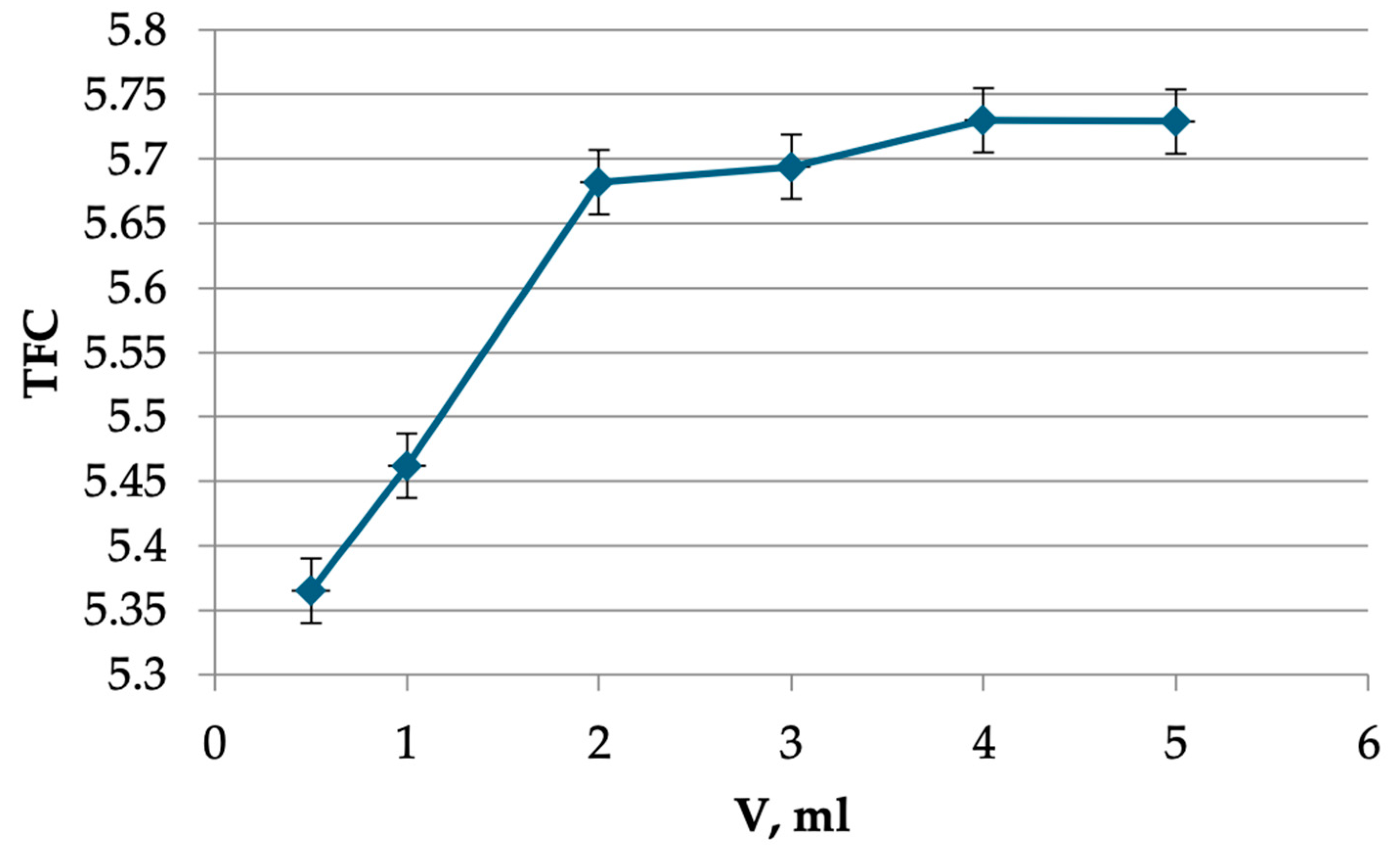
4.4. Ultrasound-Assisted Extraction Method for Flavonoid Analysis
4.5. Extraction and Qualitative Analysis of Flavonoids by UPLC/MS
- -
- Sample volume of 5 µL;
- -
- Column, 0.21 × 15.0 cm Acuity UPLC BEH C18 (1.7 µm);
- -
- Column temperature of 35 °C;
- -
- Flow rate of 0.25 mL/min;
- -
- Gradient chromatography mode formed by mixing mobile phases A and B according to the scheme involving 0 min: %B 5, 30 min: %B 50, 32 min: %B 100, 33 min: %B 5, and 36 min: %B 5;
- -
- UV detection: 220–500 nm.
- -
- MS detection in positive ion mode;
- -
- Detector parameters: capillary voltage of +3 kV; cone voltage of 50 V; capillary temperature of 450 °C; source temperature of 120 °C; drying gas flow rate of 800 L/h; gas flow rate of 50 L/h in the cone; and scanning in the mass range of 100 to 1500 units;
- -
- MS detection in negative ion mode;
- -
- Detector parameters: capillary voltage of −3 kV; cone voltage of −30 V; capillary temperature of 350 °C; source temperature of 120 °C; drying gas flow rate of 500 L/h; gas flow rate of 50 L/h in the cone; and scanning in the mass range of 100 to 1500 units.
4.6. Antimicrobial Activity
4.7. Anti-Inflammatory Activity
4.8. Anti-Aggregation, Anticoagulation, and Antioxidant/Prooxidant Activity
- 3,7-dimethyl-1-(5-oxohexyl)xanthine—pentoxifylline;
- 2-acetyloxybenzoic acid—acetylsalicylic acid;
- Heparin sodium;
- Ascorbic acid;
- Unrefined filtered sunflower oil.
- Coagulation test kits produced by “Technologiya-Standard”:
- -
- Tech-APTV-El-test, -Tech-Fibrinogen-test, -Techplastin-test (R).
- 2.
- Inducers of platelet aggregation produced by “Technologiya-Standard”:
- -
- ADP;
- -
- Collagen.
4.9. Statistical Analysis
5. Conclusions
Author Contributions
Funding
Informed Consent Statement
Data Availability Statement
Acknowledgments
Conflicts of Interest
Abbreviations
| AOA | Antioxidative Activity |
| AOS | Antioxidative System |
| APTT | Activated Partial Thromboplastin Time |
| ETIH | Aqueous alcoholic extract from T. inodorum herb |
| LC-MS/MS | Liquid Chromatography-Tandem Mass Spectropmetry |
| LPO | Lipid Peroxidation |
| MA | Maximum Amplitude of platelet aggregation |
| mg Req/100 g D.W. | amount of flavonoids in terms of rutin (R) equivalent (Req) and absolutely dry weight (D.W.) of raw materials |
| PT | Prothrombin Time |
| TFC | Total Flavonoid Content |
| TQD | Tandem Quadrupole Mass Spectrometer Detector |
References
- Skilbeck, C.A.; Lynch, I.; Ellenby, M.; Spencer, M.A. Achene Morphology of British and Irish Mayweeds and Chamomiles: Implications for Taxonomy and Identification. Br. Ir. Bot. 2019, 1, 128–166. [Google Scholar] [CrossRef]
- Farhadi, F.; Khameneh, B.; Iranshahi, M.; Iranshahy, M. Antibacterial Activity of Flavonoids and Their Structure–Activity Relationship: An Update Review. Phytother. Res. 2019, 33, 13–40. [Google Scholar] [CrossRef] [PubMed]
- Bremer, K.; Humphries, C.J. Generic Monograph of the Asteraceae-Anthemideae. Bull. Nat. Hist. Mus. London Bot. 1993, 23, 71–177. [Google Scholar]
- Baličević, R.; Ravlić, M.; Ravlić, I. Allelopathic Effect of Aromatic and Medicinal Plants on Tripleurospermum inodorum (L.) C.H. Schultz. Herbologia 2015, 15, 1–14. [Google Scholar] [CrossRef]
- Brouillet, L.T.S.-B. Tanaceteen. In Flora of North America: North of Mexico; Oxford University Press: New York, NY, USA, 2006; Volume 19, ISBN 978-0-19-530563-0. [Google Scholar]
- Crossley, J.; Skilbeck, C.A. Sea Mayweed Tripleurospermum Maritimum and Scentless Mayweed T. inodorum (Asteraceae) Intermediates in Orkney. Br. Ir. Bot. 2021, 3, 297–323. [Google Scholar] [CrossRef]
- Šibul, F.; Orčić, D.; Berežni, S.; Anačkov, G.; Mimica-Dukić, N. HPLC–MS/MS Profiling of Wild-Growing Scentless Chamo-Mile. Acta Chromatogr. 2020, 32, 86–94. [Google Scholar] [CrossRef]
- Isman, M. Medicinal and Aromatic Crops: Production, Phytochemistry, and Utilization; Jeliazkov (Zheljazkov), V.D., Cantrell, C.L., Eds.; ACS Symposium Series; American Chemical Society: Washington, DC, USA, 2016; Volume 1218. [Google Scholar]
- Kay, Q.O.N. Tripleurospermum inodorum (L.) Schultz Bip. J. Ecol. 1994, 82, 681–697. [Google Scholar] [CrossRef]
- Stace, C.A.; Thompson, H. New Flora of the British Isles, 4th ed.; C&M Floristics: Middlewood Green, Suffolk, UK, 2019; ISBN 978-1-5272-2630-2. [Google Scholar]
- Ramezanali, A.M.; Pakravan, M.; Sonboli, A.; Khayati, M. Three New Records of Tripleurospermum (Asteraceae) for the Flora of Iran. Iran. J. Bot. 2021, 27, 32–41. [Google Scholar]
- Tiezzi, A.; Karpiński, T.M.; Miranda, M.; Bustamante, K.E.; Viteri, R.; Carrillo, G.; Alexandrova, R.; Beykov, P.; Roy, A.; Attre, T. New Aspects in Medicinal Plants and Pharmacognosy; Zenodo: Geneva, Switzerland, 2017. [Google Scholar]
- Wilcox, M. Examining Tripleurospermum Inodorum (scentless Mayweed) (asteraceae) Inland. Br. Ir. Bot. 2022, 4, 51–57. [Google Scholar] [CrossRef]
- Suleimen, Y.M.; Hecke, K.; Ibatayev, Z.A.; Iskakova, Z.B.; Atakan, K.; Martins, C.H.G.; Silva, T.S. Crystal Structure and Biological Activity of Matricaria Ester Isolated from Tripleurospermum inodorum (L.) Sch. Bip. J. Struct. Chem. 2018, 59, 1025–1027. [Google Scholar] [CrossRef]
- Chehregani, A.; Mohsenzadeh, F.; Mirazi, N.; Hajisadeghian, S.; Baghali, Z. Chemical Composition and Antibacterial Activi-Ty of Essential Oils of Tripleurospermum disciforme in Three Developmental Stages. Pharm. Biol. 2010, 48, 1280–1284. [Google Scholar] [CrossRef] [PubMed]
- Cushnie, T.P.T.; Lamb, A.J. Antimicrobial Activity of Flavonoids. Int. J. Antimicrob. Agents 2005, 26, 343–356. [Google Scholar] [CrossRef] [PubMed]
- Lu, T.; Cantrell, C.L.; Robbs, S.L.; Franzblau, S.G.; Fischer, N.H. Antimycobacterial Matricaria Esters and Lactones from Astereae Species. Planta Medica 1998, 64, 665–667. [Google Scholar] [CrossRef]
- Ćavar Zeljković, S.; Ayaz, F.A.; Inceer, H.; Hayirlioglu-Ayaz, S.; Colak, N. Evaluation of Chemical Profile and Antioxidant Activity of Tripleurospermum insularum, a New Species from Turkey. Nat. Prod. Res. 2015, 29, 293–296. [Google Scholar] [CrossRef] [PubMed]
- Tofighi, Z.; Molazem, M.; Doostdar, B.; Taban, P.; Shahverdi, A.R.; Samadi, N.; Yassa, N. Antimicrobial Activities of Three Medicinal Plants and Investigation of Flavonoids of Tripleurospermum disciforme. Iran. J. Pharm. Res. IJPR 2015, 14, 225–231. [Google Scholar] [PubMed]
- Ievinsh, G.; Andersone-Ozola, U.; Karlsons, A.; Osvalde, A. Tripleurospermum maritimum from a Coastal Shingle Beach: Nitrophilic Status, Tolerance to Salinity and Heavy Metals. Environ. Exp. Biol. 2021, 19, 265–273. [Google Scholar] [CrossRef]
- Mawlood, S.I. Chemical and biological study of Iraqi Kurdistan chamomile flower (Matricaria recutita L. Baghdad Sci. J. 2011, 8, 736–740. [Google Scholar] [CrossRef]
- Mortazaei, S.; Mahmoudvand, H.; Rafieian, R. Phytochemical Properties of Some Iranian Medicinal Plants. J. Chem. Pharm. Sci. 2016, 9, 3427–3432. [Google Scholar]
- Yaşar, A.; Üçüncü, O.; Güleç, C.; Inceer, H.; Ayaz, S.; Yayl, N. GC-MS Analysis of Chloroform Extracts in Flowers, Stems, and Roots of Tripleurospermum Callosum. Pharm. Biol. 2005, 43, 108–112. [Google Scholar] [CrossRef]
- Servi, H.; Yücel, Y.Y.; Polatoğlu, K. Composition and Acetylcholinesterase Inhibition Properties of Tripleurospermum inodorum (L.) Sch. Bip. Essential Oil from Istanbul. Aurum J. Health Sci. 2018, 1, 23–38. [Google Scholar]
- Kurkcuoglu, M.; Tosun, F.; Inceer, H.; Baser, K.H.C. Volatile Compounds of Tripleurospermum decipiens from Two Natural Populations in Turkey. Chem. Nat. Compd. 2019, 55, 565–567. [Google Scholar] [CrossRef]
- Velikhanova, Z.R.; Sorokina, A.A. Development of the Characteristics of the Authenticity of the Tripleurospermum inodorum. In Proceedings of the Materials of the VI International Scientific and Methodological Conference “Pharmaceutical Education–2016. Ways and Forms of Pharmaceutical Education Improvement. Creation of New Physiologically Active Bubstances”, Voronezh, Russia, 21–23 April 2016; Belenova, A.S., Ed.; VSU: Voronezh, Russia, 2016; pp. 208–211. [Google Scholar]
- Blinova, O.L.; Gileva, A.A.; Hlebnikov, A.V.; Belonogova, V.D.; Turyshev, A.Y. Development of a method for quantitative determination of the amount of flavonoids in Tripleurospermum inodorum’s flowers. Med. Pharm. J. Pulse 2021, 23, 157–166. (In Russian) [Google Scholar] [CrossRef]
- Al-Wahaibi, L.H.; Mahmood, A.; Khan, M.; Alkhathlan, H.Z. Phytochemical Analysis and Bioactivity Screening of Three Medicinal Plants of Saudi Arabia. Trop. J. Pharm. Res. 2020, 19, 371–376. [Google Scholar] [CrossRef]
- Al-Saleem, M.S.; Awaad, A.S.; Alothman, M.R.; Alqasoumi, S.I. Phytochemical Standardization and Biological Activities of Certain Desert Plants Growing in Saudi Arabia. Saudi Pharm. J. 2018, 26, 198–204. [Google Scholar] [CrossRef]
- Puchkova, T.V. Cosmeceuticals: Modern Intensive Cosmetics; School of Cosmetic Chemists: Moscow, Russia, 2010. [Google Scholar]
- Göger, G.; Yavaş, İ.; Yur, S.; Köse, Y.B.; Özek, G. Volatiles and Fatty Acid Analyzes of Tripleurospermum decipiens (Fisch & C. A. Mey) Bornm and Investigation of the Extracts for Antimicrobial and Enzyme Inhibitory Activities. J. Res. Pharm. 2021, 25, 429–440. [Google Scholar] [CrossRef]
- Velikhanova, Z.R.; Marakhova, A.I.; Sorokina, A.A. The content of biologically active substances in the flowers of scentless mayweed (Tripleurospermum perforatum). Pharmacy 2017, 66, 9–12. (In Russian) [Google Scholar]
- Kumar, A.; Khan, F.; Saikia, D. Phenolic Compounds and Their Biological and Pharmaceutical Activities. In The Chemistry inside Spices & Herbs: Research and Development; Chaurasia, P.K., Bharati, S.L., Eds.; Bentham Science Publishers: Sharjah, United Arab Emirates, 2022; pp. 206–236. ISBN 978-981-5039-56-6. [Google Scholar]
- Chen, M.; He, X.; Sun, H.; Sun, Y.; Li, L.; Zhu, J.; Xia, G.; Guo, X.; Zang, H. Phytochemical Analysis, UPLC-ESI-Orbitrap-MS Analysis, Biological Activity, and Toxicity of Extracts from Tripleurospermum limosum (Maxim. ) Pobed. Arab. J. Chem. 2022, 15, 103797. [Google Scholar] [CrossRef]
- Maroun, R.G.; Rajha, H.N.; El Darra, N.; El Kantar, S.; Chacar, S.; Debs, E.; Vorobiev, E.; Louka, N. 8—Emerging Technologies for the Extraction of Polyphenols from Natural Sources. In Polyphenols: Properties, Recovery, and Applications; Galanakis, C.M., Ed.; Woodhead Publishing: Cambridge, UK, 2018; pp. 265–293. [Google Scholar]
- Górniak, I.; Bartoszewski, R.; Króliczewski, J. Comprehensive Review of Antimicrobial Activities of Plant Flavonoids. Phy-tochem. Rev. 2019, 18, 241–272. [Google Scholar] [CrossRef]
- Ștefan, M.; Bîrsă, M.L. Flavonoids—An Amazing Group of Compounds with Potent Antimicrobial Properties. Mem. Sci. Sect. Rom. Acad. 2019, 42, 111–139. [Google Scholar]
- Dobiáš, P.; Pavlíková, P.; Adam, M.; Eisner, A.; Beňová, B.; Ventura, K. Comparison of Pressurised Fluid and Ultrasonic Ex-Traction Methods for Analysis of Plant Antioxidants and Their Antioxidant Capacity. Open Chem. 2010, 8, 87–95. [Google Scholar] [CrossRef]
- Uzel, R.A. Impact of Ultrasound-Assisted Extraction on Supercritical Recover of Valuable Compounds from Dry Pine Needles. Int. J. Food Eng. 2018, 4, 8–13. [Google Scholar] [CrossRef]
- Lavilla, I.; Bendicho, C. Fundamentals of Ultrasound-Assisted Extraction. In Water Extraction of Bioactive Compounds; Elsevier: Amsterdam, The Netherlands, 2017; pp. 291–316. ISBN 978-0-12-809380-1. [Google Scholar]
- Turrini, F.; Boggia, R.; Leardi, R.; Borriello, M.; Zunin, P. Optimization of the Ultrasonic-Assisted Extraction of Phenolic Compounds from Oryza sativa L. ‘Violet Nori’ and Determination of the Antioxidant Properties of its Caryopses and Leaves. Molecules 2018, 23, 844. [Google Scholar] [CrossRef] [PubMed]
- Pinchao-Pinchao, Y.A.; Ordoñez-Santos, L.E.; Osorio-Mora, O. Evaluation of the Effect of Different Factors on the Ultrasound Assisted Extraction of Phenolic Compounds of the Pea Pod. Dyna 2019, 86, 211–215. [Google Scholar] [CrossRef]
- Sivakumar, V.; Ravi Verma, V.; Rao, P.G.; Swaminathan, G. Studies on the Use of Power Ultrasound in Solid–Liquid Myrob-Alan Extraction Process. J. Clean. Prod. 2007, 15, 1813–1818. [Google Scholar] [CrossRef]
- Tiwari, B.K. Ultrasound: A Clean, Green Extraction Technology. TrAC Trends Anal. Chem. 2015, 71, 100–109. [Google Scholar] [CrossRef]
- Altemimi, A.; Watson, D.G.; Choudhary, R.; Dasari, M.R.; Lightfoot, D.A. Ultrasound Assisted Extraction of Phenolic Com-Pounds from Peaches and Pumpkins. PLoS ONE 2016, 11, e0148758. [Google Scholar] [CrossRef] [PubMed]
- Madhu, B.; Srinivas, M.S.; Srinivas, G.; Jain, S.K. Ultrasonic Technology and Its Applications in Quality Control, Processing and Preservation of Food: A Review. Curr. J. Appl. Sci. Technol. 2019, 32, 1–11. [Google Scholar] [CrossRef]
- Chemat, F.; Rombaut, N.; Sicaire, A.-G.; Meullemiestre, A.; Fabiano-Tixier, A.-S.; Abert-Vian, M. Ultrasound Assisted Extraction of Food and Natural Products. Mechanisms, Techniques, Combinations, Protocolsand Applications. A Review. Ultrason. Sonochemistry 2017, 34, 540–560. [Google Scholar] [CrossRef] [PubMed]
- Vernès, L.; Vian, M.; Chemat, F. Ultrasound and Microwave as Green Tools for Solid-Liquid Extraction. In Liquid-Phase Ex-traction; Elsevier: Amsterdam, The Netherlands, 2020; pp. 355–374. ISBN 978-0-12-816911-7. [Google Scholar]
- Yang, X.; Li, Y.; Li, S.; Oladejo, A.O.; Ruan, S.; Wang, Y.; Huang, S.; Ma, H. Effects of Ultrasound Pretreatment with Different Frequencies and Working Modes on the Enzymolysis and the Structure Characterization of Rice Protein. Ultrason. Sonochemistry 2017, 38, 19–28. [Google Scholar] [CrossRef] [PubMed]
- Uddin, M.K.; Juraimi, A.S.; Ali, M.E.; Ismail, M.R. Evaluation of Antioxidant Properties and Mineral Composition of Purslane (Portulaca oleracea L.) at Different Growth Stages. Int. J. Mol. Sci. 2012, 13, 10257–10267. [Google Scholar] [CrossRef] [PubMed]
- Olabode, I.R.; Sachivkina, N.; Karamyan, A.; Mannapova, R.; Kuznetsova, O.; Bobunova, A.; Zhabo, N.; Avdonina, M.; Gurina, R. In Vitro Activity of Farnesol against Malassezia Pachydermatis Isolates from Otitis Externa Cases in Dogs. Animals 2023, 13, 1259. [Google Scholar] [CrossRef] [PubMed]
- Sachivkina, N.; Podoprigora, I.; Bokov, D. Morphological Characteristics of Candida Albicans, Candida Krusei, Candida Guilliermondii, and Candida Glabrata Biofilms, and Response to Farnesol. Vet. World 2021, 14, 1608–1614. [Google Scholar] [CrossRef] [PubMed]
- Sachivkina, N.; Vasilieva, E.; Lenchenko, E.; Kuznetsova, O.; Karamyan, A.; Ibragimova, A.; Zhabo, N.; Molchanova, M. Reduction in Pathogenicity in Yeast-like Fungi by Farnesol in Quail Model. Animals 2022, 12, 489. [Google Scholar] [CrossRef] [PubMed]
- Arsene, M.M.J.; Viktorovna, P.I.; Sergei, G.V.; Hajjar, F.; Vyacheslavovna, Y.N.; Vladimirovna, Z.A.; Aleksandrovna, V.E.; Nikolayevich, S.A.; Sachivkina, N. Phytochemical Analysis, Antibacterial and Antibiofilm Activities of Aloe Vera Aqueous Extract against Selected Resistant Gram-Negative Bacteria Involved in Urinary Tract Infections. Fermentation 2022, 8, 626. [Google Scholar] [CrossRef]
- Sachivkina, N.; Karamyan, A.; Semenova, V.; Ignatiev, A.; Abdurasulov, A.; Muratova, R.; Emilbekova, D.; Ermatova, V.; Risvanli, A.; Salykov, R. The Effects of Angelica Ternata Extract from Kyrgyzstan on the Formation of Candida Albicans ATCC 10231 Biofilms. Appl. Sci. 2023, 13, 12042. [Google Scholar] [CrossRef]
- Balaji, T.; Manushankar, C.M.; Al-Ghanim, K.A.; Kamaraj, C.; Thirumurugan, D.; Thanigaivel, S.; Nicoletti, M.; Sachivkina, N.; Govindarajan, M. Padina Boergesenii-Mediated Copper Oxide Nanoparticles Synthesis, with Their Antibacterial and An-Ticancer Potential. Biomedicines 2023, 11, 2285. [Google Scholar] [CrossRef] [PubMed]
- Tharani, M.; Rajeshkumar, S.; Al-Ghanim, K.A.; Nicoletti, M.; Sachivkina, N.; Govindarajan, M. Terminalia Chebula-Assisted Silver Nanoparticles: Biological Potential, Synthesis, Characterization, and Ecotoxicity. Biomedicines 2023, 11, 1472. [Google Scholar] [CrossRef] [PubMed]
- Ramasubbu, K.; Padmanabhan, S.; Al-Ghanim, K.A.; Nicoletti, M.; Govindarajan, M.; Sachivkina, N.; Rajeswari, V.D. Green Synthesis of Copper Oxide Nanoparticles Using Sesbania Grandiflora Leaf Extract and Their Evaluation of Anti-Diabetic, Cy-Totoxic, Anti-Microbial, and Anti-Inflammatory Properties in an In-Vitro Approach. Fermentation 2023, 9, 332. [Google Scholar] [CrossRef]
- European Convention for the Protection of Vertebrate Animals Used for Experimental and Other Scientific Purposes (ETS No. 123). Available online: https://www.coe.int/en/web/conventions/full-list?module=treaty-detail&treatynum=123 (accessed on 20 September 2023).
- Born, G. Aggregation of Blood Platelets by Adenosine Diphosphate and Its Reversal. Nature 1962, 194, 927–929. [Google Scholar] [CrossRef] [PubMed]


| Concentration of Ethyl Alcohol, % | Weight of Raw Materials, g: Volume of Extractant, mL | Extraction Time, min | TFC, % |
|---|---|---|---|
| Parameter—extractant concentration | |||
| 20 | 1:200 | 20 | 3.844 ± 0.003 |
| 40 | 4.285 ± 0.001 | ||
| 50 | 4.845 ± 0.003 | ||
| 60 | 4.901 ± 0.002 | ||
| 70 | 5.025 ± 0.003 | ||
| 95 | 3.104 ± 0.003 | ||
| Parameter—ratio of raw material mass, g, and extractant volume, mL | |||
| 70 | 1:50 | 40 | 5.424 ± 0.002 |
| 1:100 | 5.564 ± 0.003 | ||
| 1:150 | 5.684 ± 0.002 | ||
| 1:200 | 5.681 ± 0.003 | ||
| Parameter—extraction time, min | |||
| 70 | 1:150 | 20 | 5.025 ± 0.003 |
| 30 | 5.165 ± 0.002 | ||
| 40 | 5.682 ± 0.003 | ||
| 50 | 5.591 ± 0.004 | ||
| 60 | 5.426 ± 0.001 | ||
| Name | tR | Mol. Weight | UV Absorption Wavelength, nm | Structure |
|---|---|---|---|---|
| Chlorogenic acid | 4.2 | 354 | 326 |  |
| 5-O-p-coumaroylquinic acid | 5.77 | 338 | 312 |  |
| 1-O-p-coumaroylquinic acid | 7.21 | 338 | 307 |  |
| Quercetin-3-O-glucoside | 8.57 | 464 | 370 |  |
| Luteolin-7-O-glucoside | 8.88 | 448 | 348 |  |
| Luteolin-7-O-rutinoside | 8.97 | 594 | 349 |  |
| 3,5-O-dicaffeoylquinic acid | 10.12 | 516 | 327 |  |
| Quercetin-3-O-malonylglucoside | 10.62 | 550 | 370 |  |
| Apigenin-7-O-glucoside | 10.72 | 432 | 344 |  |
| Luteolin-3-malonylglucoside | 10.99 | 534 | 348 | 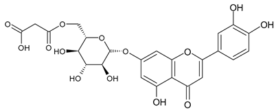 |
| Tsinarin | 11.13 | 516 | 329 |  |
| Rhamnetin-3-(O-dimethyl ramnosylglucosy glucoside) | 13.07 | 814 | 370 |  |
| Luteolin | 14.51 | 286 | 349 | 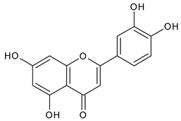 |
| Sample | Zone of Inhibition St. aur., mm | Zone of Inhibition C. alb., mm | Zone of Inhibition E. coli, mm |
|---|---|---|---|
| ETIH | 22 ± 2 | 12 ± 1 | 14 ± 2 |
| Alcohol 70% | 6 ± 1 | 5 ± 1 | 7 ± 2 |
| Sample | APTT Change, % to Control | Change in PT, % to Control | Fibrinogen, % to Control |
|---|---|---|---|
| ETIH solution | +7.1 (7.3 ± 0.1) | 0.0 (0) | 0.0 (0) |
| Heparin sodium | +20.3 (20.6 ± 0.3) | 0.0 (0) | 0.0 (0) |
| Sample | Latent Period, % to Control | Maximum Amplitude, % to Control | Aggregation Rate, % to Control | Time to Reach MA, % to Control | Disaggregation, % to Control |
|---|---|---|---|---|---|
| ETIH solution | +4.2 (4.8 ± 0.2) ††,# | −6.5 (6.7 ± 0.1) *,††,# | −9.6 (10.8 ± 0.2) *,†† | +9.6 (10.2 ± 0.3) *,†,# | 0.0 (0) †† |
| Acetylsalicylic acid | −2.1 (1.9 ± 0.1) †† | −13.7 (13.6 ± 0.3) *,†† | −10.5 (10.0 ± 0.2) *,†† | +10.5 (10.2 ± 0.1) *,†† | 0.0 (0.0–0.0) †† |
| Pentoxifylline | +32.4 (28.7–35.6) * | −48.4 (42.7–56.5) ** | −34.9 (34.1 ± 0.5) ** | + 32.1 (31.1 ± 0.4) ** | 13.6 (13.8 ± 0.2) ** |
| Sample | Model | Light Sum | Flash |
|---|---|---|---|
| ETIH solution | I | −16.1 (16.4 ± 0.3) *,a | −11.4 (12.9 ± 0.4) *,a |
| II | −32.6 (31.2 ± 0.4) **,a | −7.2 (9.8 ± 0.2) *,a | |
| Ascorbic acid | I | −84.5 (83.6 ± 0.5) ** | −91.7 (88.9 ± 0.7) ** |
| II | −78.1 (75.9 ± 0.5) ** | −86.8 (86.5 ± 0.5) ** |
| Sample | Paw Diameter (mm) | Severity of Inflammatory Reaction, % | Suppression of Edema at Peak of Inflammation, % | |||
|---|---|---|---|---|---|---|
| 0 h | 4 h | 24 h | 4 h | 24 h | 4 h | |
| Control | 2.9 (3.3 ± 0.1) | 4.7 (4.6 ± 0.1) a | 4.1 (3.8 ± 0.3) a | 62.07 | 41.38 | - |
| ETIH solution | 2.7 (2.8 ± 0.1) | 3.3 (3.3 ± 0.1) * | 3.1 (3.2 ± 0.1) * | 22.22 ± 3.56 | 14.81 ± 2.15 | 66.67 |
| Diclofenac sodium, 10 mg/kg | 3.0 (2.9 ± 0.2) | 3.5 (3.6 ± 0.2) *,a | 3.2 (3.2 ± 0.1) * | 16.67 ± 2.78 | 6.67 ± 1.05 | 72.22 |
Disclaimer/Publisher’s Note: The statements, opinions and data contained in all publications are solely those of the individual author(s) and contributor(s) and not of MDPI and/or the editor(s). MDPI and/or the editor(s) disclaim responsibility for any injury to people or property resulting from any ideas, methods, instructions or products referred to in the content. |
© 2024 by the authors. Licensee MDPI, Basel, Switzerland. This article is an open access article distributed under the terms and conditions of the Creative Commons Attribution (CC BY) license (https://creativecommons.org/licenses/by/4.0/).
Share and Cite
Marakhova, A.; Zhilkina, V.Y.; Elapov, A.; Sachivkina, N.; Samorodov, A.; Pupykina, K.; Krylova, I.; Kezimana, P.; Stoynova, A.M.; Venkatesan, R.; et al. The Development of a Method for Obtaining Tripleurospermum inodorum (L.) Sch. Bip. Herb Extract Enriched with Flavonoids and an Evaluation of Its Biological Activity. Plants 2024, 13, 1629. https://doi.org/10.3390/plants13121629
Marakhova A, Zhilkina VY, Elapov A, Sachivkina N, Samorodov A, Pupykina K, Krylova I, Kezimana P, Stoynova AM, Venkatesan R, et al. The Development of a Method for Obtaining Tripleurospermum inodorum (L.) Sch. Bip. Herb Extract Enriched with Flavonoids and an Evaluation of Its Biological Activity. Plants. 2024; 13(12):1629. https://doi.org/10.3390/plants13121629
Chicago/Turabian StyleMarakhova, Anna, Vera Yu. Zhilkina, Alexander Elapov, Nadezhda Sachivkina, Alexander Samorodov, Kira Pupykina, Irina Krylova, Parfait Kezimana, Anastasia M. Stoynova, Raja Venkatesan, and et al. 2024. "The Development of a Method for Obtaining Tripleurospermum inodorum (L.) Sch. Bip. Herb Extract Enriched with Flavonoids and an Evaluation of Its Biological Activity" Plants 13, no. 12: 1629. https://doi.org/10.3390/plants13121629