Review

Exploring Aluminum Tolerance Mechanisms in Plants with Reference to Rice and Arabidopsis: A Comprehensive Review of Genetic, Metabolic, and Physiological Adaptations in Acidic Soils

1
Plant Physiology and Molecular Biology Research Unit, Department of Botany, University of Kalyani, Kalyani 741235, West Bengal, India
2
Department of Botany, University of Gour Banga, Malda 732103, West Bengal, India
3
Department of Agronomy, Faculty of Agriculture, Shar-e-Bangla Agricultural University, Dhaka 1207, Bangladesh
4
Kyung Hee University, 26 Kyungheedae-ro, Dongdaemun-gu, Seoul 02447, Republic of Korea
*
Authors to whom correspondence should be addressed.
Plants 2024, 13(13), 1760; https://doi.org/10.3390/plants13131760
Submission received: 20 May 2024 / Revised: 15 June 2024 / Accepted: 21 June 2024 / Published: 25 June 2024
(This article belongs to the Special Issue Plant Responses and Tolerance to Metal/Metalloid Toxicity III)

Abstract

:
Aluminum (Al) makes up a third of the Earth’s crust and is a widespread toxic contaminant, particularly in acidic soils. It impacts crops at multiple levels, from cellular to whole plant systems. This review delves into Al’s reactivity, including its cellular transport, involvement in oxidative redox reactions, and development of specific metabolites, as well as the influence of genes on the production of membrane channels and transporters, alongside its role in triggering senescence. It discusses the involvement of channel proteins in calcium influx, vacuolar proton pumping, the suppression of mitochondrial respiration, and the initiation of programmed cell death. At the cellular nucleus level, the effects of Al on gene regulation through alterations in nucleic acid modifications, such as methylation and histone acetylation, are examined. In addition, this review outlines the pathways of Al-induced metabolic disruption, specifically citric acid metabolism, the regulation of proton excretion, the induction of specific transcription factors, the modulation of Al-responsive proteins, changes in citrate and nucleotide glucose transporters, and overall metal detoxification pathways in tolerant genotypes. It also considers the expression of phenolic oxidases in response to oxidative stress, their regulatory feedback on mitochondrial cytochrome proteins, and their consequences on root development. Ultimately, this review focuses on the selective metabolic pathways that facilitate Al exclusion and tolerance, emphasizing compartmentalization, antioxidative defense mechanisms, and the control of programmed cell death to manage metal toxicity.

1. Introduction

Acidic soils, comprising nearly 40% of the Earth’s crust, include about 50% of arable land. These soils can support certain crop species, though they often require altered physiological responses. In acidic soils, Al is the most abundant element, significantly impacting rice cultivation, which occupies 13% of acidic land [1]. Al exists in various forms, including aluminum silicate and sulfate, and can dissolve into active trivalent Al ions (Al3+), particularly under low-pH conditions in the rhizosphere. Dominant forms like aluminum hydroxide cations [Al(OH)2+, Al(OH)3+] can cause oxidative stress. Both inorganic and organic residues in the rhizosphere contribute to Al release into acidic water bodies [2]. Exchangeable Al3+ binds with silicates, clays, and other residues, affecting mineral availability to plants. High Al concentrations, depending on soil pH, can damage root systems.
In acidic soil, roots are impaired with membrane permeability, hydraulic conductivity, mineral acquisition, and the replacement of other nutrients, which are collectively responsible for plant growth and productivity [3]. Al toxicity involves inducible gene expression and two primary stress response mechanisms. Bio-exclusion of Al involves both internal and external mechanisms for Al, and those essentially need organic acid-based ligand formation.
The effects of Al accumulation are more notable in the root apex, root cap, and extended permanent regions of roots, where altered carbohydrate metabolism is directly related to metal uptake. In Al-tolerant genotypes, the uptake of metal may not be related to impeded ion uptake for nitrate (NO3)-like ions [4]. In cereals, the inhibition of such ion uptake is reversed by ammonium (NH4+) treatment, which suggests the co-transport of Al with other cations. Still, the interaction of Al with NO3 is a possible clue for mineral imbalances under acidic soil. The increase in amino acids under Al treatment is also assumed for an enhanced degradation of proteins in the root zone. Likewise, a proportion of amines (asparagines, glutamine) are the product of Al-induced protein hydrolysis or oxidation, and thereby, the accumulation of free amino acids serves as a bio-indicator of acidic soil [5]. On the other hand, uptake of ions is reduced due to competition with Al according to the pH of the solution. Thus, reduction in NO3 absorption releases more protons (H+) into the rhizosphere that otherwise favor acidic soil pH and more Al absorption. A decline in NO3 uptake influences signal(s) for rhizosphere secretion by Al-sensitive species to release some special metabolites. Thus, NO3 uptake would be a preliminary symptom of Al toxicity within a short period of application of the metal, particularly in tolerant plants.
Toxicity to Al is another path irrespective of tolerant and susceptible species where different chemicals may function as elicitors of defense mechanisms. In relation to crop species grown in acidic soil, there are various mechanisms of Al tolerance [6]. Few predominant chemical species (malate, oxaloacetate, citrate, and a few other organic acids) are released into the rhizosphere and act as Al chelators. This is an extracellular strategy of Al avoidance by specific reactions in different tolerant genotypes with variations in the sequestration of metal [7]. In intracellular pathways, the same strategy for metal chelation is operative, where H+ pump-mediated cytoplasmic and vacuolar acidity is accomplished. Thus, Al sensitivity covers acidolysis of the cytoskeleton and biomolecules to different degrees depending on reactions in non-tolerant species. The subsequent imprisonment of the metal into non-cellular or apoplastic spaces also sets up another tolerance mechanism. Moreover, Al toxicity also leads to a change in the membrane potential, which is often correlated with the membrane surface binding capacities of other cations [8]. A change in plasma membrane potential induced by Al-mediated depolarization can also regulate calcium ion (Ca2+) uptake, influencing the growth of the plants. The effect of cytoplasmic Ca2+ concentration directly regulates many processes of cell metabolism where Al injury is responsible. Al-dependent flux of Ca2+ from extracellular spaces regulates the inhibitions of root elongation, which may be reversed by acidification of the soil [9]. Therefore, the cellular pH of the roots is the determining factor for the availability of some signaling molecules (e.g., Ca2+) for the interaction of Al in soil. A pH-dependent synthesis of complex polysaccharides (like 1-3-glucan) in the roots influences metabolism of calcium, where the roles of calmodulin (CaM)-like binding proteins are important [10].
Despite the significant body of literature describing the toxicity of Al in plants, little is deciphered about specific ion effects like oxidative stress. It was reported earlier that besides water relations and other cellular anomalies induced by Al as a metal in general, changes in redox are the prime key to Al vulnerability. The accumulation of reactive oxygen species (ROS) and, thereby, the induction of antioxidation machineries are the keys to Al tolerance [11]. A change in cellular redox, inducing oxidative stress, is the initiation of different chemical reactions like the peroxidation of lipids, the carbonylation of protein, the tautomerization of nucleic acids, all of which are processes of the degeneration of plants under Al toxicity. Furthermore, Al-induced inhibitions of cellular respiration and adenosine triphosphate (ATP) depletion can compel the plants to bypass the electron flow from the electron transport chain (both in chloroplasts and mitochondria) into molecular oxygen. At the cellular level, in roots, the depletion of energy-utilizing pathways and the dysfunction of the organic acid cycle in the mitochondria lead to the loss of viability of roots [12]. Thus, oxidative stress is set as the key underlying mechanism of Al toxicity where interference from other redox metals (iron, chromium, zinc) in the tissue is complementary to the degenerative process of toxicity. More insights are yet to be known for ROS production through other pathways linked to cellular reactions as major determining factors for root growth inhibition under Al contamination.
This review aims to thoroughly understand the Al resistance mechanisms in tolerant genotypes and other species in acidic soils. It emphasizes the need to develop cultivars or identify specific traits to support adaptation to Al-rich acidic soils. Understanding increased Al3+ sensitivity is crucial due to the constraints of food production in specific agro-climatic zones; it is imperative to understand the tolerance mechanisms of crops for breeding purposes. Metal detoxification primarily relies on exclusion mechanisms, with organic acids forming stable complexes with Al3+ [13]. Genetic studies on rice, a crop highly sensitive to acidic soils, have identified several gene families (ALMT, MATE, ABC) and transcription factors regulatinggenes (ART1, ASR5, OsWRKY22) that are crucial for Al exclusion and tolerance [14]. This review also reveals the importance of omics technologies like genomics, proteomics, and metabolomics to understand the complex traits in crops under Al toxicity using intradisciplinary approaches in crop science. It may cover the generation of metabolites under metal stress with their qualitative and quantitative distribution, biosynthesis, catabolism, and the functional network in combination for tolerance. It also finally discusses the development of selective markers based on specific metabolomes and their roles in metal tolerance in plants through gene overexpression and chemical elicitation.

2. Aluminum Bioavailability in Different Forms to Sensitize the Plants

Highly complex effluents from industrial outlets composed of different Al compounds are present in varying quantities and are responsible for toxicity in acidic soil. In soil with pH < 4, increasing crop susceptibility to metal toxicity due to a high concentration of Al can cause soil acidification, which is more targeted towards failure of crop productivity [15]. In spite of this, soil acidification is achieved by other agronomic factors through the accumulation of basic cations: K+, Na+, Mg2+, Ca2+, etc. Al is the major component, covering >7% of metallic elements in the Earth’s crust after silicon, yet showing no significant roles in plant growth and development [16]. Al is precipitated in soil from major ores like aluminosilicate that undergo initial solubilization (at pH < 5.0), resolving in an eventual phytotoxic form. Trivalent aluminum ions can also accompany different hydroxides of metal like aluminum hydroxide cations like Al(OH)2+, Al(OH)3, and [Al(OH)4] in partial to full solubilization for plants’ absorption. This is based on a gradual depletion of soil pH that corresponds to a better solubility of Al salts with different valences (Figure 1). Out of the different valences, Al3+ is the most toxic and stress-producing form, particularly when distributed through apoplastic and symplastic spaces in roots. Plant species irrespective of taxa when subjected to intense and prolonged durations of Al exposure undergo variable levels of modification through intra- and intercellular components [17]. Physiologically, these cover changes in cell wall components, modification of transport processes across the membrane, disruption of cytoskeletons, altered cell signaling energy-yielding metabolism, and, finally, the mutation of nuclear material [18]. Notably, plants that respond to Al3+ toxicity are distinctly categorized as either tolerant or sensitive species. Thereby, a search is underway for the tolerance mechanisms of plants in acidic soil when plant growth is vulnerable at physiological and genetic levels. The most predominant line of investigation is based on the absorption of Al through the root system, which changes root physiology and the biochemistry and molecular biology of cells. The second line of research is based on an analysis of induced oxidative stress with specific Al3+ ions at the cellular level [19]. Different oxidative metabolites, their varying expression through tolerant and sensitive cultivars, and regulation through biochemical reactions are the major domains of Al-induced oxidative stress in research. This is more intricate than other findings, where partially known or hypothetical ligands are reported to form complex compounds with free Al in the cytosol [20]. In this view, biosynthetic and catabolic genes are under study, concerning tolerant cultivars. Therefore, the acquired information may be relevant for the application of molecular breeding and biotechnological devices for the development of tolerant genotypes against Al toxicity.

3. Aluminum-Induced Oxidative Stress and Metabolic Alterations

Industrial processes and human activities are primary contributors to the dispersal of toxic forms of Al in the Earth’s crust. These activities release various toxic molecular forms of Al, posing significant threats to both the environment and plants’ lives. Sources of these toxic forms include the processing of ores, the use of fertilizers and herbicides, and the disposal of solid wastes such as sewage sludge and alloy emissions from different industries [21]. A key indicator of Al toxicity in plant cells is the generation of ROS. In particular, roots are highly sensitive to Al exposure, leading to the production of various ROS. This generation of ROS occurs not only in the cytosol but also within cellular organelles such as chloroplasts, mitochondria, and peroxisomes, which are rich in electron transport chains in cells [22]. In the nucleus, peroxidation of the membrane can initiate cascades of free radical generation, including hydroperoxides and peroxy radicals, which interact with nucleotides like macromolecules. Research has identified several genes activated in the nucleus that act upon tolerance mechanisms through cytoplasmic and organellar compartments. Key genes include ADP-ribosyltransferase 1, ferric reductase defective like 4, acetolactate synthase 1, and Nramp family 1 (ART1, FRDL4, ALS1, NRAT1), along with various antioxidants and proteins involved in the ascorbate (AsA)–glutathione (GSH) cycle [23].
Mitochondria can also be significant sources of ROS, especially where malate and organic acid transporters facilitate the entry of Al ion (Al3+). The inter-membrane spaces, housing the electron transport chain, are prone to ROS generation, particularly hydroxyl radicals, resulting in oxidative decarboxylation for DNA. The redox status is crucial for understanding the metabolic impact of Al on tissues, with pyridine nucleotides (NADH/NAD+, NADPH/NADP+, FADH2/FAD+) playing a key role in signaling through ROS [24]. These ROS trigger various harmful reactions including lipid peroxidation, protein carbonylation, and the formation of bulky nucleotide adducts. The dual roles of ROS as critical signaling residues as well as in inducing oxidative stress are more contextual for signaling against Al ions (Al3+). Root tips affected with Al3+ are demonstrated with detoxification mechanisms through cell wall modification. ROS and their exposure on the outer surface of the plasma membrane induce a complex network of wall-modifying enzymes [25]. There are a few noticeable overexpressed proteins like expansions, xyloglucan endo-transglucosylase, and pectin acetyl esterase, the latter of which is responsible for cell wall elasticity as well as crosslinking with secondary deposition. ROS, particularly hydrogen peroxide (H2O2), are the most important inducers in the reaction where Al3+ is chelated with complex carbohydrate residues. The formation of superoxide radicals on apoplastic and membrane-bound NAD(P)H oxidase (NOX) and respiratory burst oxidase homolog (RBOH) is the most important due to their activities in signaling processes. A number of enzymes like superoxide dismutase, xanthine oxidase, and, finally, a few classes III wall-bound peroxidases are important for different ROS/free radicals in sensitization to Al. In the presence of Al3+, the crosslinking of cell wall residues, mostly xyloglucans, increases the simultaneous formation of ROS and the overexpression of variants of peroxidases. ROS on the cell wall and in apoplastic spaces are protruded with membrane transporters like nod26-like intrinsic protein (NIP), Halimione portulacoides plasma membrane amino acid transporter 1(HmPALT1), and nitrate transporter 1 (NRAT1), with simultaneous restriction of Al3+ entry [26]. In the latter stages of metal-ion-induced toxicity in the cytosol, ROS signal membrane transporters to overexpress organic acid biosynthesis. A few phenolic residues are also induced to chelate Al3+ in the rhizosphere to prevent the entry of ions through the root system. The path of ROS development under Al3+ toxicity is quite consistent with other external stimuli. The intricate control mechanism employing RBOH happens to be most useful in metal-affected tissues for the transmission of signals following its transduction [27]. Additionally, NOX-derived ROS are also accompanied by a number of apoplast-harboring proteins, mostly peroxidases (PRXs), polyamine oxidases (PAOs), quinone reductase, and other metal-bearing anime oxidases. An oxidative signal, mainly through altered mitochondrial electron pathways, also promotes ROS generation [28]. Finally, within the nuclear membrane, ROS exposure activates gene encoding for antioxidative cascades for the regulation of oxidative stress. The translocation of ROS as a signal through the cytosol can also induce a number of membrane-bound transporters like Oryza sativa L.-activated malate transporter 1 (OsALS1) and vacuolar amino acid transporter 1 (VALT1) for conjugations of Al3+ as well as direct detoxification with non-thiol residues and phytochelatin [29]. ROS produced by herbicides and pesticides, like xenobiotics, may function in reliable biomarking for specific plant species. The metals inserted within the xenobiotics have a distinct path to be detoxicated in the plant system, initially by the spatio-temporal transitory accumulation of free radicals (e.g., H2O2) that may act in the evocation of signals. This is followed by the compartmentalization of metal in cytosolic or even apoplastic spaces in conjugation with glutathione-like compounds [30]. Elicitors like melatonin are effective in the chelation of pesticide-like residues following transport into vacuoles by ATP-binding cassette (ABC) transporters, as found in the detoxification of Al3+.Therefore, ROS sensing and signal transduction would be dependent on the type of stimuli and plants’ genotypic potential to minimize their effects, either by direct sequestering or by antioxidation.
Among the plethora of free radicals, H2O2 stands out for its dual role in signaling, potentially leading to either beneficial or detrimental outcomes in the context of Al toxicity. The synthesis of macromolecules such as lignin, which relies on peroxidase activity on H2O2, is an example of an immediate response to Al stress. Oxidative stress, indicated by the accumulation of H2O2, fuels the peroxidase activity that is necessary for lignin biosynthesis [31]. Concurrently, plants exercise a defense system through a combination of both enzymatic and non-enzymatic antioxidants to mitigate a H2O2-induced peroxidation reaction.
In chloroplasts, Al toxicity triggers ROS production through mechanisms involving the electron transport chain in Photosystems I and II. Various ROS forms are generated, including singlet oxygen, superoxide, hydrogen peroxide, and hydroxyl radicals. The uptake of Al3+ induces the expression of certain genes in the chloroplast genome, which include membrane-bound ATP-independent proteases that play a significant role in Al tolerance mechanisms [32]. The mechanisms of Al tolerance are complex, involving the synergistic contributions of metal compartmentalization, chloroplast fluorescence, mitochondrial oxidative reactions, and peroxisomal carbon oxidation (Figure 2). Therefore, Al tolerance is a multifaceted approach that essentially underscores the intricate networks of responses of plants against oxidative stress.
Al profoundly impacts root growth and extends its detrimental effects to other plant organs. Many species show signs of toxicity at different organizational levels, from morphological changes to nuclear alterations, largely due to their oxidative sensitivity to the metal [33]. The solubilization of Al3+ in the rhizosphere, a result of soil acidification, leads to the formation of various chemical species of Al. This complicates agricultural practices in such soils, especially with the co-presence of other metals like ferrous ion (Fe2+) and various Al salt intermediates. In regions like the root meristem, endodermis, and cortical tissues, exposure to Al3+ causes lipid peroxidation and the accumulation of ROS, as demonstrated in maize cultivars. This results in significant inhibition of tissue elongation, highlighting the root as the primarily affected organ [34]. Al toxicity triggers oxidative stress, which alters cellular and membrane integrity by disrupting redox balance within cells. Different ROS types can initiate peroxidation reactions on macromolecules such as lipids, proteins, and nucleic acids, compromising cell viability.
Although Al is not a transition metal and does not directly catalyze redox reactions, it can still contribute to the oxidation of biomolecules, leading to oxidative stress. This often involves Al forming electrostatic bonds with biomolecular groups such as PO43− or COO on plasma membrane pectins [35]. The formation of callose within cell walls serves as an adaptive strategy to mitigate free radical damage. Transition metals, in conjunction with Al, contribute to the pool of ROS. Excessive Al on the membrane surface can increase the uptake of redox-active iron (Fe) into the rhizosphere, creating a vicious cycle of ROS production in plant tissues [36]. Crop species, including barley, rice, and gram, suffer from Al toxicity at their root apices, leading to growth inhibition due to lipoxygenase activity. In maize, factors such as early senescence and altered water relations, alongside oxidative stress, amplify the detrimental effects [37].
In acidic soils, Al accumulation can inhibit the function of cellular proton-ATPase pumps (H+/ATPase), leading to the efflux of potassium and other essential elements from the membrane. The impact of Al-induced oxidative stress varies among crop species, depending on their genetic makeup. Metabolomic responses in susceptible species are critical in acidic conditions, where micromolar concentrations of Al3+ have specific toxic effects, impairing water and nutrient uptake. Combating soil acidification, a mounting global concern, involves selecting Al-tolerant genotypes and identifying and cloning specific genes related to Al tolerance [38]. A comprehensive understanding of cellular responses and nuclear regulatory mechanisms is vital for developing Al-tolerant crop lines. Metabolomics, reflecting changes in metabolite expression triggered by various Al species, needs further investigation. The interaction between nitrate and Al in root uptake significantly affects primary metabolomes [39]. Gene expression changes involve transcription factors like suppressor of gamma response 1 (SOG1), as well as DNA damage signaling proteins such as Ataxia-telangiectasia-mutated, and Rad3-related (ATR). In acidic environments where Al toxicity is prevalent, a higher ratio of ammonium ions to nitrate ions (NH4+ to NO3) exists. Plants resistant to Al exhibit a preference for NH4+ over NO3, facilitating better solubilization of Al in the rhizosphere (Figure 3).

4. Metabolome Induction to Aluminum Responses in Plants

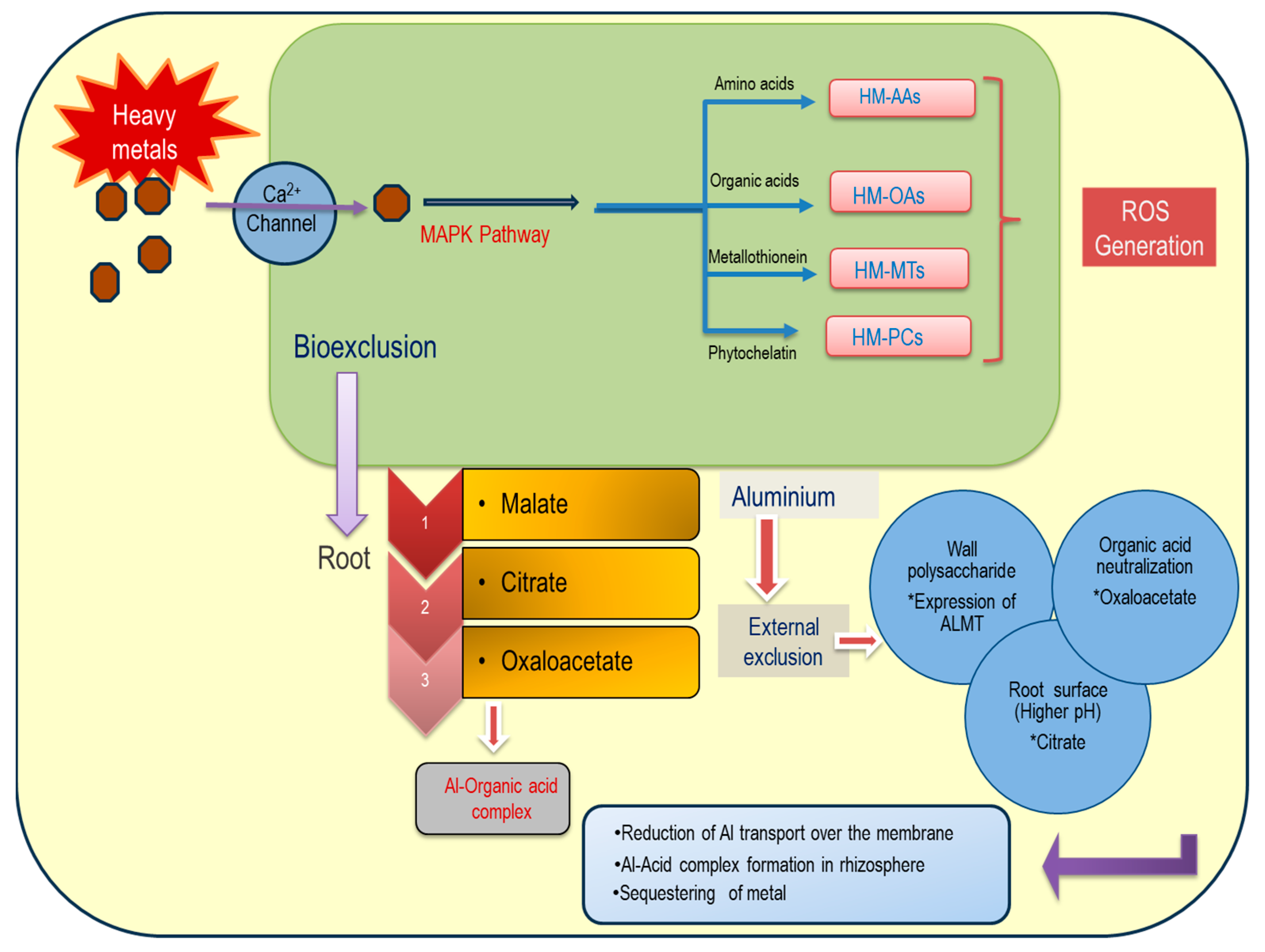
The response to Al toxicity in plants involves two primary pathways: exclusion and oxidative tolerance [40]. Exclusion mechanisms involve several channel proteins on cell membranes and tonoplasts, including anion transporters (like malate/hydroxide ion), that facilitate Al sequestration into vacuoles or apoplastic spaces [41]. The solubility of various organic acid anions like citrate, lactate, oxalate, and succinate in acidic environments also plays a role [42]. Carbohydrate biosynthesis, particularly for cell wall components such as pectin, acidic heteropolysaccharides, D-galacturonic acid, and glycoside-containing residues like rhamnose and galactose, is crucial. Pectin, a prevalent residue for Al3+ chelation, is recycled from primary metabolites. Monosaccharides like galactose and cellobiose bind Al3+ on the cell wall, while lignin, among the heteropolysaccharides, is pivotal for covalent Al binding, influencing metal compartmentalization [43]. Al-tolerant plant genotypes exhibit enhanced lignin biosynthesis, supporting anatomical adaptations like vascular lumen thickening under metal stress [44]. Lignin and phenolics like coumarins, derived from the phenylpropanoid or shikimic acid pool, are significant [45]. Key enzymes overexpressed under Al toxicity include phenylalanine ammonia lyase (PAL) and coumarate CoA ligase [46], especially in rice cultivars. In plants, a multistep bio-exclusion of Al3+ from root cells is understood specifically for tolerant species where ions are entered through the symplast of tissues crossing the cell wall (Figure 4).
Al tolerance also involves malate transporters (AlMTs) in wheat and Arabidopsis, essential for Al exclusion [47]. Increased cytoplasmic Ca2+ is another aspect of Al tolerance, affecting cell division inhibition in roots [48]. In wheat root apices, Al toxicity inhibits phospholipase C (PLC), affecting downstream phosphatidyl inositol 4, 5 bisphosphate (PIP2) signaling. PIP2 and its associated genes, along with actin proteins, play roles in Ca2+ signaling under Al stress [49]. The source of Ca2+ in Al toxicity is still uncertain, but it influences downstream metabolism and plant sensitivity [50].
Al toxicity impacts secondary metabolite metabolism, particularly in cell walls and phloem tissue [51]. Genes for transporters like glucosyl transferase and antioxidative secondary metabolites like hydroxy ferulic acids have been identified [52]. Hormonal biosynthesis genes and metal chelators are also implicated in various secondary metabolite pathways [53]. Metabolites overexpressed under Al toxicity include carotenoid and indole alkaloid biosynthesis and pinene degradation, indicating distinct mechanisms for Al exclusion and tolerance. Adenosine triphosphate binding box (ABC) proteins play roles in energy-dependent Al transport and ATP hydrolysis [54]. Al toxicity also correlates with increased lipid and copper transporters, affecting ROS homeostasis and gene expression related to malondialdehyde and protein carbonyl content [55].

5. Root Phenotypes and Quantitative Trait Loci for Aluminum Toxicity

Soil acidification, particularly from ammonium fertilizer use, has been a major issue in recent decades. This is especially true in areas with inorganic fertilizers and lowland rice fallows [56]. Studies show that excess Al3+ in soil disrupts root growth, primarily by hindering microtubule formation in root hairs. Therefore, tolerant cultivars should possess several quantitative trait loci (QTLs) that enable the sensing of soil acidity, the induction of receptor proteins on cell membranes, the perception of Al-induced redox changes, and the amplification of antioxidation pathways [57]. Major QTLs related to Al toxicity might also be linked to water or other metal stresses, often involving channel proteins, gateway proteins, and other transmembrane domains [58].
Other vital QTLs include transcription factors for genes of cell wall modifications, such as pectin methyl esterase. These genes, upregulated within six hours, help form stable complexes with Al. Their enzyme activity also relates to the metal’s adsorption capacity, suggesting bio-sequestering [59]. Al stress remodels cell wall structures, involving sugar residues and special QTLs with glucosyl transferase on the cell wall, impacting not only glucose but also galactose, glucuronic acid, and xylose modifications [60,61].
Recent developments have introduced strategies to modulate complex interactions of environmental stresses, including the use of bio-stimulants to modify root growth under Al toxicity [62]. For instance, pyroligneous acid, a common bio-stimulant, can reduce Al-induced root growth inhibition, enhancing resilience to metal toxicity and restoring yield [63]. This compound, derived from carbonated low-oxygen conditions, is a mix of bioactive water-soluble moieties, such as sugar and alcohol derivatives, and phenolics and organic acids.
In crops like Triticum, certain QTLs, like those for anaerobic carbohydrate metabolism (alcohol dehydrogenase, lactate dehydrogenase, pyruvate decarboxylase), have been identified for Al toxicity resistance [64]. Bio-stimulants can also enhance antioxidative enzymes like peroxidases and promote transcription factors (auxin response factor) in seedlings pre-treated for Al toxicity. These QTLs link carbon concentration mechanisms to Al toxicity, focusing on the development of primary metabolites like organic acids and glucosyl residues [65]. Al tolerance also benefits plants through improved photosynthetic efficiency, as well as the acquisition of reserve carbohydrates, particularly in C4 plants.

6. Metabolite Shifting from Central Carbon Metabolism

Plants have adapted cellular and metabolic strategies to mitigate Al toxicity. Typically, the citric acid pool and other organic acids form complexes with Al, aiding in its exclusion [66]. This mechanism involves slow Al entry into root cells, followed by acid secretion, acid ionization, and the formation of chelate compounds with Al at the center [67]. In the rhizosphere, plants secrete malic acids and siderophores to chelate metals in extracellular spaces [68]. Tolerant cultivars are capable of bio-exclusion of intracellular Al by forming non-toxic aluminum–chlorohydrate (Al-COOH) complexes. This also complements enhanced carbon concentration mechanisms.
In both C4 and C3 species, stable carboxylated products like 3-phosphoglyceraldehyde are crucial for Al interaction [69]. Central carbon metabolism supplies carbon intermediates for osmolytes, antioxidants, and other secondary metabolites aiding in Al tolerance [70]. The Calvin cycle, glycolysis, hexose monophosphate shunt, and the citric acid cycle contribute to respiratory substrates, growth, and yield under stress, requiring substantial adenosine triphosphate (ATP) consumption [71]. Studies indicate metabolic shifts from primary metabolism to transitory pathways with specialized metabolites, particularly shikimic acid pathway-derived flavonoids in roots [72].
Mitochondrial metabolism of pyruvate, involving oxidative decarboxylation and oxidative phosphorylation, is enhanced for ATP biosynthesis in response to Al influx in roots. Glycolytic and other anaplerotic reactions produce pyruvate, influencing downstream organic acid pathways under the control of respiratory flux in mitochondria. These acids, particularly malic and citric, can lower intracellular and rhizospheric pH, reducing Al solubility [73]. Enhanced photosynthetic activity in Al-tolerant cultivars is linked to these downstream residues from the central carbon pool [74].
Identifying specific root metabolomes and shifts in other reactions are essential for sustaining photosynthesis and respiration in tolerant species to Al. Recent advancements in omics, especially metabolomics as well as proteomics, offer identifications of complex reaction webs under Al stress [75]. Analyzing qualitative and quantitative aspects of Al-induced metabolomes helps in understanding signal transduction and genome alteration, with rice varieties serving as effective models for studying root-based metabolite changes [76]. Both constitutive (ALMT families) and inductive (MATE families) genes for metabolomes play significant roles in Al tolerance, whether through exclusion or chemical detoxification.

7. Aluminum-Induced Signaling for Reactive Oxygen Species Development

Al toxicity is most severe in its soluble form, particularly as free Al3+, which is prevalent in acidic environments as aluminum hydroxide ions [Al(OH)+ and Al(OH)2+] [77]. Al-induced phytotoxicity primarily starts from specific ion effects, leading to ROS generation and various cellular functions. ROS generation often results in programmed cell death (PCD) characterized by caspase-like activation, chromatin condensation, nuclear dehydration, and chromatin fragmentation [78]. Al tolerance in plants involves successful regulation of cellular redox, enabling root growth even with significant metal accumulation [79]. This tolerance is a genotypic trait linked to antiapoptotic processes that balance cellular redox.
In mitochondria, tolerant species regulate mitochondria-dependent PCD, including cytochrome c release and caspase protease activation [80]. ROS generation in plants under Al stress is a key factor in oxidative damage, similar to that observed under biotic and abiotic stress [81]. Al toxicity triggers oxidative bursts in chloroplast and mitochondrial compartments, disrupting normal redox reactions [82]. Although Al is not a transition metal and does not catalyze redox reactions like Fenton’s reaction, it still induces ROS formation in mitochondria. This is facilitated through processes like the univalent reduction of O2 [83]. Subsequent reactions for the reduction of superoxide into H2O2 are cytotoxic, but also play a dual role in signaling for acclimation and tolerance, yet at low concentrations. Thus, higher H2O2 concentrations accelerate PCD and senescence.
The relationship between Al toxicity, ROS bursts, and antioxidative enzyme synthesis is crucial for understanding plant survival under oxidative stress [84]. This understanding can help mitigate lipid peroxidation in cellular membranes and compartments. Current research focuses on the correlation between Al toxicity and plant survival in acidic soils, where Al3+ bioaccumulation is a concern. Agronomic measures to neutralize soil acidity, such as the use of amending chemical residues, are essential for managing this issue.

8. Comprehensive Genomics for Aluminum Toxicity

Al tolerance in different species, including Oryza sativa L., which is more tolerant than other cereals like maize, rye, sorghum, and wheat, involves varied gene expressions in roots against the metal [20]. This includes specific Al-responsive gene expressions involving transcription factors (TFs), particularly basic leucine zipper (bZip) and regulatory enzymes like protein kinases [85]. Al toxicity is also understood to involve post-translational modifications such as phosphorylation and adenylation of TFs, which are responsible for gene functioning [86]. This mechanism includes the repression, activation, and co-regulation of genes related to metal tolerance.
Al signaling involves different chemical species interactions with root proteomes, leading to changes in gene expression and root activities [87]. Molecular studies in rice have identified and cloned several QTLs like signal transduction and activation of RNA 1, Nramp family 1, Oryza sativa acetolactate synthase 1, and Oryza sativa magnesium transporter 1 (STAR2, Nrat1, OsAlS1, and OsMGT1), which are significant in the context of Al ion (Al3+) tolerance [88]. These genes are involved in the generation of superoxides, free radicals, and other ROS-producing genes.
In Arabidopsis, salicylic acid-induced nicotinamide adenine dinucleotide phosphate hydrogen [NADP(H)] oxidase reactions are common for specific ROS production, regulating Al toxicity metabolomes [89]. Al3+ and other ions initially induce nitrogen oxidase (NOX) activity, linked to superoxide production. Two pore segment channel 1 (TPC1) channel protein activation on the membrane increases Ca2+ influx [90]. This influx triggers salicylic acid synthesis and accumulation, which in turn boosts NADP(H) production in root tissues, affecting cellular redox [91].
Vacuole-mediated cell death, a genome-responsive pathway for Al toxicity, is another mechanism, differing from mitochondria-associated cellular death where redox-responsive genes are crucial [92,93]. This process, regulated by proteolytic genes, leads to vacuole collapse and membrane disruption in roots, especially under Al exposure [94]. Protease activity, NADP(H) oxidase, and TPC1 activation are considered key paths for Al resistance [95].
Al phytotoxicity also involves vacuolar membrane lysis, leading to vacuole collapse. In tissues with excessive Al, caspase-like activities trigger vacuolar processing enzyme (VPE) functioning, disrupting cell membranes. VPE functions are also linked to root growth sensitivity, particularly in meristematic regions sensitive to Al toxicity. Numerous genes including VPE1 QTLs are upregulated in response to Al toxicity, indicating their involvement in cell death events [96].

9. Special Metabolomic Pathways to Register Aluminum Toxicity

Mitochondrial pathways in response to Al toxicity involve a specific branch point at the ubiquinone (ubQ) site, triggering alternative pathways. The upregulation of genes related to the vacuolar processing enzyme 1 (VPE1) QTL has been linked to cell death events under Al toxicity. Alternative oxidase (AOX) diverts electrons from the main transport chain to reduce molecular oxygen without energy production, thereby reducing reactive oxygen species (ROS) burden under Al toxicity [97]. Excessive Al causes complex II and III to be bypassed, leading to a reduced ubQ pool, indicating oxidative redox activity [98].
Under high Al toxicity, there is a significant increase in alternative oxidase (AOX) transcripts, regulated both transcriptionally and translationally. In non-stressed plants, both transcript abundance and enzyme activity of AOX are low. Al accumulation downregulates root respiration through complexes III and IV, diminishes succinate-dependent electron transfer, and reduces cytochrome oxidase activity [99]. The results of increased ROS and free radicals can consume energy to reduce ROS with the participation of nicotinamide adenine dinucleotide phosphate NADP(H), flavin adenine dinucleotide (FADH2), and other reducing equivalents. Plant response to Al is finally accomplished at the nuclear level where a number of gene expressions ensure various intermediate products that influence the cellular phenomena either singly or in combination (Figure 5).
Differential gene expression shows that major TCA cycle enzymes like succinate dehydrogenase, alpha-ketoglutarate dehydrogenase, and malate synthase are targeted to generate NADP(H), bypassing complexes III and IV to AOX [100]. Al-treated tobacco roots experience a significant increase in ROS and a consequent decline in ATP synthesis [101]. This suggests that Al-induced inhibition of respiration downregulates mitochondrial ATP production, affects the bioenergetics status of tissues, and potentially induces the signaling of programmed cell death (PCD) in roots. This effect may extend to shoot tissues, where Al toxicity leads to early senescence and foliage abscission [102].
Furthermore, the mitochondrial genome under Al toxicity overexpresses special proteins on the outer membrane. Excess Al toxicity opens mitochondrial transition pores, leading to the leakage of matrix-based proteins, loss of matrix potential (ΔΨm), and impaired ATP generation [103]. Al toxicity stimulates these pores, releasing cytochrome c oxidase proteins, ultimately triggering PCD [104]. Oxidative stress, therefore, is linked to mitochondrial metabolomics, potentially driven by elevated Ca2+ concentration and increased ROS, leading to further peroxidation reactions. Mitochondrial permeability transition, influenced by Ca2+ under Al toxicity, involves specific membrane proteins in the process of cell death.

10. Special Metabolites and Their Contribution to Al Tolerance in Plants

Tobacco cells exposed to Al toxicity accumulate caffeic acid and chlorogenic acids via the phenylpropanoid pathway. A key enzyme in this process is L-phenylalanine ammonia-lyase (PAL), whose activity is linked to phosphate availability in plant tissues. Aluminum toxicity induces phosphate deprivation, increasing PAL activity [105]. The phenolics produced by this pathway act as antioxidants, protecting against lipid and protein peroxidation and macromolecule carbonylation. When exposed to a combination of Al3+ and Fe2+, plant cells activate these antioxidants, indicating tolerance against this joint combination of metals. This suggests that the combined effects of metals are mediated by the induction of phenolic antioxidants like phenylpropanoid residues and supersedes the individual effects of ions [106].
Organic acids from the TCA cycle are other metabolites reducing rhizotoxicity in Al-contaminated soil. Al ions (Al3+), prevalent in acidic soils, induce proton (H+) secretion in some sensitive species. The acidic pH, with its high H+ concentration, enhances solubilization in the soil, mitigating phytotoxic impacts. A lower pH can also reduce the negative charge of biological membranes, decreasing Al binding affinity [107].
In Arabidopsis, citrate induction is crucial for the expression of the transcription factor Arabidopsis thaliana SENSITIVE TO PROTON RHIXOTOXICITY 1 gene (AtSTOP1), associated with specific H+ transporters. Mutations in STOP1 can reduce Al tolerance due to inadequate citrate production, indicating AtSTOP1’s role in activating other genes, including those in antioxidative cascades, for co-tolerance against Al3+.
Other metabolites include methylated residues formed during reactions to Al toxicity, contributing to H+ de-acidification. A protein similar to glycerophosphodiesterase (GPD) is overexpressed at the transcript level in tobacco soon after Al exposure, which is necessary for the methylation of CCGG islands in the promoter regions. This methylation pattern changes with Al exposure, similar to the response to herbicide-induced ROS [108]. Since ROS are common in metal induction, demethylation may occur through the development of oxygen radicals. Cytosine residue methylation decreases upon Al3+ application in roots, indicating that metal induction can cause total DNA methylation, potentially leading to genotoxicity.

11. Metabolomes under Regulation of Signal Transduction and Protein Turnover

Several metabolic residues are believed to be involved in signaling and perception pathways through the cell membrane during Al transportation. Specific guanosine triphosphate (GTP) binding proteins, isolated from cell membranes, are known to function in ion channel opening, cell volume proliferation, and energy-mediated processes [109]. Various species, including cereals and Arabidopsis, show changes in mass proteome expression in roots exposed to Al, indicating alterations of global gene expression related to antioxidation. Vacuolar hydrogen ion ATPase (H+/ATPase) activity in roots is suggested to create an electrochemical gradient for Al3+ activation through a proton antiporter system [110]. In Al-tolerant Triticum species, a tonoplast ATPase protein is overexpressed, linked to metal absorption, though reports vary on ATPase expression levels across different crop species irrespective of Al sensitivity and tolerance. This suggests that Al compartmentalization is an avoidance strategy, with plants variably responding to H+/ATPase proteome expression for exclusion [111].
Mitochondrial ATPase activity, isolated from mitochondrial inter-membrane space, varies with protein subunit expression. For instance, soybean shows reduced membrane-bound ATPase activity but increased mitochondrial ATPase under Al toxicity. Parallel expression of organic acid-secreting proteins and P-type ATPase suggests dual roles in sequestering Al in vacuoles or apoplastic spaces following esterification. In legumes like soybean, an increase in citrate synthase activity from the TCA cycle correlates with co-transport by ATPase-driven activity.
Homeo-domain proteins from the MCM1, AGAMOUS, DEFICIENS and SRF-Box (MADS-Box) family in soybean have distinct DNA binding domains for regulatory factors in Al tolerance genes [112]. Metabolomes contributing anaplerotic reactions with citrate, malate, and oxaloacetate are crucial for chelating metals in apoplastic spaces. In some rice cultivars, tolerance to Al is linked to overexpression of MADS-Box factors, enhancing expression of vacuolar processing enzyme (VPE) genes and H+ extrusion [113].
Protein regulation through lysis is also significant in Al toxicity, where root growth retardation is linked to protein lysis [114]. In rice and other crops, the induction of proteasomes and heat shock proteins (HSPs) occurs under varying Al3+ concentrations where HSPs are involved in protein folding, transport, stress resistance, and gene expression [115]. This suggests the necessity of protein post-translational modification under Al toxicity. The induction of HSP70 and its derivatives might be a key factor in Al tolerance, with protease activity indicating tolerance through the turnover of degenerative proteins. In tolerant cultivars, protease-mediated lysis of misfolded proteins may alleviate degradation from root membrane lysis and growth inhibition [116].

12. Transcriptional Control of Aluminum Tolerance in Roots under Acidic Cytosol

An acidic environment is the most favorable key to Al sensitivity in root physiology, where a number of gene activations are directly or indirectly involved through transcription factors. Transcription factors have been known of for the last few decades with regard to plant responses to different stimuli. Factors like ADP-ribosyltransferase 1 (ART1) in rice are most important for Al tolerance, as already mentioned earlier [117]. Now, there is a quest to identify a few interacting proteins with ART1. Already, nine of the downstream genes induced by ART1 factors have been functionally characterized for Al tolerance exclusively in rice [118]. Moreover, tolerant species to Al have also been scanned for a few QTLs and used as molecular markers. Still, the fine mapping and cloning of these QTLs are yet to be deciphered for breeding. Acidic soil is a major problem for cultivated crops in which transcriptome-wide association studies combined with genome-wide association studies are required to identify the direct involvement of gene versus trait association. In addition, for the well-characterized ART1-regulated gene, the membrane localized polypeptides of 53 amino acid residues are identified [109]. The gene C-terminal domain phosphatase (CTD3) is essentially metal-specific and exhibits no transport ability for Al but directly binds to Al in roots [119]. Moreover, CTD3 from rice (OsCTD3) is specifically required with a strong promoter for its expression on membranes to detoxify Al. External detoxification in roots for Al also involves a lot of acid secretion in the rhizosphere rather than internal secretion of the metal. In a number of cases, biomolecules like pectin, hemicelluloses, and other polysaccharides on the root symplast, as well as endodermal casparian strips, are required for sequestration of the metal [120].
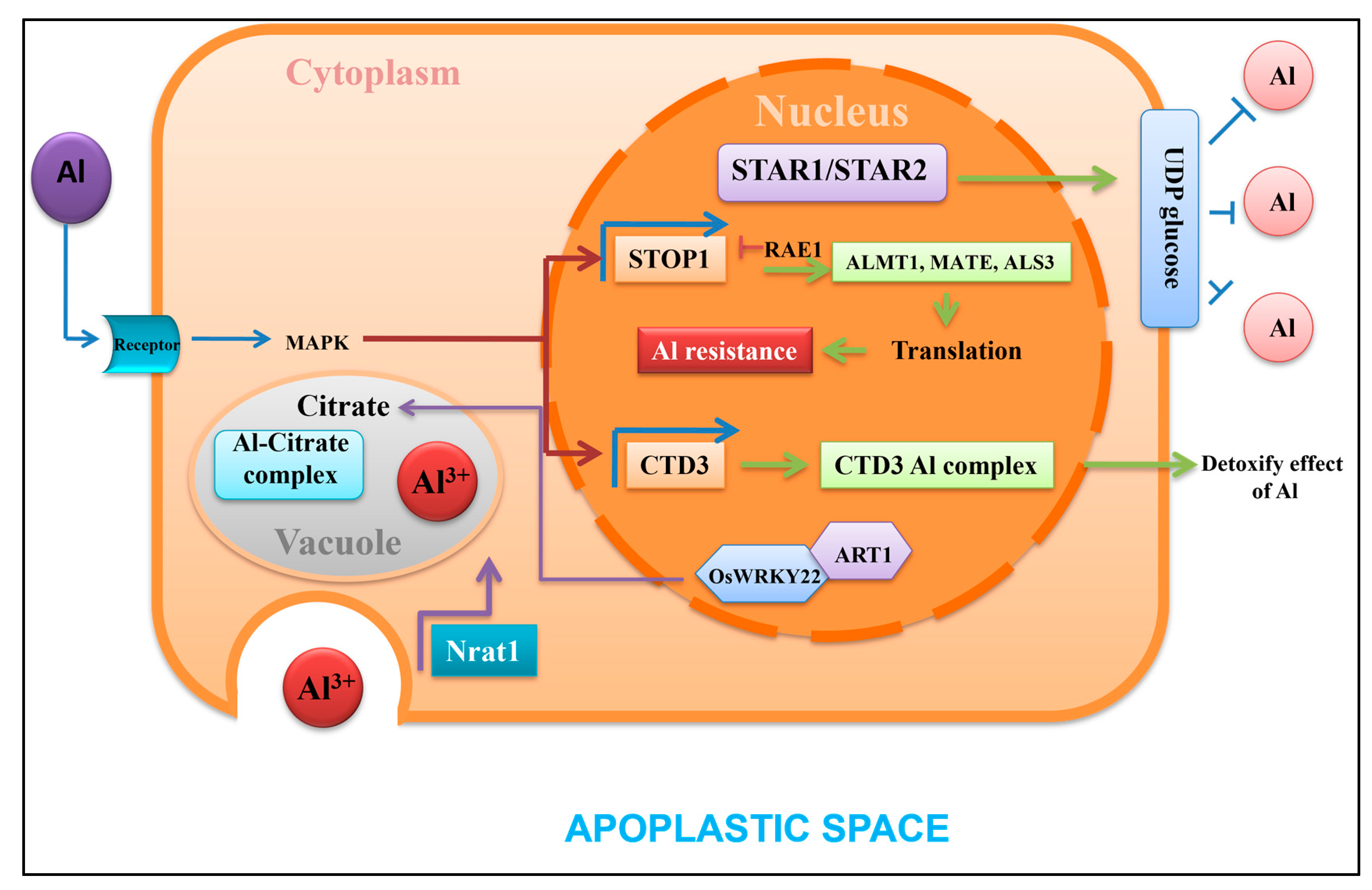
The modality of gene regulation with transcription factors through phosphorylation–dephosphorylation and adenylation–deadenylation is important for cell signaling with Al. In acidic soil, a C2H2 zinc-finger protein homologue to SENSITIVE TO PROTON RHIXOTOXICITY 1 (STOP1), as found in Arabidopsis, can regulate malate transporters Arabidopsis thaliana multidrug and toxic compound extrusion 1 and Arabidopsis thaliana aluminum activated malate transporter 1 genes (AtMATE1, AtALMT1). These two factors can bind the basic upstream elements [GGN(T/g/a/C)V(C/A/g)S(C/G)] where the downstream genes would be malate, citrate, oxalate, and other organic acids [121]. This promoter can control a minimum of 30 genes covering expansin A10, magnesium transporter 1, signal transduction and activation of RNA 1, and ferric reductase defective like 4/2, (EXPA10, MGT1, STAR1/2, FRDL4, FRDL2), expressed in extracellular or intracellular spaces [122]. The specific ATP-binding cassettes and membrane binding domain, similar to E. coli ABC transporters, highlights that the two basic transcription factors (STAR1-2) of the ALMT1 gene are more characterized. The homeodomain complex STAR1-STAR2, localized at the membrane, transports pyrimidine nucleotide (UDP-glucose), which is required for the modification of the cell wall and callose [123]. Transcription factors like FRDL4 are required for the secretion of organic acids under Al toxicity in roots for internal detoxification. These factors include (cys-cys)n, a homologue peptide that can directly bind with Al and reduce Al detoxification, although externally. On the other hand, magnesium transport and upregulation of MGT1 can reduce internal Al concentration, superseding other metals [124]. For the vacuolar sequestration of Al, other members of Nramp gene family 1 (Nrat1) are important, specifically for aluminum ions (Al3+). For the same species, a tonoplast-bound ABC transporter is recognized from rice, Oryza sativa acetolactate synthase 1 (OsALS1), which takes part in the activation of promoters under low pH. There exists a strong correlation for Al tolerance with the expression of OsALS1 and OsFRDL4-like factors in roots under acidic pH of soil [125]. Along with the normal ability in organic acid transportation, genes encoding expansion-like proteins for cell wall elongation in root tips are dependent on the activation of transcription factors. From the most sensitive cereals to Al (e.g., rice), another few transcription factors like ART2 are also important to interact with other factors (Figure 6). Likewise, wrinkled transcription factor (WRKY) domains, which are characterized by a zinc-finger structure (either Cx4-5Cx22-23HxH or Cx7Cx23HxC) on the upstream sequence containing W-BOX promoter, are also applicable for Al sensitivity [126]. Many factors against Al toxicity like STAR1/2 can interact with WRKY in a dimer configuration, where the cis-element is represented with a consensus sequence of TTGACC/T [127]. The expression of OsFRDL4 is controlled by OsWRKY22, where the localization of expression is the plasma membrane, nucleus, tonoplast, etc. Sometimes, co-expression of OsWRKY22 and ART1 is found in dimer configuration within the nucleus for citrate secretion. A few factors like abscisic acid stress and ripening (ASR) genes are identified in response to wider activation of Al-tolerant genes. These ASR factors mostly act as chaperones and transcription factors and are amenable to the ASR5 gene, involved in Al tolerance in rice [128]. ASR5 is nucleus- and cytoplasm-specific in expression and regulates the co-expression of different genes from Al-induced stress tolerance. In more recent versions of genome-wide arrays of activating genes, the ASR5 trans-factor is shown to bind the STAR1 promoter and, downstream, can also regulate Nrat1. Also from rice, ALS1 encodes an ABC transporter belonging to transporter-associated antigen processing protein (TAP), which can be induced only by Al3+ but not other metals at low pH [129]. These are further supported by the expression of transcription factors for the genes expressed in rice—Oryza sativa pectin methylesterase (OsPME)—required for pectin methyl esterase in Al detoxification for apoplastic spaces. Conclusively, transcriptomes for Al tolerance are mostly based on the sensitization of transcription factors where proteins, either singly or in combination, can regulate the expression where molecular mechanisms are concerned.

13. Phytohormone Signaling and Functioning for Aluminum Tolerance

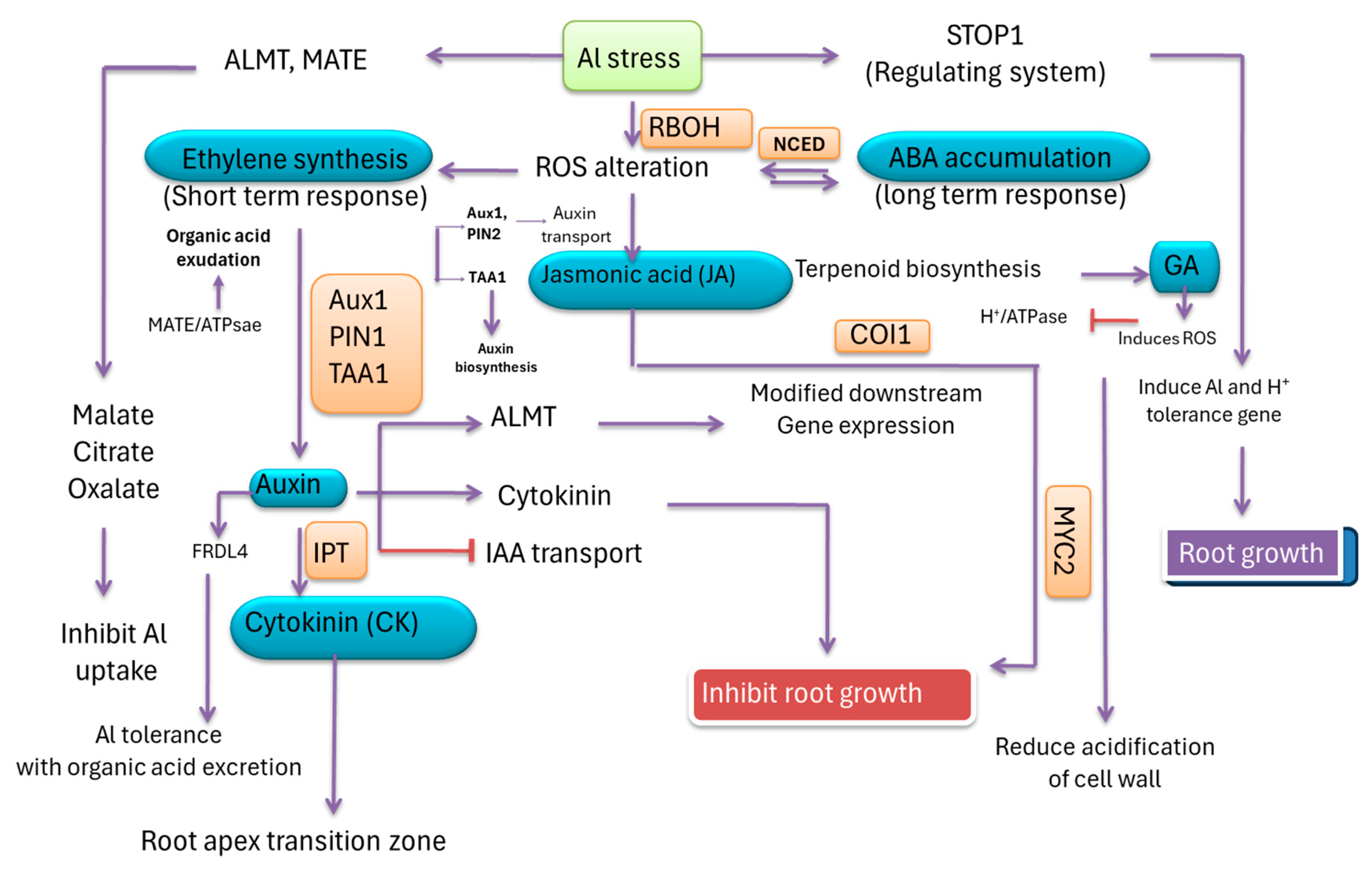
Al undoubtedly invites metal stress and is also coincided by plant hormones including auxin (Aux), gibberellins (GA), ethylene (ET), abscisic acid (ABA), jasmonic acid (JA), and cytokinin (CK), as well as other secondary elicitors like nitric oxide [130]. External applications of plant growth regulators and their synthetic analogs, in most cases, record similar effects. Rice under acidic soil shows a strong affinity for Al3+ sensitivity and thereby induces specific transcription factors like ADP-Ribosyltransferase 1 (ART1), a C2H2 zinc-finger motif. This factor specifically plays a role in the regulation of 31 downstream genes under Aux control [131]. The genes include rice aluminum transporters like nicotinate riboside transporter 1/Oryza sativa nicotinate riboside transporter 4 (NRAT1/OsNRAMP4), which are characteristically Al tolerance genes where Aux metabolism is involved. Since root growth is the most vulnerable to Al toxicity due to inhibition, changes in Aux synthesis and translocation may be important, as recorded in Brassica [132]. Aux signaling, with its principle function being stress-induced root growth inhibition, has demonstrated the expression of Aux transporter proteins like Oryza sativa pin formed 3 (OsPIN3t). This transporter activity is also observed with the reduction in adventitious growth in Arabidopsis. The toxicity of Al inhibits the expression of the pin formed 2 (PIN2) gene encoding protein for auxin transport towards the roots. Downregulation of its expression can reduce endogenous Aux concentrations in roots, and thereby, sensitivity to Al is more marked, impeding root growth [133]. Other transporter genes for Aux, including PIN2, ferric reductase defective like 4 (FRDL4), and auxin resistant 1 (AUX1), are also proposed to correlate with ethylene production. ET undoubtedly happens to be a good signaling residue, resulting in impeded root growth under Al toxicity [134]. ET has a direct relationship with Al3+ stress, where RBOH-mediated ROS alteration can induce its synthesis. ET is also a direct signaling residue for auxin biosynthesis, where genes like Aux1, PIN1, and tryptophan aminotransferase related 1 (TAA1) are involved in root growth inhibition under Al stress [135]. ABA is typically recognized as a stress hormone, also cloned from rice, where a few homologues of the abscisic acid-stress-ripening (ASR) family are demonstrated under ABA and Al3+ stress simultaneously. The increase in synthesized ABA is directly related to cellular signaling, where H2O2 is the most important ROS. The signaling cascade under Al stress, as perceived through cell membrane-bound receptors, is also co-induced with 9-cis-epoxycarotinoid dioxygenase (NCED). This gene encodes the protein that splits the carotenoids for ABA biosynthesis. Higher expression of NCED accumulates intracellular ABA in support of osmotic adjustment under Al3+-stressed crops [136]. ABA synthesis in Al-treated root cells can induce ATP-binding cassette transporter (ABC) and nitrate transporter1/peptide transporter (NPF) for its translocation in different organs. Subsequently, ABA serves as a stimulus for the resistance mechanism against Al toxicity by increasing water use efficiency and decreasing transpiration rates [137]. Additionally, ABA accumulation in some root tissues induces the formation of a protein complex via receptors like pyrabactin resistance 1/pyrabactin resistance-like (PYR/PYL) and protein phosphatase 2C (PP2C). This complex induces the activation of some protein kinases which otherwise activate by the phosphorylation of specific anion channels to release malate against Al accumulation [138]. This family encodes specific transcription factors such as amino-terminal enhancer of split-related 5 (OsAsR5), responsible for the co-expression of signal transduction and activation of RNA 1 (START1) in the Nramp family of factors [139]. Another gene, FRDL4, has also been cloned from roots sensitive to Al3+, where the tolerance to the metal is manifested by the development of ROS. The ROS would otherwise be an inducer for different genes related to Al tolerance, more specifically the synthesis of organic acids. The organic acids in turn are also induced by some growth regulators like ethylene under Al stress [140]. ET in wheat has also been reported with negative control for Al activated malate transporter (ALMT1) that reduces the root secretion of organic acids. Thereby, mutation of this gene would be sufficient for sensitivity to Al toxicity [141]. ET has also been reported for Al tolerance, yet in crosstalk with Aux interference. The expression of ethylene biosynthetic genes like 1-aminocyclopropane-1-carboxylic acid synthase (ACSs) and 1-aminocyclopropane-1-carboxylic acid oxidase (ACOs) induces a direct promotion of ethylene biosynthesis under Al3+ toxicity in sensitive cultivars [142]. On signaling pathways, ET activates the expression of transcription factors like ethylene insensitive 3 and 1 (EIN3 and EIN1), which otherwise control root growth. EIN3 has a good binding ability to the promoters of a few genes (YUCCA 9, YUC9) to interfere with the growth of the root apex transition zone under Al toxicity [143]. The toxicity of Al in root sensitization finds the expression of a few genes (TAA1, YUCs [YUC3/5/7/8/9]) for transcription factors in the regulation of ethylene biosynthesis in contrast to Aux. Therefore, the toxicity of Al in root growth moderation would be a supplementary effect, where Aux and ET expression in effected tissues is simultaneous. ET is also responsible for the regulation of both local Aux biosynthesis and Aux transport by the induction of genes like TAA1/YUCs and PIN2, and AUX1, respectively [144]. The distinct action of ET in the reversal of Aux sensitivity is variable through plant species when exposed to Al3+; still, their molecular mechanisms are obscure. GAs are implicated in metal tolerance through the preservation of carbohydrate metabolism and vegetative growth. In that context, Al3+-induced inhibitions of root elongation following sugar translocation to roots are also reported as possible pathways for metal tolerance. Still, downregulation of Al-induced ROS accumulation and changes in root cell wall deposition may be apprehended as membrane-localized Al sequestering [145]. The function of GAs with multiple attributes may collectively include the biosynthesis of bioactive GAs in tomatoes, improving growth and pigment content, as well as the CO2 fixation rate in soybean under metal stress [146]. CT is less explored under Al toxicity but is also related to cell wall alkalization, an influx of H+ over the membrane, and, in a few cases, alteration of expression for cell wall modifying genes. CT biosynthesis is directly influenced by Aux mediation, where transcription factors like transport inhibitor response 1 (TIR1) and auxin signaling f-box (AFB) are responsible for encoding other factors on upstream binding sequences. These factors most importantly include auxin response factors (ARFs) 7–19, which are otherwise involved in isopentenyl pyrophosphate (IPT) residue biosynthesis as a precursor of CT [147]. Growth substances like JA also play a differential role against Al toxicity. JA application in Al-sensitive root species induces adaptive responses in plants by the regulation of ROS metabolism (Figure 7). Two genes, coronatine insensitive 1 (COI1) and myeloblastosis viral oncogene homologue 2 (MYC2), are found to be overexpressed in root growth inhibition by altering the ET concentration. Phenotypic analysis with mutants insensitive to JA biosynthesis and signaling was recorded in response to Al3+ with direct involvement for ET accumulation. The inhibition of formation and depolymerization of cortical microtubules in those mutants are responsible for COI1 gene expression under Al stress [148]. Moreover, malate transporter expression by aluminum activated malate transporter (ALMT) is also dependent on the regulation of JA signaling where COI1 is involved. Therefore, phytohormones, a crucial signaling residue in favor of Al tolerance, play pivotal roles, mostly in metal sequestering, ion complex formation, water balance, and ROS homeostasis.

14. Conclusions and Future Perspectives

This discussion highlights that Al toxicity impacts plants in two main phases: the establishment of metal-induced specific ion effects and the development of an oxidizing redox environment prone to peroxidation reactions. Initially, physiological and cellular events are affected, including reduced root membrane permeability, increased dehydration, vacuolar disintegration, nutrient release over the tonoplast membrane, cytosolic pH alterations, caspase activity acceleration leading to PCD, and dissolution of cellular metabolites into soluble products. Conversely, oxidative redox changes can lead to biomolecule degradation, including lipid peroxidation, protein carbonylation, and demethylation of specific coding regions or gene promoters. Altered metabolite fluxes, particularly in organic acid turnover, can enhance Al toxicity tolerance. Another key tolerance mechanism is the pattern change in secondary metabolites, especially the development of complex polysaccharides like callose, preferred in selective cultivars. This development is also linked to alternative oxidase (AOX) expression and ATPase activities, which help prevent electron misfiring from cellular organelles and stimulate energy-producing pathways. Additionally, specialized transcriptional regulation by MCM1, AGAMOUS, DEFICIENS, and SRF-Box (MADS Box) proteins and other transcription factors is crucial for activating specific genes under Al toxicity. This process is complemented by protein degradation mechanisms that impede root growth, countered by the expression of heat shock proteins (HSPs) or chaperones, which aid in refolding misfolded proteins. Therefore, Al tolerance is a multifaceted process involving both enzymatic antioxidation cascades and bio-exclusion of the metal through diverse gene expressions. Future prospects include profiling different proteomes in Al-tolerant species to understand and manage various pathways under Al stress. For instance, examining proteomes involved in cysteine and methionine metabolism could shed light on methyl cycling and glutathione metabolism for antioxidation. Recent advances in proteomic sequencing are poised to provide deeper insights into precise protein expression and its role in Al tolerance. Over the past few decades, people have been on a quest to develop strategies for crops under Al contamination. Many aspects of Al cytotoxicity remain uncovered and could be targeted along with suitable screening methods for crop species that accumulate less Al. In sustainable breeding, further identification of specific QTLs governing Al tolerance should be the focus area. Along with the advent of physical technologies that are useful in environmental decontamination and the restoration of soil fertility, the development of Al-resilient crops would be a pathway for food security against acidic soil in countries in the Global South.

Author Contributions

Conceptualization, M.H. and M.K.A.; writing—original draft preparation, N.C., A.D., S.P., S.R. and S.K.S.; writing—review and editing, N.C., M.K.A. and M.H.; visualization, M.K.A. and M.H.; supervision, M.H. All authors have read and agreed to the published version of the manuscript.

Funding

N.C., A.D., S.P. and S.R. acknowledge financial support from a URS fellowship, UGC NET JRF, SVMCM, and a UGC NET JRF fellowship, respectively. M.K.A. acknowledges support from DST PURSE II, applicable to the University of Kalyani.

Data Availability Statement

All information is available in this article.

Acknowledgments

The authors acknowledge Rummana Samiha for her critical review of the draft and necessary formatting.

Conflicts of Interest

The authors declare no conflicts of interest.

References

  1. Tyagi, W.; Yumnam, J.S.; Sen, D.; Rai, M. Root transcriptome reveals efficient cell signaling and energy conservation key to aluminum toxicity tolerance in acidic soil adapted rice genotype. Sci. Rep. 2020, 10, 4580. [Google Scholar] [CrossRef] [PubMed]
  2. Banet, T.; Massey, M.S.; Zohar, I.; Litaor, M.I.; Ippolito, J.A. Assessing modified aluminum-based water treatment residuals as a plant-available phosphorus source. Chemosphere 2020, 247, 125949. [Google Scholar] [CrossRef] [PubMed]
  3. Abdelaal, K.; AlKahtani, M.; Attia, K.; Hafez, Y.; Király, L.; Künstler, A. The role of plant growth-promoting bacteria in alleviating the adverse effects of drought on plants. Biology 2021, 10, 520. [Google Scholar] [CrossRef] [PubMed]
  4. Kar, D.; Pradhan, A.A.; Datta, S. The role of solute transporters in aluminum toxicity and tolerance. Physiol. Plant. 2021, 171, 638–652. [Google Scholar] [CrossRef] [PubMed]
  5. Haghighi, T.M.; Saharkhiz, M.J.; Kavoosi, G.; Jowkar, A. Monitoring amino acid profile and protein quality of licorice (Glycyrrhiza glabra L.) under drought stress, silicon nutrition and mycorrhiza inoculation. Sci. Hortic. 2022, 295, 110808. [Google Scholar] [CrossRef]
  6. Rahman, R.; Upadhyaya, H. Aluminium toxicity and its tolerance in plant: A review. J. Plant. Biol. 2021, 64, 101–121. [Google Scholar] [CrossRef]
  7. Dhiman, S.; Khanna, K.; Kumar, P.; Bhardwaj, T.; Devi, K.; Sharma, N.; Sharma, P.; Arora, P.; Kapoor, N.; Sharma, A.; et al. Divulging molecular perspectives of plant defense machinery under heavy metal toxicity. J. Plant Growth Regul. 2023, 9, 1–37. [Google Scholar] [CrossRef]
  8. Ofoe, R.; Thomas, R.H.; Asiedu, S.K.; Wang-Pruski, G.; Fofana, B.; Abbey, L. Aluminum in plant: Benefits, toxicity and tolerance mechanisms. Front. Plant Sci. 2023, 13, 1085998. [Google Scholar] [CrossRef] [PubMed]
  9. Siqueira, J.A.; Silva, M.F.; Wakin, T.; Nunes-Nesi, A.; Araújo, W.L. Metabolic and DNA checkpoints for the enhancement of Al tolerance. J. Hazard. Mater. 2022, 430, 128366. [Google Scholar] [CrossRef]
  10. Zarattini, M.; Corso, M.; Kadowaki, M.A.; Monclaro, A.; Magri, S.; Milanese, I.; Jolivet, S.; de Godoy, M.O.; Hermans, C.; Fagard, M.; et al. LPMO-oxidized cellulose oligosaccharides evoke immunity in Arabidopsis conferring resistance towards necrotrophic fungus B. cinerea. CommunBiol 2021, 4, 727. [Google Scholar] [CrossRef]
  11. Zulfiqar, F.; Ashraf, M. Antioxidants as modulators of arsenic-induced oxidative stress tolerance in plants: An overview. J. Hazard. Mater. 2022, 427, 127891. [Google Scholar] [CrossRef] [PubMed]
  12. Che-Othman, M.H.; Jacoby, R.P.; Millar, A.H.; Taylor, N.L. Wheat mitochondrial respiration shifts from the tricarboxylic acid cycle to the GABA shunt under salt stress. New Phytol. 2020, 225, 1166–1180. [Google Scholar] [CrossRef] [PubMed]
  13. Szurman-Zubrzycka, M.; Chwiałkowska, K.; Niemira, M.; Kwaśniewski, M.; Nawrot, M.; Gajecka, M.; Larsen, P.B.; Szarejko, I. Aluminum or low pH–which is the bigger enemy of barley? transcriptome analysis of barley root meristem under Al and low pH stress. Front. Genet. 2021, 12, 675260. [Google Scholar] [CrossRef] [PubMed]
  14. Guan, K.; Yang, Z.; Zhan, M.; Zheng, M.; You, J.; Meng, X.; Li, H.; Gao, J. Two Sweet Sorghum (Sorghum bicolor L.) WRKY Transcription factors promote aluminum tolerance via the reduction in callose deposition. Int. J. Mol. Sci. 2023, 24, 10288. [Google Scholar] [CrossRef] [PubMed]
  15. Shetty, R.; Vidya, C.S.N.; Prakash, N.B.; Lux, A.; Vaculik, M. Aluminum toxicity in plants and its possible mitigation in acid soils by biochar: A review. Sci. Total Environ. 2021, 765, 142744. [Google Scholar] [CrossRef] [PubMed]
  16. Schaller, J.; Puppe, D.; Kaczorek, D.; Ellerbrock, R.; Sommer, M. Silicon cycling in soils revisited. Plants 2021, 10, 295. [Google Scholar] [CrossRef] [PubMed]
  17. Johnston, N.R.; Strobel, S.A. Principles of fluoride toxicity and the cellular response: A review. Arch. Toxicol. 2020, 94, 1051–1069. [Google Scholar] [CrossRef] [PubMed]
  18. Yadav, B.; Jogawat, A.; Lal, S.K.; Lakra, N.; Mehta, S.; Shabek, N.; Narayan, O.P. Plant mineral transport systems and the potential for crop improvement. Planta 2021, 253, 45. [Google Scholar] [CrossRef] [PubMed]
  19. Chauhan, D.K.; Yadav, V.; Vaculík, M.; Gassmann, W.; Pike, S.; Arif, N.; Tripathi, D.K. Aluminum toxicity and aluminum stress-induced physiological tolerance responses in higher plants. Crit. Rev. Biotechnol. 2021, 41, 715–730. [Google Scholar] [CrossRef] [PubMed]
  20. Garcia-Caparros, P.; De Filippis, L.; Gul, A.; Hasanuzzaman, M.; Ozturk, M.; Altay, V.; Lao, M.T. Oxidative stress and antioxidant metabolism under adverse environmental conditions: A review. Bot. Rev. 2021, 87, 421–466. [Google Scholar] [CrossRef]
  21. Dahiya, V. Heavy metal toxicity of drinking water: A silent killer. GSC Biol. Pharm. Sci. 2022, 19, 020–025. [Google Scholar] [CrossRef]
  22. Mansoor, S.; Wani, O.A.; Lone, J.K.; Manhas, S.; Kour, N.; Alam, P.; Ahmad, A.; Ahmad, P. Reactive oxygen species in plants: From source to sink. Antioxidants 2022, 11, 225. [Google Scholar] [CrossRef] [PubMed]
  23. Worthington, M.; Perez, J.G.; Mussurova, S.; Silva-Cordoba, A.; Castiblanco, V.; Cardoso Arango, J.A.; Jones, C.; Fernandez-Fuentes, N.; Skot, L.; Dyer, S.; et al. A new genome allows the identification of genes associated with natural variation in aluminium tolerance in Brachiaria grasses. J. Exp. Bot. 2021, 72, 302–319. [Google Scholar] [CrossRef] [PubMed]
  24. Jardim-Messeder, D.; Margis-Pinheiro, M.; Sachetto-Martins, G. Salicylic acid and adenine nucleotides regulate the electron transport system and ROS production in plant mitochondria. Biochim. Biophys. Acta. Bioenerg. 2022, 1863, 148559. [Google Scholar] [CrossRef] [PubMed]
  25. Vera-Villalobos, H.; Lunario-Delgado, L.; Pérez-Retamal, D.; Román, D.; Leiva, J.C.; Zamorano, P.; Wulff-Zottele, C. Sulfate nutrition improves short-term Al3+-stress tolerance in roots of Lolium perenne L. Plant. Physiol. Biochem. 2020, 148, 103–113. [Google Scholar] [CrossRef] [PubMed]
  26. Sarkar, B.; Bandyopadhyay, P.; Das, A.; Pal, S.; Hasanuzzaman, M.; Adak, M.K. Abscisic acid priming confers salt tolerance in maize seedlings by modulating osmotic adjustment, bond energies, ROS homeostasis, and organic acid metabolism. Plant Physiol. Biochem. 2023, 202, 107980. [Google Scholar] [CrossRef] [PubMed]
  27. Ahammed, G.J.; Li, Z.; Chen, J.; Dong, Y.; Qu, K.; Guo, T.; Li, X. Reactive oxygen species signaling in melatonin-mediated plant stress response. Plant Physiol. Biochem. 2024, 207, 108398. [Google Scholar] [CrossRef] [PubMed]
  28. Dourmap, C.; Roque, S.; Morin, A.; Caubrière, D.; Kerdiles, M.; Béguin, K.; Couée, I. Stress signalling dynamics of the mitochondrial electron transport chain and oxidative phosphorylation system in higher plants. Ann. Bot. 2020, 125, 721–736. [Google Scholar] [CrossRef] [PubMed]
  29. Zhu, X.F.; Shen, R.F. Towards sustainable use of acidic soils: Deciphering aluminum-resistant mechanisms in plants. Fundam. Res. 2023, 14, 41. [Google Scholar] [CrossRef]
  30. Peng, X.; Wang, N.; Sun, S.; Geng, L.; Guo, N.; Liu, A.; Ahammed, G.J. Reactive oxygen species signaling is involved in melatonin-induced reduction of chlorothalonil residue in tomato leaves. J. Hazard. Mater. 2023, 443, 130212. [Google Scholar] [CrossRef]
  31. Han, X.; Zhao, Y.; Chen, Y.; Xu, J.; Jiang, C.; Wang, X.; Zhuo, R.; Lu, M.-Z.; Zhang, J. Lignin biosynthesis and accumulation in response to abiotic stresses in woody plants. J. For. Res. 2022, 2, 9. [Google Scholar] [CrossRef]
  32. Adamiec, M.; Dobrogojski, J.; Wojtyla, Ł.; Luciński, R. Stress-related expression of the chloroplast EGY3 pseudoprotease and its possible impact on chloroplasts’ proteome composition. Front Plant Sci. 2022, 13, 965143. [Google Scholar] [CrossRef] [PubMed]
  33. Angulo-Bejarano, P.I.; Puente-Rivera, J.; Cruz-Ortega, R. Metal and metalloid toxicity in plants: An overview on molecular aspects. Plants 2021, 10, 635. [Google Scholar] [CrossRef] [PubMed]
  34. Ahmed, B.; Rizvi, A.; Syed, A.; Rajput, V.D.; Elgorban, A.M.; Al-Rejaie, S.S.; Minkina, T.; Khan, M.S.; Lee, J. Understanding the phytotoxic impact of Al3+, nano-size, and bulk Al2O3 on growth and physiology of maize (Zea mays L.) in aqueous and soil media. Chemosphere 2022, 300, 134555. [Google Scholar] [CrossRef] [PubMed]
  35. Sun, N.; Wang, Y.; Bao, Z.; Cui, P.; Wang, S.; Lin, S. Calcium binding to herring egg phosphopeptides: Binding characteristics, conformational structure and intermolecular forces. Food Chem. 2020, 310, 125867. [Google Scholar] [CrossRef]
  36. Nivetha, N.; Srivarshine, B.; Sowmya, B.; Rajendiran, M.; Saravanan, P.; Rajeshkannan, R.; Dragoi, E.-N. A comprehensive review on bio-stimulation and bio-enhancement towards remediation of heavy metals degeneration. Chemosphere 2023, 312, 137099. [Google Scholar] [CrossRef]
  37. Sah, R.P.; Chakraborty, M.; Prasad, K.; Pandit, M.; Tudu, V.K.; Chakravarty, M.K.; Moharana, D. Impact of water deficit stress in maize: Phenology and yield components. Sci. Rep. 2020, 10, 2944. [Google Scholar] [CrossRef]
  38. Miftahudin, M.; Roslim, D.I.; Fendiyanto, M.H.; Satrio, R.D.; Zulkifli, A.; Umaiyah, E.I.; Chikmawati, T.; Sulistyaningsih, Y.-C.; Suharsono, S.; Hartana, A.; et al. OsGERLP: A novel aluminum tolerance rice gene isolated from a local cultivar in Indonesia. Plant Physiol. Biochem. 2021, 162, 86–99. [Google Scholar] [CrossRef]
  39. Pereira-Lima, Í.A.; Batista-Silva, W.; Siqueira, J.A.; Silva, M.F.; Medeiros, D.B.; Cavalcanti, J.H.; de Carvalho Gonçalves, J.F.; Ribeiro, D.M.; Fernie, A.R.; Nunes-Nesi, A.; et al. Differential Aluminum tolerance in Arabidopsis thaliana ecotypes is seemingly related to metabolite changes. Environ. Exp. Bot. 2023, 214, 105472. [Google Scholar] [CrossRef]
  40. Chandra, J.; Keshavkant, S. Mechanisms underlying the phytotoxicity and genotoxicity of aluminum and their alleviation strategies: A review. Chemosphere 2021, 278, 130384. [Google Scholar] [CrossRef]
  41. Saha, I.; Ghosh, A.; Dolui, D.; Fujita, M.; Hasanuzzaman, M.; Adak, M.K. Differential impact of nitric oxide and abscisic acid on the cellular and physiological functioning of sub1A QTL bearing rice genotype under salt stress. Plants 2022, 11, 1084. [Google Scholar] [CrossRef] [PubMed]
  42. Kaya, O.; Kose, C.; Esıtken, A.; Turan, M.; Utku, O. Can organic acid and sugar compositions be used to predict cell death point limits? Receptacle and pistil organs of apricot (Prunus armeniaca L.). Rend. Lincei. Sci. Fis. Nat. 2021, 32, 493–509. [Google Scholar] [CrossRef]
  43. Moreno, A.; Morsali, M.; Sipponen, M.H. Catalyst-free synthesis of lignin vitrimers with tunable mechanical properties: Circular polymers and recoverable adhesives. ACS Appl. Mater. Interfaces 2021, 13, 57952–57961. [Google Scholar] [CrossRef] [PubMed]
  44. Karumanchi, A.R.; Sivan, P.; Kummari, D.; Rajasheker, G.; Kumar, S.A.; Reddy, P.S.; Kishor, P.K. Root and leaf anatomy, ion accumulation, and transcriptome pattern under salt stress conditions in contrasting genotypes of sorghum bicolor. Plants 2023, 12, 2400. [Google Scholar] [CrossRef] [PubMed]
  45. Mansinhos, I.; Gonçalves, S.; Rodríguez-Solana, R.; Ordóñez-Díaz, J.L.; Moreno-Rojas, J.M.; Romano, A. Impact of temperature on phenolic and osmolyte contents in in vitro cultures and micropropagated plants of two mediterranean plant species, Lavandula viridis and Thymus lotocephalus. Plants 2022, 11, 3516. [Google Scholar] [CrossRef]
  46. Pappi, P.; Nikoloudakis, N.; Fanourakis, D.; Zambounis, A.; Delis, C.; Tsaniklidis, G. Differential triggering of the phenylpropanoid biosynthetic pathway key genes transcription upon cold stress and viral infection in tomato leaves. Horticulturae 2021, 7, 448. [Google Scholar] [CrossRef]
  47. Zhou, Y.; Neuhäuser, B.; Neumann, G.; Ludewig, U. LaALMT1 mediates malate release from phosphorus-deficient white lupin root tips and metal root to shoot translocation. Plant Cell Environ. 2020, 43, 1691–1706. [Google Scholar] [CrossRef] [PubMed]
  48. Wiley, C.D.; Campisi, J. The metabolic roots of senescence: Mechanisms and opportunities for intervention. Nat. Metab. 2021, 3, 1290–1301. [Google Scholar] [CrossRef] [PubMed]
  49. Vasilev, F.; Ezhova, Y.; Chun, J.T. Signaling enzymes and ion channels being modulated by the actin cytoskeleton at the plasma membrane. Int. J. Mol. Sci. 2021, 22, 10366. [Google Scholar] [CrossRef]
  50. Bhattacharjee, B.; Ali, A.; Tuteja, N.; Gill, S.; Pattanayak, A. Identification and expression pattern of aluminium-responsive genes in roots of rice genotype with reference to Al-sensitivity. Sci. Rep. 2023, 13, 12184. [Google Scholar] [CrossRef]
  51. Ap Rees, T.; Hill, S.A. Metabolic control analysis of plant metabolism. Plant Cell Environ. 1994, 17, 587–599. [Google Scholar] [CrossRef]
  52. Guo, M.; Li, C.; Huang, R.; Qu, L.; Liu, J.; Zhang, C.; Ge, Y. Ferulic acid enhanced resistance against blue mold of Malus domestica by regulating reactive oxygen species and phenylpropanoid metabolism. Postharvest Biol. Technol. 2023, 202, 112378. [Google Scholar] [CrossRef]
  53. Tang, J.; Ding, Y.; Nan, J.; Yang, X.; Sun, L.; Zhao, X.; Jiang, L. Transcriptome sequencing and ITRAQ reveal the detoxification mechanism of Bacillus GJ1, a potential biocontrol agent for Huanglongbing. PLoS ONE 2018, 13, 0200427. [Google Scholar] [CrossRef] [PubMed]
  54. Dahuja, A.; Kumar, R.R.; Sakhare, A.; Watts, A.; Singh, B.; Goswami, S.; Praveen, S. Role of ATP-binding cassette transporters in maintaining plant homeostasis under abiotic and biotic stresses. Physiol. Plant 2021, 171, 785–801. [Google Scholar] [CrossRef] [PubMed]
  55. Kaur, R.; Das, S.; Bansal, S.; Singh, G.; Sardar, S.; Dhar, H.; Ram, H. Heavy metal stress in rice: Uptake, transport, signaling, and tolerance mechanisms. Physiol. Plant. 2021, 173, 430–448. [Google Scholar] [CrossRef] [PubMed]
  56. Pan, X.; Baquy, M.A.A.; Guan, P.; Yan, J.; Wang, R.; Xu, R.; Xie, L. Effect of soil acidification on the growth and nitrogen use efficiency of maize in Ultisols. J. Soils. Sediments 2020, 20, 1435–1445. [Google Scholar] [CrossRef]
  57. Ranjan, A.; Sinha, R.; Lal, S.K.; Bishi, S.K.; Singh, A.K. Phytohormone signalling and cross-talk to alleviate aluminium toxicity in plants. Plant Cell Rep. 2021, 40, 1331–1343. [Google Scholar] [CrossRef]
  58. Jamla, M.; Khare, T.; Joshi, S.; Patil, S.; Penna, S.; Kumar, V. Omics approaches for understanding heavy metal responses and tolerance in plants. Curr. Plant. Biol. 2021, 27, 100213. [Google Scholar] [CrossRef]
  59. Buanafina, M.M.D.O.; Morris, P. The impact of cell wall feruloylation on plant growth, responses to environmental stress, plant pathogens and cell wall degradability. Agronomy 2022, 12, 1847. [Google Scholar] [CrossRef]
  60. Horst, W.J.; Wang, Y.; Eticha, D. The role of the root apoplast in aluminium-induced inhibition of root elongation and in aluminium resistance of plants: A review. Ann. Bot. 2010, 106, 185–197. [Google Scholar] [CrossRef]
  61. Šípošová, K.; Labancová, E.; Hačkuličová, D.; Kollárová, K.; Vivodová, Z. The changes in the maize root cell walls after exogenous application of auxin in the presence of cadmium. Environ. Sci. Pollut. Res. 2023, 30, 87102–87117. [Google Scholar] [CrossRef] [PubMed]
  62. Trivedi, P.; Batista, B.D.; Bazany, K.E.; Singh, B. Plant–microbiome interactions under a changing world: Responses, consequences and perspectives. New Phytol. 2022, 234, 1951–1959. [Google Scholar] [CrossRef] [PubMed]
  63. Murtaza, G.; Ahmed, Z.; Eldin, S.M.; Ali, I.; Usman, M.; Iqbal, R.; Tariq, A. Biochar as a green sorbent for remediation of polluted soils and associated toxicity risks. A critical review. Separations 2023, 10, 197. [Google Scholar] [CrossRef]
  64. Adak, M.K.; Das, A.; Kundu, A.; Chatterjee, M.; Hasanuzzaman, M. Molecular mechanisms in understanding anoxia tolerance in rice seeds under submergence and their implication in rice biotechnology. Seeds 2023, 2, 246–258. [Google Scholar] [CrossRef]
  65. Mashabela, M.D.; Piater, L.A.; Steenkamp, P.A.; Dubery, I.A.; Tugizimana, F.; Mhlongo, M.I. Comparative metabolite profiling of wheat cultivars (Triticum aestivum) reveals signatory markers for resistance and susceptibility to stripe rust and aluminium (Al3+) toxicity. Metabolites 2022, 12, 98. [Google Scholar] [CrossRef] [PubMed]
  66. Zuluaga, M.Y.A.; de Oliveira, A.L.M.; Valentinuzzi, F.; Jayme, N.S.; Monterisi, S.; Fattorini, R.; Pii, Y. An insight into the role of the organic acids produced by Enterobacter sp. strain 15S in solubilizing tricalcium phosphate: In situ study on cucumber. BMC Microbiol. 2023, 23, 184. [Google Scholar] [CrossRef] [PubMed]
  67. Samanta, S.; Singh, A.; Banerjee, A.; Roychoudhury, A. Exogenous supplementation of melatonin alters representative organic acids and enzymes of respiratory cycle as well as sugar metabolism during arsenic stress in two contrasting indica rice cultivars. J. Biotechnol. 2020, 324, 220–232. [Google Scholar] [CrossRef] [PubMed]
  68. Sade, H.; Meriga, B.; Surapu, V.; Gadi, J.; Sunita, M.S.L.; Suravajhala, P.; Kishor, P.B.K. Toxicity and tolerance of aluminum in plants: Tailoring plants to suit to acid soils. Biometals 2016, 29, 187–210. [Google Scholar] [CrossRef] [PubMed]
  69. Halim, M.A.; Rahman, M.M.; Megharaj, M.; Naidu, R. Cadmium immobilization in the rhizosphere and plant cellular detoxification: Role of plant-growth-promoting rhizobacteria as a sustainable solution. J. Agric. Food. Chem. 2020, 68, 13497–13529. [Google Scholar] [CrossRef]
  70. Lyu, J.; Jin, L.; Meng, X.; Jin, N.; Wang, S.; Hu, L.; Yu, J. Exogenous Si mitigates the effects of cinnamic-acid-induced stress by regulating carbon metabolism and photosynthetic pigments in cucumber seedlings. Agronomy 2022, 12, 1569. [Google Scholar] [CrossRef]
  71. Yadav, B.; Jogawat, A.; Rahman, M.S.; Narayan, O.P. Secondary metabolites in the drought stress tolerance of crop plants: A review. Gene Rep. 2021, 23, 101040. [Google Scholar] [CrossRef]
  72. Pinto, V.B.; Almeida, V.C.; Pereira-Lima, Í.A.; Vale, E.M.; Araújo, W.L.; Silveira, V.; Viana, J.M.S. Deciphering the major metabolic pathways associated with aluminum tolerance in popcorn roots using label-free quantitative proteomics. Planta 2021, 254, 132. [Google Scholar] [CrossRef] [PubMed]
  73. Anand, P.; Akhter, Y. A review on enzyme complexes of electron transport chain from Mycobacterium tuberculosis as promising drug targets. Int. J. Biol. Macromol. 2022, 212, 474–494. [Google Scholar] [CrossRef] [PubMed]
  74. Vasconcelos, C.V.; Costa, A.C.; Müller, C.; Castoldi, G.; Costa, A.M.; de Paula Barbosa, K.; Da Silva, A.A. Potential of calcium nitrate to mitigate the aluminum toxicity in Phaseolus vulgaris: Effects on morphoanatomical traits, mineral nutrition and photosynthesis. Ecotoxicology 2020, 29, 203–216. [Google Scholar] [CrossRef] [PubMed]
  75. Bano, K.; Kumar, B.; Tenguria, R.K.; Alsahli, A.A.; Chen, Y. Salicylic acid and sulfur synergism ameliorates arsenic toxicity in Brassica napus through regulating carbohydrate accumulation and ethylene production. S. Afr. J. Bot. 2023, 160, 246–259. [Google Scholar] [CrossRef]
  76. Ambrosino, L.; Colantuono, C.; Diretto, G.; Fiore, A.; Chiusano, M.L. Bioinformatics resources for plant abiotic stress responses: State of the art and opportunities in the fast evolving-omics era. Plants 2020, 9, 591. [Google Scholar] [CrossRef]
  77. Botté, A.; Zaidi, M.; Guery, J.; Fichet, D.; Leignel, V. Aluminium in aquatic environments: Abundance and ecotoxicological impacts. Aquat. Ecol. 2022, 56, 751–773. [Google Scholar] [CrossRef]
  78. Jalili, S.; Ehsanpour, A.A.; Javadirad, S.M. The role of melatonin on caspase-3-like activity and expression of the genes involved in programmed cell death (PCD) induced by in vitro salt stress in alfalfa (Medicago sativa L.) roots. Bot. Stud. 2022, 63, 19. [Google Scholar] [CrossRef] [PubMed]
  79. Podar, D.; Maathuis, F.J. The role of roots and rhizosphere in providing tolerance to toxic metals and metalloids. Plant Cell Environ. 2022, 45, 719–736. [Google Scholar] [CrossRef]
  80. Munir, R.; Yasin, M.U.; Afzal, M.; Jan, M.; Muhammad, S.; Jan, N.; Gan, Y. Melatonin alleviated cadmium accumulation and toxicity by modulating phytohormonal balance and antioxidant metabolism in rice. Chemosphere 2024, 346, 140590. [Google Scholar] [CrossRef]
  81. Petrov, V.; Hille, J.; Mueller-Roeber, B.; Gechev, T.S. ROS-mediated abiotic stress-induced programmed cell death in plants. Front. Plant. Sci. 2015, 6, 69. [Google Scholar] [CrossRef] [PubMed]
  82. Sychta, K.; Słomka, A.; Kuta, E. Insights into plant programmed cell death induced by heavy metals—Discovering a terra incognita. Cells 2021, 10, 65. [Google Scholar] [CrossRef] [PubMed]
  83. Rajput, V.D.; Harish; Singh, R.K.; Verma, K.K.; Sharma, L.; Quiroz-Figueroa, F.R.; Mandzhieva, S. Recent developments in enzymatic antioxidant defence mechanism in plants with special reference to abiotic stress. Biology 2021, 10, 267. [Google Scholar] [CrossRef] [PubMed]
  84. Choudhary, A.; Kumar, A.; Kaur, N. ROS and oxidative burst: Roots in plant development. Plant Divers. 2020, 42, 33–43. [Google Scholar] [CrossRef] [PubMed]
  85. Jeyasri, R.; Muthuramalingam, P.; Satish, L.; Pandian, S.K.; Chen, J.T.; Ahmar, S.; Ramesh, M. An overview of abiotic stress in cereal crops: Negative impacts, regulation, biotechnology and integrated omics. Plants 2021, 10, 1472. [Google Scholar] [CrossRef] [PubMed]
  86. Devanna, B.N.; Mandlik, R.; Raturi, G.; Sudhakaran, S.S.; Sharma, Y.; Sharma, S.; Deshmukh, R. Versatile role of silicon in cereals: Health benefits, uptake mechanism, and evolution. Plant Physiol. Biochem. 2021, 165, 173–186. [Google Scholar] [CrossRef] [PubMed]
  87. Hussain, S.; Tai, B.; Hussain, A.; Jahan, I.; Yang, B.; Xing, F. Genome-wide identification and expression analysis of the basic leucine zipper (bZIP) transcription factor gene family in Fusarium graminearum. Genes 2022, 13, 607. [Google Scholar] [CrossRef] [PubMed]
  88. Jan, N.; Rather, A.M.U.D.; John, R.; Chaturvedi, P.; Ghatak, A.; Weckwerth, W.; Mir, R.R. Proteomics for abiotic stresses in legumes: Present status and future directions. Crit. Rev. Biotechnol. 2023, 43, 171–190. [Google Scholar] [CrossRef]
  89. Dumanović, J.; Nepovimova, E.; Natić, M.; Kuča, K.; Jaćević, V. The significance of reactive oxygen species and antioxidant defense system in plants: A concise overview. Front. Plant Sci. 2020, 11, 552969. [Google Scholar] [CrossRef]
  90. Xie, L.; Li, H.; Zhong, Z.; Guo, J.; Hu, G.; Gao, Y.; Zhang, P. Metabolome analysis under aluminum toxicity between aluminum-tolerant and-sensitive rice (Oryza sativa L.). Plants 2022, 11, 1717. [Google Scholar] [CrossRef]
  91. Park, J.C.; Yoo, Y.; Lim, H.; Yun, S.; Win, K.T.Y.S.; Kim, K.M.; Lee, S.W. Intracellular Ca2+ accumulation triggered by caffeine provokes resistance against a broad range of biotic stress in rice. Plant Cell Environ. 2022, 45, 1049–1064. [Google Scholar] [CrossRef]
  92. Kour, J.; Khanna, K.; Singh, A.D.; Dhiman, S.; Bhardwaj, T. Calcium’s multifaceted functions: From nutrient to secondary messenger during stress. S. Afr. J. Bot. 2023, 152, 247–263. [Google Scholar] [CrossRef]
  93. Valenti, M.; Molina, M.; Cid, V.J. Heterologous expression and auto-activation of human pro-inflammatory caspase-1 in saccharomyces cerevisiae and comparison to caspase-8. Front. Immunol. 2021, 12, 668602. [Google Scholar] [CrossRef]
  94. Kuroyanagi, M.; Yamada, K.; Hatsugai, N.; Kondo, M.; Nishimura, M.; Hara-Nishimura, I. Vacuolar processing enzyme is essential for mycotoxin-induced cell death in Arabidopsis thaliana. J. Biol. Chem. 2005, 280, 32914–32920. [Google Scholar] [CrossRef]
  95. Van Aken, O. Mitochondrial redox systems as central hubs in plant metabolism and signaling. Plant Physiol. 2021, 186, 36–52. [Google Scholar] [CrossRef] [PubMed]
  96. Samperna, S.; Masi, M.; Vurro, M.; Evidente, A.; Marra, M. Cyclopaldic acid, the main phytotoxic metabolite of Diplodia cupressi, induces programmed cell death and autophagy in Arabidopsis thaliana. Toxins 2022, 14, 474. [Google Scholar] [CrossRef] [PubMed]
  97. Labudda, M.; Różańska, E.; Prabucka, B.; Muszyńska, E.; Marecka, D.; Kozak, M.; Sobczak, M. Activity profiling of barley vacuolar processing enzymes provides new insights into the plant and cyst nematode interaction. Mol. Plant Pathol. 2020, 21, 38–52. [Google Scholar] [CrossRef]
  98. Amintas, S.; Dupin, C.; Boutin, J.; Beaumont, P.; Moreau-Gaudry, F.; Bedel, A.; Dabernat, S. Bioactive food components for colorectal cancer prevention and treatment: A good match. Crit. Rev. Food Sci. Nutr. 2023, 63, 6615–6629. [Google Scholar] [CrossRef] [PubMed]
  99. Challabathula, D.; Analin, B.; Mohanan, A.; Bakka, K. Differential modulation of photosynthesis, ROS and antioxidant enzyme activities in stress-sensitive and-tolerant rice cultivars during salinity and drought upon restriction of COX and AOX pathways of mitochondrial oxidative electron transport. J. Plant Physiol. 2022, 268, 153583. [Google Scholar] [CrossRef]
  100. Thome, T.; Kim, K.; Dong, G.; Ryan, T.E. The role of mitochondrial and redox alterations in the skeletal myopathy associated with chronic kidney disease. Antioxid Redox Signal 2023, 38, 318–337. [Google Scholar] [CrossRef]
  101. Akbari, B.; Baghaei-Yazdi, N.; Bahmaie, M.; Mahdavi, A.F. The role of plant-derived natural antioxidants in reduction of oxidative stress. BioFactors 2022, 48, 611–633. [Google Scholar] [CrossRef]
  102. Hernansanz-Agustín, P.; Enríquez, J.A. Generation of reactive oxygen species by mitochondria. Antioxidants 2021, 10, 415. [Google Scholar] [CrossRef]
  103. Hassan, N.; Krieg, T.; Zinser, M.; Schröder, K.; Kröger, N. An overview of scaffolds and biomaterials for skin expansion and soft tissue regeneration: Insights on zinc and magnesium as new potential key elements. Polymers 2023, 15, 3854. [Google Scholar] [CrossRef] [PubMed]
  104. Mittal, I.; Jhanji, S.; Dhatt, K.K. Efficacy of sodium nitroprusside, a nitric oxide donor, on vase life and postharvest attributes of gladiolus spikes. Acta. Physiol. Plant 2021, 43, 108. [Google Scholar] [CrossRef]
  105. Şirin, S.; Aslım, B. Determination of antioxidant capacity, phenolic acid composition and antiproliferative effect associated with phenylalanine ammonia lyase (PAL) activity in some plants naturally growing under salt stress. Med. Chem. Res. 2019, 28, 229–238. [Google Scholar] [CrossRef]
  106. Wani, A.K.; Akhtar, N.; Mir, T.U.G.; Singh, R.; Jha, P.K.; Mallik, S.K.; Prakash, A. Targeting apoptotic pathway of cancer cells with phytochemicals and plant-based nanomaterials. Biomolecules 2023, 13, 194. [Google Scholar] [CrossRef]
  107. Ahmad, B.; Dar, T.A.; Khan, M.; Ahmad, A.; Rinklebe, J.; Chen, Y.; Ahmad, P. Oligochitosan fortifies antioxidative and photosynthetic metabolism and enhances secondary metabolite accumulation in arsenic-stressed peppermint. Front Plant Sci. 2022, 13, 987746. [Google Scholar] [CrossRef]
  108. Goncharuk, E.A.; Zagoskina, N.V. Heavy metals, their phytotoxicity, and the role of phenolic antioxidants in plant stress responses with focus on cadmium. Molecules 2023, 28, 3921. [Google Scholar] [CrossRef] [PubMed]
  109. Yan, L.; Riaz, M.; Liu, J.; Yu, M.; Cuncang, J. The aluminum tolerance and detoxification mechanisms in plants; recent advances and prospects. Crit. Rev. Environ. Sci. Technol. 2022, 52, 1491–1527. [Google Scholar] [CrossRef]
  110. Niedziela, A.; Domżalska, L.; Dynkowska, W.M.; Pernisová, M.; Rybka, K. Aluminum stress induces irreversible proteomic changes in the roots of the sensitive but not the tolerant genotype of triticale seedlings. Plants 2022, 11, 165. [Google Scholar] [CrossRef]
  111. Conde, A.; Chaves, M.M.; Gerós, H. Membrane transport, sensing and signaling in plant adaptation to environmental stress. Plant Cell Physiol. 2011, 52, 1583–1602. [Google Scholar] [CrossRef] [PubMed]
  112. Sarraf, M.; Vishwakarma, K.; Kumar, V.; Arif, N.; Das, S.; Johnson, R.; Hasanuzzaman, M. Metal/metalloid-based nanomaterials for plant abiotic stress tolerance: An overview of the mechanisms. Plants 2022, 11, 316. [Google Scholar] [CrossRef] [PubMed]
  113. Kosakivska, I.V.; Babenko, L.M.; Romanenko, K.O.; Korotka, I.Y.; Potters, G. Molecular mechanisms of plant adaptive responses to heavy metals stress. Cell. Biol. Int. 2021, 45, 258–272. [Google Scholar] [CrossRef] [PubMed]
  114. Fang, G.; Yang, J.; Sun, T.; Wang, X.; Li, Y. Evidence that synergism between potassium and nitrate enhances the alleviation of ammonium toxicity in rice seedling roots. PLoS ONE 2021, 16, 0248796. [Google Scholar] [CrossRef]
  115. Ojeda-Rivera, J.O.; Oropeza-Aburto, A.; Herrera-Estrella, L. Dissection of root transcriptional responses to low pH, aluminum toxicity and iron excess under Pi-limiting conditions in Arabidopsis wild-type and stop1 seedlings. Front. Plant Sci. 2020, 11, 01200. [Google Scholar] [CrossRef] [PubMed]
  116. Wang, J.; Su, C.; Cui, Z.; Huang, L.; Gu, S.; Jiang, S.; Zhao, M. Transcriptomics and metabolomics reveal tolerance new mechanism of rice roots to Al stress. Front. Genet. 2023, 13, 1063984. [Google Scholar] [CrossRef]
  117. Yamaji, N.; Huang, C.F.; Nagao, S.; Yano, M.; Sato, Y.; Nagamura, Y.; Ma, J.F. A zinc finger transcription factor ART1 regulates multiple genes implicated in aluminum tolerance in rice. Plant Cell 2009, 21, 3339–3349. [Google Scholar] [CrossRef]
  118. Rasheed, A.; Fahad, S.; Hassan, M.U.; Tahir, M.M.; Aamer, M.; Wu, Z.M. A review on aluminum toxicity and quantitative trait loci mapping in rice (Oryza sativa L.). Appl. Ecol. Environ. Res. 2020, 18, 3951. [Google Scholar] [CrossRef]
  119. Moussa, H.; Quezada, E.; Viña, D.; Riadi, H.; Gil-Longo, J. Redox-Active Phenolic Compounds Mediate the Cytotoxic and Antioxidant Effects of Carpodesmia tamariscifolia (Cystoseira tamariscifolia). Chem. Biodivers. 2020, 17, 2000121. [Google Scholar] [CrossRef]
  120. Sterckeman, T.; Thomine, S. Mechanisms of cadmium accumulation in plants. Crit. Rev. Plant Sci. 2020, 39, 322–359. [Google Scholar] [CrossRef]
  121. Jin, J.F.; He, Q.Y.; Li, P.F.; Lou, H.Q.; Chen, W.W.; Yang, J.L. Genome-wide identification and gene expression analysis of acyl-activating enzymes superfamily in tomato (Solanum lycopersicum) under aluminum stress. Front. Plant Sci. 2021, 12, 754147. [Google Scholar] [CrossRef]
  122. Liu, F.; Ma, D.; Yu, J.; Meng, R.; Wang, Z.; Zhang, B.; Xia, J. Overexpression of an ART1-Interacting Gene OsNAC016 Improves Al Tolerance in Rice. Int. J. Mol. Sci. 2023, 24, 17036. [Google Scholar] [CrossRef] [PubMed]
  123. Wang, X.; Komatsu, S. Subcellular proteomics to elucidate soybean response to abiotic stress. Plants 2023, 12, 2865. [Google Scholar] [CrossRef]
  124. Tan, R.J.; Yang, Y.; Yan, Y.W.; Mao, D.D.; Yuan, H.M.; Wang, C.; Zhao, F.G.; Luan, S. Two transporters mobilize magnesium from vacuolar stores to enable plant acclimation to magnesium deficiency. Plant Physiol. 2022, 190, 1307–1320. [Google Scholar]
  125. Sun, L.M.; Che, J.; Ma, J.F.; Shen, R.F. Expression level of transcription factor ART1 is responsible for differential aluminum tolerance in Indica rice. Plants 2021, 10, 634. [Google Scholar] [CrossRef]
  126. Maheen, N.; Shafiq, M.; Sadiq, S.; Farooq, M.; Ali, Q.; Habib, U.; Ali, F. Genome Identification and Characterization of WRKY Transcription Factor Gene Family in Mandarin (Citrus reticulata). Agriculture 2023, 13, 1182. [Google Scholar] [CrossRef]
  127. Canellas, L.P.; Canellas, N.O.A.; Irineu, L.E.S.D.S.; Olivares, F.L.; Piccolo, A. Plant chemical priming by humic acids. Chem. Biol. Technol. Agric. 2020, 7, 12. [Google Scholar] [CrossRef]
  128. Jingguang, C.; Qi, L.; Baiquan, Z.; Longbiao, G.; Guoyou, Y. Progress on molecular mechanism of aluminum resistance in rice. Rice Sci. 2020, 27, 454–467. [Google Scholar] [CrossRef]
  129. Feng, Y.; Bayaer, E.; Qi, Y. Advances in the biological functions of auxin transporters in rice. Agriculture 2022, 12, 989. [Google Scholar] [CrossRef]
  130. Arif, Y.; Sami, F.; Siddiqui, H.; Bajguz, A.; Hayat, S. Salicylic acid in relation to other phytohormones in plant: A study towards physiology and signal transduction under challenging environment. Environ. Exp. Bot. 2020, 175, 104040. [Google Scholar] [CrossRef]
  131. Wei, H.; Jing, Y.; Zhang, L.; Kong, D. Phytohormones and their crosstalk in regulating stomatal development and patterning. J. Exp. Bot. 2021, 72, 2356–2370. [Google Scholar] [CrossRef]
  132. Le Poder, L.; Mercier, C.; Février, L.; Duong, N.; David, P.; Pluchon, S.; Desnos, T. Uncoupling aluminum toxicity from aluminum signals in the STOP1 pathway. Front. Plant Sci. 2022, 13, 785791. [Google Scholar] [CrossRef]
  133. Kopittke, M.P. Role of phytohormones in aluminium rhizotoxicity. Plant Cell Environ. 2016, 39, 2319–2328. [Google Scholar] [CrossRef]
  134. Khan, S.; Sehar, Z.; Albaqami, M.; Khan, N.A. Ethylene crosstalk with isoprenoid-derived signaling molecules in the context of salinity tolerance. Environ. Exp. Bot. 2023, 212, 105379. [Google Scholar] [CrossRef]
  135. Karlova, R.; Boer, D.; Hayes, S.; Testerink, C. Root plasticity under abiotic stress. Plant Physiol. 2021, 187, 1057–1070. [Google Scholar] [CrossRef]
  136. Baek, D.; Kim, W.Y.; Cha, J.Y.; Park, H.J.; Shin, G.; Park, J.; Yun, D.J. The GIGANTEA-ENHANCED EM LEVEL complex enhances drought tolerance via regulation of abscisic acid synthesis. Plant Physiol. 2020, 184, 443–458. [Google Scholar] [CrossRef]
  137. Das, S.; Ganesan, M. Aluminum induced malate transporter (ALMT1) is regulating the Aluminum stress tolerance responses of mungbean seedlings. Plant Gene 2022, 32, 100388. [Google Scholar] [CrossRef]
  138. Gavassi, M.A.; Silva, G.S.; da Silva, C.D.M.S.; Thompson, A.J.; Macleod, K.; Oliveira, P.M.R.; Habermann, G. NCED expression is related to increased ABA biosynthesis and stomatal closure under aluminum stress. Environ. Exp. Bot. 2021, 185, 104404. [Google Scholar] [CrossRef]
  139. Cáceres, C.; Quintana, J.; Nunes-Nesi, A.; Cohen, J.D.; Delgado, M.; Ribera-Fonseca, A.; Reyes-Díaz, M. Interplay of phytohormone signaling with aluminum and drought-stress resistance mechanisms: An integrated perspective amidst climate change. Environ. Expl. Bot. 2023, 23, 105575. [Google Scholar] [CrossRef]
  140. Kabir, A.H.; Tahura, S.; Elseehy, M.M.; El-Shehawi, A.M. Molecular characterization of Fe-acquisition genes causing decreased Fe uptake and photosynthetic inefficiency in Fe-deficient sunflower. Sci. Rep. 2021, 11, 5537. [Google Scholar] [CrossRef]
  141. Fan, Y.; Ouyang, Y.; Pan, Y.; Hong, T.; Wu, C.; Lin, H. Effect of aluminum stress on the absorption and transportation of aluminum and macronutrients in roots and leaves of Aleurites montana. For. Ecol. Manag. 2020, 458, 117813. [Google Scholar] [CrossRef]
  142. Mukherjee, A.; Gaurav, A.K.; Singh, S.; Yadav, S.; Bhowmick, S.; Abeysinghe, S.; Verma, J.P. The bioactive potential of phytohormones: A review. Biotechnol. Rep. 2022, 35, 00748. [Google Scholar] [CrossRef]
  143. Jin, J.; Essemine, J.; Xu, Z.; Duan, J.; Shan, C.; Mei, Z.; Cai, W. Arabidopsis Ethylene Insensitive 3 directly regulates the expression of PG1β-like family genes in response to aluminum stress. J. Exp. Bot. 2022, 73, 4923–4940. [Google Scholar] [CrossRef]
  144. Ahammed, G.J.; Gantait, S.; Mitra, M.; Yang, Y.; Li, X. Role of ethylene crosstalk in seed germination and early seedling development: A review. Plant Physiol. Biochem. 2020, 151, 124–131. [Google Scholar] [CrossRef]
  145. Ulhassan, Z.; Bhat, J.A.; Zhou, W.; Senan, A.M.; Alam, P.; Ahmad, P. Attenuation mechanisms of arsenic induced toxicity and its accumulation in plants by engineered nanoparticles: A review. Environ. Pollut. 2022, 302, 119038. [Google Scholar] [CrossRef] [PubMed]
  146. Shaffique, S.; Kang, S.-M.; Hoque, M.I.U.; Imran, M.; Aaqil Khan, M.; Lee, I.-J. Research Progress in Soybean by Phytohormone Modulation and Metal Chelation over the Past Decade. Agriculture 2023, 13, 1325. [Google Scholar] [CrossRef]
  147. Tu, T.; Zheng, S.; Ren, P.; Meng, X.; Zhao, J.; Chen, Q.; Li, C. Coordinated cytokinin signaling and auxin biosynthesis mediates arsenate-induced root growth inhibition. Plant Physiol. 2021, 185, 1166–1181. [Google Scholar] [CrossRef]
  148. Yuan, J.; Cheng, L.; Li, H.; An, C.; Wang, Y.; Zhang, F. Physiological and protein profiling analysis provides insight into the underlying molecular mechanism of potato tuber development regulated by jasmonic acid in vitro. BMC Plant Biol. 2022, 22, 481. [Google Scholar] [CrossRef]
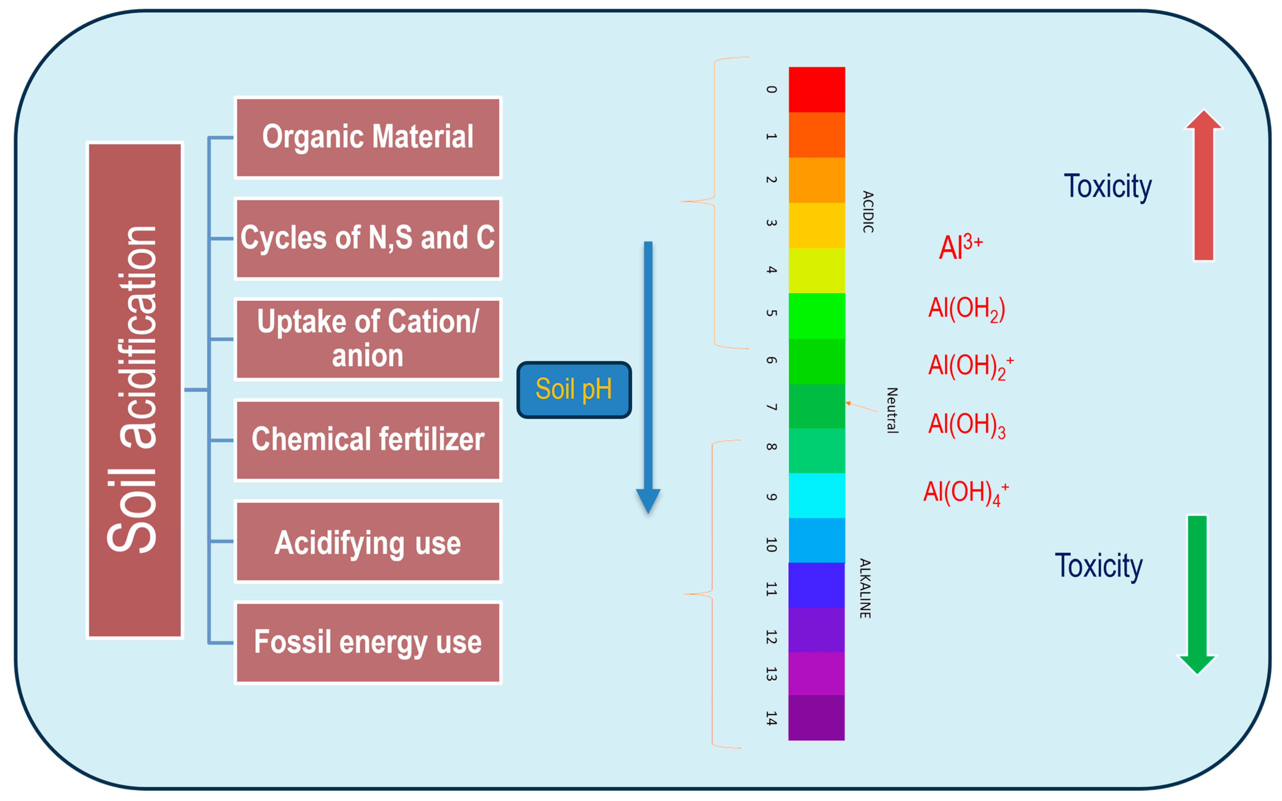
Figure 1. Illustration of soil acidification’s impact on Al chemistry, highlighting its transformation into various inorganic forms. Al reactivity varies with solubility, heavily influenced by ionic changes. Different Al species are formed based on acidic pH levels, with Al ions (Al3+) being the most predominant in acidic conditions, forming stable complexes with sulfate ions (SO42−), hydroxide (OH), phosphate ions (PO42−), and silicon (Si). As pH increases, aluminum toxicity diminishes, leading to diverse complexes like aluminum hydroxide ions [Al(OH)3, [Al(OH)4], Al(H2O)63+] and other insoluble hydroxides. Al3+ actively enters plant roots in environments with a pH lower than 5. At neutral pH (~7), Al(OH)3 forms, characterized by higher insolubility and non-toxicity. In environments with a pH greater than 7, aluminate [Al(OH)4] or aluminate anion species occur, often complexing with other molecular species such as AlO4Al12(OH)24(H2O)127+.
Plants 13 01760 g001
Figure 2. Integrated approach for ROS-mediated cellular tolerance of plants to Al toxicity. Al shares a common path for organic acid transporters (OA: Al3+) where the ions diffuse inside the cytosol and interact with the mitochondrial oxidative cycle. Malate dehydrogenase (MDH) and succinate dehydrogenase (SDH) are the major enzymatic sources where nicotinamide adenine dinucleotide [NAD(H) + H+] contributes electrons from O2 into superoxide anions (O2•−) and hydrogen peroxide (H2O2). In sensitive cultivars, H2O2 leads the formation of hydroxide ions (OH) with the Fenton reaction. On the other hand, the nucleus is activated in nuclear membrane oxidation, which also produces free radicals and the induction of other genes: angiotensin II receptor type 1, nitrate transporter 1 (ATR1, NRT1, etc.). Al-induced release of cytochrome c inhibits caspase-like activity, forwarding cell apoptosis. Chloroplasts’ reactions in another form can also induce ROS formation through the introduction of ethylene-dependent gravitropism-deficient and yellow-green light 3 (EGY3)-like metalloproteases that are efficient in the production of H2O2, which is retrograded in downstream signaling. The EGY3 can also induce malate transporters, where Al3+ can invade PSI. The latter is induced to develop O2•−/OH-like free radicals and are engaged in chloroplast membrane oxidation. Al3+ can also induce a chain of oxidative reactions where nucleotides are released freely by the action of xanthine oxidase-like activities.
Plants 13 01760 g002
Figure 3. Depiction of NH4+ and NO3 absorption by Al at root–soil interfaces. NH4+ lowers the rhizospheric pH, increasing the inhibition of metal uptake in plant roots due to competitive inhibition between Al3+ and H+. Conversely, rhizospheric pH becomes more alkaline with NO3+ accumulation, aiding in the desorption of metals into the rhizosphere but enhancing their transport into roots. Nitrate addition increases the negative electrical potential on the root surface, facilitating the conversion of NH4+ to NO3. Excess NH4+/H+ can displace soluble NO3 in roots. In sensitive cultivars, NH4+ can influence the binding of ataxia telangiectasia mutated (ATM) and ataxia telangiectasia mutated rad3-related (ATR) with suppressor of gamma response 1 (SOG1) for DNA damage recognition, leading to arrested cell growth in roots under high Al stress. The caption also references key enzymes and transporters involved in nitrogen metabolism: nitrite reductase (NiR), nitrate reductase (NR), glutamine synthase (GS), glutamate dehydrogenase (GDH), glutamine oxoglutarate aminotransferase (GOGAT), nitrate transporter 1/peptide transporter family (NPF), and nitrate transporter 2 family (NRT2).
Plants 13 01760 g003
Figure 4. Illustration of Al tolerance mechanisms in plants through bio-exclusion during stress. The plant’s response to Al stress includes the formation of Al–organic acid (* oxaloacetate and * citrate as predominat residues) complexes, leading to increased alkalization of the root surface. This process involves the induction of Al-malate transporters (* ALMT), activation of proton ATPases, and alterations in cell wall polysaccharide composition. These adaptations aim to minimize Al accumulation within cells, thereby mitigating oxidative damages through enhanced antioxidation pathways.
Plants 13 01760 g004
Figure 5. Depicting the genotoxic effects of Al through various intermediate gene expressions that individually or synergistically influence cellular phenomena. Initially, Al3+ stimulates ATP production, while simultaneously downregulating the expression of other electron transport carriers, leading to the dismutation of oxygen into free radical ROS. This ROS production triggers the tonoplast membrane, where Ca2+ activates mitochondrial permeability transition (MPT) channels. Concurrently, vacuole-mediated cell death is facilitated by the overexpression of vacuole-localized cysteine protease genes like vacuolar processing enzyme genes (VPE1, VPE1b, VPE2, VPE3), releasing Al from the vacuole into the cytosol. The over-activation of MPT channels disrupts the proton motive force (∆ψm) and causes spillage of cytochrome c oxidase, underlying the oxidative damage. The TCA cycle is activated, triggering anaplerotic reactions and inducing genes for malate synthase, citrate synthase, and oxaloacetate decarboxylase. Mitochondrial membrane leakage, linked to the overexpression of mitochondrial membrane permeability transition pore (MPTP) proteins and increased protease activity, also contributes to this process. Collectively, these events culminate in programmed cell death (PCD) of the roots, completing the cycle of Al toxicity.
Plants 13 01760 g005
Figure 6. A hypothetical model for control of organic acid (citrate) exudation in roots against Al ion (Al3+) toxicity. At the nuclear level, activation of SENSITIVE TO PROTON RHIXOTOXICITY 1 (STOP1) initiates the signaling pathway, which otherwise activates multidrug and toxic compound extrusion 1, Arabidopsis thaliana aluminum activated malate transporter 1, and amylotrophic lateral sclerosis 3 (AIMTE, MATE, ALS3) genes. STOP1 also upregulates the expression of RNA export 1 (RAE1), which, in feedback regulation, reduces STOP1 through degradation and ubiquitination. Signal transduction and activation of RNA 1 and 2 (STAR1, STAR2) otherwise induce UDP glucose-like residues, which conjugate with Al for extracellular detoxification. Transcription factors like Oryza sativa wrinkled transcription factor (OsWRKY22) form dimers with ADP-ribosyltransferase 1 (ART1) and collectively induce citrate biosynthesizing genes. Expressed proteins from the Nramp family (Nrat1) on the cellular membrane engulf Al3+ within the apoplastic space for chelation.
Plants 13 01760 g006
Figure 7. The involvement and crosstalk of plant growth substances against Al sensitivity in plants. Growth hormones like Aux, GA, ABA, CK, JA, and ET induce several cellular processes in roots of plants exposed to Al hyper-accumulation. Both external and internal detoxification by exclusion, sequestering, and chelation reactions are important under hormonal influence. Reduction of H+ by inhibition of H+/ATPase activity is important to downregulate the acidification of apoplasts of roots to reduce Al ions (Al3+). ET induces auxin accumulation as well as auxin transportation by pin formed 2 (PIN2)-mediated polar transport that results in root growth inhibition. Acidification of cell wall by organic acids (malate, citrate, oxalate) is important for ligand formation with metal. ABA and ET have synergistic actions with negative regulation, where RBOH and short-term response, respectively, are important. ABA can also induce ROS biosynthesis, which directly influences Al tolerance genes. Other substances like JA and CK are distantly related to Al tolerance by overexpression of coronatine insensitive 1 and myeloblastosis viral oncogene homologue 2 (COI1, MYC2) and direct inhibition of root growth.
Plants 13 01760 g007
Disclaimer/Publisher’s Note: The statements, opinions and data contained in all publications are solely those of the individual author(s) and contributor(s) and not of MDPI and/or the editor(s). MDPI and/or the editor(s) disclaim responsibility for any injury to people or property resulting from any ideas, methods, instructions or products referred to in the content.