Article

Genome-Wide Analysis of DNA Demethylases in Land Plants and Their Expression Pattern in Rice

State Key Laboratory of Hybrid Rice, College of Life Sciences, Wuhan University, Wuhan 430072, China
*
Authors to whom correspondence should be addressed.
These authors contributed equally to this work.
Plants 2024, 13(15), 2068; https://doi.org/10.3390/plants13152068
Submission received: 31 May 2024 / Revised: 20 July 2024 / Accepted: 24 July 2024 / Published: 26 July 2024
(This article belongs to the Special Issue Bioinformatics and Functional Genomics in Modern Plant Science)

Abstract

:
DNA demethylation is a very important biochemical pathway regulating a group of biological processes, such as embryo development, fruit ripening, and response to stress. Despite the essential role of DNA demethylases, their evolutionary relationship and detailed biological functions in different land plants remain unclear. In this study, 48 DNA demethylases in 12 land plants were identified and classified. A phylogenetic tree was constructed to demonstrate the evolutionary relationships among these DNA demethylases, indicating how they are related across different species. Conserved domain, protein motif, and gene structure analysis showed that these 48 DNA demethylases fell into the presently identified four classes of DNA demethylases. Amino acid alignment revealed conserved catalytic sites and a previously less-studied protein region (referred to as domain A) within the DNA demethylases. An analysis showed a conserved pattern of gene duplication for DNA demethylases throughout their evolutionary history, suggesting that these genes had been maintained due to their importance. The examination of promoter cis-elements displayed potential signaling and regulating pathways of DNA demethylases. Furthermore, the expression profile was analyzed to investigate the physiological role of rice DNA demethylase in different developmental stages, in tissues, and in response to stress and various phytohormone signals. The findings offer a deeper insight into the functional regions of DNA demethylases and their evolutionary relationships, which can guide future research directions. Understanding the role of DNA demethylases can lead to improved plant stress resistance and contribute to the development of better crop and fruit varieties.

1. Introduction

DNA methylation, as a very important type of epigenetic modification, plays an essential role in many biological processes. Theoretically, epigenetic markers like cytosine DNA methylation lead to the alteration of chromatin structure [1]. In higher plants, according to studies on Arabidopsis thaliana, there are three main sites, CG, CHG, and asymmetric CHH (H referring to A, C, and T), for the existing methylated nucleotide 5-methyl-deoxycytidine (5-meC) in DNA molecules [2,3]. The main protein family responsible for DNA methylation establishment in plants is the DRM (Domains rearranged methyltransferase) family [4,5], whose mammalian orthologous counterpart is the DNMT3 (DNA methyltransferase 3) family [5]. For the maintenance of different methylated sequence contexts, plants utilize different enzymes for correspondent DNA contexts (for example, MET1 (Methyltransferase 1) for CG and CMT3 (Chromomethylase 3) for CHG) [6] and the methylation at the asymmetric site CHH (for example, CMT2 with DRM2) must be established de novo [5]. There are two potential DNA demethylation pathways: active and passive. DNA semiconservative replication allows for passive DNA demethylation to automatically occur in newly synthesized strands without the help of additional enzymes or factors. In newly synthesized DNA strands, all the bases lose any kind of modification like methylation. Thus, if there are no specific enzymes to create methylation on the newly synthesized strands and maintain DNA methylation level in the cell, the DNA methylation soon fades away after several rounds of DNA replication [7]. In active demethylation, it can take place independently from DNA replication but requires the participation of demethylases. Researchers have reported a couple of possible mechanisms of DNA demethylation [5,8]. However, all DNA demethylases including DME, ROS1, DML2, DML3 (Demeter for DME, Repressor of silencing, 1 for ROS1, and DME-like for DML) discovered in A. thaliana are reported to show a base excision-repair mechanism [9,10]. In the base excision-repair pathway, a specialized glycosylase cleaves the glycosidic bond, generating an AP (apurinic/apyrimidinic site) site; then, an endonuclease recognizes the AP site and removes the left deoxyribose. Eventually, DNA polymerase and ligase mend the generated gap [5].
The first reported enzyme for DNA demethylation in A. thaliana is ROS1 [9,11]. To be specific, the A. thaliana ROS1 gene contains a highly conversed endonuclease III domain, which also features DNA glycosylase function [9]. A. thaliana DME bears a DNA glycoslyase/AP lyase bifunctional domain conserved to that of ROS1 [12]. According to current studies, those previously identified demethylases are all bifunctional enzymes [5,11]. Many DNA demethylases have been already identified in some other land plant species as well. For example, in Oryza sativa, the enzymes in the DNG (DNA glycosylase) gene family are reported to carry out DNA demethylase activity [13], as well as DML proteins in A. thaliana. In previous studies, orthologues of AtDML2 and AtROS1, which are OsDNG701 and OsDNG704, OsDNG702 (also known as OsROS1a) and OsDNG703, respectively, have already been identified [14,15]. However, there is no gene closely related to AtDME detected in rice [14]. An investigation into the physiological role of DNG family genes in plants demonstrated the indispensability of DNG702 in rice gametophyte development [16] and other important functions of DNG family genes in gametes, zygotes [13] and pollen vegetative cells [17]. Many DNA demethylases like the ZmROS1 gene in maize (Zea mays) have also been discovered and are believed to have important functions in maize endosperms [18]. These DNA demethylases play a crucial role in gene expression regulation and plant development. The function of ROS1 has been discovered to regulate trans-genes and some endogenous genes through the suppression of methylation-regulated silencing of genomes [19,20]. In A. thaliana, mutations in ROS1 causing loss of activity may lead to many unwanted methylations of some genes and, thus, the unnecessary silencing of gene expression [9]. Expression of the ROS1 gene is widespread within all plant tissues [9], and, in comparison, the DME gene is only transiently expressed in the central cell and synergid in the development of female gametophytes of dicots [10]. However, among four A. thaliana proteins, only the loss of function mutations in the DME gene generate severe growth defects in plants [10]. Mutations in the DME gene may bring about abnormal endosperm formation and deficient embryo development, consequently leading to seed abortion [21]. The DML2 and DML3 proteins can avoid DNA hypermethylation within vegetative tissues of A. thaliana [11]. These studies show that the process of DNA demethylation itself features several important biological meanings. First of all, demethylation is necessary for the early developmental stages of plants, without which impaired embryo development would occur [22,23]. Second, other reports have revealed that DNA demethylation has regulatory functions in the response of plants to abiotic stress, including drought [24], high concentration of salt [25], extreme temperature [26,27,28], and lack of nutrition [29,30]. The predicted mechanism of how DNA demethylases are related to stress response is that DNA demethylation at certain promoter sites triggers the transcription of downstream responding genes [31]. In addition, DNA demethylation also plays a role during the process of fruit maturation according to the proposed data from plants such as tomato and strawberry [32]. These reports demonstrate that investigations into DNA demethylation in plants have promising prospects in crop production improvement. However, recent studies on specific DNA demethylases mainly have mainly focused on A. thaliana and O. sativa [13,17,31].
Besides all the biological roles of DNA demethylase, it is also important to note that some DNA demethylases are reported to participate in plant–microbe interactions and the pathogen resistance of plants. To begin with, it was found that a protein called βC1, encoded by a satellite, affects the enzymatic activity of AtDME and NbROS1L in Nicotiana benthamiana, damaging the microbe-resistance of plants [33]. Another study discovered that dme mutants of A. thaliana have an increased susceptibility to both the bacterial infection of Pseudomonas Syringae (Pst DC3000) and the fungal infection of Verticlium Dahliae, and it concluded that DME plays a more prominent role in anti-pathogen response than other DNA demethylases, as well [34]. The role of ROS1 in plant resistance, especially in A. thaliana, has also been greatly investigated in many studies. For example, in contrast to the acquired resistance derived from plant–microbe interactions (or so-called systematic acquired resistance, SAR), a transcriptome analysis revealed that ROS1 controlled demethylation functions in transgenerational acquired resistance (TAR) against Pst DC3000 [35]. From the same research, the mechanism of the ROS1-dependent resistance was also proposed: a switch between demethylation and methylation on sialic acid (SA)-dependent gene alters their expression level [35]. Thus, it is very likely that the resistance is established based on the phytohormone, SA, and regulated pathways, similar to the case in abiotic resistance [36]. These reports, combined, indicate that in plants, DNA demethylases are largely involved in plant–pathogen interactions and are very likely to exert influences through phytohormone pathways. But whether the expression level of DNA demethylases is altered in response to a specific phytohormone remains unclear.
At present, the development of bioinformatics approaches and the accumulation of bioinformation provides us with lots of methods and a tremendous resource pool to further explore more demethylase genes in a larger group of land plants. In this study, we identified and classified 48 genes in 12 land plants and then performed evolutionary analysis, amino acid alignment, gene structure, conserved domain, motif, promotor, and expression pattern analysis to discover the structure (catalytic sites and conserved domain), function (physiological role), and evolutionary pattern of DNA demethylase genes in different species. These data provide us with a better insight into further studies on DNA demethylases in land plants.

2. Materials and Methods

2.1. Identification of DNA Demethylases in 12 Land Plants

The genomes and annotated data of 12 land plants including 6 monocots [Brachypodium distachyon (v3.2), Musa acuminata (v1), Oryza sativa (v7.0), Sorghum bicolor (v3.1.1), Setaria italica (v2.2), and Zea mays (Refgen_v4)] and 6 eudicots [Anacardium occidentale (v0.9), Arabidopsis thaliana (Araport11), Fragaria vesca (v4.0.a2), Prunus persica (v2.1), Solanum lycopersicum (ITAG4.0), and Vitis vinifera (v2.1)] were downloaded from the Phytozome website (https://phytozome-next.jgi.doe.gov; accessed on 1 April 2024). The protein sequences of A. thaliana were used as queries to identify the sequences of potential DNA demethylases in these plants through the BlastP method. The longest transcripts of the BLAST results were preserved, while others were discarded. For the verification of the acquired protein sequences, the Batch Web CD-Search Tool on the NCBI CDD website (https://www.ncbi.nlm.nih.gov; accessed on 4 April 2024; [37]) was used to compare the conserved domains in BLAST results with control sequences in A. thaliana. Protein sequences with incomplete conserved domains were eliminated.

2.2. Sequence Alignment and Construction of the Phylogenetic Tree

All protein sequences of different DNA demethylase candidates in 12 land plants were aligned within a corresponding protein family using the MEGA11 software (Version 11.0.13). After the alignment of all sequences, the MEGA11 software was then used to construct phylogenetic trees of those protein sequences through neighbor-joining (NJ) methods based on the Poisson correction model, with pairwise deletion option. Meanwhile, the option of the bootstrap test was set to 1000 replicates [38]. A visualization of completed trees in NWK format was carried out by the iTOL website (https://itol.embl.de; accessed on 13 May 2024).

2.3. Protein Structure and Property Analysis

The conserved domains in our DNA demethylase candidates were analyzed on the NCBI conserved domain database (CDD) website using the Batch Web CD-Search Tool (https://www.ncbi.nlm.nih.gov; accessed on 4 April 2024; [37]). The MEME tool (http://meme-suite.org/tools/meme; accessed on 5 April 2024) on the MEME suite website was utilized to discover conserved motif patterns in those protein sequences. The results were visualized by the TBtools software (v2.084; [39]). The properties of target proteins, including pI and molecular weight, were predicted by the TBtools Seqcat function. The visualization of predicted peptide 3-dimensional structures was performed on the SWISS-MODEL website (https://swissmodel.expasy.org/interactive; accessed on 8 April 2024). The statistical data were graphed by the ChiPlot website (https://www.chiplot.online; accessed on 10 April 2024).

2.4. Gene Structure Analysis

The structure of DNA methylase candidate genes was analyzed and visualized on the GSDS (Gene Structure Display Server; http://gsds.gao-lab.org; accessed on 4 April 2024) website.

2.5. Gene Duplication Analysis of DNA Demethylase Families

The gene duplication analysis of candidate sequences was conducted on the MCScanX software (https://github.com/wyp1125/MCScanX, accessed on 4 April 2024) using the Blast P method, with the E-value option set to 1 × 10⁻⁵ [40]. The DnaSP6.0 software (v6.12.03) was applied to calculate both the nonsynonymous (Ka) and synonymous (Ks) substitutions and ratios of Ka/Ks [41]. The divergence time calculation of duplicated DNA demethylase candidate gene pairs was based on the formula T = Ks/(2 × 9.1 × 10−9) × 10−6 millions of years ago (MYA; [42]).

2.6. Promoter Analysis

To investigate into the cis-elements within promoter regions of candidate genes, the 2000 bp upstream of the gene translation start site sequences of corresponding genes was extracted from the plant genome sequences based on gff3 files. The identification of cis-elements was carried on the PlantCARE website [43]. Finally, the promoter patterns of targeted genes were visualized by the TBtools software (v2.084; [39]), and the statistical data were graphed by the ChiPlot website (https://www.chiplot.online; accessed on 11 April 2024).

2.7. Expression Pattern Analysis

Gene expression data of OsDNG704, OsDNG703, OsDML3a, OsDNG701, and OsROS1 were obtained from the RiceXPro databank (https://ricexpro.dna.affrc.go.jp; accessed on 12 April 2024), including those of different developmental stages and those responding to phytohormones. All expression arrays were subjected to 75 percentile normalization, and the Heatmap was visualized by TBtools [39].

3. Results

3.1. Identification and Classification of 48 DNA Demethylases in 12 Plants

The accumulation of growing amounts of plant genomic data enabled us to discover a large range of DNA demethylase candidates among lots of plant species. In previous studies, the major four DNA demethylases (ROS1, DME, DML2, and DML3) in A. thaliana have already been identified, and, thus, sequences of those genes and their corresponding proteins were set as positive controls and blast queries in our study. The genomic information of other 11 land plants including A. occidentale, B. distachyon, F. vesca, M. acuminata, O. sativa, P. prunus, S. bicolor, S. lycopersicum, S. italica, V. vinifera, and Z. mays was used as data sources for the identification of potential plant DNA demethylases.
A previous study showed that enzymes performing DNA demethylase function contain three functional domains [44]. At present, the pattern and characteristics of DME domain and HhH-GPD domain (to be specific, Nth domain) are already stored on the NCBI CDD website, while information about N-terminal domain A remains unclear. Consequently, protein sequences, bearing both DME and glycosylase domains, were identified as DNA demethylases. Overall, we identified 48 genes (Table 1) which are predicted to be capable of demethylase activity. Among them, four O. sativa DNA demethylases (OsDNG701, OsDNG702, OsDNG703, and OsDNG704), three tomato (S. lycopercicum) genes (SlDML, SlROS1L, and SlDML2) [13], and four maize (Z. mays) genes (ZmDNG105, ZmDNG102, ZmROS1, and ZmMDR1) were already discovered. Then, all 48 identified protein sequences were subjected to MEGA software (Version 11.0.13) for alignment and phylogenetic analysis. Due to the uncertainty of the algorithm and model and far relationships between some genes, the tree of the genes has three primary clades instead of two clades as usual (Figure 1), and the bootstrap value above 50% is shown. The second clade is divided into two classes, which are designated DMLa, containing AtDML2 and AtROS1, and DME, containing AtDME. The other two clades are classified into two groups, with the name of DMLb and DMLc. The phylogenetic tree demonstrates that the DMLc is the largest group among all four classes. One of the two clades in class DMLb is only composed of monocotyledonous genes, while the other comprises solely dicotyledonous genes (Figure 1). In addition, DNA demethylases in M. acuminata are only found in the DMLc class, while those of V. vinifera only exist in the DMLa and DME classes (Figure 1).

3.2. Recent WGD Events Generate Gene Diversification in Few Gene Pairs from Target DNA Demethylases

Gene paralogues are mainly derived from gene duplication events [45], which indicates the great contribution from gene duplication to gene and protein diversification. There are different types of evolutionary events that contribute to gene diversification, but in our study, we mainly focused on whole-genome duplication (WGD) events. MCScanX software (https://github.com/wyp1125/MCScanX, accessed on 4 April 2024) was used to discover gene duplicative pairs of DNA demethylases in 12 land plants’ genomes, and eventually, only 6 gene duplicative pairs were found within 48 DNA demethylase genes. Among them, only one pair was from monocots (MaDML2 versus MaDML3) without a single one in Gramineae plants, while five were found in eudicots. This result indicates that, in monocots, or to be specific, in Gramineae, the diversification of DNA demethylases might mainly come from species differentiation instead of intraspecies duplication events.
The estimated dates of duplication events in the six pairs of genes are relatively far away from the modern period (Table 2). Five pairs were estimated to be duplicated before 70 million years ago, when the differentiation of monocot and eudicot took place. One duplicative pair (SlROS1L vs. SlDML), however, emerged much later: about 34 million years ago.

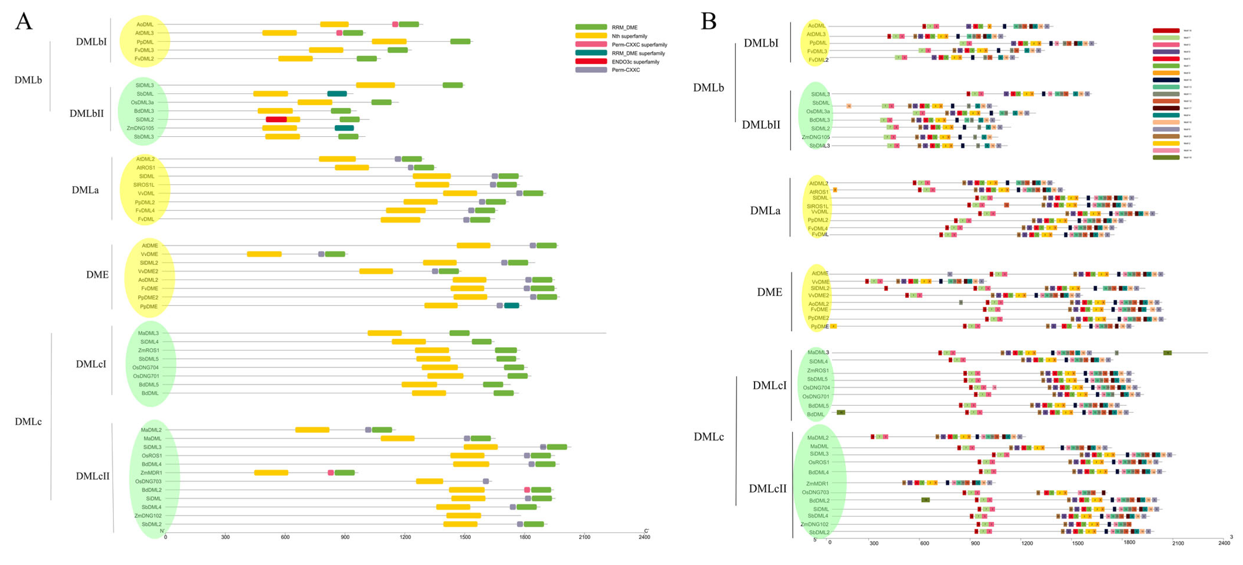
3.3. Protein Structure and Motif Analysis Verify the Classification of DNA Demethylases

To study the structural properties of DNA demethylase gene encoded proteins, all identified protein sequences were submitted to the Batch Web CD-Search Tool on NCBI. The result (Figure 2A) demonstrates that all demethylases possess the Nth domain (COG0177), and almost all have the RRM_DME Domain (pfam15628). The Nth domain is an endonuclease III domain, which is generally predicted to be responsible for DNA replication, recombination, and repair; while the RRM-DME domain is believed to be specified for DML proteins and is predicted to facilitate the recognition of specific DNA sequences through the guide of ssDNA or RNA [37,46]. Some of them also feature a Perm-CXXC domain (pfam15629) tandem with the C-terminal Nth domain. Incidentally, a very long and complete permease domain (Xan_ur_permease, cl23746) exists in a protein in M. acuminata, which may function as a transporter. This indicates that this protein might have multiple functions other than those of the DNA demethylase. The evolutionary relationships of DNA demethylases among species are seen from the phylogenetic analysis, as well. Proteins in DMLa containing AtROS1 and AtDML2 and DME including AtDME all feature a conserved pattern with three predicted domains (RRM_DME, Perm-CXXC, and Nth in order of from C-terminal to N-terminal). In contrast, the case in DMLb and DMLc is more complicated. Two enzymes (AtDML3 and AoDML) from eudicot in the DMLb group are expected to have the Perm-CXXC domain, while others in that class are not. One clade of DMLc (DMLcI) is only composed of two-domain proteins, and another mainly consists of proteins with three domains, which contain an additional Perm-CXXC domain apart from DME and Nth domains (Figure 2A). In DMLcII, all possess three domains, except OsDNG703 and ZmDNGI02. These results not only further verify our construction of the phylogenetic tree, but also indicate some subtle details of potential evolutionary events, such as gene duplication and functional differentiation.
The information of the previously reported domain A, required for DNA binding and 5 meC excision [44], has not been thoroughly studied and is, thus, presently not stored in the database on NCBI. The motif analysis, however, shows a possible domain A region. The MEME suite was used to discover the most likely 20 motifs in our identified protein sequences, and all 20 potential motifs were visualized by TBtools. Most identified DNA demethylase proteins share a very similar motif composition, with motif pattern 16-7-3, at the N-terminal side of the Nth domain region (Figure 2B), although some proteins in the DMLb class do not bear motif 16, solely featuring tandem motif 7 and motif 3. One protein in Z. mays is the only exception with neither 16-7-3 nor 7-3 patterns (Figure 2B). According to previous research [44], this region containing such motif pattern is very likely to be that so called domain A due to its position in the protein sequence. From the diagram, we can also conclude that the Nth domain of DNA demethylases is highly conserved in evolution because practically all Nth domains in different proteins have a motif composition in the order of 20-9-5-1-2-8. At the C-terminal end of most DNA demethylases in classes DMLa, DMLc, and DME, there exists a longer motif pattern with nine tandem motifs (19-14-13-11-12-17-4-10-6) (Figure 2B). This region covers both the DME domain and the small Perm-CXXC domain, and also even includes a large part of IDR (intrinsic disordered region) between Nth and DME. In fact, there is a small gap between motifs 12 and 17 in most DMLa, DME, and DMLc proteins, which separates this region featuring long conserved motif patterns into two parts. And from the sequence and domain information, the latter part (motifs 17-4-10-6) is the predicted Perm-CXXC and DME regions. In DMLb, most proteins, in comparison, lack an entire 9-motif composition pattern, but all show a 4-10-6 motif pattern, which very likely comprises the DME domain at the C-terminal end of our identified DNA demethylases (Figure 2B). Moreover, motif 19 is possessed by all DNA demethylases in IDR, which may play an unknown role in the enzyme activity or regulation of the protein itself.

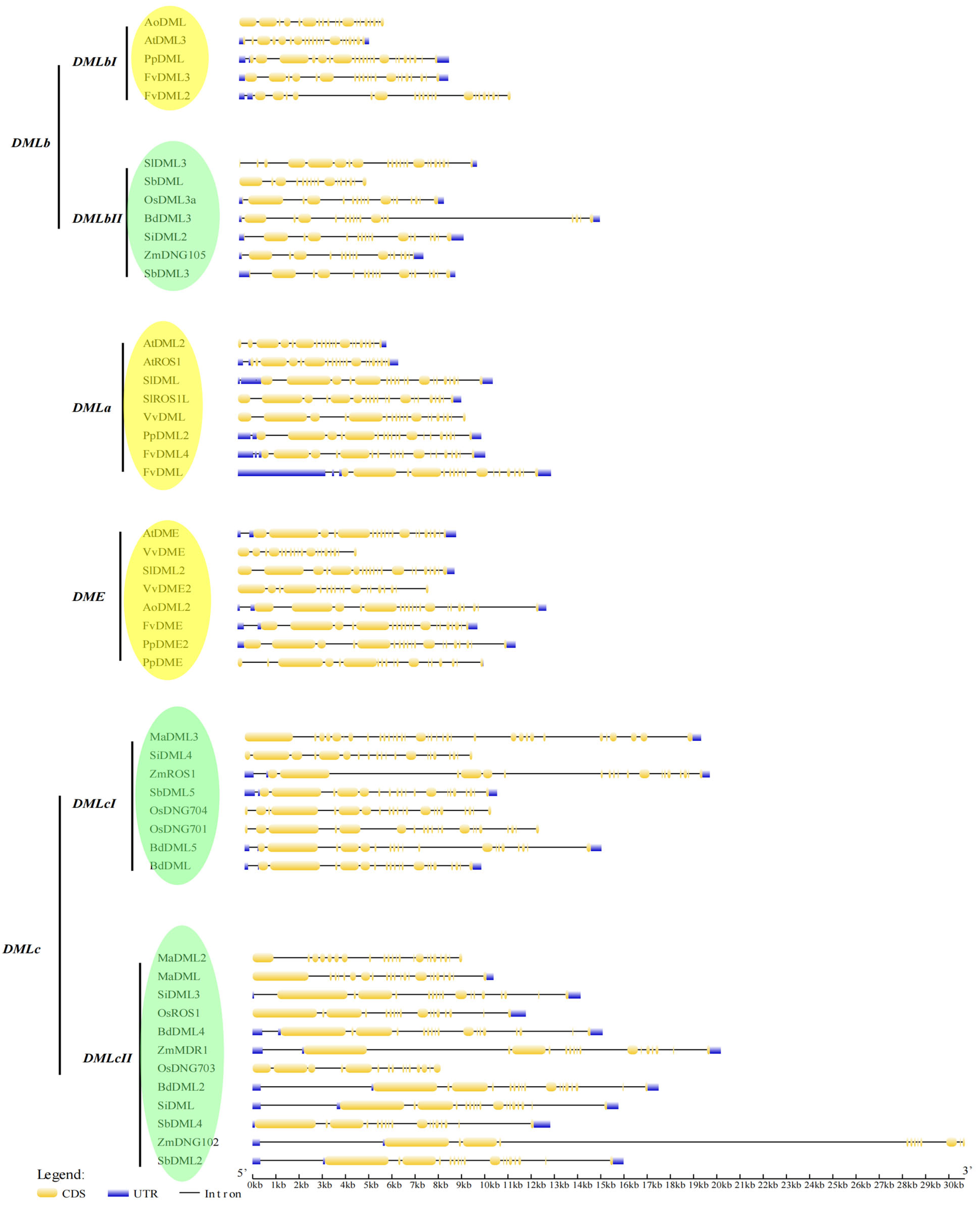
3.4. Gene Structure Analysis Shows the Exon–Intron Patterns of DNA Demethylases

An investigation of the gene structure shows some interesting trends in our identified DNA demethylase genes. The most common pattern in one clade of DMLc class (DMLcII) is two long exons at the 5′ end with a small exon between them, followed by six tiny exons, one intermediate exon, and then six to seven small exons again (Figure 3). In comparison, in another clade of this class (DMLcI), the two long exons are separately divided into a short exon and a long exon, while the rest have the same pattern. However, M. acuminata genes in DMLc bear an exon–intron pattern with some differences. In DMLcII, the second long exon in two M. acuminata genes is totally cut up by introns into a group of 7 small exons, and for that in subclass 1, it has additional 11 small exons at the 3′ end apart from the cut second large exons. The result of extra exons in the M. acuminata DMLcI gene is consistent with our previous domain and motif analysis (Figure 2A,B), meaning that these extra exons encode a different functional domain (permease domain). The pattern of small exons at the 3′ end of the DMLa and DME genes is quite similar to the pattern of the DMLc genes. But most of their first large exons are flanked by two additional small exons. The pattern of DMLb is like a combination of both previously mentioned patterns. One clade has almost exactly the same pattern as DMLcII, with shorter exons, while another has extra flanking exons as DME and DMLa. In conclusion, this similar exon–intron pattern may indicate the very conserved evolutionary relationship of DNA demethylase genes.

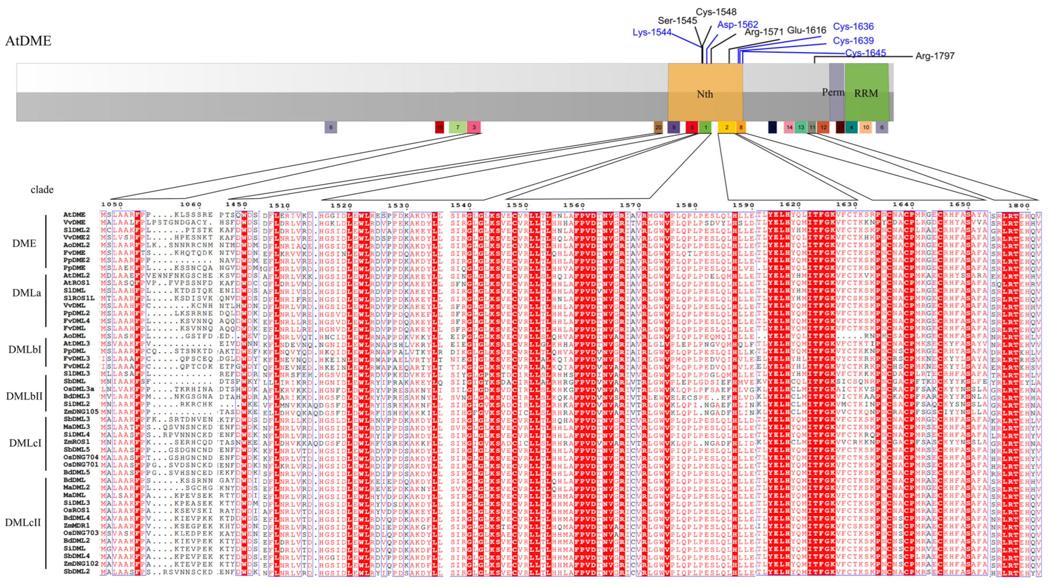
3.5. Sequences Alignment Demonstrates the Conserved Enzymatic Activity Sites

The potential enzymatic activity sites of glycosylases have already been identified in AtDME and AtROS1 domains [12,44]. Three sites, a lysine residue (Lys-1544) and an aspartic residue (Asp-1562) in AtDME (a transcript version with a protein length of 1987 aa), and four cysteines constituting a 4Fe-4S cluster nearby, are crucial to the glycosylase/lyase activity of the glycosylase domain [12]. These sites are predicted to be highly conserved in glycosylase domains of other enzymes in humans [12] and bacteria [12,44]. In addition, the investigation into detailed mechanisms of ROS1 shows that some hydrophobic residues form a pocket to accommodate 5 meC [47]. In our analysis, it is shown that all of the identified DNA demethylases have the same conserved lysine and aspartate residues in the glycosylase domain (Figure 4) and almost all have the 4Fe-4S cluster, only with one exception of AoDML which features a 3Fe-3S cluster. Thus, the result indicates that those sites are essential for glycosylase domain function, which is also supported by empirical data [12,47]. However, according to our analysis, there are many other completely conserved sites that are sequentially proximate to those three sites in aligned protein sequences. These sites might constitute the enzyme pocket which functions to accommodate the substrate 5-meC of DNA molecules and recognizes the DNA molecule.
Domain A (recognized as protein region from 980 to 1080 of a AtDME transcript version with a protein length of 1987aa) in AtDME is composed of a mixed charge cluster, which is likely to function as DNA binding sites and facilitates 5 meC excision, according to the previous reports [44,47], and the result of the sequence alignment of 48 DNA demethylases is in agreement with that in AtDME (Figure 5A). In this study, a statistical analysis demonstrates that among all Demeter-like proteins (only with the exception of ZmMDR1), the charge amino acid composition in a potential Domain A region is significantly higher than the whole protein or common proteins (Rice Rubisco) in plants (Figure 5A), and it is in correspondence with that of the DNA binding region of the rice histone-fold domain (HFD_H4). The predicted three-dimensional structure of this region shows that this region folds into a regular helix–turn–helix DNA binding structure (taking AtDME and SlDML2 as examples) (Figure 5C,D), which is very similar to that of HFD_H4 (Figure 5B). This conserved pattern may work as a supplement for the assumption that domain A may be necessary for protein–DNA interactions between DNA and DNA demethylases.

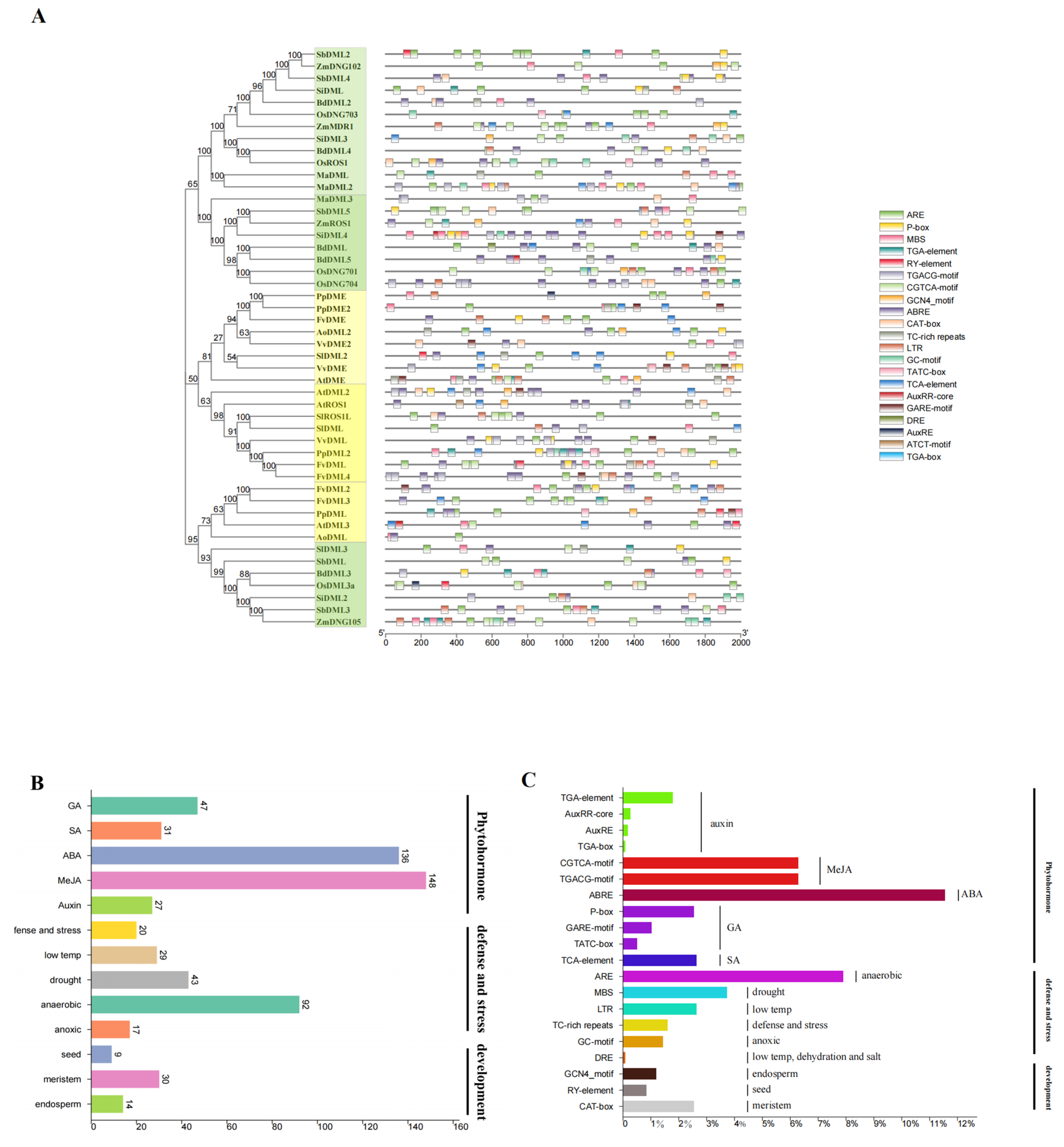
3.6. Cis-Element Analysis Shows Implication for the Future Research of DNA Demethylases in Plants

According to previous studies, DNA demethylation mainly functions in plants’ early development [10,21] and response to abiotic and biotic stresses [25,27,29]; thus our study is focused on cis-elements that participate in those areas, like elements that are susceptible to phytohormone. Among all the targeted cis-elements that showed up in promoter regions of 48 genes, the most abundant class is that responsive to light. In total, we discovered 528 light-responsive elements with 27 different types in the promoter region of 48 DNA demethylases, which include G-box, Box 4, GT1-motif, etc. Elements responsive to phytohormone were also very common in the promoter region of our genes. We identified a total 11 kinds of different cis-elements that correspond to 5 major phytohormones (Methyl Jasmonate-MeJA, Gibberellic acid-GA, Abscisic acid-ABA, Salicylic acid-SA, and Auxin; Table 3). Among them, elements responsive to ABA (ABRE element with 136 times) and GA have the highest frequency (CGTCA-motif and TGACG-motif both with 74 times; Table 3 and Figure 6C). In addition, we also discovered some elements specifically responsible for stress and plant development. For example, ARE elements that respond to anaerobic induction were widespread among 48 genes with only a few exceptions. Elements induced by low temperature (LTR) and drought (MBS) were less common but still appeared in those promoter regions with a frequency of 44 times for MBS and 31 times for LTR, which cannot be ignored. Elements such as CAT-Box, GCN4_motif, and RY-element corresponding to meristem development, endosperm formation, and seed development were also discovered, which confirm the role of DNA demethylation in plant development (Table 3 and Figure 6A–C).

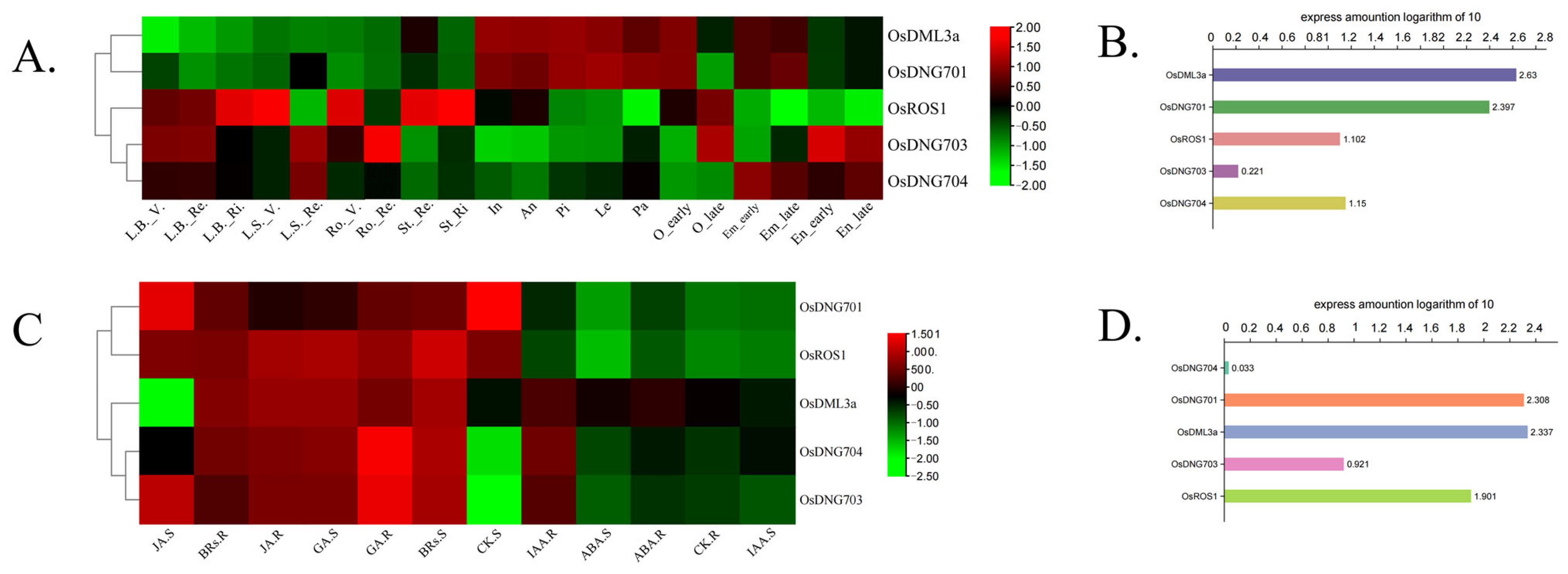
3.7. Expression Patterns of Rice DNA Demethylase Genes in Different Developmental Stages and Tissues and in Response to Phytohormones

Characteristics of gene expression in different organs and tissues and through different developmental stages empower us to thoroughly understand gene function [48]. Based on data collected from rice plants grown in standard conditions and treated by specific factors, including phytohormones and pathogen inoculation, the construction of standard expression profiles enables us to find subtle clues for the functional differentiation of different genes [49]. This analysis demonstrates that, overall, OsDML3a and OsDNG701 feature obviously higher expression levels than OsDNG704, OsDNG703, and OsROS1, indicating the potentially more significant role in the organism of the former two (Figure 7B,D). Through different rice tissues, the expression level of OsDML3a and OsDNG701 in some reproductive tissues, such as anther and pistil, is generally higher than in vegetative organs (root, leaf blade, etc.) and embryo and endosperm, while the expression pattern of OsDNG704, OsDNG703, and OsROS1 is reversed (Figure 7A). However, the expression alteration of OsDMLs during different reproductive tissue developmental stages is not obvious (Figure 7A).
From the results, it is clearly illustrated that the expression level of all five rice DNA demethylases shows a relative accordant pattern in response to different phytohormones. In general, the expression of these DNA demethylases is upregulated both in shoots and roots under the treatment of JA, BR (brassinolide), and GA (with the only apparent exception of OsDML3a in response to JA in the shoot) (Figure 7C). In comparison, the expression of five rice DNA demethylase genes is downregulated or remains at a low level in response to the treatment of CK (cytokinin), IAA (indoleacetic acid), and ABA in shoots and roots (Figure 7C). However, exceptions still exist. In rice shoots, the expression level of OsDNG701 and OsROS1 is high rather than low in response to CK, whilst the expression of OsDML3a, OsDNG704, and OsDNG703 is upregulated instead of downregulated in rice roots under the treatment of IAA (Figure 7C).

4. Discussion

4.1. Construction of Phylogenetic Tree and Further Verification Lead to New Classes of DNA Demethylases

In previous studies, the phylogenetic analysis of plant DNA demethylases was either restricted to a few species or only roughly classified as the potential DNA demethylase genes without further verification and analysis [31,50]. But, in this study, the 48 genes are identified and clearly classified into four distinct classes (DME, DMLa/b/c; Figure 1). Within the same class, DNA demethylases show obvious consensus, such as common protein properties like pI value (Table 4), conserved motif composition (Figure 2B), and gene structure (Figure 3). For example, the class DMLc has the longest exons (Figure 3), compared to DMLa, DMLb, and DME, at the 5′ end of the DNA molecule. In contrast, the DMLb class genes at that end have exons with the shortest length. This indicates that the exon and intron distribution pattern within a single class is relatively conserved. However, the expected gene structure generated from bioinformatical algorithm may need further modification through experiments.
The construction of a phylogenetic tree also demonstrates an interesting result that two classes are eudicot-specific (DMLa and DME) (Table 1; Figure 1) and one is restricted in monocots (DMLc) (Table 1). The conclusion that genes closely related to DME are eudicot-specific is also supported by a published report [50]. From the phylogenetic tree (Figure 1), DNA demethylases from F. vesca and P. persica in classes DMLa, DME, and DMLbI are all closely related and occur in pairs (FvDME with PpDME2 and PpDME in class DME, PpDML2 with FvDML and FvDML4 in class DMLa, and PpDML with FvDML3 and FvDML2 in class DMLbI; Figure 1). These results are plausible because the two species come from one family (Table 1); thus, they have a close evolutionary relationship. This indicates that DNA demethylases from the two species were derived from evolutionary events slightly before the divergence between F. vesca and P. persica about 69 million years ago [51] or may just come from the species differentiation of F. vesca and P. persica. There are two main subclasses within DMLc, which both contain different DNA demethylases from all six monocots. The result may indicate that DNA demethylases of this class in the same species are paralogous and underwent duplication in their evolution history. However, just as the case of DNA demethylases in F. vesca and P. persica, some DNA demethylases from S. bicolor and Z. mays also occur in pairs in class DMLc (for example, ZmROS1 with SbDML5 in DMLcI and ZmDNG102 with SbDML2 in DMLcII). Among all the six monocots, Z. mays and S. bicolor do have the closest relationship according to the estimated divergence time using the Timetree website [51]. Thus, we may locate the evolutionary origin of these DNA demethylases at a date of about 16 million years ago (the divergence time of Z. mays and S. bicolor; [51]). From the observation that DMLbII is only composed of monocotyledonous genes, while DMLbI comprises solely dicotyledonous genes, we can conclude that these two clades may share orthologs and are derived from relative ancient species differentiation. The classification provides a new way for further discovery of more plant DNA demethylases through exploring the orthologs of already identified genes in the corresponding class. In addition, it also equips researchers with a deep insight into the evolution history of DNA demethylases or even species themselves.

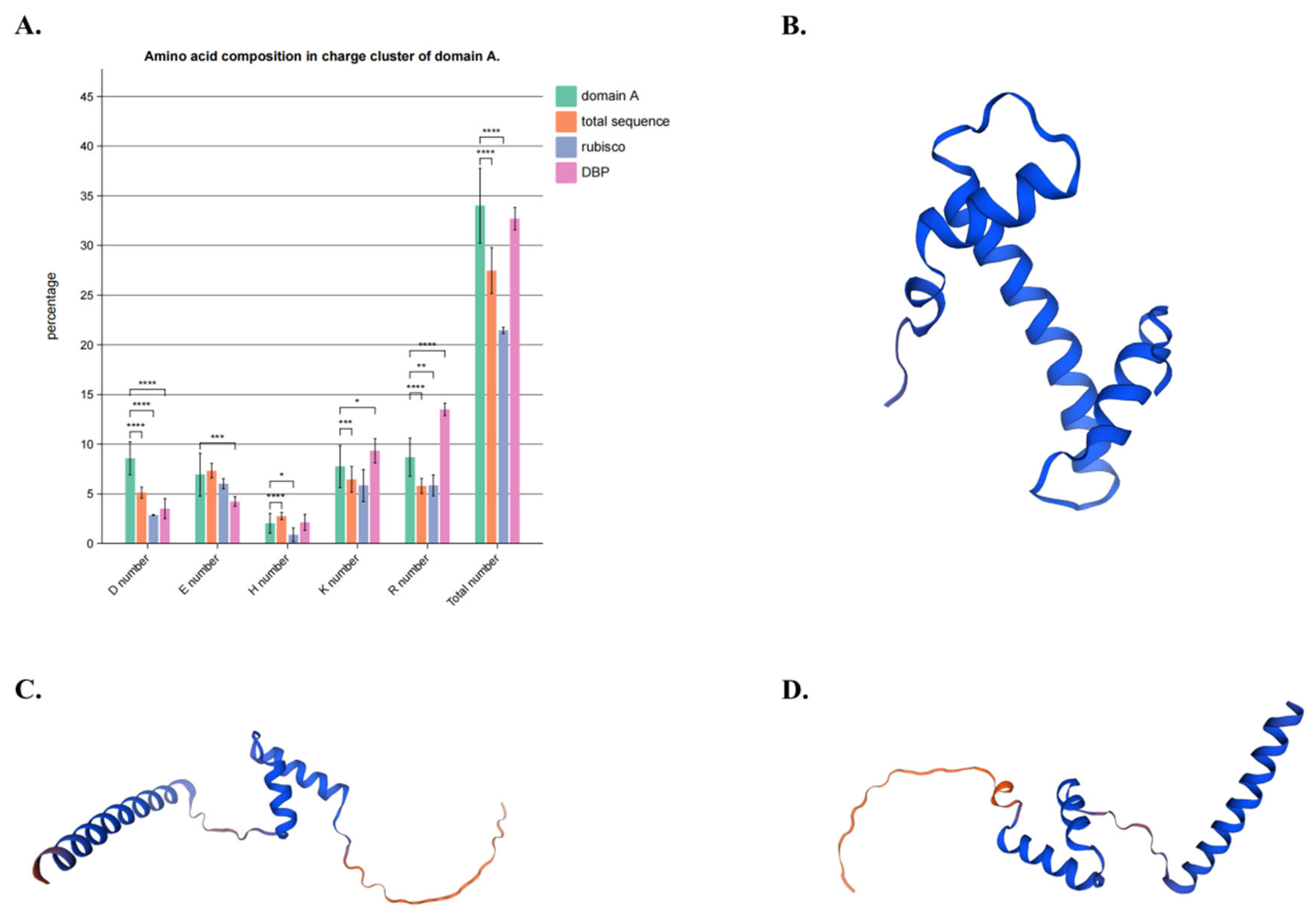
4.2. Multiple Sequence Alignment and Domain and Motif Analysis Discovered a New Functional Region of DNA Demethylases

Although domain A of DNA demethylases proximate to N-terminal of the enzyme has been long considered as functional [44], it has not been profoundly understood. Some conserved sites sequentially near that region may participate in the formation of substrates accommodating pockets [47] of glycosylase and were, thus, investigated, but other poorly conserved amino acids sites were ignored in many studies. However, motif analysis shows that a conserved motif pattern and composition appear in the region of all DNA demethylases of 12 land plants, forming a potential domain. A high frequency of the appearance of charge amino acids in this region also indicates that this region is very likely to play a role in DNA binding (Figure 5A) [44], which may play an important role in the stabilization or recognition of DNA substrates. The visualization of the predicted protein 3-dimensional structure of the potential domain A region also indicates that this DNA binding region is very likely to have a traditional helix-turn-helix pattern (Figure 5B–D).
The high frequency of the appearance of red color-designated conserved amino acid sites in the glycosylase/lyase domain region in DNA demethylases (Figure 4) also demonstrates that the bifunctional domain is highly conserved during the species differentiational events in evolutionary history, and the function of a single amino acid is relatively irreplaceable.

4.3. Cis-Element Analysis Shows the Future Investigation Field of the Role of DNA Demethylase in Plant Development Regulation and Stress Response

It has already been known that the expression of DNA demethylases is regulated by DNA methylation levels [52,53,54]. Some studies have also revealed the function of DNA demethylases in plant development [10,55], fruit ripening [56], and response to extreme conditions [36]. However, these studies mainly focused on the pathway and reaction regulated by DNA demethylation, or the influence of methylation on the expression of the DNA demethylase. But, by looking into promoter regions of identified DNA demethylase genes, it is found that there are some of cis-elements related to many different conditions and phytohormones (Table 2), which indicates the latent and possible direct regulation by phytohormones and external factors. Consequently, this provides a research direction: for example, an investigation into the possible mechanism of condition-induced transcription activation or suppression of DNA demethylase genes. Due to the considerable frequency of the appearance of anaerobic and anoxic resistance-responsible cis-elements (ARE and GC-motif) in the promoter region (Figure 6C), the DNA demethylases may, to some extent, be predicted to play a plausible role in plant tolerance against waterlogging stress and the rescue of drowned roots [57]. Moreover, since phytohormones such as auxin and GA also involve the rescue of waterlogging conditions [58], DNA demethtylases may participate in such response indirectly. The frequent appearance of MeJA, which, in rice and other plant species, acts in defense of fungal pathogens [59,60], regulating cis-elements (Figure 6C), proves that the DNA demethylases are very likely to have the potential to be involved in anti-fungi processes. As mentioned previously, there is a recent report proving that AtROS1 may participate in the anti-pathogen process of plants through the SA regulated pathways [35], and our results may provide a new research direction for the study of the role of another phytohormone, MeJA, in the DNA demethylase-regulated pathway against fungal pathogens (Figure 6B,C; Table 3). The abundance of ABA-responsible elements also indicates that DNA demethylases may participate in plant resistance to changing environments, in accordance with a previous study [31].

4.4. Expression Patterns Analysis of OsDMLs Shows the Separated Role of Different Rice DNA Demethylases in Response to Specific Phytohormones and in Different Tissues

The expression profile of rice DML genes of different developmental stages demonstrates that DNA demethylases in rice are very likely to undergo functional diversification (Figure 7A). OsDML3a and OsDNG701 feature a higher expression level in reproductive tissues (anther, pistil, etc.) (Figure 7A), which indicates that this pair of genes may participate in the formation of gamete and flowering. In comparison, the expression of OsDNG704 and OsDNG703 is higher in the embryo and endosperm, leading to a conclusion that they may be involved in the regulation of the embryo and endosperm’s development. Thus, such DNA demethylase genes may perform a very similar function as DME in A. thaliana, which is mainly expressed in reproductive tissues [61]. Furthermore, OsROS1 is expressed more in vegetative organs, and this consequently shows that it may mainly function in response to stress and changing environments. The fact that OsROS1 has a considerable expression level in vegetative tissues is similar to the case of the ROS1 gene in A. thaliana. Previous reports show that some DNA demethylases like ROS1 in A. thaliana are expressed mainly in vegetative tissues and play an important role in disease and stress response [31,62]. These results show that OsROS1 might have a very similar role as its A. thaliana counterparts in disease and stress resistance.
The next heatmap (Figure 7C) illustrates that the expression level of DNA demethylases in rice do respond to several specific phytohormone treatments and, thus, may give a hint that the results are in accordance with the results from cis-element analysis (Figure 6; Table 3). It is noticed that, especially for JA (or MeJA), the high expression level of rice DNA demethylases seen from the heatmap may be the consequence of the direct regulation by those cis-elements in their promoter regions (except OsDNG703, which does not contain related cis-elements) (Figure 7C; Table 3). Thus, these results may further verify the assumption that rice DNA demethylases are involved in fungal pathogen resistance. There are no recent detailed investigations into specific DNA demethylases in response to a particular phytohormone; however, there is research demonstrating that the level of DNA methylation and demethylation is altered in response to phytohormone [63]. Moreover, the data collected from predictive bioinformatic analysis still need further verification through wet experiments. For example, there is a considerable amount of cis-elements responding to phytohormone ABA in the promoter region of OsROS1 (Table 3), but it is seen from the expression profile that the expression of OsROS1 remains low under the treatment of ABA (Figure 7C). Such results may derive from more complicated situations or regulations, which must be further studied through detailed mechanism investigations.

5. Conclusions

In this study, 48 DNA demethylases in 12 land plants including A. thaliana, A. occidentale, B. distachyon, F. vesca, M. acuminata, O. sativa, P. prunus, S. bicolor, S. lycopersicum, S. italica, V. vinifera, and Z. mays were compared and analyzed through bioinformatical tools. Their evolutionary relationships were uncovered through the construction of a phylogenetic tree, and four main classes (DME and DMLa/b/c) were established. Among them, class DMLb is monocot-specific. Conserved domain, protein motif pattern, and gene structure analyses further verified the similarities and relationships of DNA demethylases within the same class. The sequence alignment of DNA demethylase proteins showed a conserved and highly structured region, which was believed to be domain A and might have a role in DNA binding, facilitating the recognition or stabilization of DNA–protein interaction. The cis-element analysis of the promoter region in DNA demethylase genes provides future directions in studying the role of DNA demethylases in stress and defense response or in response to phytohormone, while the expression profile construction in rice, to some extent, supports the general role of DNA demethylases in organisms through pointing out their expression alternation and difference in response to different phytohormones and in different tissues. And for better crop varieties creation, future investigations may be conducted into crops with an enhanced function of DNA demethylases.

Supplementary Materials

The following supporting information can be downloaded at: https://www.mdpi.com/article/10.3390/plants13152068/s1, Table S1: The protein motif sequence information derived from the MEME analysis; Table S2: Protein domain analysis of all Demeter-like protein sequences; Table S3: The detailed description of all DNA demethylase genes.The gene synonyms of DNA demethylase in B. distachyon, M. acuminata, O. sativa, S. bicolor, S. italica, Z. mays, A. occidentale, A. thaliana, F. vesca, P. persica, S. lycopersicum, and V. vinifera were named according to their orders of appearance on chromosomes, and the gene synonyms of rice DNA demethylase were named from the rice annotation project database (RAP-DB; https://rapdb.dna.affrc.go.jp/index.html; accessed on 14 April 2024); the gene synonyms of maize DNA demethylase were named from the MaizeGDB (https://www.maizegdb.org/; accessed on 14 May 2024). The gene locus ID with a indicates these genes with partial domains, the gene locus with b indicates genome annotated errors, and the gene locus with c indicates that the gene was selected for further analysis. Abbreviations are as follows: Amino acid (aa), Isoelectric point (pI). Other references are as follows: [9,10,64].

Author Contributions

J.H. and L.L. conceived the ideas. S.M. performed data curation, formal analysis, methodology, and original draft. J.X. performed data curation, formal analysis, methodology, and original draft. Y.Z. performed data curation and formal analysis. J.H. and L.L. supervised the research and revised the article. All authors have read and agreed to the published version of the manuscript.

Funding

This research was supported by grants from the National Natural Science Foundation of China (No. 31871238), the China Postdoctoral Science Foundation (2024M752481), and the Postdoctoral Fellowship Program of CPSF (GYB20240579).

Data Availability Statement

The datasets used and/or analyzed during the current study are available from the corresponding author on reasonable request.

Conflicts of Interest

The authors declare no conflicts of interest.

References

  1. Li, S.; Peng, Y.; Panchenko, A.R. DNA methylation: Precise modulation of chromatin structure and dynamics. Curr. Opin. Struct. Biol. 2022, 75, 102430. [Google Scholar] [CrossRef] [PubMed]
  2. Jones, P.A. Functions of DNA methylation: Islands, start sites, gene bodies and beyond. Nat. Rev. Genet. 2012, 13, 484–492. [Google Scholar] [CrossRef] [PubMed]
  3. Henderson, I.R.; Jacobsen, S.E. Epigenetic inheritance in plants. Nature 2007, 447, 418–424. [Google Scholar] [CrossRef] [PubMed]
  4. Read, A.; Weiss, T.; Crisp, P.A.; Liang, Z.; Noshay, J.; Menard, C.C.; Wang, C.; Song, M.; Hirsch, C.N.; Springer, N.M.; et al. Genome-wide loss of CHH methylation with limited transcriptome changes in Setaria viridis DOMAINS REARRANGED METHYLTRANSFERASE (DRM) mutants. Plant J. 2022, 111, 103–116. [Google Scholar] [CrossRef] [PubMed]
  5. Zhu, J.K. Active DNA demethylation mediated by DNA glycosylases. Annu. Rev. Genet. 2009, 43, 143–166. [Google Scholar] [CrossRef]
  6. Shim, S.; Lee, H.G.; Seo, P.J. MET1-Dependent DNA methylation represses light signaling and influences plant regeneration in arabidopsis. Mol. Cells 2021, 44, 746–757. [Google Scholar] [CrossRef] [PubMed]
  7. Parrilla-Doblas, J.T.; Roldán-Arjona, T.; Ariza, R.R.; Córdoba-Cañero, D. Active DNA demethylation in plants. Int. J. Mol. Sci. 2019, 20, 4683. [Google Scholar] [CrossRef] [PubMed]
  8. Tsukada, Y.; Fang, J.; Erdjument-Bromage, H.; Warren, M.E.; Borchers, C.H.; Tempst, P.; Zhang, Y. Histone demethylation by a family of JmjC domain-containing proteins. Nature 2006, 439, 811–816. [Google Scholar] [CrossRef] [PubMed]
  9. Gong, Z.; Morales-Ruiz, T.; Ariza, R.R.; Roldán-Arjona, T.; David, L.; Zhu, J.K. ROS1, a repressor of transcriptional gene silencing in Arabidopsis, encodes a DNA glycosylase/lyase. Cell 2002, 111, 803–814. [Google Scholar] [CrossRef] [PubMed]
  10. Choi, Y.; Gehring, M.; Johnson, L.; Hannon, M.; Harada, J.J.; Goldberg, R.B.; Jacobsen, S.E.; Fischer, R.L. DEMETER, a DNA glycosylase domain protein, is required for endosperm gene imprinting and seed viability in arabidopsis. Cell 2002, 110, 33–42. [Google Scholar] [CrossRef] [PubMed]
  11. Zhu, J.; Kapoor, A.; Sridhar, V.V.; Agius, F.; Zhu, J.K. The DNA glycosylase/lyase ROS1 functions in pruning DNA methylation patterns in Arabidopsis. Curr. Biol. 2007, 17, 54–59. [Google Scholar] [CrossRef] [PubMed]
  12. Morales-Ruiz, T.; Ortega-Galisteo, A.P.; Ponferrada-Marín, M.I.; Martínez-Macías, M.I.; Ariza, R.R.; Roldán-Arjona, T. DEMETER and Repressor of Silencing 1 encode 5-methylcytosine DNA glycosylases. Proc. Natl. Acad. Sci. USA 2006, 103, 6853–6858. [Google Scholar] [CrossRef] [PubMed]
  13. Zhou, S.; Li, X.; Liu, Q.; Zhao, Y.; Jiang, W.; Wu, A.; Zhou, D.X. DNA demethylases remodel DNA methylation in rice gametes and zygote and are required for reproduction. Mol. Plant 2021, 14, 1569–1583. [Google Scholar] [CrossRef] [PubMed]
  14. Zemach, A.; Kim, M.Y.; Silva, P.; Rodrigues, J.A.; Dotson, B.; Brooks, M.D.; Zilberman, D. Local DNA hypomethylation activates genes in rice endosperm. Proc. Natl. Acad. Sci. USA 2010, 107, 18729–18734. [Google Scholar] [CrossRef] [PubMed]
  15. La, H.; Ding, B.; Mishra, G.P.; Zhou, B.; Yang, H.; Bellizzi Mdel, R.; Chen, S.; Meyers, B.C.; Peng, Z.; Zhu, J.K.; et al. A 5-methylcytosine DNA glycosylase/lyase demethylates the retrotransposon Tos17 and promotes its transposition in rice. Proc. Natl. Acad. Sci. USA 2011, 108, 15498–15503. [Google Scholar] [CrossRef] [PubMed]
  16. Ono, A.; Yamaguchi, K.; Fukada-Tanaka, S.; Terada, R.; Mitsui, T.; Iida, S. A null mutation of ROS1a for DNA demethylation in rice is not transmittable to progeny. Plant J. 2012, 71, 564–574. [Google Scholar] [CrossRef] [PubMed]
  17. Kim, M.Y.; Ono, A.; Scholten, S.; Kinoshita, T.; Zilberman, D.; Okamoto, T.; Fischer, R.L. DNA demethylation by ROS1a in rice vegetative cells promotes methylation in sperm. Proc. Natl. Acad. Sci. USA 2019, 116, 9652–9657. [Google Scholar] [CrossRef] [PubMed]
  18. Xu, Q.; Wu, L.; Luo, Z.; Zhang, M.; Lai, J.; Li, L.; Springer, N.M.; Li, Q. DNA demethylation affects imprinted gene expression in maize endosperm. Genome Biol. 2022, 23, 77. [Google Scholar] [CrossRef] [PubMed]
  19. Gehring, M.; Huh, J.H.; Hsieh, T.F.; Penterman, J.; Choi, Y.; Harada, J.J.; Goldberg, R.B.; Fischer, R.L. DEMETER DNA glycosylase establishes MEDEA polycomb gene self-imprinting by allele-specific demethylation. Cell 2006, 124, 495–506. [Google Scholar] [CrossRef] [PubMed]
  20. Kapoor, A.; Agius, F.; Zhu, J.K. Preventing transcriptional gene silencing by active DNA demethylation. FEBS Lett. 2005, 579, 5889–5898. [Google Scholar] [CrossRef] [PubMed]
  21. Park, J.S.; Frost, J.M.; Park, K.; Ohr, H.; Park, G.T.; Kim, S.; Eom, H.; Lee, I.; Brooks, J.S.; Fischer, R.L.; et al. Control of DEMETER DNA demethylase gene transcription in male and female gamete companion cells in Arabidopsis thaliana. Proc. Natl. Acad. Sci. USA 2017, 114, 2078–2083. [Google Scholar] [CrossRef] [PubMed]
  22. Schoft, V.K.; Chumak, N.; Choi, Y.; Hannon, M.; Garcia-Aguilar, M.; Machlicova, A.; Slusarz, L.; Mosiolek, M.; Park, J.S.; Park, G.T.; et al. Function of the DEMETER DNA glycosylase in the Arabidopsis thaliana male gametophyte. Proc. Natl. Acad. Sci. USA 2011, 108, 8042–8047. [Google Scholar] [CrossRef] [PubMed]
  23. Huh, J.H.; Bauer, M.J.; Hsieh, T.F.; Fischer, R.L. Cellular programming of plant gene imprinting. Cell 2008, 132, 735–744. [Google Scholar] [CrossRef]
  24. Sadhukhan, A.; Prasad, S.S.; Mitra, J.; Siddiqui, N.; Sahoo, L.; Kobayashi, Y.; Koyama, H. How do plants remember drought? Planta 2022, 256, 7. [Google Scholar] [CrossRef] [PubMed]
  25. Singroha, G.; Kumar, S.; Gupta, O.P.; Singh, G.P.; Sharma, P. Uncovering the epigenetic marks involved in mediating salt stress tolerance in plants. Front. Genet. 2022, 13, 811732. [Google Scholar] [CrossRef] [PubMed]
  26. Perrella, G.; Bäurle, I.; van Zanten, M. Epigenetic regulation of thermomorphogenesis and heat stress tolerance. New Phytol. 2022, 234, 1144–1160. [Google Scholar] [CrossRef] [PubMed]
  27. Luo, X.; He, Y. Experiencing winter for spring flowering: A molecular epigenetic perspective on vernalization. J. Integr. Plant Biol. 2020, 62, 104–117. [Google Scholar] [CrossRef] [PubMed]
  28. Hereme, R.; Galleguillos, C.; Morales-Navarro, S.; Molina-Montenegro, M.A. What if the cold days return? Epigenetic mechanisms in plants to cold tolerance. Planta 2021, 254, 46. [Google Scholar] [CrossRef] [PubMed]
  29. Secco, D.; Whelan, J.; Rouached, H.; Lister, R. Nutrient stress-induced chromatin changes in plants. Curr. Opin. Plant Biol. 2017, 39, 1–7. [Google Scholar] [CrossRef]
  30. Fan, X.; Peng, L.; Zhang, Y. Plant DNA methylation responds to nutrient stress. Genes 2022, 13, 992. [Google Scholar] [CrossRef] [PubMed]
  31. Zhang, H.; Gong, Z.; Zhu, J.K. Active DNA demethylation in plants: 20 years of discovery and beyond. J. Integr. Plant Biol. 2022, 64, 2217–2239. [Google Scholar] [CrossRef] [PubMed]
  32. Cheng, J.; Niu, Q.; Zhang, B.; Chen, K.; Yang, R.; Zhu, J.K.; Zhang, Y.; Lang, Z. Downregulation of RdDM during strawberry fruit ripening. Genome Biol. 2018, 19, 212. [Google Scholar] [CrossRef] [PubMed]
  33. Gui, X.; Liu, C.; Qi, Y.; Zhou, X. Geminiviruses employ host DNA glycosylases to subvert DNA methylation-mediated defense. Nat. Commun. 2022, 13, 575. [Google Scholar] [CrossRef] [PubMed]
  34. Zeng, W.; Huang, H.; Lin, X.; Zhu, C.; Kosami, K.I.; Huang, C.; Zhang, H.; Duan, C.G.; Zhu, J.K.; Miki, D. Roles of DEMETER in regulating DNA methylation in vegetative tissues and pathogen resistance. J. Integr. Plant Biol 2021, 63, 691–706. [Google Scholar] [CrossRef]
  35. López Sánchez, A.; Stassen, J.H.; Furci, L.; Smith, L.M.; Ton, J. The role of DNA (de)methylation in immune responsiveness of Arabidopsis. Plant J. Cell Mol. Biol. 2016, 88, 361–374. [Google Scholar] [CrossRef] [PubMed]
  36. Yang, L.; Lang, C.; Wu, Y.; Meng, D.; Yang, T.; Li, D.; Jin, T.; Zhou, X. ROS1-mediated decrease in DNA methylation and increase in expression of defense genes and stress response genes in Arabidopsis thaliana due to abiotic stresses. BMC Plant Biol. 2022, 22, 104. [Google Scholar] [CrossRef] [PubMed]
  37. Wang, J.; Chitsaz, F.; Derbyshire, M.K.; Gonzales, N.R.; Gwadz, M.; Lu, S.; Marchler, G.H.; Song, J.S.; Thanki, N.; Yamashita, R.A.; et al. The conserved domain database in 2023. Nucleic Acids Res. 2023, 51, D384–D388. [Google Scholar] [CrossRef] [PubMed]
  38. Tamura, K.; Stecher, G.; Kumar, S. MEGA11: Molecular evolutionary genetics analysis version 11. Mol. Biol. Evol. 2021, 38, 3022–3027. [Google Scholar] [CrossRef] [PubMed]
  39. Chen, C.; Chen, H.; Zhang, Y.; Thomas, H.R.; Frank, M.H.; He, Y.; Xia, R. TBtools: An integrative toolkit developed for interactive analyses of big biological data. Mol. Plant 2020, 13, 1194–1202. [Google Scholar] [CrossRef] [PubMed]
  40. Wang, Y.; Tang, H.; Debarry, J.D.; Tan, X.; Li, J.; Wang, X.; Lee, T.H.; Jin, H.; Marler, B.; Guo, H.; et al. MCScanX: A toolkit for detection and evolutionary analysis of gene synteny and collinearity. Nucleic Acids Res. 2012, 40, e49. [Google Scholar] [CrossRef] [PubMed]
  41. Rozas, J.; Ferrer-Mata, A.; Sánchez-DelBarrio, J.C.; Guirao-Rico, S.; Librado, P.; Ramos-Onsins, S.E.; Sánchez-Gracia, A. DnaSP 6: DNA Sequence Polymorphism Analysis of Large Data Sets. Mol. Biol. Evol. 2017, 34, 3299–3302. [Google Scholar] [CrossRef] [PubMed]
  42. Hou, J.; Ren, R.; Xiao, H.; Chen, Z.; Yu, J.; Zhang, H.; Shi, Q.; Hou, H.; He, S.; Li, L. Characteristic and evolution of HAT and HDAC genes in Gramineae genomes and their expression analysis under diverse stress in Oryza sativa. Planta 2021, 253, 72. [Google Scholar] [CrossRef] [PubMed]
  43. Lescot, M.; Déhais, P.; Thijs, G.; Marchal, K.; Moreau, Y.; Van de Peer, Y.; Rouzé, P.; Rombauts, S. PlantCARE, a database of plant cis-acting regulatory elements and a portal to tools for in silico analysis of promoter sequences. Nucleic Acids Res. 2002, 30, 325–327. [Google Scholar] [CrossRef]
  44. Mok, Y.G.; Uzawa, R.; Lee, J.; Weiner, G.M.; Eichman, B.F.; Fischer, R.L.; Huh, J.H. Domain structure of the DEMETER 5-methylcytosine DNA glycosylase. Proc. Natl. Acad. Sci. USA 2010, 107, 19225–19230. [Google Scholar] [CrossRef] [PubMed]
  45. Koonin, E.V. Orthologs, paralogs, and evolutionary genomics. Annu. Rev. Genet. 2005, 39, 309–338. [Google Scholar] [CrossRef] [PubMed]
  46. Iyer, L.M.; Abhiman, S.; Aravind, L. Natural history of eukaryotic DNA methylation systems. Prog. Mol. Biol. Transl. Sci. 2011, 101, 25–104. [Google Scholar] [PubMed]
  47. Du, X.; Yang, Z.; Xie, G.; Wang, C.; Zhang, L.; Yan, K.; Yang, M.; Li, S.; Zhu, J.K.; Du, J. Molecular basis of the plant ROS1-mediated active DNA demethylation. Nat. Plants 2023, 9, 271–279. [Google Scholar] [CrossRef] [PubMed]
  48. Sato, Y.; Takehisa, H.; Kamatsuki, K.; Minami, H.; Namiki, N.; Ikawa, H.; Ohyanagi, H.; Sugimoto, K.; Antonio, B.A.; Nagamura, Y. RiceXPro version 3.0: Expanding the informatics resource for rice transcriptome. Nucleic Acids Res. 2013, 41, D1206–D1213. [Google Scholar] [CrossRef]
  49. Sato, Y.; Antonio, B.A.; Namiki, N.; Takehisa, H.; Minami, H.; Kamatsuki, K.; Sugimoto, K.; Shimizu, Y.; Hirochika, H.; Nagamura, Y. RiceXPro: A platform for monitoring gene expression in japonica rice grown under natural field conditions. Nucleic Acids Res. 2011, 39, D1141–D1148. [Google Scholar] [CrossRef] [PubMed]
  50. Gu, T.; Ren, S.; Wang, Y.; Han, Y.; Li, Y. Characterization of DNA methyltransferase and demethylase genes in Fragaria vesca. Mol. Genet. Genom. 2016, 291, 1333–1345. [Google Scholar] [CrossRef]
  51. Kumar, S.; Suleski, M.; Craig, J.M.; Kasprowicz, A.E.; Sanderford, M.; Li, M.; Stecher, G.; Hedges, S.B. (Timetree 5: An expanded resource for species divergence times. Mol. Biol. Evol. 2022, 39, msac174. [Google Scholar] [CrossRef] [PubMed]
  52. Gao, Z.; Liu, H.L.; Daxinger, L.; Pontes, O.; He, X.; Qian, W.; Lin, H.; Xie, M.; Lorkovic, Z.J.; Zhang, S.; et al. An RNA polymerase II- and AGO4-associated protein acts in RNA-directed DNA methylation. Nature 2010, 465, 106–109. [Google Scholar] [CrossRef] [PubMed]
  53. Huettel, B.; Kanno, T.; Daxinger, L.; Aufsatz, W.; Matzke, A.J.; Matzke, M. Endogenous targets of RNA-directed DNA methylation and Pol IV in Arabidopsis. EMBO J. 2006, 25, 2828–2836. [Google Scholar] [CrossRef] [PubMed]
  54. Mathieu, O.; Reinders, J.; Caikovski, M.; Smathajitt, C.; Paszkowski, J. Transgenerational stability of the Arabidopsis epigenome is coordinated by CG methylation. Cell 2007, 130, 851–862. [Google Scholar] [CrossRef]
  55. Khouider, S.; Borges, F.; LeBlanc, C.; Ungru, A.; Schnittger, A.; Martienssen, R.; Colot, V.; Bouyer, D. Male fertility in Arabidopsis requires active DNA demethylation of genes that control pollen tube function. Nat. Commun. 2021, 12, 410. [Google Scholar] [CrossRef]
  56. Tang, D.; Gallusci, P.; Lang, Z. Fruit development and epigenetic modifications. New Phytol. 2020, 228, 839–844. [Google Scholar] [CrossRef]
  57. Arora, K.; Panda, K.K.; Mittal, S.; Mallikarjuna, M.G.; Thirunavukkarasu, N. In silico characterization and functional validation of cell wall modification genes imparting waterlogging tolerance in maize. Bioinform. Biol. Insights 2017, 11, 1177932217747277. [Google Scholar] [CrossRef]
  58. Pan, J.; Sharif, R.; Xu, X.; Chen, X. Mechanisms of Waterlogging Tolerance in Plants: Research Progress and Prospects. Front. Plant Sci. 2021, 11, 627331. [Google Scholar] [CrossRef] [PubMed]
  59. Dinolfo, M.I.; Martínez, M.; Castañares, E.; Vanzetti, L.S.; Rossi, F.; Stenglein, S.A.; Arata, A.F. Interaction of methyl-jasmonate and Fusarium poae in bread wheat. Fungal Biol. 2022, 126, 786–792. [Google Scholar] [CrossRef] [PubMed]
  60. Li, Y.; Nie, Y.; Zhang, Z.; Ye, Z.; Zou, X.; Zhang, L.; Wang, Z. Comparative proteomic analysis of methyl jasmonate-induced defense responses in different rice cultivars. Proteomics 2014, 14, 1088–1101. [Google Scholar] [CrossRef] [PubMed]
  61. Bauer, M.J.; Fischer, R.L. Genome demethylation and imprinting in the endosperm. Curr. Opin. Plant Biol. 2011, 14, 162–167. [Google Scholar] [CrossRef] [PubMed]
  62. Yu, A.; Lepère, G.; Jay, F.; Wang, J.; Bapaume, L.; Wang, Y.; Abraham, A.L.; Penterman, J.; Fischer, R.L.; Voinnet, O.; et al. Dynamics and biological relevance of DNA demethylation in Arabidopsis antibacterial defense. Proc. Natl. Acad. Sci. USA 2013, 110, 2389–2394. [Google Scholar] [CrossRef] [PubMed]
  63. Bennett, M.; Cleaves, K.; Hewezi, T. Expression patterns of DNA methylation and demethylation genes during plant development and in response to phytohormones. Int. J. Mol. Sci. 2021, 22, 9681. [Google Scholar] [CrossRef] [PubMed]
  64. Cao, D.; Ju, Z.; Gao, C.; Mei, X.; Fu, D.; Zhu, H.; Luo, Y.; Zhu, B. Genome-wide identification of cytosine-5 DNA methyltransferases and demethylases in Solanum lycopersicum. Gene 2014, 550, 230–237. [Google Scholar] [CrossRef]
Figure 1. Phylogenetic analysis of Demeter-like proteins in 12 land plants (Unrooted NJ tree of 48 Demeter-like proteins). Bootstrap test values >50 are shown next to the branch. Classes and clades are shown on the right side. The green color indicates that the class is monocot-specific. The yellow color indicates that the class is eudicot-specific. The scale bar stands for the number of nucleotide replacements per site. The corresponding gene Locus ID and name are shown in Table S3.
Plants 13 02068 g001
Figure 2. Diagram of major predicted conserved domains and motif patterns in Demeter-like proteins. (A) Illustration of conserved domains in Demeter-like proteins. Classes of different genes are displayed on the left. Color green indicates that the class is monocot-specific. Color yellow indicates that the class is eudicot-specific. A scale bar with units of one amino acid from N’ to C’ is shown at the bottom. Domain data were obtained from NCBI CDD. The Locus ID of a gene for its corresponding name can be found in Table S3. (B) Illustration of protein motif patterns of Demeter-like proteins. The rectangular bars represent every single motif, and the number of the motif is placed on the bars. Classes of different genes are displayed on the left and a motif with its number on the right. Color green indicates that the class is monocot-specific. Color yellow indicates that the class is eudicot-specific. A scale bar with units of one amino acid from N’ to C’ is shown at the bottom. Genes in A. thaliana are set as control. The Locus ID of a gene for its corresponding name can be found in Table S3.
Plants 13 02068 g002
Figure 3. Diagram of gene structure of Demeter-like proteins in 12 land plants. The yellow thick bars stand for exons, and blue rectangular bars stand for UTR, while the thin lines represent introns. Classes of different genes are displayed on the left. Color green indicates that the class is monocot-specific. Color yellow indicates that the class is eudicot-specific. A scale bar with units of bp is shown at the bottom. Genes in A. thaliana are set as control.
Plants 13 02068 g003
Figure 4. Conserved amino acid sites in 48 Demeter-like proteins. The light-gray- and dark-gray-colored bar at the top is a simplified representative of the AtDME protein, and important conserved sites are designated above the bar. Squares with different color and number below the gray bar represent different motifs of AtDME protein and motif numbers are consistent with those in Figure 2. For the diagram of aligned amino acid sites in the protein sequences, the amino acids designated in white letters and red background are completely conserved among all 48 Demeter-like proteins, and those designated in red letters are partially conserved. Non-conserved sites are designated in black letters.
Plants 13 02068 g004
Figure 5. Illustration of potential domain A in Demeter-like proteins. (A) Statistics of charge amino acid composition in the potential domain A region. The charge amino acid compositions in entire Demeter-like proteins and rice rubisco are set as negative control, while those of the DNA binding region of the histone-fold domain are set as positive control (designated as DBP). (B) Predicted 3-dimensional structure of DBP. (C) Predicted 3-dimensional structure of the AtDME potential domain A region. (D) Predicted 3-dimensional structure of the SlDML2 potential domain A region. *, **, ***, **** indicate significant differences of charge amino acid composition between different groups: * p < 0.05, ** p < 0.01, *** p < 0.001, **** p < 0.0001.
Plants 13 02068 g005
Figure 6. Illustration of phytohormone, defense and stress, and developmental stage response cis-elements in Demeter-like gene promoter region. (A) Position of cis-elements in the promoter region of Demeter-like genes in 12 land plants. Colored rectangular bars stand for cis-elements with equal lengths of 40 bp. Classes of different genes are displayed on the left. Color green indicates that the class is monocot-specific. Color yellow indicates that the class is eudicot-specific. A scale bar with units of bp is shown at the bottom. (B) Sum-numbers of different cis-elements in response to phytohormone, defense and stress, and developmental stage in Demeter-like gene promoter region of 12 land plants. (C) Percentage of total targeted cis-elements in Demeter-like gene promoter region of 12 land plants. GA, gibberellin; MeJA, methyl jasmonic acid; SA, salicylic acid; ABA, abscisic acid; low temp, low temperature.
Plants 13 02068 g006
Figure 7. Expression profiles of DNA demethylase genes of different developmental stages and tissues and different hormone treatments. (A) Expression heatmap is constructed based on different developmental stages and tissues. The rice DNA demethylase genes are designated on the right. The values in the color scale represent normalized FPKM; red/green indicates the relatively high level/low level of transcript abundance. Tissue types and developmental stages of Oryza sativa are indicated at the top of the column. L. leaves; B. blade; S. sheath; Ro. root; St. stem; In. inflorescence; An. anther; Pi. pisti; Le. lemma; Pa. palea; O. ovary; Em. embryo; En. endosperm; Re. reproductive; V. vegetative; Ri. Ripening. (B) Relative expression amount of rice DNA demethylases. All data were taken as a logarithm of 10. (C) Expression heatmap constructed based on different phytohormone treatments. Maximum alteration was collected as the expression profile. The rice DNA demethylase genes are designated on the right. The values in the color scale represent normalized Cy5/Cy3 ratios. Phytohormone names are indicated at the bottom. JA. represents jasmonic acid; GA. represents gibberellic acid; BR. represents brassinolide; CK. represents cytokinin; ABA. represents abscisic acid; IAA. represents indoleacetic acid; S. represents shoot response; R. represents root response. (D) Relative expression amount of rice DNA demethylases. All data were taken as a logarithm of 10.
Plants 13 02068 g007
Table 1. Identification of Demeter-like proteins in 12 land plants.
MonocotsEudicotsTotal
MusaceaeGramineaeBrassicaceaeAnacardiaceaeRosaceaePoaceaeVitaceae
MaBdOsSbSiZmAtAoFvPpSlVv
Demethylase
DMLb class01121111211012
DMLa class0000002021218
DME class 0000001112128
DMLc class34433300000020
 Total35554442444348
Bd. represents Brachypodium distachyon; Ma represents Musa acuminata; Os represents Oryza sativa; Sb represents Sorghum bicolor; Si represents Setaria italica; Zm represents Zea mays; Ao represents Anacardium occidentale; At represents Arabidopsis thaliana; Fv represents Fragaria vesca; Pp represents Prunus persica; Sl represents Solanum lycopersicum; Vv represents Vitis vinifera.
Table 2. Ka/Ks and the divergence time of WGD gene pairs in 12 land plants.
SpeciesGene PairKa/KsDate (Mya)
AoAoDML vs. AoDML20.192689736166.914916
MaMaDML2 vs. MaDML30.23278007490.99861036
PpPpDML vs. PpDML20.295172933108.1189753
SlSlROS1L vs. SlDML0.39364966234.13422417
SlSlROS1L vs. SlDML20.188789595149.3784079
VvVvDME vs. VvDML0.20786223199.7488283
Ma represents Musa acuminata; Ao represents Anacardium occidentale; Pp represents Prunus persica; Sl represents Solanum lycopersicum; Vv represents Vitis vinifera. Ka/Ks represents ratio of non-synonymous mutation rate to synonymous mutation rate. Mya represents million years.
Table 3. Statistics of the light, phytohormone and stress, and defense response cis-elements in Demeter-like gene promoter regions.
Gene NameLightAuxinGASAABAMeJADefense and StressLow TempDroughtSeedMeristemEndospermAnaerobicAnoxic
AoDML130001000100010
AtDML3111023200000000
PpDML131202001110120
FvDML351021001000060
FvDML2160226011100040
SlDML391101210100010
SbDML70101400000020
OsDML3a71000800112030
BdDML3102203201200000
SiDML2150002201001012
ZmDNG105113002802201023
SbDML3101004402202020
AtDML22400210210002100
AtROS1151013410001101
SlDML81012201100010
SlROS1L110003401001040
VvDML140304810000020
PpDML293213200202031
FvDML4170108811021020
FvDML130207411231030
AtDME101101430200120
VvDME50521211001120
SlDML2120131010110030
VvDME240101400102010
AoDML2160122010000130
FvDME110111002000020
PpDME2140220010200020
PpDME71000001100120
MaDML390001600101010
SiDML4141406210500101
ZmROS1141112200101100
SbDML590111801101040
OsDNG704161006801002010
OsDNG70190013602100121
BdDML5101106010000001
BdDML91014211001020
MaDML2602211001301031
MaDML71001211200010
SiDML380011201001122
OsROS1/DNG7021701011600001103
BdDML4100103202001010
ZmMDR190123601100130
OsDNG70371210000000031
BdDML2190003410101000
SiDML101100011001030
SbDML41402011200101000
ZmDNG10270100400200130
SbDML271100000110070
GA represents gibberellin; ABA represents Abscisic Acid; MeJA represents Methyl Jasmonate; SA represents Salicylic acid; Low temp represents low temperature.
Table 4. Summary of protein properties of Demeter-like proteins in 12 land plants.
Total No.Protein Length (aa)Molecular Weight (kDa)pINo. of Genes with pI > 7
DME gene family81793 ± 371196 ± 407.5 ± 0.67
DMLa gene family81680 ± 212188 ± 226.8 ± 0.92
DMLb gene family121179 ± 210133 ± 247.9 ± 1.010
DMLc gene family201775 ± 283197 ± 316.5 ± 0.51
aa represents amino acid; kDa represents kilo-Dalton; pI represents isoelectric point.
Disclaimer/Publisher’s Note: The statements, opinions and data contained in all publications are solely those of the individual author(s) and contributor(s) and not of MDPI and/or the editor(s). MDPI and/or the editor(s) disclaim responsibility for any injury to people or property resulting from any ideas, methods, instructions or products referred to in the content.

