Article

Ziziphus mauritiana Lam. Bark and Leaves: Extraction, Phytochemical Composition, In Vitro Bioassays and In Silico Studies

1
Physiology and Biochemistry Laboratory, Department of Biology, Science Faculty, Selcuk University, Konya 42130, Turkey
2
Department of Pharmacy, Botanic Garden “Giardino dei Semplici”, Università degli Studi “Gabriele d’Annunzio”, 66100 Chieti, Italy
3
Department of Pharmaceutical and Pharmacological Sciences, University of Padova, 35131 Padua, Italy
4
Department of Molecular Biology and Genetics, Istanbul AREL University, Istanbul 34537, Turkey
5
Laboratoire de Botanique et Valorisation de la Diversite Vegetale, UFR Science de la Nature, Universite Nangui Abrogoua, 22 BP 1428 Abidjan 22, Abidjan 83111, Côte d’Ivoire
6
Department of Human Nutrition, The University of Agriculture Peshawar, Peshawar 25000, Khyber Pakhtunkhwa, Pakistan
*
Author to whom correspondence should be addressed.
Plants 2024, 13(16), 2195; https://doi.org/10.3390/plants13162195
Submission received: 21 July 2024 / Revised: 1 August 2024 / Accepted: 5 August 2024 / Published: 8 August 2024
(This article belongs to the Special Issue Advances in Plant and Food Metabolomics)

Abstract

:
In this work, homogenizer-assisted extraction (HAE) and maceration (MAE) were applied on leaves and bark of Ziziphus mauritiana using water and methanol (MeOH) as solvents. HAE and MAE extracts were compared through liquid chromatography coupled with mass spectrometry (LC-MS) and evaluating the antioxidant activity, and enzyme inhibition against acetylcholinesterase (AChE), butrylcholinesterase (BChE), tyrosinase, α-amylase, and α-glucosidase. Considering the phytochemical contents and the bioassays results, the HAE extracts resulted favorably with larger content of phenolics and higher antioxidant activity. The MeOH extracts displayed the highest α-amylase inhibitory activity, with HAE MeOH leaf extract leading at 0.78 mmol acarbose equivalent (ACAE)/g. In conclusion, the study highlights that HAE can increase the extraction of phenolic and flavonoid from Z. mauritiana plant materials compared to maceration. Further research could explore the potential therapeutic applications of Z. mauritiana extracts, especially HAE MeOH leaf extracts, for their notable antioxidant and enzyme inhibitory activities, facilitating the way for the development of novel pharmaceutical interventions.

1. Introduction

Free radicals are consistently generated as natural byproducts of oxygen metabolism during mitochondrial oxidative phosphorylation, the mitochondrion one being the primary origin of free radicals [1,2]. The involvement of free radicals in numerous biochemical reactions as well as in disease states has been thoroughly established [3,4]. Unbalanced oxidative stress is related to various disease conditions, such as cerebrovascular disease, cancer, arteriosclerosis, heart disease, ulcers, osteoporosis, rheumatoid arthritis, diabetes mellitus, neurodegenerative diseases (e.g., Parkinsonism and Alzheimer’s disease), and many more [4,5]. In recent years, significant attention has been directed toward exploring the potential therapeutic benefits of antioxidants in managing degenerative diseases associated with substantial oxidative damage. Numerous plant extracts and various classes of phytochemicals have demonstrated noteworthy antioxidant properties [6,7,8], and plants of the genus Ziziphus (Family: Rhamnaceae) are well known in this regard. Ziziphus species exhibit growth as either shrublets, shrubs, or trees characterized by thorny branches [9]. The genus Ziziphus includes about 100 deciduous or evergreen tree and shrub species found in tropical and subtropical regions [9]. Certain species, such as Z. mauritiana Lam. and Z. spinachristi (L.) wild, are found on nearly every continent. These species are known for their highly nutritious fruits that are typically consumed fresh [10]. Various plant parts of Ziziphus have a history of traditional use in treating conditions such as asthma, allergies, depression, bronchial disorders, measles, and ulcers [11,12,13,14,15].
Z. mauritiana Lam. is a spiny shrub native to southern Asia and eastern Africa [10], and distribution greatly expanded by humans, also introduced in America. The plants are shrubs or trees up to 15 m tall, spiny or rarely unarmed, and have gray and fissured barks. It has a blade ovate to elliptic or suborbicular leaves [16]. The leaves are applied to treat sores, and the roots are employed to prevent and cure skin diseases. This plant is a rich source of proteins, fats, dietary fiber, and various inorganic elements such as calcium, phosphorus, magnesium, potassium, sodium, chlorine, and sulfur [9,17]. Additionally, the aerial parts of the plant contain cetyl alcohol, alkaloids like protopine and berberine, flavonoids such as quercetin and kaempferol, and sterols including sitosterol, stigmasterol, lanosterol, and diosgenin [9,18].Various studies have demonstrated a high antioxidant capacity in Z. jujuba under different physiological conditions [19,20].
Extraction is a crucial step in the production of plant extracts. In this sense, in recent years, it has been reported that several green extraction techniques, such as microwave- or ultrasound-assisted extractions, are being replaced by traditional techniques such as maceration and Soxhlet [21,22]. Among green extraction techniques, homogenizer-assisted extraction (HAE) is gaining increasing interest in phytochemical studies. HAE is a method in which the sample is rapidly rotated, introduced into the dispersing head in a straight line, and then pushed outward through slots in the rotor assembly. HAE has several advantages. For example, in this process, the amount of solvent used is lower than in other techniques and thus ensures lower energy consumption and a shorter extraction time. This also provides reducing the particle size of the plant material, which facilitates the release of phytochemicals into the medium [23].
The antioxidant capacity of a plant extract can be related to the plant part used, the extraction method, and the type of solvent used for extraction [24]. Different extraction methods and solvents may yield results with low correlation due to variations in antioxidant mechanisms. Moreover, the choice of antioxidant capacity analysis depends on the free radical generator or oxidant, as well as the applied technology [25]. Therefore, comparing different antioxidant methods can provide valuable insights into understanding the relationship between the bioactive compound profiles in different parts of the plant and their antioxidant activity.
In this work, we aimed to determine the differences between Z. mauritiana leaf and bark extracts using homogenization and maceration techniques in terms of chemical composition and biological activities (antioxidant and enzyme inhibitory properties). The results were also evaluated using multivariate and in silico analyses to gain further insights. The obtained results could shed light on the biopharmaceutical potential of Z. mauritiana.

2. Results and Discussion

2.1. Total Phenolics and Flavonoids Content

The results of spectrophotometrical assays are reported in Table 1 and show that Z. mauritiana is rich in phenolics and flavonoids. In the comparison of the two extraction methods, homogenizer-assisted extraction (HAE) using MeOH as the solvent yields higher total phenolic content (TPC) than macerated-assisted extraction (MAC) with methanol (MeOH). Specifically, for leaves, HAE-MeOH extracts contain 112.01 mg GAE/g of phenolics, while MAC MeOH extracts contain 100.24 mg GAE/g. For bark, HAE MeOH extracts contain 105.99 mg GAE/g, while MAC MeOH extracts contain 101.02 mg GAE/g. Furthermore, the choice of solvent also impacts the TPC. In MAC extracts, MeOH results in higher phenolic content compared to water extracts. However, in HAE extracts, the water extract yields a higher TPC than MeOH. For example, in HAE, water extracts from leaves exhibited 49.26 mg GAE/g, whereas MAC water extracts contain 34.57 mg GAE/g. Similarly, for bark, HAE water extracts contain 45.48 mg GAE/g, while MAC water extracts contain 42.78 mg GAE/g of phenolics.
A similar pattern is observed in the TFC as well. Among the leaves extracts, HAC MeOH extracts showed the highest TFC at 51.91 mg RE/g, followed by MAC MeOH extracts at 48.09 mg RE/g. However, all bark extracts had lower TFC compared to the leaves extracts. Among these, HAC MeOH exhibited the highest TFC at 10.33 mg RE/g, while MAC water extracts had the lowest flavonoid content at 2.23 mg RE/g.
Phenolic compounds, found in plants, are the primary bioactive components with free radical scavenging and antioxidant capabilities [26]. Thus, polyphenol-rich foods and edibles are deemed health-enhancing [27]. The Ziziphus species are both medicinally important and nutritionally rich, serving various beneficial purposes like cooling, stimulating appetite, and aiding digestion [10,11,12]. Numerous studies have concurred with the present investigation’s findings, revealing substantial phenolic and flavonoid content in Ziziphus. For example, Uddin et al. conducted a study involving six genotypes of Z. nummularia and reported TPC values ranging from 82.063 to 88.893 mg GAE/100 g, along with TFC ranging from 63.350 to 76.083 mg QE/100 g in the MeOH extract [28]. Riaz et al. found that drought conditions favor a 3.6% increase in total phenols and a 3.9% increase in flavonoids in Ziziphus species compared to irrigated conditions, highlighting the significant impact of growing conditions on phytochemical content [29]. In another prior study, a MeOH extract of Z. jujuba exhibited the highest phenolic content (218.33 GAE μg/mg) among various non-polar extracts [27].

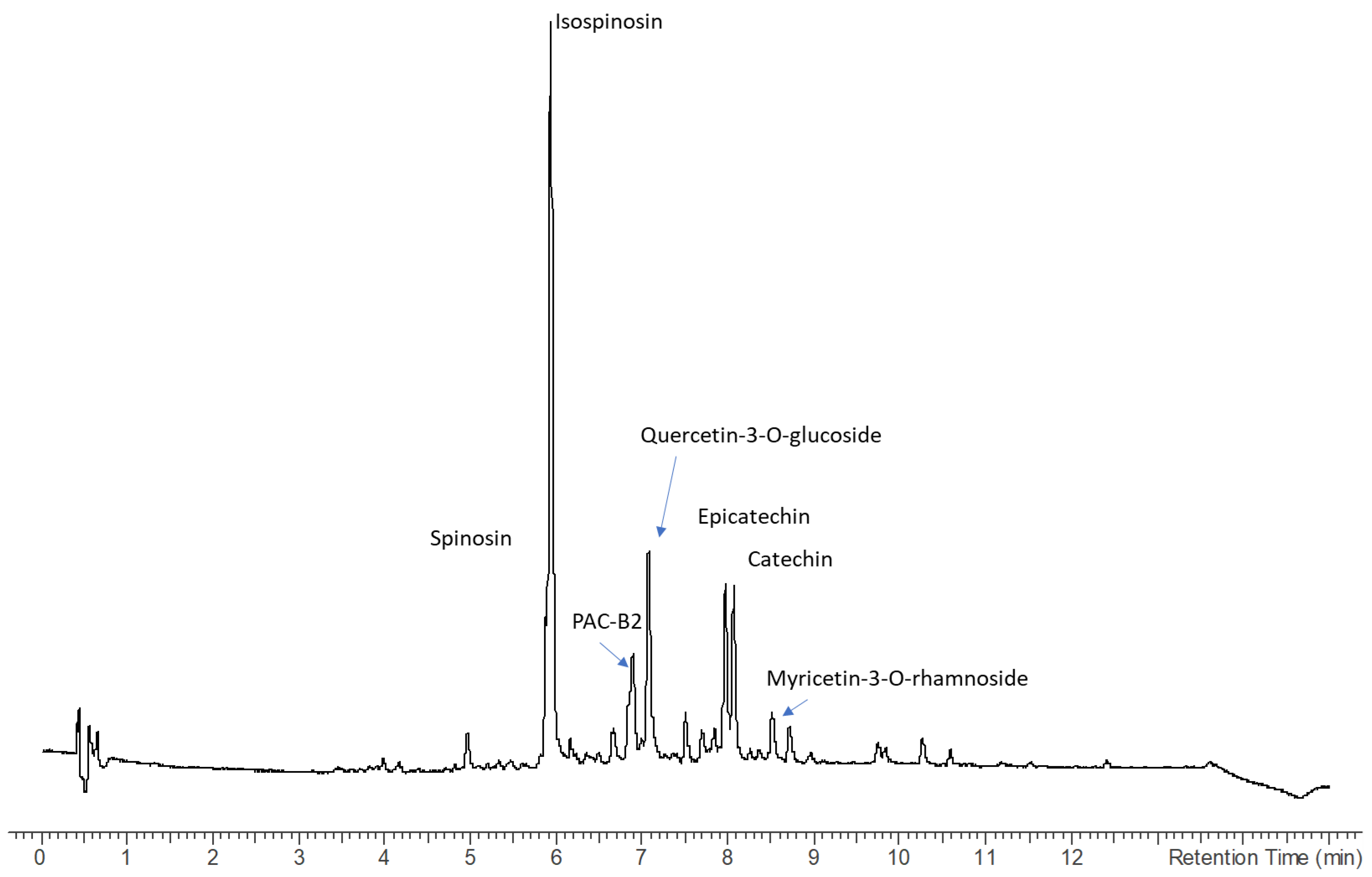
2.2. Chemical Composition by the Comprehensive Analysis of High-Resolution QTOF and Multiple-Stage Mass Spectrometry in Ion Trap

Identification of phytoconstituents was obtained combining the observation of diode array spectra for the compounds presenting significant UV absorption, high-resolution and MS/MS data from QTOF, as well as multiple-stage mass spectrometry fragmentation pathways (MSn) from the Ion Trap. Bark and leaves analysis showed the presence of several classes of secondary metabolites, and different compositions were observed for the two plant parts. The complete characterization is reported in Table 2, where the presence of each of the identified compounds in the different extracts obtained from bark and leaves is indicated.
More in detail, stem bark extract obtained in methanol (Figure 1) present intense signals at high retention time that can be ascribed to lipid fractions, mostly phospholipid derivatives. Several less intense signals can be ascribed to numerous compounds belonging to different classes of phytoconstituents and plant metabolites. A first group of compounds present low retention times and one compound showing molecular formula of C7H13NO, being tentatively assigned on the basis of the HR-MS and the fragment at m/z 71 to pyrrolidine alkaloid named Norhygrine (1-(pyrrolidin-2-yl)propan-2-one) [30]; this alkaloid has been also reported from the bark of the root of Punica granatum [31].
The UV spectra of the peaks observed in the stem bark extract mostly were related to flavan-3-ols presenting UV maximum at 280 nm. Procyanidin B2 was identified from the molecular formula C30H27O12 and the diagnostic fragments supporting the reaction of quinone methide fission (leading to m/z 289 and 291), retro Diels–Alder fission (leading to m/z 427 and 409), and heterocyclic ring fission (leading to m/z 453). Furthermore, the identity of compounds was confirmed by co-injection of an authentic standard. Peaks showing similar UV were also identified as flavan-3-ols, as catechin and epicatechin that were identified thanks to the observation in MS2 of m/z 139 [18], and also confirmed by standard injection. A derivative presenting molecular formula of C17H18O6 was tentatively assigned as a dimethyl derivative of catechin (or epicatechin). Root extract presents three isobaric peaks that have been tentatively identified as Secoisolariciresinol-sesquilignans derivatives on the basis of their mass spectra [32] but are not detectable in leaves. Phloridzin was also identified on the basis of MS spectrum [33] and confirmed by comparison with reference compounds. Other compounds that were identified both in bark and leaves, and are known constituents of ziziphus, are the isoquinoline alkaloid Magnoflorine [34] and the cyclopeptide Nummularine [35].
Leaves extract presents a higher number of compounds, and several peaks present UV ascribable not only to flavan-3-ols but also flavonols with UV maximum at 350–365 nm. Furthermore, peaks can be ascribed to coumarin derivatives based on the observed UV maximum at 320–330 nm. Compounds were identified from the HR-MS and fragments observed in Ion Trap and confirmed, when possible, with reference compounds. Identified compounds have been luteolin and myricetin-3-O-rhamnoside. Other relevant constituents of the leaves were the flavonoids spinosin, isospinosin, quercetin-3-O-glucoside, quercetin-3-O-rhamnoside, and myricetin-3-O-rhamnoside. All these phenolics and others reported in the table were previously described for ziziphus [18,36]. Further tentatively identified compounds are the pterosupin a C-glycoside phenol derivative that was previously isolated from Pterocarpum marsupium [37]. Also identified was the magnoflorine, an aporphyne alkaloid that was previously detected in many other medicinal species [38]. On the other hand, as previously reported in the literature [35,39,40] N-formylcyclopeptide alkaloids were detected, namely the Nummularin F, B, and U, both in bark and leaves. The chromatograms are given in Figure 1 and Figure 2.
As reported in Table 2, several classes of constituents were identified by comparing the obtained data with the literature; some compounds were annotated on the basis of HR-MS data and MSn fragmentation, others also confirmed by injection of reference compounds. Many constituents of Z. mauritania were reported in the recent paper by Qin et al. [34], namely the flavonoids, the phenylpropanoids, and the alkaloids, and our findings are in agreement with the previously reported identifications. Furthermore, we also identified the presence of some cyclopeptides, namely the nummularins that were described in Ziziphus [41]. The identification of the nummularins was supported by comparison of previously published mass spectrometric data [35]. Norhygrine is a pyrrolidine derivative previously reported from Asian Sedum species [30], but we tentatively identify it for the first time in Z. mauritania. Catechin and derivatives and procyanidins were detected and mass spectra compared with the literature [42,43] and finally compared with reference compounds as Phloridzin that also was previously identified in Ziziphus [33]. Other constituents previously described in this specie were the coumarins [44]. Secolignans were tentatively identified by comparison with mass spectrometric data [32]. Leaves present a larger number of identified constituents, with spinosin, isospinosin, myricetin, and quercetin derivatives only detected in the leaves, while secolignans were only detected in the bark.
The most abundant constituents were also quantified using a semiquantitative approach generating calibration curves with the appropriate reference standard. The most relevant bioactive phytoconstituents that can be extracted from the bark are catechin, epicatechin, and procyanidin B2, while flavonoids were prevalent in the leaves. Data are summarized in Table 3, and results are reported as mg/g of dried extract. An exemplificative chromatogram for the leaves extract is reported in Figure 2. Differences were observed in the extracts obtained with the homogenization (H) and maceration (Ma) with the different solvents. The comparison of extracts obtained with the same solvent but with the two techniques allows to observe a significant effect of the homogenization approach. Considering HAE and MAC for the flavonoids spinosyn, isospinosin, Quercetin-3-O-glucoside, Quercetin-3-O-rhamnoside, and Myricetin-3-O-rhamnoside, for these derivatives we observed a significant increase in the extraction using HAE, with changes of 5–10% compared to MAE. Several compounds present increased extraction using HAE, showing the improved efficiency compared to traditional maceration.
Such comparison also offered the opportunity to compare the extraction in water and with methanol. Aqueous extraction is in general less efficient compared to methanol in the observed dataset. Several phytoconstituents present limited water solubility, thus offering a less favorable solvent for extraction.
The observed differences comparing the homogenization and maceration results on the selected compounds are limited. A general, more efficient extraction using the homogenization approach is observed with average increment of the extraction of compounds in the range 5–10%. This result should be considered also taking into consideration the long time of the extraction for the two approaches. For homogenization, the extraction time was 5 h and for maceration was 24 h, thus a long time for possible extraction and equilibration of the compound’s concentration into and outside from the plant matrix.

2.3. Antioxidant Capacity

Aging and various human diseases, including inflammatory disorders, cancer, neurodegenerative conditions, and digestive ailments, have been linked to the excessive production of reactive oxygen species (ROS) and other free radicals [45,46]. Consequently, there has been a growing interest in seeking natural exogenous sources of antioxidants, with plant-based sources being considered a safer alternative to synthetic antioxidants [47]. In pursuit of this objective, previous study investigated the antioxidant efficacy of Ziziphus species, employing various strategies for assessing the antioxidant potential of test samples [48].
The current investigation results illustrate a strong correlation between antioxidant activity and TPC in the extracts. For instance, the HAE method using MeOH as the solvent demonstrated significantly higher antioxidant activity across all tested assays compared to MAE with MeOH. The highest antioxidant activity in leaves HAE MeOH extracts was observed in the ABTS assay, with a value of 747.25 mg TE/g, followed by the CUPRAC assay 698.46 mg TE/g, DPPH assay 414.30 mg TE/g, FRAP assay 325.59 mg TE/g, and PDB assay 3.91 mmol TE/g (Table 4). Similarly, the MeOH extracts from the bark displayed higher antioxidant activity, with the highest value recorded in the CUPRAC assay, 741.14 mg TE/g. The findings align with the information presented in Table 1 and Table 2, wherein the MeOH extracts from both leaves and bark exhibited elevated levels of secondary metabolites. Notably, spinosin, isospinosin, and quercetin derivatives were identified as the predominant compounds, with the highest concentration observed in the MeOH extract of leaves. Prior investigations also reported heightened antioxidant activity associated with spinosin [49,50]. Among the water extracts, HAE leaves extract exhibited the highest CUPRAC and FRAP activity, measuring 262.89 mg TE/g and 119.78 mg TE/g, respectively. In contrast, HAE bark extract displayed the highest ABTS and DPPH scavenging activity, measuring 189.52 mg TE/g and 103.25 mg TE/g, respectively. In MAC water extracts, both bark and leaves extracts showed the lowest PDB activity, with values of 0.84 mmol TE/g and 0.81 mmol TE/g, respectively.
A previous study reported that Z. mauritiana leaf extract has shown effectiveness in alleviating oxidative liver damage, possibly achieved through the scavenging of free radicals by antioxidant enzymes [48]. This plant species is notably abundant in carotenes and various phenolic compounds, including caffeic acid, p-hydroxybenzoic acid, ferulic acid, and p-coumaric acid [51], with naringenin as a predominant flavonoid [52]. Additionally, Ziziphus mauritiana fruit exhibits elevated levels of tannin and phytate [53], both of which possess antioxidant properties [54,55]. A study claimed that stressful desert conditions significantly improved the phytochemistry of Ziziphus species, leading to increased phenols and flavonoids in leaves and fruits, along with enhanced antioxidant activity [29]. Additionally, a separate investigation revealed that polar solvent extracts exhibited higher antioxidant activity compared to non-polar solvent extracts in Z. jujuba. Notably, the MeOH extract demonstrated the most potent free radical-scavenging activity, with an IC50 value of 20.44 ± 0.18 μg/mL [27]. The MAC activity of the leaves extracted using MeOH was comparable to the MAC activity reported in a previous study on Z. jujuba fruits (11.04 mg EDTAE/g).

2.4. Enzyme Inhibitory Activity

2.4.1. Neuroprotective Effects

Alzheimer’s disease (AD) is a neurodegenerative disorder marked by the progressive loss of memory and cognitive function. The anticipated increase in its frequency among the elderly demographic is expected to increase 3-fold by 2050 [56]. AD resides within the category of conditions that continue to present challenges to medicinal chemists. The quest for highly efficacious drugs to address AD remains an ongoing endeavor. In this context, the inhibition of cholinesterase stands as a promising strategy to elevate acetylcholine levels in the brain [57]. The two inhibitors, AChE and BChE, show immense promise and have attracted considerable attention from researchers.
In the present investigation, the AChE and BChE inhibitory activity was determined. The results indicated in Table 5 showed enzyme inhibitory activity: the HAE MeOH extract from the leaves displayed the highest AChE inhibitory activity, measuring 2.55 mg GALAE/g, followed closely by the bark MAC MeOH extract at 2.41 mg GALAE/g, indicating its potential in neuroprotective effect. In contrast, the HAE water extract from the leaves exhibited the lowest AChE inhibitory activity, measuring only 0.09 mg GALAE/g. Interestingly, there was no inhibition observed in the MAC water extracts from both leaves and bark, as well as in the HAE water extracts from the bark (Table 5).
For BChE inhibition, the MeOH extracts from the bark in both the HAE and MAC methods showed values of 1.57 mg GALAE/g and 1.14 mg GALAE/g, respectively. However, none of the other extracts from both parts of the plant exhibited any BChE inhibitory activity.
The MeOH extracts examined in this study were found to contain abundant spinosin and isospinosin (refer to Table 2), suggesting a potential explanation for the observed AChE) inhibition. Recent research indicates that spinosin may offer benefits in addressing learning and memory deficits associated with AD through its impact on multiple targets [58]. Additionally, Wang et al. reported in their study that a spinosin derivative isolated from Z. mauritiana demonstrated the inhibition of AChE [59]. Secondary metabolites isolated from Z. oxyphylla roots inhibited AChE and BChE at different concentrations. Ziziphus is rich in bioactive compounds; a study found peptids of Z. jujuba responsible for the inhibition of AChE and BChE [60].

2.4.2. Dermatoprotective Effects

Tyrosinase plays a pivotal role as the key enzyme in pigment synthesis, initiating a series of reactions that convert the amino acid tyrosine into the melanin biopolymer [61]. Tyrosinase inhibitors hold particular significance in cosmetic applications for their skin-whitening effects [62]. The production of abnormal melanin pigmentation, resulting in a dark complexion, presents a significant aesthetic concern for individuals [63]. Given that plants offer a rich source of bioactive chemicals with a minimal risk of adverse side effects, there has been a growing interest in harnessing them as a natural source of tyrosinase inhibitors. This heightened interest stems from the fact that tyrosinase is a crucial enzyme in the melanin synthesis pathway known as melanogenesis, making it the primary and successful target for inhibitors that directly impede its catalytic activity. It is noteworthy that a majority of commercially available cosmetics and skin-lightening agents are, in fact, tyrosinase inhibitors [64].
In terms of tyrosinase inhibitory activity, the MeOH extracts from both leaves and bark, processed by HAE, demonstrated the highest tyrosinase inhibitory activity levels at 75.97 mg KAE/g and 75.48 mg KAE/g, respectively (Table 5). These results were closely followed by the MAC MeOH extracts from the bark at 74.95 mg KAE/g and from the leaves at 73.79 mg KAE/g, respectively. In contrast, the MAC water extracts from both leaves and bark displayed lower tyrosinase activity when compared to the tested MeOH extracts. Among these, the bark extract showed the lowest tyrosinase activity, measuring only 13.55 mg KAE/g. According to Moon et al., the inhibitory effects of certain flavonoids, including spinosin, quercetin, and kaempferol isolated from Z. jujuba, were investigated. Among these compounds, spinosin demonstrated the most potent tyrosinase inhibitory activity [65].

2.4.3. Antidiabetic Effect

Hyperglycemia, an excess of blood sugar, is a serious concern. Elevated post-meal glucose levels can predict diabetes complications [66,67]. Most synthetic antidiabetic drugs target type 2 diabetes by addressing insulin issues, but some like metformin have adverse effects at high doses [68]. Thus, the main goal in antidiabetic research is to find safe and effective agents without side effects. In pursuit of this objective, researchers have turned to traditional medicines and foods from diverse cultures, exploring them for potential clues and insights that could lead to the development of new therapeutic drugs [26,69,70]. Targeting enzymes like α-amylase and α-glucosidase can help reduce post-meal hyperglycemia [71,72].
Table 4 shows inhibition of glucose digestive enzymes. The MeOH extracts demonstrated the highest α-amylase inhibitory activity. The HAE-MeOH extract from the leaves exhibited an inhibition value of 0.78 mmol ACAE/g, followed closely by the MAC-MeOH extract from the leaves at 0.76 mmol ACAE/g, and the HAE MeOH extract from the bark at 0.72 mmol ACAE/g, suggesting its potential in modulating cholinergic neurotransmission [73]. The MAC-MeOH extract from the bark showed an inhibition value of 0.69 mmol ACAE/g. On the other hand, the water extracts from the leaves, obtained through MAC, exhibited a lower inhibition value of 0.15 mmol ACAE/g, while the water extracts from the bark, obtained through HAE, had a lower inhibition value of 0.13 mmol ACAE/g. In terms of α-glucosidase inhibitory activity, the MeOH extracts from the leaves, both from MAC and HAE, exhibited higher and similar levels of inhibition, measuring 2.11 mmol ACAE/g. However, their bark extracts showed no inhibition. The water extracts from the bark, obtained through both HAE and MAC, exhibited similar inhibition values of 1.87 mmol ACAE/g.
Our discovery regarding the MeOH extracts from Ziziphus exhibiting inhibition against α-amylase and α-glucosidase aligns with prior research findings [74,75]. Suksamrarne and colleagues demonstrated in their study that the MeOH extract of various Ziziphus plants displayed superior α-glucosidase inhibitory activity compared to their other extracts [74].

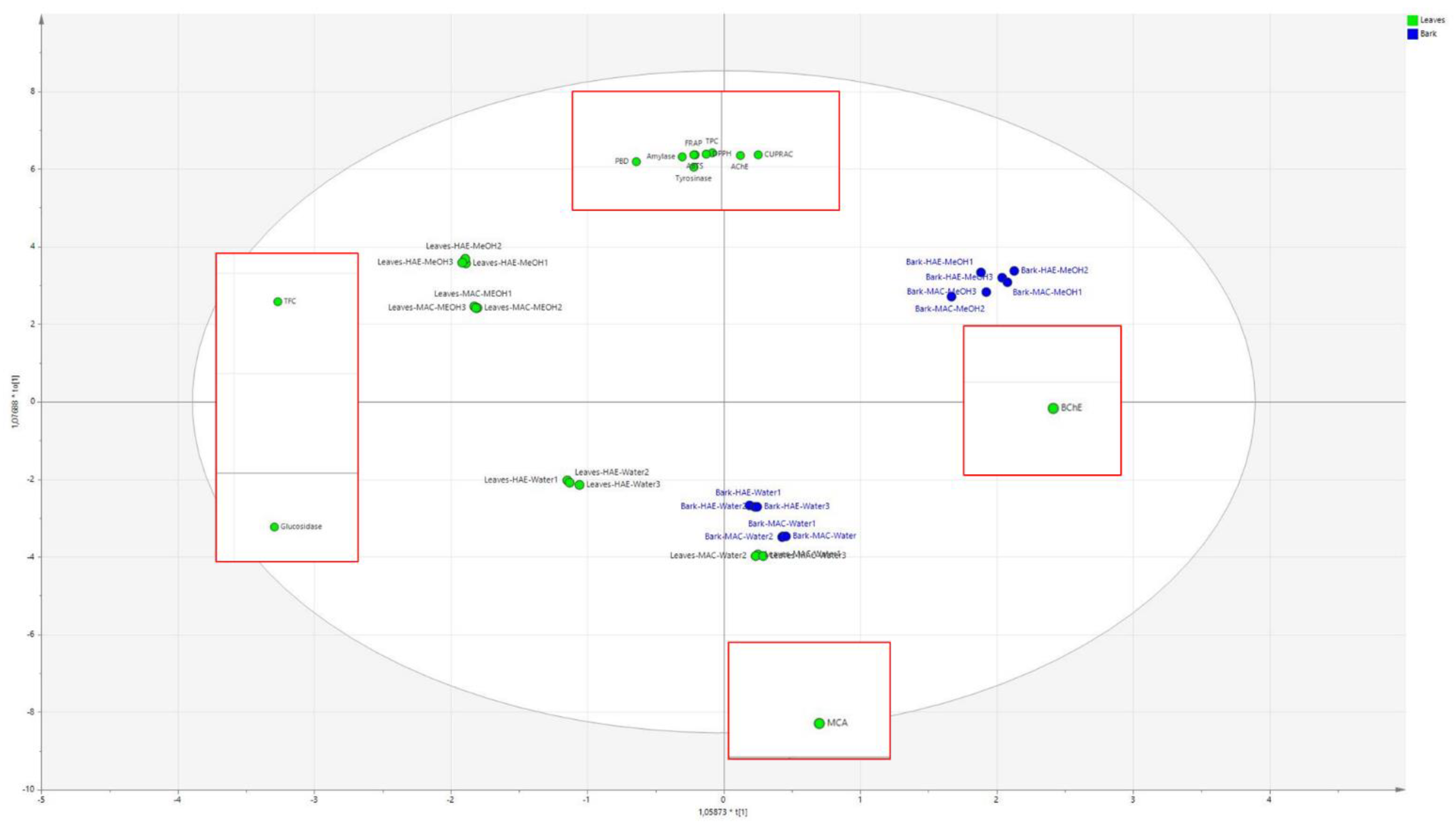
2.5. Multivariate Data Analysis

In this work, we generated large amounts of experimental data as results of spectrophotometric assays, enzymatic inhibitions. Thus, to establish correlations between observed bioactivities and chemical constituents, we adopted multivariate data analysis using PCA and OPLS-DA. We can observe that the extracts on the basis of the spectrophotometric and enzyme inhibitory assays can be divided into three similar groups, one that is in the –x; +y part of the plot is formed by the Leaves HAE methanol extract and this group presents TFC and glucosidase activities as the most discriminant descriptors. On the opposite side (+x; +y), the group was formed by bark HAE and MAC with methanol, which strongly correlates with butyrlcholinesterase inhibition. On the lower side of the plot (–y) is a group formed by leaves HAE water and bark HAE water that is more correlated with MCA. In the end, the obtained data can help in the description of the observed activities and spectrophotometric determination obtained for the extracts. Namely, methanol solvent, when used both with the maceration or the HAE, is strongly associated with good results in almost all the antioxidant assays, namely TRAP, TPC, DPPH, CUPRAC, and ABTS. The most relevant activities on the observed enzymes are the tyrosinase and amylase and acetylcholinesterase. This information shows that the extraction with methanol from the leaves and the bark is able to yield compounds with high antioxidant capacity and significant inhibitory activities. At the same time, a different behavior is observed considering leaves and bark. In fact, the leaves are strongly correlated also with TPC and glucosidase, while bark mostly with butyrlcholinesterase inhibition. On the other hand, the samples extracted with water are more related to MCA activity. All these results are summarized in Figure 3 that presents the superimposition of the loading plot of the OPLS-DA, and in the red square, the corresponding score plot showing the most relevant variables for the group of extracts.
Considering the results of the chemical analysis, we can observe that the leaves are rich in spinosyn and isopsinosin compared to the bark and present quercetin-3-O-rhamnoside as well as luteolin. Furthermore, only the leaves contain magnoflorin, caftaric acid, and gallocatechin, and all these compounds may present some specific activities on the observed enzymes. Bark is, on the other hand, characterized by the presence of some secoisolariciresionl-sesquilignan derivatives that are not detected in the leaves.

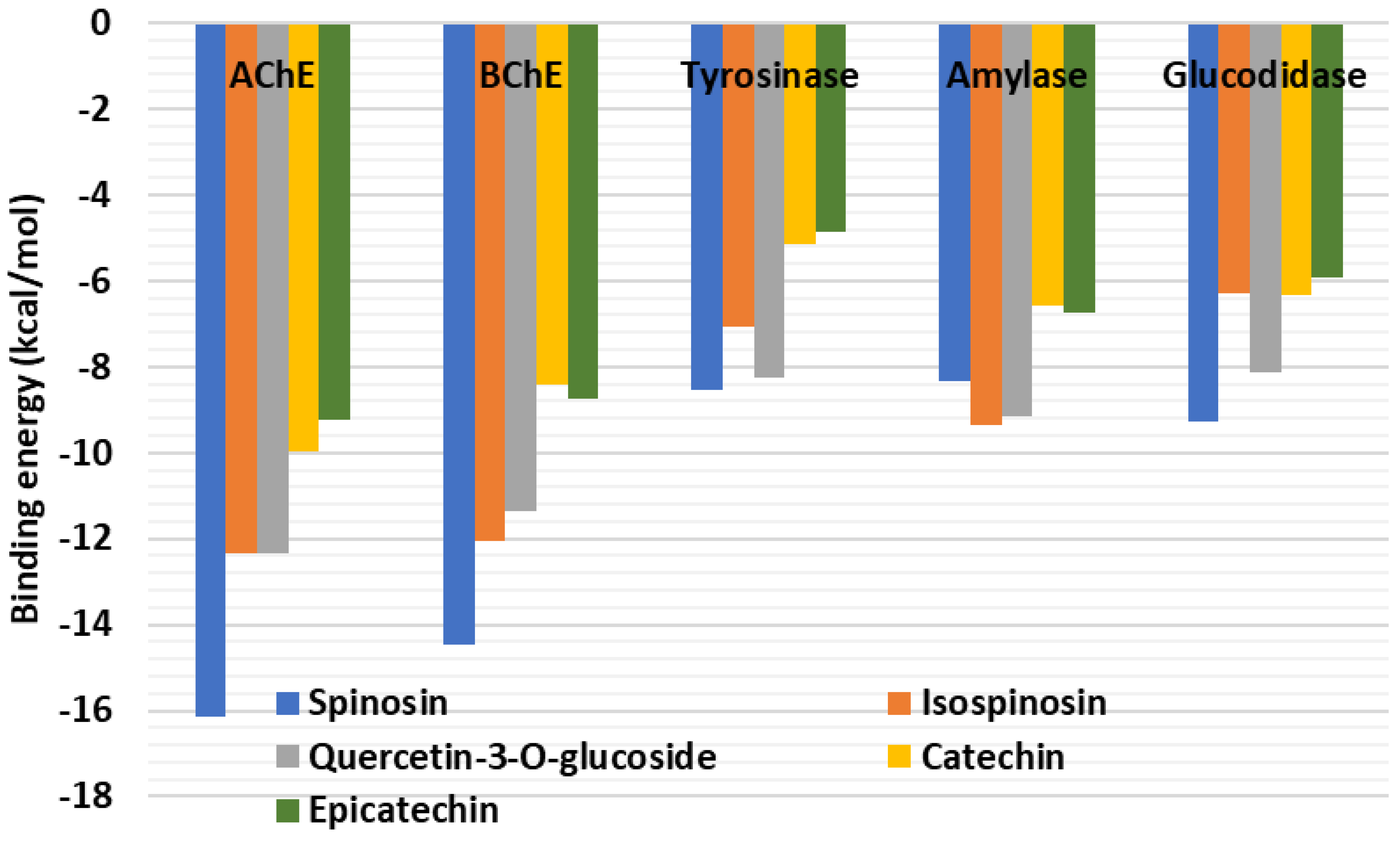
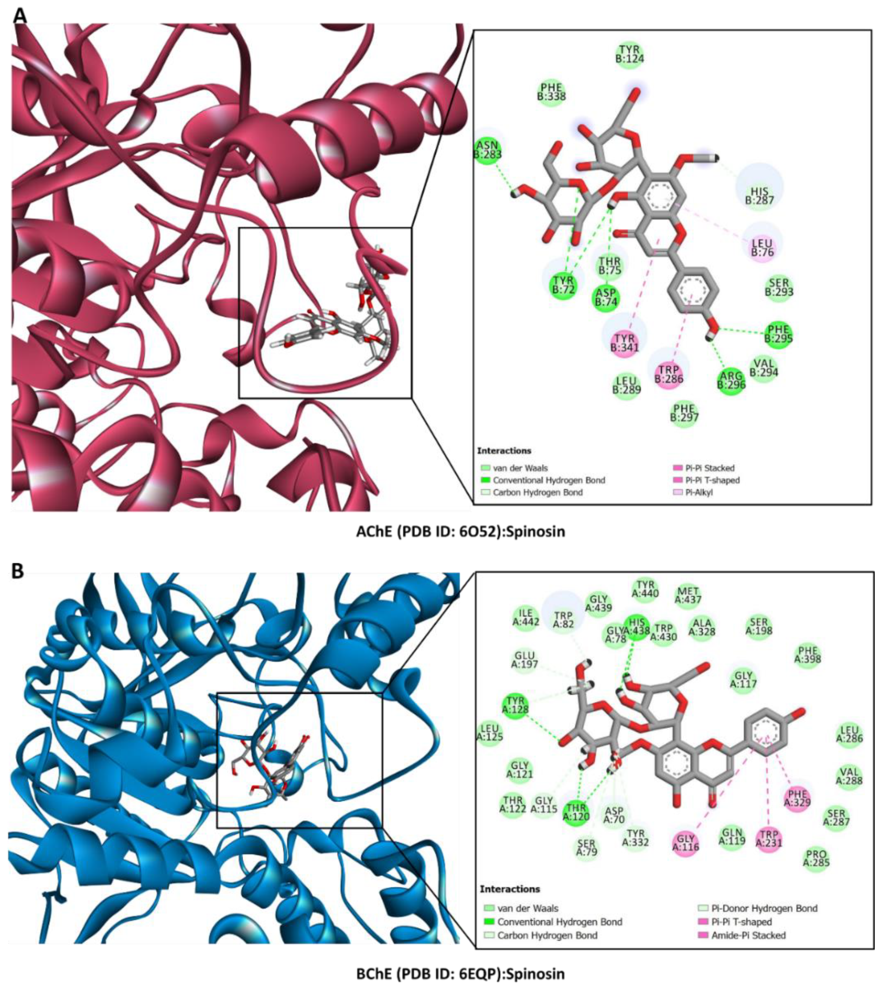
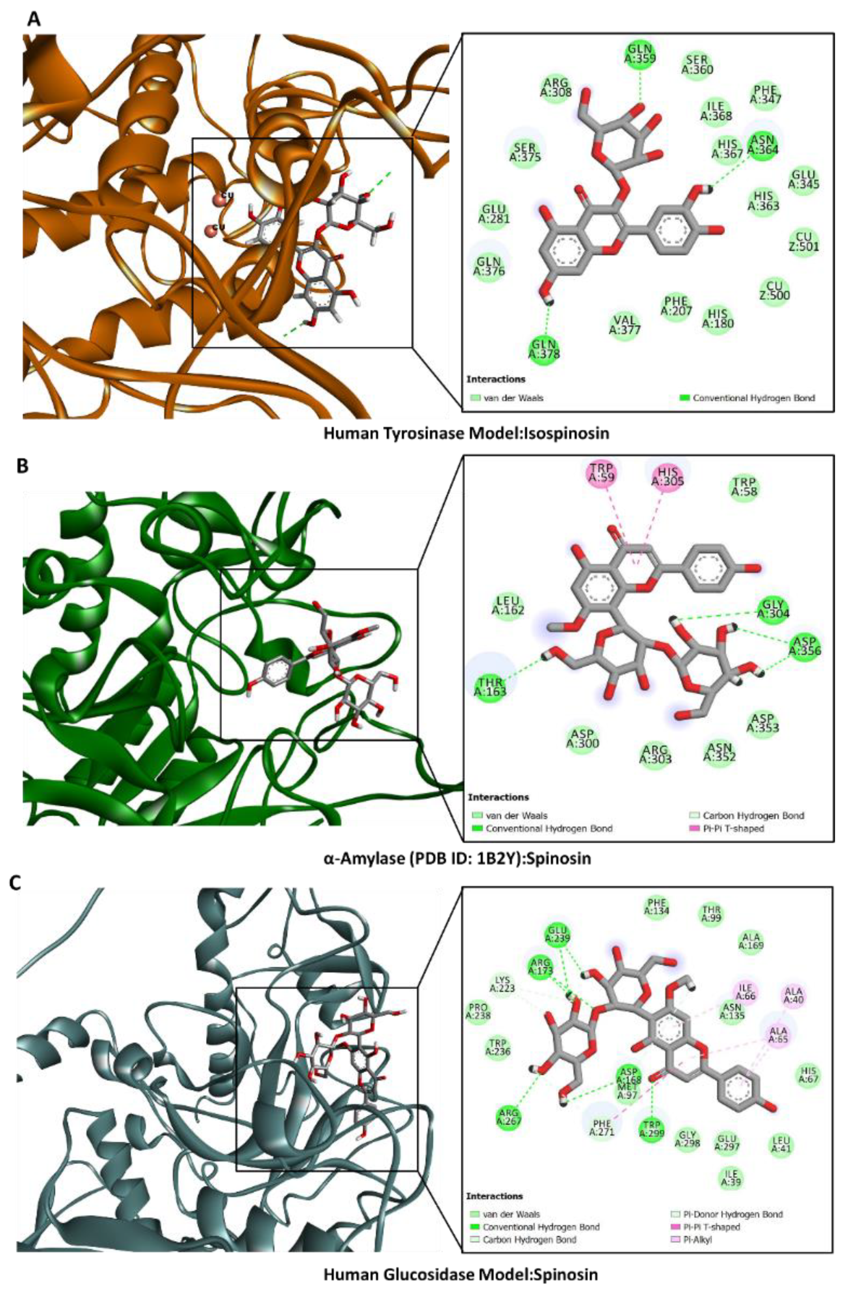
2.6. Molecular Docking

Molecular docking was used to examine how the main components in the Z. mauritiana extracts bound to the selected target enzymes. Figure 4 displays the ligands’ binding energy scores. Spinosin, isospinosin, and quercetin-3-O-glucoside were predicted to have the highest binding energy against the target enzymes. For instance, spinosin strongly bound to the active site of AChE mainly via H-bonding Tyr72, Asp74, Asn283, Phe295, and Arg296; as well as π–π stacked and π–π T-shaped with Trp286 and Tyr341, respectively (Figure 5A). Similarly, the same compound formed firm interactions with BChE mainly via H-bonding with Thr120, Tyr128, and His438 deep inside the active site supported by the π–π T-shaped and amide–π stacked interactions close to the entrance to the channel (Figure 6B). Interestingly, the key interactions between the built human tyrosinase model and isospinosin were H-bonds with Gln359, Asn364, and Gln378. Other supporting interactions, like in the above complexes, were multiple van der Waals interactions, including those with the catalytically essential active site cupper ions (Figure 6A). Furthermore, spinosin was accommodated in the catalytic cavity of amylase via the formation of H-bonds with Thr163, Gly304, and Asp356, as well as π–π T-shaped interactions with Trp59 and His304 (Figure 6B). Finally, the key interactions between the model of human glucosidase and spinosin were also H-bonds with Asp168, Arg173, Glu239, Arg267, strengthened by hydrophobic contacts with Ala40, Ala65, and Ile66, as well as multiple van der Waals interactions all over the active site (Figure 6C). The dominant compounds in the extracts of Ziziphus mauritiana bark and leaves extracts may therefore be interacting with the chosen proteins to inhibit their functions.

3. Materials and Methods

3.1. Plant Material

In the summer of 2021, the leaves (almost 500 g) and barks (almost 200 g) of Z. mauritiana were gathered in the vicinity of Bouake (Côte d’Ivoire) by the botanist Ouattara Katinan Etienne. Voucher specimens were deposited at the herbarium in the Nangui Abrogoua University (KIS-21-175). Prior to extraction, the plant materials (leaves and stem barks) were carefully washed with tap and distilled water to eliminate any soil and contaminants. After being air-dried for 10 days (in shade at room temperature), the plant samples were powdered.

3.2. Sample Preparation

Two extraction techniques were used to prepare the extracts, namely homogenizer-assisted (HAE) and maceration (MAC). Two solvents (methanol and water) were used in these techniques. To prepare HAE, 5 g of plant samples were extracted with the solvents (100 mL) at 6000 g in a homogenizer for 5 h (IKA-Ultraturrax). Regarding MAC, the plant samples (5 g) were macerated with the solvents (100 mL) for 24 h at room temperature. After extraction, all extracts were filtered, and the methanol extracts were dried using a rotary evaporator. As for the water extracts, after filtration, the extracts were dried under vacuum using a lyophilizer. All extracts were stored at 4 °C until analysis.

3.3. Assay for Total Phenolic and Flavonoid Contents

According to the methods specified by [76], total phenolics and flavonoids were quantified. Gallic acid (GA) and rutin (RE) served as standards in the assays, and the outcomes were reported as gallic acid equivalents (GAEs) and rutin equivalents. All experimental details are given in the Supplementary Materials.

3.4. High-Resolution LC-QTOF-MS and Multiple-Stage Mass Spectrometry by Ion Trap Analysis

The phytochemical analysis was performed combining ultraperformance liquid chromatography with diode array and electrospray Quadrupole Time Of Flight Mass Spectrometry (Agilent Technology, Santa Clara, CA, USA) and Multiple-Stage fragmentation in Ion Trap (Agilent Technology). An Agilent 1290 UPLC system equipped with an autosampler and 1290 series Diode Array was used as chromatograph. After the column, the flow was split with a passive T junction, and liquid was sent to diode array or mass spectrometer. The Agilent 6530 QTOF (Agilent Technology) was used as a detector; the instrument is equipped with a Jet Stream source and was operating in positive ion mode. During the acquisition, the mass values were calibrated using the Agilent calibration mixture. The parameters of the MS were as follows: gas temperature 350 °C, fragmentor 250 V, skimmer 45 V, drying gas 6 L/min, nebulizer 25 psig, Shealth temp 275 °C.
As stationary phase, an Agilent SB C18 (3 × 100 mm; 1.7 micron) was used. Eluents were water 0.1% formic acid (A), and acetonitrile with 0.1% formic acid (B). The flow rate was set to 0.5 mL/min. Gradient started with 90% of A remaining isocratical for 1 min then going to 85% B in 10 min, and then reached 90% B in 12 min and remained isocratical up to 14 min. For the multiple-stage mass spectrometry, an Agilent 1260 chromatograph was used. As stationary phase, an Agilent SB C-18 (Agilent Technology) was used (4.6 × 50; 1.8 micron). As eluents, water 1% formic acid (A), Acetonitrile (B), and methanol (C) were used. Gradient started with 95% A and 5% B, then in 2.5 min 85% A and 15% B, then in 12 min 80% A, 18% B and 2% methanol, and in 15.5 min 50% A, 40% B, and 10%C, then at 19 min 20% A, 70% B, and 10% C. The flow rate was 0.750 μL/min. Flow after the column was split by a T passive junction, and half was sent to diode array and half to a Varian MS500 (Varian Technology, Ontario, ON, Canada) equipped with an ESI source that was used and operated in turbo DDS mode acquiring MSn spectra of eluted species. Drying gas was set at 45 psi, while nebulizer at 18 psi. The drying gas temperature was 340 °C at the beginning of the chromatography and decreased at 5 °C/min for 20 min. Capillary was set to 80 °C, RF loading was 85%. For semiquantitative analysis, reference compounds were used selecting compounds belonging to flavan-3-ols, flavonoids, and coumarins. Calibration curves for catechin, epicatechin, gallocatechin, and procyanidin B2 were obtained using solution at four different concentrations of the four reference standards, at 2 µg/mL, 10 µg/mL, 20 µg/mL, and 50 µg/mL, and collecting chromatogram at 280 nm. Quercetin-3-O-glucoside was used for quantification of the flavonoids, and a calibration curve was obtained using solutions obtained at 2 µg/mL, 10 µg/mL, 20 µg/mL, and 50 µg/mL and collecting the chromatogram at 350 nm. For the coumarin quantification, auraptene and geranyl umbelliferone were used, generating calibration curves using solutions at 5 µg/mL, 10 µg/mL, 15 µg/mL, and 30 µg/mL and collecting the chromatogram at 330 nm. An exemplificative chromatogram of leaves extract is reported in Figure 3.
Data obtained from the QTOF (high-resolution) and Ion Trap (low-resolution) mass spectrometry were combined to establish the identity of the eluted compounds.

3.5. In Vitro Bioactivity Assays

The assessment of the biological activity of the obtained extracts was carried out by measuring the antioxidant [77], and enzyme-inhibitory potential [77]. With the aim of comprehensive insight into the real antioxidant potential, six different tests were used (DPPH, ABTS, FRAP, CUPRAC, PBD, and MCA tests). The enzyme-inhibitory potential of obtained extracts was estimated towards five different clinically important enzymes: tyrosinase, amylase, glucosidase, acetylcholinesterase, and butrylcholinesterase. All experimental details are given in the Supplementary Materials.

3.6. Molecular Modeling

The X-ray crystal structures of the following target proteins were downloaded from the protein data bank (https://www.rcsb.org/ accessed on 20 May 2024) [78]: AChE (PDB ID: 6O52) [79] α-amylase (PDB ID: 1B2Y) [80], BChE (PDB ID: 6EQP) [81]. A homology model of human tyrosinase constructed using UniProt entry P14679 as the target sequence and using the crystal structure of tyrosinase from Priestia megaterium (PDB ID: 6QXD) [82] as the template was retrieved [83]. Moreover, the homology model of human α-glucosidase built using the crystal structure of Mus musculus glucosidase (PDB ID: 7KBJ) [84] as a template, and using the UniProt entry P0DUB6 as the target sequence, was obtained from the same study [83]. Protein preparation was performed according to the protocol described previously [85]. Each ligand’s three-dimensional structure was retrieved from the ChemSpider database (https://www.chemspider.com/ accessed on 20 May 2024). The UCSF Chimera tool was used to optimize the geometry of the ligand’s three-dimensional structures [86]. With the help of MGLTools 1.5.6 software, all docking grid files were created utilizing the cocrystal ligand in each crystal structure. This program combines all hydrogen atoms and gives all protein atoms Gasteiger partial charges. The π Lamarckian genetic method included in AutoDock 4.2.6 software was used to simulate docking (https://autodock.scripps.edu/ accessed on 20 May 2024) [87], implementing the docking protocol as detailed in Ref. [88]. Using Biovia DS Visualizer v4.5, each ligand’s binding energy scores were calculated, and protein–ligand interactions were investigated (BIOVIA, San Diego, CA, USA).

3.7. Statistical Analysis

Statistical analysis was performed using Xl Stat (Version 16). All analyses were conducted in triplicates (n = 3) and presented as mean values with their standard deviation (mean value ± std). Differences between samples were examined using one-way analysis of variance (AVOVA) and Tukey’s post hoc test with significance level set at p < 0.05. For multivariate analysis, the data from the spectrophotometrical assays and enzyme inhibitory assays were used to generate a table containing the extracts and the respective results. The matrix was loaded in SIMCA 12 and pareto-scaled. Data initially were used to obtain a PCA, then an OPLS-DA to describe the different obtained extracts.

4. Conclusions

The comparison of extraction methods for Z. mauritiana bark and leaves showed that homogenization-assisted extraction (HAE) using MeOH was more effective, resulting in higher phenolic content and antioxidant activity compared to maceration extraction (MAE) with MeOH. It is important to note that the MeOH and water extracts from the leaves contained higher levels of secondary metabolites, such as spinosin and isospinosin. Based on the chemical structures of the most abundant compounds, we can infer that the flavonols present contributed to the antioxidant and enzyme-inhibitory activities observed in the extracts. In terms of industrial insights, our results can be valuable, and we have suggested that the HAE technique can be more effective in producing functional applications, including novel pharmaceuticals or cosmeceuticals, by using Z. mauritiana. In future studies, the main components of Z. mauritiana extracts will be isolated, with the aim of investigating in more detail the role of the different classes of phytoconstituents in the observed bioactivities. However, researchers should consider the toxicity of methanol, and we suggest the use of ethanol or an ethanol/water mixture in future studies.

Supplementary Materials

The following supporting information can be downloaded at: https://www.mdpi.com/article/10.3390/plants13162195/s1, Reference [89] is cited in the supplementary materials.

Author Contributions

N., K.I.S., S.D., S.S. and G.Z. performed the biological activity and chemical composition analysis. A.I.U. performed molecular docking analysis. K.I.S. and O.K.E. performed a field study for plant collection. C.F. and J.A. performed multivariate analysis. All authors have read and agreed to the published version of the manuscript.

Funding

This research received no external funding.

Data Availability Statement

Data are contained within the article and Supplementary Materials.

Conflicts of Interest

The authors declare no conflicts of interest.

References

  1. Ziada, A.S.; Smith, M.-S.R.; Côté, H.C. Updating the free radical theory of aging. Front. Cell Dev. Biol. 2020, 8, 575645. [Google Scholar] [CrossRef]
  2. Wawi, M.J.; Mahler, C.; Inguimbert, N.; Marder, T.B.; Ribou, A.-C. A new mitochondrial probe combining pyrene and a triphenylphosphonium salt for cellular oxygen and free radical detection via fluorescence lifetime measurements. Free Radic. Res. 2022, 56, 258–272. [Google Scholar] [CrossRef] [PubMed]
  3. Maddu, N. Diseases Related to Types of Free Radicals; IntechOpen: London, UK, 2019. [Google Scholar]
  4. Hawkins, C.L.; Davies, M.J. Role of myeloperoxidase and oxidant formation in the extracellular environment in inflammation-induced tissue damage. Free Radic. Biol. Med. 2021, 172, 633–651. [Google Scholar] [CrossRef]
  5. Peña-Bautista, C.; Baquero, M.; Vento, M.; Cháfer-Pericás, C. Free radicals in Alzheimer’s disease: Lipid peroxidation biomarkers. Clin. Chim. Acta 2019, 491, 85–90. [Google Scholar] [CrossRef] [PubMed]
  6. Acquaviva, A.; Nilofar; Bouyahya, A.; Zengin, G.; Di Simone, S.C.; Recinella, L.; Leone, S.; Brunetti, L.; Uba, A.I.; Cakilcioğlu, U. Chemical Characterization of Different Extracts from Artemisia annua and Their Antioxidant, Enzyme Inhibitory and Anti-Inflammatory Properties. Chem. Biodivers. 2023, 20, e202300547. [Google Scholar] [CrossRef]
  7. Barrientos, R.E.; Ahmed, S.; Cortés, C.; Fernández-Galleguillos, C.; Romero-Parra, J.; Simirgiotis, M.J.; Echeverría, J. Chemical fingerprinting and biological evaluation of the endemic Chilean fruit Greigia sphacelata (Ruiz and Pav.) Regel (Bromeliaceae) by UHPLC-PDA-orbitrap-mass spectrometry. Molecules 2020, 25, 3750. [Google Scholar] [CrossRef]
  8. Nilofar; Duran, T.; Uba, A.I.; Cvetanović Kljakić, A.; Božunović, J.; Gašić, U.; Bouyahya, A.; Yıldiztugay, E.; Ferrante, C.; Zengin, G. Extractions of aerial parts of Hippomarathrum scabrum with conventional and green methodologies: Chemical profiling, antioxidant, enzyme inhibition, and anti-cancer effects. J. Sep. Sci. 2023, 47, 2300678. [Google Scholar] [CrossRef] [PubMed]
  9. Prakash, O.; Usmani, S.; Singh, R.; Singh, N.; Gupta, A.; Ved, A. A panoramic view on phytochemical, nutritional, and therapeutic attributes of Ziziphus mauritiana Lam.: A comprehensive review. Phytother. Res. 2021, 35, 63–77. [Google Scholar] [CrossRef]
  10. Grice, A. Seed production, dispersal and germination in Cryptostegia grandiflora and Ziziphus mauritiana, two invasive shrubs in tropical woodlands of northern Australia. Aust. J. Ecol. 1996, 21, 324–331. [Google Scholar] [CrossRef]
  11. El Maaiden, E.; El Kharrassi, Y.; Qarah, N.A.; Essamadi, A.K.; Moustaid, K.; Nasser, B. Genus Ziziphus: A comprehensive review on ethnopharmacological, phytochemical and pharmacological properties. J. Ethnopharmacol. 2020, 259, 112950. [Google Scholar] [CrossRef]
  12. Alsayari, A.; Wahab, S. Genus Ziziphus for the treatment of chronic inflammatory diseases. Saudi J. Biol. Sci. 2021, 28, 6897–6914. [Google Scholar] [CrossRef] [PubMed]
  13. Tahergorabi, Z.; Abedini, M.R.; Mitra, M.; Fard, M.H.; Beydokhti, H. “Ziziphus jujuba”: A red fruit with promising anticancer activities. Pharmacogn. Rev. 2015, 9, 99. [Google Scholar] [PubMed]
  14. Ebrahimi, S.; Mollaei, H.; Hoshyar, R. Ziziphus Jujube: A review study of its anticancer effects in various tumor models invitro and invivo. Cell. Mol. Biol. 2017, 63, 122–127. [Google Scholar] [CrossRef]
  15. Da Costa Mousinho, N.M.; van Tonder, J.J.; Steenkamp, V. In vitro anti-diabetic activity of Sclerocarya birrea and Ziziphus mucronata. Nat. Prod. Commun. 2013, 8, 1934578X1300800924. [Google Scholar] [CrossRef]
  16. Available online: https://powo.science.kew.org/taxon/urn:lsid:ipni.org:names:719349-1 (accessed on 12 May 2024).
  17. Delfanian, M.; Esmaeilzadeh Kenari, R.; Sahari, M.A. Utilization of Jujube fruit (Ziziphus mauritiana Lam.) extracts as natural antioxidants in stability of frying oil. Int. J. Food Prop. 2016, 19, 789–801. [Google Scholar] [CrossRef]
  18. Zozio, S.; Servent, A.; Cazal, G.; Mbéguié-A-Mbéguié, D.; Ravion, S.; Pallet, D.; Abel, H. Changes in antioxidant activity during the ripening of jujube (Ziziphus mauritiana Lamk). Food Chem. 2014, 150, 448–456. [Google Scholar] [CrossRef] [PubMed]
  19. Li, J.-W.; Fan, L.-P.; Ding, S.-D.; Ding, X.-L. Nutritional composition of five cultivars of Chinese jujube. Food Chem. 2007, 103, 454–460. [Google Scholar] [CrossRef]
  20. Zhang, H.; Jiang, L.; Ye, S.; Ye, Y.; Ren, F. Systematic evaluation of antioxidant capacities of the ethanolic extract of different tissues of jujube (Ziziphus jujuba Mill.) from China. Food Chem. Toxicol. 2010, 48, 1461–1465. [Google Scholar] [CrossRef]
  21. Belwal, T.; Ezzat, S.M.; Rastrelli, L.; Bhatt, I.D.; Daglia, M.; Baldi, A.; Devkota, H.P.; Orhan, I.E.; Patra, J.K.; Das, G.; et al. A critical analysis of extraction techniques used for botanicals: Trends, priorities, industrial uses and optimization strategies. TrAC Trends Anal. Chem. 2018, 100, 82–102. [Google Scholar] [CrossRef]
  22. Picot-Allain, C.; Mahomoodally, M.F.; Ak, G.; Zengin, G. Conventional versus green extraction techniques—A comparative perspective. Curr. Opin. Food Sci. 2021, 40, 144–156. [Google Scholar] [CrossRef]
  23. Márquez, K.; Márquez, N.; Ávila, F.; Cruz, N.; Burgos-Edwards, A.; Pardo, X.; Carrasco, B. Oleuropein-Enriched Extract From Olive Mill Leaves by Homogenizer-Assisted Extraction and Its Antioxidant and Antiglycating Activities. Front. Nutr. 2022, 9, 895070. [Google Scholar] [CrossRef]
  24. Yang, C.-H.; Li, R.-X.; Chuang, L.-Y. Antioxidant activity of various parts of Cinnamomum cassia extracted with different extraction methods. Molecules 2012, 17, 7294–7304. [Google Scholar] [CrossRef] [PubMed]
  25. Munteanu, I.G.; Apetrei, C. Analytical methods used in determining antioxidant activity: A review. Int. J. Mol. Sci. 2021, 22, 3380. [Google Scholar] [CrossRef] [PubMed]
  26. Shakeel, A.; Kouadio Ibrahime, S.; Nilofar; Claudio, F.; Ozan Emre, E.; Ouattara Katinan, E.; Gokhan, Z. Online-HPLC Strategies, Antioxidant and Enzyme Inhibitory Activities of Different Extracts of Mondia whitei Leaves. J. Biol. Regul. Homeost. Agents 2023, 37, 6029–6039. [Google Scholar]
  27. Al-Reza, S.M.; Bajpai, V.K.; Kang, S.C. Antioxidant and antilisterial effect of seed essential oil and organic extracts from Zizyphus jujuba. Food Chem. Toxicol. 2009, 47, 2374–2380. [Google Scholar] [CrossRef] [PubMed]
  28. Uddin, N.; Ali, N.; Uddin, Z.; Nazir, N.; Zahoor, M.; Rashid, U.; Ullah, R.; Alqahtani, A.S.; Alqahtani, A.M.; Nasr, F.A. Evaluation of cholinesterase inhibitory potential of different genotypes of Ziziphus nummularia, their HPLC-UV, and molecular docking analysis. Molecules 2020, 25, 5011. [Google Scholar] [CrossRef] [PubMed]
  29. Riaz, M.U.; Raza, M.A.; Saeed, A.; Ahmed, M.; Hussain, T. Variations in morphological characters and antioxidant potential of different plant parts of four Ziziphus Mill. species from the Cholistan. Plants 2021, 10, 2734. [Google Scholar] [CrossRef]
  30. Kim, J.H.; Hart, H.T.; Stevens, J.F. Alkaloids of some Asian Sedum species. Phytochemistry 1996, 41, 1319–1324. [Google Scholar] [CrossRef]
  31. Kumar, B.; Khan, S.A.; Akhtar, M.J. Phytochemicals and therapeutic potential of Punica granatum L. Herbs Spices Their Roles Nutraceuticals Funct. Foods 2023, 1, 171–209. [Google Scholar]
  32. Du, J.; Zhong, B.; Subbiah, V.; Barrow, C.J.; Dunshea, F.R.; Suleria, H.A. Lc-esi-qtof-ms/ms profiling and antioxidant activity of phenolics from custard apple fruit and by-products. Separations 2021, 8, 62. [Google Scholar] [CrossRef]
  33. Xue, X.; Zhao, A.; Wang, Y.; Ren, H.; Du, J.; Li, D.; Li, Y. Composition and content of phenolic acids and flavonoids among the different varieties, development stages, and tissues of Chinese Jujube (Ziziphus jujuba Mill.). PLoS ONE 2021, 16, e0254058. [Google Scholar] [CrossRef] [PubMed]
  34. Qin, S.H.; Yan, F.; E, S.; Xiong, P.; Tang, S.N.; Yu, K.Q.; Zhang, M.; Cheng, Y.C.; Cai, W. Comprehensive characterization of multiple components of Ziziphus jujuba Mill using UHPLC-Q-Exactive Orbitrap Mass Spectrometers. Food Sci. Nutr. 2022, 10, 4270–4295. [Google Scholar] [CrossRef] [PubMed]
  35. Tuenter, E.; Foubert, K.; Staerk, D.; Apers, S.; Pieters, L. Isolation and structure elucidation of cyclopeptide alkaloids from Ziziphus nummularia and Ziziphus spina-christi by HPLC-DAD-MS and HPLC-PDA-(HRMS)-SPE-NMR. Phytochemistry 2017, 138, 163–169. [Google Scholar] [CrossRef] [PubMed]
  36. Liao, L.; Won, T.H.; Kang, S.S.; Shin, J. Simultaneous analysis of bioactive metabolites from Ziziphus jujuba by HPLC–DAD–ELSD–MS/MS. J. Pharm. Investig. 2012, 42, 21–31. [Google Scholar] [CrossRef]
  37. Singh, P.; Bajpai, V.; Gupta, A.; Gaikwad, A.N.; Maurya, R.; Kumar, B. Identification and quantification of secondary metabolites of Pterocarpus marsupium by LC–MS techniques and its in-vitro lipid lowering activity. Ind. Crops Prod. 2019, 127, 26–35. [Google Scholar] [CrossRef]
  38. Xu, T.; Kuang, T.; Du, H.; Li, Q.; Feng, T.; Zhang, Y.; Fan, G. Magnoflorine: A review of its pharmacology, pharmacokinetics and toxicity. Pharmacol. Res. 2020, 152, 104632. [Google Scholar] [CrossRef]
  39. Singh, B.; Pandey, V.B. An N-formyl cyclopeptide alkaloid from Zizyphus nummularia bark. Phytochemistry 1995, 38, 271–273. [Google Scholar] [CrossRef] [PubMed]
  40. Dwivedi, S.P.D.; Pandey, V.B.; Shah, A.H.; Eckhardt, G. Cyclopeptide Alkaloids from Zizyphus nummularia. J. Nat. Prod. 1987, 50, 235–237. [Google Scholar] [CrossRef]
  41. Tuenter, E.; Ahmad, R.; Foubert, K.; Amin, A.; Orfanoudaki, M.; Cos, P.; Maes, L.; Apers, S.; Pieters, L.; Exarchou, V. Isolation and Structure Elucidation by LC-DAD-MS and LC-DAD-SPE-NMR of Cyclopeptide Alkaloids from the Roots of Ziziphus oxyphylla and Evaluation of Their Antiplasmodial Activity. J. Nat. Prod. 2016, 79, 2865–2872. [Google Scholar] [CrossRef]
  42. de Souza, L.M.; Cipriani, T.R.; Iacomini, M.; Gorin, P.A.J.; Sassaki, G.L. HPLC/ESI-MS and NMR analysis of flavonoids and tannins in bioactive extract from leaves of Maytenus ilicifolia. J. Pharm. Biomed. Anal. 2008, 47, 59–67. [Google Scholar] [CrossRef]
  43. Symma, N.; Hensel, A. Advanced analysis of oligomeric proanthocyanidins: Latest approaches in liquid chromatography and mass spectrometry based analysis. Phytochem. Rev. 2022, 21, 809–833. [Google Scholar] [CrossRef]
  44. Xie, P.-J.; You, F.; Huang, L.-X.; Zhang, C.-H. Comprehensive assessment of phenolic compounds and antioxidant performance in the developmental process of jujube (Ziziphus jujuba Mill.). J. Funct. Foods 2017, 36, 233–242. [Google Scholar] [CrossRef]
  45. Liu, Z.; Ren, Z.; Zhang, J.; Chuang, C.-C.; Kandaswamy, E.; Zhou, T.; Zuo, L. Role of ROS and nutritional antioxidants in human diseases. Front. Physiol. 2018, 9, 477. [Google Scholar] [CrossRef] [PubMed]
  46. Slika, H.; Mansour, H.; Wehbe, N.; Nasser, S.A.; Iratni, R.; Nasrallah, G.; Shaito, A.; Ghaddar, T.; Kobeissy, F.; Eid, A.H. Therapeutic potential of flavonoids in cancer: ROS-mediated mechanisms. Biomed. Pharmacother. 2022, 146, 112442. [Google Scholar] [CrossRef] [PubMed]
  47. Lourenço, S.C.; Moldão-Martins, M.; Alves, V.D. Antioxidants of natural plant origins: From sources to food industry applications. Molecules 2019, 24, 4132. [Google Scholar] [CrossRef] [PubMed]
  48. Dahiru, D.; Obidoa, O. Evaluation of the antioxidant effects of Ziziphus mauritiana Lam. leaf extracts against chronic ethanol-induced hepatotoxicity in rat liver. Afr. J. Tradit. Complement. Altern. Med. 2008, 5, 39–45. [Google Scholar] [CrossRef]
  49. Zhang, X.; Wang, J.; Gong, G.; Ma, R.; Xu, F.; Yan, T.; Wu, B.; Jia, Y. Spinosin inhibits aβ1-42 production and aggregation via activating Nrf2/HO-1 pathway. Biomol. Ther. 2020, 28, 259. [Google Scholar] [CrossRef] [PubMed]
  50. Lesjak, M.; Beara, I.; Simin, N.; Pintać, D.; Majkić, T.; Bekvalac, K.; Orčić, D.; Mimica-Dukić, N. Antioxidant and anti-inflammatory activities of quercetin and its derivatives. J. Funct. Foods 2018, 40, 68–75. [Google Scholar] [CrossRef]
  51. Koley, T.K.; Walia, S.; Nath, P.; Awasthi, O.; Kaur, C. Nutraceutical composition of Zizyphus mauritiana Lamk (Indian ber): Effect of enzyme-assisted processing. Int. J. Food Sci. Nutr. 2011, 62, 276–279. [Google Scholar] [CrossRef]
  52. El-Shahir, A.A.; El-Wakil, D.A.; Abdel Latef, A.A.H.; Youssef, N.H. Bioactive Compounds and Antifungal Activity of Leaves and Fruits Methanolic Extracts of Ziziphus spina-christi L. Plants 2022, 11, 746. [Google Scholar] [CrossRef]
  53. Tsegaye, M.; Alemu, T.; Dilnessa, A.; Tolessa, A.; Tantu, T.; Bekalu, Y.; Haile, F. Effect of storage condition on the nutritional and anti-nutritional composition of kurkura (Ziziphus mauritiana Lam.) fruit from North-Eastern Ethiopia. Heliyon 2023, 9, e17380. [Google Scholar] [CrossRef] [PubMed]
  54. Dare, R.G.; Nakamura, C.V.; Ximenes, V.F.; Lautenschlager, S.O. Tannic acid, a promising anti-photoaging agent: Evidences of its antioxidant and anti-wrinkle potentials, and its ability to prevent photodamage and MMP-1 expression in L929 fibroblasts exposed to UVB. Free Radic. Biol. Med. 2020, 160, 342–355. [Google Scholar] [CrossRef] [PubMed]
  55. Kumar, V.; Sinha, A.K.; Makkar, H.P.; Becker, K. Dietary roles of phytate and phytase in human nutrition: A review. Food Chem. 2010, 120, 945–959. [Google Scholar] [CrossRef]
  56. Small, D.H.; Mok, S.S.; Bornstein, J.C. Alzheimer’s disease and Aβ toxicity: From top to bottom. Nat. Rev. Neurosci. 2001, 2, 595–598. [Google Scholar] [CrossRef] [PubMed]
  57. Mehfooz, H.; Saeed, A.; Sharma, A.; Albericio, F.; Larik, F.A.; Jabeen, F.; Channar, P.A.; Flörke, U. Dual Inhibition of AChE and BChE with the C-5 substituted derivative of Meldrum’s acid: Synthesis, structure elucidation, and molecular docking studies. Crystals 2017, 7, 211. [Google Scholar] [CrossRef]
  58. Xu, F.; He, B.; Xiao, F.; Yan, T.; Bi, K.; Jia, Y.; Wang, Z. Neuroprotective effects of spinosin on recovery of learning and memory in a mouse model of Alzheimer’s disease. Biomol. Ther. 2019, 27, 71. [Google Scholar] [CrossRef]
  59. Wang, B.; Zhu, H.-T.; Wang, D.; Yang, C.-R.; Xu, M.; Zhang, Y.-J. New spinosin derivatives from the seeds of Ziziphus mauritiana. Nat. Prod. Bioprospecting 2013, 3, 93–98. [Google Scholar] [CrossRef]
  60. Zare-Zardini, H.; Tolueinia, B.; Hashemi, A.; Ebrahimi, L.; Fesahat, F. Antioxidant and cholinesterase inhibitory activity of a new peptide from Ziziphus jujuba fruits. Am. J. Alzheimer’s Dis. Other Dement. 2013, 28, 702–709. [Google Scholar] [CrossRef] [PubMed]
  61. Del Marmol, V.; Beermann, F. Tyrosinase and related proteins in mammalian pigmentation. FEBS Lett. 1996, 381, 165–168. [Google Scholar] [CrossRef]
  62. Mermer, A.; Demirci, S. Recent advances in triazoles as tyrosinase inhibitors. Eur. J. Med. Chem. 2023, 259, 115655. [Google Scholar] [CrossRef]
  63. Priestley, G.C. Molecular Aspects of Dermatology; John Wiley & Sons: Hoboken, NJ, USA, 1993. [Google Scholar]
  64. Kim, Y.-J.; Uyama, H. Tyrosinase inhibitors from natural and synthetic sources: Structure, inhibition mechanism and perspective for the future. Cell. Mol. Life Sci. CMLS 2005, 62, 1707–1723. [Google Scholar] [CrossRef] [PubMed]
  65. Moon, K.M.; Hwang, Y.-H.; Yang, J.-H.; Ma, J.Y.; Lee, B. Spinosin is a flavonoid in the seed of Ziziphus jujuba that prevents skin pigmentation in a human skin model. J. Funct. Foods 2019, 54, 449–456. [Google Scholar] [CrossRef]
  66. Ratner, R.E. Controlling postprandial hyperglycemia. Am. J. Cardiol. 2001, 88, 26–31. [Google Scholar] [CrossRef] [PubMed]
  67. McCue, P.; KWON, Y.I.; Shetty, K. Anti-amylase, anti-glucosidase and anti-angiotensin i-converting enzyme potential of selected foods. J. Food Biochem. 2005, 29, 278–294. [Google Scholar] [CrossRef]
  68. Ohmura, C.; Tanaka, Y.; Mitsuhashi, N.; Atsum, Y.; Matsuoka, K.; Onuma, T.; Kawamori, R. Efficacy of low-dose metformin in Japanese patients with type 2 diabetes mellitus. Curr. Ther. Res. 1998, 59, 889–895. [Google Scholar] [CrossRef]
  69. Nilofar; Eyupoglu, O.E.; Nazzaro, F.; Fratianni, F.; Ahmed, S.; Ferrante, C.; Senkardes, I.; Zengin, G. An analytical framework combining online high-performance liquid chromatography methodologies and biological properties of different extracts of Leonurus cardiaca. J. Sep. Sci. 2023, 47, 2300695. [Google Scholar] [CrossRef]
  70. Rahman, M.M.; Dhar, P.S.; Anika, F.; Ahmed, L.; Islam, M.R.; Sultana, N.A.; Cavalu, S.; Pop, O.; Rauf, A. Exploring the plant-derived bioactive substances as antidiabetic agent: An extensive review. Biomed. Pharmacother. 2022, 152, 113217. [Google Scholar] [CrossRef] [PubMed]
  71. Papoutsis, K.; Zhang, J.; Bowyer, M.C.; Brunton, N.; Gibney, E.R.; Lyng, J. Fruit, vegetables, and mushrooms for the preparation of extracts with α-amylase and α-glucosidase inhibition properties: A review. Food Chem. 2021, 338, 128119. [Google Scholar] [CrossRef] [PubMed]
  72. Gong, L.; Feng, D.; Wang, T.; Ren, Y.; Liu, Y.; Wang, J. Inhibitors of α-amylase and α-glucosidase: Potential linkage for whole cereal foods on prevention of hyperglycemia. Food Sci. Nutr. 2020, 8, 6320–6337. [Google Scholar] [CrossRef] [PubMed]
  73. Small, D.H.; Michaelson, S.; Sberna, G. Non-classical actions of cholinesterases: Role in cellular differentiation, tumorigenesis and Alzheimer’s disease. Neurochem. Int. 1996, 28, 453–483. [Google Scholar] [CrossRef]
  74. Suksamrarn, S.; Saenkham, A.; Nakapong, S.; Lomchoey, N. Potent a–Glucosidase inhibitory activity of some Ziziphus plants. KKU Sci. J. 2017, 45, 438–446. [Google Scholar]
  75. Khaleel, S. Anti-oz-glucosidase, anti-o-amylase and antiinflammatory effects of leaf extracts of Ziziphus spina-christi (sedr) grown in Jordan. Res. J. Biol. Sci. 2018, 13, 1–7. [Google Scholar]
  76. Slinkard, K.; Singleton, V.L. Total phenol analysis: Automation and comparison with manual methods. Am. J. Enol. Vitic. 1977, 28, 49–55. [Google Scholar] [CrossRef]
  77. Uysal, S.; Zengin, G.; Locatelli, M.; Bahadori, M.B.; Mocan, A.; Bellagamba, G.; De Luca, E.; Mollica, A.; Aktumsek, A. Cytotoxic and enzyme inhibitory potential of two Potentilla species (P. speciosa L. and P. reptans Willd.) and their chemical composition. Front. Pharmacol. 2017, 8, 290. [Google Scholar] [CrossRef] [PubMed]
  78. Kurumbail, R.G.; Stevens, A.M.; Gierse, J.K.; McDonald, J.J.; Stegeman, R.A.; Pak, J.Y.; Gildehaus, D.; Iyashiro, J.M.; Penning, T.D.; Seibert, K.; et al. Structural basis for selective inhibition of cyclooxygenase-2 by anti-inflammatory agents. Nature 1996, 384, 644–648. [Google Scholar] [CrossRef] [PubMed]
  79. Gerlits, O.; Ho, K.-Y.; Cheng, X.; Blumenthal, D.; Taylor, P.; Kovalevsky, A.; Radić, Z. A new crystal form of human acetylcholinesterase for exploratory room-temperature crystallography studies. Chem.-Biol. Interact. 2019, 309, 108698. [Google Scholar] [CrossRef] [PubMed]
  80. Maurus, R.; Begum, A.; Williams, L.K.; Fredriksen, J.R.; Zhang, R.; Withers, S.G.; Brayer, G.D. Alternative Catalytic Anions Differentially Modulate Human α-Amylase Activity and Specificity. Biochemistry 2008, 47, 3332–3344. [Google Scholar] [CrossRef] [PubMed]
  81. Rosenberry, T.; Brazzolotto, X.; Macdonald, I.; Wandhammer, M.; Trovaslet-Leroy, M.; Darvesh, S.; Nachon, F. Comparison of the Binding of Reversible Inhibitors to Human Butyrylcholinesterase and Acetylcholinesterase: A Crystallographic, Kinetic and Calorimetric Study. Molecules 2017, 22, 2098. [Google Scholar] [CrossRef] [PubMed]
  82. Ielo, L.; Deri, B.; Germanò, M.P.; Vittorio, S.; Mirabile, S.; Gitto, R.; Rapisarda, A.; Ronsisvalle, S.; Floris, S.; Pazy, Y.; et al. Exploiting the 1-(4-fluorobenzyl)piperazine fragment for the development of novel tyrosinase inhibitors as anti-melanogenic agents: Design, synthesis, structural insights and biological profile. Eur. J. Med. Chem. 2019, 178, 380–389. [Google Scholar] [CrossRef]
  83. Omer, H.A.A.; Caprioli, G.; Abouelenein, D.; Mustafa, A.M.; Uba, A.I.; Ak, G.; Ozturk, R.B.; Zengin, G.; Yagi, S. Phenolic Profile, Antioxidant and Enzyme Inhibitory Activities of Leaves from Two Cassia and Two Senna Species. Molecules 2022, 27, 5590. [Google Scholar] [CrossRef]
  84. Karade, S.S.; Hill, M.L.; Kiappes, J.L.; Manne, R.; Aakula, B.; Zitzmann, N.; Warfield, K.L.; Treston, A.M.; Mariuzza, R.A. N-Substituted Valiolamine Derivatives as Potent Inhibitors of Endoplasmic Reticulum α-Glucosidases I and II with Antiviral Activity. J. Med. Chem. 2021, 64, 18010–18024. [Google Scholar] [CrossRef]
  85. Birgül, K.; Yıldırım, Y.; Karasulu, H.Y.; Karasulu, E.; Uba, A.I.; Yelekçi, K.; Bekçi, H.; Cumaoğlu, A.; Kabasakal, L.; Yılmaz, Ö.; et al. Synthesis, molecular modeling, in vivo study and anticancer activity against prostate cancer of (+) (S)-naproxen derivatives. Eur. J. Med. Chem. 2020, 208, 112841. [Google Scholar] [CrossRef] [PubMed]
  86. Pettersen, E.F.; Goddard, T.D.; Huang, C.C.; Couch, G.S.; Greenblatt, D.M.; Meng, E.C.; Ferrin, T.E. UCSF Chimera—A visualization system for exploratory research and analysis. J. Comput. Chem. 2004, 25, 1605–1612. [Google Scholar] [CrossRef] [PubMed]
  87. Morris, G.M.; Huey, R.; Lindstrom, W.; Sanner, M.F.; Belew, R.K.; Goodsell, D.S.; Olson, A.J. AutoDock4 and AutoDockTools4: Automated docking with selective receptor flexibility. J. Comput. Chem. 2009, 30, 2785–2791. [Google Scholar] [CrossRef] [PubMed]
  88. Han, M.İ.; Bekçi, H.; Uba, A.I.; Yıldırım, Y.; Karasulu, E.; Cumaoğlu, A.; Karasulu, H.Y.; Yelekçi, K.; Yılmaz, Ö.; Küçükgüzel, Ş.G. Synthesis, molecular modeling, in vivo study, and anticancer activity of 1,2,4-triazole containing hydrazide–hydrazones derived from (S)-naproxen. Arch. Der Pharm. 2019, 352, 1800365. [Google Scholar] [CrossRef]
  89. Grochowski, D.M.; Uysal, S.; Aktumsek, A.; Granica, S.; Zengin, G.; Ceylan, R.; Locatelli, M.; Tomczyk, M. In vitro enzyme inhibitory properties, antioxidant activities, and phytochemical profile of Potentilla thuringiaca. Phytochem. Lett. 2017, 20, 365–372. [Google Scholar] [CrossRef]
Figure 1. The methanol extract of stem bark, BPI chromatogram Ion Trap detector, negative ion mode.
Plants 13 02195 g001
Figure 2. The methanol extract of leaves observed at 254 nm used for the semiquantification of compounds, main derivatives are highlighted.
Plants 13 02195 g002
Figure 3. Combination of the loading plot of the OPLS-DA of the different obtained extracts of ziziphus leaves and barks and the corresponding score plot that represents the results of the spectrophotometric assays and enzyme inhibitory results.
Plants 13 02195 g003
Figure 4. Docking score of dominant compounds in the extracts of Ziziphus mauritiana bark and leaves extracts.
Plants 13 02195 g004
Figure 5. Protein–ligand interaction: (A) AChE and spinosin, (B) BChE and spinosin.
Plants 13 02195 g005
Figure 6. Protein–ligand interaction: (A) tyrosinase and isospinosin, (B) amylase and spinosin, and (C) glucosidase and spinosin.
Plants 13 02195 g006
Table 1. Total phenolic and flavonoid contents in the tested extracts.
PartsMethodsSolventTPC (mg GAE/g)TFC (mg RE/g)
LeavesHAEMeOH112.01 ± 1.08 a51.91 ± 0.15 a
MACMeOH100.24 ± 0.86 c48.09 ± 0.20 b
HAEWater49.26 ± 0.10 d20.80 ± 0.45 c
MACWater34.57 ± 0.53 g11.52 ± 0.40 c
BarkHAEMeOH105.99 ± 0.92 b10.33 ± 0.06 d
MACMeOH101.02 ± 1.20 c6.95 ± 0.09 e
HAEWater45.48 ± 0.57 e4.35 ± 0.06 f
MACWater42.78 ± 0.28 f2.23 ± 0.32 g
Values are reported as mean ± SD of three parallel experiments. HAE: Homogenizer-assisted extraction; MAC: Maceration; TPC: Total phenolic content; TFC: Total flavonoid content. GAE: Gallic acid equivalent; RE: Rutin equivalent. Different letters indicate significant differences in the tested extracts (p < 0.05).
Table 2. Chemical characterization of the tested extracts: the symbol “x” indicates the presence of the compound in the extract, * indicates confirmation with comparison of authentic standard. Quantification of the most abundant constituents was performed as described in the Materials and Methods.
No.RtMSFormulaNameStem Bark MethanolStem Bark WaterLeaves MethanolLeaves Water
10.56173.0806C8H12O4octene dioic acidxxxx
20.65127.0752C7H13NONorhygrine xxxx
32.65289.1762C10H20N6O4Asparagylargininxxxx
43.35609.1811C28H33O15Spinosin xx
53.46121.0508C7H7NOBenzamidexxxx
63.63609.1811C28H33O15Isospinosin xxx
73.81579.1952C30H27O12Procyanidin B2 *xxxx
83.9465.1833C21H21O12Quercetin-3-O-glucoside * xx
94.03429.152C23H32N4O4 Nummularine Fxxxx
104.08319.117C17H18O6dimethyl catechinxxxx
114.17449.1952C21H21O11Quercetin-3-O-rhamnoside * xx
124.29593.123 C32H41N5O6Nummularine Bxxxx
134.44433.1402C18H24O12Apiosylglucosyl 4-hydroxybenzoate xx
144.81291.243C15H15O6Catechin *x xxx
155.19291.243C15H15O6Epicatechin *xxxx
165.22465.139C21H21O12Myricetin-3-O-rhamnoside * xx
175.33495.1501C23H26O12Pimentolx xx
185.47337.198C12H24N4O7Fructopyranosil argininex xx
195.93489.2703C24H40O10alpha-Ionol O-[arabinosyl-(1->6)-glucoside] xx
206.35559.1371C30H38O10Secoisolariciresinol-sesquilignanxx
216.48471.2995C26H39N4O4Nummularine Uxxxx
226.61559.1371C30H38O10Secoisolariciresinol-sesquilignanxx
236.89559.1371C30H38O10Secoisolariciresinol-sesquilignanxx
247.04158.1545C11H11Ndimethylquinolinex xx
257.07313.0702C13H12O9Caftaric acid * xx
267.09279.0953C10H18N2O5SMethionyl glutamate xx
277.35343.1978C20H24NO4Magnoflorinexxxx
287.5287.056C15H10O6Luteolin * xx
297.63465.0923C21H21O13quercetin-hexoside xx
307.97327.0421C15H18O8Coumaroyl hexoside Xx
318.06469.3521C31H48O3Methyl 3-oxo-12-oleanen-28-oatexxXx
328.26559.2542C30H38O10Secoisolariciresinol-sesquilignanx
338.26595.189C27H31O15Quercetin-dirhamnoside Xx
348.36307.0821C15H15O7Gallocatechin *xxxx
358.96437.1445C21H24O10Phloridzin *xxXx
369.21337,1062C16H16O8Caffeoyl shikimic acid xx
379.74609.2123C28H33O15Isorhamnetin-diglucoside xx
389.83315.1792C16H26O6Carveol hexoside xx
399.86559.2542C30H38O10Secoisolariciresinol-sesquilignanx
4010.1327.0421C15H18O8Coumaroyl hexoside xx
4111.17301.141C14H20O7Methoxy-benzyl-hexosidex xx
4211.18149.0238C5H8O3S2-oxo-4-methylthiobutanoate xx
4311.51299.1839C19H22O3Auraptene xx
4412.41801.598C44H81O10PPG(18:3(9Z,12Z,15Z)/20:0 Phosphatidyl glycerolx
4513.5771.465C45H89NO8Phospholipidx xx
4613.6850.6961C50H92NO7PPC(o-22:1(13Z)/20:4(8Z,11Z,14Z,17Z))x
4713.61299.1839C19H22O3Geranyl umbelliferone xx
4813.8329.111C18H16O67,8,4′-Trimethylisoscutellareinxxxx
Table 3. Measured amounts of selected phytochemicals in the extracts. Values are reported as mean ± SD of three parallel experiments. HAE: homogenizer-assisted extraction; MAC: maceration. Different letters indicate statistical significance comparing the same solvent and the two different techniques (p < 0.05).
NameStem Bark Methanol HAE
(mg/g)
Stem Bark Methanol MAC
(mg/g)
Stem Bark Water HAE
(mg/g)
Stem Bark Water MAC
(mg/g)
Leaves Methanol HAE
(mg/g)
Leaves Methanol MAC
(mg/g)
Leaves Water HAE
(mg/g)
Leaves Water MAC
(mg/g)
Spinosin 8.42 ± 0.25 a7.88 b ± 0.507.25 ± 0.20 c7.02 d ± 0.30
Isospinosin 0.25 ± 0.220.21 ± 0.126.85 ± 0.22 a6.35 ± 0.22 b6.19 ± 0.11 c5.89 ± 0.32 d
Procyanidin B2 *1.22 ± 0.04 a1.04 ± 0.06 b0.54 ± 0.030.48 ± 0.130.68 ± 0.13 c0.53 ± 0.10 d0.34 ± 0.050.31 ± 0.05
Quercetin-3-O-glucoside * 6.29 ± 0.20 a6.01 ± 0.20 b4.13 ± 0.22 a3.99 ± 0.12 b
dimethyl catechin0.28 ± 0.030.29 ± 0.030.13 ± 0.010.15 ± 0.010.31 ± 0.130.25 ± 0.030.26 ± 0.010.22 ± 0.01
Quercetin-3-O-rhamnoside 1.46 ± 0.10 a1.10 ± 0.21 b1.41 ± 0.19 a0.99 ± 0.14 b
Catechin *2.51 ± 0.18 a2.23 ± 0.18 b1.41 ± 0.11 c1.24 ± 0.09 d6.89 ± 0.15 c6.35 ± 0.15 d5.71 ± 0.15 e5.16 ± 0.19 f
Epicatechin *3.62 ± 0.16 a3.19 ± 0.16 b1.58 ± 0.15 c1.30 ± 0.22 d6.72 ± 0.15 e6.20 ± 0.15 f4.77 ± 0.22 g4.19 ± 0.22 h
Myricetin-3-O-rhamnoside 3.25 ± 0.15 a3.11 ± 0.08 b2.85 ± 0.12 c2.35 ± 0.12 d
Luteolin * 0.34 ± 0.020.32 ± 0.020.12 ± 0.050.10 ± 0.05
Gallocatechin1.39 ± 0.11 a1.21 ± 0.10 b0.78 ± 0.100.64 ± 0.100.49 ± 0.05 c0.39 ± 0.02 d0.36 ± 0.050.31 ± 0.05
Auraptene 0.21 ± 0.05 a0.20 ± 0.09 a0.13 ± 0.03 b0.16 ± 0.06 b
Geranyl umbelliferone 0.13 ± 0.050.10 ± 0.050.08 ± 0.050.06 ± 0.05
7,8,4′-Trimethylisoscutellarein0.15 ± 0.020.15 ± 0.020.07 ± 0.020.07 ± 0.020.34 ± 0.080.28 ± 0.070.07 ± 0.020.06 ± 0.02
* Confirmed by standards.
Table 4. Antioxidant effects of the tested extracts.
PartsMethodsSolventDPPH (mg TE/g)ABTS (mg TE/g)CUPRAC (mg TE/g)FRAP (mg TE/g)PBD (mmol TE/g)MCA (mg EDTAE/g)
LeavesHAEMeOH414.30 ± 4.73 a747.25 ± 5.34 a698.46 ± 4.48 b325.59 ± 2.99 a3.91 ± 0.25 a10.17 ± 0.62 d
MACMeOH331.83 ± 4.49 d545.97 ± 5.01 d693.98 ± 2.92 b265.05 ± 3.19 c3.16 ± 0.10 b11.79 ± 0.64 d
HAEWater92.32 ± 9.99 e226.51 ± 3.67 e262.89 ± 2.47 c119.78 ± 3.07 d1.29 ± 0.11 d15.18 ± 0.61 c
MACWater33.90 ± 1.29 f67.96 ± 1.65 g102.33 ± 0.50 e36.00 ± 0.44 f0.81 ± 0.04 e31.71 ± 0.72 b
BarkHAEMeOH365.67 ± 3.74 b630.63 ± 6.87 b761.14 ± 22.58 a279.64 ± 4.44 b3.11 ± 0.25 bc14.60 ± 0.72 c
MACMeOH348.11 ± 1.53 c601.00 ± 9.43 c747.38 ± 5.34 a277.69 ± 5.74 b2.67 ± 0.20 c14.90 ± 1.35 c
HAEWater103.25 ± 6.25 e189.52 ± 11.48 f259.43 ± 1.67 c114.96 ± 1.47 d1.07 ± 0.07 de30.71 ± 0.10 b
MACWater13.92 ± 1.43 g65.58 ± 4.19 g203.86 ± 3.60 d88.11 ± 0.91 e0.84 ± 0.02 e35.23 ± 0.27 a
Values are reported as mean ± SD of three parallel experiments. TE: Trolox equivalent; EDTAE: EDTA equivalent. Different letters indicate significant differences in the tested extracts (p < 0.05).
Table 5. Enzyme inhibitory effects of the tested extracts.
PartsMethodsSolventAChE (mg GALAE/g)BChE (mg GALAE/g)Tyrosinase (mg KAE/g)Amylase (mmol ACAE/g)Glucosidase (mmol ACAE/g)
LeavesHAEMeOH2.55 ± 0.02 aNa75.97 ± 0.73 a0.78 ± 0.01 a2.07 ± 0.01 b
MACMeOH2.14 ± 0.08 cNa73.79 ± 0.44 a0.76 ± 0.01 a2.11 ± 0.01 a
HAEWater0.09 ± 0.01 dNa47.53 ± 2.08 b0.25 ± 0.01 d2.11 ± 0.01 a
MACWaterNaNa33.47 ± 0.30 c0.15 ± 0.01 e0.20 ± 0.02 d
BarkHAEMeOH2.39 ± 0.06 b1.57 ± 0.13 a75.48 ± 0.08 a0.72 ± 0.02 bna
MACMeOH2.41 ± 0.02 b1.14 ± 0.36 b74.95 ± 0.74 a0.69 ± 0.02 cna
HAEWaterNaNa28.56 ± 3.02 d0.13 ± 0.01 e1.87 ± 0.01 c
MACWaterNaNa13.55 ± 0.42 e0.16 ± 0.01 e1.87 ± 0.01 c
Values are reported as mean ± SD of three parallel experiments. GALAE: Galanthamine equivalent; KAE: Kojic acid equivalent; ACAE: Acarbose equivalent; na: not active. Different letters indicate significant differences in the tested extracts (p < 0.05).
Disclaimer/Publisher’s Note: The statements, opinions and data contained in all publications are solely those of the individual author(s) and contributor(s) and not of MDPI and/or the editor(s). MDPI and/or the editor(s) disclaim responsibility for any injury to people or property resulting from any ideas, methods, instructions or products referred to in the content.

