Article

Dentatacid A: An Unprecedented 2, 3-Seco-arbor-2, 3-dioic Triterpenoid from the Invasive Plant Euphorbia dentata, with Cytotoxicity Effect on Colon Cancer

1
Department of Pharmaceutical Engineering, School of Chemistry and Chemical Engineering and Jiangsu Province Hi-Tech Key Laboratory for Biomedical Research, Southeast University, Nanjing 211189, China
2
Changshu Institute for Products Quality Supervision and Inspection, Changshu Measurement and Testing Center, Suzhou 215500, China
*
Author to whom correspondence should be addressed.
Plants 2024, 13(17), 2533; https://doi.org/10.3390/plants13172533
Submission received: 19 August 2024 / Revised: 2 September 2024 / Accepted: 5 September 2024 / Published: 9 September 2024
(This article belongs to the Section Phytochemistry)

Abstract

:
Euphorbia dentata Michx. is an invasive plant species in China, known for its toxicity and potential to reduce crop yields, posing numerous threats. To gain a deeper understanding of this invasive plant, phytochemical methods were employed to isolate 13 terpenoids (111, 19, 20) and 7 sterols (1218) from the ethanol extract of E. dentata, identifying one new compound and 19 known compounds. Within spectroscopic methods such as NMR, HR-ESI-MS, and ECD, the structures and absolute configurations of these compounds were established. Among them, dentatacid A (11) possesses an unprecedented 2, 3-seco-arbor-2, 3-dioic skeleton within the potential biosynthetic pathway proposed. Dentatacid A also exhibited excellent anti-proliferative activity against the HT-29 (human colorectal adenocarcinoma) cell line, with an IC50 value of 2.64 ± 0.78 μM, which was further confirmed through network pharmacology and molecular docking. This study significantly expands the chemical diversity of E. dentata and offers new insights into the resource utilization and management of this invasive plant from the perspective of natural product discovery.

1. Introduction

Plant invasion is a global phenomenon associated with human activities and socio-economic driving factors [1]. With the advancement of globalization, plant invasions have increasingly presented negative impacts on human societies [2]. These invasions not only affect the ecological systems of the invaded areas but also pose significant threats to the economic and cultural lives of the local populations. Euphorbia dentata Michx., commonly known as the toothed spurge, is an invasive plant species belonging to the genus Euphorbia in the family Euphorbiaceae. Originally native to North America, its distribution has expanded to South America [3], Oceania [4], East Asia [4,5], and throughout Europe due to the phenomenon of plant invasion [6]. Research indicates that the white latex produced by E. dentata possesses a certain level of toxicity [7]. Moreover, when this species acts as a weed in agricultural fields, such as those growing soybeans, it significantly reduces soybean yields, thereby causing considerable distress to farmers [8,9].
Compared to the recognition of E. dentata as an invasive species, many plants from the Euphorbiaceae family and the Euphorbia genus have received considerable acclaim in China. Plants of the Euphorbia genus, such as Langdu (E. fischeriana auct. non Steud.), Jingangzuan (E. neriifolia L.), Zeqi (E. helioscopia L.), Xusuizi (E. lathyris L.), Feiyangcao (E. hirta L.), and Dijincao (E. humifusa Willd. ex Schltdl.) are widely used in traditional Chinese medicine due to their ability to clear heat and detoxify. Additionally, contemporary methods of separation and extraction have revealed that diterpenoids (such as ingenane type and tigliane type) from the Euphorbia genus, representative of the genus, exhibit a rich array of natural product components, including terpenes, sterols, and flavonoids [10,11]. Further pharmacological research has demonstrated that natural products derived from plants in the Euphorbia genus exhibit exceptional anti-inflammatory, antioxidant, antiviral, and cytotoxic activities [10,12,13]. Therefore, investigating the chemical constituents of E. dentata through phytochemical approaches could not only aid in the discovery of novel bioactive molecules but also contribute to the research on the resource utilization of invasive plants, thereby mitigating issues related to biological invasion.
In previous studies, An et al. specifically selected the toxic white latex of E. dentata as the subject of their research, identifying several sesquiterpenes with significant activity against Helicoverpa armigera [7]. This study, however, approaches the investigation from a different perspective by examining the entire plant of E. dentata (as shown in Figure 1). Ultimately, 11 structurally diverse triterpenoids, 7 sterols, a diterpenoid, and a sesquiterpenoid were successfully isolated and identified from E. dentata (as shown in Figure 2). Among these, dentatacid A is a novel entity featuring an unprecedented overoxidized 2, 3-seco-arborinane triterpenoid skeleton. The structure of the dentatacid A was elucidated through HR-ESI-MS, 1D/2D NMR spectroscopy, and ECD calculations. Additionally, the cytotoxicity of these compounds against four types of cancer cells, 143B (human osteosarcoma cell line 143B), A549 (human alveolar basal epithelial), HepG2 (human liver carcinoma cell line), and HT-29 (human colorectal adenocarcinoma) cell lines, was assessed using the CCK-8 assay. Furthermore, network pharmacology and molecular docking approaches were employed to further analyze the exceptional cytotoxicity of these compounds.

2. Result and Discussion

Dentatacid A (11) was obtained as a white powder (chloroform/methanol). Its molecular formula was determined to be C30H48O4 with 7 degrees of unsaturation (DoUs), based on the (+)-HR-ESI-MS ion peak at m/z 495.3441 [M + Na]+ (calcd for C30H48NaO4+, 495.3446) and the 13C-NMR data (as shown in Table 1). Further analysis of the DEPT-135 data (as shown in Figure S5) revealed that 11 contain 8 quaternary carbons, together with 6 CH signals, 8 CH2 signals, and 8 CH3 signals. The presence of 8 characteristic methyl groups, δH 1.10 (s, 3H, H-24), 1.09 (s, 6H, H-23, 25), 0.85 (d, J = 6.5 Hz, 3H, H-30), 0.79 (d, J = 6.5 Hz, 3H, H-29), 0.74 (s, 3H, H-26), 0.71 (s, 3H, H-27), and 0.70 (s, 3H, H-28), suggests that the compound is likely a triterpenoid. The signals at δC 145.9 and 114.5 in 13C-NMR, along with δH 5.23 (d, J = 5.7 Hz, 1H, H-11), indicate the presence of a double bond within the compound. δC 172.4 and 180.6 reveal the presence of two carbonyl groups. Based on the chemical formula, the compound contains four oxygen atoms. However, no hydroxyl hydrogen signals were observed in the proton spectrum, nor were any oxygen-bonded carbon signals (hydroxyl or ether groups) detected in the carbon spectrum. Considering δH 11.91 (s, 2H, H-2 COOH) and the infrared (IR) absorption bands at 3443.28 cm−1, it is inferred that the compound contains two carboxyl groups. From the analysis of unsaturation, the pentacyclic triterpenoid has an unsaturation degree of 5, one double bond accounts for an unsaturation degree of 1, and two carboxyl groups account for an unsaturation degree of 2, giving a total unsaturation degree of 8, which does not match the observed value. It is known that methyl groups in triterpenoids can easily be oxidized to carboxyl groups, whereas 1H-NMR data of this compound reveal the presence of 8 methyl groups, none of which are oxidized. Therefore, it is inferred that this compound possesses a rare open-chain triterpenoid skeleton, with ring cleavage sites being unusually over-oxidized to form two carboxyl groups.
In the HMBC spectrum (as shown in Figure S7), the carboxyl carbon signal at δC 172.4 correlates with H-1 and H-5, and the signal at δC 180.6 correlates with H-5, H-23, and H-24, suggesting that the A ring has been oxidatively cleaved, which forms a 2,3-seco triterpenoid. The signal at δC 145.9 correlates with H-1, H-5, and H-25, while the proton signal at δH 5.23 (d, J = 5.7 Hz, 1H, H-11) correlates with C-8, C-10, and C-13, thus confirming C-9 and C-11 are connected by a carbon-carbon double bond. The proton signal at δH 1.09 (s, H-25) correlates with δC 42.0 and δC 145.9, confirming that Me-25 is attached to C-10. The proton signal at δH 0.74 (s, H-26) correlates with C-8 and C-15, confirming that Me-26 is attached to C-14. The proton signal at δH 0.71 (s, H-27) correlates with C-18, and the proton signal at δH 1.59 (d, J = 2.9 Hz, H-12) correlates with C-27, confirming that Me-27 is attached to C-13. The signal at δC 13.8 correlates with H-16, H-18, and H-21, confirming that Me-28 is attached to C-17. The proton signal at δH 1.52 (m, H-18) correlates with C-13, C-17, and C-19, while the signals at δH 1.28 (m, H-19), δH 1.76 (m, H-20), and δH 0.94 (q, J = 9.4 Hz, H-21) all correlate with C-17, confirming that the E ring is a five-membered ring. The signal at δC 59.0 correlates with H-29 and H-30, while the signal at δC 30.3 correlates with H-21, H-29, and H-30.
Additionally, the 1H-1H COSY spectrum (as shown in Figure 3 and Figure S8) shows correlations between H-21, H-20, and H-22, confirming the presence of an isopropyl group attached to C-21 in the triterpenoid. Concurrently, correlations are observed between H-5/H-6, H-6/H-7, and H-7/H-8, as well as between H-11 and H-12, H-15 and H-16, and H-20 and H-21. These correlations, combined with the aforementioned HMBC data, confirm the linkage of the B, C, D, and E rings.
In the 1H-1H NOESY spectrum (as shown in Figure 4 and Figure S9), H-5 shows correlations with H-7α, H-26 correlates with H-6α/H-12α/H-15α, and H-18 correlates with H-21. Additionally, H-8 correlates with H-7β/H-12β/H-25, H-27 correlates with H-15β, H-28 correlates with H-15β/H-22, and H-30 correlates with H-16β. Based on these correlations, H-8, Me-24, Me-27, and Me-28 are in the same plane and are β-substituents, while H-5, Me-26, H-18, and H-21 are in the same plane and are α-substituents. Thus, the relative configuration of 11 can be determined as an arborinane-type triterpenoid.
Moreover, TDDFT ECD calculation was performed to confirm the absolute configuration of 11 at the B3LYP/6-311++G(2d,2p) level. The ECD curves of 11 showed very similar tendencies in the range 190 to 400 nm (as shown in Figure 5), indicating identical absolute configurations of 5R, 8S, 10S, 13R, 14S, 17S, 18S, and 21S. Ultimately, 11 has been determined as (5R, 8S, 10S, 13R, 14S, 17S, 18S, 21S)-2, 3-seco-arbor-9-en-2, 3-dioic acid, designated as dentata A.
Unprecedentedly, dentata A is the first-reported arborinane-type triterpenoid with the structure of 2, 3-seco-2, 3-dioic acid, which skeleton is represented by fewer than 30 entries in CAS SciFinder (https://scifinder-n.cas.org/, accessed on 1 August 2024) [14].
In addition to Dentata A, 19 known analogs, including β-Amyrin ferulate (1) [15], isofouquierol (2) [16], 9,19-Cyclolanost-25-ene-3β,24-diol (3) [17], 9,19-Cyclolanost-25-ene-3β,24R-diol (4) [17], Ursolic acid (5) [18], cycloart-23-ene-3β,25-diol (6) [19], 11-Oxo-α-amyrin (7) [20], 11-Oxo-β-amyrin (8) [21], Oleanolic acid (9) [22], 24-methylcycloartane-3β,24,241-triol (10) [23], β-sitosterol (12) [24], 24-hydroxystigmasta-4,28-dien-3-one (13) [25], 7β-hydroxysitosterol (14) [26], 7α-hydroxysitosterol (15) [26], saringosterol (16) [27], 3β-hydroxy-stigmast-5,22-dien-7-one (17) [28], 3β-hydroxystigmast-5-en-7-one (18) [29], phytyldiol (19) [30] and caryophyllene oxide (20) [30], were isolated and identified by comparing NMR and MS data with the previously reported (as shown in the NMR data for known compounds from Supplementary Materials). Additionally, 19 known compounds have been isolated from E. dentata for the first time.

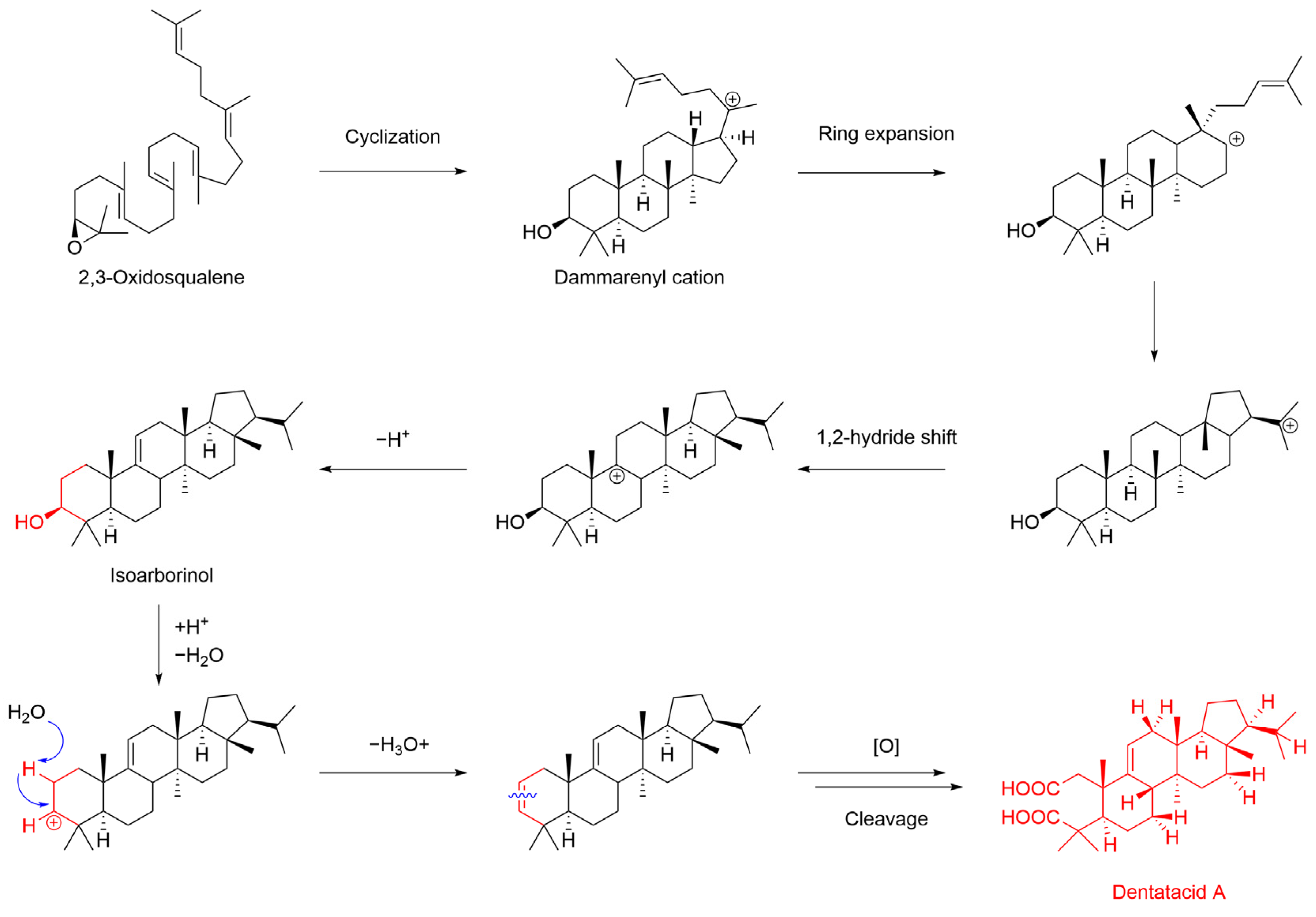
2.1. Hypothetical Biosynthesis for Dentatacid A

Due to the novelty of the chemical structure of dentatacid A, we propose a plausible biosynthetic pathway for dentatacid A. 2,3-Oxidosqualene (OSC) is a recognized precursor to triterpenoid derivatives [31]. As shown in Figure 6, OSC undergoes cyclization to form the dammarenyl cation, which, through a series of ring expansions and carbon cation migrations, yields isoarborinol, the arborinane-type triterpenoid skeleton compound [32]. In a potentially aqueous and mildly acidic environment in nature, isoarborinol undergoes dehydration and undergoes an affinity elimination reaction to form a 2,3-en triterpenoid skeleton [33,34]. This is followed by a series of oxidative cleavages and ring openings, ultimately leading to the formation of dentatacid A.

2.2. Cytotoxicity Assay

Small molecules derived from natural products have played a crucial role in the discovery of anti-tumor compounds, with triterpenoids bearing carboxyl groups being among the most significant classes of these natural products [35,36]. Our laboratory has been dedicated to the study of the cytotoxicity of natural terpenoids with novel structures [37,38,39,40]. Based on this focus, with HCPT as the control group, the CCK-8 assay is employed to preliminary investigate the cytotoxic effects of 11 triterpenoid compounds on tumor cell lines 143B, A549, HepG2, and HT-29.
In the cytotoxicity assay, all triterpenoids from E. dentata exhibited varying degrees of cytotoxicity against the four tumor cell lines, with half of the compounds showing IC50 values less than 20 μM (as shown in Table 2). For the A549 cell line, compounds 4, 5, 7, and 8, along with the novel 11, demonstrated excellent cytotoxicity, with IC50 values ranging from 4.74 to 10.04 μM. Notably, compounds 4 and 11 had IC50 values of 4.74 ± 2.61 and 4.85 ± 1.14 μM, respectively, which are very close to the cytotoxicity of HCPT, with an IC50 value of 2.89 ± 1.47 μM. However, for the HepG2 and 143B cell lines, the triterpenoids showed less promising cytotoxicity, with many compounds having IC50 values greater than 50 μM. The IC50 values of 11 were 10.65 ± 2.17 and 18.82 ± 0.60 μM for HepG2 and 143B, respectively. Interestingly, in the cytotoxicity assay against the HT-29 cell line, the triterpenoids exhibited outstanding performance. Compounds 4 and 5 had IC50 values of 8.92 ± 2.82 and 5.94 ± 2.13 μM, respectively, while compound 11 showed even more remarkable activity with an IC50 value of 2.64 ± 0.78 μM, surpassing HCPT’s IC50 value of 2.70 ± 0.16 μM for HT-29.
However, due to experimental constraints, the effects of these compounds on non-tumorigenic cells require further in-depth investigation.

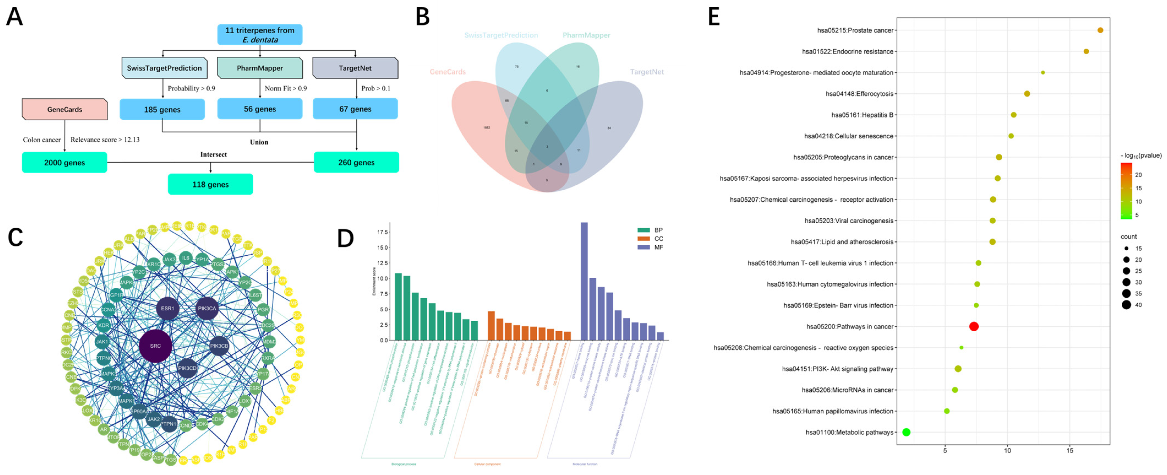
2.3. Bioinformatics Analysis

Given the exceptional cytotoxicity exhibited by compound 11 against the HT-29 cell line, we decided to conduct a bioinformatics analysis using network pharmacology to investigate the potential targets through which triterpenoids exert their cytotoxic effects. As shown in Figure 7A, within uploading the structures of the 11 triterpenoids, 185, 56, and 67 potential targets were collected from Swiss Target Prediction, PharmMapper, and TargetNet, respectively. Additionally, through GeneCards, we identified 2000 potential gene targets related to human colon cancer. By performing an intersection analysis using Venny 2.1.0, we identified 118 intersecting targets between the triterpenoid compounds and colon cancer (as shown in Figure 7B). Constructing a PPI network via STRING revealed prominent interactions among SRC, ESR, and PIK3CA (as shown in Figure 7C). Subsequently, we conducted GO and KEGG enrichment analyses. As shown in Figure 7D and 7E, the GO analysis results indicated that the intersecting targets are involved in processes such as protein phosphorylation, protein-containing complex assembly, and heme binding. According to the KEGG analysis, the intersecting targets are enriched in the PI3K-Akt signaling pathway.

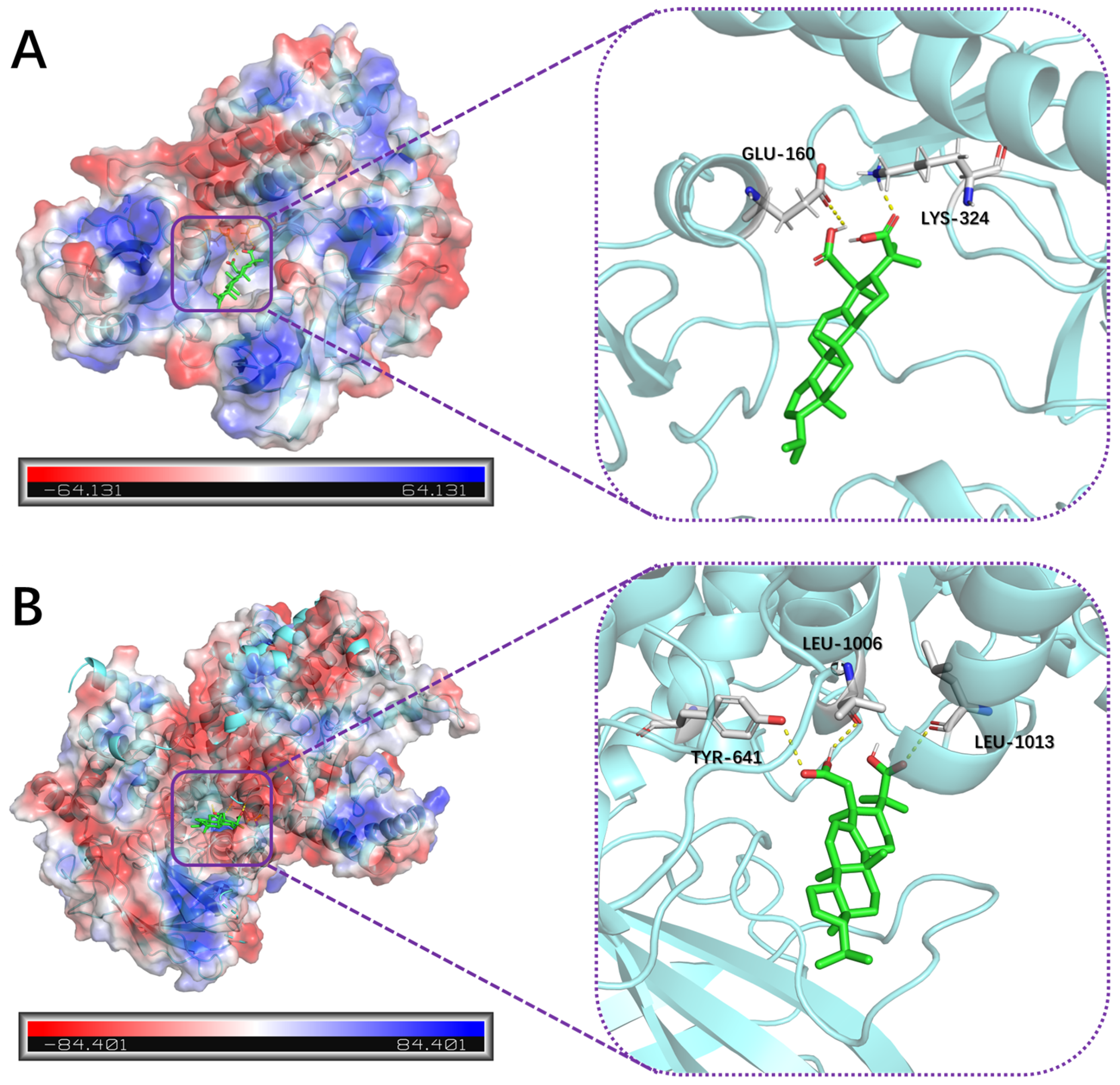
2.4. Molecular Docking Analysis

Relevant studies have shown that one of the key pathways regulating the proliferation of HT-29 cells is the SRC signaling pathway, and the downstream PI3K/Akt phosphorylation signaling pathway of SRC is also a crucial pathway for the proliferation of cancer cells [41,42,43]. Based on the results of bioinformatics analysis, 11 is likely to exert its cytotoxic effects on HT-29 cells by modulating the SRC/PI3K/Akt signaling pathway. To further validate this hypothesis, we selected the SRC protein (PDB ID: 8JN8) and PIK3CA protein (PDB ID: 7R9V) for molecular docking with 11 to investigate their interactions.
The molecular docking results revealed that 11 successfully entered the active sites of SRC and PIK3CA proteins and formed hydrogen bonds with the target proteins. In the molecular docking with the SRC protein (as shown in Figure 8A), the hydroxyl hydrogen of 2-COOH of 11 formed a hydrogen bond with the carbonyl oxygen of GLU-160, and the carbonyl oxygen of 3-COOH formed a hydrogen bond with a hydrogen atom of LYS-324. In the molecular docking with the PIK3CA protein (as shown in Figure 8B), the carbonyl oxygen of 2-COOH of 11 formed a hydrogen bond with the phenolic hydroxyl hydrogen of TYR-641, the hydroxyl hydrogen of 2-COOH formed a hydrogen bond with the carbonyl oxygen of LEU-1006, and the carbonyl oxygen of 3-COOH formed a hydrogen bond with a hydrogen atom of LEU-1013.
These preliminary results suggest that compound 11 may exert its antitumor effects through the SRC/PI3K/Akt signaling pathway. However, more definitive results require further experimental validation, such as Western blotting.

3. Experiment

3.1. General Experimental Procedures
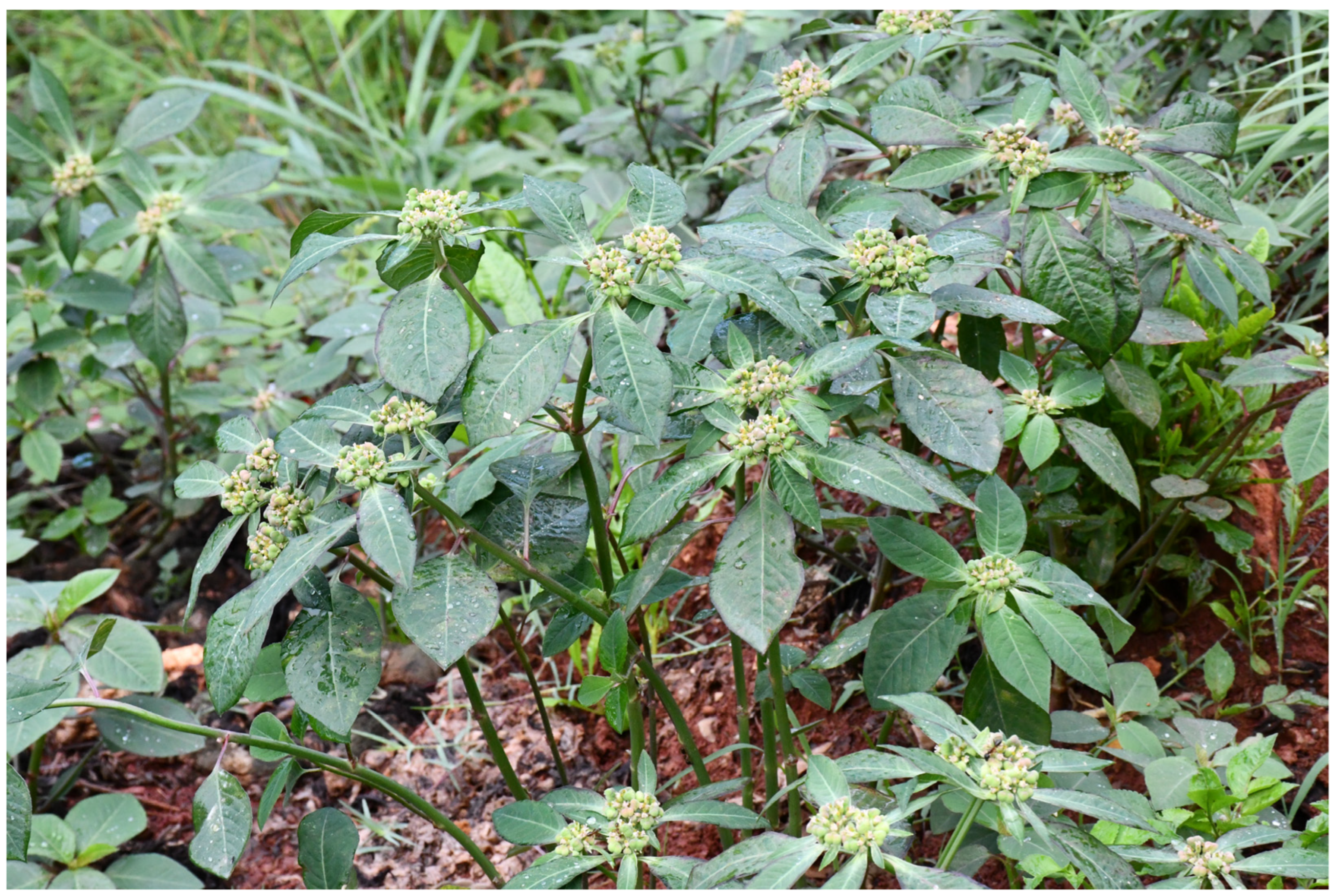
All 1D (1H and 13C NMR, DEPT-135) and 2D (HSQC, HMBC, COSY, and NOESY) NMR data were obtained from the Bruker Avance DRX600. All NMR data were analyzed with the MestReNova v14.0 software. The chemical shift values were presented in δ (ppm) based on the Dimethyl Sulfoxide-d6 (δC 39.5; δH 2.50, 3.33) resonance as references, whereas the coupling constants were expressed as J in Hz. All HR-ESI-MS data were obtained from the Agilent 1260–6224 liquid phase high-resolution time-of-flight mass spectrometer (Agilent Technologies, Inc., Beijing, China). All UV spectra were obtained with the Shimadzu UV-2550 spectrophotometer (Shimadzu (China) Co., Ltd, Shanghai, China). All IR spectra were obtained with the Nicolet 5700 FT-IR Spectrometer (Thermo Fisher Scientific Inc., Shanghai, China). All CD spectra were obtained with the JASCO J-1500 Circular Dichroism Spectrophotometer (JASCO China (Shanghai) Co., Ltd, Shanghai, China). All optical rotations were obtained with the INESA SGW-2 (Shanghai INESA Physico-Optical Instrument Co., Ltd, Shanghai, China). HPLC separations were performed with the Shimadzu LC-20AR (Shimadzu (China) Co., Ltd, Shanghai, China) equipped with a Fisher Wharton Xbridge C18 column (5 µm, 10 × 250 mm, Waters (Shanghai) Co., Ltd, Shanghai, China).
All HPLC-grade solvents in HPLC separations were from Honeywell (China) Co., Ltd (Shanghai, China). Column chromatography (CC) was performed with silica gel (80–100 mesh, 200–300 mesh, Qingdao Haiyang Chemical Co., Ltd., Qingdao, China), MCI GEL CHP20P (Mitsubishi Chemical Corporation, Tokyo, Japan), D101-macroporous absorption resin (Shanghai Yuanye Bio-Technology Co., Ltd., Qingdao, China), and Sephadex LH-20 (Sigma-Aldrich (Shanghai) Trading Co., Ltd., Qingdao, China). Thin-layer chromatography (TLC) was performed with precoated silica gel GF254 plates (Qingdao Haiyang Chemical Co., Ltd., Qingdao, China). The TLC analysis was performed with 10% sulfuric acid-ethanol as the chromogenic agent. All analytical grade solvents in CC and TLC were from Sinopharm Chemical Reagent Co., Ltd. (Shanghai, China). All deuterated solvents (Dimethyl Sulfoxide-d6, 99.0%) were purchased from Shanghai Titan Scientific Co., Ltd., Shanghai, China.

3.2. Plant Material

The whole-plant sample of E. dentata was collected in November, 2020, from Yunnan Province, People’s Republic of China. The sample was identified and classified by Anrui Lou, the senior engineer from Kunming Plant Biotechnology Co., Ltd. (Kunming, China), with reference to specimens in the Herbarium of the School of Life Sciences at Beijing Normal University. The sample of E. dentata was air-dried naturally and stored under cool and dry conditions. A voucher specimen (accession number: CLDJ-2020-11-01) was deposited at the Natural Products Chemistry Laboratory, College of Chemistry and Chemical Engineering, Southeast University, Nanjing, China.

3.3. Extraction and Isolation

The entire dried plant of E. dentata (11.25 kg) was subjected to thermal reflux extraction with 85% EtOH (6 h × 3 × 40 L) at 80 °C, which yielded 1.47 kg of ethanol extract. The crude extract of E. dentata was then dispersed in warm deionized water (3 L) and sequentially extracted three times each with petroleum ether (3 × 5 L), ethyl acetate (3 × 5 L), and n-butanol (3 × 5 L). After concentration under reduced pressure, the PE-fraction (254.4 g), EA-fraction (148.3 g), and nB-fraction (157.8 g) extracts were obtained.
PE-fraction (254.4 g) was separated on the silica gel column (200–300 mesh, PE/EA, 20:1 to 0:1, v/v) to give PE-fraction1 to PE-fraction9. PE-fraction3 was separated on the MCI GEL CHP20P (MeOH/H2O, 7:3 to 1:0, v/v) to give PE-fraction3-1 to PE-fraction3-5. PE-fraction3-1 (2.67 g) was separated with recrystallizing to obtain 12 (164.2 mg). PE-fraction4 was separated on the silica gel column (200–300 mesh, DCM/MeOH, 1:0 to 0:1, v/v) to give PE-fraction4-1 to PE-fraction4-9. PE-fraction4-2 (59.3 mg) was separated with preparative TLC (PE/AC, 3:1, v/v) to obtain 1 (3.4 mg). PE-fraction4-7 was separated on the silica gel column (200–300 mesh, PE/AC, 1:0 to 0:1, v/v) to give PE-fraction4-7-1 to PE-fraction4-7-18. PE-fraction4-7-3 (0.93 g) was separated on the silica gel column (200–300 mesh, DCM/MeOH, 1:0 to 0:1, v/v) to obtain 13 (17.0 mg). PE-fraction4-7-5 (1.15 g) was separated on the silica gel column (200–300 mesh, DCM/MeOH, 20:1 to 0:1, v/v) to obtain 2 (34.7 mg), 7 (11.3 mg), and 8 (5.9 mg). PE-fraction4-7-8 (355.3 mg) was separated on the silica gel column (200–300 mesh, DCM/MeOH, 1:0 to 0:1, v/v) to obtain 3 (1.7 mg), 4 (3.3 mg), 16 (9.9 mg), and 10 (12.6 mg). Based on the TLC analysis, PE-fraction5 and PE-fraction6 were combined for further separation. PE-fraction5/6 (12.71 g) was separated on the D101-macroporous absorption resin (EtOH/H2O, 7:3 to 1:0, v/v) to give PE-fraction5-1 to PE-fraction5-5. PE-fraction5-2 (2.94 g) was separated on the silica gel column (200–300 mesh, PE/EA, 30:1 to 0:1, v/v) to give PE-fraction5-2-1 to PE-fraction5-2-13 together with obtaining 5 (3.1 mg). PE-fraction5-2-13 (0.23 g) was separated on semipreparative HPLC (85% CH3CN in H2O, 3 mL/min) to obtain 19 (tR = 41.3 min, 49.0 mg). PE-fraction5-4 (1.1 g) was separated on the silica gel column (200–300 mesh, PE/AC, 30:1 to 0:1, v/v) to obtain 6 (10.8 mg) and 9 (4.3 mg). Based on the TLC analysis, PE-fraction7 and PE-fraction8 were combined for further separation. PE-fraction7/8 (23.01 g) was separated on the D101-macroporous absorption resin (EtOH/H2O, 7:3 to 1:0, v/v) to give PE-fraction7-1 to PE-fraction7-4. PE-fraction7-3 (5.8 g) was separated on the Sephadex LH-20 (MeOH) to give PE-fraction7-3-1 to PE-fraction7-3-7. PE-fraction7-3-5 (2.7 g) was separated on the silica gel column (200–300 mesh, PE/EA, 10:1 to 0:1, v/v) to give PE-fraction7-3-5-1 to PE-fraction7-3-5-19 together with obtaining 14 (146.7 mg), 15 (50.7 mg), and 20 (9.0 mg). PE-fraction7-3-5-10 (0.4 g) was separated on semipreparative HPLC (98% CH3CN in H2O, 3 mL/min) to obtain 17 (tR = 26.1 min, 21.3 mg) and 18 (tR = 29.8 min, 73.8 mg). PE-fraction7-3-6 (95 mg) was purified on semipreparative HPLC (95% CH3CN in H2O, 3 mL/min) to obtain 11 (tR = 29.5 min, 6.7 mg).

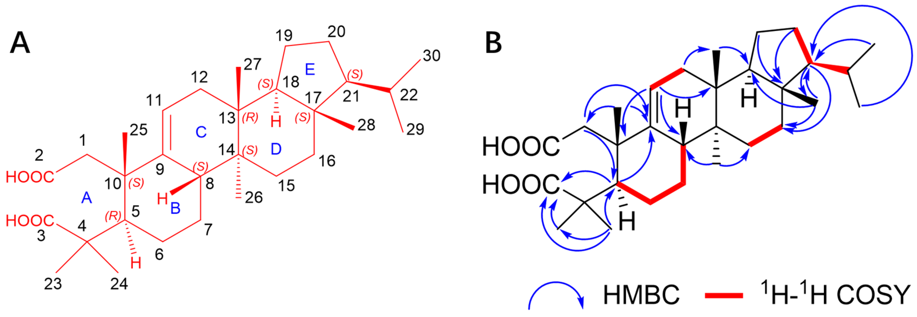
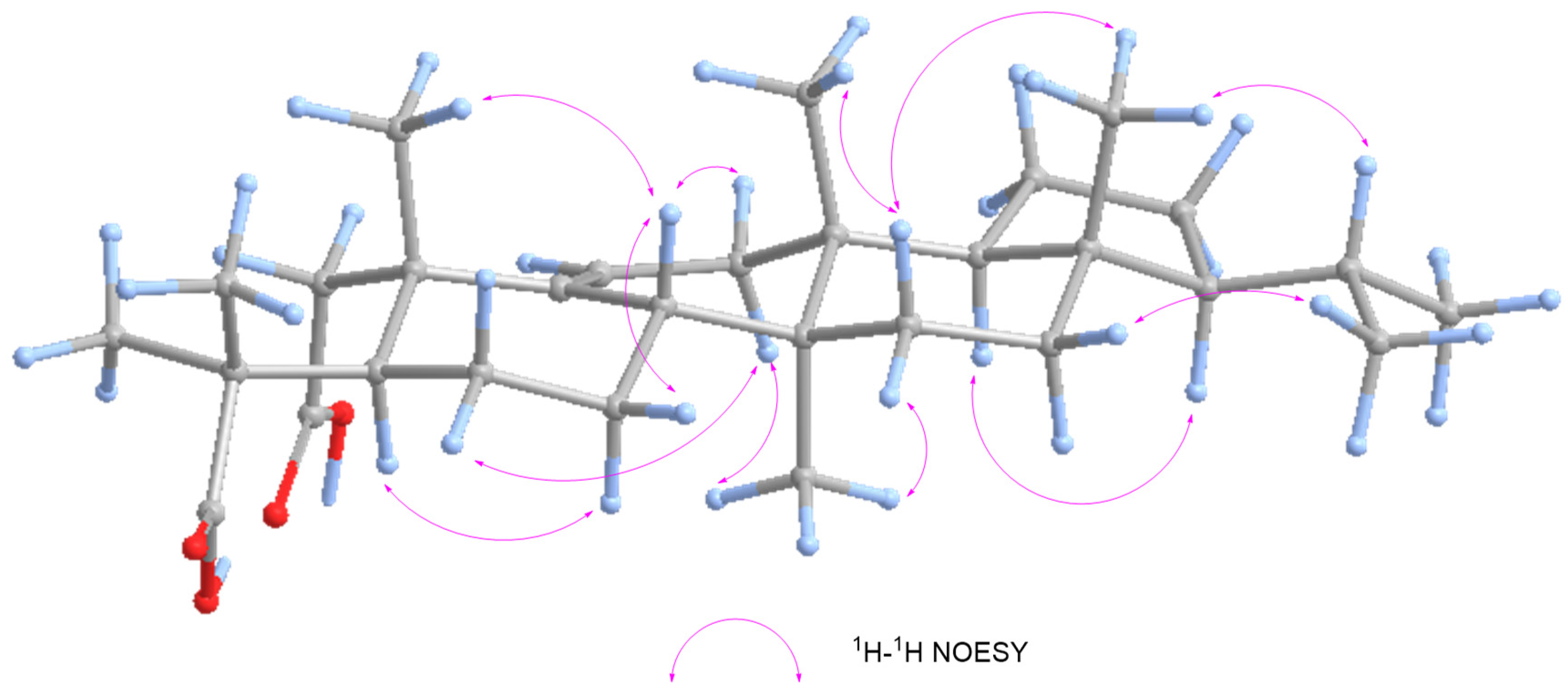
3.4. Compounds Characterization

Dentatacid A (11): white powder (chloroform/methanol). [α ] D 20 +86 (c 0.005, MeOH). UV (MeOH) λmax (log ε) 195 (7.14), 198 (7.02) nm. ECD (MeOH) λε) 200 (+5.78), 222 (+16.478), 258 (−0.18) nm; IR (KBr) νmax 3443, 1592,1384, 1351, 618 cm−1; 1H and 13C NMR data (Dimethyl Sulfoxide-d6), as shown in Table 1. (+)-HR-ESI-MS m/z 495.3441 [M + Na]+ (calcd for C30H48NaO4+, 495.3446). All data of dentatacid A’s characterization are shown in Figures S1–S13.

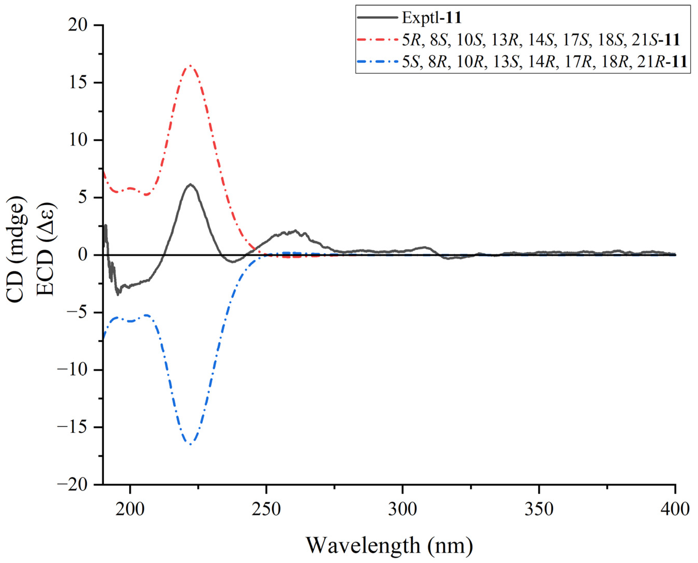
3.5. ECD Calculations

Conformational analysis was performed with Monte Carlo searching in the MMFF94 molecular mechanics force field in Spartan ‘14 V1.1.4 [44]. The conformers with the Boltzmann distribution greater than 0.05 were considered for further DFT calculations.
Subsequently, the conformers were optimized further using DFT at the B3LYP/6-31G(d) level in the methanol with the GAUSSIAN 16 [45]. The theoretically calculated ECD spectra of 11 were established using the time-dependent density functional theory (TDDFT) method at the B3LYP/6-311++G(2d,2p) level in methanol, and further Boltzmann averaged to compare with experimentally obtained ECD spectra by SpecDis v1.71 [46,47]. The sigma/gamma ratio for processing the calculated ECD was 0.3 eV. All data of ECD calculations are shown in Figure S14 and Tables S1 and S2.

3.6. Methods of Cytotoxicity Assay

3.6.1. Materials

Dulbecco’s modified Eagle’s medium (DMEM), fetal bovine serum (FBS), and antibiotics (penicillin/streptomycin) were purchased from Jiangsu KeyGEN Bio TECH Corp., Ltd. (Nanjing, China). Cell Counting Kit-8 (CCK-8) was purchased from Beyotime Biotech, Inc. (Shanghai, China). 143B, A549, HepG2, and HT-29 were purchased from the National Collection of Authenticated Cell Cultures (Shanghai, China).

3.6.2. Cell Culture

All 143B, A549, HepG2, and HT-29 were cultured with DMEM containing 10% FBS and 1% antibiotics (penicillin/streptomycin) at 37 °C incubator in a humidified atmosphere of 5% CO2.
When cells reach 80–90% confluency within a single field of view, the cells in the logarithmic growth phase will undergo subculture. Digestion of the cells is performed using 0.25% trypsin with EDTA, and the digestion process is terminated using a complete DMEM medium. The subcultured cells will be maintained at a density of 1–2 × 10⁶ cells per milliliter of medium.

3.6.3. Cell Viability

Cell viability of 143B, A549, HepG2, and HT-29 was measured by the CCK-8 assay. Cells were seeded in 96-well plates at a density of 5 × 10⁶ cells/well for 24 h and subsequently treated with the tested compounds for another 24 h. After exposure, the medium was replaced with 100 µL complete DMEM medium containing 10% CCK-8 and maintained the culture for 4 h. Then the absorbance was measured at 450 nm using the Thermo Scientific Multiskan GO (Thermo Fisher Scientific, Waltham, MA, USA). GraphPad Prism 10 was used to conduct all statistical analyses.

3.7. Methods of Bioinformatics Analysis

3.7.1. Target Prediction of Triterpenoids from E. dentata

The targets of triterpenoids of E. dentata and colon cancer were collected from Swiss Target Prediction (http://www.swisstargetprediction.ch/, accessed on 1 August 2024) [48], PharmMapper (https://lilab-ecust.cn/pharmmapper/index.html, accessed on 1 August 2024) [49] and TargetNet (http://targetnet.scbdd.com/, accessed on 1 August 2024) [50]. The targets from the Swiss Target Prediction were with a probability > 0.1. The targets from PharmMapper were with the Norm Fit > 0.9. The targets from TargetNet were with the Prob > 0.1.

3.7.2. Screening of Potential Targets in Colon Cancer

The potential targets of colon cancer were collected from GeneCards (https://www.genecards.org/, accessed on 1 August 2024) with colon cancer as the keyword [51]. The targets from GeneCards had a relevance score of > 12.13.

3.7.3. Construction of the Target PPI Network between Triterpenoids from E. dentata and Colon Cancer

Venny 2.1.0 (https://bioinfogp.cnb.csic.es/tools/venny/, accessed on 1 August 2024) was employed to intersect the targets of triterpenoids from E. dentata and colon cancer. The intersection targets were submitted to STRING11.0 (https://cn.string-db.org/, accessed on 1 August 2024) [52] to construct the Protein-Protein Interaction (PPI) network model. The PPI network model was constructed in multiple proteins of STRING11.0 with Homo sapiens of the Organisms and with the medium confidence (0.400) of the minimum required interaction score. CytoScape3.10.1 (Free Software Foundation, Inc., Boston, MA, USA) was employed to visualize the PPI network. The PPI network was analyzed with Degree, Closeness, and Betweenness generated from the app CentiScaPe 2.2 in CytoScape3.10.1.

3.7.4. Enrichment Analysis of the Intersection Targets

The targets of intersection targets were submitted to the Database for Annotation, Visualization, and Integrated Discovery (DAVID) Bioinformatics (https://david.ncifcrf.gov/, accessed on 1 August 2024), and the main metabolic pathways were analyzed and enrichment analysis was performed [53]. The result of the enrichment analysis was submitted to WeiShengXin (https://www.bioinformatics.com.cn/, accessed on 1 August 2024) to visualize the GO enrichment analysis and KEGG pathway enrichment analysis.

3.8. Molecular Docking

The lowest energy conformations of the compound were from the ECD result with the highest Boltzmann distribution, further optimizing with MM2—Minimize Energy in Chem3D 20.0 (PerkinElmer, Inc., Waltham, MA, USA). The crystal structure of SRC protein (PDB code: 8JN8) and PI3K protein (PDB code: 7R9V) was obtained from the protein data bank (PDB, http://www.wwpdb.org, accessed on 1 August 2024). The structure of the SRC and PI3K proteins was optimized using Open-Source PyMOL (Schrödinger, LLC, New York, NY, USA), including removal of water molecules, hydrogenation, and energy minimization. The molecular docking between the compounds and two proteins was performed using AutoDockTools-1.5.7 and AutoDock vina [54]. The visualization of the molecular docking results was also performed using Open-Source PyMOL.

4. Conclusions

In summary, we isolated 11 triterpenoids, 7 sterols, a diterpenoid, and a sesquiterpenoid from the invasive plant E. dentata, including a structurally unprecedented 2, 3-seco-arbor-2, 3-dioic triterpenoid. Through preliminary investigations involving cytotoxicity assays, bioinformatics analysis, and molecular docking, we have made significant progress in exploring the antitumor activity of these triterpenoids. Among them, compound 11 showed notably more compelling cytotoxicity with an IC50 value of 2.64 ± 0.78 µM, surpassing HCPT’s IC50 value of 2.70 ± 0.16 µM for HT-29. These findings greatly enhance the understanding of the secondary metabolites and biological activities of E. dentata and offer new insights into the resource utilization and management of this invasive plant from the perspective of natural product discovery.

Supplementary Materials

The following supporting information can be downloaded at: https://www.mdpi.com/article/10.3390/plants13172533/s1. Figure S1: The structure (A), the key HMBC and 1H–1H COSY correlations (B) of 11; Figure S2. Key 1H-1H NOESY correlations correlations of 11; Figure S3. 1H NMR spectrum of 11 in Dimethyl Sulfoxide-d6; Figure S4. 13C NMR spectrum of 11 in Dimethyl Sulfoxide-d6; Figure S5. DEPT-135 spectrum of 11 in Dimethyl Sulfoxide-d6; Figure S6. HSQC spectrum of 11 in Dimethyl Sulfoxide-d6; Figure S7. HMBC spectrum of 11 in Dimethyl Sulfoxide-d6; Figure S8. 1H-1H COSY spectrum of 11 in Dimethyl Sulfoxide-d6; Figure S9. 1H-1H NOESY spectrum (A) and the partial enlarged detail (B) of 11 in Dimethyl Sulfoxide-d6; Figure S10. (+)-HR-ESI-MS spectrum (A) and the partial enlarged detail (B) of 11; Figure S11. UV spectrum of 11; Figure S12. Calculated and experimental ECD spectra for 11; Figure S13. IR spectrum of 11; Figure S14. Conformers of isomer 11; Table S1. Important thermodynamic parameters and conformational analysis of 11 at the B3LYP/6-311++G(2d,2p) level with CPCM solvent model in methanol; Table S2. Cartesian coordinates for the reoptimized conformers of 11 at the B3LYP/6-311++G(2d,2p) level with CPCM solvent model in methanol; The NMR data for known compounds.

Author Contributions

C.-S.X.: Data curation, Formal analysis, Visualization, Writing—original draft and Writing—review and editing. Y.-L.S.: Investigation. Q.L.: Methodology. Y.Z.: Validation. H.-W.W.: Methodology. H.-L.Y.: Supervision. Y.-Y.S.: Supervision. J.Z.: Validation. C.W.: Validation. Z.-X.L.: Conceptualization, Funding acquisition, Project administration, Resources, and Writing—review and editing. All authors have read and agreed to the published version of the manuscript.

Funding

This study was supported by the National Natural Science Foundation of China (32370063).

Institutional Review Board Statement

Not applicable.

Informed Consent Statement

Not applicable.

Data Availability Statement

Data are contained within the article and Supplementary Materials.

Conflicts of Interest

The authors declare that they have no known competing financial interests or personal relationships that could have appeared to influence the work reported in this paper.

References

  1. Gioria, M.; Hulme, P.E.; Richardson, D.M.; Pyšek, P. Why Are Invasive Plants Successful? Annu. Rev. Plant Biol. 2023, 74, 635–670. [Google Scholar] [CrossRef] [PubMed]
  2. Augusthy, S.; Nizam, A.; Kumar, A. The diversity, drivers, consequences and management of plant invasions in the mangrove ecosystems. Sci. Total Environ. 2024, 945, 173851. [Google Scholar] [CrossRef]
  3. Marchessi, J.E.; Subils, R.; Scaramuzzino, R.L.; Crosta, H.N.; Eseiza, M.F.; Saint André, H.M.; Juan, V.F. Presencia de Euphorbia davidii Subils (Euphorbiaceae) en la Provincia de Buenos Aires: Morfología y anatomía de la especie. Kurtziana 2011, 36, 45–53. [Google Scholar]
  4. Liberato, J.R.; Wright, J.G.; Seth, M.; Shivas, R.G. Oidiopsi (Erysiphaceae) on Euphorbia spp. in Australia and Vanuatu. Australas. Plant Pathol. 2005, 34, 409–411. [Google Scholar] [CrossRef]
  5. Xue, H.-G.; Zhou, S.-D.; He, X.-J.; Yu, Y. Karyotype in fifteen populations belonging to thirteen species of Euphorbia (Euphorbiaceae) in China. Acta Phytotaxon. Sin. 2007, 45, 619–626. [Google Scholar] [CrossRef]
  6. Barina, Z.; Shevera, M.; Sirbu, C.; Pinke, G. Current distribution and spreading of Euphorbia davidii (E. dentata agg.) in Europe. Cent. Eur. J. Biol. 2013, 8, 87–95. [Google Scholar] [CrossRef]
  7. An, T.; Cao, D.; Zhang, Y.; Han, X.; Yu, Z.; Liu, Z. Norsesquiterpenes from the Latex of Euphorbia dentata and Their Chemical Defense Mechanisms against Helicoverpa armigera. Molecules 2023, 28, 7681. [Google Scholar] [CrossRef]
  8. Juan, V.F.; Saint-Andre, H.; Fernandez, R.R. Competencia de lecheron (Euphorbia dentata) en soja. Planta Daninha 2003, 21, 175–180. [Google Scholar] [CrossRef]
  9. Tanveer, A.; Khaliq, A.; Javaid, M.M.; Chaudhry, M.N.; Awan, I. Implications of weeds of genus euphorbia for crop production: A review. Planta Daninha 2013, 31, 723–731. [Google Scholar] [CrossRef]
  10. Rojas-Jiménez, S.; Valladares-Cisneros, M.G.; Salinas-Sánchez, D.O.; Pérez-Ramos, J.; Sánchez-Pérez, L.; Pérez-Gutiérrez, S.; Campos-Xolalpa, N. Anti-Inflammatory and Cytotoxic Compounds Isolated from Plants of Euphorbia Genus. Molecules 2024, 29, 1083. [Google Scholar] [CrossRef]
  11. Shi, Q.-W.; Su, X.-H.; Kiyota, H. Chemical and pharmacological research of the plants in genus Euphorbia. Chem. Rev. 2008, 108, 4295–4327. [Google Scholar] [CrossRef] [PubMed]
  12. Yang, S.; Yuan, C.; Chen, Y. New Advance on the Research of Chemical Constituents and Pharmacological Activities of Diterpenoids from the Plants of Genus Euphorbia. Chin. Wild Plant Resour. 2020, 39, 53–60. [Google Scholar]
  13. Zhao, H.; Sun, L.; Kong, C.; Mei, W.; Dai, H.; Xu, F.; Huang, S. Phytochemical and pharmacological review of diterpenoids from the genus Euphorbia Linn (2012–2021). J. Ethnopharmacol. 2022, 298, 115574. [Google Scholar] [CrossRef]
  14. Wei, X.-F.; Wang, Y.-K.; Liu, R.-T.; Wu, J.-P.; Xu, K.P. Review of natural plant-derived seco-triterpenoids and derived saponins from 2020 to 2023: New compounds, distributions, diverse activities and structure–activity relationships. Phytochem. Rev. 2024. [Google Scholar] [CrossRef]
  15. Tewtrakul, S.; Tansakul, P.; Daengrot, C.; Ponglimanont, C.; Karalai, C. Anti-inflammatory principles from Heritiera littoralis bark. Phytomedicine 2010, 17, 851–855. [Google Scholar] [CrossRef]
  16. Xu, M.; Liu, J.; Sun, W.; Chen, Y.; Wang, M.; Zhou, Y. New dammarane-type triterpenoids from the whole plant of Euphorbia hypericifolia. Nat. Prod. Res. 2024, 1–6. [Google Scholar] [CrossRef]
  17. Fang, C.H.; Li, Y.P.; Li, Y.; Yue, J.M.; Bao, J.; Yu, J.H. Triterpenoids with multi-skeletons as 11 beta -HSD1 inhibitors from Euphorbia sikkimensis. Phytochemistry 2023, 211, 113684. [Google Scholar] [CrossRef] [PubMed]
  18. Silva, M.G.; Vieira, I.G.; Mendes, F.N.; Albuquerque, I.L.; dos Santos, R.N.; Silva, F.O.; Morais, S.M. Variation of ursolic acid content in eight Ocimum species from northeastern Brazil. Molecules 2008, 13, 2482–2487. [Google Scholar] [CrossRef] [PubMed]
  19. Liu, J.J.; Hao, J.J.; Tan, M.; Liao, C.C.; Liu, D.; Li, H.M.; Li, R.T. Iridoids and other constituents from the leaves and stems of Valeriana officinalis var. latifolia. Phytochemistry 2024, 218, 113934. [Google Scholar] [CrossRef]
  20. Sun, H.-B.; Zou, H.-F.; Xia, B.; Gu, Y.-C.; Wu, W.-L.; Zhou, Y. Targeted isolation of ursane-type triterpenoids from Tragopogon porrifolius based on Feature-Based Molecular Networking. Phytochem. Lett. 2023, 57, 67–72. [Google Scholar] [CrossRef]
  21. Seki, H.; Ohyama, K.; Sawai, S.; Mizutani, M.; Ohnishi, T.; Sudo, H.; Akashi, T.; Aoki, T.; Saito, K.; Muranaka, T. Licorice β-amyrin 11-oxidase, a cytochrome P450 with a key role in the biosynthesis of the triterpene sweetener glycyrrhizin. Proc. Natl. Acad. Sci. USA 2008, 105, 14204–14209. [Google Scholar] [CrossRef] [PubMed]
  22. Kemertelidze, E.P.; Gvazava, L.N.; Alania, M.D.; Kikoladze, V.S.; Tsitsishvili, V.G. Digitoside, a novel triterpene glycoside from Digitalis ciliata. J. Nat. Prod. 1992, 55, 217–220. [Google Scholar] [CrossRef]
  23. Akihisa, T.; Watanabe, K.; Yoneima, R.; Suzuki, T.; Kimura, Y. Biotransformation of cycloartane-type triterpenes by the fungus Glomerella fusarioides. J. Nat. Prod. 2006, 69, 604–607. [Google Scholar] [CrossRef]
  24. Lizazman, M.A.; Jong, V.Y.M.; Chua, P.; Lim, W.K.; Karunakaran, T. Phytochemicals from Calophyllum canum Hook f. ex T. Anderson and their neuroprotective effects. Nat. Prod. Res. 2023, 37, 2043–2048. [Google Scholar] [CrossRef] [PubMed]
  25. Zhou, Q.-M.; Peng, C.; Yang, H.; Liu, L.-S.; Yang, Y.-T.; Xie, X.-F.; Guo, L.; Liu, Z.-H.; Xiong, L. Steroids from the aerial parts of Leonurus japonicus. Phytochem. Lett. 2015, 12, 287–290. [Google Scholar] [CrossRef]
  26. Zhu, Y.; Soroka, D.; Sang, S. Oxyphytosterols as active ingredients in wheat bran suppress human colon cancer cell growth: Identification, chemical synthesis, and biological evaluation. J. Agric. Food Chem. 2015, 63, 2264–2276. [Google Scholar] [CrossRef]
  27. Ayyad, S.E.N.; Sowellim, S.Z.A.; El-Hosini, M.S.; Abo-Atia, A. The structural determination of a new steroidal metabolite from the brown alga Sargassum asperifolium. Z. Naturforschung C J. Biosci. 2003, 58, 333–336. [Google Scholar] [CrossRef]
  28. Foley, D.A.; O’Callaghan, Y.; O’Brien, N.M.; McCarthy, F.O.; Maguire, A.R. Synthesis and characterization of stigmasterol oxidation products. J. Agric. Food Chem. 2010, 58, 1165–1173. [Google Scholar] [CrossRef]
  29. Schabdach, H.; Johne, S.; Steiner, U.; Seifert, K. Plant-disease resistance inducing activity of 7-oxosterols and 7-hydroxysterols. Z. Naturforschung C J. Biosci. 1995, 50, 257–262. [Google Scholar] [CrossRef]
  30. Brown, G.D.; Liang, G.Y.; Sy, L.K. Terpenoids from the seeds of Artemisia annua. Phytochemistry 2003, 64, 303–323. [Google Scholar] [CrossRef]
  31. Thimmappa, R.; Geisler, K.; Louveau, T.; O’Maille, P.; Osbourn, A. Triterpene biosynthesis in plants. Annu. Rev. Plant Biol. 2014, 65, 225–257. [Google Scholar] [CrossRef] [PubMed]
  32. Xu, R.; Fazio, G.C.; Matsuda, S.P. On the origins of triterpenoid skeletal diversity. Phytochemistry 2004, 65, 261–291. [Google Scholar] [CrossRef] [PubMed]
  33. Wang, F.; Ren, F.C.; Liu, J.K. Alstonic acids A and B, unusual 2,3-secofernane triterpenoids from Alstonia scholaris. Phytochemistry 2009, 70, 650–654. [Google Scholar] [CrossRef]
  34. Vo, H.Q.; Viet Pham, T.; Le, A.T.; Hoang, H.N.T.; Nguyen, P.Q.D.; Doan, L.N.T.; Nguyen, H.T.; Ho, D.V. Two New Lupane-Type Triterpenes from the Stems and Leaves of Buxus latistyla Gagnep. and Their Cytotoxic Activity. Nat. Prod. Commun. 2024, 19, 1–5. [Google Scholar] [CrossRef]
  35. Luo, H.; Vong, C.T.; Chen, H.; Gao, Y.; Lyu, P.; Qiu, L.; Zhao, M.; Liu, Q.; Cheng, Z.; Zou, J.; et al. Naturally occurring anti-cancer compounds: Shining from Chinese herbal medicine. Chin. Med. 2019, 14, 48. [Google Scholar] [PubMed]
  36. Castellano, J.M.; Ramos-Romero, S.; Perona, J.S. Oleanolic Acid: Extraction, Characterization and Biological Activity. Nutrients 2022, 14, 623. [Google Scholar] [CrossRef]
  37. Li, Q.; Wang, Z.W.; Wang, M.X.; Yu, H.L.; Chen, L.; Cai, Z.; Zhang, Y.; Gu, M.M.; Shao, Y.L.; Han, H.P.; et al. Brunonianines A-C, C(20)-diterpenoid alkaloids with cyano group from Delphinium brunonianum Royle. Phytochemistry 2024, 219, 113987. [Google Scholar] [CrossRef]
  38. Li, Q.; Gu, M.-M.; Wu, H.-W.; Xu, C.-S.; Yu, H.-L.; Zhang, Y.; Su, Y.-Y.; Han, H.-P.; Liao, Z.-X. Brunonianines D-F, three new C19-diterpenoid alkaloids from the Delphinium brunonianum, with therapeutic effect on ovarian cancer in vitro and in vivo. Bioorganic Chem. 2024, 148, 107478. [Google Scholar] [CrossRef]
  39. Shao, Y.; Li, Q.; Wang, M.; Wang, C.; Zhang, Y.; Xu, C.; Liao, Z.; Han, H. Triterpenoid acids characterized by the oxidation of the C-27-methy from the roots of Astilbe grandis Stapf ex Wils. Fitoterapia 2023, 168, 105556. [Google Scholar] [CrossRef]
  40. Shao, Y.; Xu, C.; Li, Q.; Zhang, Y.; Cai, Z.; Yu, H.; Gu, M.; Su, Y.; Han, H.; Liao, Z. Structures and Tumor Cell Lines Proliferation Activities of Triterpenes Isolated from Astilbe grandis. Chem. Biodivers. 2024, 21, e202400100. [Google Scholar] [CrossRef]
  41. Wu, H.; Li, Q.; Yu, H.; Gu, M.; Wang, Y.; Xu, C.; Liao, Z. Comprehensive utilization of Hainan cashew nut shell: Process optimization of cashew nut shell liquid extraction and cardanol refinement by catalytic transfer hydrogenation. Ind. Crops Prod. 2023, 203, 117168. [Google Scholar] [CrossRef]
  42. Li, X.; Cao, Q.; Xu, N.; Hu, Y.; Zhu, J.; Liu, M.; Li, L. The proliferation mechanism of colon cancer cell lines HT-29 and Colo-205. J. Toxicol. 2022, 36, 130–136. [Google Scholar]
  43. Zhang, X.; Xu, H.; Bi, X.; Hou, G.; Liu, A.; Zhao, Y.; Wang, G.; Cao, X. Src acts as the target of matrine to inhibit the proliferation of cancer cells by regulating phosphorylation signaling pathways. Cell Death Dis. 2021, 12, 931. [Google Scholar] [CrossRef] [PubMed]
  44. Wavefunction, Inc. Spartan ’14; Wavefunction, Inc.: Irvine, CA, USA, 2014. [Google Scholar]
  45. Frisch, M.J.; Trucks, G.W.; Schlegel, H.B.; Scuseria, G.E.; Robb, M.A.; Cheeseman, J.R.; Scalmani, G.; Barone, V.; Petersson, G.A.; Nakatsuji, H.; et al. Gaussian 16; Gaussian Inc.: Wallingford, CT, USA, 2016. [Google Scholar]
  46. Bruhn, T.; Schaumlöffel, A.; Hemberger, Y.; Bringmann, G. SpecDis: Quantifying the Comparison of Calculated and Experimental Electronic Circular Dichroism Spectra. Chirality 2013, 25, 243–249. [Google Scholar] [CrossRef] [PubMed]
  47. Pescitelli, G.; Bruhn, T. Good Computational Practice in the Assignment of Absolute Configurations by TDDFT Calculations of ECD Spectra. Chirality 2016, 28, 466–474. [Google Scholar] [CrossRef]
  48. Gfeller, D.; Grosdidier, A.; Wirth, M.; Daina, A.; Michielin, O.; Zoete, V. SwissTargetPrediction: A web server for target prediction of bioactive small molecules. Nucleic Acids Res. 2014, 42, W32–W38. [Google Scholar] [CrossRef]
  49. Wang, X.; Shen, Y.; Wang, S.; Li, S.; Zhang, W.; Liu, X.; Lai, L.; Pei, J.; Li, H. PharmMapper 2017 update: A web server for potential drug target identification with a comprehensive target pharmacophore database. Nucleic Acids Res. 2017, 45, W356–W360. [Google Scholar] [CrossRef]
  50. Yao, Z.-J.; Dong, J.; Che, Y.-J.; Zhu, M.-F.; Wen, M.; Wang, N.-N.; Wang, S.; Lu, A.-P.; Cao, D.-S. TargetNet: A web service for predicting potential drug–target interaction profiling via multi-target SAR models. J. Comput. -Aided Mol. Des. 2016, 30, 413–424. [Google Scholar] [CrossRef]
  51. Stelzer, G.; Rosen, N.; Plaschkes, I.; Zimmerman, S.; Twik, M.; Fishilevich, S.; Stein, T.I.; Nudel, R.; Lieder, I.; Mazor, Y.; et al. The GeneCards Suite: From Gene Data Mining to Disease Genome Sequence Analyses. Curr. Protoc. Bioinform. 2016, 54, 1.30.1–1.30.33. [Google Scholar] [CrossRef]
  52. Szklarczyk, D.; Gable, A.L.; Nastou, K.C.; Lyon, D.; Kirsch, R.; Pyysalo, S.; Doncheva, N.T.; Legeay, M.; Fang, T.; Bork, P. The STRING database in 2021: Customizable protein–protein networks, and functional characterization of user-uploaded gene/measurement sets. Nucleic Acids Res. 2021, 49, D605–D612. [Google Scholar] [CrossRef]
  53. Sherman, B.T.; Hao, M.; Qiu, J.; Jiao, X.; Baseler, M.W.; Lane, H.C.; Imamichi, T.; Chang, W. DAVID: A web server for functional enrichment analysis and functional annotation of gene lists (2021 update). Nucleic Acids Res. 2022, 50, W216–W221. [Google Scholar] [CrossRef]
  54. Eberhardt, J.; Santos-Martins, D.; Tillack, A.F.; Forli, S. AutoDock Vina 1.2.0: New Docking Methods, Expanded Force Field, and Python Bindings. J. Chem. Inf. Model. 2021, 61, 3891–3898. [Google Scholar] [CrossRef] [PubMed]
Figure 1. Euphorbia dentata Michx. in Yunnan Province, People’s Republic of China.
Plants 13 02533 g001
Figure 2. Structures of compounds 120.
Plants 13 02533 g002
Figure 3. The structure (A), the key HMBC, and 1H–1H COSY correlations (B) of 11.
Plants 13 02533 g003
Figure 4. Key NOESY correlations of 11.
Plants 13 02533 g004
Figure 5. Calculated and experimental ECD spectra for 11.
Plants 13 02533 g005
Figure 6. Plausible biogenetic pathways for dentatacid A (1).
Plants 13 02533 g006
Figure 7. Target prediction and enrichment analysis results of triterpenoids from E. dentata and colon cancer. (A) The flowchart about the targets of triterpenoids and the target prediction of colon cancer. (B) The Venn diagram of triterpenoids—colon cancer targets. (C) The PPI network of the intersection target. (D) The GO enrichment analysis of the intersection targets. The green, orange, and purple columns respectively represent the intersecting targets related to Biological Process (BP), Cellular Components (CC), and Molecular Functions (MF). (E) The KEGG pathway enrichment analysis of common targets. The bubble size represents the number of intersecting targets enriched in a signaling pathway while the color of the bubble represents the p-value.
Plants 13 02533 g007
Figure 8. Molecular docking analysis of 11 with SRC protein (PDB ID: 8JN8, as shown in (A)) and PIK3CA protein (PDB ID: 7R9V, as shown in (B)).
Plants 13 02533 g008
Table 1. 1H (600 MHz) and 13C (150 MHz) NMR data of 11 in Dimethyl Sulfoxide-d6.
No.δCδC, TypeδH, (mult, J)
142.0CH22.67 (d, J = 18.0 Hz),
2.56 (d, J = 18.0 Hz)
2172.4C-
3180.6C-
445.6C-
546.7CH2.52 (dd, J = 11.9, 3.2 Hz)
623.6CH2α 1.65 a (m),
β 1.50 a (m)
724.5CH2β 1.68 a (m),
α 1.17 a (m)
841.8CH1.91 (d, J = 13.1 Hz)
9145.9C-
1043.4C-
11114.5CH5.23 (d, J = 5.7 Hz)
1236.2CH2α 1.59 (d, J = 2.9 Hz),
β 1.39 a (m)
1335.9C-
1437.9C-
1529.0CH2β 1.21 (s),
α 1.27 a (m)
1635.5CH2α 1.34 (dd, J = 13.3, 2.9 Hz),
β 1.59 (dd, J = 13.3, 2.9 Hz)
1742.4C-
1851.5CH1.52 a (m)
1919.7CH21.28 a (m)
2027.8CH21.15 a (m),
1.76 (m)
2159.0CH0.94 (q, J = 9.4 Hz)
2230.3CH1.39 a (m)
2327.3CH31.09 a (s)
2423.5CH31.10 (s)
2525.3CH31.09 a (s)
2616.5CH30.74 (s)
2715.3CH30.71 (s)
2813.8CH30.70 (s)
2922.9CH30.79 (d, J = 6.5 Hz)
3022.0CH30.85 (d, J = 6.5 Hz)
a Multiplicity not determined due to overlapping signals.
Table 2. Cytotoxic assay of compounds 111 with IC50 values (µM).
CompoundsA549 (μM) aHepG2 (μM) aHT-29 (μM) a143B (μM) a
122.79 ± 2.86>5025.10 ± 1.8719.98 ± 3.16
222.69 ± 1.4624.32 ± 1.4015.66 ± 0.38>50
3>5012.29 ± 0.1111.87 ± 2.7319.11 ± 5.53
44.74 ± 2.6110.10 ± 1.048.92 ± 2.8226.34 ± 1.06
531.56 ± 6.5726.68 ± 4.005.94 ± 2.13>50
6>5024.82 ± 1.1322.77 ± 3.6626.98 ± 0.65
710.04 ± 2.22>5018.90 ± 1.56>50
89.63 ± 2.25>5021.48 ± 4.1519.27 ± 1.95
929.68 ± 3.6923.68 ± 1.7215.07 ± 13.5617.01 ± 2.73
10>5031.52 ± 4.4213.01 ± 1.14>50
114.85 ± 1.1410.65 ± 2.172.64 ± 0.7818.82 ± 0.60
HCPT b2.89 ± 1.473.19 ± 0.462.70 ± 0.162.56 ± 1.41
a The values presented are the mean ± SD of triplicate experiments. b The positive control. The bold means different compounds.
Disclaimer/Publisher’s Note: The statements, opinions and data contained in all publications are solely those of the individual author(s) and contributor(s) and not of MDPI and/or the editor(s). MDPI and/or the editor(s) disclaim responsibility for any injury to people or property resulting from any ideas, methods, instructions or products referred to in the content.

