Article

Impact of Quorum Sensing on the Virulence and Survival Traits of Burkholderia plantarii

1
Department of Integrated Biological Science, Pusan National University, Pusan 46241, Republic of Korea
2
Institute of System Biology, Pusan National University, Pusan 46241, Republic of Korea
3
Department of Plant Pathology, Faculty of Agriculture, Cairo University, Giza 12613, Egypt
*
Author to whom correspondence should be addressed.
These authors contributed equally to this work.
Plants 2024, 13(18), 2657; https://doi.org/10.3390/plants13182657
Submission received: 16 August 2024 / Revised: 21 September 2024 / Accepted: 21 September 2024 / Published: 23 September 2024
(This article belongs to the Section Plant Protection and Biotic Interactions)

Abstract

:
Quorum sensing (QS) is a mechanism by which bacteria detect and respond to cell density, regulating collective behaviors. Burkholderia plantarii, the causal agent of rice seedling blight, employs the LuxIR-type QS system, common among Gram-negative bacteria, where LuxI-type synthase produces QS signals recognized by LuxR-type regulators to control gene expression. This study aimed to elucidate the QS mechanism in B. plantarii KACC18965. Through whole-genome analysis and autoinducer assays, the plaI gene, responsible for QS signal production, was identified. Motility assays confirmed that C8-homoserine lactone (C8-HSL) serves as the QS signal. Physiological experiments revealed that the QS-defective mutant exhibited reduced virulence, impaired swarming motility, and delayed biofilm formation compared to the wild type. Additionally, the QS mutant demonstrated weakened antibacterial activity against Escherichia coli and decreased phosphate solubilization. These findings indicate that QS in B. plantarii significantly influences various pathogenicity and survival traits, including motility, biofilm formation, antibacterial activity, and nutrient acquisition, highlighting the critical role of QS in pathogen virulence and adaptability.

1. Introduction

Bacteria utilize a mechanism known as quorum sensing (QS) to detect cell density in their environment and regulate gene expression cooperatively. QS influences the transcriptional regulation of various genes involved in processes such as bioluminescence, exoenzymes production, biofilm formation, and pathogenicity [1,2,3]. Understanding QS is crucial for studying the physiology and pathogenicity of bacterial pathogens.
Among the different QS systems, many Proteobacteria use acylated homoserine lactone (AHL) signaling systems, while most Gram-positive bacteria rely on oligopeptide signaling systems [4]. The AHL-mediated QS system is the most common and well-studied in Gram-negative bacteria [5]. In these systems, LuxI-type synthase enzymes produce extracellular signal molecules called autoinducers, which are detected by LuxR-type receptors to assess bacterial density in the surrounding environment [2].
Burkholderia plantarii, the causal agent of rice seedling blight, was first identified in 1987 [6]. In South Korea, B. plantarii was first reported in 2016 [7]. This pathogen has several virulence factors, with tropolone being a major one, leading to seedling wilt, root growth suppression, and chlorosis [6]. B. plantarii is genetically similar to Burkholderia glumae, the causative agent of bacterial panicle blight in rice [8]. B. glumae has a well-characterized QS system involving the LuxIR-type system. TofI synthesizes QS signal molecules, which are recognized by TofR. Subsequently, QsmR, the quorum-sensing master regulator, controls the production of various metabolic products [9,10,11,12]. In 2006, Solis et al. first identified and characterized the QS system in B. plantarii, demonstrating that the plaI gene, a homolog of the luxI family, regulates QS via N-acyl homoserine lactones (AHLs). Their study showed that the plaI insertion mutant exhibited a significant reduction in virulence in rice seedling blight, establishing the foundational role of QS in the pathogenicity of this bacterium [13].
Genomic studies have provided significant insights into the unique features of Burkholderia species, revealing their high genetic versatility and adaptability to various ecological niches [14]. The ability of these bacteria to thrive in diverse environments is attributed to their large multi-replicon genomes, which are rich in insertion sequences and genomic islands. These genomic elements not only enhance their metabolic diversity but also contribute to their ability to adapt to changing environmental conditions. Comparative genomic studies on B. plantarii have further emphasized these characteristics, demonstrating the extensive genetic resources this species possesses. This genetic complexity is linked to the presence of numerous virulence factors, which play crucial roles in the pathogenicity of B. plantarii. These studies have identified various genes and regulatory mechanisms that enable B. plantarii to infect host plants, evade immune responses, and cause disease symptoms. Understanding these genomic traits is essential for developing strategies to manage the diseases caused by this pathogen and for exploring its potential applications in biotechnology and agriculture [15].
Despite the significant impact of B. plantarii on rice productivity, comprehensive studies on this pathogen are scarce. To address this gap, an extensive analysis of the B. plantarii KACC18965 genome was conducted, comparing it with B. glumae BGR1 to identify the gene responsible for QS signal molecule biosynthesis. Various QS-regulated physiological traits, including virulence, motility, biofilm formation, interbacterial interactions, and phosphate solubilization were examined. This study aims to elucidate the QS mechanism in B. plantarii and its role in regulating various physiological traits, thus providing a foundation for developing strategies to mitigate its pathogenic effects on rice seedlings.

2. Results

2.1. Selection of plaI Candidate Genes

To identify the gene responsible for QS signal synthesis in B. plantarii, homologous genes of tofI in the genome of B. plantarii KACC18965 were screened. Three candidate genes, plaI, plaI2, and plaI3, were identified based on their high sequence homology with tofI (Table 1). Each of these genes contained motifs associated with AI synthase function. Notably, only plaI and plaI2 were located adjacent to the tofR homologous gene (plaR), as observed in B. glumae.

2.2. Gene Involved in Producing a QS Signal Molecule

LuxI-type synthases are known to produce AHLs, which act as QS signal molecules in Gram-negative bacteria [2]. To confirm which plaI gene is involved in AHL production, an autoinducer (AI) thin-layer chromatography (TLC) assay was conducted on B. plantarii wild-type and plaI deletion mutants [9]. The AI TLC assay revealed the presence of N-hexanoyl homoserine lactone (C6-HSL) and N-octanoyl homoserine lactone (C8-HSL) in the wild-type, plaI2 deletion mutant, and plaI3 deletion mutant, but not in the plaI deletion mutant (Figure 1A). To verify that the absence of AHL production was due to the deletion of plaI, an AI assay with a plaI complemented strain was performed. The complemented strain restored AHL production, confirming that plaI is the gene responsible for QS signal synthesis in B. plantarii (Figure 1B).

2.3. Reduced Virulence in plaI Deletion Mutant

B. plantarii is known to cause bacterial seedling blight in rice and can also infect rice during the flowering stage, leading to symptoms in rice panicles. To investigate the role of QS in the virulence of B. plantarii, both seedling and flowering tests using the wild type, plaI deletion mutant, and plaI complemented strain were conducted.
In the seedling test, rice panicles were inoculated with B. plantarii KACC18965 wild type, plaI deletion mutant, and complemented strain. After 7 days, the lengths of the shoots and roots of the seedlings were measured, and the severity of the disease was compared (Figure 2A). The plaI deletion mutant exhibited a slight reduction in virulence compared to the wild type and complemented strain. Specifically, the root length was significantly reduced in the wild type and complemented strain compared to the plaI deletion mutant, indicating a significant decrease in virulence only in the roots (Figure 2B).
Additionally, in the flowering test, symptoms were observed 1 week after inoculating rice panicles with the wild type, plaI deletion mutant, and complemented strain using the dipping method at the flowering stage (Figure 2C). Consistent with the seedling test results, the plaI deletion mutant showed reduced virulence compared to the wild type and complemented strain (Figure 2D,E).

2.4. QS-Dependent Differences of Collective Movement

Motility is a key mechanism regulated by QS in diverse bacteria [16]. To investigate whether QS influences the collective movement of B. plantarii, motility assays were conducted. For individual movement, assessed by swimming assays, all strains displayed similar levels of movement (Figure 3). However, in collective movement, evaluated by swarming assays, the plaI deletion mutant exhibited significantly reduced movement compared to the wild type and plaI complemented strain (Figure 3). To further identify which AHL acts as the primary QS signal, a swarming assay on the plaI deletion mutant supplemented with 1 µM C8-HSL was performed. The addition of C8-HSL restored the swarming ability of the plaI deletion mutant to the level observed in the wild type (Figure 3).

2.5. QS-Dependent Differences in Biofilm Formation

To determine the relationship between biofilm formation and QS, B. plantarii wild type, plaI deletion mutant, and plaI complemented strain were cultured under static conditions in PDB. Biofilm formation at the air–liquid interface was observed at 24 h intervals. The wild type and plaI complemented strain initiated biofilm formation at 24 h, with the amount of biofilm stabilizing after 48 h. In contrast, the plaI deletion mutant began biofilm formation at 72 h, with the amount of biofilm increasing up to 96 h (Figure 4A). To further investigate the impact of QS on biofilm formation, the mass of air-dried biofilms formed at the air–liquid interface after 48 h of static incubation was quantified. The plaI deletion mutant exhibited a significantly reduced biofilm mass compared to both the wild type and the complemented strain (Figure 4B). This substantial reduction underscores the critical role of QS in regulating biofilm formation, highlighting the connection between QS and the ability of B. plantarii to form robust biofilms under static conditions. Microscopic observations of the biofilms at 48 h post-incubation further supported these findings. The wild type and plaI complemented strains displayed dense, continuous biofilm structures, while the plaI deletion mutant showed a fragmented and less dense biofilm with scattered cell clusters (Figure 4C). These results highlight the critical role of QS in maintaining biofilm architecture and support the quantitative data, showing that the disruption of the plaI gene severely impairs biofilm formation.

2.6. Weakened Antibacterial Activity in plaI Deletion Mutant

A bacterial competition assay was performed to assess whether QS influences the antibacterial activity of B. plantarii. The survival of E. coli cells was evaluated after co-culturing with B. plantarii wild type, plaI deletion mutant, and complemented strain. E. coli cells co-cultured with the B. plantarii wild-type or plaI complemented strain exhibited no survival, as indicated by the absence of colonies. In contrast, E. coli cells co-cultured with the plaI deletion mutant showed significant survival, demonstrating reduced antibacterial activity in the absence of the plaI gene (Figure 5).

2.7. Phosphate Solubilization Activity Modulated by QS

To investigate further activities regulated by QS in B. plantarii, the wild type and plaI deletion mutant were cultured on various detection media, including siderophore detection plates, skim milk agar for proteinase activity, and oxidative stress tolerance assays. However, none of these tests revealed any significant differences in the deletion mutant. Notably, the diameter of the halo produced by the plaI deletion mutant on PKV agar medium was significantly smaller compared to the wild type and complemented strain (Figure 6). This finding indicates that QS plays a critical role in modulating phosphate solubilization activity in B. plantarii.

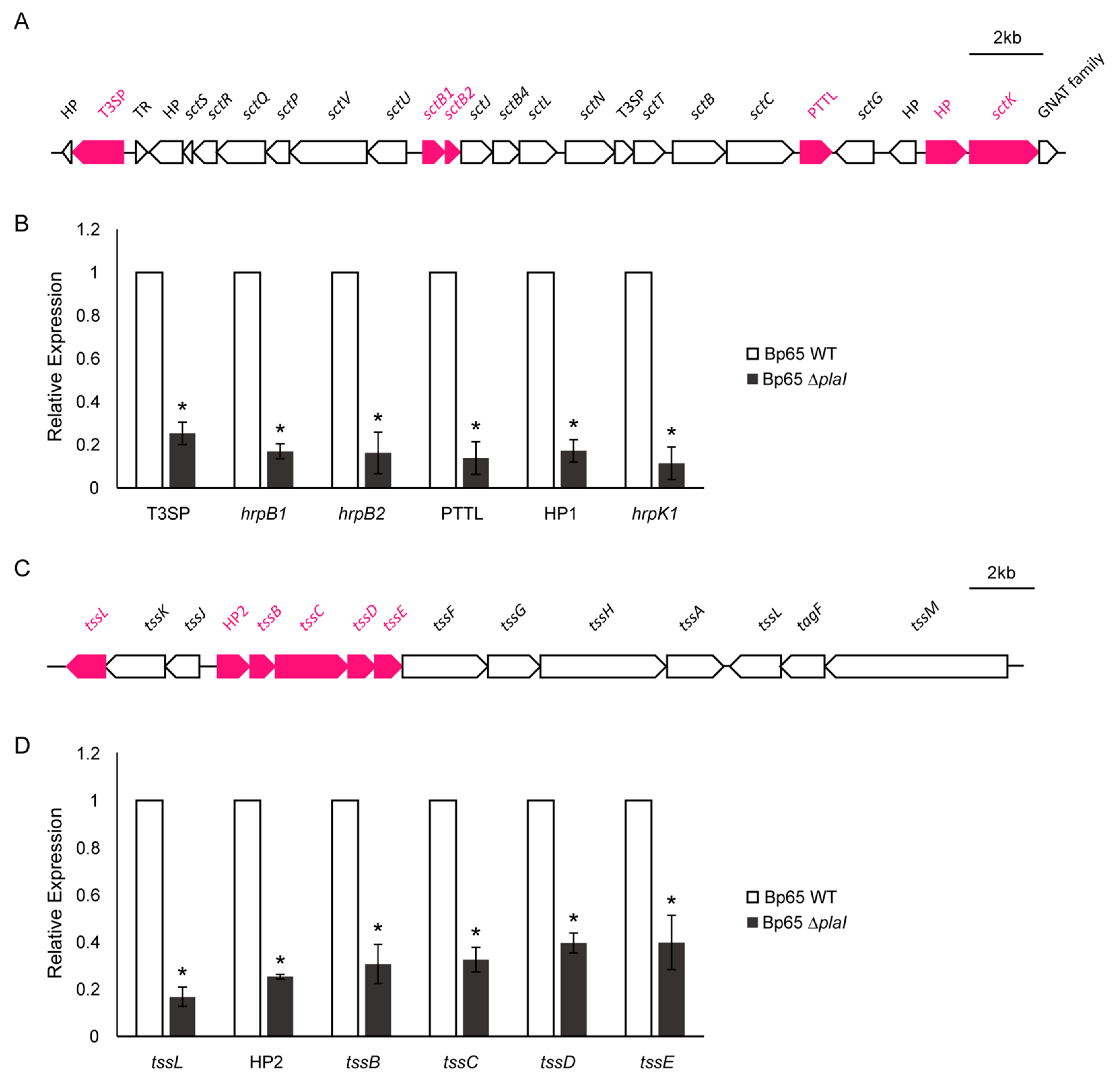
2.8. Changes in Expression of T3SS and T6SS Genes Regulated by QS

Both Type III Secretion System (T3SS) and Type VI Secretion System (T6SS) are integral to bacterial pathogenicity and antimicrobial activity [17,18,19]. To investigate the role of QS in regulating the T3SS and T6SS, we focused on six key genes from each of these clusters. The qPCR results demonstrated a significant reduction in the expression levels of all selected T3SS and T6SS genes in the plaI deletion mutant compared to the wild-type strain (Figure 7). This highlights the crucial role of QS in modulating the expression of T3SS and T6SS genes, which are essential for the pathogenic and interbacterial interactions of B. plantarii.

3. Discussion

Quorum sensing is a crucial communication process in bacteria that allows them to coordinate their behavior by releasing and detecting signal molecules called autoinducers in the environment [3]. QS is integral to various bacterial physiological processes such as virulence, secondary metabolism, DNA competence, and biofilm formation, making it essential for bacterial adaptation and survival in different environments [20,21]. This study focuses on the identification and physiological impacts of QS in B. plantarii, a pathogen causing rice seedling blight. The findings of this study support the earlier work by Solis et al. [13], who first demonstrated the role of QS in B. plantarii ATCC 43,733 virulence through an insertion mutant of the plaI gene. Similar to their findings, a reduction in virulence in the plaI deletion mutant was observed, further confirming the importance of QS in regulating pathogenicity. However, the current study extends these insights by exploring additional QS-regulated traits, such as biofilm formation, motility, and antibacterial activity in KACC18965 strain. The deletion mutant in this study displayed a notable reduction in biofilm formation and swarming motility, along with weakened antibacterial activity. These results highlight that QS in B. plantarii not only governs virulence but also plays a crucial role in its ecological fitness and survival, particularly in competitive interbacterial interactions.
The current investigation began with identifying the QS signal synthase gene in B. plantarii. By analyzing homologous genes to tofI, a QS signal synthase in B. glumae, three candidate genes (plaI, plaI2, and plaI3) were selected. Deletion mutants for each gene were created, and autoinducer detection assays confirmed that plaI functions as the QS signal synthase in B. plantarii KACC18965. Furthermore, swarming motility tests showed that the addition of C8-HSL restored the motility of the plaI deletion mutant to wild-type levels, indicating that B. plantarii uses C8-HSL as a QS signal.
Several studies have shown that QS mutations reduce bacterial virulence by disrupting the regulation of genes critical for pathogenicity and survival in host environments [5]. In our virulence assays, the QS-defective plaI mutant of B. plantarii exhibited a significant reduction in disease severity compared to the wild type and complemented strain during both the seedling and flowering stages of rice infection. This reduction in virulence is consistent with previous findings that QS is essential for coordinating the expression of virulence factors that enable successful infection and colonization. However, despite the attenuation in virulence, the plaI mutant still displayed a considerable level of pathogenicity, suggesting that other virulence mechanisms independent of QS may also contribute to the infection process of B. plantarii. This observation is supported by the recent genomic analysis of B. plantarii, which identified multiple virulence traits [15]. Additionally, the reduction in root length observed in the seedling test indicates that QS plays a more significant role in root pathogenicity, possibly due to its involvement in regulating factors critical for root invasion and colonization, such as motility, biofilm formation, and the secretion of degradative enzymes. The plaI mutant’s reduced ability to form biofilms and its impaired motility further underscore the importance of QS in facilitating the establishment of B. plantarii in root tissues.
In motility assays, the degree of swarming in the QS-defective mutant was significantly reduced compared to the wild type, whereas there was no significant difference in the swimming motility between the wild type and the QS-defective mutant. Swarming motility is highly dependent on the QS system because it requires a critical cell density to initiate. Prior research indicates that QS mutations frequently impair swarming in various bacterial species [16]. The observed lack of difference in swimming motility at 28 °C, which is the optimal growth temperature for B. plantarii, corroborates findings in B. glumae where swimming motility is significantly affected by QS at higher temperatures but not at 28 °C [10]. Additionally, swarming motility has been linked to the invasion and colonization capabilities of pathogens. Studies have shown that reduced swarming motility can lead to decreased virulence, as swarming is essential for effective pathogen dissemination and tissue colonization [10,22]. Therefore, the observed reduction in swarming motility in the QS-defective mutant likely contributes to its decreased virulence, affecting its ability to invade and colonize host tissues effectively.
The biofilm formation assays revealed a notable weight reduction and delay in the initiation and establishment of biofilms in the QS-defective mutant compared to the wild type and complemented strain. This delay is significant as biofilm formation is a crucial factor in the pathogenicity and environmental persistence of bacteria. QS plays a pivotal role in the regulation of biofilm formation across various Gram-negative bacteria, including Pseudomonas aeruginosa and members of the Burkholderia cepacia complex. These biofilms provide bacteria with enhanced resistance to environmental stressors and antimicrobial agents, thus contributing to their survival and virulence [23,24,25,26]. In P. aeruginosa, QS regulates the production of extracellular polysaccharides that form the biofilm matrix, which is essential for the structural integrity and function of the biofilm [23]. This is consistent with our observation of reduced biofilm formation in the QS-defective mutant of B. plantarii. Similar findings in B. cepacia complex species suggest that QS not only facilitates the initial adhesion and biofilm formation but also influences the development of mature, structured biofilms that are critical for chronic infections [24,25,26]. The delayed biofilm formation observed in the B. plantarii QS-defective mutant is likely due to the impaired production of QS-regulated factors that are essential for initiating and maintaining biofilms. The restoration of biofilm formation in the complemented strain further supports the role of QS in this process.
Moreover, the antibacterial activity of B. plantarii was assessed by co-culturing it with E. coli and it was found that the QS-defective mutant had reduced antibacterial activity. QS is known to regulate antibiotic production in some bacteria [4]. In B. glumae, T6SS cluster 1, involved in antibacterial activity, is QS-regulated [27,28]. The findings of the current study suggest a similar QS regulation of antibacterial activity in B. plantarii.
Phosphate solubilization assays revealed that the QS-defective mutant had decreased phosphate solubilization ability compared to the wild type. Phosphate-solubilizing bacteria typically dissolve calcium phosphate through gluconic acid-mediated degradation via membrane-bound glucose dehydrogenase (GDH) [29,30]. The reduced phosphate solubilization in the QS-defective mutant indicates QS involvement in this metabolic pathway, although further research is needed to confirm the specific enzymes involved in B. plantarii.
Given the established roles of T3SS and T6SS in bacterial pathogenicity and antimicrobial activity in B. glumae [17,18,19], their expression in B. plantarii was investigated. The qPCR analysis focused on six genes from each secretion system cluster, revealing significant reductions in their expression levels in the QS-defective mutant compared to the wild type. The transcription of T3SS genes, essential for the secretion of virulence factors directly into host cells, has been shown to be regulated by QS in various bacteria, including enteropathogenic E. coli, in which the expression of T3SS genes is induced by QS, enhancing the pathogen ability to colonize and cause disease in the host [31]. Similarly, the current study findings indicate that QS positively regulates the expression of T3SS genes in B. plantarii. Moreover, QS also modulates the expression of T6SS, which plays a critical role in interbacterial competition and virulence. In P. aeruginosa, the QS-dependent transcriptional regulator MvfR positively influences the expression of T6SS HIS-II genes [32]. This regulation enhances the bacterium ability to outcompete other microbial species and establish infections. Similarly, the present study shows that the QS system in B. plantarii significantly impacts the expression of T6SS genes, thereby influencing its antibacterial activity. In Aeromonas hydrophila, QS has been shown to control the secretion of T6SS effectors, which are crucial for the pathogenicity and survival [33]. The results of the current study are consistent with these findings, as diminished antibacterial activity in the QS-defective mutant of B. plantarii was observed when co-cultured with E. coli. This reduced activity could, at least in part, be related to the role of QS in regulating T6SS and its associated functions in bacterial interactions. Overall, the significant downregulation of T3SS and T6SS genes in the QS-defective mutant of B. plantarii highlights the integral role of QS in modulating these critical secretion systems. This modulation is crucial for the pathogen’s ability to infect host plants and compete with other bacterial species, thus contributing to its pathogenicity and ecological fitness.

4. Materials and Methods

4.1. Bacterial Strains and Culture Conditions

The bacterial strains used in this study are listed in Table 2. The GenBank accession number for B. plantarii KACC18965 is GCA_030644525.1. B. plantarii strains were grown in Luria-Bertani (LB) broth [1% tryptone (w/v), 0.5% yeast extract (w/v), 1% NaCl (w/v)] and cultured at 28 °C. When necessary, selective antibiotics (100 μg/mL rifampicin; 50 μg/mL kanamycin; 100 μg/mL apramycin) were added to the media.

4.2. Construction of B. plantarii plaI Deletion Mutants and Complementation of plaI

Deletion mutants for three plaI candidate genes (ΔplaI, ΔplaI2, and ΔplaI3) and complemented strains (CplaI) were generated following established protocols. Briefly, standard molecular biology procedures [38] were employed for DNA amplification, recombinant DNA construction, and the generation of mutant strains. For the construction of plaI deletion mutants, the upstream (L fragment) and downstream (R fragment) regions of the gene were amplified from B. plantarii KACC18965 genomic DNA using Solgent Pfu-X DNA polymerase and primers with appropriate restriction sites. Specific primers were designed for each gene: plaI-LF, plaI-LR, plaI-RF, and plaI-RR for plaI deletion; plaI2-LF, plaI2-LR, plaI2-RF, and plaI2-RR for plaI2 deletion; and plaI3-LF, plaI3-LR, plaI3-RF, and plaI3-RR for plaI3 deletion (Table S1).
The amplified fragments were ligated into the pK18mobsacB suicide vector, which had been digested with the appropriate restriction enzymes [36]. The recombinant plasmids were first introduced into E. coli DH5α λpir cells for replication, then transferred to E. coli S17-1 λpir (donor strain), and, subsequently, conjugated into B. plantarii KACC18965 (recipient strain) [39]. Deletion mutants were selected using the appropriate antibiotics, and double-crossover recombination was induced by subculturing in LB medium supplemented with 20% sucrose for counter-selection.
For complementation, the full open reading frame of plaI, including approximately 273 bp of the upstream promoter region, was cloned into the pBBR1MCS-2 expression vector [37]. The resulting recombinant vectors were introduced into each mutant strain by conjugation. Polymerase chain reaction (PCR) was used to confirm both the deletion mutants and the complemented strains. The strains and plasmids used in this study are listed in Table 2.

4.3. Purification and Detection of Autoinducers

The QS signals were purified and detected as described previously [9]. B. plantarii KACC18965 wild type, deletion mutants (ΔplaI, ΔplaI2, ΔplaI3), and complemented strain (CplaI) were subcultured for 18 h. After mixing the culture medium, excluding the bacteria and an equal amount of ethyl acetate, the supernatant was separated through centrifugation and evaporated to obtain QS signals. The concentrated QS signal samples were developed on Merck silica gel 60 F254 precoated plates with a methanol/water solvent system (70:30, v/v). After development, the LB agar medium containing the indicator strain Chromobacterium violaceum CV026 was placed on a TLC plate and the QS signal was confirmed by reacting for 1 day in a 28 °C incubator.

4.4. In Vivo Virulence Assay

To investigate the correlation between QS and virulence in B. plantarii, both seedling and flowering tests were conducted using B. plantarii KACC18965 wild type, plaI deletion mutant, and plaI complemented strain as previously described [15].
For the seedling test, 30 Dongjinchal rice panicles (Korea Seed & Variety Service, Republic of Korea) were soaked in 15 mL of bacterial solution at an OD600 of 0.8, cultured for 24 h at 28 °C with shaking, and, then, transferred to new containers for germination in an incubator at 28 °C for 3 days. After 3 days of germination, the rice seedlings were cultured for 7 days under conditions of 25 °C for 12 h of daylight and 22 °C for 12 h of night. After 7 days of culturing, the lengths of the shoots and roots were measured.
For the flowering test, Dongjin rice plants were grown at the Department of Southern Area Crop Science of the National Institute of Crop Science (Miryang, Republic of Korea) under greenhouse conditions (32 °C during the day and 25 °C at night). A bacterial suspension with an OD600 of 0.8 was inoculated into the flowering-stage Dongjin rice plants using the dipping method for 1 min. After a week, the plants were harvested, and the virulence degree of the panicles was assessed using the following scale: 0—healthy panicle; 1—0 to 20% diseased panicle; 2—20 to 40% diseased panicle; 3—40 to 60% diseased panicle; 4—60 to 80% diseased panicle; and 5—80 to 100% diseased panicle. Disease severity for each plant was calculated using the formula: disease severity = (number of samples per each scale × scale value)/total number of panicles. The virulence assays were repeated in three independent replicates.

4.5. Motility Assay

For the motility assays, 1 mL of bacterial culture at an OD600 of 0.5 was concentrated to 100 μL. The assays were performed on 0.3% LB agar medium for swimming and 0.5% LB agar medium for swarming. The degree of movement of each sample was observed after 48 h for swimming and 24 h for swarming. The motility assays were repeated in three independent replicates.

4.6. Biofilm Formation Assay

For biofilm formation assays, bacteria were cultured for 12 h in potato dextrose broth (PDB) at 28 °C. The cell culture was adjusted to an OD600 of 0.05 with PDB, and static culture was carried out in 3 mL volumes in a 12-well plate (SPL Life Sciences, Pochon, Republic of Korea) at 28 °C. Biofilm formation at the air–liquid interface was checked at 24, 48, 72, and 96 h. The biofilm formation assays were repeated with three independent replicates. To further quantify the pellicle biomass, a parallel set of experiments was conducted using larger volumes. Bacterial cultures (OD600 = 0.05) were inoculated into 30 mL of PDB in 50 mL glass beakers and incubated under static conditions at 28 °C for 48 h. Following incubation, the pellicle formed at the air–liquid interface was carefully harvested from each replicate. The pellicles were then air-dried for 3 h after all liquid was removed and weighed to quantify the biomass. This experiment was also repeated three times independently to ensure consistent results. Microscopic observations of biofilm formation were conducted after 48 h of static incubation. Biofilms from the wild type, ΔplaI deletion mutant, and complemented strains were stained with safranin and observed using an Optinity KB-600F microscope equipped with CoolLED pE300-WHITE illumination (CoolLED). Images were acquired with a KCS3-23S CMOS camera (Korea LabTech, Seoul, Republic of Korea). All images were taken at 100× magnification, and representative images were selected for analysis.

4.7. In Vitro Interbacterial Competition Assay

The in vitro interbacterial competition assay was performed as described previously [17]. The B. plantarii strains were co-incubated with E. coli DH5α harboring the pCRISPomyces-2 plasmid, which confers survival in apramycin and allows alpha complementation for β-galactosidase, at a 1:1 ratio on LB agar for 6 h at 28 °C. After collecting the patches cultured together, the number of surviving E. coli colonies was measured on LB plates containing apramycin (100 μg/mL) and 5-bromo-4-chloro-indolyl-β-d-galactopyranoside (X-Gal, 40 μg/mL) to confirm the antibacterial activity of B. plantarii. The in vitro interbacterial competition assays were repeated with three independent replicates.

4.8. Phosphate Solubilization Assay

To assess the phosphate solubilization ability of B. plantarii strains, Pikovskaya’s (PKV) agar medium [1% glucose (w/v), 0.5% Ca3(PO4)2 (w/v), 0.05% (NH4)2SO4 (w/v), 0.05% yeast extract (w/v), 0.02% KCl (w/v), 0.01% MgSO4·7H2O (w/v), 0.0002% MnSO4·H2O (w/v), 0.0002% FeSO4·H2O (w/v), 1.5% agar (w/v)] was used. Twenty microliters of culture fluid from each strain were dropped in the center of PKV agar and cultured in an incubator for 5 days at 28 °C. The diameter of the halo zone was then measured. The phosphate solubilization assays were repeated with three independent replicates.

4.9. RNA Extraction, and qPCR

The wild-type B. plantarii KACC18965 and plaI deletion mutant strains were cultured in LB medium at 28 °C for 10 h. Total RNA was extracted using the RNeasy Mini Kit (Qiagen, Valencia, CA, USA). Complementary DNA (cDNA) synthesis and quantitative PCR (qPCR) were performed using an Enzynomics kit according to the manufacturer’s protocol (Enzynomics, Daejeon, Republic of Korea). Primers listed in Table S2 were utilized for qPCR to quantify gene expression levels, and the 16S rRNA gene was used as a housekeeping gene for qPCR with 785F and 907R universal primers [40]. This study focused on the T3SS and T6SS, given their roles in pathogenicity and antibacterial activity. Following the identification and comparison of gene clusters with previous research, we selected six genes from each cluster for detailed analysis. The primary objective was to determine whether these two critical secretion systems are regulated by QS. To this end, the gene expression levels of the selected genes using qPCR was measured, aiming to assess the influence of QS on the regulation of T3SS and T6SS.

4.10. Statistical Analysis

Statistical analyses were carried out using the Statistical Analysis Systems (SAS). For the data analysis, variance was assessed using the General Linear Model (GLM) procedures. Mean comparisons were conducted using Duncan’s multiple range test or the least significant difference (LSD) test at a significance level of p < 0.05.

5. Conclusions

The current study demonstrates that the QS system in B. plantarii, which uses C8-HSL as a signal molecule, regulates various physiological traits related to pathogenicity and survival, including virulence, motility, biofilm formation, antibacterial activity, phosphate solubilization, and secretion system expression. This research enhances the understanding of B. plantarii pathogenicity and group behavior, providing a foundation for developing targeted strategies to manage rice seedling blight. This study provides a comprehensive understanding of the QS mechanism in B. plantarii, offering valuable insights into its group behavior and pathogenicity. These findings lay the groundwork for developing targeted strategies to manage rice seedling blight and exploring potential biotechnological applications.

Supplementary Materials

The following supporting information can be downloaded at: https://www.mdpi.com/article/10.3390/plants13182657/s1, Table S1: Primers for mutant construction used in this study; Table S2: Primers for qPCR used in this study.

Author Contributions

Conceptualization, Y.-S.S.; Methodology, M.K. and D.L.; Validation, M.K., D.L., M.M., G.H., H.C., S.L., and T.-J.K.; formal analysis, M.K., D.L., M.M., G.-H.L., and Y.-S.S.; data curation, M.M., G.H., and S.-W.K.; writing—original draft preparation, M.K. and Y.-S.S.; writing—review and editing, D.L., M.M., and Y.-S.S.; project administration, Y.-S.S.; funding acquisition, Y.-S.S. All authors have read and agreed to the published version of the manuscript.

Funding

This research was supported by the Basic Science Research Program of the National Research Foundation (NRF), funded by the Ministry of Education (2022R1A2C1003190 and 2022R1A4A5031503), Republic of Korea.

Data Availability Statement

The data that support the findings of this study are available within the paper and its Supplementary Materials online.

Conflicts of Interest

The authors declare no conflicts of interest.

References

  1. Schuster, M.; Sexton, D.J.; Diggle, S.P.; Greenberg, E.P. Acyl-homoserine lactone quorum sensing: From evolution to application. Annu. Rev. Microbiol. 2013, 67, 43–63. [Google Scholar] [CrossRef] [PubMed]
  2. Whiteley, M.; Diggle, S.P.; Greenberg, E.P. Progress in and promise of bacterial quorum sensing research. Nature 2017, 551, 313–320. [Google Scholar] [CrossRef] [PubMed]
  3. Papenfort, K.; Bassler, B.L. Quorum sensing signal–response systems in Gram-negative bacteria. Nat. Rev. Microbiol. 2016, 14, 576–588. [Google Scholar] [CrossRef] [PubMed]
  4. Abisado, R.G.; Benomar, S.; Klaus, J.R.; Dandekar, A.A.; Chandler, J.R. Bacterial quorum sensing and microbial community interactions. MBio 2018, 9, e02331-17. [Google Scholar] [CrossRef] [PubMed]
  5. Geske, G.D.; O’Neill, J.C.; Blackwell, H.E. Expanding dialogues: From natural autoinducers to non-natural analogues that modulate quorum sensing in Gram-negative bacteria. Chem. Soc. Rev. 2008, 37, 1432–1447. [Google Scholar] [CrossRef]
  6. Azegami, K.; Nishiyama, K.; Watanabe, Y.; Kadota, I.; Ohuchi, A.; Fukazawa, C. Pseudomonas plantarii sp. nov., the causal agent of rice seedling blight. Int. J. Syst. Evol. Microbiol. 1987, 37, 144–152. [Google Scholar]
  7. Ra, J.E.; Kang, M.H.; Seo, S.J.; Lee, B.C.; Choi, N.J.; Chung, I.M.; Kim, S.M. First report of bacterial grain rot caused by Burkholderia plantarii in Republic of Korea. J. Plant Pathol. 2016, 98, 3. [Google Scholar]
  8. Maeda, Y.; Shinohara, H.; Kiba, A.; Ohnishi, K.; Furuya, N.; Kawamura, Y.; Ezaki, T.; Vandamme, P.; Tsushima, S.; Hikichi, Y. Phylogenetic study and multiplex PCR-based detection of Burkholderia plantarii, Burkholderia glumae and Burkholderia gladioli using gyrB and rpoD sequences. Int. J. Syst. Evol. Microbiol. 2006, 56, 1031–1038. [Google Scholar] [CrossRef]
  9. Kim, J.; Kim, J.G.; Kang, Y.; Jang, J.Y.; Jog, G.J.; Lim, J.Y.; Kim, S.; Suga, H.; Nagamatsu, T.; Hwang, I. Quorum sensing and the LysR-type transcriptional activator ToxR regulate toxoflavin biosynthesis and transport in Burkholderia glumae. Mol. Microbiol. 2004, 54, 921–934. [Google Scholar] [CrossRef]
  10. Kim, J.; Kang, Y.; Choi, O.; Jeong, Y.; Jeong, J.E.; Lim, J.Y.; Kim, M.; Moon, J.S.; Suga, H.; Hwang, I. Regulation of polar flagellum genes is mediated by quorum sensing and FlhDC in Burkholderia glumae. Mol. Microbiol. 2007, 64, 165–179. [Google Scholar] [CrossRef]
  11. Goo, E.; Kang, Y.; Kim, H.; Hwang, I. Proteomic analysis of quorum sensing-dependent proteins in Burkholderia glumae. J. Proteome Res. 2010, 9, 3184–3199. [Google Scholar] [CrossRef] [PubMed]
  12. An, J.H.; Goo, E.; Kim, H.; Seo, Y.S.; Hwang, I. Bacterial quorum sensing and metabolic slowing in a cooperative population. Proc. Natl. Acad. Sci. USA 2014, 111, 14912–14917. [Google Scholar] [CrossRef] [PubMed]
  13. Solis, R.; Bertani, I.; Degrassi, G.; Devescovi, G.; Venturi, V. Involvement of quorum sensing and RpoS in rice seedling blight caused by Burkholderia plantarii. FEMS Microbiol. Lett. 2006, 259, 106–112. [Google Scholar] [CrossRef] [PubMed]
  14. Eberl, L.; Vandamme, P. Members of the genus Burkholderia: Good and bad guys. F1000Research 2016, 5, F1000 Faculty Rev-1007. [Google Scholar] [CrossRef] [PubMed]
  15. Mannaa, M.; Lee, D.; Lee, H.H.; Han, G.; Kang, M.; Kim, T.J.; Park, J.; Seo, Y.S. Exploring the comparative genome of rice pathogen Burkholderia plantarii: Unveiling virulence, fitness traits, and a potential type III secretion system effector. Front. Plant Sci. 2024, 15, 1416253. [Google Scholar] [CrossRef]
  16. Verstraeten, N.; Braeken, K.; Debkumari, B.; Fauvart, M.; Fransaer, J.; Vermant, J.; Michiels, J. Living on a surface: Swarming and biofilm formation. Trends Microbiol. 2008, 16, 496–506. [Google Scholar] [CrossRef]
  17. Kim, N.; Kim, J.J.; Kim, I.; Mannaa, M.; Park, J.; Kim, J.; Lee, H.H.; Lee, S.B.; Park, D.S.; Sul, W.J.; et al. Type VI secretion systems of plant-pathogenic Burkholderia glumae BGR1 play a functionally distinct role in interspecies interactions and virulence. Mol. Plant Pathol. 2020, 21, 1055–1069. [Google Scholar] [CrossRef]
  18. Kang, Y.; Kim, J.; Kim, S.; Kim, H.; Lim, J.Y.; Kim, M.; Kwak, J.; Moon, J.S.; Hwang, I. Proteomic analysis of the proteins regulated by HrpB from the plant pathogenic bacterium Burkholderia glumae. Proteomics 2008, 8, 106–121. [Google Scholar] [CrossRef]
  19. Kim, N.; Han, G.; Jung, H.; Lee, H.H.; Park, J.; Seo, Y.S. T6SS accessory proteins, including DUF2169 domain-containing protein and pentapeptide repeats protein, contribute to bacterial virulence in T6SS group_5 of Burkholderia glumae BGR1. Plants 2021, 11, 34. [Google Scholar] [CrossRef]
  20. Azimi, S.; Klementiev, A.D.; Whiteley, M.; Diggle, S.P. Bacterial quorum sensing during infection. Annu. Rev. Microbiol. 2020, 74, 201–219. [Google Scholar] [CrossRef]
  21. Mukherjee, S.; Bassler, B.L. Bacterial quorum sensing in complex and dynamically changing environments. Nat. Rev. Microbiol. 2019, 17, 371–382. [Google Scholar] [CrossRef] [PubMed]
  22. Wang, Q.; Frye, J.G.; McClelland, M.; Harshey, R.M. Gene expression patterns during swarming in Salmonella typhimurium: Genes specific to surface growth and putative new motility and pathogenicity genes. Mol. Microbiol. 2004, 52, 169–187. [Google Scholar] [CrossRef] [PubMed]
  23. Davies, D.G.; Parsek, M.R.; Pearson, J.P.; Iglewski, B.H.; Costerton, J.W.; Greenberg, E.P. The involvement of cell-to-cell signals in the development of a bacterial biofilm. Science 1998, 280, 295–298. [Google Scholar] [CrossRef] [PubMed]
  24. Huber, B.; Riedel, K.; Hentzer, M.; Heydorn, A.; Gotschlich, A.; Givskov, M.; Molin, S.; Eberl, L. The cep quorum-sensing system of Burkholderia cepacia H111 controls biofilm formation and swarming motility. Microbiology 2001, 147, 2517–2528. [Google Scholar] [CrossRef] [PubMed]
  25. Tseng, B.S.; Majerczyk, C.D.; Passos da Silva, D.; Chandler, J.R.; Greenberg, E.P.; Parsek, M.R. Quorum sensing influences Burkholderia thailandensis biofilm development and matrix production. J. Bacteriol. 2016, 198, 2643–2650. [Google Scholar] [CrossRef]
  26. Savoia, D.; Zucca, M. Clinical and environmental Burkholderia strains: Biofilm production and intracellular survival. Curr. Microbiol. 2007, 54, 440–444. [Google Scholar] [CrossRef]
  27. Majerczyk, C.; Schneider, E.; Greenberg, E.P. Quorum sensing control of Type VI secretion factors restricts the proliferation of quorum-sensing mutants. eLife 2016, 5, e14712. [Google Scholar] [CrossRef]
  28. Russell, A.B.; Peterson, S.B.; Mougous, J.D. Type VI secretion system effectors: Poisons with a purpose. Nat. Rev. Microbiol. 2014, 12, 137–148. [Google Scholar] [CrossRef]
  29. Babu-Khan, S.; Yeo, T.C.; Martin, W.L.; Duron, M.R.; Rogers, R.D.; Goldstein, A.H. Cloning of a mineral phosphate-solubilizing gene from Pseudomonas cepacia. Appl. Environ. Microbiol. 1995, 61, 972–978. [Google Scholar] [CrossRef]
  30. Song, O.R.; Lee, S.J.; Lee, Y.S.; Lee, S.C.; Kim, K.K.; Choi, Y.L. Solubilization of insoluble inorganic phosphate by Burkholderia cepacia DA23 isolated from cultivated soil. Braz. J. Microbiol. 2008, 39, 151–156. [Google Scholar] [CrossRef]
  31. Sircili, M.P.; Walters, M.; Trabulsi, L.R.; Sperandio, V. Modulation of enteropathogenic Escherichia coli virulence by quorum sensing. Infect. Immun. 2004, 72, 2329–2337. [Google Scholar] [CrossRef] [PubMed]
  32. Maura, D.; Hazan, R.; Kitao, T.; Ballok, A.E.; Rahme, L.G. Evidence for direct control of virulence and defense gene circuits by the Pseudomonas aeruginosa quorum sensing regulator, MvfR. Sci. Rep. 2016, 6, 34083. [Google Scholar] [CrossRef] [PubMed]
  33. Khajanchi, B.K.; Sha, J.; Kozlova, E.V.; Erova, T.E.; Suarez, G.; Sierra, J.C.; Popov, V.L.; Horneman, A.J.; Chopra, A.K. N-acylhomoserine lactones involved in quorum sensing control the type VI secretion system, biofilm formation, protease production, and in vivo virulence in a clinical isolate of Aeromonas hydrophila. Microbiology 2009, 155, 3518–3531. [Google Scholar] [CrossRef] [PubMed]
  34. Kalogeraki, V.S.; Winans, S.C. Suicide plasmids containing promoterless reporter genes can simultaneously disrupt and create fusions to target genes of diverse bacteria. Gene 1997, 188, 69–75. [Google Scholar] [CrossRef]
  35. McClean, K.H.; Winson, M.K.; Fish, L.; Taylor, A.; Chhabra, S.R.; Camara, M.; Daykin, M.; Lamb, J.H.; Swift, S.; Bycroft, B.W.; et al. Quorum sensing and Chromobacterium violaceum: Exploitation of violacein production and inhibition for the detection of N-acylhomoserine lactones. Microbiology 1997, 143, 3703–3711. [Google Scholar] [CrossRef]
  36. Schäfer, A.; Tauch, A.; Jäger, W.; Kalinowski, J.; Thierbach, G.; Pühler, A. Small mobilizable multi-purpose cloning vectors derived from the Escherichia coli plasmids pK18 and pK19: Selection of defined deletions in the chromosome of Corynebacterium glutamicum. Gene 1994, 145, 69–73. [Google Scholar] [CrossRef]
  37. Kovach, M.E.; Elzer, P.H.; Hill, D.S.; Robertson, G.T.; Farris, M.A.; Roop, R.M., II; Peterson, K.M. Four new derivatives of the broad-host-range cloning vector pBBR1MCS, carrying different antibiotic-resistance cassettes. Gene 1995, 166, 175–176. [Google Scholar] [CrossRef]
  38. Sambrook, J.; Fritsch, E.F.; Maniatis, T. Molecular Cloning: A Laboratory Manual, 2nd ed.; Cold Spring Harbor Laboratory Press: Cold Spring Harbor, NY, USA, 1989. [Google Scholar]
  39. Simon, R.; Priefer, U.; Pühler, A. A broad host range mobilization system for in vivo genetic engineering: Transposon mutagenesis in gram-negative bacteria. Nat. Biotechnol. 1983, 1, 784–791. [Google Scholar] [CrossRef]
  40. Ziesemer, K.A.; Mann, A.E.; Sankaranarayanan, K.; Schroeder, H.; Ozga, A.T.; Brandt, B.W.; Zaura, E.; Waters-Rist, A.; Hoogland, M.; Salazar-Garcia, D.C.; et al. Intrinsic challenges in ancient microbiome reconstruction using 16S rRNA gene amplification. Sci. Rep. 2015, 5, 16498. [Google Scholar] [CrossRef]
Figure 1. Autoinducers (AIs) production of B. plantarii KACC18965 strains. Detection of C8-HSL and C6-HSL was performed using the biosensor strain Chromobacterium violaceum on thin-layer chromatography (TLC). (A) AI production was undetectable in the plaI deletion mutant (Bp65 ∆plaI) compared to the wild type (Bp65 WT) and other plaI deletion mutants (Bp65 ∆plaI2 and Bp65 ∆plaI3). (B) AI production was absent in the plaI deletion mutant (Bp65 ∆plaI) but was restored in the complemented strain (Bp65 CplaI).
Plants 13 02657 g001
Figure 2. Virulence of B. plantarii KACC18965 strains. (A) Photographs representing the seedlings infected with B. plantarii strains, including the wild type (Bp65 WT), plaI deletion mutant (Bp65 ∆plaI), and plaI complemented strain (Bp65 CplaI), compared to a negative control. (B) Quantitative analysis of shoot and root lengths of seedlings. Seedlings infected with the plaI deletion mutant (Bp65 ∆plaI) exhibited longer root lengths compared to those infected with the wild type or plaI complemented strain. Significant differences according to LSD test in root lengths are indicated by an asterisk (*, p < 0.05). (C) Photographs representing the disease symptoms on rice panicles caused by the different B. plantarii strains and the negative control. (D) Disease severity index of rice panicles infected with the B. plantarii strains. The plaI deletion mutant (Bp65 ∆plaI) showed a lower disease severity index compared to the wild type and plaI complemented strain. Different letters on the error bar indicate statistically significant differences between groups according to LSD test at (p < 0.05). (E) Distribution of disease severity indices among different treatment groups, illustrating the proportion of panicles within each severity category.
Plants 13 02657 g002
Figure 3. Motility of B. plantarii KACC18965 strains. Swarming and swimming motilities of B. plantarii strains, including wild type (Bp65 WT), plaI deletion mutant (Bp65 ∆plaI), plaI complemented strain (Bp65 CplaI), and plaI deletion mutant supplemented with 1 µM C8-HSL. Swarming motility (top row) was significantly reduced in the plaI deletion mutant (Bp65 ∆plaI) compared to the wild type (Bp65 WT) and the plaI complemented strain (Bp65 CplaI). The addition of 1 µM C8-HSL to the plaI deletion mutant restored swarming motility to the wild-type level. Swimming motility (bottom row) showed no significant difference among the wild type, plaI deletion mutant, and plaI complemented strain.
Plants 13 02657 g003
Figure 4. Biofilm formation of B. plantarii KACC18965 strains. (A) Biofilm formation in static cultures of B. plantarii strains over time. Representative images show biofilm formation at the air–liquid interface at 24, 48, 72, and 96 h for the wild type (Bp65 WT), plaI deletion mutant (Bp65 ∆plaI), and plaI complemented strain (Bp65 CplaI). The plaI deletion mutant exhibited delayed biofilm formation compared to the wild type and complemented strain. (B) Quantification of air-dried biofilm mass produced by the tested strains. Bars represent the mean ± standard error of the mean from three independent replicates (n = 3). Different letters above the error bars indicate statistically significant differences as determined by the Duncan’s multiple range test (p < 0.05). (C) Microscopic images of biofilms formed by B. plantarii strains 48 h after incubation, stained with safranin. Left: Wild-type (Bp65 WT) biofilm shows a dense, continuous structure. Middle: plaI deletion mutant (Bp65 ∆plaI) biofilm is fragmented with significantly reduced biofilm mass. Right: plaI complemented strain (Bp65 CplaI) shows restored biofilm formation similar to the wild type. Scale bar = 10 μm.
Plants 13 02657 g004
Figure 5. Antibacterial activity of B. plantarii KACC18965 strains. (A) Co-culture assay showing the survival of E. coli when co-cultured with B. plantarii strains. Serial dilutions (100 to 10−5) of E. coli were plated after co-culture with wild type (Bp65 WT), plaI deletion mutant (Bp65 ∆plaI), and plaI complemented strain (Bp65 CplaI). E. coli co-cultured with the wild type or complemented strain did not survive, while E. coli co-cultured with the plaI deletion mutant showed significant survival. (B) Quantitative analysis of E. coli survival in co-culture assays. The log (cfu/mL) values of E. coli are shown. E. coli only (control) had the highest survival rate, while E. coli co-cultured with the wild type or complemented strain had significantly lower survival rates. Different letters on the error bar indicate statistically significant differences between according to LSD test at (p < 0.05).
Plants 13 02657 g005
Figure 6. Phosphate solubilizing activity of B. plantarii KACC18965 strains. (A) Photographs showing the phosphate solubilizing activity of B. plantarii strains on Pikovskaya’s agar media. The diameter of the halo around the colonies indicates the extent of phosphate solubilization. The plaI deletion mutant (Bp65 ∆plaI) formed a smaller halo compared to the wild type (Bp65 WT) and the plaI complemented strain (Bp65 CplaI). (B) Quantitative analysis of the diameter of phosphate solubilization halos. The diameter of the halos was measured, showing that the plaI deletion mutant (Bp65 ∆plaI) had significantly reduced phosphate solubilizing activity compared to the wild type and complemented strain. Different letters on the error bar indicate statistically significant differences according to LSD test between groups at (p < 0.05).
Plants 13 02657 g006
Figure 7. Gene clusters and relative expression levels of T3SS and T6SS genes in B. plantarii KACC18965 strains. (A) Schematic representation of the B. plantarii T3SS gene cluster, highlighting the selected genes analyzed in this study (in red), including T3SP, hrpB1, hrpB2, PTTL, HP1, and hrpK1. (B) Quantitative PCR (qPCR) analysis showing relative expression levels of the selected T3SS genes in the wild-type strain (Bp65 WT) and the plaI deletion mutant (Bp65 ∆plaI). Expression levels of these genes are significantly lower in the plaI deletion mutant compared to the wild type. Significant differences in gene expression between the wild type and the mutant are indicated by an asterisk (*, p < 0.05). (C) Schematic representation of the B. plantarii T6SS gene cluster, highlighting the selected genes analyzed (in red), including tssL, HP2, tssB, tssC, tssD, and tssE. (D) qPCR analysis demonstrating the relative expression levels of these T6SS genes in the wild type (Bp65 WT) versus the Bp65 ∆plaI. Significant differences in gene expression between the wild type and the mutant are indicated by an asterisk (*, p < 0.05).
Plants 13 02657 g007
Table 1. Characteristics of plaI genes in B. plantarii KACC18965.
Locus TagGene NameNCBI FunctionIdentityCoverageE-Value
GIY62_33880plaIGNAT family N-acetyltransferase99.841000
GIY62_30580plaI2Cation transporter99.571000
GIY62_14315plaI3Hypothetical protein99.831000
Table 2. Bacterial strains and plasmids used in this study.
Bacterial StrainDescriptionSource
Escherichia coli Lab collection
DH5α λpirF 80dlacZ△M15 (lacZYA-argF) U169 recA1 endA1hsdR17 (rk-, mk+) phoAsupE44-thi-1 gyrA96 relA1[34]
S17-1 λpirhsdR recA pro RP4-2 (Tc::Mu; Km::Tn7) (λpir)
Chromobacterium violaceum [35]
CV026Autoinducer indicator strain
Burkholderia plantarii KACC
KACC18965Wild typeThis study
KACC18965 △plaIB. plantarii KACC18965 derivative, deletion of 503 bp within GIY62_33880This study
KACC18965 △plaI2B. plantarii KACC18965 derivative, deletion of 490 bp within GIY62_30580This study
KACC18965 △plaI3B. plantarii KACC18965 derivative, deletion of 484 bp within GIY62_14315This study
KACC18965 CplaIB. plantarii GIY62_33880 deletion mutant containing pCplaIThis study
Plasmids
pK18mobsacBAllelic exchange suicide vector, sacB KmR[36]
pDplaI1pK18mobsacB::LR fragment of GIY62_33880 restricted by BamHI and HindIIIThis study
pDplaI2pK18mobsacB::LR fragment of GIY62_30580 restricted by BamHI and HindIIIThis study
pDplaI3pK18mobsacB::LR fragment of GIY62_14315 restricted by BamHI and HindIIIThis study
pBBR1MCS2Broad-host-range plasmid, KmR, for construction of complemented strain[37]
pCplaIpBBR1MCS2::CDS of GIY62_33880 and 273 bp upstream sequence of GIY62_33880This study
Abbreviations: RifR—rifampicin resistance; KmR—kanamycin resistance.
Disclaimer/Publisher’s Note: The statements, opinions and data contained in all publications are solely those of the individual author(s) and contributor(s) and not of MDPI and/or the editor(s). MDPI and/or the editor(s) disclaim responsibility for any injury to people or property resulting from any ideas, methods, instructions or products referred to in the content.

