Review

Prion–like Proteins in Plants: Key Regulators of Development and Environmental Adaptation via Phase Separation

1
Faculty of Arts and Sciences, Beijing Normal University, Zhuhai 519087, China
2
Center for Biological Science and Technology, Guangdong Zhuhai–Macao Joint Biotech Laboratory, Advanced Institute of Natural Science, Beijing Normal University, Zhuhai 519087, China
*
Author to whom correspondence should be addressed.
Plants 2024, 13(18), 2666; https://doi.org/10.3390/plants13182666
Submission received: 22 August 2024 / Revised: 15 September 2024 / Accepted: 18 September 2024 / Published: 23 September 2024
(This article belongs to the Special Issue Responses of Plant Molecular Physiology to Environments)

Abstract

:
Prion–like domains (PrLDs), a unique type of low–complexity domain (LCD) or intrinsically disordered region (IDR), have been shown to mediate protein liquid–liquid phase separation (LLPS). Recent research has increasingly focused on how prion–like proteins (PrLPs) regulate plant growth, development, and stress responses. This review provides a comprehensive overview of plant PrLPs. We analyze the structural features of PrLPs and the mechanisms by which PrLPs undergo LLPS. Through gene ontology (GO) analysis, we highlight the diverse molecular functions of PrLPs and explore how PrLPs influence plant development and stress responses via phase separation. Finally, we address unresolved questions about PrLP regulatory mechanisms, offering prospects for future research.

1. Introduction

Prions, derived from the term “proteinaceous infectious particles”, represent a distinct class of proteins found predominantly in mammals [1,2]. Prions exist in dual conformational states, which are fundamental to their biological functions and roles in disease pathology. The normal cellular form, known as PrPC, performs vital neurophysiological functions, while the misfolded and pathogenic isoform, PrPSc, is characterized by its infectivity [3,4,5]. PrPSc has self–replicating capabilities, triggering the conversion of PrPC into its pathogenic form [3,4,5]. This conversion leads to the aggregation of misfolded proteins into amyloid deposits, resulting in a cascade of neurodegenerative diseases [2,6,7]. Prion–like proteins (PrLPs), which contain prion–like domains (PrLDs) with structural and amino acid composition similarities to prions [8,9,10,11,12], are found across a spectrum of organisms, including animals, yeasts, and plants [2,13,14,15].
Although it used to be considered that PrLPs are associated with diseases, their prion–like properties are now recognized as playing a pivotal role in cellular physiology, particularly their ability to undergo phase separation. Phase separation is a cellular physiological phenomenon involving the concentration of proteins and other biomacromolecules into membrane–less organelles (MLOs) distinct from the surrounding cellular environment [16,17]. This process orchestrates the spatial arrangement and regulation of cellular biochemistry, allowing the precise modulation of biochemical reactions independent of membrane–bound organelles [17,18]. Protein liquid–liquid phase separation (LLPS), leading to the formation of condensates, is common in cells and generally mediated by multivalence interactions involving proteins that contain low–complexity domains (LCDs) or intrinsically disordered regions (IDRs) [19,20]. Recent studies indicate that PrLDs, a unique type of LCD or IDR, are crucial for driving protein phase separation [21,22,23]. In this review, we elucidate the mechanisms underlying the phase separation of PrLPs and their implications for plant developmental processes and adaptation to environmental stresses.

2. Structural Features of PrLPs and Mechanisms in Forming Phase Separation

PrLPs typically comprise one or more PrLDs along with functional domains. PrLDs are often enriched in polar amino acids such as glutamine and asparagine, which facilitate β–sheet formation and amyloid fibril nucleation [2,7,23]. However, only a few PrLPs have been shown to form amyloid fibrils [11,24]. In addition to glutamine and asparagine, other polar amino acids, including serine and tyrosine, are also enriched in PrLDs, while hydrophobic amino acids are generally lacking [23]. This unique amino acid composition gives PrLPs low sequence complexity and a largely unfolded state, contributing to their prion–like behavior [23]. Studies indicate that the prion–like behavior of these domains is determined by their amino acid compositions rather than by their specific amino acid sequence [25,26]. The identification of PrLPs has been facilitated by tools such as the PLAAC (Prion–Like Amino Acid Composition) algorithm, based on a hidden Markov model (HMM) trained on known yeast prion sequences [27]. The application of this tool has revealed that PrLPs not only exist in yeast and mammalian genomes but are also extensively present across the plant kingdom [14,27].
One key feature of PrLPs is their capacity to mediate phase separation through multivalent interactions, which occur either between proteins or between proteins and nucleic acids and are crucial for phase separation [20,28]. PrLPs can mediate intramolecular or intermolecular interactions, promoting homotypic phase separation [29]. Additionally, PrLPs can also form scaffolds to recruit other proteins, leading to condensate formation, a process known as heterotypic phase separation [29,30]. Based on research into prion–like RNA–binding proteins (PrL–RBPs), a “stickers–and–spacers” model has been proposed to elucidate phase separation mediated by PrLPs [23,31,32,33]. In this model, specific physicochemical properties of the amino acids modulate the phase behavior and material characteristics of the biomolecular condensates [23,31]. Stickers are motifs that mediate cohesive interactions, forming reversible and non–covalent crosslinks [32,34]. Specifically, aromatic residues such as tyrosine and phenylalanine, along with charged residues such as arginine and lysine within RNA–binding domains (RBDs), are identified as primary stickers [23,32]. These residues engage in π–π and cation–π interactions, which are critical for forming the crosslinks that drive phase separation [23,35,36,37]. Among these, tyrosine is a more potent sticker than phenylalanine [23]. Arginine functions as an auxiliary binder, with its effectiveness influenced by the environmental context [37]. Conversely, lysine reduces the interactions between stickers [23,37]. Spacers provide a structural scaffold for stickers and are crucial in determining the effective solvation volume (ves) [32,34]. The ves is calculated as the average volume per residue allocated for interactions with the solvent, affecting the solubility and phase behavior of the protein [32]. Spacer compositions, typically glycine, glutamine, and serine, play significant roles [23]. Glycine maintains the liquidity of the condensates, while glutamine and serine promote condensate hardening [23]. The distribution of stickers and the positioning of spacers can influence the characteristics of condensates [33].
Furthermore, RNA plays a regulatory role in the phase separation of PrLPs, particularly in PrL–RBPs where RNA–binding domains (RBDs) are prevalent [38]. Structured RNAs can act as scaffolds within condensates, facilitating nucleation and driving condensate formation [38,39]. For instance, stress granules (SGs) are rich in structured mRNAs, which potentially serve as nucleation scaffolds for SG formation [39]. Low RNA concentrations can promote condensate formation, whereas high RNA concentrations or high RNA/protein ratios can inhibit condensate assembly [39]. The negatively charged RNA can interact with cationic peptides upon reaching a state of charge neutralization. These multivalent interactions facilitate the formation of liquid–like condensates [40,41].

3. The Molecular Function of PrLP Condensates

Due to the diverse functional domains within PrLPs, these proteins have been shown to exhibit various regulatory roles in physiological and biochemical processes in yeast and animal cells. To understand the function of PrLPs in plants, Gene Ontology (GO) analysis of PrLP genes across ten common monocot and dicot plant species reveals that many of their molecular functions are conserved (Figure 1). This analysis indicates that PrLPs are enriched in nucleic acid binding and nuclear compartments, where they often function as RBPs involved in gene transcription and RNA processing, consistent with their roles in yeast and mammalian cells [21]. Additionally, they are also enriched in membrane–less, nucleic acid–rich organelles such as the RISC complex, ribonucleoprotein (RNP) granules, and processing bodies (P–bodies, PBs), which have been shown to form by phase separation. Moreover, PrLPs are implicated in vesicle–mediated transport and vesicle budding from membranes. Correspondingly, they are found in intracellular membrane–bounded organelles and clathrin–coated vesicles, indicating their widespread role in the regulation of membrane dynamics (Appendix A).

3.1. Transcriptional Regulation

GO analysis reveals that numerous PrLP genes encode transcription factors or transcriptional regulators. These factors can form condensates under specific conditions, which may have contrasting effects on gene transcription. On the one hand, the condensate can sequester transcription factors and/or disrupt transcriptional regulatory complex formation, thereby compromising its functionality [42]. On the other hand, some PrLPs form condensates that concentrate transcription–related proteins, either enhancing or sustaining the transcription of downstream genes [43,44,45,46]. Furthermore, the LLPS of specific PrLPs can localize to specific gene loci and facilitate the formation of specialized transcriptional regulatory structures such as R−loops [47]. The R−loop structure comprises RNA–DNA hybrids and single−stranded DNA [48]. HRLP (hnRNP R−LIKE PROTEIN), along with splicing factor SR45 (ARGININE/SERINE−RICH45), forms condensates that enhance R−loop formation, which in turn affects the recruitment of transcription−related proteins such as RNA polymerase II (RNA Pol II) and impedes co−transcriptional splicing of the pre−mRNA located within the R−loop [47,48]. The intricate interplay between PrLPs and their LLPS−driven condensates highlights the sophisticated regulation of gene expression in response to various cellular signals [46,47,49].

3.2. RNA Processing

Numerous PrLPs possess unique RBDs that enable their interaction with RNA molecules [50]. These domains include YTH (YT521−B homology) [51], RRM (RNA recognition motif) [52], DEAD−box [53], KH (K homology) [54], and ALBA (acetylation lowers binding affinity) domains [55]. These PrL−RBPs can undergo LLPS, either independently or by recruiting other RNA−processing factors, to form condensates. The condensates gather RNA and other RNA−processing factors into membrane−less cellular compartments, which enhance the efficiency and precision of RNA−processing events, including alternative splicing [54,56] and polyadenylation [51,52]. This coordination contributes to the precise regulation of gene expression.
RNA metabolic processes and mRNA processing are enriched in GO terms across various plant species. PrL−RBPs can undergo phase separation with mRNA, thereby participating in mRNA processing [23,39]. PrL−RBPs with RRM domains, such as FCA (FLOWERING CONTROL LOCUS A) and FPA (FLOWERING LOCUS PA), form condensates that participate in mRNA 3′ proximal site selection and polyadenylation processing [52]. Additionally, PrLPs also contribute to the biogenesis and maturation of small noncoding RNAs. These nuclear RNP condensates, known as dicing bodies (D−bodies), small interfering RNA (siRNA) bodies, and Cajal bodies, facilitate different stages of RNA processing. D−bodies convert pri−miRNA (primary microRNA) into pre−miRNA (precursor microRNA), while siRNA bodies and Cajal bodies produce siRNA. Cajal bodies are also involved in the processing of snRNA (small nuclear RNA) [53,57,58,59]. PrL−RBPs such as DEAD−box RH (RNA helicase) 8/12 facilitate the formation of D−bodies and siRNA bodies, while NRPD1b recruits siRNA in Cajal bodies [53,58,59].

3.3. Formation of Cytoplasmic RNP Granules

GO analysis indicates that PrLPs play a crucial role in the assembly of RNP granules in the cytoplasm, such as SGs and PBs, which are essential for mRNA regulation. SGs are dynamic MLOs that rapidly assemble under stress conditions [60,61,62]. SGs participate in the post−transcriptional regulation and translational processes of mRNAs, particularly those encoding stress−responsive genes [63]. mRNAs can be temporarily stored in these condensates, where their translation is suppressed. PrL−RBPs with YTH domains, such as ECT1 (EVOLUTIONARILY CONSERVED C−TERMINAL REGION 1) and ECT8, act as N6−methyladenosine (m6A) readers, binding to m6A−modified RNA and forming condensates to sequester these m6A−modified mRNAs, serving as a protective mechanism to stress [64,65,66,67,68].
PBs are another type of cytoplasmic RNP granules that exhibit properties of liquid−like condensates. They function as sites for RNA sequestration and degradation [64,69,70,71]. Key mRNA decapping factors, including DCP1 (DECAPPING1), DCP2, and DCP5, are integral components of PBs. Notably, DCP1 and DCP5 have been identified as PrLPs [45,72,73]. The assembly and disassembly of PBs are linked to the activation of plant immune response, where mRNAs of immune−suppressive genes are degraded [74]. SGs and PBs frequently share several components, suggesting potential for protein and RNA exchange between these organelles, as supported by recent research [75,76]. For instance, PrL−RBPs RH6/8/12 are present in both SGs and PBs. The rh6/rh8/rh12 triple mutant shows defects in the formation of PBs and SGs [77]. Additionally, PBs share common components with siRNA bodies, a type of RNP granule involved in the biogenesis of siRNA [59,78].

3.4. Membrane−Related Regulation

GO analysis suggests a potential link between PrLPs and the regulation of membrane behaviors, such as vesicle−mediated transport and vesicle budding. Although the involvement of PrLPs in these processes remains poorly understood in plants, other IDPs have been shown to participate through LLPS. During endocytosis and vesicle formation, the LLPS of IDPs can induce membrane invagination, which enhances membrane curvature [79,80]. Membranes also serve as scaffolds for the assembly or disassembly of condensates [73,81,82]. The PB component DCP1, a PrLP, can interact with the SCAR (the suppressor of the cAMP receptor)–WAVE (WASP family verprolin homolog) complex at cell edges and vertices. DCP1 undergoes LLPS with the SCAR–WASP complex, promoting PB disassembly and actin nucleation on the plasma membrane [73]. Additionally, condensate formation facilitates the enveloping of membrane structures, which is beneficial for processes such as substance transport and the formation of autophagosomes [82,83]. Further research is warranted to elucidate the membrane−associated functions of PrLPs in plants.

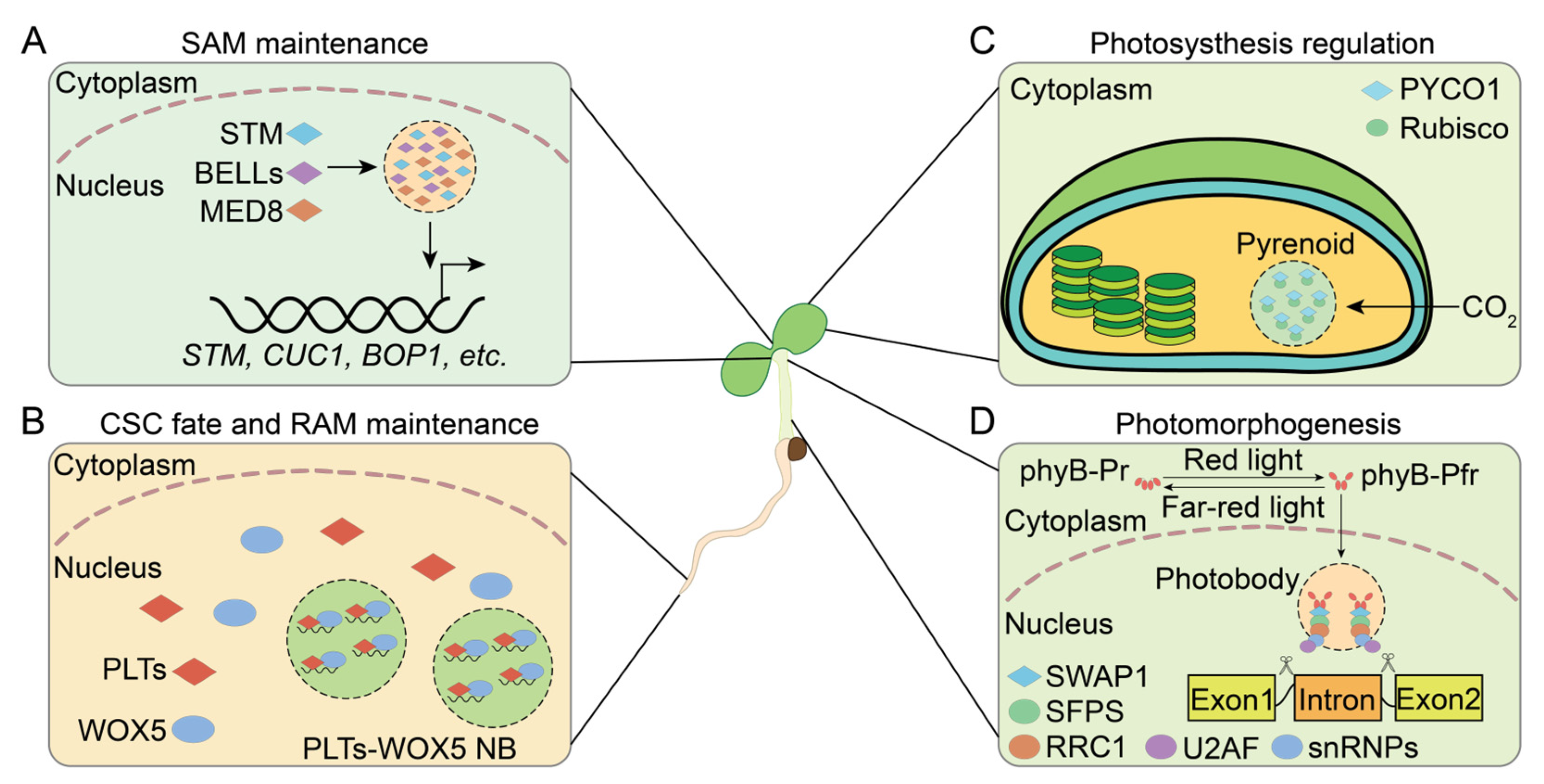
4. Functional Roles of PrLPs in Plant Development

Throughout the plant life cycle, from seed germination to flowering and fruiting, various PrLPs are pivotal in modulating developmental pathways. These proteins also enhance the plant’s ability to perceive environmental changes, thereby facilitating the rapid adaptation of developmental strategies.

4.1. Meristem Maintenance

Plant growth relies on the meristem [84,85]. The root apical meristem (RAM) and shoot apical meristem (SAM) are regions containing pluripotent stem cells that maintain the stem cell niches (SCNs), enabling differentiation into root and shoot tissues [86,87]. Precise control of gene expression by key transcription factors is essential for maintaining stem cell activity in these meristem regions. Recent studies indicate that PrLD−containing transcription factors are crucial for RAM and SAM maintenance by modulating transcriptional regulation through promoting LLPS [43,88].
In the RAM, the SCN consists of quiescent center (QC) cells, which are essential for maintaining surrounding stem cells in a non−autonomous manner. This maintenance ensures the continuous production of new cells for root growth and tissue differentiation [86,87,89,90]. The PLT (PLETHORA) and the WOX (WUSCHEL−RELATED HOMOLOGY BOX) transcription factor families are integral to this regulatory process [91,92]. WOX5 and the PLTs, including PLT1/2/3/4, are co–expressed in the SCN and work in concert to preserve the stem cell identity of the QC [88,92,93,94]. The translocation of WOX5 to the columella stem cells (CSCs) results in its incorporation into nuclear bodies (NBs) by PLTs, thereby inhibiting its activity. Each PLT has PrLDs at its C−terminus, endowing it with the ability to undergo LLPS and form NBs. These NBs sequester WOX5 and RNA, crucial for sustaining the stem cell properties of CSCs and guiding their developmental fate [88] (Figure 2B).
The maintenance of the SAM relies on KNOX1 (CLASS 1 KNOTTED1−LIKE HOMEOBOX) transcription factors, which are crucial for preventing cell differentiation and sustaining the meristematic state [95]. In Arabidopsis, four KNOX1 proteins, including STM (SHOOT MERISTEMLESS), play an important role in SAM initiation and maintenance [43,96,97]. STM contains a PrLD and forms nuclear condensates, a process that is facilitated by PrLD−containing BELL (BEL1−like) proteins. Additionally, the Mediator complex subunit MED8, which contains a PrLD and is part of the transcriptional machinery, is associated with the STM condensates. Condensate formation enhances the transcriptional regulatory activity of STM and strengthens its ability to maintain the meristem. STM exhibits significantly stronger meristematic activity than the other three KNOX1 proteins, which lack PrLDs and do not undergo phase separation. This difference highlights the importance of PrLD in STM condensate formation and meristematic activity [43] (Figure 2A).

4.2. Light Signaling Regulation

Light is a crucial environmental signal, serving both as an essential factor for photosynthesis and as a cue for plant photomorphogenesis. During photosynthesis, the MLO pyrenoids increase the local concentration of CO2 and promote the aggregation of rubisco [98]. In Chlamydomonas reinhardtii, the EPYC1 (essential pyrenoid component 1), featuring IDRs, interacts with rubisco and undergoes LLPS to form pyrenoids [99,100,101,102]. Similarly, PrLP PYCO1 (pyrenoid component 1) typically interacts with rubisco in microalgae [30]. PYCO1 undergoes LLPS via its sticker motifs, particularly in the R6 region rather than the PrLD. PYCO1 forms pyrenoids that offer a structural scaffold for rubisco, thereby enhancing its CO2 fixation efficiency [30] (Figure 2C).
Plant cells discern various light qualities via a spectrum of photoreceptors, modulating plant growth and development [103]. Recent findings indicate that red and far−red light act as a key regulatory switch for the alternative splicing of pre−mRNA in plants [56]. In darkness, phytochrome phyB resides in the cytoplasm as an inactive red light−absorbing form (phyB−Pr) [104]. Exposure to red light induces phyB to transition into its active far−red light−absorbing form (phyB−Pfr), which then relocates to the nucleus to form photobodies. The PrLP SWAP1 (SUPPRESSOR OF WHITE APRICOT/SURP RNA−BINDING DOMAIN CONTAINING PROTEIN1) interacts with the splicing factors SFPS (SPLICING FACTOR FOR PHYTOCHROME SIGNALING) and RRC1 (REDUCED RED–LIGHT RESPONSES IN CRY1CRY2 BACKGROUND1) to form the SFPS–RRC1–SWAP1 complex for pre–mRNA splicing. This complex is recruited by photobodies and interacts with the spliceosome to mediate the alternative splicing of pre−mRNA [56,105,106] (Figure 2D).

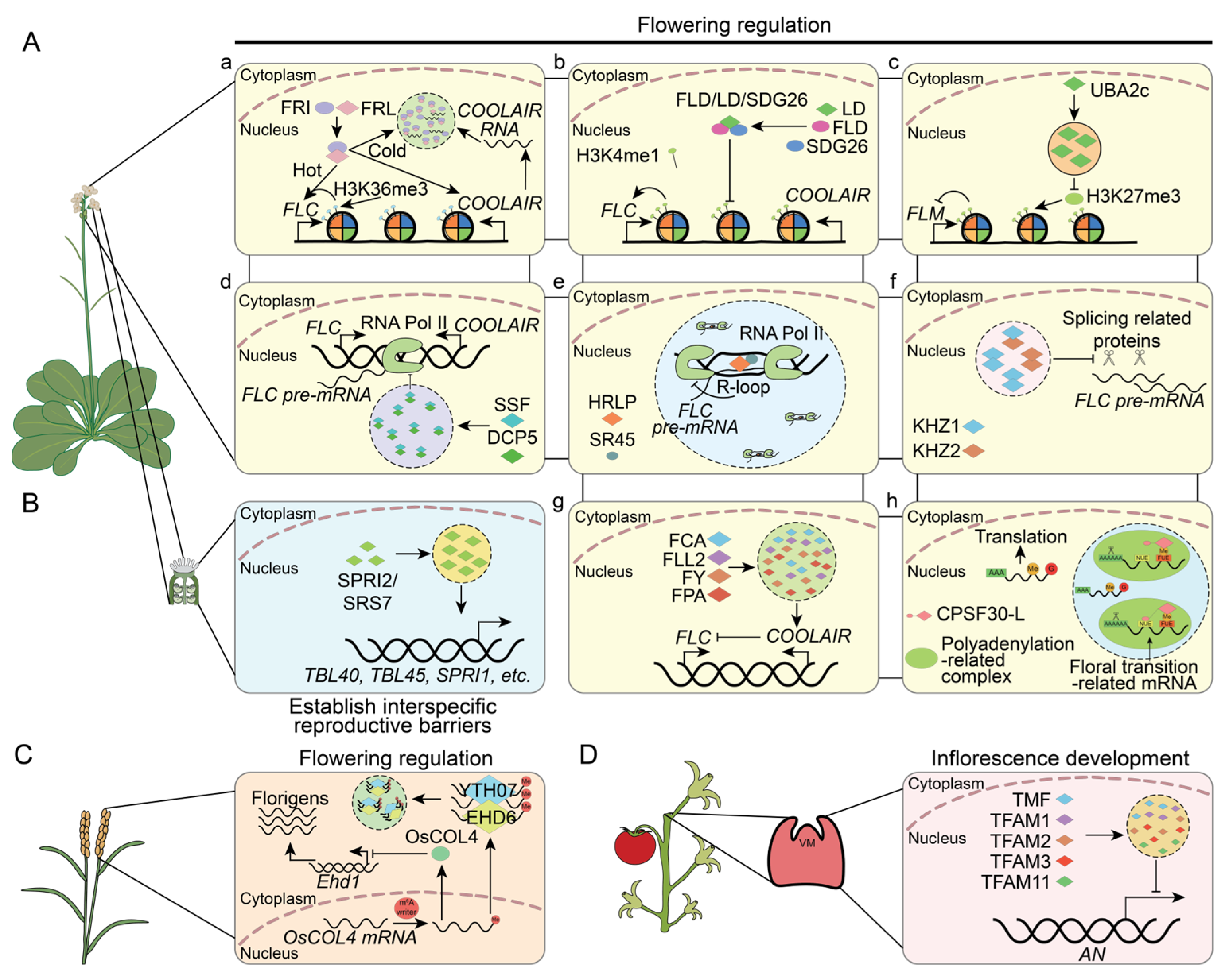
4.3. Reproductive Growth

Plant reproductive growth, including flowering and fruiting, represents a crucial stage in the life span of flowering plants. Flowering time is tightly regulated by both genetic factors and environmental signals, with at least six pathways involved in this regulatory network [107,108]. FT (FLOWERING LOCUS T), a major component of florigen, controls the transition from vegetative to reproductive growth [109,110]. Additionally, FLC (FLOWERING LOCUS C), a MADS–box transcription factor, is a critical inhibitor of FT transcription in the autonomous and vernalization pathways [111,112]. The expression of FLC is regulated at the epigenetic, transcriptional, and post–transcriptional levels, with PrLPs reported to participate in these processes [111,113,114].
Under warm conditions, the activation of FLC expression is controlled by the trimethylation of histone H3 at lysine 36 and lysine 4 (H3K36me3 and H3K4me3), which depends on the transcriptional activator FRIGIDA (FRI) complex [115,116]. This complex is composed of FRI, FRL1 (FRIGIDA–LIKE 1), and several chromatin modifiers and transcriptional co–activators. In cold temperatures, FRI and FRL1 form nuclear condensates that sequester FRI away from the FLC promoter. The PrLP FRL1 participates in the formation of these condensates, and the disruption of the FRI condensate occurs in the frl11 mutant [117] (Figure 3(Aa)). LD (LUMINIDEPENDENS) interacts with SDG26 (SET DOMAIN GROUP 26) and the H3K4 demethylase FLD (FLOWERING LOCUS D) to form the FLD/LD/SDG26 complex. The complex acts as a negative regulator of FLC expression, thus inhibiting the accumulation of H3K4me1 within the FLC gene body [118] (Figure 3(Ab)). As a PrLP, LD can form condensates in the nucleus [14]. However, whether its phase separation is involved in the assembly of the FLD/LD/SDG26 complex requires further investigation. Additionally, the MADS–box transcription factor FLM (FLOWERING LOCUS M) inhibits FT transcription to prevent early flowering [119,120]. The transcription of FLM is regulated by the PrLP UBA2c. UBA2c can bind directly to FLM chromatin and inhibit the trimethylation of H3K27, a marker associated with transcriptional repression [121]. Interestingly, UBA2c forms gel–like speckles in the nucleus, dependent on its PrLD [121]. This function cannot be substituted by the PrLD of FUS, which is known to drive LLPS, suggesting that the gel–like condensates formed by the UBA2c PrLD domain play a distinct role in Arabidopsis [121] (Figure 3(Ac)).
At the transcriptional level, two PrLPs, the P–body component DCP5 and the floral repressor SSF (SISTER OF FCA), work together to regulate the transcription of FLC. SSF serves as a “scaffold” protein anchored to the FLC locus, facilitating the recruitment of DCP5 to the FLC chromatin, where it undergoes LLPS. This LLPS depends on the presence of the PrLD in both SSF and DCP5. The formation of these nuclear condensates is essential for the repression of FLC transcription. Lacking the PrLDs in either DCP5 or SSF leads to failure in repressing FLC transcription and an inability to rescue the flowering time phenotype [45] (Figure 3(Ad)). Additionally, the PrL–RBP HRLP suppresses FLC transcription by promoting R–loop formation near the intron of FLC, resulting in reduced recruitment of RNA Pol II. HRLP forms a complex with the splicing factor SR45 and undergoes LLPS to form nuclear condensates. These condensates are necessary for inhibiting the co–transcriptional splicing and transcription of FLC (Figure 3(Ae)). Notably, the LLPS of HRLP is mediated not by its C–terminal PrLD but by IDRs in its N–terminal region [47].
In addition, PrLPs also regulate post–transcription processes to affect flowering. The splicing efficiency of the FLC pre–mRNA is a critical determinant in producing mature FLC mRNA [54]. Two PrL–RBPs, KHZ1 (CCCH zinc–finger and KH domain–containing proteins 1) and KHZ2, can form homodimers or heterodimers. Their accumulation in nuclear speckles is associated with the reduced splicing efficiency of the FLC pre–mRNA [54] (Figure 3(Af)). COOLAIR, the long antisense RNAs transcribed from the downstream region of the FLC locus, are key modulators of the chromatin environment at the FLC locus [122,123]. The PrL–RBP FCA can form nuclear condensates in collaboration with the polymerase and nuclease components of the RNA 3’–end processing machinery and FLL2, a PrLP with coiled–coil domains. These condensates regulate polyadenylation at specific genomic sites, including proximal sites of COOLAIR nascent transcripts, thereby reducing transcriptional read–through. FLL2 promotes the formation of these condensates, and its overexpression leads to an increase in the size and number of these condensates. Other PrLD–containing proteins, such as FLL1, FPA, and FY, have been identified within the FCA condensates, indicating a complex interplay among these factors in gene expression regulation through phase–separated nuclear condensates [52] (Figure 3(Ag)). Moreover, PrLPs are involved in regulating mRNA stability through the recognition of m6A–modified mRNAs. In Arabidopsis, CPSF30–L enhances the formation of liquid–like nuclear bodies, primarily recognizing m6A–modified far–upstream elements (FUEs), and is crucial for controlling polyadenylation site choice by recruiting the polyadenylation complex. The cpsf30–l mutant leads to an elongation of the 3′ untranslated region (3′ UTR) of specific transcripts, including the floral transition–related gene SOC1 (SUPPRESSOR OF OVEREXPRESSION OF CONSTANS1), resulting in delayed flowering [51] (Figure 3(Ah)). Similarly, in rice, the PrLP EHD6 (EARLY HEADING DATE6) recruits YTH07, a PrLP recognized as an m6A reader, to form RNP granules. These granules facilitate the sequestration of the mRNA of the flowering repressor OsCOL4 (CONSTANSlike 4) in rice, which is critical for reducing OsCOL4 abundance and accelerating flowering [124] (Figure 3C).
Inflorescence formation is a prerequisite for flowering. In tomatoes, the PrLP ALOG transcription factor family plays a significant role in both the regulation of flowering time and the development of inflorescence. The TMF (TERMINATING FLOWER) and its paralogous TFAMs (TMF FAMILY MEMBERs) form heterotypic transcriptional condensates. These condensates collaboratively repress the expression of the floral identity gene AN (ANANTHA). This repression fine–tunes the transition of SAM maturation and the development of inflorescences. Loss–of–function mutations in single or multiple combinations of TMF and TFAMs result in varying degrees of early flowering and alterations in inflorescence structure [125] (Figure 3D).
Prezygotic interspecific incompatibility prevents the formation of unfavorable hybrids between species [126,127,128]. The PrLP SPRI2 (STIGMATIC PRIVACY 2), a member of the SHI (SHORT–INTERNODES) transcription factor family, along with its paralogue SRS5 (SPRI2–like 5), is crucial for this process. SPRI2 and SRS5 are essential for the rejection of pollen from other species on pistils by activating the transcription of genes related to cell wall modification. Notably, SPRI2 localizes within nuclear condensates through LLPS, which is governed by its PrLD [128] (Figure 3B).

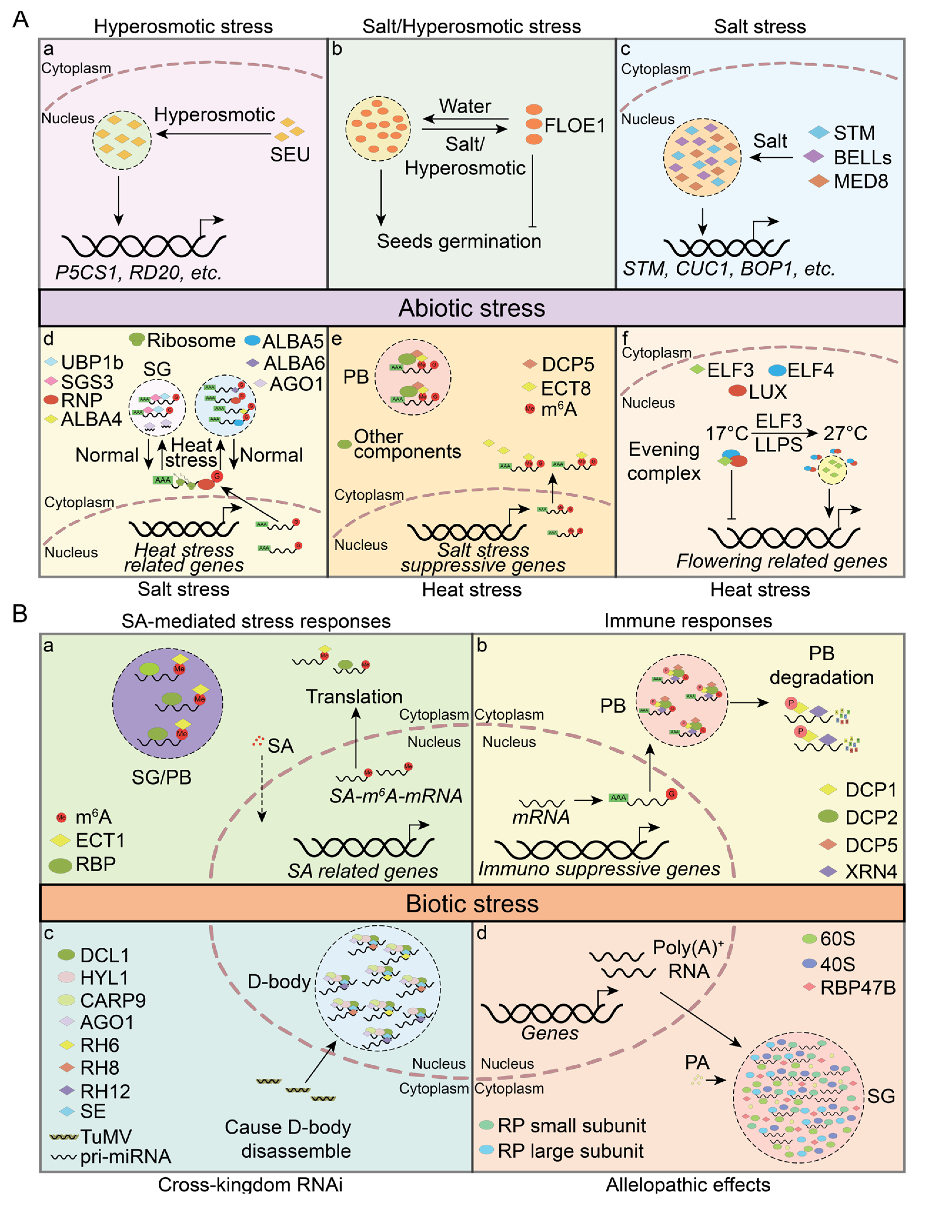
5. PrLPs Involved in Environmental Perception

After sensing changes in both intracellular and extracellular environments, including pH, ion concentration, and temperature, PrLPs can rapidly form condensates via phase separation [129,130,131]. These condensates are typically dynamic and reversible, enabling precise regulation of intracellular processes when the environment changes.

5.1. Abiotic Stress

Hyperosmotic stress causes cellular dehydration, which diminishes cell volume and subsequently increases intracellular macromolecular crowding. This increased crowding enhances the likelihood of molecular interactions, potentially serving as a cellular sensing mechanism to detect hyperosmotic stress. In Arabidopsis, the transcriptional regulator SEUSS (SEU) has been identified as a sensor of molecular crowding. SEU contains an N–terminal PrLD (also referred to as IDR1), which enables SEU to form condensates under hyperosmotic conditions. SEU promotes the upregulation of genes involved in osmotic adjustment, thereby enhancing the plant’s resilience to hyperosmotic stress [132] (Figure 4(Aa)).
Seed maturation involves an essential dehydration phase that protects embryos from external stresses and maintains dormancy [133]. Under favorable conditions, seeds rapidly absorb water, hydrate, and resume cellular activities, leading to germination. FLOE1, a key water–sensing PrLP in seeds, can undergo reversible hydration–dependent phase separation. This process results in the formation of cytoplasmic condensates during seed imbibition, which is essential for the embryo to detect water availability and initiate germination under optimal conditions. Under salt or hyperosmotic stresses, FLOE1 may suppress germination by reducing condensate formation, thereby enhancing the plant’s survival in adverse environments. Notably, the seeds of floe11 mutants exhibit increased germination rates under salt and hyperosmotic stress. FLOE1 contains two IDRs: the N–terminal DS domain rich in aspartic acid (D) and serine (S), and the C–terminal QPS domain, which is a PrLD enriched in glutamine (Q), proline (P), and serine (S). Deleting the QPS domain prevents phase separation, resulting in a loss–of–function phenotype. However, deleting the DS domain results in a gel–like condensate, exhibiting a gain–of–function phenotype that promotes germination under stress. Furthermore, natural variation in FLOE1 suggests that different isoforms may modulate germination timing in natural populations, aligning with diverse climatic adaptations [134,135] (Figure 4(Ab)).
The transcription factor STM plays a pivotal role in sensing salt stress. During salt stress, STM forms condensates, then recruits the Mediator complex subunit MED8 and BELLs proteins, including ATH1 (ARABIDOPSIS THALIANA HOMEOBOX 1), RPL (REPLUMLESS, also known as PENNYWISE and BELLRINGER), and PNF (POUNDFOOLISH). Upon exposure to elevated salt stress, plant cells exhibit enhanced STM/BELLs/MED8 condensate formation. These condensates amplify the STM’s transcriptional activity and stimulate the expression of downstream target genes, thereby promoting salt tolerance and increased shoot branching. Notably, the proteins within this complex are all classified as PrLPs, with RPL likely playing a key role in condensate formation [43] (Figure 4(Ac)).
RNA m6A modification is a crucial epigenetic modification that regulates various aspects of plant development and stress responses [136,137,138]. The ECT8, an m6A reader with a PrLD, is involved in regulating mRNA stability under salt stress. Both transcriptional and translational levels of ECT8 are upregulated in response to salt stress. ECT8 directly interacts with decapping protein DCP5 and undergoes LLPS to form PBs, which are crucial for the degradation of m6A–modified RNAs that encode negative regulators of the salt stress response [139] (Figure 4(Ad)).
Heat stress impedes normal developmental processes and reduces plant productivity [140,141]. To mitigate the effects of heat, plants activate heat stress transcription factors (HSFs), which respond to elevated temperatures by initiating the transcription of heat–responsive genes [142,143]. In Arabidopsis, ALBA proteins, specifically the PrLPs ALBA4 and ALBA6, undergo LLPS under heat stress, leading to the formation of PBs or SGs. These cytoplasmic granules interact with HSF mRNAs and other transcripts, stabilizing them under high–temperature conditions and ensuring their translation when conditions return to normal [55]. Additionally, OLIGOURIDYLATE BINDING PROTEIN UBP1b, a PrLP and another component of SGs, serves a similar function. UBP1b can bind mRNAs and undergo LLPS during heat stress to form SGs, thus protecting heat stress–related mRNAs from degradation [144]. Concurrently, PrLP ARGONAUTE1 (AGO1) forms condensates and accumulates within SGs under heat stress. The sequestration of AGO1 within SGs protects AGO1 and ensures that the RNA–silencing activity of AGO1 remains unaffected by the stress [145] (Figure 4(Ae)).
Elevated temperatures prompt plants to flower prematurely, an adaptive strategy that ensures the timely completion of their reproductive cycle [146,147]. Under normal conditions, the evening complex, comprising ELF3 (EARLY FLOWERING 3), ELF4, and LUX (LUX ARRHYTHMO), binds to the promoter regions of flowering–associated genes and inhibits their transcription [42,148,149]. Under heat stress, the PrLP ELF3 undergoes LLPS to form ELF3 condensates, which sequester ELF3 and prevent its assembly with ELF4 and LUX within the evening complex. As a result, the transcriptional repression of flowering genes is alleviated, accelerating the transition to the flowering stage [42]. ELF3 represents a novel thermosensory mechanism, allowing plants to dynamically adjust their growth and developmental programs in response to environmental stimuli (Figure 4(Af)).

5.2. Biotic Stress

During growth and development, plants are frequently attacked by various pathogens and insects. To combat these biotic stresses, plants have developed various defense mechanisms, including the production of defense–related hormones and secondary metabolites and rapid gene expression regulation [150]. Salicylic acid (SA) is a key defense–related hormone that stimulates the expression of stress–responsive genes to promote immune responses [151,152,153]. However, plants must balance an overly robust immune response to avoid irreversible cellular damage [154,155]. ECT1, an m6A reader with a PrLD at its N–terminus, is a key regulator in this process. ECT1 binds to m6A–modified SA–induced mRNAs and undergoes LLPS to form condensates. These condensates may transform into PBs or SGs, which are involved in mRNA degradation or storage, thereby reducing mRNA levels and negatively regulating the immune response [156] (Figure 4(Ba)). Additionally, ECT1 interacts with its homolog ECT9 to undergo phase separation, modulating immune responses to non–pathogenic stimuli and preventing an overactive immune system [157]. Components of PBs, including DCP1, DCP2, and XRN4, participate in the mRNA decay process triggered by microbe–associated molecular patterns (MAMPs). MAMP–activated MAP kinases (MPK3 and MPK6) phosphorylate DCP1, enhancing its interaction with XRN4, a 5′–3′ RNA exonuclease responsible for the degradation of decapped mRNAs [74] (Figure 4(Bb)).
Nucleotide–binding leucine–rich repeat receptors (NLRs) are pivotal intracellular immune receptors for disease resistance [158]. However, the expression of NLRs under normal conditions may trigger autoimmunity, requiring tight regulation of their transcription [159]. The PrL–RBP FPA is involved in this regulatory mechanism. FPA can interact with RNA cleavage and polyadenylation factors, leading to the premature cleavage and polyadenylation of NLR transcripts. This process controls the functional expression of NLRs and affects immunity [160]. Although FPA contains a PrLD, its capacity to undergo LLPS and form condensates during RNA processing remains to be determined.
RNA interference (RNAi) is a crucial mechanism for effecting the expression of immune–related genes in response to pathogens and viruses [161,162]. Several PrLPs are involved in small RNA biosynthesis and gene silencing. D–bodies, a type of nuclear RNP granule, are crucial for microRNA (miRNA) biosynthesis and processing [57]. DEAD–box RNA helicases, including PrLPs RH6, RH8, and RH12, interact with SERRATE (SE), a key component of D–bodies, and promote LLPS to form D–bodies. Upon Turnip mosaic virus (TuMV) infection, the accumulation of these helicases in the nucleus decreases, leading to a reduction in D–body levels. This reduction correlates with the negative regulation of TuMV infections [53] (Figure 4(Bc)). Furthermore, the PrLP SGS3 undergoes LLPS and recruits RDR6 to form the SGS3/RDR6 body (also known as the siRNA body) [78,163]. The SGS3/RDR6 body modulates the production of siRNAs, thereby facilitating plant defense against viral infection [163]. Additionally, the core components of the RNA–induced silencing complex (RISC), known as ARGONAUTE proteins (AGO1, AGO2, AGO3, and AGO5), contain PrLDs and are potential phase–separating proteins [78,164].
Plants produce unique biochemical substances known as allelopathic compounds, which inhibit the normal development of neighboring plants in the competitive struggle for survival [165]. Phenolic acid (PA), commonly secreted by plant roots, can enter the plant cell nucleus. The PrLP RBP47B acts as a PA receptor. Upon binding to PAs, RBP47B undergoes LLPS, forming SGs. These SGs incorporate mRNA, ribosomes, rRNA, and various other cellular components, leading to the comprehensive suppression of translation in plant cells affected by allelopathic substances [166] (Figure 4(Bd)).

6. Conclusions and Future Prospects

In this review, we focus on the structural characteristics of PrLPs and their crucial roles in regulating phase separation. We also discuss the molecular functions of PrLPs and their involvement in various biological processes, highlighting their broad impact on cellular function. While most research has been conducted using the model organism Arabidopsis, our understanding of PrLPs in other plant species remains limited. Further studies could shed light on PrLP functions across the plant kingdom. Recent findings have also underscored the interaction between MLOs and cellular membranes, which may influence membrane properties and processes [80]. Although PrLPs are enriched in many membrane–associated processes, their specific roles in these processes remain largely unexplored. Elucidating these functions could significantly enhance our understanding of cellular dynamics. Additionally, investigating PrLPs from an evolutionary perspective could uncover their new roles in environmental adaptations and evolutionary processes. Such an approach may reveal conserved and complex mechanisms, thereby deepening our understanding of the evolutionary significance of PrLPs.

Supplementary Materials

The following supporting information can be downloaded at: https://www.mdpi.com/article/10.3390/plants13182666/s1, Table S1: Biological Processes of Plants; Table S2: Molecular Functions of Plants; Table S3: Cellular Components of Plants.

Author Contributions

Conceptualization, writing—original draft preparation, review, and editing, Y.L. and P.W.; supervision and funding acquisition, Y.L.; software, formal analysis, data curation, visualization, and validation, P.W. All authors have read and agreed to the published version of the manuscript.

Funding

This work was supported by grants from the National Natural Science Foundation of China (32300572 to Y.L.).

Data Availability Statement

The data presented in this study are available in Tables S1–S3.

Conflicts of Interest

The authors declare no conflicts of interest.

Appendix A. The Methods and Materials of Gene Ontology Enrichment Analysis

We performed a gene ontology (GO) enrichment analysis to investigate the functional roles of PrLP candidates across a diverse set of ten plant species, including five monocots (Brachypodium distachyon, Hordeum vulgare, Oryza sativa, Sorghum bicolor, and Triticum aestivum) and five dicots (Arabidopsis thaliana, Glycine max, Helianthus annuus, Prunus persica, and Solanum lycopersicum). Protein sequences for these species were obtained from the EnsemblPlants database (http://plants.ensembl.org/index.html. accessed on 17 September 2024). The presence of PrLDs within these sequences was determined using the PLAAC algorithm program analysis [27]. The identified PrLP genes were then subjected to the PANTHER classification system (https://pantherdb.org/) accessed on 29 June 2024 to conduct the GO enrichment analysis [167,168,169]. We focused on the top 50 GO terms from each category and selected 10 terms that exhibited significant enrichment across all ten species. To visually represent these findings, we employed the ggplot2 package [170] within the R software environment (version 4.4.3) [171] to create bubble plots that highlight the most highly enriched GO terms.

References

  1. Prusiner, S.B. Novel proteinaceous infectious particles cause scrapie. Science 1982, 216, 136–144. [Google Scholar] [CrossRef] [PubMed]
  2. Shorter, J.; Lindquist, S. Prions as adaptive conduits of memory and inheritance. Nat. Rev. Genet. 2005, 6, 435–450. [Google Scholar] [CrossRef]
  3. Riesner, D. Biochemistry and structure of PrPC and PrPSc. Br. Med. Bull. 2003, 66, 21–33. [Google Scholar] [CrossRef] [PubMed]
  4. Prusiner, S.B. Prions. Proc. Natl. Acad. Sci. USA 1998, 95, 13363–13383. [Google Scholar] [CrossRef] [PubMed]
  5. Eghiaian, F.; Grosclaude, J.; Lesceu, S.; Debey, P.; Doublet, B.; Tréguer, E.; Rezaei, H.; Knossow, M. Insight into the PrPC → PrPSc conversion from the structures of antibody–bound ovine prion scrapie–susceptibility variants. Proc. Natl. Acad. Sci. USA 2004, 101, 10254–10259. [Google Scholar] [CrossRef]
  6. Prusiner, S.B. A unifying role for prions in neurodegenerative diseases. Science 2012, 336, 1511–1513. [Google Scholar] [CrossRef] [PubMed]
  7. King, O.D.; Gitler, A.D.; Shorter, J. The tip of the iceberg: RNA–binding proteins with prion–like domains in neurodegenerative disease. Brain Res. 2012, 1462, 61–80. [Google Scholar] [CrossRef]
  8. Krishnan, R.; Lindquist, S.L. Structural insights into a yeast prion illuminate nucleation and strain diversity. Nature 2005, 435, 765–772. [Google Scholar] [CrossRef]
  9. Santoso, A.; Chien, P.; Osherovich, L.Z.; Weissman, J.S. Molecular basis of a yeast prion species barrier. Cell 2000, 100, 277–288. [Google Scholar] [CrossRef]
  10. Sabate, R.; Rousseau, F.; Schymkowitz, J.; Ventura, S. What makes a protein sequence a prion? PLoS Comput. Biol. 2015, 11, e1004013. [Google Scholar] [CrossRef]
  11. Alberti, S.; Halfmann, R.; King, O.; Kapila, A.; Lindquist, S. A systematic survey identifies prions and illuminates sequence features of prionogenic proteins. Cell 2009, 137, 146–158. [Google Scholar] [CrossRef] [PubMed]
  12. Gil-Garcia, M.; Iglesias, V.; Pallarès, I.; Ventura, S. Prion–like proteins: From computational approaches to proteome–wide analysis. FEBS Open Biol. 2021, 11, 2400–2417. [Google Scholar] [CrossRef] [PubMed]
  13. Liebman, S.W.; Chernoff, Y.O. Prions in yeast. Genetics 2012, 191, 1041–1072. [Google Scholar] [CrossRef] [PubMed]
  14. Chakrabortee, S.; Kayatekin, C.; Newby, G.A.; Mendillo, M.L.; Lancaster, A.; Lindquist, S. Luminidependens (LD) is an Arabidopsis protein with prion behavior. Proc. Natl. Acad. Sci. USA 2016, 113, 6065–6070. [Google Scholar] [CrossRef]
  15. Garai, S.; Citu; Singla-Pareek, S.L.; Sopory, S.K.; Kaur, C.; Yadav, G. Complex networks of prion–like proteins reveal cross talk between stress and memory pathways in plants. Front. Plant Sci. 2021, 12, 707286. [Google Scholar] [CrossRef]
  16. Emenecker, R.J.; Holehouse, A.S.; Strader, L.C. Biological phase separation and biomolecular condensates in plants. Annu. Rev. Plant Biol. 2021, 72, 17–46. [Google Scholar] [CrossRef]
  17. Boeynaems, S.; Alberti, S.; Fawzi, N.L.; Mittag, T.; Polymenidou, M.; Rousseau, F.; Schymkowitz, J.; Shorter, J.; Wolozin, B.; Van Den Bosch, L.; et al. Protein phase separation: A new phase in cell biology. Trends Cell Biol. 2018, 28, 420–435. [Google Scholar] [CrossRef]
  18. Banani, S.F.; Lee, H.O.; Hyman, A.A.; Rosen, M.K. Biomolecular condensates: Organizers of cellular biochemistry. Nat. Rev. Mol. Cell Biol. 2017, 18, 285–298. [Google Scholar] [CrossRef]
  19. Molliex, A.; Temirov, J.; Lee, J.; Coughlin, M.; Kanagaraj, A.P.; Kim, H.J.; Mittag, T.; Taylor, J.P. Phase separation by low complexity domains promotes stress granule assembly and drives pathological fibrillization. Cell 2015, 163, 123–133. [Google Scholar] [CrossRef]
  20. Zhang, H.; Ji, X.; Li, P.; Liu, C.; Lou, J.; Wang, Z.; Wen, W.; Xiao, Y.; Zhang, M.; Zhu, X. Liquid–liquid phase separation in biology: Mechanisms, physiological functions and human diseases. Sci. China Life Sci. 2020, 63, 953–985. [Google Scholar] [CrossRef]
  21. Wiedner, H.J.; Giudice, J. It’s not just a phase: Function and characteristics of RNA–binding proteins in phase separation. Nat. Struct. Mol. Biol. 2021, 28, 465–473. [Google Scholar] [CrossRef] [PubMed]
  22. Gotor, N.L.; Armaos, A.; Calloni, G.; Torrent Burgas, M.; Vabulas, R.M.; De Groot, N.S.; Tartaglia, G.G. RNA–binding and prion domains: The Yin and Yang of phase separation. Nucleic Acids Res. 2020, 48, 9491–9504. [Google Scholar] [CrossRef] [PubMed]
  23. Wang, J.; Choi, J.M.; Holehouse, A.S.; Lee, H.O.; Zhang, X.; Jahnel, M.; Maharana, S.; Lemaitre, R.; Pozniakovsky, A.; Drechsel, D.; et al. A molecular grammar governing the driving forces for phase separation of prion–like RNA binding proteins. Cell 2018, 174, 688–699.e16. [Google Scholar] [CrossRef] [PubMed]
  24. Fomicheva, A.; Ross, E.D. From prions to stress granules: Defining the compositional features of prion–like domains that promote different types of assemblies. Int. J. Mol. Sci. 2021, 22, 1251. [Google Scholar] [CrossRef] [PubMed]
  25. Ross, E.D.; Edskes, H.K.; Terry, M.J.; Wickner, R.B. Primary sequence independence for prion formation. Proc. Natl. Acad. Sci. USA 2005, 102, 12825–12830. [Google Scholar] [CrossRef]
  26. Ross, E.D.; Baxa, U.; Wickner, R.B. Scrambled prion domains form prions and amyloid. Mol. Cell. Biol. 2004, 24, 7206–7213. [Google Scholar] [CrossRef]
  27. Lancaster, A.K.; Nutter-Upham, A.; Lindquist, S.; King, O.D. PLAAC: A web and command–line application to identify proteins with prion–like amino acid composition. Bioinformatics 2014, 30, 2501–2502. [Google Scholar] [CrossRef]
  28. Li, P.; Banjade, S.; Cheng, H.-C.; Kim, S.; Chen, B.; Guo, L.; Llaguno, M.; Hollingsworth, J.V.; King, D.S.; Banani, S.F.; et al. Phase transitions in the assembly of multivalent signalling proteins. Nature 2012, 483, 336–340. [Google Scholar] [CrossRef]
  29. Farag, M.; Borcherds, W.M.; Bremer, A.; Mittag, T.; Pappu, R.V. Phase separation of protein mixtures is driven by the interplay of homotypic and heterotypic interactions. Nat. Commun. 2023, 14, 5527. [Google Scholar] [CrossRef]
  30. Oh, Z.G.; Ang, W.S.L.; Poh, C.W.; Lai, S.-K.; Sze, S.K.; Li, H.-Y.; Bhushan, S.; Wunder, T.; Mueller-Cajar, O. A linker protein from a red–type pyrenoid phase separates with Rubisco via oligomerizing sticker motifs. Proc. Natl. Acad. Sci. USA 2023, 120, e2304833120. [Google Scholar] [CrossRef]
  31. Martin, E.W.; Holehouse, A.S.; Peran, I.; Farag, M.; Incicco, J.J.; Bremer, A.; Grace, C.R.; Soranno, A.; Pappu, R.V.; Mittag, T. Valence and patterning of aromatic residues determine the phase behavior of prion–like domains. Science 2020, 367, 694–699. [Google Scholar] [CrossRef] [PubMed]
  32. Bremer, A.; Farag, M.; Borcherds, W.M.; Peran, I.; Martin, E.W.; Pappu, R.V.; Mittag, T. Deciphering how naturally occurring sequence features impact the phase behaviours of disordered prion–like domains. Nat. Chem. 2021, 14, 196–207. [Google Scholar] [CrossRef] [PubMed]
  33. Holehouse, A.S.; Ginell, G.M.; Griffith, D.; Böke, E. Clustering of aromatic residues in prion–like domains can tune the formation, state, and organization of biomolecular condensates. Biochemistry 2021, 60, 3566–3581. [Google Scholar] [CrossRef] [PubMed]
  34. Semenov, A.N.; Rubinstein, M. Thermoreversible gelation in solutions of associative polymers. 1. Statics. Macromolecules 1998, 31, 1373–1385. [Google Scholar] [CrossRef]
  35. Dougherty, D.A. Cation–pi interactions in chemistry and biology: A new view of benzene, Phe, Tyr, and Trp. Science 1996, 271, 163–168. [Google Scholar] [CrossRef]
  36. Dougherty, D.A. The cation–pi interaction. Acc. Chem. Res. 2013, 46, 885–893. [Google Scholar] [CrossRef]
  37. Gallivan, J.P.; Dougherty, D.A. Cation–pi interactions in structural biology. Proc. Natl. Acad. Sci. USA 1999, 96, 9459–9464. [Google Scholar] [CrossRef]
  38. Langdon, E.M.; Qiu, Y.; Ghanbari Niaki, A.; McLaughlin, G.A.; Weidmann, C.A.; Gerbich, T.M.; Smith, J.A.; Crutchley, J.M.; Termini, C.M.; Weeks, K.M.; et al. mRNA structure determines specificity of a polyQ–driven phase separation. Science 2018, 360, 922–927. [Google Scholar] [CrossRef]
  39. Maharana, S.; Wang, J.; Papadopoulos, D.K.; Richter, D.; Pozniakovsky, A.; Poser, I.; Bickle, M.; Rizk, S.; Guillén-Boixet, J.; Franzmann, T.M.; et al. RNA buffers the phase separation behavior of prion–like RNA binding proteins. Science 2018, 360, 918–921. [Google Scholar] [CrossRef]
  40. Gomes, E.; Shorter, J. The molecular language of membraneless organelles. J. Biol. Chem. 2019, 294, 7115–7127. [Google Scholar] [CrossRef]
  41. Sharp, P.A.; Chakraborty, A.K.; Henninger, J.E.; Young, R.A. RNA in formation and regulation of transcriptional condensates. RNA 2022, 28, 52–57. [Google Scholar] [CrossRef] [PubMed]
  42. Jung, J.-H.; Barbosa, A.D.; Hutin, S.; Kumita, J.R.; Gao, M.; Derwort, D.; Silva, C.S.; Lai, X.; Pierre, E.; Geng, F.; et al. A prion–like domain in ELF3 functions as a thermosensor in Arabidopsis. Nature 2020, 585, 256–260. [Google Scholar] [CrossRef] [PubMed]
  43. Cao, X.; Du, Q.; Guo, Y.; Wang, Y.; Jiao, Y. Condensation of STM is critical for shoot meristem maintenance and salt tolerance in Arabidopsis. Mol. Plant 2023, 16, 1445–1459. [Google Scholar] [CrossRef] [PubMed]
  44. He, J.; Huo, X.; Pei, G.; Jia, Z.; Yan, Y.; Yu, J.; Qu, H.; Xie, Y.; Yuan, J.; Zheng, Y.; et al. Dual–role transcription factors stabilize intermediate expression levels. Cell 2024, 187, 2746–2766.e25. [Google Scholar] [CrossRef]
  45. Wang, W.; Wang, C.; Wang, Y.; Ma, J.; Wang, T.; Tao, Z.; Liu, P.; Li, S.; Hu, Y.; Gu, A.; et al. The P–body component DECAPPING5 and the floral repressor SISTER OF FCA regulate FLOWERING LOCUS C transcription in Arabidopsis. Plant Cell 2023, 35, 3303–3324. [Google Scholar] [CrossRef]
  46. Demmerle, J.; Hao, S.; Cai, D. Transcriptional condensates and phase separation: Condensing information across scales and mechanisms. Nucleus 2023, 14, 2213551. [Google Scholar] [CrossRef]
  47. Zhang, Y.; Fan, S.; Hua, C.; Teo, Z.W.N.; Kiang, J.X.; Shen, L.; Yu, H. Phase separation of HRLP regulates flowering time in Arabidopsis. Sci. Adv. 2022, 8, eabn5488. [Google Scholar] [CrossRef]
  48. Garcia-Muse, T.; Aguilera, A. R loops: From physiological to pathological roles. Cell 2019, 179, 604–618. [Google Scholar] [CrossRef]
  49. Stortz, M.; Presman, D.M.; Levi, V. Transcriptional condensates: A blessing or a curse for gene regulation? Commun. Biol. 2024, 7, 187. [Google Scholar] [CrossRef]
  50. March, Z.M.; King, O.D.; Shorter, J. Prion–like domains as epigenetic regulators, scaffolds for subcellular organization, and drivers of neurodegenerative disease. Brain Res. 2016, 1647, 9–18. [Google Scholar] [CrossRef]
  51. Song, P.; Yang, J.; Wang, C.; Lu, Q.; Shi, L.; Tayier, S.; Jia, G. Arabidopsis N(6)–methyladenosine reader CPSF30–L recognizes FUE signals to control polyadenylation site choice in liquid–like nuclear bodies. Mol. Plant 2021, 14, 571–587. [Google Scholar] [CrossRef] [PubMed]
  52. Fang, X.; Wang, L.; Ishikawa, R.; Li, Y.; Fiedler, M.; Liu, F.; Calder, G.; Rowan, B.; Weigel, D.; Li, P.; et al. Arabidopsis FLL2 promotes liquid–liquid phase separation of polyadenylation complexes. Nature 2019, 569, 265–269. [Google Scholar] [CrossRef] [PubMed]
  53. Li, Q.; Liu, N.; Liu, Q.; Zheng, X.; Lu, L.; Gao, W.; Liu, Y.; Liu, Y.; Zhang, S.; Wang, Q.; et al. DEAD–box helicases modulate dicing body formation in Arabidopsis. Sci. Adv. 2021, 7, eabc6266. [Google Scholar] [CrossRef]
  54. Yan, Z.; Shi, H.; Liu, Y.; Jing, M.; Han, Y. KHZ1 and KHZ2, novel members of the autonomous pathway, repress the splicing efficiency of FLC pre–mRNA in Arabidopsis. J. Exp. Bot. 2020, 71, 1375–1386. [Google Scholar] [CrossRef] [PubMed]
  55. Tong, J.; Ren, Z.; Sun, L.; Zhou, S.; Yuan, W.; Hui, Y.; Ci, D.; Wang, W.; Fan, L.M.; Wu, Z.; et al. ALBA proteins confer thermotolerance through stabilizing HSF messenger RNAs in cytoplasmic granules. Nat. Plants 2022, 8, 778–791. [Google Scholar] [CrossRef] [PubMed]
  56. Kathare, P.K.; Xin, R.; Ganesan, A.S.; June, V.M.; Reddy, A.S.N.; Huq, E. SWAP1–SFPS–RRC1 splicing factor complex modulates pre–mRNA splicing to promote photomorphogenesis in Arabidopsis. Proc. Natl. Acad. Sci. USA 2022, 119, e2214565119. [Google Scholar] [CrossRef]
  57. Xie, D.; Chen, M.; Niu, J.; Wang, L.; Li, Y.; Fang, X.; Li, P.; Qi, Y. Phase separation of SERRATE drives dicing body assembly and promotes miRNA processing in Arabidopsis. Nat. Cell Biol. 2021, 23, 32–39. [Google Scholar] [CrossRef]
  58. Li, C.F.; Pontes, O.; El-Shami, M.; Henderson, I.R.; Bernatavichute, Y.V.; Chan, S.W.; Lagrange, T.; Pikaard, C.S.; Jacobsen, S.E. An ARGONAUTE4–containing nuclear processing center colocalized with Cajal bodies in Arabidopsis thaliana. Cell 2006, 126, 93–106. [Google Scholar] [CrossRef]
  59. Tan, H.; Luo, W.; Yan, W.; Liu, J.; Aizezi, Y.; Cui, R.; Tian, R.; Ma, J.; Guo, H. Phase separation of SGS3 drives siRNA body formation and promotes endogenous gene silencing. Cell Rep. 2023, 42, 111985. [Google Scholar] [CrossRef]
  60. Weber, C.; Nover, L.; Fauth, M. Plant stress granules and mRNA processing bodies are distinct from heat stress granules. Plant J. 2008, 56, 517–530. [Google Scholar] [CrossRef]
  61. Sorenson, R.; Bailey-Serres, J. Selective mRNA sequestration by OLIGOURIDYLATE–BINDING PROTEIN 1 contributes to translational control during hypoxia in Arabidopsis. Proc. Natl. Acad. Sci. USA 2014, 111, 2373–2378. [Google Scholar] [CrossRef] [PubMed]
  62. Wheeler, J.R.; Matheny, T.; Jain, S.; Abrisch, R.; Parker, R. Distinct stages in stress granule assembly and disassembly. Elife 2016, 5, e18413. [Google Scholar] [CrossRef] [PubMed]
  63. Youn, J.Y.; Dyakov, B.J.A.; Zhang, J.; Knight, J.D.R.; Vernon, R.M.; Forman-Kay, J.D.; Gingras, A.C. Properties of stress granule and P–body proteomes. Mol. Cell 2019, 76, 286–294. [Google Scholar] [CrossRef] [PubMed]
  64. Chantarachot, T.; Bailey-Serres, J. Polysomes, stress granules, and processing bodies: A dynamic triumvirate controlling cytoplasmic mRNA fate and function. Plant Physiol. 2018, 176, 254–269. [Google Scholar] [CrossRef]
  65. Anderson, P.; Kedersha, N. RNA granules: Post–transcriptional and epigenetic modulators of gene expression. Nat. Rev. Mol. Cell Biol. 2009, 10, 430–436. [Google Scholar] [CrossRef]
  66. Vanderweyde, T.; Youmans, K.; Liu-Yesucevitz, L.; Wolozin, B. Role of stress granules and RNA–binding proteins in neurodegeneration: A mini–review. Gerontology 2013, 59, 524–533. [Google Scholar] [CrossRef]
  67. Buchan, J.R.; Parker, R. Eukaryotic stress granules: The ins and outs of translation. Mol. Cell 2009, 36, 932–941. [Google Scholar] [CrossRef]
  68. Iserman, C.; Desroches Altamirano, C.; Jegers, C.; Friedrich, U.; Zarin, T.; Fritsch, A.W.; Mittasch, M.; Domingues, A.; Hersemann, L.; Jahnel, M.; et al. Condensation of Ded1p promotes a translational switch from housekeeping to stress protein production. Cell 2020, 181, 818–831.e819. [Google Scholar] [CrossRef]
  69. Jang, G.J.; Yang, J.Y.; Hsieh, H.L.; Wu, S.H. Processing bodies control the selective translation for optimal development of Arabidopsis young seedlings. Proc. Natl. Acad. Sci. USA 2019, 116, 6451–6456. [Google Scholar] [CrossRef]
  70. Decker, C.J.; Parker, R. P–bodies and stress granules: Possible roles in the control of translation and mRNA degradation. Cold Spring Harb. Perspect. Biol. 2012, 4, a012286. [Google Scholar] [CrossRef]
  71. Aizer, A.; Kalo, A.; Kafri, P.; Shraga, A.; Ben-Yishay, R.; Jacob, A.; Kinor, N.; Shav-Tal, Y. Quantifying mRNA targeting to P–bodies in living human cells reveals their dual role in mRNA decay and storage. J. Cell Sci. 2014, 127, 4443–4456. [Google Scholar] [CrossRef] [PubMed]
  72. Schiaffini, M.; Chicois, C.; Pouclet, A.; Chartier, T.; Ubrig, E.; Gobert, A.; Zuber, H.; Mutterer, J.; Chicher, J.; Kuhn, L.; et al. A NYN domain protein directly interacts with DECAPPING1 and is required for phyllotactic pattern. Plant Physiol. 2022, 188, 1174–1188. [Google Scholar] [CrossRef] [PubMed]
  73. Liu, C.; Mentzelopoulou, A.; Muhammad, A.; Volkov, A.; Weijers, D.; Gutierrez-Beltran, E.; Moschou, P.N. An actin remodeling role for Arabidopsis processing bodies revealed by their proximity interactome. EMBO J. 2023, 42, e111885. [Google Scholar] [CrossRef] [PubMed]
  74. Yu, X.; Li, B.; Jang, G.J.; Jiang, S.; Jiang, D.; Jang, J.C.; Wu, S.H.; Shan, L.; He, P. Orchestration of processing body dynamics and mRNA decay in Arabidopsis immunity. Cell Rep. 2019, 28, 2194–2205.e6. [Google Scholar] [CrossRef]
  75. Jang, G.J.; Jang, J.C.; Wu, S.H. Dynamics and functions of stress granules and processing bodies in plants. Plants 2020, 9, 1122. [Google Scholar] [CrossRef]
  76. Maruri-Lopez, I.; Figueroa, N.E.; Hernandez-Sanchez, I.E.; Chodasiewicz, M. Plant stress granules: Trends and beyond. Front. Plant Sci. 2021, 12, 722643. [Google Scholar] [CrossRef]
  77. Chantarachot, T.; Sorenson, R.S.; Hummel, M.; Ke, H.; Kettenburg, A.T.; Chen, D.; Aiyetiwa, K.; Dehesh, K.; Eulgem, T.; Sieburth, L.E.; et al. DHH1/DDX6–like RNA helicases maintain ephemeral half–lives of stress–response mRNAs. Nat. Plants 2020, 6, 675–685. [Google Scholar] [CrossRef]
  78. Kim, E.Y.; Wang, L.; Lei, Z.; Li, H.; Fan, W.; Cho, J. Ribosome stalling and SGS3 phase separation prime the epigenetic silencing of transposons. Nat. Plants 2021, 7, 303–309. [Google Scholar] [CrossRef]
  79. Schiano Lomoriello, I.; Sigismund, S.; Day, K.J. Biophysics of endocytic vesicle formation: A focus on liquid–liquid phase separation. Curr. Opin. Cell Biol. 2022, 75, 102068. [Google Scholar] [CrossRef]
  80. Hatzianestis, I.H.; Mountourakis, F.; Stavridou, S.; Moschou, P.N. Plant condensates: No longer membrane–less? Trends Plant Sci. 2023, 28, 1101–1112. [Google Scholar] [CrossRef]
  81. Snead, W.T.; Gladfelter, A.S. The control centers of biomolecular phase separation: How membrane surfaces, PTMs, and active processes regulate condensation. Mol. Cell 2019, 76, 295–305. [Google Scholar] [CrossRef] [PubMed]
  82. Zhao, Y.G.; Zhang, H. Phase separation in membrane biology: The interplay between membrane–bound organelles and membraneless condensates. Dev. Cell 2020, 55, 30–44. [Google Scholar] [CrossRef] [PubMed]
  83. Buchan, J.R.; Kolaitis, R.M.; Taylor, J.P.; Parker, R. Eukaryotic stress granules are cleared by autophagy and Cdc48/VCP function. Cell 2013, 153, 1461–1474. [Google Scholar] [CrossRef] [PubMed]
  84. Wang, B.; Smith, S.M.; Li, J. Genetic regulation of shoot architecture. Annu. Rev. Plant Biol. 2018, 69, 437–468. [Google Scholar] [CrossRef] [PubMed]
  85. Petricka, J.J.; Winter, C.M.; Benfey, P.N. Control of Arabidopsis root development. Annu. Rev. Plant Biol. 2012, 63, 563–590. [Google Scholar] [CrossRef]
  86. van den Berg, C.; Willemsen, V.; Hendriks, G.; Weisbeek, P.; Scheres, B. Short–range control of cell differentiation in the Arabidopsis root meristem. Nature 1997, 390, 287–289. [Google Scholar] [CrossRef]
  87. Lu, R.; Canher, B.; Bisht, A.; Heyman, J.; De Veylder, L. Three–dimensional quantitative analysis of the Arabidopsis quiescent centre. J. Exp. Bot. 2021, 72, 6789–6800. [Google Scholar] [CrossRef]
  88. Burkart, R.C.; Strotmann, V.I.; Kirschner, G.K.; Akinci, A.; Czempik, L.; Dolata, A.; Maizel, A.; Weidtkamp-Peters, S.; Stahl, Y. PLETHORA–WOX5 interaction and subnuclear localization control Arabidopsis root stem cell maintenance. EMBO Rep. 2022, 23, e54105. [Google Scholar] [CrossRef]
  89. Dolan, L.; Janmaat, K.; Willemsen, V.; Linstead, P.; Poethig, S.; Roberts, K.; Scheres, B. Cellular organisation of the Arabidopsis thaliana root. Development 1993, 119, 71–84. [Google Scholar] [CrossRef]
  90. Benfey, P.N.; Scheres, B. Root development. Curr. Biol. 2000, 10, R813–R815. [Google Scholar] [CrossRef]
  91. Aida, M.; Beis, D.; Heidstra, R.; Willemsen, V.; Blilou, I.; Galinha, C.; Nussaume, L.; Noh, Y.-S.; Amasino, R.; Scheres, B. The PLETHORA genes mediate patterning of the Arabidopsis root stem cell niche. Cell 2004, 119, 109–120. [Google Scholar] [CrossRef] [PubMed]
  92. Sarkar, A.K.; Luijten, M.; Miyashima, S.; Lenhard, M.; Hashimoto, T.; Nakajima, K.; Scheres, B.; Heidstra, R.; Laux, T. Conserved factors regulate signalling in Arabidopsis thaliana shoot and root stem cell organizers. Nature 2007, 446, 811–814. [Google Scholar] [CrossRef] [PubMed]
  93. Forzani, C.; Aichinger, E.; Sornay, E.; Willemsen, V.; Laux, T.; Dewitte, W.; Murray, J.A.H. WOX5 suppresses CYCLIN D activity to establish quiescence at the center of the root stem cell niche. Curr. Biol. 2014, 24, 1939–1944. [Google Scholar] [CrossRef]
  94. Pi, L.; Aichinger, E.; van der Graaff, E.; Llavata-Peris, C.I.; Weijers, D.; Hennig, L.; Groot, E.; Laux, T. Organizer–derived WOX5 signal maintains root columella stem cells through chromatin–mediated repression of CDF4 expression. Dev. Cell 2015, 33, 576–588. [Google Scholar] [CrossRef] [PubMed]
  95. Hay, A.; Tsiantis, M. KNOX genes: Versatile regulators of plant development and diversity. Development 2010, 137, 3153–3165. [Google Scholar] [CrossRef]
  96. Long, J.A.; Barton, M.K. The development of apical embryonic pattern in Arabidopsis. Development 1998, 125, 3027–3035. [Google Scholar] [CrossRef]
  97. Long, J.A.; Moan, E.I.; Medford, J.I.; Barton, M.K. A member of the KNOTTED class of homeodomain proteins encoded by the STM gene of Arabidopsis. Nature 1996, 379, 66–69. [Google Scholar] [CrossRef]
  98. Meyer, M.T.; Whittaker, C.; Griffiths, H. The algal pyrenoid: Key unanswered questions. J. Exp. Bot. 2017, 68, 3739–3749. [Google Scholar] [CrossRef]
  99. Mackinder, L.C.M.; Meyer, M.T.; Mettler-Altmann, T.; Chen, V.K.; Mitchell, M.C.; Caspari, O.; Freeman Rosenzweig, E.S.; Pallesen, L.; Reeves, G.; Itakura, A.; et al. A repeat protein links Rubisco to form the eukaryotic carbon–concentrating organelle. Proc. Natl. Acad. Sci. USA 2016, 113, 5958–5963. [Google Scholar] [CrossRef]
  100. Freeman Rosenzweig, E.S.; Xu, B.; Kuhn Cuellar, L.; Martinez-Sanchez, A.; Schaffer, M.; Strauss, M.; Cartwright, H.N.; Ronceray, P.; Plitzko, J.M.; Förster, F.; et al. The eukaryotic CO2–concentrating organelle is liquid–like and exhibits dynamic reorganization. Cell 2017, 171, 148–162.e19. [Google Scholar] [CrossRef]
  101. He, S.; Chou, H.-T.; Matthies, D.; Wunder, T.; Meyer, M.T.; Atkinson, N.; Martinez-Sanchez, A.; Jeffrey, P.D.; Port, S.A.; Patena, W.; et al. The structural basis of Rubisco phase separation in the pyrenoid. Nat. Plants 2020, 6, 1480–1490. [Google Scholar] [CrossRef] [PubMed]
  102. Wunder, T.; Cheng, S.L.H.; Lai, S.-K.; Li, H.-Y.; Mueller-Cajar, O. The phase separation underlying the pyrenoid–based microalgal Rubisco supercharger. Nat. Commun. 2018, 9, 5076. [Google Scholar] [CrossRef] [PubMed]
  103. Paik, I.; Huq, E. Plant photoreceptors: Multi–functional sensory proteins and their signaling networks. Semin. Cell Dev. Biol. 2019, 92, 114–121. [Google Scholar] [CrossRef] [PubMed]
  104. Legris, M.; Ince, Y.C.; Fankhauser, C. Molecular mechanisms underlying phytochrome–controlled morphogenesis in plants. Nat. Commun. 2019, 10, 5219. [Google Scholar] [CrossRef] [PubMed]
  105. Xin, R.; Zhu, L.; Salome, P.A.; Mancini, E.; Marshall, C.M.; Harmon, F.G.; Yanovsky, M.J.; Weigel, D.; Huq, E. SPF45–related splicing factor for phytochrome signaling promotes photomorphogenesis by regulating pre–mRNA splicing in Arabidopsis. Proc. Natl. Acad. Sci. USA 2017, 114, E7018–E7027. [Google Scholar] [CrossRef]
  106. Xin, R.; Kathare, P.K.; Huq, E. Coordinated regulation of pre–mRNA splicing by the SFPS–RRC1 complex to promote photomorphogenesis. Plant Cell 2019, 31, 2052–2069. [Google Scholar] [CrossRef]
  107. Cho, L.H.; Yoon, J.; An, G. The control of flowering time by environmental factors. Plant J. 2017, 90, 708–719. [Google Scholar] [CrossRef]
  108. Song, Y.H.; Ito, S.; Imaizumi, T. Flowering time regulation: Photoperiod– and temperature–sensing in leaves. Trends Plant Sci. 2013, 18, 575–583. [Google Scholar] [CrossRef]
  109. Pin, P.A.; Nilsson, O. The multifaceted roles of FLOWERING LOCUS T in plant development. Plant Cell Environ. 2012, 35, 1742–1755. [Google Scholar] [CrossRef]
  110. Wickland, D.P.; Hanzawa, Y. The FLOWERING LOCUS T/TERMINAL FLOWER 1 gene family: Functional evolution and molecular mechanisms. Mol. Plant 2015, 8, 983–997. [Google Scholar] [CrossRef]
  111. Whittaker, C.; Dean, C. The FLC locus: A platform for discoveries in epigenetics and adaptation. Annu. Rev. Cell Dev. Biol. 2017, 33, 555–575. [Google Scholar] [CrossRef] [PubMed]
  112. Michaels, S.D.; Amasino, R.M. FLOWERING LOCUS C encodes a novel MADS domain protein that acts as a repressor of flowering. Plant Cell 1999, 11, 949–956. [Google Scholar] [CrossRef] [PubMed]
  113. He, Y.; Amasino, R.M. Role of chromatin modification in flowering–time control. Trends Plant Sci. 2005, 10, 30–35. [Google Scholar] [CrossRef] [PubMed]
  114. Helliwell, C.A.; Anderssen, R.S.; Robertson, M.; Finnegan, E.J. How is FLC repression initiated by cold? Trends Plant Sci. 2015, 20, 76–82. [Google Scholar] [CrossRef]
  115. Choi, K.; Kim, J.; Hwang, H.-J.; Kim, S.; Park, C.; Kim, S.Y.; Lee, I. The FRIGIDA complex activates transcription of FLC, a strong flowering repressor in Arabidopsis, by recruiting chromatin modification factors. Plant Cell 2011, 23, 289–303. [Google Scholar] [CrossRef]
  116. Ko, J.-H.; Mitina, I.; Tamada, Y.; Hyun, Y.; Choi, Y.; Amasino, R.M.; Noh, B.; Noh, Y.-S. Growth habit determination by the balance of histone methylation activities in Arabidopsis. EMBO J. 2010, 29, 3208–3215. [Google Scholar] [CrossRef]
  117. Zhu, P.; Lister, C.; Dean, C. Cold–induced Arabidopsis FRIGIDA nuclear condensates for FLC repression. Nature 2021, 599, 657–661. [Google Scholar] [CrossRef]
  118. Fang, X.; Wu, Z.; Raitskin, O.; Webb, K.; Voigt, P.; Lu, T.; Howard, M.; Dean, C. The 3′ processing of antisense RNAs physically links to chromatin–based transcriptional control. Proc. Natl. Acad. Sci. USA 2020, 117, 15316–15321. [Google Scholar] [CrossRef]
  119. Posé, D.; Verhage, L.; Ott, F.; Yant, L.; Mathieu, J.; Angenent, G.C.; Immink, R.G.H.; Schmid, M. Temperature–dependent regulation of flowering by antagonistic FLM variants. Nature 2013, 503, 414–417. [Google Scholar] [CrossRef]
  120. Lee, J.H.; Ryu, H.-S.; Chung, K.S.; Posé, D.; Kim, S.; Schmid, M.; Ahn, J.H. Regulation of temperature–responsive flowering by MADS–box transcription factor repressors. Science 2013, 342, 628–632. [Google Scholar] [CrossRef]
  121. Zhao, N.; Su, X.M.; Liu, Z.W.; Zhou, J.X.; Su, Y.N.; Cai, X.W.; Chen, L.; Wu, Z.; He, X.J. The RNA recognition motif–containing protein UBA2c prevents early flowering by promoting transcription of the flowering repressor FLM in Arabidopsis. New Phytol. 2021, 233, 751–765. [Google Scholar] [CrossRef] [PubMed]
  122. Wu, Z.; Fang, X.; Zhu, D.; Dean, C. Autonomous pathway: FLOWERING LOCUS C repression through an antisense–mediated chromatin–silencing mechanism. Plant Physiol. 2020, 182, 27–37. [Google Scholar] [CrossRef] [PubMed]
  123. Csorba, T.; Questa, J.I.; Sun, Q.; Dean, C. Antisense COOLAIR mediates the coordinated switching of chromatin states at FLC during vernalization. Proc. Natl. Acad. Sci. USA 2014, 111, 16160–16165. [Google Scholar] [CrossRef] [PubMed]
  124. Cui, S.; Song, P.; Wang, C.; Chen, S.; Hao, B.; Xu, Z.; Cai, L.; Chen, X.; Zhu, S.; Gan, X.; et al. The RNA binding protein EHD6 recruits the m(6)A reader YTH07 and sequesters OsCOL4 mRNA into phase–separated ribonucleoprotein condensates to promote rice flowering. Mol. Plant 2024, 17, 935–954. [Google Scholar] [CrossRef]
  125. Huang, X.; Xiao, N.; Zou, Y.; Xie, Y.; Tang, L.; Zhang, Y.; Yu, Y.; Li, Y.; Xu, C. Heterotypic transcriptional condensates formed by prion–like paralogous proteins canalize flowering transition in tomato. Genome Biol. 2022, 23, 78. [Google Scholar] [CrossRef]
  126. Tsuchimatsu, T.; Fujii, S. The selfing syndrome and beyond: Diverse evolutionary consequences of mating system transitions in plants. Philos. Trans. R. Soc. B Biol. Sci. 2022, 377, 20200510. [Google Scholar] [CrossRef]
  127. Moreira-Hernández, J.I.; Muchhala, N. Importance of pollinator–mediated interspecific pollen transfer for angiosperm evolution. Annu. Rev. Ecol. Evol. Syst. 2019, 50, 191–217. [Google Scholar] [CrossRef]
  128. Fujii, S.; Yamamoto, E.; Ito, S.; Tangpranomkorn, S.; Kimura, Y.; Miura, H.; Yamaguchi, N.; Kato, Y.; Niidome, M.; Yoshida, A.; et al. SHI family transcription factors regulate an interspecific barrier. Nat. Plants 2023, 9, 1862–1873. [Google Scholar] [CrossRef]
  129. Alberti, S.; Gladfelter, A.; Mittag, T. Considerations and challenges in studying liquid–liquid phase separation and biomolecular condensates. Cell 2019, 176, 419–434. [Google Scholar] [CrossRef]
  130. Xu, X.; Zheng, C.; Lu, D.; Song, C.P.; Zhang, L. Phase separation in plants: New insights into cellular compartmentalization. J. Integr. Plant Biol. 2021, 63, 1835–1855. [Google Scholar] [CrossRef]
  131. Emenecker, R.J.; Holehouse, A.S.; Strader, L.C. Emerging roles for phase separation in plants. Dev. Cell 2020, 55, 69–83. [Google Scholar] [CrossRef] [PubMed]
  132. Wang, B.; Zhang, H.; Huai, J.; Peng, F.; Wu, J.; Lin, R.; Fang, X. Condensation of SEUSS promotes hyperosmotic stress tolerance in Arabidopsis. Nat. Chem. Biol. 2022, 18, 1361–1369. [Google Scholar] [CrossRef] [PubMed]
  133. Sano, N.; Rajjou, L.; North, H.M.; Debeaujon, I.; Marion-Poll, A.; Seo, M. Staying alive: Molecular aspects of seed longevity. Plant Cell Physiol. 2016, 57, 660–674. [Google Scholar] [CrossRef] [PubMed]
  134. Dorone, Y.; Boeynaems, S.; Flores, E.; Jin, B.; Hateley, S.; Bossi, F.; Lazarus, E.; Pennington, J.G.; Michiels, E.; De Decker, M.; et al. A prion–like protein regulator of seed germination undergoes hydration–dependent phase separation. Cell 2021, 184, 4284–4298.e27. [Google Scholar] [CrossRef] [PubMed]
  135. Penfield, S. Water sensing in seeds by FLOE1 phase transitions. Dev. Cell 2021, 56, 2140–2141. [Google Scholar] [CrossRef] [PubMed]
  136. Liang, Z.; Riaz, A.; Chachar, S.; Ding, Y.; Du, H.; Gu, X. Epigenetic modifications of mRNA and DNA in plants. Mol. Plant 2020, 13, 14–30. [Google Scholar] [CrossRef]
  137. Zhou, L.; Gao, G.; Tang, R.; Wang, W.; Wang, Y.; Tian, S.; Qin, G. m6A–mediated regulation of crop development and stress responses. Plant Biotechnol. J. 2022, 20, 1447–1455. [Google Scholar] [CrossRef]
  138. Tang, J.; Chen, S.; Jia, G. Detection, regulation, and functions of RNA N6–methyladenosine modification in plants. Plant Commun. 2023, 4, 100546. [Google Scholar] [CrossRef]
  139. Cai, Z.; Tang, Q.; Song, P.; Tian, E.; Yang, J.; Jia, G. The m6A reader ECT8 is an abiotic stress sensor that accelerates mRNA decay in Arabidopsis. Plant Cell 2024, 36, 2908–2926. [Google Scholar] [CrossRef]
  140. Hasanuzzaman, M.; Nahar, K.; Alam, M.M.; Roychowdhury, R.; Fujita, M. Physiological, biochemical, and molecular mechanisms of heat stress tolerance in plants. Int. J. Mol. Sci. 2013, 14, 9643–9684. [Google Scholar] [CrossRef]
  141. Sato, H.; Mizoi, J.; Shinozaki, K.; Yamaguchi-Shinozaki, K. Complex plant responses to drought and heat stress under climate change. Plant J. 2024, 117, 1873–1892. [Google Scholar] [CrossRef] [PubMed]
  142. Ding, Y.; Shi, Y.; Yang, S. Molecular regulation of plant responses to environmental temperatures. Mol. Plant 2020, 13, 544–564. [Google Scholar] [CrossRef] [PubMed]
  143. Guo, M.; Liu, J.-H.; Ma, X.; Luo, D.-X.; Gong, Z.-H.; Lu, M.-H. The plant heat stress transcription factors (HSFs): Structure, regulation, and function in response to abiotic stresses. Front. Plant Sci. 2016, 7, 114. [Google Scholar] [CrossRef] [PubMed]
  144. Nguyen, C.C.; Nakaminami, K.; Matsui, A.; Kobayashi, S.; Kurihara, Y.; Toyooka, K.; Tanaka, M.; Seki, M. Oligouridylate binding protein 1b plays an integral role in plant heat stress tolerance. Front. Plant Sci. 2016, 7, 853. [Google Scholar] [CrossRef] [PubMed]
  145. Blagojevic, A.; Baldrich, P.; Schiaffini, M.; Lechner, E.; Baumberger, N.; Hammann, P.; Elmayan, T.; Garcia, D.; Vaucheret, H.; Meyers, B.C.; et al. Heat stress promotes Arabidopsis AGO1 phase separation and association with stress granule components. Iscience 2024, 27, 109151. [Google Scholar] [CrossRef]
  146. Khan, S.; Anwar, S.; Ashraf, M.Y.; Khaliq, B.; Sun, M.; Hussain, S.; Gao, Z.-Q.; Noor, H.; Alam, S. Mechanisms and adaptation strategies to improve heat tolerance in rice. A review. Plants 2019, 8, 508. [Google Scholar] [CrossRef]
  147. Tun, W.; Yoon, J.; Jeon, J.-S.; An, G. Influence of climate change on flowering time. J. Plant Biol. 2021, 64, 193–203. [Google Scholar] [CrossRef]
  148. Nieto, C.; López-Salmerón, V.; Davière, J.-M.; Prat, S. ELF3–PIF4 interaction regulates plant growth independently of the evening complex. Curr. Biol. 2015, 25, 187–193. [Google Scholar] [CrossRef]
  149. Nusinow, D.A.; Helfer, A.; Hamilton, E.E.; King, J.J.; Imaizumi, T.; Schultz, T.F.; Farré, E.M.; Kay, S.A. The ELF4–ELF3–LUX complex links the circadian clock to diurnal control of hypocotyl growth. Nature 2011, 475, 398–402. [Google Scholar] [CrossRef]
  150. Wang, Y.; Pruitt, R.N.; Nürnberger, T.; Wang, Y. Evasion of plant immunity by microbial pathogens. Nat. Rev. Microbiol. 2022, 20, 449–464. [Google Scholar] [CrossRef]
  151. Pokotylo, I.; Hodges, M.; Kravets, V.; Ruelland, E. A menage a trois: Salicylic acid, growth inhibition, and immunity. Trends Plant Sci. 2022, 27, 460–471. [Google Scholar] [CrossRef]
  152. Peng, Y.; Yang, J.; Li, X.; Zhang, Y. Salicylic Acid: Biosynthesis and signaling. Annu. Rev. Plant Biol. 2021, 72, 761–791. [Google Scholar] [CrossRef] [PubMed]
  153. Vlot, A.C.; Dempsey, D.M.A.; Klessig, D.F. Salicylic acid, a multifaceted hormone to combat disease. Annu. Rev. Phytopathol. 2009, 47, 177–206. [Google Scholar] [CrossRef] [PubMed]
  154. Rivas-San Vicente, M.; Plasencia, J. Salicylic acid beyond defence: Its role in plant growth and development. J. Exp. Bot. 2011, 62, 3321–3338. [Google Scholar] [CrossRef] [PubMed]
  155. Radojičić, A.; Li, X.; Zhang, Y. Salicylic acid: A double–edged sword for programed cell death in plants. Front. Plant Sci. 2018, 9, 1133. [Google Scholar] [CrossRef]
  156. Lee, K.P.; Liu, K.; Kim, E.Y.; Medina-Puche, L.; Dong, H.; Di, M.; Singh, R.M.; Li, M.; Qi, S.; Meng, Z.; et al. The m6A reader ECT1 drives mRNA sequestration to dampen salicylic acid–dependent stress responses in Arabidopsis. Plant Cell 2024, 36, 746–763. [Google Scholar] [CrossRef] [PubMed]
  157. Wang, H.; Niu, R.; Zhou, Y.; Tang, Z.; Xu, G.; Zhou, G. ECT9 condensates with ECT1 and regulates plant immunity. Front. Plant Sci. 2023, 14, 1140840. [Google Scholar] [CrossRef] [PubMed]
  158. Jones, J.D.G.; Vance, R.E.; Dangl, J.L. Intracellular innate immune surveillance devices in plants and animals. Science 2016, 354, aaf6395. [Google Scholar] [CrossRef]
  159. Lai, Y.; Eulgem, T. Transcript–level expression control of plant NLR genes. Mol. Plant Pathol. 2018, 19, 1267–1281. [Google Scholar] [CrossRef]
  160. Parker, M.T.; Knop, K.; Zacharaki, V.; Sherwood, A.V.; Tomé, D.; Yu, X.; Martin, P.G.P.; Beynon, J.; Michaels, S.D.; Barton, G.J.; et al. Widespread premature transcription termination of Arabidopsis thaliana NLR genes by the spen protein FPA. Elife 2021, 10, e65537. [Google Scholar] [CrossRef]
  161. Zhao, J.H.; Guo, H.S. Trans–kingdom RNA interactions drive the evolutionary arms race between hosts and pathogens. Curr. Opin. Genet. Dev. 2019, 58–59, 62–69. [Google Scholar] [CrossRef] [PubMed]
  162. Ding, S.-W. RNA–based antiviral immunity. Nat. Rev. Immunol. 2010, 10, 632–644. [Google Scholar] [CrossRef] [PubMed]
  163. Han, Y.; Zhang, X.; Du, R.; Shan, X.; Xie, D. The phase separation of SGS3 regulates antiviral immunity and fertility in Arabidopsis. Sci. China Life Sci. 2023, 66, 1938–1941. [Google Scholar] [CrossRef]
  164. Iwakawa, H.O.; Tomari, Y. Life of RISC: Formation, action, and degradation of RNA–induced silencing complex. Mol. Cell 2022, 82, 30–43. [Google Scholar] [CrossRef] [PubMed]
  165. Hierro, J.L.; Callaway, R.M. The ecological importance of allelopathy. Annu. Rev. Ecol. Evol. Syst. 2021, 52, 25–45. [Google Scholar] [CrossRef]
  166. Xie, Z.; Zhao, S.; Li, Y.; Deng, Y.; Shi, Y.; Chen, X.; Li, Y.; Li, H.; Chen, C.; Wang, X.; et al. Phenolic acid–induced phase separation and translation inhibition mediate plant interspecific competition. Nat. Plants 2023, 9, 1481–1499. [Google Scholar] [CrossRef]
  167. Mi, H.; Muruganujan, A.; Ebert, D.; Huang, X.; Thomas, P.D. PANTHER version 14: More genomes, a new PANTHER GO–slim and improvements in enrichment analysis tools. Nucleic Acids Res. 2019, 47, D419–D426. [Google Scholar] [CrossRef]
  168. The Gene Ontology Consortium. Expansion of the gene ontology knowledgebase and resources. Nucleic Acids Res. 2017, 45, D331–D338. [Google Scholar] [CrossRef]
  169. Mi, H.; Muruganujan, A.; Huang, X.; Ebert, D.; Mills, C.; Guo, X.; Thomas, P.D. Protocol update for large–scale genome and gene function analysis with the PANTHER classification system (v.14.0). Nat. Protoc. 2019, 14, 703–721. [Google Scholar] [CrossRef]
  170. Ginestet, C. ggplot2: Elegant graphics for data analysis. J. R. Stat. Soc. Ser. A Stat. Soc. 2011, 174, 245–246. [Google Scholar] [CrossRef]
  171. R Core Team. R: A Language and Environment for Statistical Computing; R Core Team: Vienna, Austria, 2024. [Google Scholar]
Figure 1. Gene Ontology (GO) analysis of prion–like protein (PrLP) genes across 10 plant species (Brachypodium distachyon, Hordeum vulgare, Oryza sativa, Sorghum bicolor, Triticum aestivum, Arabidopsis thaliana, Glycine max, Helianthus annuus, Prunus persica, and Solanum lycopersicum). Bubble size represents the number of genes, while color variation represents different p–values.
Plants 13 02666 g001
Figure 2. Prion−like proteins (PrLPs) regulate plant meristem maintenance and light signaling. (A) STM, BELLs, and MED8 form condensates to maintain the SAM. (B) PLTs form NBs with WOX5 and RNA to control the destiny of the CSC and sustain the RAM. (C) PYCO1 forms pyrenoids to bind rubisco, enhancing the efficiency of CO2 fixation in photosynthesis. (D) The SWAP/SFPS/RRC1 complex interacts with photobodies to regulate alternative splicing of pre–mRNAs. Diamonds indicate PrLPs; ellipses represent other proteins; dashed circles indicate droplet–like condensates; solid–line circles represent gel–like condensates. BELL: BEL1−like; CSC: columella stem cell; NB: nuclear body; Pfr: physiological active far−red form; phyB: phytochrome B; PLT: PLETHORA; Pr: physiological inactive red form; PYCO1: pyrenoid component 1; RAM: root apical meristem; RRC1: REDUCED RED−LIGHT RESPONSES IN CRY1CRY2 BACKGROUND1; SAM: shoot apical meristem; SFPS: SPLICING FACTOR FOR PHYTOCHROME SIGNALING; snRNP: small nuclear ribonucleoproteins; STM: SHOOT MERISTEMLESS; SWAP1: SUPPRESSOR OF WHITE APRICOT/SURP RNA−BINDING DOMAIN CONTAINING PROTEIN1; WOX5: WUSCHEL−RELATED HOMEOBOX 5.
Plants 13 02666 g002
Figure 3. Prion–like proteins (PrLPs) regulate plant reproductive growth. (A) PrLPs regulate flowering and fruit development. (B) SPRI2/SRS7 condensates facilitate the development of interspecific reproductive barriers. (C) YTH07 and EHD6 bind to m6A–modified mRNA and form condensates to initiate flowering in rice. (D) TMF and TFAM1/2/3/11 form condensates to modulate the development of inflorescences in tomatoes. Diamonds indicate PrLPs; ellipses represent other proteins; dashed circles indicate droplet–like condensates; solid–line circles represent gel–like condensates. AN: ANANTHA; DCP5: DECAPPING 5; EHD6: EARLY HEADING DATE 6; FCA: FLOWERING CONTROL LOCUS A; FLC: FLOWERING LOCUS C; FLD: FLOWERING LOCUS D; FLM: FLOWERING LOCUS M; FRI: FRIGIDA; FRL: FRIGIDA like; FT: FLOWERING LOCUS T; H3K4me1: monomethylated H3K4; H3K27me3: trimethylated H3K27; H3K36me3: trimethylated H3K36; HRLP: hnRNP R–LIKE PROTEIN; LD: LUMINIDEPENDENS; m6A: N6–methyladenosine; OsCOL4: CONSTANS–like 4; RNA Pol II: RNA polymerase II; SDG26: SET DOMAIN GROUP 26; SPRI2: STIGMATIC PRIVACY 2; SR45: SERINE/ARGININE–RICH 45; SSF: SISTER OF FCA; TFAM: TMF family member; TMF: TERMINATING FLOWER.
Plants 13 02666 g003
Figure 4. PrLPs regulate plant adaptation to stresses. (A) PrLPs coordinate the plant’s response to abiotic stress. (a,b). PrLPs modulate plant responses to hyperosmotic stress. (bd). PrLPs modulate plant responses to salt stress. (e,f). PrLPs govern plant responses to heat stress. (B) PrLPs exhibit responses to biotic stressors. (a,b). SGs or PB for immune responses. (c). D–bodies involved in pri–miRNA processing. (d). SGs assemble in response to allelopathic effects. Diamonds indicate PrLPs; ellipses represent other proteins; dashed circles indicate droplet–like condensates. AGO1: ARGONAUTE1; BELL: BEL1–like; CARP9: CONSTITUTIVE ALTERATIONS IN THE SMALL RNA PATHWAYS9; D–body: dicing body; DCL1: Dicer–like1; DCP: DECAPPING; ECT: EVOLUTIONARILY CONSERVED C–TERMINAL REGION; ELF3: EARLY FLOWERING 3; ELF4: EARLY FLOWERING 4; HYL1: Hyponastic Leaves1; m6A: N6–methyladenosine; PA: phenolic acid; PB: processing body; pri–miRNA: primary microRNA; RBP: RNA–binding protein; RBP47B: RNA–binding protein 47B; RH: RNA helicase; RNAi: RNA interference; RNP: ribonucleoprotein; RP: ribosome protein; SA: salicylic acid; SE: SERRATE; SEU: SEUSS; SG: stress granule; SGS3: SUPPRESSOR OF GENE SILENCING 3; STM: SHOOT MERISTEMLESS; TuMV: Turnip mosaic virus; UBP1b: OLIGOURIDYLATE BINDING PROTEIN 1b; VCS: VARICOSE; XRN: 5′–to–3′ exonuclease exoribonuclease.
Plants 13 02666 g004
Disclaimer/Publisher’s Note: The statements, opinions and data contained in all publications are solely those of the individual author(s) and contributor(s) and not of MDPI and/or the editor(s). MDPI and/or the editor(s) disclaim responsibility for any injury to people or property resulting from any ideas, methods, instructions or products referred to in the content.

