Article

Physiological Mechanism through Which Al Toxicity Inhibits Peanut Root Growth

1
Department of Biotechnology, College of Coastal Agricultural Sciences, Guangdong Ocean University, Zhanjiang 524088, China
2
Department of Agronomy, College of Coastal Agricultural Sciences, Guangdong Ocean University, Zhanjiang 524088, China
*
Author to whom correspondence should be addressed.
Plants 2024, 13(2), 325; https://doi.org/10.3390/plants13020325
Submission received: 7 January 2024 / Revised: 18 January 2024 / Accepted: 18 January 2024 / Published: 22 January 2024
(This article belongs to the Special Issue Molecular Genetics and Breeding of Oilseed Crops)

Abstract

:
Al (Aluminum) poisoning is a significant limitation to crop yield in acid soil. However, the physiological process involved in the peanut root response to Al poisoning has not been clarified yet and requires further research. In order to investigate the influence of Al toxicity stress on peanut roots, this study employed various methods, including root phenotype analysis, scanning of the root, measuring the physical response indices of the root, measurement of the hormone level in the root, and quantitative PCR (qPCR). This research aimed to explore the physiological mechanism underlying the reaction of peanut roots to Al toxicity. The findings revealed that Al poisoning inhibits the development of peanut roots, resulting in reduced biomass, length, surface area, and volume. Al also significantly affects antioxidant oxidase activity and proline and malondialdehyde contents in peanut roots. Furthermore, Al toxicity led to increased accumulations of Al and Fe in peanut roots, while the contents of zinc (Zn), cuprum (Cu), manganese (Mn), kalium (K), magnesium (Mg), and calcium (Ca) decreased. The hormone content and related gene expression in peanut roots also exhibited significant changes. High concentrations of Al trigger cellular defense mechanisms, resulting in differentially expressed antioxidase genes and enhanced activity of antioxidases to eliminate excessive ROS (reactive oxygen species). Additionally, the differential expression of hormone-related genes in a high-Al environment affects plant hormones, ultimately leading to various negative effects, for example, decreased biomass of roots and hindered root development. The purpose of this study was to explore the physiological response mechanism of peanut roots subjected to aluminum toxicity stress, and the findings of this research will provide a basis for cultivating Al-resistant peanut varieties.

1. Introduction

With the continuous growth of industrial activities, the emission of acid gases has increased, leading to a more significant effect of acid deposition on the environment. Consequently, the prevalence and severity of acid deposition in certain regions have increased [1,2]. Global statistics indicate that about thirty percent of the land in the world consists of acidic soil [3]. Previous studies have demonstrated that soil acidity and high cation exchange capacity can elevate the soil aluminum (Al) concentration [4]. As soil acidification worsens, Al dissolves in the soil, causing Al toxicity to become a major factor restricting the growth of plants in acid soil [5].
Al is the largest quantity of metallic element found in the earthcrust [6]. In acidic soil, the different forms of Al that affect plant growth include aluminum ions (Al3+), Al(OH)2+, Al(OH)4, and Al(OH)2+ [5,7]. Excessive Al can hinder plant growth in multiple ways. First, it restrains the growth of the root and hinders the absorption of water and nutrients [8]. For instance, high-Al treatment reduces the levels of P, Ca, N, Mg, K, Fe, and other elements in the ground and roots of Brassica napus [8]. Second, exposure to Al toxicity stress increases the content of Al3+ in plant cell walls and the cytoplasm, leading to the suppression of cell division and elongation [9]. Studies have shown that rice (Oryza sativa) cells exhibit various chromosomal abnormalities and abnormal cell division when exposed to Al toxicity, including chromosome adhesion, hysteresis, micronuclei, binuclei, and multinuclei cells [9]. Third, Al toxicity stress can damage the photosynthetic system and reduce pigment content, resulting in decreased photosynthesis in leaves [10]. Research has demonstrated that, compared with those of other organelles, Al-sensitive rye (Secale cereale) exhibit enlarged chloroplasts when subjected to Al toxicity stress, with increased Al accumulation. This leads to a decrease in chlorophyll content and an impairment of photosynthetic function [10]. Additionally, excessive accumulation of Al can lead to an increase in ROS (reactive oxygen species), which results in the peroxidation of lipids and dysfunction of organelles in the plasma membrane [11]. Consequently, plant roots produce major antioxidases, such as CAT (catalase), SOD (superoxide dismutase), and POD (peroxidase), to counteract the increase in ROS and free radicals caused by Al toxicity-related stress [11]. For example, the liveness of CAT, POD, SOD, and APX (ascorbate peroxidase) is significantly increased in the root tip cells of the Al-resistant Chinese fir (Cunninghamia lanceolata) variety YX01 after exposure to Al stress [11]. Research shows that the ROS burst induced by Al stress triggers mitochondria-dependent programmed cell death (PCD) in peanut root tip cells [7]. Other studies have shown that the quantity of fruit, mass of seeds, and yield of Al-sensitive varieties of Brassica napus are significantly lower than those of Al-resistant varieties under Al poisoning [12]. Therefore, when crops are exposed to Al toxicity stress, it severely affects their normal growth and reduces crop yield [10,11,12,13].
Peanut (Arachis hypogaea) is a significant source crop with high levels of fat and protein [14]. Its production is crucial for increasing the supply of oil and protein, thereby promoting agricultural production [15]. Currently, China has a peanut cultivation region of approximately 4.8 million hectares, with a yield of approximately 18 million tons. This has greatly contributed to China’s oil production and ensured an effective supply of vegetable oil [16]. Studies have shown that Al toxicity has a significant role in restricting peanut development on acidic soil [14]. However, there is a lack of research on peanut roots subjected to Al toxicity stress, particularly regarding the physiological response mechanism of peanut roots to Al toxicity stress.
Phytohormones play an important role in adjusting the growth of plants and helping roots adapt to stress [17]. Certain exogenously added regulatory factors (EARF), such as nutrient elements and phytohormones, play an important role in lowering Al toxicity in plants [13]. Phytohormones such as ABA (abscisic acid), JA (jasmonic acid), SA (salicylic acid), and GA (gibberellinic acid) have effects on cell elongation, differentiation, and proliferation in the roots of plants [18]. Additionally, JA participates in the formation and growth of plant root hairs [19], while high levels of SA can hinder the growth of taproots and lateral roots [19,20]. Moreover, ABA inhibits cell division and differentiation of stem cells, thereby restraining the growth of primary roots in plants [21,22]. GA regulates the area size of the root meristem in plants and is particularly important for GA biosynthesis in the cortex and inner cortex [23,24]. GA at the root tip stimulates the development and proliferation of root cells [24,25]. Despite the key role of plant hormones in the growth of roots, the regulatory mechanisms of plant hormones on peanut root growth under Al toxicity stress have not been investigated.
The peanut roots are essential for getting nutrients [26]. However, the physiological response mechanism of peanut roots to Al toxicity stress has not yet been established. However, the physiological regulatory pathways through which peanut roots respond to Al toxicity stress still need to be explored. The goal of this study was to investigate the physiological response mechanism of peanut roots to aluminum toxicity stress, and the findings would serve as a foundation for the development of novel Al-resistant peanut types.

2. Results

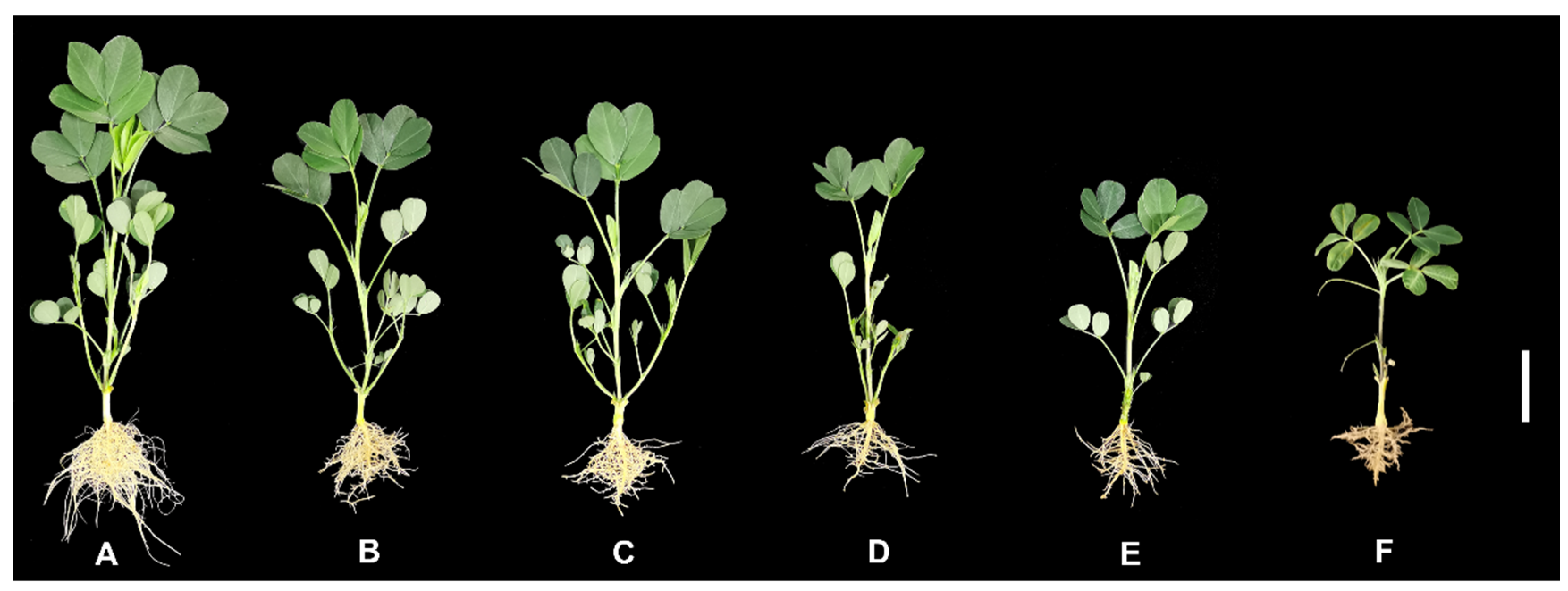
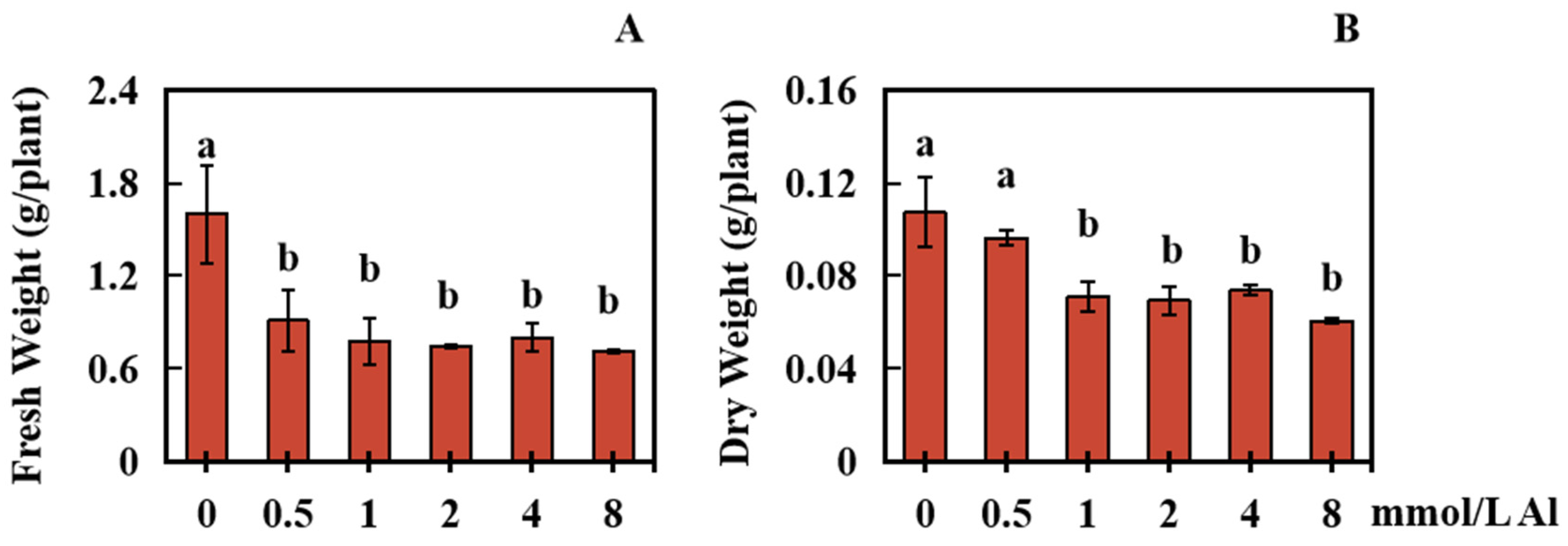
2.1. Effects of Al Poisoning on the Biomass of Roots in Peanut Plants

To analyze the influences of Al toxicity stress on the biomass of peanut roots, changes in the dry weight and fresh weight of peanut roots were measured under varying concentrations of Al. The results showed significant inhibition of peanut root biomass under Al toxicity stress (Figure 1A–F and Figure 2A,B). Initially, the peanut root biomass decreased as the concentration of Al increased. However, once the Al toxicity stress reached a certain level, there was no longer a significant decrease in peanut root biomass at the different Al concentrations (Figure 1A–F and Figure 2A,B).
In the experimental setup, a control group was treated with 0 mmol/L Al ions, while exogenous Al was applied at concentrations of 0.5, 1.0, 2.0, 4.0, and 8.0 mmol/L for a duration of 20 days. In comparison to the control group, the fresh weight of the peanut roots in the treatment groups markedly decreased by 43.05%, 51.43%, 53.37%, 49.86%, and 55.49%, respectively, with increasing concentrations of Al (Figure 2A).
Furthermore, in comparison to those in the control group, the dry weight of peanut roots treated with 0.5 mmol/L Al decreased by 10.25%. Although this decrease was not statistically significant, higher concentrations of Al (1.0, 2.0, 4.0, and 8.0 mmol/L) caused significant decreases in peanut root dry weight—33.85%, 35.40%, 31.37%, and 43.48%, respectively (Figure 2B).

2.2. Influences of Al Poisoning on Development of Peanut Roots

For analyzing the influences of Al toxicity stress on the development of peanut roots, the changes in peanut root growth under various concentrations of Al were measured. Exposure to Al toxicity had a certain effect on peanut root growth and development (Figure 3A–E). Exogenous Al was administered at concentrations of 0, 0.5, 1.0, 2.0, 4.0, and 8.0 mmol/L for a duration of 20 days. Comparatively, the control group (0 mmol/L Al) exhibited significant decreases in total root length of 51.63%, 61.75%, 67.41%, 69.96%, and 75.94%, respectively (Figure 3A). Furthermore, the average root diameter significantly increased by 38.30%, 32.02%, 37.66%, 39.77%, and 58.36%, respectively (Figure 3B). The total root volume also significantly decreased by 37.98%, 47.96%, 45.33%, 48.80%, and 48.51%, respectively (Figure 3C). Additionally, the total root surface area decreased considerably, by 44.89%, 51.96%, 64.60%, 63.91%, and 66.66% (Figure 3D), while the total number of root tips also significantly decreased by 53.62%, 66.43%, 70.77%, 70.83%, and 79.12%, respectively (Figure 3E).

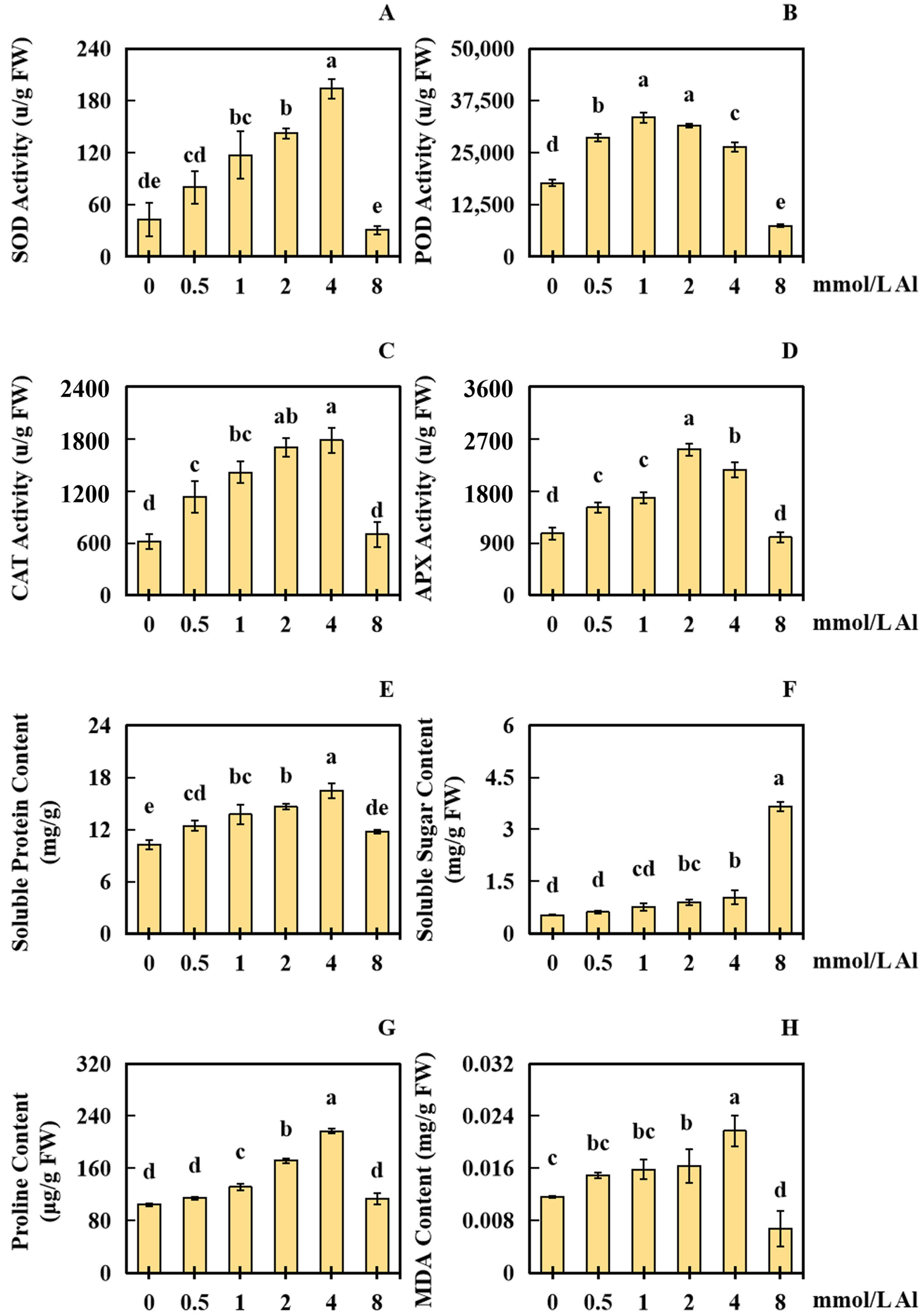
2.3. Results of Al Poisoning on the Physical Response Indices of Peanut Roots

To analyze the influences of Al poisoning on the physical response of peanut roots, this study evaluated various physiological response indices of peanut roots under different concentrations of Al. The findings revealed that the physical response indexes of peanut roots were significantly influenced by exposure to Al toxicity stress. Using 0 mmol/L Al as the control, the application of exogenous Al at concentrations of 0.5, 1.0, 2.0, 4.0, and 8.0 mmol/L for a period of 20 days had a notable effect on the physiological response of peanut roots. In comparison with that in the control group, the SOD liveness in the 0.5 and 8.0 mmol/L Al-treated groups was not markedly different. However, the SOD activity in the 1.0, 2.0, and 4.0 mmol/L Al-treated groups improved significantly, by 172.50%, 231.25%, and 351.25%, respectively (Figure 4A).
The POD activity in the 8.0 mmol/L Al-treated group reduced markedly by 58.12%, whereas the POD activity in the 0.5, 1.0, 2.0, and 4.0 mmol/L Al-treated groups increased significantly by 61.41%, 88.94%, 77.65%, and 48.71%, respectively (Figure 4B). No significant change in CAT activity was observed in the 8.0 mmol/L Al-treated group, whereas CAT activity in the 0.5, 1.0, 2.0, and 4.0 mmol/L Al-treated groups improved significantly by 83.78%, 129.73%, 175.68%, and 189.19%, respectively (Figure 4C). The APX activity in the 8.0 mmol/L treatment group showed no significant change, while the APX activity in the 0.5, 1.0, 2.0, and 4.0 mmol/L Al-treated groups elevated significantly by 42.19%, 57.81%, 135.94%, and 103.13%, respectively (Figure 4D).
Regarding biochemical parameters, the soluble protein content in the 8.0 mmol/L treatment group remained relatively constant, but in the 0.5, 1.0, 2.0, and 4.0 mmol/L Al-treated groups, it elevated significantly by 21.35%, 33.98%, 42.44%, and 60.80%, respectively (Figure 4E). The soluble sugar content in the 0.5 mmol/L Al-treated group did not obviously change, while in the 1.0, 2.0, 4.0, and 8.0 mmol/L Al-treated groups, it enhanced significantly by 46.96%, 72.22%, 99.25%, and 600.66%, respectively (Figure 4F). The proline content in the 0.5 and 8.0 mmol/L Al-treated groups exhibited no significant change, whereas in the 1.0, 2.0, and 4.0 mmol/L Al-treated groups, it increased markedly by 25.77%, 64.43%, and 107.26%, respectively (Figure 4G). The malondialdehyde (MDA) content in the 8.0 mmol/L Al-treated group reduced observably by 42.17%, and no obvious change was surveyed in the content of MDA in the 0.5 and 1.0 mmol/L Al-treated groups (Figure 4H). However, the content of MDA in the 2.0 and 4.0 mmol/L Al-treated groups enhanced significantly, by 40.26% and 86.13%, respectively (Figure 4H).

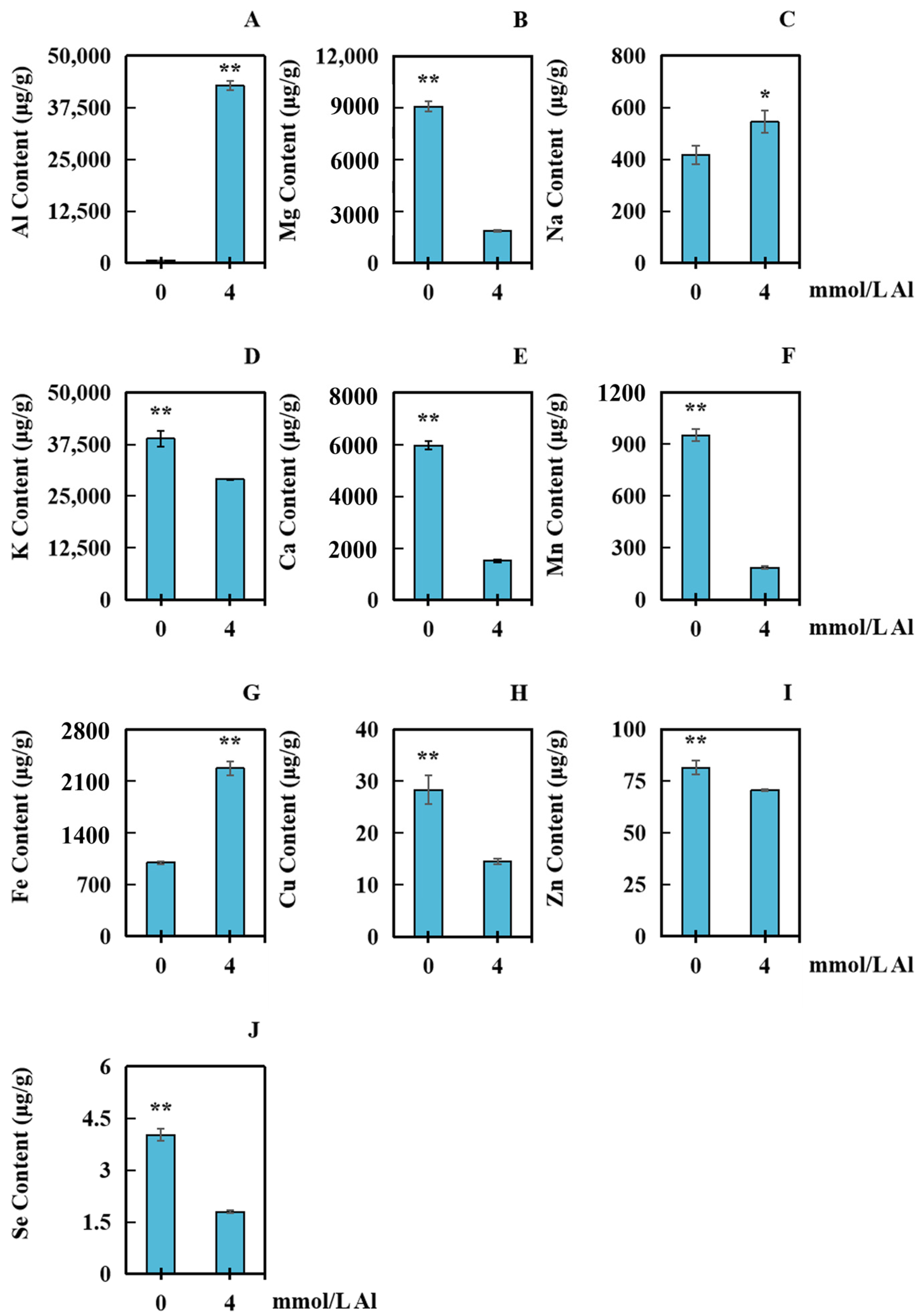
2.4. Influences of Al Poisoning on the Accumulation of Several Elements in Peanut Roots

The ion contents of ten different elements in peanut roots with varying Al concentrations were measured to analyze the effect of aluminum toxicity stress on ion accumulation. The control group received a 0 mmol/L aluminum treatment, while the samples were taken and dried after a 20-day treatment with 4.0 mmol/L exogenous aluminum. The contents of Al, K, Fe, Na, Mn, Cu, Mg, Zn, Se, and Ca were determined in peanut roots. In comparison with that in the control group, the content of Na in the Al-treated group was significantly elevated by 30.54% (Figure 5C). The contents of Al and Fe increased markedly by 18,840.51% and 127.13%, respectively (Figure 5A,G). Mg, K, Ca, Mn, Cu, Zn, and Se decreased significantly, by 79.42%, 25.23%, 74.62%, 80.55%, 48.78%, 13.46%, and 55.33%, respectively (Figure 5B,D–F,H–J).

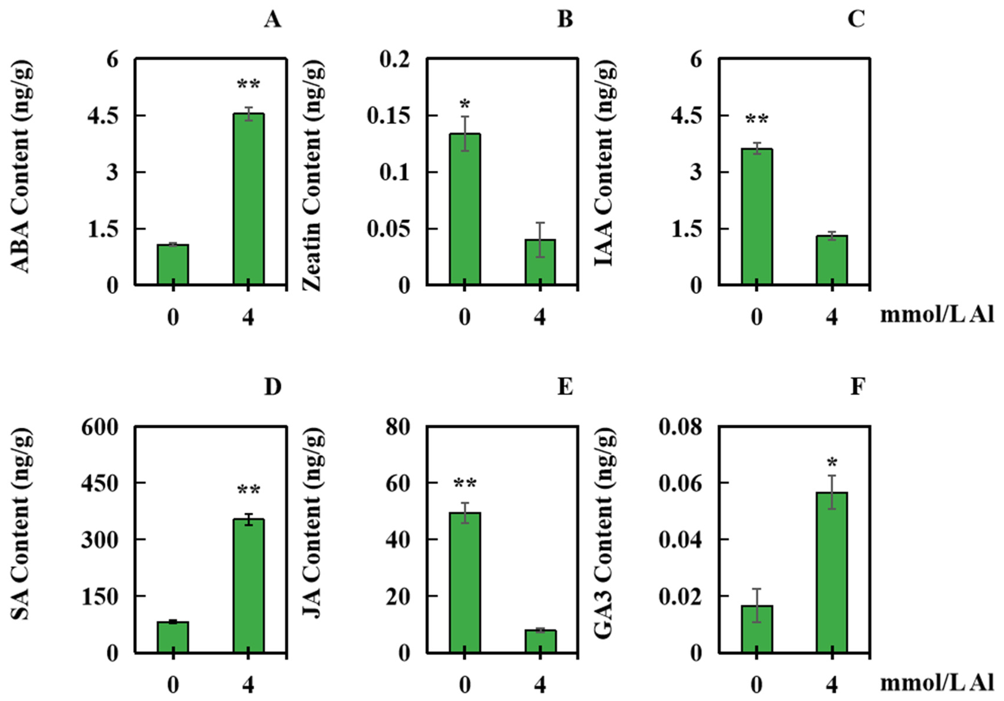
2.5. Effects of Al Toxicity Stress on the Contents of Several Hormones in Peanut Roots

The contents of six different hormones in peanut roots were detected to investigate the influence of aluminum toxicity stress on hormone synthesis. The samples were taken under normal aluminum conditions at 4 mmol/L of Al. In comparison with those in the control group, the contents of ABA, SA, and gibberellin 3 (GA3) in the treatment groups significantly increased by 322.67%, 329.20%, and 240.00%, respectively (Figure 6A,D,F). However, zeatin, indole acetic acid (IAA), and JA significantly decreased by 70.00%, 63.80%, and 83.86%, respectively (Figure 6B,C,E).

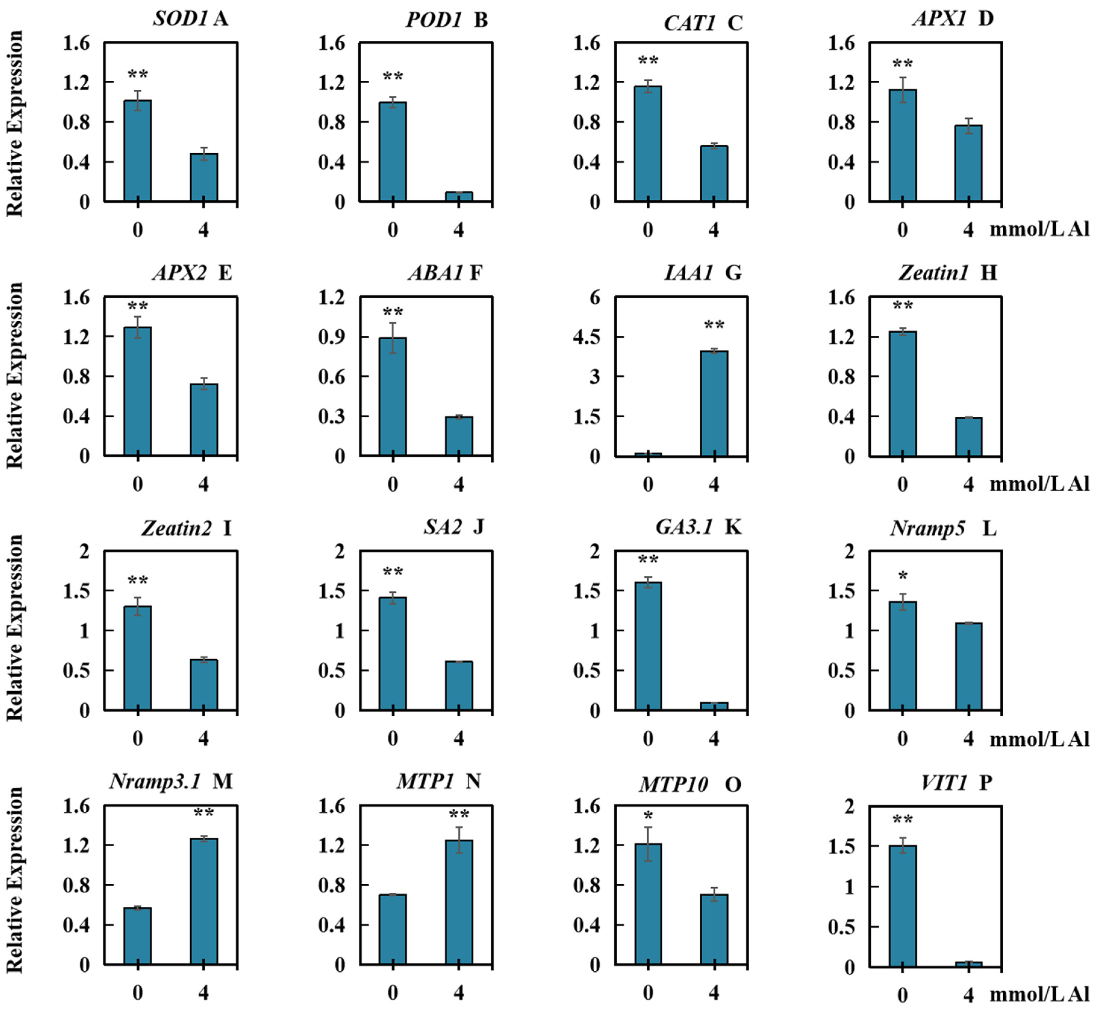
2.6. Effects of Al Toxicity Stress on the Relative Transcript Levels of Several Types of Genes in Roots of Peanut

To analyze the influences of Al toxicity on relative expression of the genes in connection with physiological responses (SOD1, POD1, CAT1, APX1, and APX2; hormone synthesis (ABA1, IAA1, Zeatin1, Zeatin2, SA2, and GA3.1); and ion transport (Nramp5, Nramp3.1, MTP1, MTP10, and VIT1), we analyzed the expression of 15 genes under Al toxicity stress. The results demonstrated that, in comparison with those in the controls, the genes whose relative expression decreased significantly included SOD1 (Figure 7A), POD1 (Figure 7B), CAT1 (Figure 7C), APX1 (Figure 7D), APX2 (Figure 7E), ABA1 (Figure 7F), Zeatin1 (Figure 7H), Zeatin2 (Figure 7I), SA2 (Figure 7J), GA3.1 (Figure 7K), Nramp5 (Figure 7L), MTP10 (Figure 7O), and VIT1 (Figure 7P). The genes whose relative expression increased significantly were IAA1 (Figure 7G), Nramp3.1 (Figure 7M), and MTP1 (Figure 7N).

3. Discussion

In acidic soils, active Al ions are the primary factor that limits plant growth [27]. The main effectiveness of Al poisoning on plants is the suppression of root growth [28]. Our study demonstrated significant reductions in the fresh weight, dry weight, length of total root, surface area of total root, surface area of total root, and root tip number in the roots of peanuts subjected to Al poisoning, indicating severe suppression of the elongation of roots. Interestingly, the average diameter of the peanut roots increased significantly following exposure to Al toxicity. This may be caused by the close combining of Al with the cell wall of root cells in plants, which alters the structure and flexibility of the cell wall, reducing its ductility and disrupting the normal function of the plant cytoskeleton. Consequently, the root tip expands [29]. Similar findings have been reported in Eucalyptus clones, in which Al toxicity stress led to taproot enlargement and root tip shortening and thickening [30]. These changes may be due to alterations in the cell wall cellulose content under Al stress, which causes cell deformation and a shift in growth direction from vertical to horizontal, resulting in the expansion and thickening of the root tip base [30].
Under conditions of severe aluminum toxicity stress, there can be an imbalance between the generation and elimination of free radicals and ROS, leading to an oxidative stress response, enzyme inactivation, and peroxidation of cell membranes [31]. The antioxidant defense system and osmoregulation system play significant roles in plants’ ability to resist oxidative stress [32]. Plants produce various enzymes, including SOD, POD, CAT, and APX, which help them cope with oxidative stress [32]. Additionally, components such as soluble proteins, proline, and soluble sugars can help reduce the effects of Al poisoning [32]. The content of MDA is an essential indicator of lipid peroxidation in plant cell membranes [33]. Hence, the measurement of antioxidase activity and osmotic adjustment components is crucial for reflecting the metabolism activity and health level of cells [34].
The SOD family genes found in plants are categorized into three groups based on their metal cofactors: Cu/ZnSOD, MnSOD, and FeSOD [35]. SOD plays a crucial role as the first line of defense in plant antioxidant systems. In Brassica napus L., the genes BnCuSOD1, BnCuSOD3, BnCuSOD14, BnFeSOD4, BnFeSOD5, BnFeSOD6, BnMnSOD2, and BnMnSOD10 were observed to be significantly upregulated under various abiotic stress conditions, such as cold, salt, drought, and flood [36]. In the present study, the differential expression of peanut SOD1 and the activity of SOD in peanut roots may be influenced by variations in the absorption of Fe, Cu, Mn, and Zn affected by the presence of Al3+. The POD gene family refers to the class III peroxidase gene family, which is responsible for directing the synthesis of plant-specific REDOX enzymes in grapevine (Vitis vinifera) [37]. Previous research has demonstrated that treatment with H2O2 induces the upregulation of MePOD13, MePOD17, MePOD23, and MePOD85 expression in Manihot esculenta [38]. PODs not only help remove ROS but also contribute to the accumulation of lignin in cell walls, triggering significant thickening and hardening of the cell wall, as observed in watermelon (Citrullus lanatus) [39]. The genes ClPOD51 and ClPOD54 are believed to play key roles in lignin synthesis in watermelon rinds [39]. In the present study, Al poisoning induced differential expression of POD1, which may not only impact the antioxidant capacity of peanuts but also influence root growth inhibition. The expression of CAT in certain plants is influenced by plant hormones [40]. For example, the expression of AtCAT1 in Arabidopsis thaliana is induced by ABA [40]. In Brassica napus, the genes BnCAT1, BnCAT3, BnCAT11, and BnCAT13 were significantly upregulated under low temperature, salt, ABA, and GA treatments [41]. Previous reports identified a total of 166 members of the peanut APX gene family, with AhAPX4, AhAPX17, and AhAPX19 showing significant upregulation under abiotic stress (drought and cold) and treatment with plant hormones such as ABA and SA, indicating that the synthesis of APXs is induced by abiotic stress and that their differential expression is associated with hormone levels in peanut cells [42]. Thus, the changes in CAT and APX activities observed in this study are likely related not only to the oxidative damage caused by Al poisoning but also to alterations in hormone levels in peanut root cells.
In this study, there was a clear pattern observed in the liveness of SOD, CAT, POD, and APX, with a beginning enhancing followed by a reducing. When the concentration of Al was low, the liveness of those antioxidases was elevated, indicating that the peanut cell membrane system experienced some damage due to Al toxicity. POD, SOD, APX, and CAT are known to play a significant role in protecting the plant cell membrane system during aluminum poisoning [43]. However, as the concentration of Al increased to a certain extent, the activities of these enzymes started to decline. This decline might be attributed to the depletion of SOD, POD, CAT, and APX due to their involvement in protecting the peanut membrane system [44]. Alternatively, the decline could be a result of an excessively high Al concentration, which seriously damages the oxidation defense system of the peanut plants, as evidenced by the reduced activities of the enzymes [45]. Similar decreases in physiological response indices have also been observed under other abiotic stress conditions, such as in soybean (Glycine max) treated with microplastics [45] and in broad bean (Vicia faba) treated with lead [46].
The experiment also revealed trends in the levels of proline, soluble sugar, and soluble protein. The soluble protein and proline contents initially increased, followed by a decrease, while the soluble sugar content continuously increased. Notably, at the highest concentration tested, compared to those at the previous concentration, the soluble protein and proline levels decreased, while the soluble sugar level significantly increased. This finding suggests that peanuts respond synergistically to Al toxicity stress by adjusting the contents of osmotic adjustment components such as soluble protein, proline, and soluble sugar [43]. MDA, a byproduct of lipid peroxidation caused by oxidative damage, reflects the extent of peroxidation damage to the cytomembrane [47]. In this study, the MDA content exhibited an initial enhancement followed by a reduction. As the concentration of Al increased, additional peroxidative damage occurred in the peanuts due to Al toxicity, leading to a gradual increase in the MDA content. However, at the highest Al concentration, the MDA content decreased significantly compared to that at the previous concentration. This suggested that at higher Al concentrations, the antioxidant defense system in peanut root cells was severely damaged and possibly dysfunctional.
Based on these results, it can be inferred that under lower concentrations of Al toxicity stress, the antioxidant defense system and osmotic regulation system of peanuts can protect peanut root cells and minimize oxidative damage. However, under higher concentrations of Al toxicity stress, the antioxidant defense system of peanuts is severely impaired, while the osmotic regulation system continues to play a role.
In comparison with that in the control group, the accumulation of Al in peanut roots significantly increased after exposure to the Al treatment. This increase could be the cause of Al poisoning in peanut roots. Previous studies suggest that Al ions have a greater affinity for unmethylated pectin in the cytoderm, leading to a substantial buildup of Al in the root cell wall of plants. This accumulation results in increased rigidity in the cell wall and abnormal cytoskeletal structure [48]. Additionally, excessive Al in the cytoplasm can cause various chromosomal abnormalities, including chromosome adhesion, hysteresis, micronucleus formation, binucleus, and multinucleus cells. Consequently, these abnormalities can affect the division and differentiation of plant cells [9].
Notably, in this research, the contents of Mg, Ca, and Mn in the roots of peanuts decreased markedly. Research findings indicate an antagonistic relationship between Mg and Al in plants [49]. There is also competition between Mg and Al for binding sites on the cell wall and membrane. Inside the cell, Mg and Al compete for binding sites on oxygen-contributing compounds involved in energy metabolism, protein synthesis, and photosynthesis [49]. Similarly, Mn and Al also exhibit antagonistic effects [50]. When combined, the Al and Mn treatments mitigated the decrease in plant biomass caused by Al or Mn alone. This suggests that the two ions have antagonistic interactions that affect plant growth [51]. Ca is another component of pectin in plant cell walls and influences the fluidity of cell membranes [52]. Calcium ions serve as signaling molecules for physiological regulation, and they can compete with Al ions to bind to sites in pectin as cations [53]. In the present study, the decreases in the contents of Mg, Ca, and Mn in peanut roots may be attributed to the high concentration of Al ions occupying a significant number of binding sites in peanut root cells.
Furthermore, an increase in the Fe content was detected in peanut roots subjected to Al toxicity stress. Similar findings have been observed in soybeans exposed to Mn poisoning [54]. It is possible that the accumulation of Al in peanut roots also leads to an increase in Fe content. There may be a certain synergistic accumulation of these two ions in peanuts, and an increase in Fe content may contribute to improving peanut resistance to Al toxicity.
The NRAMP (natural resistance-associated macrophage protein) family plays a vital role in metal absorption and transportation in plants, with most AhNRAMPs being primarily expressed in androecia, roots, and seeds [55]. NRAMPs are crucial for the transport of metal ions across cell membranes. For instance, the expression levels of GmNRAMP5a and GmNRAMP1a are upregulated under Cu toxicity stress in Glycine max [56]. Metal-tolerance proteins (MTPs) also play a significant role in the transport of bivalent cations in plants [57]. For example, the expression levels of GmMTP1.1 in soybean leaves and GmMTP4.3 in roots significantly increased under treatment with bivalent Cd, Fe, Co, Zn, and Mn [58]. Previous studies have focused mainly on the involvement of the NRAMP and MTP families in the induction of bivalent metal ions, such as Fe2+ and Cd2+ [55,57]. In the present study, certain members of the peanut NRAMP and MTP families (AhNramp5, AhNramp3.1, AhMTP1, and AhMTP10) exhibited differential expression in peanut roots exposed to aluminum poisoning. These findings suggested that the peanut NRAMP and MTP families probably also have a critical role in peanut resistance to Al poisoning. Moreover, transporters associated with bivalent metal ions may not only participate in the transport of Mg, Fe, Ca, Mn, Cu, and Zn but also contribute to the transport of trivalent Al3+.
ABA is a hormone that is crucial for plant resistance to metal poisoning [59]. In the root system, ABA functions primarily in the meristem and elongation regions [59]. Abscisic aldehyde oxidase, 9-cis-epoxycarotenoid dioxygenase, and alcohol dehydrogenase (ABA2) impact ABA synthesis in plants in response to abiotic stress [59,60,61]. Research has revealed that the NGATHA1 transcription factor in Arabidopsis thaliana positively regulates ABA production during abiotic stress by activating NCED3, which encodes a key ABA biosynthesis agent [62]. Under specific concentrations of metal ions, ABA promotes primary root growth while inhibiting lateral root growth [63,64]. Plants can regulate ABA content under abiotic stress by modulating the transcription levels of several genes associated with ABA biosynthesis [59]. Metal stress, such as cadmium, nickel, zinc, and Al exposure, impacts the ABA content in plants [61]. In the present study, significant increases in ABA content were observed when peanut roots suffered from Al poisoning. This difference may be attributed to the induction of genes in connection with ABA biosynthesis in peanut roots by Al, suggesting that peanuts enhance the ABA content to counter Al toxicity stress.
Zeatin is typically produced by rapidly dividing cells, such as the root tip and bud, and plays a vital role in promoting plant cell division, root extension, leaf expansion, and seed germination [65,66]. Isopentenyltransferases (IPTs) are pivotal enzymes that catalyze cytokinin biosynthesis, while IAA influences Zeatin biosynthesis by regulating IPT expression [67]. Studies have demonstrated that Arabidopsis AtIPT9 stimulates cytokinin synthesis and enhances root growth [68]. In the current study, the zeatin level in the roots of peanuts was significantly reduced under Al poisoning. It is possible that the induction of Al ions influenced the expression of relevant genes, hindered Zeatin synthesis, and inhibited cell division and root growth in peanut roots.
IAA is involved in regulating all aspects of normal plant growth and development, including synthesis, metabolism, polar transport, and self-signal transduction [69]. The stability of IAA levels also represents the coping strategies of plants for changes in the environment [70,71]. The auxin-related genes such as PINFORMED (PIN) and AUXIN1/LIKE AUX1 (AUX1/LAX) involve adjusting the polar transportation of growth hormones, thus further controlling and optimizing the growth hormone distribution in root tips [72,73]. The metabolism and dynamic polarity distribution of IAA are influenced by metal ions [69]. In this study, the IAA level in peanut roots under Al toxicity stress was lower than that under normal conditions, indicating that Al toxicity inhibited peanut root growth.
SA regulates biological and abiotic stress responses in plants, promotes ROS production through antioxidant defense systems, and induces gene expression [74]. Under stress, a low SA concentration can facilitate the growth of plants, while a high SA concentration can inhibit plant growth [75,76]. This is because a high SA concentration affects the gene expression of PIN2, which acts as a vital gene for adjusting the synthesis of hormone, and downregulates its expression, thereby reducing hormone production in the root tip meristem and conclusively restraining the growth of roots and the development of root hair in plants [75]. In this study, the SA level in peanut roots significantly increased under Al toxicity stress. It was possible that SA synthesis induced by Al toxicity stress led to an increase in the SA level in the roots of peanuts. As a result, the growth hormone content and distribution in peanut roots were affected, leading to peanut root dysplasia.
When plants experience stress, JA can assist in their adaptation to stress [77]. In the case of blueberries (Vaccinium corymbosum) exposed to Al toxicity, the application of exogenous methyl jasmonate (MeJA) has been found to mitigate damage caused by Al toxicity stress [78]. There is an antagonistic relationship between JA and SA, where SA inhibits the expression of JA response genes, and JA may weaken the immune response induced by SA [79]. JA also participates in the gene expression of certain metal ion transporters, such as FRO2, and IRT1, which impact the accumulation of metal ions in roots [80]. In the present study, the JA level in the roots of peanuts was substantially decreased under Al toxicity stress. This reduction may be attributed to the antagonistic effect between JA and SA, as an increase in SA content results in a decrease in JA content.
GA3 plays a significant role in promoting plant germination, stem extension, leaf growth, flowering, and fruit development [81]. In Arabidopsis, GA2-oxidase (GA2ox), GA3ox (GA3-oxidase), and GA20ox (GA 20-oxidase) have been identified as key genes involved in the synthesis and degradation of GA3 [82]. An antagonistic relationship exists between GA3 and ABA, which regulates various plant development processes, including root growth [83]. Previous studies have demonstrated that exposure to Al leads to a reduction in GA3 content and an increase in ABA content in the roots of Pinus massoniana [84]. However, in the present study, the GA3 level in peanut roots increased with increasing ABA content under Al toxicity stress. These findings contradict previous research results and may be attributed to species-specific coping strategies [82]. It is possible that Al toxicity stress influences the expression of genes associated with GA3 synthesis in peanut roots, thereby enabling peanuts to increase their tolerance to Al toxicity stress by increasing the GA3 content in their roots.
In this study, the DEGs were categorized into three main groups: genes related to physiological indices (SOD1, POD1, CAT1, APX1, and APX2), hormone-related genes (ABA1, IAA1, Zeatin1, Zeatin2, SA2, and GA3.1), and ion transport genes (Nramp5, Nramp3.1, MTP1, MTP10, and VIT1). Physiological index response-related genes and hormone-related genes have crucial roles in adjusting plant responses to abiotic stress and recognizing ROS [85]. The differential expression of physiological index response-related genes and hormone-related genes in the presence of Al suggests that plants respond to the stress of Al toxicity by activating these genes to resist stress and eliminate excessive ROS in roots. The NRAMP family is highly important for metal absorption and transportation in plants, with most AhNRAMPs being primarily expressed in androecia, roots, and immature seeds [55]. MTPs are also vital for bivalent cation transport in plants [86]. Previous studies have linked NRAMPs and MTPs to the transportation of bivalent metal ions such as Fe2+ and Cd2+ [55,87]. In the present study, several peanut AhNRAMPs (Nramp5 and Nramp3.1) and AhMTPs (MTP1 and MTP10) exhibited differential expression in peanut roots exposed to Al toxicity, suggesting that the peanut NRAMP and MTP families may also play significant roles in peanut resistance to Al toxicity stress. Furthermore, transporters associated with bivalent metal ions might be involved in the transport of trivalent Al3+, providing new perspectives for studying peanut Al tolerance.
On account of the experimental results of this research, the physiological regulatory mechanism of peanut roots in responding to Al toxicity is summarized in Figure 8. First, under Al poisoning and high levels of exogenous Al, there was an increase in the absorption of Al by root cells. This excess Al led to intracellular oxidative damage (Figure 8I), while cells regulated their internal environment through osmotic substances (Figure 8II) to maintain stability. A high Al concentration activated cellular defense mechanisms, resulting in differential gene expression of antioxidase-encoding genes (Figure 8III). Consequently, the activities of antioxidases were enhanced (Figure 8VI), effectively eliminating excess ROS. Under high-Al conditions, phytohormone-relevant genes were expressed differentially (Figure 8IV), and ion transshipment-relevant genes were also expressed differentially (Figure 8V), causing changes in plant hormones (Figure 8VII) and a reduced absorption capacity for mineral nutrients (Figure 8VIII). Ultimately, this led to phenotypes such as decreased biomass of roots and inhibited growth of roots in peanut plants.

4. Materials and Methods

4.1. Plant Materials and Hydroponic Treatment

The plant material was the peanut variety Zhanyou 62, bred by the ZAAS (Zhanjiang Academy of Agricultural Sciences) in Zhanjiang, Guangdong Province, China. It was cultivated in the plant culture room of the College of Coastal Agricultural Sciences at Guangdong Ocean University (E: 110.30311, N: 21.15005). Peanut seeds with complete seed coats and uniform sizes were selected. The plants were sterilized with 1% NaClO (Solarbio, Beijing, China) and planted in silica sand. The peanut seeds were buried and cultured in sand for 10 days. Afterward, the uniformly grown peanut plants were transferred to a 15 L plastic box and provided with an improved Hoagland nutrient solution [87] for hydroponic cultivation. The hydroponic nutrient solution contained 25 µmol/L MgCl2, 400 µmol/L NH4NO3, 1500 µmol/L KNO3, 40 µmol/L Fe-EDTA (Na), 1200 µmol/L Ca(NO3)2·4H2O, 500 µmol/L MgSO4·7H2O, 1.5 µmol/L ZnSO4·7H2O, 0.5 µmol/L CuSO4·5H2O, 300 µmol/L (NH4)2SO4, 300 µmol/L K2SO4, 1.5 µmol/L MnSO4·H2O, 0.16 µmol/L (NH4)5MoO24·4H2O, 2.5 µmol/L NaB4O7·10H2O, and 500 µmol/L KH2PO4. All chemical reagents used were of analytical grade (Kermel, Tianjin, China). Al2(SO4)3·18H2O (Shanghai Reagent, Shanghai, China) was used as the source of Al, and Al3+ was added to the nutrient solution at concentrations of 0, 0.5, 1.0, 2.0, 4.0, and 8.0 mmol/L for hydroponic treatment of peanuts. The control group was treated with an Al3+ concentration of 0 mmol/L. Each treatment was repeated three times. The peanut plants were cultivated under the following conditions: a temperature of 25–30 °C during the daytime and 18–22 °C at nighttime, with a light period of approximately 12 h/d and a light intensity of 2000 lux. The nutrient solutions were changed every 5 days. After 20 days of Al treatment, the peanut roots were collected.

4.2. Measurement of Dry and Fresh Weight of Roots in Peanut

The root fresh weight of each peanut plant was measured using an BS124S electronic balance (Sartorius, Göttingen, Germany). Then, the root samples were moved to an electric blast constant temperature oven (Yiheng, Shanghai, China) and dried at a constant temperature of 60 °C for one week [88]. The dry weight of each treated sample was measured for three biological replicates.

4.3. Determination of Peanut Root Morphological Indices

A WinRHIZO LA6400 XL root scanner (Regent, Vancouver, BC, Canada) was used to scan the morphological indices of the peanut roots and capture images of each treated peanut root system. WinRHIZO software (WinRHIZO 2013e Professional Edition) was subsequently used to analyze and obtain data concerning the surface area, volume, total length, and number of roots [89]. Three measurements were obtained for each treatment.

4.4. Determination of Peanut Root Physiological Response Indices

As mentioned previously, peanut roots were treated with Al2(SO4)3·18H2O as an aluminum source for 20 days using various concentrations of Al3+ (0, 0.5, 1.0, 2.0, 4.0, and 8 mmol/L). After the treated peanut roots were harvested, eight physiological response indices were measured. The proline level was determined by means of the sulfosalicylic acid method [90]. The level of soluble protein was tested by applying the Coomassie bright blue staining technology [85]. The soluble sugar level was tested by means of the anthrone-sulfuric acid colorimetry method [91]. The level of MDA was measured by applying the thiobarbituric acid method [92]. POD liveness was determined by means of the guaiacol way [93]. SOD liveness was tested by application of the nitrogen blue tetrazole method [94]. CAT life was tested by means of a spectrophotometer (Yuexi UV-5100B, Shanghai, China) [95]. The activity of APX was tested by applying the experimental technology demonstrated by Li et al. [96].

4.5. Determination of Ion Content in Peanut Roots

Peanut roots were treated with 0 (control group) or 4.0 (al poison treatment group) mmol/L Al3+. The dry root samples (0.15 g) were completely dissolved in nitric acid (Guangzhou Reagent, Guangzhou, China). The levels of Al, Mg, Na, K, Fe, Ca, Mn, Zn, Cu, and Se in peanut roots were tested using PS7800 ICP–AES (inductively coupled plasma atomic emission spectrometry) (Hitachi, Tokyo, Japan) [86,97]. Each index was analyzed using three biological replicates.

4.6. Determination of the 6 Kinds of Hormones in Peanut Roots

Peanut roots were treated with 0 or 4.0 mmol/L Al3+ for 20 days of culture, after which endogenous hormones were extracted from the roots. The contents of endogenous hormones, including ABA, IAA, zeatin (ZT), SA, JA, and GA3, in peanut roots were measured using high-performance liquid chromatography (HPLC) (AGLIENT 1290, Santa Clara, CA, USA) combined with SCIEX-6500Qtrap MS/MS (tandem mass spectrometry) (AB, Madison, WI, USA). Internal reference materials were supplemented with the extract to correct the determination outcome [54,98]. The external standard substances used, such as zeatin, SA, ABA, JA, IAA, and GA3, were chromatography-pure reagents (Sigma, Saint Louis, MI, USA). The internal standard substances used were deuterated zeatin (D-zeatin), deuterated SA (D-SA), deuterated ABA (D-ABA), deuterated JA (D-JA), deuterated IAA (D-IAA), and deuterated GA3 (D-GA3) (Sigma, Saint Louis, MI, USA). C18 QuECherS (Amperex, Shanghai, China) was used as a filler for the column. The CH3OH and C2H3N used in the test were chromatography-pure (Merck, Darmstadt, Germany). Preparation of the operating fluid and calculation of the hormone standard curve, as well as extraction of hormones from peanut roots, were conducted following the methods reported in reference [85]. Supplementary Tables S1–S3 present the HPLC gradient parameters, mass spectrometry parameters, and selected monitoring conditions for protonated or deprotonated plant hormone reactions, respectively.

4.7. Fluorescence Quantitative PCR (qPCR) Analysis

In accordance with the results of transcriptome sequencing in peanuts, several genes differentially expressed in response to metal ion stress and potentially linked to antioxidant and hormone synthesis were identified [88]. In this study, qPCR was adopted to analyze gene expression in response to aluminum toxicity stress in peanuts. Peanut roots were collected after 20 days of dealing with 0 or 4.0 mmol/L of Al. The RNA was isolated from the roots of peanuts via the Kit of MolPure Plant RNA (Yeasen, Shanghai, China). The cDNA was obtained through reverse transcription by means of the Kit of HifairII 1st Strand cDNA Synthesis (Yeasen, Shanghai, China). The expression of genes in the peanut roots was detected using Hieff UNICON Universal Blue qPCR SYBR Green Master Mix reagent (Yeasen, Shanghai, China) and Bio-Rad fluorescence quantitative PCR (CFX Connect Optics Module, Hercules, CA, USA) [99].
For qPCR, a 20 μL reaction mixture was utilized, consisting of 10 μL of Hieff UNICON Universal Blue qPCR SYBR Green Master Mix reagent, 1 μL of cDNA template, 0.5 μL of downstream primers, 0.5 μL of upstream primers, and 8 μL of nuclease-free water. The qPCR reaction procedures were performed as follows: 95 °C for 10 min; 95 °C for 15 s; 60 °C for 1 min; and 40 cycles [100]. AhUbiquitin (DQ887087.1) was selected as the internal control gene [101]. The gene expression levels were determined via the following formula: gene relative expression level = expression level of targeted gene/expression level of internal control gene [72]. The primers used for qPCR are displayed in Table S4.

4.8. Data Analysis

Microsoft Excel 2010 (Microsoft, Redmond, WA, USA) and SPSS (Statistic Package for Social Science) software version 19.0 (IBM Corporation, New York, NY, USA) were applied to statistical analysis. A student’s t test was adopted for statistical comparison and significance analysis between the two groups of data. The Waller-Duncan test was used to compare the significance of differences across multiple groups of data [85].

5. Conclusions

Based on the aforementioned results, an analysis was conducted on the physiological regulatory mechanism of peanut roots in responding to Al poisoning. Initially, the entrance of Al into peanut root cells increased due to the high presence of exogenous Al during Al toxicity stress. Throughout this process, the increased levels of Al activated cellular defense mechanisms, thereby altering the expression of antioxidase-encoding genes and enhancing the activity of antioxidases. Consequently, the excess ROS were effectively eliminated. Additionally, exposure to Al ions during Al toxicity stress led to the induction of genes associated with plant hormones and metal ion transport. Consequently, this induction caused changes in the expression levels of plant hormones and metal ion transport-related genes, ultimately resulting in a range of symptoms, such as decreased biomass of roots, restrained growth of roots, and abnormal accumulation of metal ions in peanut roots. The consequences of this study establish a foundation for further research into the physiological and molecular mechanisms referred to in the peanut root responding to Al poisoning. Moreover, these findings offer a theoretical grounding for the development of new Al-resistant peanut varieties through breeding efforts.

Supplementary Materials

The following supporting information can be downloaded at: https://www.mdpi.com/article/10.3390/plants13020325/s1, Table S1: The detailed gradient parameters for HPLC; Table S2: mass spectrum parameters; Table S3: the selected monitoring indicators for protonation or deprotonation reactions of phytohormones; Table S4: the primers applied to qPCR in this study.

Author Contributions

J.S. and Y.L. conceived and designed the experiments. J.S., M.Z., F.Z., D.F., S.Y., Y.X. and Y.L. performed the experiments. J.S. and Y.L. analyzed the data. J.S. and Y.L. wrote the manuscript. All authors have read and agreed to the published version of the manuscript.

Funding

This research was funded by the Project of Marine Young Talent Innovation of Zhanjiang City (210818004542097), the Project of Rural Science and Technology Team “One-to-One” Service to Help the Town Full Coverage of Action Funding in Zhanjiang City (A23081), the Postgraduate Education Innovation Project of Guangdong Ocean University-Excellent Dissertation Cultivation Project (040502752327), the Project of Social Services of Rural Science and Technology Specialists of Guangdong Ocean University (080503052212), the National Natural Science Foundation of China (32002131), and Maintenance Expenses of Key Construction Disciplines in High-level University of Crop Science (080508052201).

Institutional Review Board Statement

Not applicable.

Informed Consent Statement

Not applicable.

Data Availability Statement

Data is contained within the article and Supplementary Material.

Acknowledgments

We would like to thank Analytical and Testing Center of Guangdong Ocean University for determination of ion content.

Conflicts of Interest

The authors declare no conflict of interest.

References

  1. Qi, S.Q.; Li, X.X.; Luo, J.; Han, R.F.; Chen, Q.Q.; Shen, D.S.; Shentu, J.L. Soil heterogeneity influence on the distribution of heavy metals in soil during acid rain infiltration: Experimental and numerical modeling. J. Environ. Manag. 2022, 322, 116144. [Google Scholar] [CrossRef] [PubMed]
  2. Wu, Z.F.; Sun, X.M.; Sun, Y.Q.; Yan, J.Y.; Zhao, Y.F.; Chen, J. Soil acidification and factors controlling topsoil pH shift of cropland in central China from 2008 to 2018. Geoderma 2022, 408, 115586. [Google Scholar] [CrossRef]
  3. Han, T.F.; Liu, K.L.; Huang, J.; Ma, C.B.; Zheng, L.; Wang, H.Y.; Qu, X.L.; Ren, Y.; Yu, Z.K.; Zhang, H.M. Spatio-temporal evolution of soil pH and its driving factors in the main Chinese farmland during past 30 years. J. Plant Nutr. Fertil. 2020, 26, 2137–2149. [Google Scholar]
  4. Whitley, A.E.; Moir, J.L.; Almond, P.C. A meta-analysis of exchangeable aluminium in New Zealand soils using the National Soils Database. Soil Res. 2019, 57, 113–123. [Google Scholar] [CrossRef]
  5. Bojórquez-Quintal, E.; Escalante-Magaña, C.; Echevarría-Machado, I.; Martínez-Estévez, M. Aluminum, a friend or foe of higher plants in acid soils. Front. Plant Sci. 2017, 8, 1767. [Google Scholar] [CrossRef]
  6. Li, C.X.; Hou, C.C.; Chen, L.Y.; Kaskel, S.; Xu, Q. Rechargeable Al-ion batteries. EnergyChem 2021, 3, 100049. [Google Scholar] [CrossRef]
  7. Huang, W.J.; Yang, X.D.; Yao, S.C.; LwinOo, T.; He, H.Y.; Wang, A.Q.; Li, C.Z.; He, L.F. Reactive oxygen species burst induced by aluminum stress triggers mitochondria-dependent programmed cell death in peanut root tip cells. Plant Physiol. Biochem. 2014, 82, 76–84. [Google Scholar] [CrossRef]
  8. Xiong, J.; Ding, G.; Li, S.Y.; Chen, L.L.; Song, L.Q. Effects of aluminum stress on growth, development and nutrient uptake of different aluminum-tolerant rapeseed cultivars at seedling stage. Acta Agric. Boreali-Sin. 2020, 35, 165–171. (In Chinese) [Google Scholar]
  9. Mohanty, S.; Das, A.B.; Das, P.; Mohanty, P. Effect of a low dose of aluminum on mitotic and meiotic activity, 4C DNA content, and pollen sterility in rice, Oryza sativa L. cv. Lalat. Ecotoxicol. Environ. Saf. 2004, 59, 70–75. [Google Scholar] [CrossRef]
  10. Sousa, A.D.; AbdElgawad, H.; Fidalgo, F.; Teixeira, J.; Matos, M.; Tamagnini, P.; Fernandes, R.; Figueiredo, F.; Azenha, M.; Teles, L.O.; et al. Subcellular compartmentalization of aluminum reduced its hazardous impact on rye photosynthesis. Environ. Pollut. 2022, 315, 120313. [Google Scholar] [CrossRef]
  11. Xu, S.S.; Wu, L.H.; Li, L.Y.; Zhong, M.H.; Tang, Y.; Cao, G.Q.; Lin, K.M.; Ye, Y.Q. Aluminum-induced alterations to the cell wall and antioxidant enzymes involved in the regulation of the aluminum tolerance of Chinese Fir (Cunninghamia lanceolata). Front. Plant Sci. 2022, 13, 891117. [Google Scholar] [CrossRef] [PubMed]
  12. Xiong, J.; Ding, G.; Chen, L.L.; Li, S.Y.; Zou, X.Y.; Song, L.Q. Effects of aluminum stress on physiological characteristics and yield components of two rapeseed varieties. J. Gansu Agric. Univ. 2019, 54, 43–50. (In Chinese) [Google Scholar]
  13. Yan, L.; Riaz, M.; Li, S.; Cheng, J.; Jiang, C.C. Harnessing the power of exogenous factors to enhance plant resistance to aluminum toxicity; a critical review. Plant Physiol. Biochem. 2023, 203, 108064. [Google Scholar] [CrossRef] [PubMed]
  14. Xiao, D.; Li, X.; Zhou, Y.Y.; Wei, L.; Keovongkod, C.; He, H.Y.; Zhan, J.; Wang, A.Q.; He, L.F. Transcriptome analysis reveals significant difference in gene expression and pathways between two peanut cultivars under Al stress. Gene 2021, 781, 145535. [Google Scholar] [CrossRef]
  15. Liao, B.S. Current situation and potential analysis of peanut production in China. Chin. J. Oil Crop Sci. 2020, 42, 161–166. (In Chinese) [Google Scholar]
  16. Yan, M.L.; Ge, W.W.; Zhang, X.; Huang, Y.N.; Zhang, Z.D.; Zhang, Y. The situation analysis and development of oil industry in China countermeasures. China Oils Fats 2023, 48, 8–18. (In Chinese) [Google Scholar]
  17. Takeuchi, J.; Fukui, K.; Seto, Y.; Takaoka, Y.; Okamoto, M. Ligand–receptor interactions in plant hormone signaling. Plant J. 2021, 105, 290–306. [Google Scholar] [CrossRef]
  18. Zluhan-Martínez, E.; López-Ruíz, B.A.; García-Gómez, M.L.; García-Ponce, B.; Sánchez, M.D.L.P.; Álvarez-Buylla, E.R.; Garay-Arroyo, A. Integrative roles of phytohormones on cell proliferation, elongation and differentiation in the Arabidopsis thaliana primary root. Front. Plant Sci. 2021, 12, 659155. [Google Scholar] [CrossRef]
  19. Li, M.X.; Zhu, Y.C.; Li, S.S.; Zhang, W.; Yin, C.X.; Lin, Y.J. Regulation of phytohormones on the growth and development of plant root hair. Front. Plant Sci. 2022, 13, 865302. [Google Scholar] [CrossRef]
  20. Pasternak, T.; Groot, E.P.; Kazantsev, F.V.; Teale, W.; Omelyanchuk, N.; Kovrizhnykh, V.; Palme, K.; Mironova, V.V. Salicylic acid affects root meristem patterning via auxin distribution in a concentration-dependent manner. Plant Physiol. 2019, 180, 1725–1739. [Google Scholar] [CrossRef]
  21. Takatsuka, H.; Umeda, M. Hormonal control of cell division and elongation along differentiation trajectories in roots. J. Exp. Bot. 2014, 65, 2633–2643. [Google Scholar] [CrossRef] [PubMed]
  22. Xie, L.L.; Chen, F.; Du, H.W.; Zhang, X.K.; Wang, X.G.; Yao, G.X.; Xu, B.B. Graphene oxide and indole-3-acetic acid cotreatment regulates the root growth of Brassica napus L. via multiple phytohormone pathways. BMC Plant Biol. 2020, 20, 101. [Google Scholar] [CrossRef] [PubMed]
  23. Baskin, T.I.; Preston, S.; Zelinsky, E.; Yang, X.L.; Elmali, M.; Bellos, D.; Wells, D.M.; Bennett, M.J. Positioning the root elongation zone is saltatory and receives input from the shoot. iScience 2020, 23, 101309. [Google Scholar] [CrossRef] [PubMed]
  24. Barker, R.; Garcia, M.N.F.; Powers, S.J.; Vaughan, S.; Bennett, M.J.; Phillips, A.L.; Thomas, S.G.; Hedden, P. Mapping sites of gibberellin biosynthesis in the Arabidopsis root tip. New Phytol. 2020, 229, 1521–1534. [Google Scholar] [CrossRef] [PubMed]
  25. Qin, H.; Pandey, B.K.; Li, Y.X.; Huang, G.Q.; Wang, J.; Quan, R.D.; Zhou, J.H.; Zhou, Y.; Miao, Y.C.; Zhang, D.B.; et al. Orchestration of ethylene and gibberellin signals determines primary root elongation in rice. Plant Cell 2022, 34, 1273–1288. [Google Scholar] [CrossRef]
  26. Zheng, Y.M.; Shen, P.; Sun, X.W.; Wu, Z.F.; Yu, T.Y.; Feng, H.; Sun, Q.Q.; Wu, J.X.; Wang, C.B.; Wu, Y. Quantifying the role of peanut root and root nodule in nitrogen absorption and fixation under four forms of N fertilizers. J. Agric. Food Res. 2022, 9, 100334. [Google Scholar] [CrossRef]
  27. Muhammad, N.; Zvobgo, G.; Zhang, G.P. A review: The beneficial effects and possible mechanisms of aluminum on plant growth in acidic soil. J. Integr. Agric. 2019, 18, 1518–1528. [Google Scholar] [CrossRef]
  28. Kochian, V.L.; Piñeros, A.M.; Liu, J.; Magalhaes, J.V. Plant adaptation to acid soils: The molecular basis for crop aluminum resistance. Annu. Rev. Plant Biol. 2015, 66, 571–598. [Google Scholar] [CrossRef]
  29. Singh, S.; Tripathi, D.K.; Singh, S.; Sharma, S.; Dubey, N.K.; Chauhan, D.K.; Vaculík, M. Toxicity of aluminium on various levels of plant cells and organism: A review. Environ. Exp. Bot. 2017, 137, 177–193. [Google Scholar] [CrossRef]
  30. Li, W.N.; Ullah, S.; Xu, Y.Y.; Bai, T.D.; Ye, S.M.; Jiang, W.X.; Yang, M. Effects of elevated aluminum concentration and distribution on root damage, cell wall polysaccharides, and nutrient uptake in different tolerant eucalyptus clones. Int. J. Mol. Sci. 2022, 23, 13438. [Google Scholar] [CrossRef]
  31. Liu, J.; Piñeros, M.A.; Kochian, L.V. The role of aluminum sensing and signaling in plant aluminum resistance. J. Integr. Plant Biol. 2014, 56, 221–230. [Google Scholar] [CrossRef] [PubMed]
  32. Pirzadah, T.B.; Malik, B.; Tahir, I.; Rehman, R.U.; Hakeem, K.R.; Alharby, H.F. Aluminium stress modulates the osmolytes and enzyme defense system in Fagopyrum species. Plant Physiol. Biochem. 2019, 144, 178–186. [Google Scholar] [CrossRef] [PubMed]
  33. Killi, D.; Raschi, A.; Bussotti, F. Lipid peroxidation and chlorophyll fluorescence of photosystem II performance during drought and heat stress is associated with the antioxidant capacities of C3 sunflower and C4 maize varieties. Int. J. Mol. Sci. 2020, 21, 4846. [Google Scholar] [CrossRef]
  34. Ru, C.; Wang, K.F.; Hu, X.T.; Chen, D.Y.; Wang, W.; Yang, H.S. Nitrogen modulates the effects of heat, drought, and combined stresses on photosynthesis, antioxidant capacity, cell osmoregulation, and grain yield in winter wheat. J. Plant Growth Regul. 2023, 42, 1681–1703. [Google Scholar] [CrossRef]
  35. Dreyer, B.H.; Schippers, J.H.M. Copper-Zinc superoxide dismutases in plants: Evolution, enzymatic properties, and beyond. Annu. Plant Rev. Online 2019, 2, 933–968. [Google Scholar]
  36. Su, W.; Raza, A.; Gao, A.; Jia, Z.; Zhang, Y.; Hussain, M.A.; Mehmood, S.S.; Cheng, Y.; Lv, Y.; Zou, X. Genome-wide analysis and expression profile of superoxide dismutase (sod) gene family in rapeseed (Brassica napus L.) under different hormones and abiotic stress conditions. Antioxidants 2021, 10, 1182. [Google Scholar] [CrossRef] [PubMed]
  37. Xiao, H.L.; Wang, C.P.; Khan, N.; Chen, M.; Fu, W.; Guan, L.; Leng, X. Genome-wide identification of the class III POD gene family and their expression profiling in grapevine (Vitis vinifera L.). BMC Genom. 2020, 21, 444. [Google Scholar] [CrossRef] [PubMed]
  38. Wu, C.L.; Ding, X.P.; Ding, Z.H.; Tie, W.W.; Yan, Y.; Wang, Y.; Yang, H.; Hu, W. The class III peroxidase (POD) gene family in cassava: Identification, phylogeny, duplication, and expression. Int. J. Mol. Sci. 2019, 20, 2730. [Google Scholar] [CrossRef]
  39. Yang, T.T.; Zhang, P.Y.; Pan, J.H.; Amanullah, S.; Luan, F.S.; Han, W.H.; Liu, H.Y.; Wang, X.Z. Genome-wide analysis of the peroxidase gene family and verification of lignin synthesis-related genes in watermelon. Int. J. Mol. Sci. 2022, 23, 642. [Google Scholar] [CrossRef]
  40. Zhang, Y.; Zheng, L.J.; Yun, L.; Ji, L.; Li, G.H.; Ji, M.C.; Shi, Y.; Zheng, X. Catalase (CAT) gene family in wheat (Triticum aestivum L.): Evolution, expression pattern and function analysis. Int. J. Mol. Sci. 2022, 23, 542. [Google Scholar] [CrossRef]
  41. Raza, A.; Su, W.; Gao, A.; Mehmood, S.S.; Hussain, M.A.; Nie, W.L.; Lv, Y.; Zhang, X.L. Catalase (CAT) gene family in rapeseed (Brassica napus L.): Genome-wide analysis, identification, and expression pattern in response to multiple hormones and abiotic stress conditions. Int. J. Mol. Sci. 2021, 22, 4281. [Google Scholar] [CrossRef] [PubMed]
  42. Raza, A.; Sharif, Y.; Chen, K.; Wang, L.H.; Fu, H.W.; Zhuang, Y.H.; Chitikineni, A.; Chen, H.; Zhang, C.; Varshney, R.K.; et al. Genome-wide characterization of ascorbate peroxidase gene family in peanut (Arachis hypogea L.) revealed their crucial role in growth and multiple stress tolerance. Front. Plant Sci. 2022, 13, 962182. [Google Scholar] [CrossRef] [PubMed]
  43. Ling, J.Y.; Tan, J.H.; Chen, H.; Yang, Z.Q.; Luo, Q.F.; Jia, J. Physiology, transcriptome and root exudates analysis of response to aluminum stress in Pinus massoniana. Forests 2023, 14, 1410. [Google Scholar] [CrossRef]
  44. Jiang, X.F.; Chen, H.; Liao, Y.C.; Ye, Z.Q.; Li, M.; Klobučar, G. Ecotoxicity and genotoxicity of polystyrene microplastics on higher plant Vicia faba. Environ. Pollut. 2019, 250, 831–838. [Google Scholar] [CrossRef]
  45. Qiu, G.K.; Han, Z.M.; Wang, Q.Y.; Wang, T.Y.; Sun, Z.H.; Yu, Y.; Han, X.R.; Yu, H.W. Toxicity effects of nanoplastics on soybean (Glycine max L.): Mechanisms and transcriptomic analysis. Chemosphere 2022, 313, 137571. [Google Scholar] [CrossRef] [PubMed]
  46. Wang, C.R.; Tian, Y.; Wang, X.R.; Geng, J.J.; Jiang, J.L.; Yu, H.X.; Wang, C. Lead-contaminated soil induced oxidative stress, defense response and its indicative biomarkers in roots of Vicia faba seedlings. Ecotoxicology 2010, 19, 1130–1139. [Google Scholar] [CrossRef] [PubMed]
  47. Li, F.; Wu, X.J.; Wu, W. Effects of malondialdehyde-induced protein oxidation on the structural characteristics of rice protein. Int. J. Food Sci. Technol. 2020, 55, 760–768. [Google Scholar] [CrossRef]
  48. Sun, C.L.; Lu, L.L.; Yu, Y.; Liu, L.J.; Hu, Y.; Ye, Y.Q.; Jin, C.W.; Lin, X.Y. Decreasing methylation of pectin caused by nitric oxide leads to higher aluminium binding in cell walls and greater aluminium sensitivity of wheat roots. J. Exp. Bot. 2016, 67, 979–989. [Google Scholar] [CrossRef]
  49. Tian, X.Y.; He, D.D.; Bai, S.; Zeng, W.Z.; Wang, Z.; Wang, M.; Wu, L.Q.; Chen, Z.C. Physiological and molecular advances in magnesium nutrition of plants. Plant Soil 2021, 468, 1–17. [Google Scholar] [CrossRef]
  50. Muhammad, N.; Cai, S.G.; Shah, J.M.; Zhang, G.P. The combined treatment of Mn and Al alleviates the toxicity of Al or Mn stress alone in barley. Acta Physiol. Plant. 2016, 38, 277. [Google Scholar] [CrossRef]
  51. Muhammad, N.; Zvobgo, G.; Fu, L.B.; Lwalaba, J.L.W.; Zhang, G.P. Physiological mechanisms for antagonistic interaction of manganese and aluminum in barley. J. Plant Nutr. 2019, 42, 466–476. [Google Scholar] [CrossRef]
  52. Zhu, J.K. Abiotic stress signaling and responses in plants. Cell 2016, 167, 313–324. [Google Scholar] [CrossRef] [PubMed]
  53. Wei, Q.Q.; Xu, Q.S.; Pan, L.; Kong, Y.L.; Zhu, L.F.; Cao, X.C.; Tian, W.H.; Liu, J.; Jin, Q.Y.; Xiang, X.J.; et al. Mechanism of interaction between calcium ions and hydrogen sulfide to alleviate the inhibitory effect of aluminum on root elongation of rice. Chin. J. Rice Sci. 2023, 37, 142–152. (In Chinese) [Google Scholar]
  54. Liu, Y.; Chen, J.Y.; Li, X.H.; Yang, S.X.; Wu, Z.W.; Xue, Y.B.; Chen, J.P. Physiological mechanisms in which manganese toxicity inhibits root growth in soybean. J. Soil Sci. Plant Nutr. 2023, 23, 4141–4156. [Google Scholar] [CrossRef]
  55. Tan, Z.J.; Li, J.X.; Guan, J.H.; Wang, C.H.; Zhang, Z.; Shi, G.R. Genome-wide identification and expression analysis reveals roles of the NRAMP gene family in iron/cadmium interactions in peanut. Int. J. Mol. Sci. 2023, 24, 1713. [Google Scholar] [CrossRef]
  56. Qin, L.; Han, P.P.; Chen, L.Y.; Walk, T.C.; Li, Y.S.; Hu, X.J.; Xie, L.H.; Liao, H.; Liao, X. Genome-wide identification and expression analysis of NRAMP family genes in soybean (Glycine max L.). Front. Plant Sci. 2017, 8, 1436. [Google Scholar] [CrossRef]
  57. Wang, X.Q.; Wang, C.H.; Zhang, Z.; Shi, G.R. Genome-wide identification of metal tolerance protein genes in peanut: Differential expression in the root of two contrasting cultivars under metal stresses. Front. Plant Sci. 2022, 13, 791200. [Google Scholar] [CrossRef]
  58. El-Sappah, A.H.; Abbas, M.; Rather, S.A.; Wani, S.H.; Soaud, N.; Noor, Z.; Huang, Q.L.; Eldomiaty, A.S.; Mir, R.R.; Li, J. Genome-wide identification and expression analysis of metal tolerance protein (MTP) gene family in soybean (Glycine max) under heavy metal stress. Mol. Biol. Rep. 2023, 50, 2975–2990. [Google Scholar] [CrossRef]
  59. Tao, Q.; Jupa, R.; Liu, Y.K.; Luo, J.P.; Li, J.X.; Kováč, J.; Li, T.Q. Abscisic acid-mediated modifications of radial apoplastic transport pathway play a key role in cadmium uptake in hyperaccumulator Sedum alfredii. Plant Cell Environ. 2019, 42, 1425–1440. [Google Scholar] [CrossRef]
  60. Sussmilch, F.C.; Atallah, N.M.; Brodribb, T.J.; Banks, J.A.; McAdam, S.A.M. Abscisic acid (ABA) and key proteins in its perception and signaling pathways are ancient, but their roles have changed through time. Plant Signal. Behav. 2017, 12, e1365210. [Google Scholar] [CrossRef]
  61. Vishwakarma, K.; Upadhyay, N.; Kumar, N.; Yadav, G.; Singh, J.; Mishra, R.K.; Kumar, V.; Verma, R.; Upadhyay, R.G.; Pandey, M.; et al. Abscisic acid signaling and abiotic stress tolerance in plants: A review on current knowledge and future prospects. Front. Plant Sci. 2017, 8, 161. [Google Scholar] [CrossRef] [PubMed]
  62. Sato, H.; Takasaki, H.; Takahashi, F.; Suzuki, T.; Iuchi, S.; Mitsuda, N.; Ohme-Takagi, M.; Ikeda, M.; Seo, M.; Yamaguchi-Shinozaki, K.; et al. Arabidopsis thaliana NGATHA1 transcription factor induces ABA biosynthesis by activating NCED3 gene during dehydration stress. Proc. Natl. Acad. Sci. USA 2018, 115, 11178–11187. [Google Scholar] [CrossRef] [PubMed]
  63. Boursiac, Y.; Léran, S.; Corratgé-Faillie, C.; Gojon, A.; Krouk, G.; Lacombe, B. ABA transport and transporters. Trends Plant Sci. 2013, 18, 325–333. [Google Scholar] [CrossRef] [PubMed]
  64. Zhang, Q.; Yuan, W.; Wang, Q.W.; Cao, Y.Y.; Xu, F.Y.; Dodd, I.C.; Xu, W.F. ABA regulation of root growth during soil drying and recovery can involve auxin response. Plant Cell Environ. 2022, 45, 871–883. [Google Scholar] [CrossRef] [PubMed]
  65. Hwang, I.; Sheen, J.; Müller, B. Cytokinin signaling networks. Annu. Rev. Plant Biol. 2012, 63, 353–380. [Google Scholar] [CrossRef]
  66. Nordström, A.; Tarkowski, P.; Tarkowska, D.; Norbaek, R.; Åstot, C.; Dolezal, K.; Sandberg, G. Auxin regulation of cytokinin biosynthesis in Arabidopsis thaliana: A factor of potential importance for auxin–cytokinin-regulated development. Proc. Natl. Acad. Sci. USA 2004, 101, 8039–8044. [Google Scholar] [CrossRef]
  67. Kotov, A.A.; Kotova, L.M.; Romanov, G.A. Signaling network regulating plant branching: Recent advances and new challenges. Plant Sci. 2021, 307, 110880. [Google Scholar] [CrossRef]
  68. Antoniadi, I.; Mateo-Bonmati, E.; Pernisova, M.; Brunoni, F.; Antoniadi, M.; Villalonga, M.G.A.; Ament, A.; Karády, M.; Turnbull, C.; Doležal, K.; et al. IPT9, a cis-zeatin cytokinin biosynthesis gene, promotes root growth. Front. Plant Sci. 2022, 13, 932008. [Google Scholar] [CrossRef]
  69. Ranjan, A.; Sinha, R.; La, S.K.; Bishi, S.K.; Singh, A.K. Phytohormone signalling and cross-talk to alleviate aluminium toxicity in plants. Plant Cell Rep. 2021, 40, 1331–1343. [Google Scholar] [CrossRef]
  70. Figueiredo, M.R.A.D.; Strader, L.C. Intrinsic and extrinsic regulators of Aux/IAA protein degradation dynamics. Trends Biochem. Sci. 2022, 47, 865–874. [Google Scholar] [CrossRef]
  71. Hu, J.; Su, H.L.; Cao, H.; Wei, H.B.; Fu, X.K.; Jiang, X.M.; Song, Q.; He, X.H.; Xu, C.Z.; Luo, K.M. AUXIN RESPONSE FACTOR7 integrates gibberellin and auxin signaling via interactions between DELLA and AUX/IAA proteins to regulate cambial activity in poplar. Plant Cell 2022, 34, 2688–2707. [Google Scholar] [CrossRef] [PubMed]
  72. Li, G.J.; Song, H.Y.; Li, B.H.; Kronzucker, H.J.; Shi, W.M. Auxin resistant1 and PIN-FORMED2 protect lateral root formation in Arabidopsis under iron stress. Plant Physiol. 2015, 169, 2608–2623. [Google Scholar] [PubMed]
  73. Zhao, J.J.; Wang, W.Y.; Zhou, H.K.; Wang, R.L.; Zhang, P.; Wang, H.C.; Pan, X.L.; Xu, J. Mn toxicity inhibited root growth by disrupting auxin biosynthesis and transport in Arabidopsis. Front. Plant Sci. 2017, 8, 272. [Google Scholar] [CrossRef] [PubMed]
  74. Budimir, J.; Trefon, K.; Nair, A.; Thurow, C.; Gatz, C. Redox–active cysteines in TGACG-BINDING FACTOR 1 (TGA1) do not play a role in salicylic acid or pathogen-induced expression of TGA1-regulated target genes in Arabidopsis thaliana. New Phytol. 2021, 230, 2420–2432. [Google Scholar] [CrossRef] [PubMed]
  75. Ke, M.Y.; Ma, Z.M.; Wang, D.Y.; Sun, Y.B.; Wen, C.J.; Huang, D.Q.; Chen, Z.C.; Yang, L.; Tan, S.T.; Li, R.X.; et al. Salicylic acid regulates PIN2 auxin transporter hyperclustering and root gravitropic growth via Remorin-dependent lipid nanodomain organisation in Arabidopsis thaliana. New Phytol. 2021, 229, 963–978. [Google Scholar] [CrossRef] [PubMed]
  76. Koo, Y.M.; Heo, A.Y.; Choi, H.W. Salicylic acid as a safe plant protector and growth regulator. Plant Pathol. J. 2020, 36, 1–10. [Google Scholar] [CrossRef]
  77. Verma, V.; Ravindran, P.; Kumar, P.P. Plant hormone-mediated regulation of stress responses. BMC Plant Biol. 2016, 16, 86. [Google Scholar] [CrossRef] [PubMed]
  78. Ulloa-Inostroza, E.M.; Alberdi, M.; Meriño-Gergichevich, C.; Reyes-Díaz, M. Low doses of exogenous methyl jasmonate applied simultaneously with toxic aluminum improve the antioxidant performance of Vaccinium corymbosum. Plant Soil 2017, 412, 81–96. [Google Scholar] [CrossRef]
  79. Zhang, N.L.; Zhou, S.; Yang, D.Y.; Fan, Z.J. Revealing shared and distinct genes responding to JA and SA signaling in Arabidopsis by meta-analysis. Front. Plant Sci. 2020, 11, 908. [Google Scholar] [CrossRef]
  80. Maurer, F.; Müller, S.; Bauer, P. Suppression of Fe defciency gene expression by jasmonate. Plant Physiol. Biochem. 2011, 49, 530–536. [Google Scholar] [CrossRef]
  81. Bagale, P.; Pandey, S.; Regmi, P.; Bhusal, S. Role of plant growth regulator “Gibberellins” in vegetable production: An overview. Int. J. Hortic. Sci. Technol. 2022, 9, 291–299. [Google Scholar]
  82. Teshome, S.; Kebede, M. Analysis of regulatory elements in GA2ox, GA3ox and GA20ox gene families in Arabidopsis thaliana: An important trait. Biotechnol. Biotechnol. Equip. 2021, 35, 1603–1612. [Google Scholar] [CrossRef]
  83. Víctor, Q. Advances in the molecular mechanisms of abscisic acid and gibberellins functions in plants. Int. J. Mol. Sci. 2021, 22, 6080. [Google Scholar]
  84. Wang, P.; Zhou, S.J.; Li, A.; Xie, L.B. Influence of aluminum at low pH on the rhizosphere processes of Masson pine (Pinus massoniana Lamb). Plant Growth Regul. 2022, 97, 499–510. [Google Scholar] [CrossRef]
  85. Liu, Y.; Pan, Y.H.; Li, J.Y.; Chen, J.Y.; Yang, S.X.; Zhao, M.; Xue, Y.B. Transcriptome sequencing analysis of root in soybean responding to Mn poisoning. Int. J. Mol. Sci. 2023, 24, 12727. [Google Scholar] [CrossRef] [PubMed]
  86. Wang, Y.Y.; Li, J.Y.; Pan, Y.H.; Chen, J.Y.; Liu, Y. Metabolic responses to manganese toxicity in soybean roots and leaves. Plants 2023, 12, 3615. [Google Scholar] [CrossRef]
  87. Liu, Y.; Xue, Y.B.; Xie, B.X.; Zhu, S.N.; Lu, X.; Liang, C.Y.; Tian, J. Complex gene regulation between young and old soybean leaves in responses to manganese toxicity. Plant Physiol. Biochem. 2020, 155, 231–242. [Google Scholar] [CrossRef]
  88. Liu, Y.; Zhao, M.; Chen, J.Y.; Yang, S.X.; Chen, J.P.; Xue, Y.B. Comparative transcriptome analysis reveals complex physiological response and gene regulation in peanut roots and leaves under manganese toxicity stress. Int. J. Mol. Sci. 2023, 24, 1161. [Google Scholar] [CrossRef]
  89. Pornaro, C.; Macolino, S.; Menegon, A.; Richardson, M. WinRHIZO technology for measuring morphological traits of bermudagrass stolons. Agron. J. 2017, 109, 3007–3010. [Google Scholar] [CrossRef]
  90. Ghaffari, H.; Tadayon, R.M.; Nadeem, M.; Cheema, M.; Razmjoo, J. Proline-mediated changes in antioxidant enzymatic activities and the physiology of sugar beet under drought stress. Acta Physiol. Plant. 2019, 41, 23. [Google Scholar] [CrossRef]
  91. Caverzan, A.; Passaia, G.; Rosa, S.B.; Ribeiro, C.W.; Lazzarotto, F.; Margis-Pinheiro, M. Plant responses to stresses: Role of ascorbate peroxidase in the antioxidant protection. Genet. Mol. Biol. 2012, 35, 1011–1019. [Google Scholar] [CrossRef] [PubMed]
  92. Chen, G.J.; Zheng, D.F.; Feng, N.J.; Zhou, H.; Mu, D.W.; Liu, L.; Zhao, L.M.; Shen, X.F.; Rao, G.H.; Li, T.Z. Effects of exogenous salicylic acid and abscisic acid on growth, photosynthesis and antioxidant system of rice. Chil. J. Agric. Res. 2022, 82, 21–32. [Google Scholar] [CrossRef]
  93. Rácz, A.; Hideg, É.; Czégény, G. Selective responses of class III plant peroxidase isoforms to environmentally relevant UV-B doses. J. Plant Physiol. 2018, 221, 101–106. [Google Scholar] [CrossRef] [PubMed]
  94. Sadak, M.S.; Abdalla, A.M.; Abd Elhamid, E.M.; Ezzo, M.I. Role of melatonin in improving growth, yield quantity and quality of Moringa oleifera L. plant under drought stress. Bull. Natl. Res. Cent. 2020, 44, 18. [Google Scholar] [CrossRef]
  95. Ekinci, M.; Ors, S.; Yildirim, E.; Turan, M.; Sahin, U.; Dursun, A.; Kul, R. Determination of physiological indices and some antioxidant enzymes of chard exposed to nitric oxide under drought stress. Russ. J. Plant Physiol. 2020, 67, 740–749. [Google Scholar] [CrossRef]
  96. Li, C.G.; Li, J.X.; Du, X.H.; Zhang, J.X.; Zou, Y.R.; Liu, Y.D.; Li, Y.; Lin, H.Y.; Li, H.; Liu, D.; et al. Chloroplast thylakoidal ascorbate peroxidase, PtotAPX, has enhanced resistance to oxidative stress in Populus tomentosa. Int. J. Mol. Sci. 2022, 23, 3340. [Google Scholar] [CrossRef]
  97. Kalogiouri, N.P.; Manousi, N.; Zachariadis, G.A. Determination of the toxic and nutrient element content of almonds, walnuts, hazelnuts and pistachios by ICP-AES. Separations 2021, 8, 28. [Google Scholar] [CrossRef]
  98. Khew, C.Y.; Mori, I.C.; Matsuura, T.; Hirayama, T.; Harikrishna, J.A.; Lau, E.T.; Mercer, Z.J.A.; Hwang, S.S. Hormonal and transcriptional analyses of fruit development and ripening in different varieties of black pepper (Piper nigrum). J. Plant Res. 2020, 133, 73–94. [Google Scholar] [CrossRef]
  99. Xue, Y.B.; Zhuang, Q.L.; Zhu, S.N.; Xiao, B.X.; Liang, C.Y.; Liao, H.; Tian, J. Genome wide transcriptome analysis reveals complex regulatory mechanisms underlying phosphate homeostasis in soybean nodules. Int. J. Mol. Sci. 2018, 19, 2924. [Google Scholar] [CrossRef]
  100. Zhuang, Q.L.; Xue, Y.B.; Yao, Z.F.; Zhu, S.N.; Liang, C.Y.; Liao, H.; Tian, J. Phosphate starvation responsive GmSPX5 mediates nodule growth through interaction with GmNF-YC4 in soybean (Glycine max). Plant J. 2021, 108, 1422–1438. [Google Scholar] [CrossRef]
  101. Liu, Y.; Zhao, M.; Shi, J.N.; Yang, S.X.; Xue, Y.B. Genome-wide identification of AhMDHs and analysis of gene expression under manganese toxicity stress in Arachis hypogaea. Genes 2023, 14, 2109. [Google Scholar] [CrossRef] [PubMed]
Figure 1. Phenotypes of peanuts after treatment with various Al concentrations. Peanuts were dealt with at various concentrations of Al for 20 days. (A) 0; (B) 0.5; (C) 1.0; (D) 2.0; (E) 4.0; and (F) 8 mmol/L (Bar = 5 cm).
Plants 13 00325 g001
Figure 2. Effects of different Al concentrations on biomass of peanut roots. The fresh and dry weight data were collected after 20 days of treatment with exogenous Al at concentrations of 0.5, 1.0, 2.0, 4.0, and 8.0 mmol/L, with the control group being treated with 0 mmol/L Al. (A) Fresh root weight; (B) Dry root weight. The results were represented by the average value and SD (standard deviation) of three repeated biological experiments. The Waller–Duncan multiple comparison test and single-factor ANOVA were adopted to make a comparison for significant differences between the control group and treatment group with each concentration of Al toxicity. Different letters on the bar chart indicated significant differences between data (p < 0.05).
Plants 13 00325 g002
Figure 3. Peanut root development under different Al concentrations. With 0 mmol/L Al treatment as the control, root data were collected after 20 days of treatment with 0.5, 1.0, 2.0, 4.0, and 8.0 mmol/L exogenous Al. (A) Length of total root; (B) average diameter of root; (C) volume of total root; (D) surface area of total root; (E) root tip number. The results were represented by the average value and SD (standard deviation) of three repeated biological experiments. The Waller–Duncan multiple comparison test and single-factor ANOVA were adopted to make a comparison for significant differences between the control group and treatment group with each concentration of Al toxicity. Different letters on the bar chart indicated significant differences between data (p < 0.05).
Plants 13 00325 g003
Figure 4. Results of the physiological response indices of peanut roots under aluminum toxicity stress. A 20-day treatment was conducted using exogenous aluminum concentrations of 0.5, 1.0, 2.0, 4.0, and 8.0 mmol/L, with the control group receiving 0 mmol/L Al treatment. Eight physiological indices were measured, namely, (A) SOD (superoxide dismutase) activity; (B) POD (peroxidase) activity; (C) CAT (catalase) activity; (D) APX (ascorbate peroxidase) activity; (E) content of soluble protein; (F) content of soluble sugar; (G) proline content, and (H) malondialdehyde (MDA) content. The results were represented by the average value and SD (standard deviation) of three repeated biological experiments. The Waller–Duncan multiple comparison test and single-factor ANOVA were adopted to make a comparison for significant differences between the control group and treatment group with each concentration of Al toxicity. Different letters on the bar chart indicated significant differences between data (p < 0.05).
Plants 13 00325 g004
Figure 5. Accumulation of 10 elements in roots of peanuts subjected to aluminum poisoning. The samples were taken and dried after a 20-day treatment with 4.0 mmol/L exogenous aluminum, with the control group receiving 0 mmol/L aluminum treatment. The concentrations of the 10 elements were determined: (A) Na; (B) Mg; (C) Al; (D) K; (E) Ca; (F) Mn; (G) Fe; (H) Cu; (I) Zn; and (J) Se. The results were represented by the average value and SD (standard deviation) of three repeated biological experiments. The significant difference between the control group and the concentration of aluminum toxicity stress group was determined using an independent sample T test. Asterisks on the bar chart indicated significant (* p < 0.05) or very significant differences (** p < 0.01) between data.
Plants 13 00325 g005
Figure 6. Changes in the levels of six hormones in roots of peanuts subjected to aluminum poisoning. The samples were taken after a 20-day treatment with 4.0 mmol/L exogenous aluminum, with the control group receiving 0 mmol/L aluminum treatment. The contents of the six hormones were determined: (A) ABA, (B) Zeatin, (C) IAA, (D) SA, (E) JA, and (F) GA3. The results were represented by the average value and SD (standard deviation) of three repeated biological experiments. The significant difference between the control group and the concentration of aluminum toxicity stress group was determined using an independent sample T test. Asterisks on the bar chart indicated significant (* p < 0.05) or very significant differences (** p < 0.01) between data.
Plants 13 00325 g006
Figure 7. Relative expression of 15 genes in roots of peanuts under Al toxicity stress. Treatment with 0 mmol/L Al was regarded as the control, and treatment with 4.0 mmol/L exogenous Al was applied for 20 days. (A) SOD1 (superoxide dismutase 1); (B) POD1 (peroxidase 1); (C) CAT1 (catalase 1); (D) APX1 (ascorbate peroxidase 1); (E) APX2 (ascorbate peroxidase 1); (F) ABA1 (abscisic acid 1); (G) IAA1 (indoleacetic acid 1); (H) Zeatin1; (I) Zeatin2; (J) SA2 (salicylic acid 2); (K) GA3.1 (gibberellic acid 3.1); (L) Nramp5 (natural resistant associated macrophage protein 5); (M) Nramp3.1 (natural resistant associated macrophage protein 3.1); (N) MTP1 (metal tolerance protein 1); (O) MTP10 (metal tolerance protein 10), (P) VIT1 (vacuole ion transporter 1). The results were represented by the average value and SD (standard deviation) of three repeated biological experiments. The significant difference between the control group and the concentration of aluminum toxicity stress group was determined using an independent sample t test. Asterisks on the bar chart indicated significant (* p < 0.05) or very significant differences (** p < 0.01) between data.
Plants 13 00325 g007
Figure 8. Regulatory pathway of aluminum poisoning in peanut roots. (I) Oxidative damage; (II) osmotic control; (III) antioxidase-related genes; (IV) hormone-related genes; (V) genes related to ion transport; (VI) antioxidant enzyme activity; (VII) hormone content; (VIII) absorption of mineral nutrients. Small red arrows indicated the increased content of substances or the increased activity of enzymes. The small green arrows indicated decreased enzyme activity, reduced substance content, or inhibition.
Plants 13 00325 g008
Disclaimer/Publisher’s Note: The statements, opinions and data contained in all publications are solely those of the individual author(s) and contributor(s) and not of MDPI and/or the editor(s). MDPI and/or the editor(s) disclaim responsibility for any injury to people or property resulting from any ideas, methods, instructions or products referred to in the content.

