Article

Integration of mRNA-miRNA Reveals the Possible Role of PyCYCD3 in Increasing Branches Through Bud-Notching in Pear (Pyrus bretschneideri Rehd.)

by
Ze-Shan An
,
Cun-Wu Zuo
,
Juan Mao
,
Zong-Huan Ma
,
Wen-Fang Li
and
Bai-Hong Chen
*
College of Horticulture, Gansu Agricultural University, Lanzhou 730070, China
*
Author to whom correspondence should be addressed.
Plants 2024, 13(20), 2928; https://doi.org/10.3390/plants13202928
Submission received: 31 August 2024 / Revised: 10 October 2024 / Accepted: 16 October 2024 / Published: 18 October 2024
(This article belongs to the Section Plant Physiology and Metabolism)

Abstract

:
Bud-notching in pear varieties with weak-branches enhances branch development, hormone distribution, and germination, promoting healthier growth and improving early yield. To examine the regulatory mechanisms of endogenous hormones on lateral bud germination in Pyrus spp. (cv. ‘Huangguan’) (Pyrus bretschneideri Rehd.), juvenile buds were collected from 2-year-old pear trees. Then, a comprehensive study, including assessments of endogenous hormones, germination and branching rates, RNA-seq analysis, and gene function analysis in these lateral buds was conducted. The results showed that there was no significant difference in germination rate between the control and bud-notching pear trees, but the long branch rate was significantly increased in bud-notching pear trees compared to the control (p < 0.05). After bud-notching, there was a remarkable increase in IAA and BR levels in the pruned section of shoots, specifically by 141% and 93%, respectively. However, the content of ABA in the lateral buds after bud-notching was not significantly different from the control. Based on RNA-seq analysis, a notable proportion of the differentially expressed genes (DEGs) were linked to the plant hormone signal transduction pathway. Notably, the brassinosteroid signaling pathway seemed to have the closest connection with the branching ability of pear with the related genes encoding BRI1 and CYCD3, which showed significant differences between lateral buds. Finally, the heterologous expression of PyCYCD3 has a positive regulatory effect on the increased Arabidopsis growth and branching numbers. Therefore, the PyCYCD3 was identified as an up-regulated gene that is induced via brassinosteroid (BR) and could act as a conduit, transforming bud-notching cues into proliferative signals, thereby governing lateral branching mechanisms in pear trees.

1. Introduction

The pear (Pyrus bretschneideri Rehd.) is recognized as one of the most significant fruit crops globally due to its abundant nutrition, high output, and substantial financial value [1]. To enhance yield and optimize profitability, the utilization of well-branched fruit trees in production is crucial [2,3]. However, the apparent apical dominance of fruit trees, the low germination rate of the lower branches, and the excess growth of the top branches have become one of the problems to be solved in the structure of fruit trees [4,5,6,7,8,9]. The bud-notching technique has been increasingly used to promote the sprouting of auxiliary branches and backbone branches of apple and pear fruit trees. Previous studies on apple and pear show that bud-notching can improve the germination rate and growth, branch strength, increase the growth amounts of branch, adjust the composition of branches, and then achieve the purpose of increasing production [10]. Interestingly, apical dominance has long been considered to be orchestrated by hormone and sugar-dependent mechanisms [11,12]. The hormonal regulatory network that controls shoot branching has been extensively studied in Arabidopsis and pea (Pisum sativum) [13,14,15]. Nevertheless, the hormonal signaling mechanism involved in promoting lateral bud growth through bud-notching in pear trees remains unclear at this stage.
Physiological studies indicate that the influence of external factors on plant growth and development is closely related to the change in endogenous hormones in plants. This is because hormonal changes influence nutrient transport and distribution within plants [16,17]. Endogenous hormones play an important role in the regulation of fruit trees at all stages of the growth cycle, including sprouting, bud differentiation, and leaf dormancy [18]. Research has suggested that indole acetic acid (IAA), gibberellin (GA), and zeatin riboside (ZR) can promote bud germination and plant growth, while abscisic acid (ABA) can promote bud dormancy and bud differentiation, belonging to the group of growth-inhibiting hormones [19]. Additionally, levels of endogenous hormones such as GA3, ABA, ZR, and cytokinin (CTK) in buds treated with bud-notching have shown significant changes [20,21,22]. Specifically, the application of CTK at a specific concentration in the spring prior to apple tree sprouting can effectively trigger the growth of dormant buds [23]. The necessity of type-A ARRs for CK-mediated bud development might elucidate the increased expression of MsARR3/5 observed during bud development [24]. The auxin signaling pathway plays a significant role in influencing cell growth, with Aux/IAA and the transcription factor ARF responding to auxin to regulate stem development through the control of cell division [25]. The suppression of abscisic acid (ABA) and subsequent activation of DREB/CBFs and GA2OX family members, in addition to the up-regulation of GIDs, GA biosynthesis genes, and the induction of genes positively regulated by GA, indicate that GA-mediated signaling likely contributes to the induction of growth [26]. In Arabidopsis, AtBRC1 was observed to bind to the AtNCED3 promoter and positively regulate abscisic acid (ABA) biosynthesis to suppress axillary bud outgrowth [27]. Evidence has identified brassinosteroid (BR) signaling as a component of axillary meristem formation [28]. In addition, downstream BR signaling factors have been shown to be shared by distinct signaling pathways, controlling different aspects of growth and development, including cell elongation, shoot branching, stomata formation, lateral root formation, and xylem differentiation [29]. The BR signal transduction pathway comprises numerous components, such as BRI1, BAK1, BKI1, BSK1, CDG1, BSU1, BIN2, and BZR1/2 [30,31,32,33,34,35]. A study demonstrated that BR induces CYCD3 transcription and promotes cell division, and that CYCD3 also mediates the BR signal [36]. In addition, in BR-treated apple trees, the expression levels of cell-growth related proteins MdCYCD1;1, MdCYCD3;2, and MdCYCB3;1 were significantly elevated compared to the control trees [37,38,39,40]. The up-regulation of cell proliferation genes such as MsCYCD3 was observed at stage “1” in MB, while MsPCNA2 was down-regulated compared to stage “0” in MB. Notably, AtCYCD3 was shown to be essential for cytokinin-mediated functions, in particular the regeneration of shoots from callus [41].
This study highlights the significance of bud-notching technology for enhancing shoot branching in pear trees. Although some previous work has been conducted on pear budburst, the mechanisms of how its pear budburst-related hormones and associated genes are regulated remain unclear. We investigated germination and branching rates, endogenous hormones, transcriptome analysis (RNA-seq), and qRT-PCR to gain insights into this process. Specifically, we analyzed transcriptomic differences in lateral buds before outgrowth in the S0, S1, and S2 of ‘Huangguan’ pear, which exhibits increased branching due to bud-notching during its growth cycle. The findings of this study suggest that the transport of auxin and BR from lateral branches to the stem may be crucial for the development of lateral buds in pear trees, with PyCYCD3 potentially serving as a key regulator in bud germination and branch growth. Based on the present results, we hypothesize that the up-regulation of the PyCYCD3 gene might be an important modulator of auxin and BR signaling during pear branching. The primary research framework of this study is illustrated in Figure 1.

2. Results

2.1. Samples Treatment and Collection

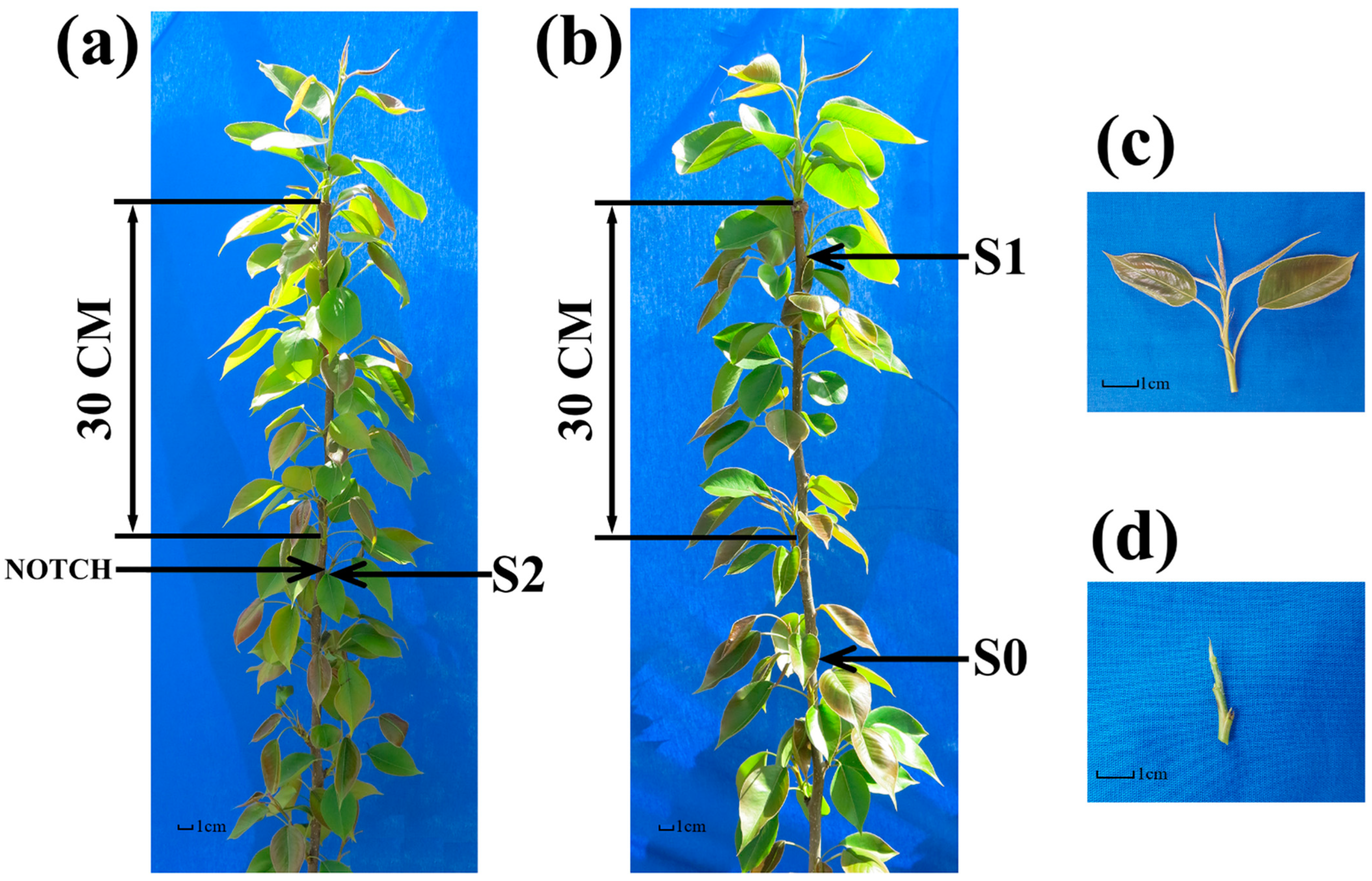
All ‘Huangguan’ pear (Pyrus bretschneideri Rehd.) trees were cultivated in the same orchard under uniform management practices and sharing an identical genetic background. Prior to budding in late March, pear trees with similar growth patterns and heights were carefully selected. Bud-notching was performed by making epidermal incisions 30 cm below the treetop, positioned 1 cm above each bud. These incisions extended on both sides of the bud, covering approximately one-third of the stem’s circumference. Lateral bud samples were collected 15 days post-notching. The first bud sample (S0), which was collected from a position 30 cm below the apical bud, demonstrates an inability to naturally promote lateral branching (Figure 2b). The second bud sample (S1), representing the first lateral bud directly below the apical bud, demonstrates a natural ability to promote lateral branching (Figure 2b). The third bud sample (S2) was collected from the first bud that underwent bud-notching 30 cm below the apical bud, and naturally exhibited branching (Figure 2a). From each position, ten bud samples were gathered, pooled collectively, and the entire procedure was reiterated thrice. An elaboration of the comprehensive sample collection process is depicted in Figure 2.

2.2. Analyzing Germination Rate, Branching Rate, and Endogenous Hormone Levels in Lateral Buds

The germination rate of pear trees by bud-notching treatment was subjected to statistical analysis. The sprouts of pear trees treated with bud-notching predominantly emerged within a 60 cm radius from the apical bud. The germination rate in the bud-notching group reached 90.01%, while the control group exhibited a germination rate of 89.53%, and there was no significant difference in germination rates between the two groups (Figure 3a, Table S1-1). The application of bud-notching treatment significantly enhanced the sprout rate of pear trees, as evidenced by the results presented in Figure 3b and Table S1-2. Specifically, the average growth promotion rate of long branches of pear trees in the bud-notching group (91.21%) was significantly higher than that of the control group (33.63%) (p < 0.01). Additionally, there was no significant difference in the average promotion rate of medium branches between the control group (4.16%) and the bud-notching group (4.18%) (p < 0.05). The mean rate of short shoot promotion in the control group was 77.60%, compared to 20.01% in the bud-notching group. The promotion rate of short branches in the control group was found to be significantly higher than that in the treatment group (p < 0.01) (Figure 3b). The comparison between the bud-notching and control groups indicated that bud-notching significantly enhanced the growth of pear trees at the initiating side buds.
The peak area values of five hormones were quantified using liquid chromatography, and the concentrations of the hormones were subsequently determined (Figure 3c, Table S2). The findings indicated that the levels of ZR, GA3, IAA, and BR in S1 and S2 were significantly elevated compared to those in S0, while the concentration of ABA did not exhibit a significant difference among S0 and S2. However, the levels of ABA in S1 were significantly elevated compared with S0 (p < 0.05). The ratio of BR/ABA in S1 was found to be statistically lower than that in S0 (p < 0.05), while the BR/ABA ratio in S2 was significantly higher than that in both S1 and S0 (p < 0.05) (Table S2). It is hypothesized that the up-regulation of growth-promoting hormones such as ZR, GA3, IAA, and BR, along with the maintenance of ABA at a stable level, may positively regulate the initiation of lateral branches following bud-notching.

2.3. Analysis of Sequencing Data and DEGs

To better understand the molecular mechanism of shoot branching by bud-notching in ‘Huangguan’ pears, we conducted a comparative transcriptomic analysis on new young shoots branching between S0, S1, and S2 groups. After quality control, 65.15 Gb of clean data were obtained, with average quality Q30 scores of >93.96% for each library, respectively, and an average GC content of 44.64%. For the three sample libraries, 68,728,487, 73,564,496, and 751,55,150 reads were mapped to the pear genome, representing 64.24–66.49% of the clean reads from the three samples (Table S3).
A total of 2154 DEGs were detected between S0 and S1, of which 1131 genes were up-regulated and 1023 genes were down-regulated. Similarly, 3036 DEGs were detected between S0 and S2, where 1339 genes were up-regulated and 1697 genes were down-regulated. Of these, 492 genes were found to be differentially expressed at both two confrontations (C1: S0_vs_S1, C2: S0_vs_S2) (Figure 4a). The comprehensive expression profile of these 492 DEGs was presented in Figure 4a, with 238 genes up-regulated and 254 genes down-regulated (Figure 4b, Table S4). Clean reads with lengths from 18 to 30 bp were obtained from S0, S1, S2. The percentage of bases Q30 reached at least 98.52% of all the samples. In addition, 505 miRNAs were identified, including 141 known and 364 new miRNAs (Table S5). Examination of miRNA expression in three samples identified 149 and 39 differentially expressed miRNAs (DEGs) at two confrontations, C1 and C2, respectively. Out of the total, 130 genes were found to be up-regulated and 19 genes were down-regulated in C1. Additionally, 27 genes were up-regulated and 12 genes were down-regulated in C2. In both confrontations, C1 and C2, nine differentially expressed genes (DEGs) exhibited up-regulation, while two DEGs displayed down-regulation (Figure 4d). Specifically, the expression profile of often differentially regulated miRNAs is presented in Figure 4c.
Functional classification of both mRNA and miRNA was conducted using the KEGG database (Figure 4e,f). For this analysis, 22 significant KEGG pathways were selected. The mRNA KEGG pathway analysis revealed the following distribution of DEGs: there were 25 and 32 DEGs from ‘Cellular Processes’, 45 and 35 DEGs from ‘Environmental Information Processing’, 53 and 86 DEGs from ‘Genetic Information Processing’, 110 and 155 DEGs from ‘Metabolism’, along with 21 and 24 DEGs from ‘Organismal Systems’ in C1 and C2, respectively (Figure 4e, Table S6). In miRNA KEGG pathway analysis, there were 7 and 10 DEGs from ‘Cellular Processes’, 13 and 11 DEGs from ‘Environmental Information Processing’, 66 and 55 DEGs from ‘Genetic Information Processing’, 60 and 35 DEGs from ‘Metabolism’, along with 13 and 8 DEGs from ‘Organismal Systems’ in C1 and C2, respectively (Figure 4f, Table S6). ‘Plant hormone signal transduction’, ‘Protein processing in endoplasmic reticulum’, and ‘Phenylpropanoid biosynthesis’ were the top three pathways with the largest number of associated DEGs in C1 and C2 (Figure 4e). A significant number of DEGs in C1 and C2 were related to ‘plant hormone signal transduction’, with 43 and 31 genes in the mRNA KEGG pathway and 9 and 6 genes in the miRNA KEGG pathway, respectively.

2.4. Plant Hormone Signaling Pathway Analysis of mRNA DEGs

Based on the KEGG pathway analysis, the ‘plant hormone signaling’ pathway was found to be essential for this research. Consequently, an in-depth analysis using the mRNA DEGs associated with this pathway was conducted. Within the auxin signaling transduction pathway, genes such as AUX1, AUX/IAA, SUAR, ARF, GH3, PP2C, ABF, BRI1, and CYCD3 were enriched (Figure 5a, Table S7). Most of the predicted auxin-induced protein genes (gene38584, 38585, 38587, 17817, 27027, newgene 29561) belonging to the SUAR family were identified to be down-regulated in both S1 and S2 when compared with S0. ARF (Pyrus_newGene_32024) was identified to be up-regulated in both S1 and S2 when compared with S0. GH3 (gene11049) was identified to be down-regulated in both S1 and S2 when compared with S0, GH3 (gene1810, gene34115, gene34117) was identified as being up-regulated in both S1 and S2 when compared with S0. Within the GA signaling transduction pathway, genes GID1 and TF were found to be enriched, of which GID1 (gene22380) and TF (gene28794) were identified to be down-regulated in both S1 and S2 when compared with S0. In the ABA signaling transduction pathway, PYR/PYL, PP2C, and ABF were enriched. Specifically, PYR/PYL (gene24413) was down-regulated in S1 and up-regulated in S2 when compared with S0, while PP2C (gene12403, gene24075, gene26342) was up-regulated in S1 and down-regulated in S2 when compared with S0. ABF (gene23064) was identified as being up-regulated in both S1 and S2 when compared with S0. Within the CTK signaling transduction pathway, AHP and B-ARR were enriched. Among these, AHP (gene22385) and B-ARR (gene9903) were identified as being up-regulated in S1 and down-regulated in S2 when compared with S0. Lastly, within the BR signaling transduction pathway, BRI1, BSK, and CYCD3 were enriched, with BRI1 (gene6361) and CYCD3 (gene1164) identified as being down-regulated in S1 and up-regulated in S2 when compared with S0, while BSK (Pyrus_newGene_23435) was up-regulated in S1 and down-regulated in S2 when compared with S0.

2.5. Analysis of the Interaction Network of miRNAs and Target mRNAs

The identification of key genes associated with shoot branching following bud-notching in pears was achieved through the KEGG pathway and miRNA-mRNA analysis. Through an analysis of the regulatory mechanisms of miRNAs and their target genes, a significant number of miRNAs were identified as targeting seven genes associated with shoot branching in pear trees (Table S8). After analysis, only one miRNA was identified to target at least two pear shoot branching-related genes, which was cpa-miR166e (GH3 and ABF). Additionally, ten miRNAs were identified as targeting only one pear shoot branching-related gene: aly-miR157d-3p (ABF), cem-miR166g (ABF), atr-miR166b (ABF), gma-miR166m (ABF), bdi-miR166e-3p (ABF), hbr-miR166a (ABF), cpa-miR166e (ABF), aly-miR166a-3p (ABF), mes-miR166i (ABF), cis-miR166a (ABF). One miRNA was identified to target one pear shoot branching-related genes, which were cpa-miR166e (GH3), gma-miR390e (SAUR), gma-miR162a (SAUR), hbr-miR156 (PP2C), aly-miR390a-5p (BRI1), and cme-miR396e (CYCD3). In addition, a regulatory network of miRNA-mRNA related to pear shoot branching-related genes was constructed (Figure 5b, Table S8). The results suggested that genes can negatively regulate the expression of miRNA. For example, ABF was up-regulated in S1, while the target ten miRNAs were identified as being down-regulated in S1; CYCD3 was up-regulated in S2, but the target miRNA (cme-miRNA396e) was identified as being down-regulated in S2.

2.6. Validation of Differential Gene Expression by qRT-PCR

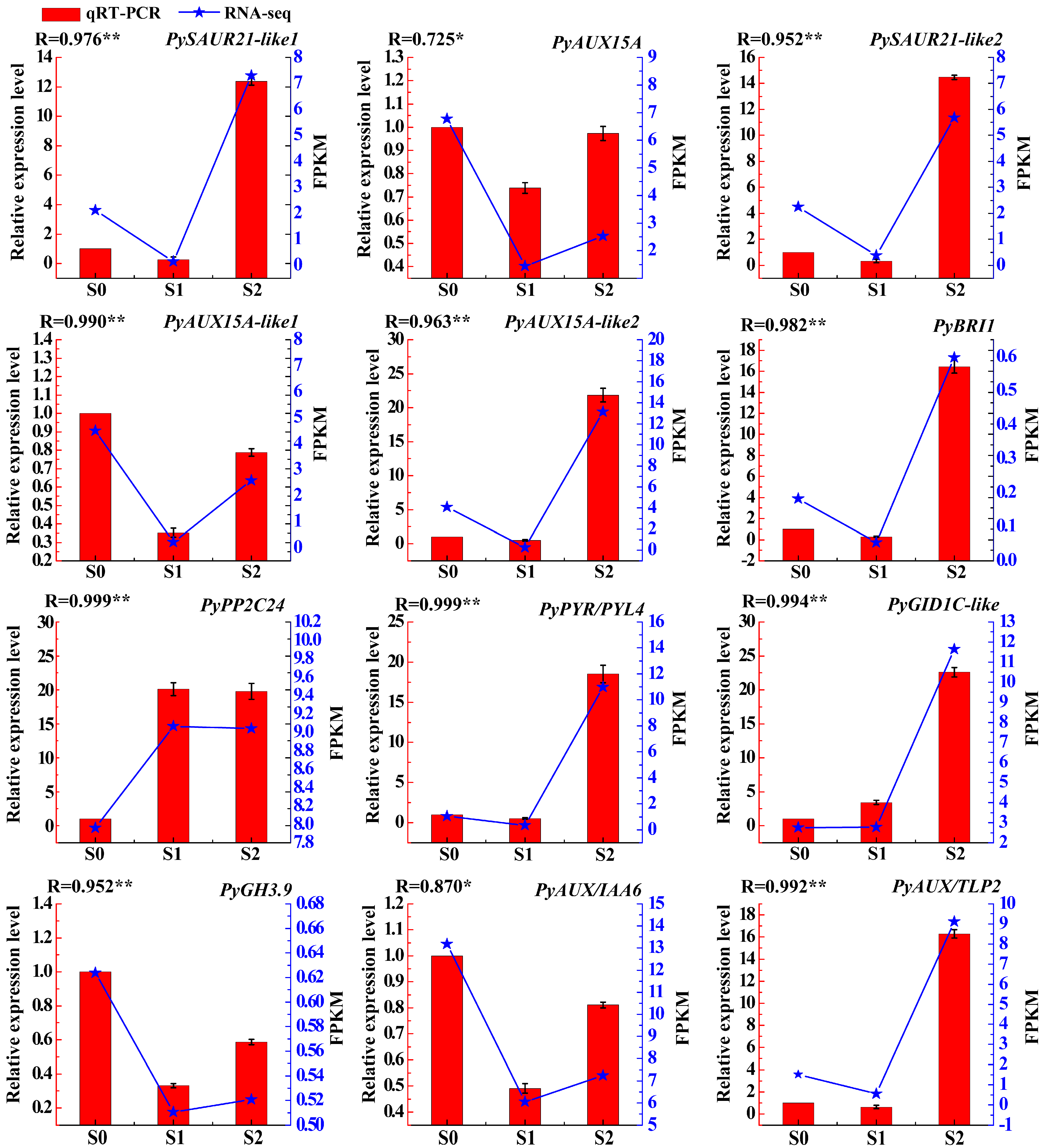
The ensure the reliability of transcriptomic data, 12 DEGs were selected for qRT-PCR validation. The accession number for these 12 DEGs were obtained through alignment with Pyrus bretschneideri (taxid:225117) in NCBI (Table S9). Specifically, we validated the expression of the following genes: PySAUR21-like1 (gene38584), PyAUX15A (gene38585), PySAUR21-like2 (gene38587), PyAUX15A-like1 (gene27027), PyAUX15A-like2 (newgene29561), PyBRI1 (gene6361), PyPP2C24 (gene24075), PyPYR/PYL4 (gene24413), PyGID1C-like (gene22380), PyGH3.9 (gene34117), PyAUX/IAA6 (gene31506), PyAUX/TLP2 (gene25464). In general, the results demonstrated that the expression patterns of RNAs discovered using qRT-PCR are comparable to those discovered using RNA-seq data, strengthening the validity of the DEGs discovered in this work (Figure 6, Table S10).

2.7. Overexpression PyCYCD3 Increased Lateral Branch Formation of Transgenic Arabidopsis

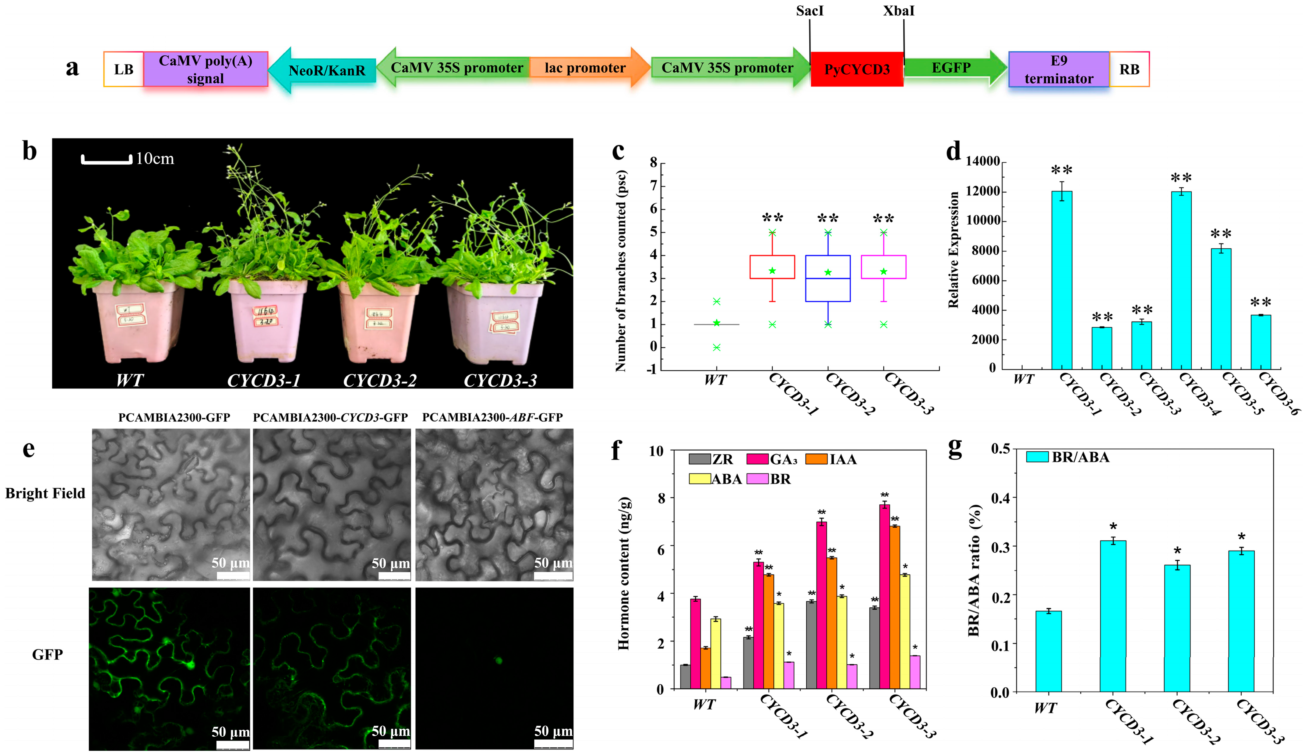
To explore the role of PyCYCD3 with respect to the induction of lateral branch formation in pear trees, the CDS of the PyCYCD3 was amplified, and the fusion protein of pCAMBIA2300-PyCYCD3-GFP was stably transformed into Arabidopsis (Figure 7a and Figure S1). Transgenic Arabidopsis was obtained by the Agrobacterium-medicated leaf disc method and PCR identification (Figure S1). CYCD3-1, CYCD3-2, CYCD3-3 were selected to explore whether PyCYCD3 promotes branching in transgenic Arabidopsis (Figure 7b). Through the photographic observation and the branching rate analysis of wild type and PyCYCD3 gene-transferred Arabidopsis plants grown for 1 month (Figure 7b,c, Table S11), it was found that the growth rate and number of branches of transgenic Arabidopsis plants were significantly higher than those of wild type Arabidopsis plants during the same period. Six transgenic Arabidopsis plant lines (CYCD3-1, CYCD3-2, CYCD3-3, CYCD3-4, CYCD3-5, CYCD3-6) were selected to observe how PyCYCD3 would be expressed by methods of the qRT-PCR (Figure 7d). The qRT-PCR results revealed that the expression level of PyCYCD3 was significantly up-regulated compared with WT (Figure 7d, Table S12).
The subcellular localization of pCAMBIA1300-PyCYCD3-GFP-transformed tobacco was studied. The results showed that the PyCYCD3 gene was mainly located in the cytoplasm, while unloaded pCAMBIA1300-GFP was expressed in both the cytoplasm and the membrane (Figure 7e). This indicates that PyCYCD3 gene plays different biological characteristics and functions in different cell physiological processes. Given its primary localization in the cytoplasm, the PyCYCD3 gene may be related to cell division, mitosis, and other related processes. In three transgenic Arabidopsis plant lines, CYCD3-1, CYCD3-2, and CYCD3-3, the ZR content were measured to be 21.57 ng/g, 36.69 ng/g, and 33.99 ng/g, respectively. The GA3 contents were 52.91 ng/g, 69.87 ng/g, and 77.12 ng/g, respectively. The contents of IAA were 47.82 ng/g, 54.89 ng/g, and 68.11 ng/g, respectively. These values were significantly higher than those of ZR (10.02 ng/g), GA3 (87.68 ng/g), and IAA (17.12 ng/g) in wild Arabidopsis (p < 0.01) (Figure 7f, Table S13). Furthermore, the BR/ABA ratios for the transgenic lines (0.31, 0.26, 0.29) were significantly higher than those of wild Arabidopsis (0.17) (p < 0.05) (Figure 7g, Table S13). These results indicate that the heterologous expression of CYCD3 may promote the synthesis of growth-promoting hormones ZR, GA, IAA, and BR and regulate the stability of growth-inhibiting endogenous hormone ABA, thereby modulating plant branch growth.

3. Discussion

3.1. The Germination, Branching Rate, and Endogenous Hormone Levels in Lateral Buds

The bud-notching technology that was used in this study provides a valuable resource for the exploration of the molecular mechanism of branching regulation in pear trees. Based on the analysis of the transcriptome of specific cells or tissues, RNA-seq is a suitable method to identify the main biological processes or pathways associated with branching [42]. Differences in the RNA-seq data of lateral buds among the S0, S1, and S2 indicated that plant hormone signal transduction was significantly enriched, providing the initial foundation for the present study. The current findings suggest that hormone signal transduction pathways involving IAA, GA, ZR, and CTK facilitate bud germination and promote plant growth, whereas ABA is associated with the induction of bud dormancy and differentiation [19,24]. Furthermore, 6-benzyladenine (6-BA) has the potential to significantly enhance the growth response of tender shoots without fallen buds on 2 or 3-year-old apple trees [9]. In this study, it was found that there was no significant difference in germination rates between the bud-notching and control groups (Figure 2a). The application of bud-notching treatment significantly enhanced the long branches rate of pear trees, as evidenced by the results presented in Figure 2b. Interestingly, the contents of the endogenous plant hormones IAA and BR showed the same increasing trend, and the contents of these two hormones were significantly higher in lateral buds (S2) sprouted by bud-notching treatment compared to the control buds (S0) (Figure 2c). Meanwhile, the effects of other hormones, such as ethylene and salicylic acid, as well as those of uncontrolled environmental factors on branching, were not thoroughly investigated in this study, which had an impact on the results of this study. We will conduct further research in future experiments.

3.2. The Genes Related to Phytohormone Signaling in Lateral Bud-Sprouting Regulation

Classical theory posits that apical dominance governs the transport of auxin along the primary stem by mediating the competition among auxin sources and subsequently regulates the activation of lateral buds [43,44]. In apples, the mechanism of auxin transport from axillary buds to stems is integral to the growth of axillary buds, which is considered essential for their development [24]. In this study, we identified the most differentially expressed genes associated with hormone signal transduction within the auxin pathway, specifically including AUX1, AUX/IAA, ARF, GH3, and SUAR, which are activated by auxin. The expression of AUX1, AUX/IAA, and GH3 was found to be up-regulated in the S2 treatment, while ARF exhibited up-regulation in both S1 and S2, but was down-regulated in S0 [45,46]. CTK may regulate the growth of apple branches by activating certain factors that indirectly suppress the expression of genes linked to branching [47,48]. Research indicates that type-B ARRs, such as ARR1 and ARR2, can activate type-A ARRs, such as ARR6, in plants [49]. ARR1, ARR2, and ARR10 trigger the transcription of ARR4, ARR5, ARR6, and ARR7 in response to CTK through AHK4 and/or CKI1 [50]. The Arabidopsis His-Asp phosphorelay network comprises 11 type-B ARR members, which are essential transcriptional regulators [51]. The current study demonstrates that the AHP and B-ARR genes are significantly enriched in the CTK signaling pathway. Specifically, Gene22385 (AHP) and Gene9903 (B-ARR) exhibited up-regulation in S1 and down-regulation in S2, suggesting that phosphorylated AHPs may translocate to the nucleus [52,53]. The finding that type-A ARRs are required for CK-mediated bud outgrowth may explain the increased expression of MsARR3/5 during bud outgrowth. It is suggested that exogenous GA spraying benefits lateral bud initiation and branch extraction [5,9,47]. This study confirmed this conclusion and identified down-regulated gibberellin genes GID1 and TF in both S1 and S2. Research has demonstrated that PYR/PYL/RCAR proteins inhibit PP2C phosphatase activity, which is a known inhibitor of the ABA signaling pathway. This suggests that PYR/PYL/RCAR and PP2C might work together to negatively regulate the ABA signaling pathway [54,55]. PYR/PYL, PP2C, and ABF genes, in the abscisic acid signaling pathway, were identified in this study. PYR/PYL (gene24413) was down-regulated in S1 and up-regulated in S2, while PP2C (gene12403, gene24075, gene26342) was up-regulated in S1 and down-regulated in S2. ABF (gene23064) was up-regulated in both S1 and S2 and down-regulated in S0. Previous research suggests that ABA indirectly promotes bud differentiation rather than playing a direct role [56,57]. Several studies have indicated that ABA increases the vacuole sugar content, leading to enhanced leaf dewatering [57,58,59]. It has also been shown that root pruning and ring cutting may enhance the differentiation process of buds [59]. Decreased IAA content and increased ABA and CTK contents in xylem SAP during flower bud differentiation may lead to increased shoot branching in pear trees through the up-regulation of genes involved in ABA signaling. Similarly, studies in tomato found significantly higher IAA content in axillary buds of wrky-b, bl, and pin4 mutant plants than in WT plants [11]. The CYCD gene, which plays a crucial role in cell division as an intermediary between endogenous and exogenous developmental signals, is co-regulated by several plant hormones, including auxin, CTK and BR [36,60,61,62]. In this study, BRI1, BSK, and CYCD3 genes were found to be enriched in the BR signaling pathway. BRI1 and CYCD3 were down-regulated in S1 and up-regulated in S2, while BSK was up-regulated in S1 and down-regulated in S2 (Figure 4a). Notably, the up-regulation of CYCD3 (gene1164) in S2 aligns with findings reported by Tan et al. in 2019. Additionally, the expression of cell proliferation genes such as CYCLIN D3 (MsCYCD3) was found to be up-regulated at stage “1” in the more-branching mutant (MB).

3.3. The Interactions between mRNAs and miRNAs

MiRNAs are short noncoding RNAs, around 19–24 nucleotides long, that can interact with longer target RNAs [63]. In ‘Huangguan’ pear (Pyrus bretschneideri Rehd.), 505 miRNAs were identified, including 143 known and 362 new ones. The interactions between miRNAs and their target genes can be one-to-many or many-to-one, rather than strictly one-to-one, introducing significant regulatory complexity (Figure 4b). This complexity is exemplified by the fact that a single miRNA can regulate multiple genes involved in branching, and conversely, a single gene can be targeted by multiple miRNAs. This intricate network underscores the elaborate control mechanisms governing branching in young pear plants. Specifically, we found ten miRNAs targeting a single gene (ABF) related to pear shoot branching, namely aly-miR157d-3p, cem-miR166g, atr-miR166b, gma-miR166m, bdi-miR166e-3p, hbr-miR166a, cpa-miR166e, aly-miR166a-3p, mes-miR166i, cis-miR166a. In addition, only one miRNA was identified to target at least two pear shoot branching-related genes: cpa-miR166e (GH3 and ABF). One miRNA was identified to target each of the pear shoot branching-related genes:cpa-miR166e (GH3), gma-miR390e (SAUR), gma-miR162a (SAUR), hbr-miR156 (PP2C), aly-miR390a-5p (BIR1), and cme-miR396e (CYCD3). CYCD3;1, a key player in cell proliferation and differentiation during plant development, is expressed in growing tissues, such as meristems and developing leaves [64]. BIR1, another important gene, prevents cell death, and its inactivation in yeast leads to cell division defects [65]. Our study revealed that the up-regulation of BIR1 and CYCD3 is crucial for shoot branching in pears. In summary, cap-miR166e was up-regulated, while GH3 (gene11049) was down-regulated in S2 compared to S0. Conversely, gma-miR162a, hbr-miR156, aly-miR390a, and cem-miR396e were down-regulated in S2, leading to the up-regulation of their respective target genes: SAUR (gene27024), PP2C (gene12403), BIR1 (gene6361), and CYCD3 (gene1164).

3.4. The Over-Expression of PyCYCD3 in Arabidopsis

The influence mechanisms of hormones such as IAA, CTK, GA on lateral bud sprouting have been extensively studied [19,24]. However, the role of BR in this process remains less well understood. Consequently, our research has concentrated on the CYCD3 gene within the BR signaling pathway. The BES1/BZR1-ARF complex supports the longstanding reported synergistic interaction between auxin and BRs, implicated in both elevated gene expression and extended hypocotyl growth [66,67]. It is speculated that ARF and CYCD3 may have similar synergy in promoting lateral bud germination. The overexpression of PtoCYCD3 promoted the cell cycle G1/S transition of perennial poplars, thus accelerating the cell division of poplar meristem, such as apical buds, axillary buds, and stem cambium; it also accelerated plant vegetative growth and produced obvious branches [68]. OsCYCD3;1 may maintain axillary meristem to promote branching in rice, possibly by regulating cell division [69]. In poplar, the upregulation of AHP and CYCD3 expression levels is closely related to branch formation [70]. This is consistent with the results of this study. Transgenic Arabidopsis leaf explants overexpressing CycD3 can initiate and maintain cell division in the absence of CTK, suggesting that CTKs activate cell division through induction of CycD3 at the G1/S transition [62]. This evidence indicates that plant D-type cyclins may function as mediators of internal and environmental stimuli to drive cell division. Our study indicated that the heterologous over-expression of PyCYCD3 has a positive regulatory effect on the increased Arabidopsis growth and branching number (Figure 7b,c). Therefore, up-regulated PyCYCD3 may play a crucial role in accumulating auxin and BR, which function as mediators of bud-notching stimuli to drive cell division and regulate lateral branching processes in pear trees.

4. Materials and Methods

4.1. Plant Materials and Sample Collection

Plant materials were obtained from 2-year-old grafted saplings of the pear variety ‘Huangguan’ (Pyrus bretschneideri Rehd.), cultivated at the National Modern Agricultural Pear Industrial Technology Experimental Station of Tiaoshan Group (Jingtai, Gansu Province, China). It is located at 103°59′~104°05′ E, 37°07′~37°09′ N, with an annual average temperature of 8.25 °C, an average frost-free period of 141 days, and an annual average precipitation of 183.1 mm. The main soil types are calcareous soil and cultivated soil. On April 5th, before the bud germination of pear seedlings, seedlings with consistent height and vigor standards were selected for budding treatment at a designated location. The specific operation method involves incising 0.4 cm above the bud body between 30–60 cm below the pear tree’s top bud, with the incision being half the stem’s circumference. The control group pear trees were not subjected to budding treatment. On the 15th day after budding, the first sample (S0) was collected from the first bud below 30 cm from the central stem to the top bud of the control tree, which was not budded and generally cannot grow into long branches. The second sample (S1) was taken from the first bud and tender shoot closest to the top bud of the control tree, which was not budded and can grow into long branches. The third sample (S2) was gathered from the first bud and tender shoot below 30 cm from the central stem to the top bud of the budded tree, which can grow into long branches after budding. All samples collected at each position had at least 30 buds and tender shoots mixed into one sample, with three biological replicates set for both the treatment and control groups [9,71].

4.2. Measurement of Emergence Rate, Branching Rate, and Hormone Levels in Lateral Buds

After the leaves fell in autumn, the number of buds and the growth length of the germination rate, the peripheral extended branch length and branch analogy (long branch > 15 cm, middle branch 5~15 cm, short branch < 5 cm) were assessed. The quantities of long, medium, and short branches, along with the average growth length of all branches were independently measured for both untreated and bud-notched pear trees.
High-performance liquid chromatography was used to quantify the contents of indole acetic acid (IAA), zeatin riboside (ZR), abscisic acid (ABA), and gibberellic acid (GA3) in 0.2 g of freshly lateral buds, collected at 15 days post-sampling, in accordance with previously described methods [19,72]. For the quantification of the brassinosteroid (BR) content, 1 g of freshly collected lateral buds at the same 15-day time point was required. Each sample set for both hormone types was composed of three biological replicates to ensure the reliability and reproducibility of the results.

4.3. RNA Preparation and Transcriptome Sequencing

Total RNA was isolated utilizing the Trizol Reagent (Invitrogen, Carlsbad, CA, USA) in accordance with the manufacturer’s guidelines. The NanoDrop 2000 Spectrophotometer was used to measure RNA concentration and purity (Thermo Fisher Scientific, Wilmington, DE). The RNA Nano 6000 Assay Kit of the Agilent Bioanalyzer 2100 System was implemented to assess RNA integrity (Agilent Technologies, Santa Clara, CA, USA). Following a manufacturer’s recommendations, sequencing libraries were generated using NEBNextR UltraTM Directional RNA Library Prep Kit for IlluminaR (NEB, USA) and the small RNA Sample Library Prep Kit for IlluminaR (NEB, Ipswich, MA, USA), and index codes were added to assign sequences to each sample. Biomarker Technologies Corporation addressed library preparation and sequencing (Beijing, China). The Agilent Bioanalyzer 2100 and qPCR were employed to analyze library quality. The RNA-seq data were submitted to the NCBI SAR database under the accession number PRJNA933274 (https://www.ncbi.nlm.nih.gov/bioproject/?term=PRJNA933274 (accessed on 9 February 2023)).

4.4. Differential Expressed Genes (DEGs) Analysis

Differential expression analysis was performed using the DESeq (2010) R package. The resulting FDR (false discovery rate) was adjusted using the PPDE (posterior probability of being DE). An FDR < 0.05 and ǀlog2(FoldChange)ǀ ≥ 1 were set as the threshold for significantly differential expression. Gene function was annotated based on the KEGG database (Kyoto Encyclopedia of Genes and Genomes). KOBAS 3.0 software (http://kobas.cbi.pku.edu.cn/kobas3) was used to test the statistical enrichment of DEGs in the KEGG pathway [73].

4.5. Quantitative Real-Time PCR (qRT-PCR) Validation

A total of twelve DEGs with significant expression patterns of the KEGG pathway were selected for qRT-PCR verification. Purification and reverse transcription of RNA were used PrimeScript™ RT reagent Kit with gDNA Eraser (TAKARA). Primers were designed by online software Primer3 input (version 4.0, http://primer3.ut.ee/ (accessed on 28 March 2023)), and the sequences are shown in Table S14. The qRT-PCR was conducted on CFX96 Touch™ Real-Time PCR Detection System (Bio-Rad, Hercules, CA, USA) by using SYBR®Premix ExTaq™ II. The data were analyzed by 2−∆∆Ct.

4.6. Overexpression PyCYCD3 in Arabidopsis and Subcellular Localization Analysis

The genomic sequences of the CDS of the PyCYCD3 gene were amplified from pear genomic DNA by the use of primers (F:AGAACACGGGGGACGAGCTCATGGGTGGTTGGGCAATTGCAG; R:ACCATGGTGTCGACTCTAGACTCCCAAATCCCAACCTCCATG), after which the sequences were cloned into pCAMBIA2300-GFP vectors and double-digested using SacI and XbaI, resulting in pCAMBIA2300-PyCYCD3-GFP constructs. The inverted trifoliate and inverted quadrifoliate leaves of Ben’s tobacco were selected for transient transformation of PyCYCD3 to determine the subcellular localization of the gene. Subsequently, the PyCYCD3 gene was transfected into Arabidopsis Columbia using the floc dipping method, and the positive plants were screened on MS plates containing 50 mg/L Kan and 20 mg/L tauramycin, after which the transgenes obtained from the screening were identified by PCR assay, and the T3 generation of pure transgenic Arabidopsis plants was obtained after two screenings. Finally, the number of transgenic Arabidopsis plants branching, the relative expression of PyCYCD3 gene (PyCYCD3-F: CGCCGTTTCGGGATTAACTA; PyCYCD3-R: GAGGTAGGAGTGAGGTGATTTG. Actin-F: GCCGACAGAATGAGCAAAGAG; Actin-R: AGGTACTGAGGGAGGCCAAGA), and its endogenous hormone content were determined.

4.7. Data Analysis

A total of thirty plants of both transgenic and wild-type Arabidopsis were selected for observation of lateral shoot growth following a period of shoot extraction. The data obtained were subjected to statistical analysis using the independent samples t-test feature in SPSS 22.0 software for comparison purposes. Two-tailed t-tests were employed to identify significant differences in the sample results. Additionally, Microsoft Excel 2019 was utilized for the statistical analysis of the raw data, while graphing was performed using Origin 9.0.

5. Conclusions

In conclusion, this study involved sequencing the transcriptome of bud-notched and control buds in pear (Pyrus bretschneideri Rehd.), focusing on plant hormone signal transduction pathways and analyzing the mRNA-miRNA interactions. A total of 15 miRNA-mRNA pairs were investigated, revealing both positive and negative correlations. Subsequently, PyCYCD3 was chosen for transformation into wild Arabidopsis plants, resulting in the generation of transgenic Arabidopsis plants. The analysis of branching patterns, endogenous hormone levels, and the relative expression of PyCYCD3 in these transgenic Arabidopsis plants suggests that PyCYCD3 may potentially play a significant role in promoting lateral branches in pear trees through bud-notching techniques. The results demonstrated that BR signaling may integrate with auxin signaling pathways and up-regulate the PyCYCD3 (down-regulate cem-miR396e) to control the formation of lateral branches by promoting lateral bud sprouting.

Supplementary Materials

The following supporting information can be downloaded at: https://www.mdpi.com/article/10.3390/plants13202928/s1, Figure S1: Amplification of PyCYCD3 and obtain the transgenic Arabidopsis plants. (a) Amplification bands of PyCYCD3. M, D5000 Marker; 1-2, Amplification bands of PyCYCD3. (b) Introduction of pCAMBIA2300-PyCYCD3-GFP into PCR bands of Agrobacterium tumefaciens. M, D5000 Marker; 1-6, PCR bands of Agrobacterium tumefaciens. (c–f) Acquisition of transgenic Arabidopsis plants of T1 generation. g, PCR test results of transgenic Arabidopsis plants of T1 generation. Note: M, D2000 Marker; 1-7, Individual transgenic Arabidopsis plants; WT, wild Arabidopsis plants; Table S1-1 Germination rate of pear tree between control and bud-notching; Table S1-2 Outgrowth rate of pear tree between control and bud-notching; Table S2: The endogenous hormone levels and ZT/ABA, GA3/ABA, IAA/ABA, BR/ABA in lateral buds of pear trees; Table S3: Categorization of reads of RNAs in the different groups; Table S4: Identified 492 mRNAs by sequences; Table S5: Identified 505 miRNAs by sequences; Table S6: mRNA and miRNA KEGG pathway analysis; Table S7: DEGs expression of plant hormone signaling pathway; Table S8: miRNAs targeting seven mRNA and expression of DEGs; Table S9: 12 DEGs accession number were obtained by Pyrus x bretschneideri (taxid:225117) alignment in NCBI; Table S10: A total of 12 DEGs were chosen for qRT-PCR validation of the RNA-seq data; Table S11: Branching counts between wild type and three transgenic Arabidopsis lines; Table S12: The relative expression of CYCD3 in six transgenic Arabidopsis plant lines; Table S13: The endogenous hormone levels and BR/ABA in branches of transgenic Arabidopsis; Table S14 Oligonucleotides used for qRT-PCR of the DEGs in lateral buds of pear trees.

Author Contributions

Conceptualization, B.-H.C.; methodology, C.-W.Z. and Z.-S.A.; investigation, Z.-S.A. and J.M.; In vitro germination, Z.-H.M. and W.-F.L.; formal analysis, Z.-S.A.; writing—review and editing, Z.-S.A. All authors have read and agreed to the published version of the manuscript.

Funding

This work was supported by the Education Science and Technology Innovation Project of Gansu Province (GSSYLXM-02), and the Science and Technology Major Project of Gansu Province (22ZD6NA045). We thank the National Modern Agricultural Pear Industrial Technology Experimental Station of Tiaoshan Group (Jingtai, Gansu Province, China) for providing us with the experimental materials.

Data Availability Statement

The data presented in this study are available on request from the corresponding author. The data are not publicly available due to the regulatory restrictions of the funding organization.

Conflicts of Interest

The authors declare no conflicts of interest.

References

  1. Wu, J.; Wang, Z.; Shi, Z.; Zhang, S.; Ming, R.; Zhu, S.; Khan, M.A.; Tao, S.; Korban, S.S.; Wang, H.; et al. The genome of the pear (Pyrus bretschneideri Rehd.). Genome Res. 2013, 23, 396–408. [Google Scholar] [CrossRef] [PubMed]
  2. Chevalier, F.; Nieminen, K.; Sanchez-Ferrero, J.C.; Rodriguez, M.L.; Chagoyen, M.; Hardtke, C.S.; Cubas, P. Strigolactone promotes degradation of DWARF14 an alpha/beta hydrolase essential for strigolactone signaling in Arabidopsis. Plant Cell 2014, 26, 1134–1150. [Google Scholar] [CrossRef] [PubMed]
  3. Teichmann, T.; Muhr, M. Shaping plant architecture. Front. Plant Sci. 2015, 6, 233. [Google Scholar] [CrossRef] [PubMed]
  4. Elfving, D.C.; Visser, D.B. Cyclanilide induces lateral branching in sweet cherry trees. Hortscience 2006, 41, 149–153. [Google Scholar] [CrossRef]
  5. Elfving, D.C.; Visser, D.B. Improving the efficacy of cytokinin applications for stimulation of lateral branch development in young sweet cherry trees in the orchard. Am. Soc. Hortic. Sci. 2007, 42, 251–256. [Google Scholar] [CrossRef]
  6. Jacyna, T. Factors influencing lateral-branch formation in woody plants. Acta Agrobot. 2002, 55, 5–25. [Google Scholar] [CrossRef]
  7. Jacyna, T.; Puchaa, A. Application of environment friendly branch promoting substances to advance sweet cherry tree canopy development in the orchard. J. Fruit Ornam. Plant Res. 2004, 12, 177–182. [Google Scholar]
  8. Zhang, L.L.; Fang, W.M.; Chen, F.D.; Song, A.P. The Role of Transcription Factors in the Regulation of Plant Shoot Branching. Plants 2022, 11, 1997. [Google Scholar] [CrossRef]
  9. McArtney, S.; Obermiller, J.D.J.H. Effect of notching 6-benzyladenine and 6-benzyladenine plus gibberellin A4+ A7 on budbreak and shoot development from paradormant buds on the leader of young apple trees. Hort Technol. 2015, 25, 233–237. [Google Scholar] [CrossRef]
  10. Aishajiang, M.; Yang, Q.; Wang, J.J.; Liu, G.J. Effects of Cutting Back, Branch-bending and Bud-notching Treatments on Endogenous Hormones in the Buds of Fuji Apple. Acta Hortic. Sin. 2013, 40, 1437–1444. [Google Scholar]
  11. Yang, H.H.; Zhou, K.; Wu, Q.F.; Jia, X.Y.; Wang, H.X.; Yang, W.H.; Lin, L.H.; Hu, X.M.; Pan, B.Q.; Li, P.; et al. The tomato WRKY-B transcription factor modulates lateral branching by targeting BLIND, PIN4, and IAA15. Hortic. Res. 2024, 11, uhae193. [Google Scholar] [CrossRef] [PubMed]
  12. Van den Ende, W. Sugars take a central position in plant growth, development and, stress responses. A focus on apical dominance. Front. Plant Sci. 2014, 5, 313. [Google Scholar] [CrossRef] [PubMed]
  13. Rameau, C.; Bertheloot, J.; Leduc, N.; Andrieu, B.; Foucher, F.; Sakr, S. Multiple pathways regulate shoot branching. Front. Plant Sci. 2015, 5, 741. [Google Scholar] [CrossRef]
  14. Barbier, F.F.; Dun, E.A.; Kerr, S.C.; Chabikwa, T.G.; Beveridge, C.A. An update on the signals controlling shoot branching. Trends Plant Sci. 2019, 24, 220–236. [Google Scholar] [CrossRef]
  15. Maurya, J.P.; Miskolczi, P.C.; Mishra, S.; Singh, R.K.; Bhalerao, R.P. A genetic framework for regulation and seasonal adaptation of shoot architecture in hybrid aspen. Proc. Natl. Acad. Sci. USA 2020, 117, 11523–11530. [Google Scholar] [CrossRef]
  16. Gonzalez-Grandio, E.; Poza-Carrion, C.; Sorzano, C.O.; Cubas, P. BRANCHED1 promotes axillary bud dormancy in response to shade in Arabidopsis. Plant Cell 2013, 25, 834–850. [Google Scholar] [CrossRef] [PubMed]
  17. Wang, M.; Le Moigne, M.-A.; Bertheloot, J.; Crespel, L.; Perez-Garcia, M.-D.; Ogé, L.; Demotes-Mainard, S.; Hamama, L.; Davière, J.-M.; Sakr, S. BRANCHED1: A key hub of shoot branching. Front. Plant Sci. 2019, 10, 76. [Google Scholar] [CrossRef]
  18. Kovaleva, L.V.; Zakharova, E.V.; Minkina, Y.V.; Timofeeva, G.V.; Andreev, I.M. Germination and In Vitro Growth of Petunia Male Gametophyte Are Affected by Exogenous Hormones and Involve the Changes in the Endogenous Hormone Leve. Russ. J. Plant Physiol. 2005, 52, 521–526. [Google Scholar] [CrossRef]
  19. Zhou, Q.; Gao, B.; Li, W.F.; Mao, J.; Yang, S.J.; Li, W.; Ma, Z.H.; Zhao, X.; Chen, B.H. Effects of exogenous growth regulators and bud picking on grafting of grapevine hard branches. Sci. Hortic. 2020, 264, 109186. [Google Scholar] [CrossRef]
  20. Müller, D.; Leyser, O. Auxin, cytokinin and the control of shoot branching. Ann. Bot. 2011, 107, 1203–1212. [Google Scholar] [CrossRef]
  21. Umehara, M.; Hanada, A.; Yoshida, S.; Akiyama, K.; Arite, T.; Takeda-Kamiya, N.; Magome, H.; Kamiya, Y.; Shirasu, K.; Yoneyama, K.; et al. Inhibition of shoot branching by new terpenoid plant hormones. Nature 2008, 455, 195–200. [Google Scholar] [CrossRef] [PubMed]
  22. Yang, Y.R.; Hu, Y.L.; Li, P.; Hancock, J.T.; Hu, X.Y. Research Progress and Application of Plant Branching. Phyton-Int. J. Exp. Bot. 2023, 92, 680–689. [Google Scholar] [CrossRef]
  23. Palmer, H.J.D.; Diack, R.; Seymour, S.; Dayatilake, D.; Tustin, D.S. New approaches to the alleviation of barewood in young apple trees. Biotechnology 2005, 80, 623–627. [Google Scholar] [CrossRef]
  24. Tan, M.; Li, G.F.; Chen, X.L.; Xing, L.B.; Ma, J.J.; Zhang, D.; Ge, H.J.; Han, M.Y.; Sha, G.L.; An, N. Role of cytokinin strigolactone and auxin export on outgrowth of axillary buds in apple. Front. Plant Sci. 2019, 10, 616. [Google Scholar] [CrossRef]
  25. Chandler, J.W. Auxin response factors. Plant Cell Environ. 2016, 39, 1014–1028. [Google Scholar] [CrossRef]
  26. Anderson, J.V.; Doğramac, M.; Horvath, D.P.; Foley, M.E. Auxin and ABA act as central regulators of developmental networks associated with paradormancy in Canada thistle (Cirsium arvense). Funct. Integr. Genom. 2012, 12, 515–531. [Google Scholar] [CrossRef] [PubMed]
  27. González-Grandío, E.; Pajoro, A.; Franco-Zorrilla, J.M.; Tarancon, C.; Immink, R.G.H.; Cubas, P. Abscisic acid signaling is controlled by a BRANCHED1/HD-ZIP I cascade in Arabidopsis axillary buds. Proc. Natl. Acad. Sci. USA 2017, 114, E245–E254. [Google Scholar] [CrossRef]
  28. Janssen, B.J.; Drummond, R.S.; Snowden, K.C. Regulation of axillary shoot development. Curr. Opin. Plant Biol. 2014, 17, 28–35. [Google Scholar] [CrossRef]
  29. Wang, Y.; Sun, S.; Zhu, W.; Jia, K.; Yang, H.; Wang, X. Strigolactone/MAX2-induced degradation of brassinosteroid transcriptional effector BES1 regulates shoot branching. Dev. Cell 2013, 27, 681–688. [Google Scholar] [CrossRef]
  30. Sun, Y.; Fan, X.-Y.; Cao, D.-M.; Tang, W.; He, K.; Zhu, J.-Y.; He, J.-X.; Bai, M.-Y.; Zhu, S.; Oh, E.; et al. Integration of brassinosteroid signal transduction with the transcription network for plant growth regulation in Arabidopsis. Dev. Cell 2010, 19, 765–777. [Google Scholar] [CrossRef]
  31. Belkhadir, Y.; Jaillais, Y. The molecular circuitry of brassinosteroid signaling. New Phytol. 2015, 206, 522–540. [Google Scholar] [CrossRef]
  32. Divi, U.K.; Rahman, T.; Krishna, P. Gene expression and functional analyses in brassinosteroid-mediated stress tolerance. Plant Biotechnol. J. 2016, 14, 419–432. [Google Scholar] [CrossRef] [PubMed]
  33. Li, Q.F.; Lu, J.; Yu, J.W.; Zhang, C.Q.; He, J.X.; Liu, Q.Q. The brassinosteroid-regulated transcription factors BZR1/BES1 function as a coordinator in multisignal-regulated plant growth. Biochim. Et Biophys. Acta (BBA)-Gene Regul. Mech. 2018, 1861, 561–571. [Google Scholar] [CrossRef]
  34. Xia, X.; Dong, H.; Yin, Y.; Song, X.; Gu, X.; Sang, K.; Zhou, J.; Shi, K.; Zhou, Y.; Foyer, C.H.; et al. Brassinosteroid signaling integrates multiple pathways to release apical dominance in tomato. Proc. Natl. Acad. Sci. USA 2021, 118, e2004384118. [Google Scholar] [CrossRef] [PubMed]
  35. Liu, X.F.; Zhao, C.D.; Gao, Y.Q.; Xu, Y.; Wang, S.J.; Li, C.S.; Xie, Y.P.; Chen, P.X.; Yang, P.Z.; Yuan, L.; et al. A multifaceted module of BRI1 ETHYLMETHANE SULFONATE SUPRESSOR1 (BES1)-MYB88 in growth and stress tolerance of apple. Plant Physiol. 2021, 185, 1903–1923. [Google Scholar] [CrossRef]
  36. Hu, Y.X.; Bao, F.; Li, J.Y. Promotive effect of brassinosteroids on cell division involves a distinct CycD3-induction pathway in Arabidopsis. Plant J. Cell Mol. 2010, 24, 693–701. [Google Scholar] [CrossRef] [PubMed]
  37. Yin, Y.; Wang, Z.; Mora-Garcia, S.; Li, J.M.; Yoshida, S.; Asami, T.; Chory, J. BES1 accumulates in the nucleus in response to brassinosteroids to regulate gene expression and promote stem elongation. Cell 2002, 109, 181–191. [Google Scholar] [CrossRef]
  38. Zheng, L.; Ma, J.; Song, C.; Zhang, L.; Gao, C.; Zhang, D.; An, N.; Mao, J.; Han, M. Genome-wide identification and expression analysis of GRF genes regulating apple tree architecture. Tree Genet. Genomes 2018, 14, 54–59. [Google Scholar] [CrossRef]
  39. Zheng, L.; Zhao, C.; Mao, J.; Song, C.; Ma, J.; Zhang, D.; Han, M.; An, N. Genome-wide identification and expression analysis of brassinosteroid biosynthesis and metabolism genes regulating apple tree shoot and lateral root growth. J. Plant Physiol. 2018, 231, 68–85. [Google Scholar] [CrossRef]
  40. Zheng, L.W.; Gao, C.; Zhao, C.D.; Zhang, L.Z.; Han, M.Y.; An, N.; Ren, X.L. Effects of brassinosteroid associated with auxin and gibberellin on apple tree growth and gene expression patterns. Hortic. Plant J. 2019, 5, 93–108. [Google Scholar] [CrossRef]
  41. Dewitte, W.; Scofield, S.; Alcasabas, A.A.; Maughan, S.C.; Menges, M.; Braun, N.; Collins, C.; Nieuwland, J.; Prinsen, E.; Sundaresan, V.; et al. Arabidopsis CYCD3 D-type cyclins link cell proliferation and endocycles and are rate-limiting for cytokinin responses. Proc. Natl. Acad. Sci. USA 2007, 104, 14537–14542. [Google Scholar] [CrossRef] [PubMed]
  42. Daccord, N.; Celton, J.-M.; Linsmith, G.; Becker, C.; Choisne, N.; Schijlen, E.; van de Geest, H.; Bianco, L.; Micheletti, D.; Velasco, R.; et al. High-quality de novo assembly of the apple genome and methylome dynamics of early fruit development. Nat. Genet. 2017, 49, 1099–1106. [Google Scholar] [CrossRef] [PubMed]
  43. Hou, M.; Wu, D.; Li, Y.; Tao, W.; Chao, L.; Zhang, Y. The role of auxin in nitrogen-modulated shoot branching. Plant Signal. Behav. 2021, 16, 1885888. [Google Scholar] [CrossRef]
  44. Prusinkiewicz, P.; Crawford, S.; Smith, R.S.; Ljung, K.; Bennett, T.; Ongaro, V.; Leyser, O. Control of bud activation by an auxin transport switch. Proc. Natl. Acad. Sci. USA 2009, 106, 17431–17436. [Google Scholar] [CrossRef]
  45. Booker, J.; Sieberer, T.; Wright, W.; Williamson, L.; Willett, B.; Stirnberg, P.; Turnbull, C.; Srinivasan, M.; Goddard, P.; Leyser, O. MAX1 encodes a cytochrome P450 family member that acts downstream of MAX3/4 to produce a carotenoid-derived branch-inhibiting hormone. Dev. Cell 2005, 8, 443–449. [Google Scholar] [CrossRef]
  46. Gonin, M.; Salas-González, I.; Gopaulchana, D.; Frene, J.P.; Rodend, S.; Poel, B.V.D.; Salta, D.E.; Castrillo, G. Plant microbiota controls an alternative root branching regulatory mechanism in plants. Proc. Natl. Acad. Sci. USA 2023, 120, e2301054120. [Google Scholar] [CrossRef]
  47. Forshey, C.G. Branching responses of young apple trees to applications of 6-benzylamino purine and gibberellin A4+7. J. Am. Soc. Hortic. Sci. 1982, 107, 538–541. [Google Scholar] [CrossRef]
  48. Li, G.; Tan, M.; Ma, J.; Cheng, F.; Li, K.; Liu, X.; Zhao, C.; Zhang, D.; Xing, L.; Ren, X.; et al. Molecular mechanism of MdWUS2-MdTCP12 interaction in mediating cytokinin signaling to control axillary bud outgrowth. J. Exp. Bot. 2021, 72, 4822–4838. [Google Scholar] [CrossRef] [PubMed]
  49. Sakai, H.; Honma, T.; Aoyama, T.; Sato, S.; Kato, T.; Tabata, S.; Oka, A. ARR1 a transcription factor for genes immediately responsive to cytokinins. Science 2001, 294, 1519–1521. [Google Scholar] [CrossRef]
  50. Hwang, I.; Sheen, J. Two-component circuitry in Arabidopsis cytokinin signal transduction. Nature 2001, 413, 383–389. [Google Scholar] [CrossRef]
  51. Tajima, Y.; Imamura, A.; Kiba, T.; Amano, Y.; Yamashino, T.; Mizuno, T. Comparative Studies on the Type-B Response Regulators Revealing their Distinctive Properties in the His-to-Asp Phosphorelay Signal Transduction of Arabidopsis thaliana. Plant Cell Physiol. 2004, 45, 28–39. [Google Scholar] [CrossRef] [PubMed]
  52. Tanaka, Y.; Suzuki, T.; Yamashino, T.; Mizuno, T. Comparative Studies of the AHP Histidine-containing Phosphotransmitters Implicated in His-to-Asp Phosphorelay in Arabidopsis thaliana. Biosci. Biotechnol. Biochem. 2014, 68, 462–465. [Google Scholar]
  53. Yamada, H.; Koizumi, N.; Nakamichi, N.; Kiba, T.; Yamashino, T.; Mizuno, T. Rapid response of Arabidopsis T87 cultured cells to cytokinin through His-to-Asp phosphorelay signal transduction. Biosci. Biotechnol. Biochem. 2004, 68, 1966–1976. [Google Scholar] [CrossRef] [PubMed]
  54. Ma, Y.; Szostkiewicz, I.; Korte, A.; Moes, D.; Yang, Y.; Christmann, A.; Grill, E. Regulators of PP2C phosphatase activity function as abscisic acid sensors. Science 2009, 324, 1064–1068. [Google Scholar] [CrossRef] [PubMed]
  55. Park, S.-Y.; Fung, P.; Nishimura, N.; Jensen, D.R.; Fujii, H.; Zhao, Y.; Lumba, S.; Santiago, J.; Rodrigues, A.; Chow, T.-F.F.; et al. Abscisic acid inhibits type 2C protein phosphatases via the PYR/PYL family of START proteins. Science 2009, 324, 1068–1071. [Google Scholar] [CrossRef]
  56. Hoad, G.V. Hormonal regulation of fruit-bud formation in fruit trees. Flower. Fruit Set Fruit Trees 1983, 149, 13–24. [Google Scholar] [CrossRef]
  57. Rakngan, J.; Gemma, H.; Iwahori, S. Flower bud formation in Japanese pear trees under adverse conditions and effects of some growth regulators. Jpn. J. Trop. Agric. 1995, 39, 1–6. [Google Scholar]
  58. Kojima, K.; Yamada, Y.; Yamamoto, M. Effects of abscisic acid injection on sugar and organic acid contents of citrus fruit. J. Jpn. Soc. Hortic. Sci. 2008, 64, 17–21. [Google Scholar] [CrossRef]
  59. Kondo, S.; Gemma, H. Relationship between abscisic acid (ABA) content and maturation of the sweet cherry. J. Jpn. Soc. Hortic. Sci. 1993, 62, 63–68. [Google Scholar] [CrossRef]
  60. Pavicic, N.B.S.; Martina, D. Effects of combined pruning treatments on fruit quality and biennial bearing of ‘elstar’ apple (Malus domestica Borkh.). J. Food Agric. Environ. 2009, 7, 510–515. [Google Scholar]
  61. Swarup, R.; Parry, G.; Graham, N.; Allen, T.; Bennett, M. Auxin cross-talk: Integration of signalling pathways to control plant development. Plant Mol. Biol. 2002, 49, 409–424. [Google Scholar] [CrossRef]
  62. Riou-Khamlichi, C.; Huntley, R.; Jacqmard, A.; Murray, J.A.H. Cytokinin Activation of Arabidopsis Cell Division Through a D-Type Cyclin. Science 1999, 283, 1541–1544. [Google Scholar] [CrossRef] [PubMed]
  63. Joo, J.Y.; Lee, J.; Ko, H.Y.; Lee, Y.S.; Lim, D.H.; Kim, E.Y.; Cho, S.; Hong, K.S.; Ko, J.J.; Lee, S.; et al. Microinjection free delivery of miRNA inhibitor into zygotes. Sci. Rep. 2014, 4, 5417. [Google Scholar] [CrossRef]
  64. Dewitte, W.; Riou-Khamlichi, C.; Scofield, S.; Healy, J.M.S.; Jacqmard, A.; Kilby, N.J.; Murraya, J.A.H. Altered cell cycle distribution hyperplasia and inhibited differentiation in Arabidopsis caused by the D-type cyclin CYCD3. Plant Cell 2002, 15, 79–92. [Google Scholar] [CrossRef]
  65. Li, F.Z.; Flanary, P.L.; Altieri, D.C.; Dohlman, H.G. Cell Division regulation by BIR1 a member of the inhibitor of apoptosis family in yeast. J. Biol. Chem. 2000, 275, 6707–6711. [Google Scholar] [CrossRef]
  66. Nemhauser, J.L.; Mockler, T.C.; Chory, J. Interdependency of brassinosteroid and auxin signaling in Arabidopsis. PLoS Biol. 2004, 2, E258. [Google Scholar] [CrossRef] [PubMed]
  67. Walcher, C.L.; Nemhauser, J.L. Bipartite promoter element required for auxin response. Plant Physiol. 2012, 158, 273–282. [Google Scholar] [CrossRef] [PubMed]
  68. Guan, C.; Xue, Y.; Jiang, P.; He, C.; Zhuge, X.; Lan, T.; Yang, H. Overexpression of PtoCYCD3;3 Promotes Growth and Causes Leaf Wrinkle and Branch Appearance in Populus. Int. J. Mol. Sci. 2021, 22, 1288. [Google Scholar] [CrossRef]
  69. Ohyama, A.; Tominaga, R.; Toriba, T.; Tanaka, W. D-type cyclin oscycd3;1 is involved in the maintenance of meristem activity to regulate branch formation in rice. J. Plant Physiol. 2022, 270, 153634. [Google Scholar] [CrossRef]
  70. Liu, X.; Zhang, Z.; Bian, W.; Duan, A.; Zhang, H. Enhancing the expression of ark1 genes in poplar leads to multiple branches and transcriptomic changes. R. Soc. Open Sci. 2020, 7, 201201. [Google Scholar] [CrossRef]
  71. Greene, D.W.; Autio, W.R. Notching techniques increase branching of young apple trees. J. Am. Soc. Hortic. Sci. 1994, 25, 678–682. [Google Scholar] [CrossRef]
  72. Xing, L.-B.; Zhang, D.; Li, Y.-M.; Shen, Y.-W.; Zhao, C.-P.; Ma, J.-J.; An, N.; Han, M.-Y. Transcription profiles reveal sugar and hormone signaling pathways mediating flower induction in apple (Malus domestica Borkh.). Plant Cell Physiol. 2015, 55, 2052–2068. [Google Scholar] [CrossRef] [PubMed]
  73. Mao, X.; Cai, T.; Olyarchuk, J.G.; Wei, L.J.B. Automated genome annotation and pathway identification using the KEGG Orthology (KO) as a controlled vocabulary. Bioinformatics 2005, 21, 3787–3793. [Google Scholar] [CrossRef] [PubMed]
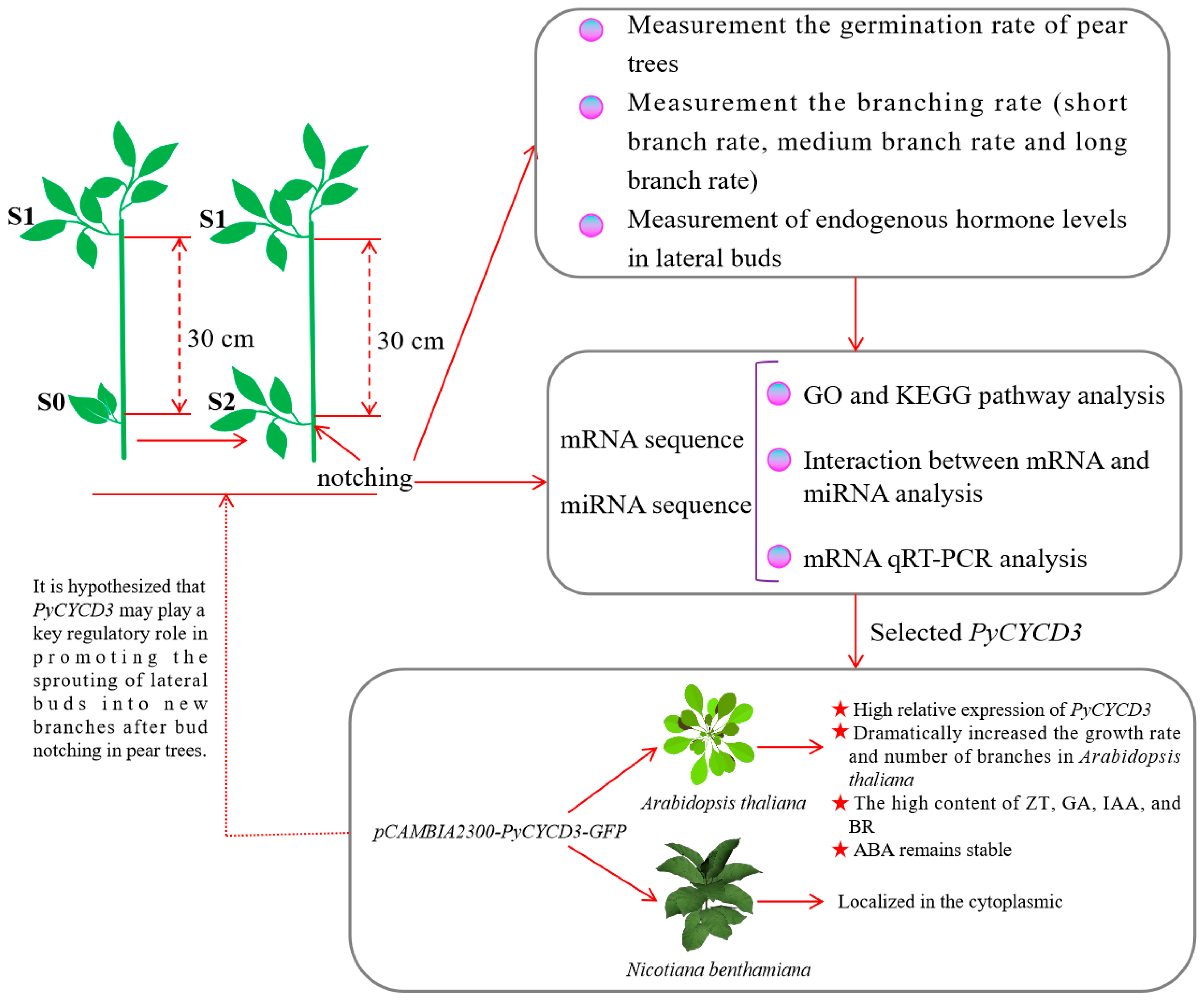
Figure 1. Simplified schematic diagram of the main research in this study.
Plants 13 02928 g001
Figure 2. Schematic diagram of sample collection. (a) S2 represents the third bud sample, which consists of lateral buds that can sprout normally after bud-notching. (b) S0 represents the first bud sample, which consists of lateral buds that can sprout normally after bud-notching. S1 represents the second bud sample, which consists of lateral buds that can sprout normally after bud-notching. (c) Illustrates either S1 or S2. (d) Represents S0.
Plants 13 02928 g002
Figure 3. Germination rate, branching rate, and endogenous hormone levels in lateral buds. (a) Sprouting rate of pear trees after bud-notching treatment. CK, control. (b) Comparison of branch formation rate between control and bud-notching treatment groups. Note: the test in Figure 3a,b is an independent samples t-test; p < 0.01 indicates a highly significant difference (**). (c) The endogenous content and ratio of hormones. Note: the hormone contents and the ratios of the hormone contents and ratios are the means of three independent samples. a, b, and c indicate a significant difference at the p < 0.05 level.
Plants 13 02928 g003
Figure 4. Differentially expressed mRNAs and miRNAs in both confrontations, C1 and C2. (a) The cluster heatmap of 492 DEGs at both confrontation C1 and C2. (b) The number of up-regulated (yellow, red) and down-regulated (green, blue) DEGs detected between C1 and C2. (c) The cluster heatmap of 11 DEGs at both confrontation C1 and C2. (d) The number of up-regulated (yellow, red) and down-regulated (green, blue) miRNAs detected between C1 and C2. (e) Pathway analysis of mRNA DEGs based on the KEGG database C1 and C2. (f) Pathway analysis of miRNA DEMs based on the KEGG database at C1 and C2. C1 is the ratio of S0_ vs. _S1, C1 is the ratio of S0_ vs. _S2.
Plants 13 02928 g004
Figure 5. Pathway analysis of mRNA DEGs and miRNA DEGs based on the KEGG database. (a) Pathway analysis of mRNA DEGs based on the KEGG database for Auxin, Gibberellin, Abscisic acid, Cytokinine, and Brassinosteroid. +p, phosphorylation; −p, dephosphorylation; +u, ubiquitination; p+, in a phosphorylated state. (b) A regulatory network of miRNA-mRNA related to pear shoot branching-related genes.
Plants 13 02928 g005
Figure 6. Real-time PCR validation of 12 DEGs in mRNA pairs. The x-axis represents RNA names, with the left y-axis represents the relative expression levels, and the right y-axis represents FPKM values. The red bars represent data yielded by qRT-PCR, while the blue star points represent data obtained by RNA sequencing; ‘r’ represents the Pearson correlation coefficient. ‘*’, correlation is significant at the 0.05 level. ‘**’, correlation is significant at the 0.01 level.
Plants 13 02928 g006
Figure 7. Overexpression PyCYCD3 increased lateral branch formation of transgenic Arabidopsis. (a) Schematic of the pCAMBIA2300-PyCYCD3-GFP fusion construct. (b) The phenotypic difference of WT and transgenic Arabidopsis thaliana. (c) Comparison of the number of outgrowths in WT and transgenic Arabidopsis thaliana. (d) The relative expression levels of PyCYCD3 in WT and transgenic lines. (e) Subcellular localization of PyCYCD3 gene in tobacco leaves. Bright field: indicates bright field of view; GFP: indicates target protein GFP signal. Bar = 50 μm. (f) The content of ZR, GA3, IAA, ABA, and BR in branches of WT and transgenic Arabidopsis. (g) The BR/ABA of hormones in branches of WT and transgenic Arabidopsis. The test in figure is an independent samples t-test; p < 0.05 indicates a significant difference (*), and p < 0.01 indicates a highly significant difference (**).
Plants 13 02928 g007
Disclaimer/Publisher’s Note: The statements, opinions and data contained in all publications are solely those of the individual author(s) and contributor(s) and not of MDPI and/or the editor(s). MDPI and/or the editor(s) disclaim responsibility for any injury to people or property resulting from any ideas, methods, instructions or products referred to in the content.