


In Vitro Collection for the Safe Storage of Grapevine Hybrids and Identification of the Presence of Plasmopara viticola Resistance Genes

 , , , , and
Abstract
1. Introduction
2. Results and Discussion
2.1. In Vitro Establishment of Grapevine Accessions
2.2. Screening for Endophytes Using Bacteriological Growth Media
2.3. In Vitro Multiplication
2.4. Presence of Plasmopara viticola Resistance Genes in Grape Accessions
3. Materials and Methods
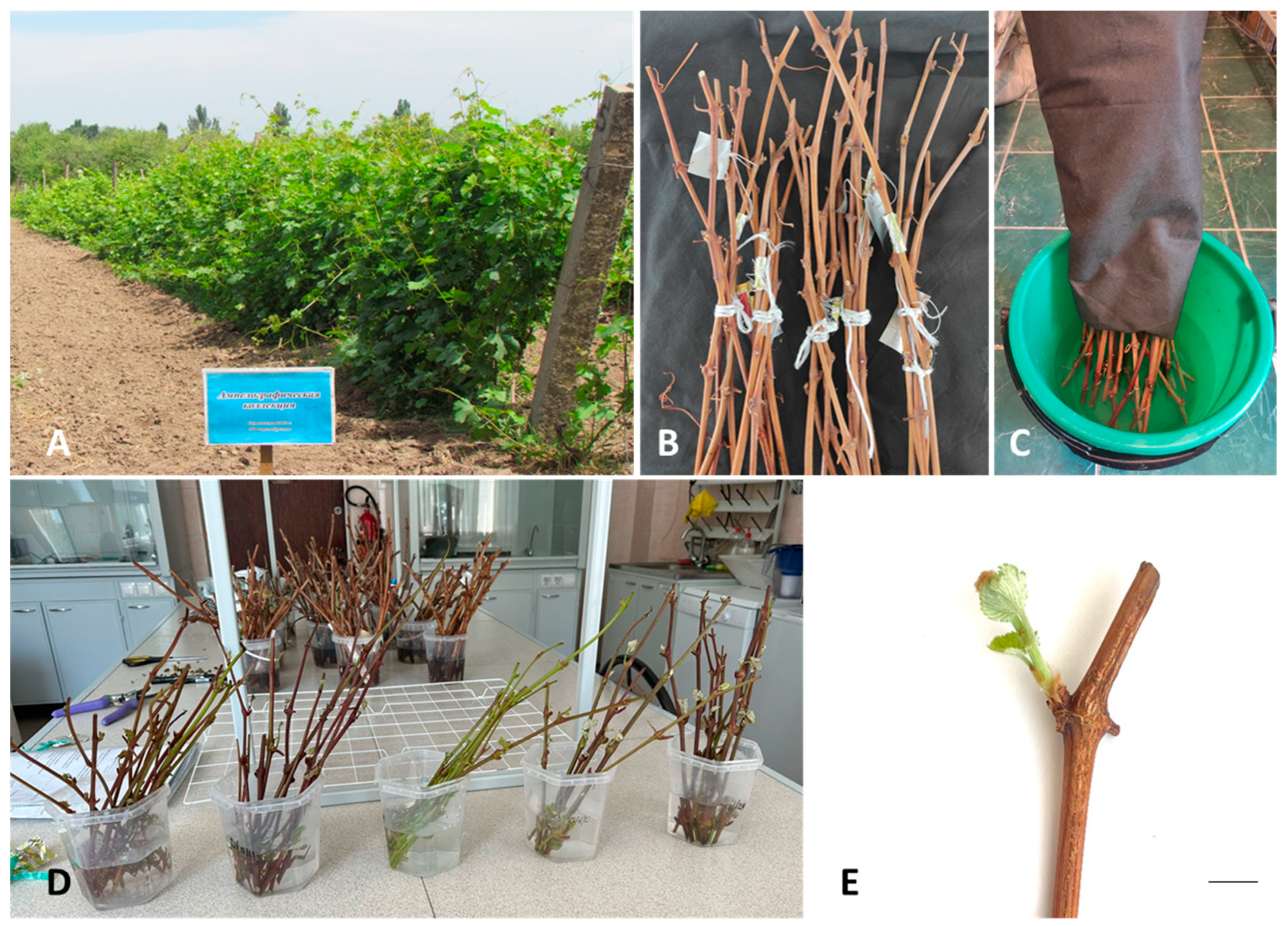
3.1. Plant Material
3.2. In Vitro Establishment and Maintenance of Grapevine Accessions
3.3. Screening for Endophytes Using Bacteriological Growth Media
3.4. In Vitro Multiplication
3.5. Testing Grape Hybrids for the Presence of Plasmopara viticola Resistance Genes
3.5.1. DNA Extraction
3.5.2. PCR Analysis
3.6. Statistical Analysis
4. Conclusions
Supplementary Materials
Author Contributions
Funding
Data Availability Statement
Acknowledgments
Conflicts of Interest
References
- OIV (International Organisation of Vine and Wine). World Statistics in 2022; International Organisation of Vine and Wine: Paris, France, 2020; Available online: https://www.oiv.int/what-we-do/global-report?oiv (accessed on 23 January 2024).
- This, P.; Lacombe, T.; Thomas, M.R. Historical origins and genetic diversity of wine grapes. Trends Genet. 2006, 22, 511–519. [Google Scholar] [CrossRef] [PubMed]
- Li, B.; Jiang, J.; Fan, X.; Zhang, Y.; Sun, H.; Zhang, G.; Liu, C. Molecular characterization of Chinese grape landraces (Vitis L.) using microsatellite DNA markers. HortScience 2017, 52, 533–540. [Google Scholar] [CrossRef]
- Smith, B.P.; Morales, N.B.; Thomas, M.R.; Smith, H.M.; Clingeleffer, P.R. Grapevine rootstocks resistant to the root-knot nematode Meloidogyne javanica. Aust. J. Grape Wine Res. 2017, 23, 125–131. [Google Scholar] [CrossRef]
- Buck, K.; Worthington, M. Genetic diversity of wild and cultivated Muscadine grapes (Vitis rotundifolia Michx.). Front. Plant Sci. 2022, 13, 852130. [Google Scholar] [CrossRef] [PubMed]
- Bettoni, J.C.; Marković, Z.; Bi, W.; Volk, G.M.; Matsumoto, T.; Wang, Q.-C. Grapevine shoot tip cryopreservation and cryotherapy: Secure storage of disease-free plants. Plants 2021, 10, 2190. [Google Scholar] [CrossRef] [PubMed]
- Carra, A.; Carimi, F.; Bettoni, J.C.; Pathirana, R. Progress and challenges in the application of synthetic seed technology for ex situ germplasm conservation in grapevine (Vitis spp.). In Synthetic Seeds; Faisal, A., Alatar, A.A., Eds.; Springer Nature: Cham, Switzerland, 2019; pp. 439–467. [Google Scholar] [CrossRef]
- Bettoni, J.; Kretzschmar, A.A.; Bonnart, R.; Shepherd, A.; Volk, G.M. Cryopreservation of 12 Vitis species using apical shoot tips derived from plants grown in vitro. HortScience 2019, 54, 976–981. [Google Scholar] [CrossRef]
- FAO (Food and Agriculture Organization of the United Nations). Crops and Livestock Products. Production in 2022. Available online: https://www.fao.org/faostat/en/#data/QCL (accessed on 23 January 2024).
- Chupradit, S.; Huy, D.T.N.; Hachem, K.; Shichiyakh, R.A.; Bokov, D.; Mahmudiono, T.; Al-Rekaby, H.Q.; Kadhim, M.M.; Thangavelu, L. Agrobiological evaluations of newly introduced grapes varieties under climatic conditions of the south of Kazakhstan. Braz. J. Biol. 2022, 84, e258275. [Google Scholar] [CrossRef]
- Syrgabek, Y.A.; Alimzhanova, M.; Amandykova, A.; Akylbekova, T.N. Soil fauna of vineyards of Southeast Kazakhstan. Soil Sci. Agrochem. 2020, 4, 57–65. [Google Scholar]
- Committee on Land Resources Management of the Ministry of Agriculture on the Condition and Use of Land for 2020. Available online: https://www.gov.kz/memleket/entities/land?lang=ru (accessed on 23 November 2023).
- Kulzhanov, S.N.; Kazybayeva, S.Z.; Tazhibaev, T.S.; Azhitaeva, L.A.; Yessenaliyeva, A. Effect of climatic conditions on the productive and biochemical characteristics of grape varieties grown on sierozem soil. Eurasian J. Soil Sci. 2022, 11, 174–183. [Google Scholar] [CrossRef]
- Yefremova, Y.M.; Urazayeva, M.V.; Kazybaeva, S.S. Rootstocks and varieties of fruits, berry crops, and grapes, used for intensive gardening in Kazakhstan. In Apple Cultivation—Recent Advances; Küden, A., Ed.; IntechOpen Limited: London, UK, 2023; pp. 1–22. [Google Scholar] [CrossRef]
- FAO (Food and Agriculture Organization of the United Nations). Crops and Livestock Products. Import and Export Quantity and Import Value in 2022. Available online: https://www.fao.org/faostat/en/#data/QCL (accessed on 23 January 2024).
- Kazybayeva, S.; Dolgikh, S.; Kulshanov, S.; Urazayeva, M.; Ushkempirova, G. The organization of the virus-tested planting material production for the grape varieties of the local and foreign selection in Kazakhstan. Bioengineering 2020, 25, 01002. [Google Scholar] [CrossRef]
- Pozharskiy, A.S.; Aubakirova, K.P.; Gritsenko, D.A.; Tlevlesov, N.I.; Karimov, N.Z.; Galiakparov, N.N.; Ryabushkina, N.A. Genotyping and morphometric analysis of Kazakhstani grapevine cultivars versus Asian and European cultivars. Genet. Mol. Res. 2020, 19, gmr18482. [Google Scholar] [CrossRef]
- Ministry of Ecology and Natural Resources of the Republic of Kazakhstan. The Fifth National Report on Progress in Implementation of the Convention on Biological Diversity. Available online: https://www.cbd.int/doc/world/kz/kz-nr-05-en.pdf (accessed on 24 January 2024).
- This, P.; Jung, A.; Boccacci, P.; Borrego, J.; Botta, R.; Costantini, L.; Crespan, M.; Dangl, G.S.; Eisenheld, C.; Ferreira-Monteiro, F.; et al. Development of a standard set of microsatellite reference alleles for identification of grape cultivars. Theor. Appl. Genet. 2004, 109, 1448–1458. [Google Scholar] [CrossRef]
- Sefc, K.M.; Lefort, F.; Grando, M.S.; Scott, K.D.; Steinkellner, H.; Thomas, M.R. Microsatellite markers for grapevine: A state of the art. In Molecular Biology & Biotechnology of the Grapevine; Roubelakis-Angelakis, K.A., Ed.; Springer: Amsterdam, The Netherlands, 2001; pp. 407–438. [Google Scholar] [CrossRef]
- Atack, A. New perspectives in grapevine (Vitis spp.) breeding. In Case Studies of Breeding Strategies in Major Plant Species; Wang, H., Ed.; IntechOpen Limited: London, UK, 2022; pp. 1–35. [Google Scholar] [CrossRef]
- Chung, P.-Y.; Liao, C.-T. Selection of parental lines for plant breeding via genomic prediction. Front. Plant Sci. 2022, 13, 934767. [Google Scholar] [CrossRef]
- López-Caamal, A.; Tovar-Sánchez, E. Genetic, morphological, and chemical patterns of plant hybridization. Rev. Chil. De Hist. Nat. 2014, 87, 16. [Google Scholar] [CrossRef]
- Egorov, E.A. Grape breeding is a key link in the development of the grapes and wine-making industry. Vavilovskii Zh. Genet. 2021, 25, 408–413. [Google Scholar] [CrossRef]
- Pérez-de-Castro, A.M.; Vilanova, S.; Cañizares, J.; Pascual, L.; Blanca, J.M.; Díez, M.J.; Prohens, J.; Picó, B. Application of genomic tools in plant breeding. Curr. Genomics 2012, 13, 179–195. [Google Scholar] [CrossRef]
- Koledenkova, K.; Esmaeel, Q.; Jacquard, C.; Nowak, J.; Clément, C.; Ait Barka, E. Plasmopara viticola the causal agent of downy mildew of grapevine: From its taxonomy to disease management. Front. Microbiol. 2022, 13, 889472. [Google Scholar] [CrossRef] [PubMed]
- Fröbel, S.; Zyprian, E. Colonization of different grapevine tissues by Plasmopara viticola—A histological study. Front. Plant Sci. 2019, 10, 951. [Google Scholar] [CrossRef] [PubMed]
- Gessler, C.; Pertot, I.; Perazzolli, M. Plasmopara viticola: A review of knowledge on downy mildew of grapevine and effective disease management. Phytopathol. Mediterr. 2011, 50, 3–44. [Google Scholar] [CrossRef]
- Gindro, K.; Alonso-Villaverde, V.; Voinesco, F.; Spring, J.-L.; Viret, O.; Dubuis, P.-H. Susceptibility to downy mildew in grape clusters: New microscopical and biochemical insights. Plant Physiol. Biochem. 2012, 52, 140–146. [Google Scholar] [CrossRef]
- Fawke, S.; Doumane, M.; Schornack, S. Oomycete interactions with plants: Infection strategies and resistance principles. Microbiol. Mol. Biol. Rev. 2015, 79, 263–280. [Google Scholar] [CrossRef] [PubMed]
- Bavaresco, L. Impact of grapevine breeding for disease resistance on the global wine industry. Acta Hortic. 2018, 1248, 7–14. [Google Scholar] [CrossRef]
- Díez-Navajas, A.M.; Wiedemann-Merdinoglu, S.; Greif, C.; Merdinoglu, D. Nonhost versus host resistance to the grapevine downy mildew, Plasmopara viticola, studied at the tissue level. Phytopathology 2008, 98, 776–780. [Google Scholar] [CrossRef]
- Cadle-Davidson, L. Variation within and between Vitis spp. for foliar resistance to the downy mildew pathogen Plasmopara viticola. Plant Dis. 2008, 92, 1577–1584. [Google Scholar] [CrossRef]
- Welter, L.J.; Göktürk-Baydar, N.; Akkurt, M.; Maul, E.; Eibach, R.; Töpfer, R.; Zyprian, E.M. Genetic mapping and localization of quantitative trait loci affecting fungal disease resistance and leaf morphology in grapevine (Vitis vinifera L.). Mol. Breed. 2007, 20, 359–374. [Google Scholar] [CrossRef]
- Venuti, S.; Copetti, D.; Foria, S.; Falginella, L.; Hoffmann, S.; Bellin, D.; Cindrić, P.; Kozma, P.; Scalabrin, S.; Morgante, M.; et al. Historical introgression of the downy mildew resistance gene Rpv12 from the Asian species Vitis amurensis into grapevine varieties. PLoS ONE 2013, 8, e61228. [Google Scholar] [CrossRef]
- Ilnitskaya, E.T.; Makarkina, M.V.; Tokmakov, S.V.; Naumova, L.G. DNA marker based identification of the Rpv3 gene determining downy mildew resistance in grapevines. Vavilovskii Zhurnal Genet. Selektsii. 2018, 22, 703–707. [Google Scholar] [CrossRef]
- Schiff, C.L.; Wilson, I.W.; Somerville, S.C. Polygenic powdery mildew disease resistance in Arabidopsis thaliana: Quantitative trait analysis of the accession Warschau-1. Plant Pathol. 2001, 50, 690–701. [Google Scholar] [CrossRef]
- Merdinouglu, D.; Scheider, C.; Prado, E.; Wiedemann-Merdinoglu, S.; Mestre, P. Breeding for durable resistance to downy and powdery mildew in grapevine. OENO One 2018, 52, 203–209. [Google Scholar] [CrossRef]
- Dalbó, M.A.; Ye, G.N.; Weeden, N.F.; Wilcox, W.F.; Reisch, B.I. Marker-assisted selection for powdery mildew resistance in grapes. J. Am. Soc. Hort. Sci. 2001, 126, 83–89. [Google Scholar] [CrossRef]
- Eibach, R.; Diehl, H.; Alleweldt, G. Investigations on the heritability of resistance to Oidium tuclceri, Pasmopara viticola and Botrytis cinerea in grapes. Vitis 1989, 28, 209–228. [Google Scholar] [CrossRef]
- Eibach, R.; Zyprian, E.; Welter, L.; Töpfer, R. The use of molecular markers for pyramiding resistance genes in grapevine breeding. Vitis 2007, 46, 120–124. [Google Scholar] [CrossRef]
- Saifert, L.; Sánchez-Mora, F.D.; Assumpção, W.T.; Zanghelini, J.A.; Giacometti, R.; Novak, E.I.; Dal Vesco, L.L.; Nodari, R.O.; Eibach, R.; Welter, L.J. Marker-assisted pyramiding of resistance loci to grape downy mildew. Pesqui. Agropecu. Bras. 2018, 53, 602–610. [Google Scholar] [CrossRef]
- Sánchez-Mora, F.D.; Saifert, L.; Zanghelini, J.; Assumpção, W.T.; Guginski-Piva, C.A.; Giacometti, R.; Novak, E.I.; Klabunde, G.H.; Eibach, R.; Dal Vesco, L.; et al. Behavior of grape breeding lines with distinct resistance alleles to downy mildew (Plasmopara viticola). Crop Breed. Appl. Biotechnol. 2017, 17, 141–149. [Google Scholar] [CrossRef]
- Foria, S.; Monte, C.; Testolin, R.; Di Gaspero, G.; Cipriani, G. Pyramidizing resistance genes in grape: A breeding program for the selection of elite cultivars. Acta Hortic. 2019, 1248, 549–554. [Google Scholar] [CrossRef]
- Collard, B.C.Y.; Jaufer, M.Z.Z.; Brouwer, J.B.; Pang, E.C.K. An introduction to markers, quantitative trait locus (QTL) mapping and marker-assisted selection for crop improvement: The basic concepts. Euphytica 2005, 142, 169–196. [Google Scholar] [CrossRef]
- Bhojwani, S.S.; Dantu, P.K. Micropropagation. In Plant Tissue Culture: An Introductory Text; Bhojwani, S.S., Dantu, P.K., Eds.; Springer: New Delhi, India, 2013; pp. 245–274. [Google Scholar] [CrossRef]
- Macedo, T.A.; Fagherazzi, A.; Bettoni, J.C.; Feldberg, N.P.; Kretzschmar, A.A. Propagação da Videira. In A Cultura da Videira: Vitivinicultura de Altitude; Rufato, L., Marcon Filho, J.L., Brighenti, A.F., Bogo, A., Kretzschmar, A.A., Eds.; UDESC: Florianópolis, Brazil, 2021; pp. 134–158. [Google Scholar] [CrossRef]
- Cassells, A.C. Pathogen and Biological Contamination Management in Plant Tissue Culture: Phytopathogens, Vitro Pathogens, and Vitro Pests. In Plant Cell Culture Protocols; Loyola-Vargas, V.M., Ochoa-Alejo, N., Eds.; Humana Press: Totowa, NJ, USA, 2012; pp. 57–80. [Google Scholar] [CrossRef]
- Ahloowalia, B.S. Tissue culture—Clonal Propagation, In Vitro. In Encyclopedia of Applied Plant Sciences; Thomas, B., Ed.; Academic Press: Cambridge, MA, USA, 2003; pp. 1360–1364. [Google Scholar] [CrossRef]
- Loberant, B.; Altman, A. Micropropagation of plants. In Encyclopedia of Industrial Biotechnology: Bioprocess, Bioseparation, and Cell Technology; Flickinger, M.C., Ed.; John Wiley & Sons, Inc.: Hoboken, NJ, USA, 2010; pp. 1–17. [Google Scholar] [CrossRef]
- Kaur, K.; Dolker, D.; Behera, S.; Pati, P.K. Critical factors influencing in vitro propagation and modulation of important secondary metabolites in Withania somnifera (L.) dunal. Plant Cell Tissue Organ. Cult. 2022, 149, 41–60. [Google Scholar] [CrossRef] [PubMed]
- Dobránszki, J.; Teixeira da Silva, J.A. Micropropagation of apple—A review. Biotechnol. Adv. 2010, 28, 462–488. [Google Scholar] [CrossRef] [PubMed]
- Bonnart, R.M.; Chen, K.Y.; Volk, G.M. Plant Tissue Culture Media Preparation. In Training in Plant Genetic Resources: Cryopreservation of Clonal Propagules; Volk, G.M., Ed.; Colorado State University: Fort Collins, CO, USA, 2022; Available online: https://colostate.pressbooks.pub/clonalcryopreservation/chapter/media/ (accessed on 11 March 2024).
- Cassells, A.C. Problems in tissue culture: Culture contamination. In Micropropagation—Technology and Application; Debergh, P.C., Zimmerman, R.H., Eds.; Kluwer Academic Publishers: Dordrecht, The Netherlands, 1991; pp. 31–44. [Google Scholar]
- Herman, E.B. Plant tissue culture contamination: Challenges and opportunities. Acta Hortic. 2017, 1155, 231–238. [Google Scholar] [CrossRef]
- Romadanova, N.V.; Tolegen, A.B.; Kushnarenko, S.V.; Zholdybayeva, E.V.; Bettoni, J.C. Effect of Plant Preservative MixtureTM on endophytic bacteria eradication from in vitro-grown apple shoots. Plants 2022, 11, 2624. [Google Scholar] [CrossRef]
- Orlikowska, T.; Nowak, K.; Reed, B. Bacteria in the plant tissue culture environment. Plant Cell Tissue Organ. Cult. 2016, 128, 487–508. [Google Scholar] [CrossRef]
- Gunson, H.E.; Spencer-Phillips, P.T.N. Latent bacterial infections: Epiphytes and endophytes as contaminants of micropropagated plants. In Physiology, Growth and Development of Plants in Culture; Lumsden, P.J., Nicholas, J.R., Davies, W.J., Eds.; Springer Nature: Cham, Switzerland, 1994; pp. 379–396. [Google Scholar] [CrossRef]
- Volk, G.M.; Bonnart, R.; Araújo de Oliveira, A.C.; Henk, A.D. Minimizing the deleterious effects of endophytes in plant shoot tip cryopreservation. Appl. Plant Sci. 2022, 10, e11489. [Google Scholar] [CrossRef] [PubMed]
- Ogbe, A.A.; Finnie, J.F.; Van Staden, J.V. The role of endophytes in secondary metabolites accumulation in medicinal plants under abiotic stress. S. Afr. J. Bot. 2020, 134, 126–134. [Google Scholar] [CrossRef]
- Kado, C.I.; Heskett, M.G. Selective media for isolation of Agrobacterium, Corynebacterium, Erwinia, Pseudomonas, and Xanthomonas. Phytopathology 1970, 60, 969–976. [Google Scholar] [CrossRef] [PubMed]
- Pasternak, T.P.; Steinmacher, D. Plant growth regulation in cell and tissue culture in vitro. Plants 2024, 13, 327. [Google Scholar] [CrossRef] [PubMed]
- Murashige, T.; Skoog, F. A revised medium for rapid growth and bioassays with tobacco tissue cultures. Physiol. Plant 1962, 15, 473–497. [Google Scholar] [CrossRef]
- Kim, S.-H.; Zebro, M.; Jang, D.-C.; Sim, J.-E.; Park, H.-K.; Kim, K.-Y.; Bae, H.-M.; Tilahun, S.; Park, S.-M. Optimization of Plant Growth Regulators for In Vitro Mass Propagation of a Disease-Free ‘Shine Muscat’ Grapevine Cultivar. Curr. Issues Mol. Biol. 2023, 45, 7721–7733. [Google Scholar] [CrossRef] [PubMed]
- Abido, A.I.A.; Sabah, M.A.M.A.; Hassanen, S.A.; Rayan, G.A. In vitro Propagation of Grapevine (Vitis vinifera L.) Muscat of Alexandria cv. For Conservation of Endangerment. Middle East J. Sci. Res. 2013, 13, 328–337. [Google Scholar] [CrossRef]
- Bettoni, J.C.; Bonnart, R.; Shepherd, A.; Kretzschmar, A.A.; Volk, G.M. Modifications to a Vitis shoot tip cryopreservation procedure: Effect of shoot tip size and use of cryoplates. Cryo Lett. 2019, 40, 103–112. [Google Scholar]
- Magyar-Tábori, K.; Dobránszki, J.; Teixeira da Silva, J.A.; Bulley, S.M.; Hudák, I. The role of cytokinins in shoot organogenesis in apple. Plant Cell Tissue Organ. Cult. 2010, 101, 251–267. [Google Scholar] [CrossRef]
- Sosnowski, J.; Truba, M.; Vasileva, V. The impact of auxin and cytokinin on the growth and development of selected crops. Agriculture 2023, 13, 724. [Google Scholar] [CrossRef]
- Souza, J.A.; Bettoni, J.C.; Dalla Costa, M.; Baldissera, T.C.; dos Passos, J.F.M.; Primieri, S. In vitro rooting and acclimatization of ‘Marubakaido’ apple rootstock using indole-3-acetic acid from rhizobacteria. Commun. Plant Sci. 2022, 12, 16–23. [Google Scholar] [CrossRef]
- Gupta, R.; Chakrabarty, S.K. Gibberellic acid in plant: Still a mystery unresolved. Plant Signal. Behav. 2013, 8, e25504. [Google Scholar] [CrossRef] [PubMed]
- Khan, N.; Ahmed, M.; Hafiz, I.; Abbasi, N.; Ejaz, S.; Anjum, M. Optimizing the concentrations of plant growth regulators for in vitro shoot cultures, callus induction and shoot regeneration from calluses of grapes. OENO One 2015, 49, 37–45. [Google Scholar] [CrossRef]
- Bettoni, J.C.; Costa, M.D.; Gardin, J.P.P.; Kretzschmar, A.A.; Souza, J.A.; Passos, J.F.M. Free culture media of growth regulators on micropropagation of grapevine (Vitis labrusca L.) ‘Bordô’ cultivar through nodal segments. Evidência 2016, 16, 59–70. [Google Scholar] [CrossRef][Green Version]
- Huh, Y.S.; Lee, J.K.; Kim, I.J.; Kang, B.G.; Lee, K.Y. Effect of biocide addition on plantlet growth and contamination occurrence during the in vitro culture of blueberry. J. Plant Biotechnol. 2015, 42, 111–116. [Google Scholar] [CrossRef]
- Bettoni, J.C.; Bonnart, R.; Shepherd, A.; Kretzschmar, A.; Volk, G.M. Cryopreservation of grapevine (Vitis spp.) shoot tips from growth chamber-sourced plants and histological observations. Vitis 2019, 58, 71–78. [Google Scholar] [CrossRef]
- Orlikowska, T.; Zawadzka, M.; Zenkteler, E.; Sobiczewski, P. Influence of the biocides PPMTM and vitrofural on bacteria isolated from contaminated plant tissue cultures and on plant microshoots grown on various media. J. Hortic. Sci. Biotechnol. 2015, 87, 223–230. [Google Scholar] [CrossRef]
- Kushnarenko, S.; Aralbayeva, M.; Rymkhanova, N.; Reed, B.M. Initiation pretreatment with Plant Preservative MixtureTM increases the percentage of aseptic walnut shoots. In Vitro Cell. Dev. Biol.-Plant 2022, 58, 964–971. [Google Scholar] [CrossRef]
- Paul, A.L.; Semer, C.; Kucharek, T.; Ferl, R.J. The fungicidal and phytotoxic properties of benomyl and PPM in supplemented agar media supporting transgenic arabidopsis plants for a Space Shuttle flight experiment. Appl. Microbiol. Biotechnol. 2001, 55, 480–485. [Google Scholar] [CrossRef]
- Ostrolucká, M.G.; Gajdošová, A.; Ondrušková, E.; Lateèková, M.; Libiaková, G. Effect of medium pH on axillary shoot proliferation of selected Vaccinium vitis-idaea L. cultivars. Acta Biol. Cracov. 2010, 52, 92–96. [Google Scholar] [CrossRef]
- Ramage, C.M.; Williams, R.R. Mineral nutrition and plant morphogenesis. In Vitro Cell. Dev. Biol.-Plant. 2002, 38, 116–124. [Google Scholar] [CrossRef]
- Chen, C.-C.; Bates, R.; Carlson, J. Effect of environmental and cultural conditions on medium pH and explant growth performance of Douglas-fir (Pseudotsuga menziesii) shoot cultures. F1000Research 2014, 3, 298. [Google Scholar] [CrossRef] [PubMed]
- Guo, G.; Xiao, J.; Jeong, B.R. Iron source and medium pH affect nutrient uptake and pigment content in Petunia hybrida ‘Madness Red’ cultured in vitro. Int. J. Mol. Sci. 2022, 23, 8943. [Google Scholar] [CrossRef] [PubMed]
- Sági-Kazár, M.; Solymosi, K.; Solti, Á. Iron in leaves: Chemical forms, signalling, and in-cell distribution. J. Exp. Bot. 2022, 73, 1717–1734. [Google Scholar] [CrossRef] [PubMed]
- Laulhere, J.P.; Briat, J.F. Iron release and uptake by plant ferritin: Effects of pH, reduction and chelation. Biochem. J. 1993, 290, 693–699. [Google Scholar] [CrossRef] [PubMed]
- Wong, K.-Y.I. In Vitro Culture of ‘Dog Ridge’ Grapevine. Senior Scholars Thesis, Texas A&M University, College Station, TX, USA, 2009. [Google Scholar]
- Kryukov, L.A.; Vodolazhsky, D.I.; Kamenetsky-Goldstein, R. Micropropagation of grapevine and strawberry from south Russia: Rapid production and genetic uniformity. Agronomy 2022, 12, 308. [Google Scholar] [CrossRef]
- Bettoni, J.C.; Costa, M.D.; Gardin, J.P.P.; Kretzschmar, A.A.; Souza, J.A. In vitro propagation of grapevine cultivars with potential for the south of Brazil. Am. J. Plant Sci. 2015, 6, 1806–1815. [Google Scholar] [CrossRef]
- Capriotti, L.; Ricci, A.; Molesini, B.; Mezzetti, B.; Pandolfini, T.; Piunti, I.; Sabbadini, S. Efficient protocol of de novo shoot organogenesis from somatic embryos for grapevine genetic transformation. Front. Plant Sci. 2023, 14, 1172758. [Google Scholar] [CrossRef]
- Bettoni, J.C.; Bonnart, R.; Volk, G.M. Challenges in implementing plant shoot tip cryopreservation technologies. Plant Cell Tissue Organ. Cult. 2021, 144, 21–34. [Google Scholar] [CrossRef]
- Bettoni, J.C.; Chen, K.; Volk, G.M. An Overview of Shoot Tip Cryopreservation. In Training in Plant Genetic Resources: Cryo-preservation of Clonal Propagules; Volk, G.M., Ed.; Colorado State University: Fort Collins, CO, USA, 2024; Available online: https://colostate.pressbooks.pub/clonalcryopreservation/chapter/an-overview-of-shoot-tip-cryopreservation/ (accessed on 13 March 2024).
- Romadanova, N.V.; Mishustina, S.A.; Matakova, G.N.; Kuhsnarenko, S.V.; Rakhimbaev, I.R.; Reed, B.M. In vitro collection of Malus shoot cultures for cryogenic bank development in Kazakhstan. Acta Hortic. 2016, 1113, 271–277. [Google Scholar] [CrossRef]
- Romadanova, N.V.; Kushnarenko, S.V. Conservation of plant biodiversity by biotechnology methods. Proc. Appl. Bot. Genet. Breed. 2023, 184, 239–248. [Google Scholar] [CrossRef]
- Kulak, V.; Longboat, S.; Brunet, N.D.; Shukla, M.; Saxena, P. In vitro technology in plant conservation: Relevance to biocultural diversity. Plants 2022, 11, 503. [Google Scholar] [CrossRef] [PubMed]
- Volk, G.M.; Walters, C. Preservation of genetic resources in the national plant germplasm clonal collections. Plant. Breed. Rev. 2003, 23, 291–344. [Google Scholar] [CrossRef]
- Wang, M.R.; Chen, L.; Teixeira da Silva, J.A.; Volk, G.M.; Wang, Q.C. Cryobiotechnology of apple (Malus spp.): Development, progress and future prospects. Plant Cell Rep. 2018, 37, 689–709. [Google Scholar] [CrossRef] [PubMed]
- Volk, G.M.; Shepherd, A.N.; Bonnart, R. Successful cryopreservation of Vitis shoot tips: Novel pre-treatment combinations applied to nine species. Cryo Lett. 2018, 39, 322–330. [Google Scholar]
- Peng, J.; Wang, X.; Wang, H.; Li, X.; Zhang, Q.; Wang, M.; Yan, J. Advances in understanding grapevine downy mildew: From pathogen infection to disease management. Mol. Plant Pathol. 2024, 25, e13401. [Google Scholar] [CrossRef] [PubMed]
- Yu, Y.; Zhang, Y.; Yin, L.; Lu, J. The mode of host resistance to Plasmopara viticola infection of grapevines. Phytopathology 2012, 102, 1094–1101. [Google Scholar] [CrossRef] [PubMed]
- Di Gaspero, G.; Copetti, D.; Coleman, C.; Castellarin, S.D.; Eibach, R.; Kozma, P.; Lacombe, T.; Gambetta, G.; Zvyagin, A.; Cindric, P.; et al. Selective sweep at the Rpv3 locus during grapevine breeding for downy mildew resistance. Theor. Appl. Genet. 2012, 124, 277–286. [Google Scholar] [CrossRef]
- Marone Fassolo, E.; Lecchi, B.; Marcianò, D.; Maddalena, G.; Toffolatti, S.L. Pathogen Adaptation to American (Rpv3-1) and Eurasian (Rpv29) Grapevine Loci Conferring Resistance to Downy Mildew. Plants 2022, 11, 2619. [Google Scholar] [CrossRef]
- Frommer, B.; Müllner, S.; Holtgräwe, D.; Viehöver, P.; Huettel, B.; Töpfer, R.; Weisshaar, B.; Zyprian, E. Phased grapevine genome sequence of an Rpv12 carrier for biotechnological exploration of resistance to Plasmopara viticola. Front. Plant Sci. 2023, 14, 1180982. [Google Scholar] [CrossRef] [PubMed]
- Foria, S.; Magris, G.; Jurman, I.; Schwope, R.; De Candido, M.; De Luca, E.; Ivanišević, D.; Morgante, M.; Di Gaspero, G. Extent of wild-to-crop interspecific introgression in grapevine (Vitis vinifera) as a consequence of resistance breeding and implications for the crop species definition. Horticult. Res. 2022, 9, uhab010. [Google Scholar] [CrossRef] [PubMed]
- Ricciardi, V.; Crespan, M.; Maddalena, G.; Migliaro, D.; Brancadoro, L.; Maghradze, D.; Failla, O.; Toffolatti, S.L.; De Lorenzis, G. Novel loci associated with resistance to downy and powdery mildew in grapevine. Front. Plant Sci. 2024, 15, 1386225. [Google Scholar] [CrossRef] [PubMed]
- Sakiroglu, M.; Brummer, E.C. Identification of loci controlling forage yield and nutritive value in diploid alfalfa using GBS-GWAS. Theor. Appl. Genet. 2017, 130, 261–268. [Google Scholar] [CrossRef] [PubMed]
- Delmotte, F.; Mestre, P.; Schneider, C.; Kassemeyer, H.H.; Kozma, P.; Richart-Cervera, S.; Rouxel, M.; Delière, L. Rapid and multiregional adaptation to host partial resistance in a plant pathogenic oomycete: Evidence from European populations of Plasmopara viticola, the causal agent of grapevine downy mildew. Infect. Genet. Evol. 2014, 27, 500–508. [Google Scholar] [CrossRef] [PubMed]
- Dinglasan, E.; Periyannan, S.; Hickey, L.T. Harnessing adult-plant resistance genes to deploy durable disease resistance in crops. Essays Biochem. 2022, 66, 571–580. [Google Scholar] [CrossRef] [PubMed]
- Zhao, Y.; Zhu, X.; Chen, X.; Zhou, J.-M. From plant immunity to crop disease resistance. J. Genet. Genom. 2022, 49, 693–703. [Google Scholar] [CrossRef] [PubMed]
- Zini, E.; Dolzani, C.; Stefanini, M.; Gratl, V.; Beltinelli, P.; Nicolini, D.; Betta, G.; Dorigatti, C.; Velasco, R.; Letschka, T.; et al. R-Loci Arrangement Versus Downy and PowderyMildew Resistance Level: A Vitis Hybrid Survey. Int. J. Mol. Sci. 2019, 20, 3526. [Google Scholar] [CrossRef] [PubMed]
- Schwander, F.; Eibach, R.; Fechter, I.; Hausmann, L.; Zyprian, E.; Töpfer, R. Rpv10: A new locus from the Asian Vitis gene pool for pyramiding downy mildew resistance loci in grapevine. Theor. Appl. Genet. 2012, 124, 163–176. [Google Scholar] [CrossRef]
- Wingerter, C.; Eisenmann, B.; Weber, P.; Dry, I.; Bogs, J. Grapevine Rpv3-, Rpv10- and Rpv12-mediated defense responses against Plasmopara viticola and the impact of their deployment on fungicide use in viticulture. BMC Plant Biol. 2021, 21, 470. [Google Scholar] [CrossRef]
- Rybakov, A.A.; Ostroukhova, S.A.; Gorbach, V.I. Viticulture: Allowance for Agricultural Works Institute, 2nd ed.; Ukituvchi: Tashkent, Uzbekistan, 1975; p. 136. (In Russian) [Google Scholar]
- Plant Preservative Mixture. Available online: https://www.plantcelltechnology.com/plant-preservative-mixture-ppm-1/ (accessed on 3 April 2024).
- Doyle, J.J.; Doyle, J.L. A rapid DNA isolation procedure for small quantities of fresh leaf tissue. Phytochem. Bull. 1987, 19, 11–15. [Google Scholar]
- Valsesia, G.; Gobbin, D.; Patocchi, A.; Vecchione, A.; Pertot, I.; Gessler, C. Development of a high-throughput method for quantification of Plasmopara viticola DNA in grapevine leaves by means of quantitative real-time polymerase chain reaction. Phytopathology 2005, 95, 672–678. [Google Scholar] [CrossRef] [PubMed]
- SYSTAT. SYSTAT 12.0, Statistics Software; SYSTAT Software, Inc.: San Jose, CA, USA, 2007; Available online: https://systatsoftware.com/ (accessed on 3 January 2024).


| Plant Accession | HgCl2 Exposure Duration (min) | |||||||||||||||||
|---|---|---|---|---|---|---|---|---|---|---|---|---|---|---|---|---|---|---|
| 5 | 7 | 10 | ||||||||||||||||
| Infected | Necrotic | Viable | Infected | Necrotic | Viable | Infected | Necrotic | Viable | ||||||||||
| IV-4/74 | 65 ± 9 | ab | 5 ± 5 | d | 30 ± 17 | bc | 70 ± 17 | ab | 10 ± 5 | cd | 20 ± 10 | c | 35 ± 13 | bc | 50 ± 18 | b | 15 ± 9 | cd |
| X-13/90 | 55 ± 14 | b | 20 ± 17 | cd | 25 ± 20 | bc | 65 ± 9 | ab | 20 ± 18 | c | 15 ± 9 | cd | 56 ± 14 | b | 44 ± 10 | bc | 0 ± 0 | d |
| IV-6/9 | 87 ± 5 | ab | 7 ± 3 | d | 7 ± 3 | d | 60 ± 19 | ab | 15 ± 17 | cd | 25 ± 20 | bc | 50 ± 14 | b | 50 ± 14 | b | 0 ± 0 | d |
| IV-14/9 | 50 ± 14 | b | 5 ± 5 | d | 5 ± 5 | d | 65 ± 11 | ab | 15 ± 17 | cd | 20 ± 18 | c | 50 ± 14 | b | 45 ± 13 | bc | 5 ± 5 | d |
| V-7/17 | 75 ± 17 | ab | 15 ± 9 | cd | 10 ± 5 | cd | 70 ± 17 | ab | 20 ± 17 | bc | 10 ± 0 | cd | 40 ± 9 | bc | 60 ± 14 | b | 0 ± 0 | d |
| DV-10/11 | 70 ± 12 | a | 5 ± 5 | d | 25 ± 10 | bc | 35 ± 13 | bc | 25 ± 10 | bc | 40 ± 9 | b | 30 ± 22 | bc | 40 ± 5 | b | 30 ± 14 | bc |
| KV-2/35 | 80 ± 17 | ab | 0 ± 0 | d | 20 ± 18 | c | 70 ± 10 | ab | 20 ± 10 | cd | 10 ± 9 | cd | 45 ± 13 | bc | 50 ± 14 | b | 5 ± 5 | d |
| III-02/22 | 65 ± 9 | ab | 5 ± 5 | d | 30 ± 17 | bc | 50 ± 14 | b | 30 ± 17 | bc | 20 ± 18 | c | 37 ± 15 | bc | 53 ± 12 | b | 10 ± 4 | cd |
| KV-2/9 | 80 ± 17 | ab | 20 ± 18 | cd | 0 ± 0 | d | 75 ±14 | ab | 10 ± 4 | cd | 15 ± 9 | cd | 55 ± 14 | b | 45 ± 14 | bc | 0 ± 0 | d |
| III-7/15 | 70 ± 14 | ab | 10 ± 4 | cd | 20 ± 12 | c | 65 ±13 | ab | 20 ± 10 | c | 15 ± 10 | cd | 35 ± 14 | bc | 60 ± 14 | b | 5 ± 5 | d |
| KV-1/10 | 80 ± 14 | ab | 7 ± 3 | c | 13 ± 9 | cd | 60 ± 15 | b | 20 ± 10 | c | 20 ± 10 | c | 40 ± 10 | bc | 55 ± 13 | b | 5 ± 5 | d |
| KII-1/29 | 87 ± 5 | a | 0 ± 0 | d | 13 ± 13 | cd | 70 ± 8 | a | 5 ± 5 | d | 25 ± 16 | bc | 35 ± 14 | bc | 50 ± 14 | b | 15 ± 9 | cd |
| VIII-3/45 | 90 ± 12 | a | 0 ± 0 | d | 10 ± 9 | cd | 60 ± 23 | ab | 20 ± 8 | cd | 20 ± 17 | c | 35 ± 13 | bc | 60 ± 15 | b | 5 ± 5 | d |
| V-7/9 | 90 ± 7 | a | 10 ± 9 | cd | 0 ± 0 | d | 60 ± 15 | b | 0 ± 0 | d | 40 ± 5 | b | 50 ± 14 | b | 45 ± 22 | bc | 5 ± 5 | d |
| XII-17/2 | 60 ± 5 | b | 10 ± 17 | cd | 25 ± 15 | bc | 60 ± 12 | b | 35 ± 13 | bc | 5 ± 5 | d | 32 ± 16 | bc | 58 ± 15 | b | 10 ± 4 | cd |
| XII-9/3 | 60 ± 15 | b | 10 ± 5 | c | 30 ± 17 | bc | 50 ± 14 | b | 15 ± 9 | cd | 35 ± 9 | bc | 25 ± 20 | bc | 45 ± 14 | bc | 30 ± 17 | bc |
| VII-6/72 | 90 ± 8 | a | 0 ± 0 | d | 10 ± 9 | cd | 45 ± 10 | bc | 45 ± 10 | bc | 10 ± 9 | cd | 45 ± 13 | bc | 55 ± 13 | b | 0 ± 0 | d |
| KIV-1/64 | 75 ± 15 | ab | 10 ± 6 | cd | 15 ± 9 | cd | 65 ± 9 | ab | 10 ± 4 | cd | 25 ± 20 | bc | 50 ± 14 | b | 45 ± 13 | bc | 5 ± 5 | d |
| Mean | 75 ± 12 | 8 ± 6 | 17 ± 11 | 61 ± 10 | 18 ± 11 | 21 ± 11 | 41 ± 9 | 51 ± 6 | 8 ± 8 | |||||||||
| Plant Accession | HgCl2 Exposure Duration (min) | |||||||||||||||||
|---|---|---|---|---|---|---|---|---|---|---|---|---|---|---|---|---|---|---|
| 5 | 7 | 10 | ||||||||||||||||
| Infected | Necrotic | Viable | Infected | Necrotic | Viable | Infected | Necrotic | Viable | ||||||||||
| IV-4/74 | 60 ± 19 | ab | 0 ± 0 | d | 40 ± 10 | bc | 30 ± 21 | bc | 60 ± 15 | b | 10 ± 9 | cd | 20 ± 17 | c | 80 ± 11 | a | 0 ± 0 | d |
| X-13/90 | 50 ± 14 | b | 20 ± 18 | c | 30 ± 14 | bc | 25 ± 20 | bc | 67 ± 17 | ab | 8 ± 5 | cd | 9 ± 6 | cd | 82 ± 12 | a | 9 ± 6 | cd |
| IV-6/9 | 50 ± 14 | b | 0 ± 0 | d | 10 ± 10 | cd | 30 ±22 | bc | 80 ± 12 | a | 0 ± 0 | d | 0 ± 0 | d | 100 ± 0 | a | 0 ± 0 | d |
| IV-14/9 | 80 ± 17 | ab | 10 ± 12 | cd | 10 ± 12 | cd | 30 ±22 | bc | 70 ± 10 | ab | 0 ± 0 | d | 0 ± 0 | d | 90 ± 16 | a | 10 ± 14 | cd |
| V-7/17 | 40 ± 9 | bc | 20 ± 18 | c | 40 ± 21 | bc | 8 ± 7 | cd | 83 ± 14 | a | 8 ± 11 | cd | 8 ± 8 | cd | 83 ± 4 | a | 8 ± 8 | cd |
| DV-10/11 | 40 ± 5 | bc | 10 ± 3 | cd | 50 ± 21 | b | 0 ± 0 | d | 60 ± 10 | b | 40 ± 23 | bc | 0 ± 0 | d | 83 ± 14 | a | 17 ± 7 | cd |
| KV-2/35 | 30 ± 14 | bc | 30 ± 14 | bc | 40 ± 22 | bc | 8 ± 13 | cd | 80 ± 16 | a | 15 ± 7 | cd | 0 ± 0 | d | 91 ± 17 | a | 9 ± 15 | cd |
| III-02/22 | 40 ± 20 | bc | 10 ± 12 | cd | 50 ± 20 | b | 0 ± 0 | d | 58 ± 24 | b | 42 ± 18 | bc | 0 ± 0 | d | 83 ± 14 | a | 8 ± 7 | cd |
| KV-2/9 | 65 ± 16 | ab | 35 ± 21 | bc | 0 ± 0 | d | 14 ± 17 | cd | 86 ± 14 | a | 0 ± 0 | d | 10 ± 16 | cd | 90 ± 17 | a | 0 ± 0 | d |
| III-7/15 | 50 ± 14 | b | 10 ± 10 | cd | 40 ± 22 | bc | 20 ± 13 | c | 67 ± 20 | ab | 13 ± 7 | cd | 8 ± 8 | cd | 92 ± 9 | a | 0 ± 0 | d |
| KV-1/10 | 90 ±17 | a | 10 ± 8 | cd | 0 ± 0 | d | 20 ± 10 | c | 80 ± 12 | a | 0 ± 0 | d | 10 ± 3 | cd | 90 ± 16 | a | 0 ± 0 | d |
| KII-1/29 | 75 ± 12 | a | 0 ± 0 | d | 25 ± 20 | bc | 33 ± 11 | bc | 50 ± 20 | b | 17 ± 8 | cd | 8 ± 6 | cd | 83 ± 12 | a | 8 ± 8 | cd |
| VIII-3/45 | 80 ± 12 | a | 0 ± 0 | d | 20 ± 13 | c | 14 ± 17 | cd | 79 ± 10 | a | 7 ± 6 | cd | 0 ± 0 | d | 92 ± 19 | a | 8 ± 6 | cd |
| V-7/9 | 100 ± 0 | a | 0 ± 0 | d | 0 ± 0 | d | 20 ± 18 | c | 70 ± 13 | ab | 10 ± 9 | cd | 0 ± 0 | d | 90 ± 17 | a | 10 ± 5 | cd |
| XII-17/2 | 57 ± 14 | b | 14 ± 8 | cd | 29 ± 13 | bc | 0 ± 0 | d | 79 ± 20 | a | 21 ± 7 | c | 0 ± 0 | d | 92 ± 14 | a | 8 ±12 | cd |
| XII-9/3 | 50 ± 9 | b | 10 ± 7 | cd | 40 ± 18 | bc | 10 ± 10 | cd | 60 ± 15 | b | 30 ± 20 | bc | 9 ± 9 | cd | 64 ± 17 | ab | 27 ± 20 | bc |
| VII-6/72 | 100 ± 0 | a | 0 ± 0 | d | 0 ± 0 | d | 26 ± 5 | bc | 75 ± 22 | a | 0 ± 0 | d | 8 ± 8 | cd | 92 ± 14 | a | 0 ± 0 | d |
| KIV-1/64 | 90 ± 10 | a | 0 ± 0 | d | 10 ± 10 | cd | 30 ± 14 | bc | 70 ± 12 | ab | 0 ± 0 | d | 10 ± 10 | cd | 90 ± 17 | a | 0 ± 0 | d |
| Mean | 65 ± 22 | 10 ± 10 | 25 ± 18 | 17 ± 11 | 71 ± 10 | 12 ± 11 | 6 ± 5 | 87 ± 7 | 7 ± 5 | |||||||||
| Plant Accession | Shoots | N | |||
|---|---|---|---|---|---|
| Infected (%) | Clean (%) | ||||
| IV-4/74 | 50 ± 9 | b | 50 ± 14 | b | 28 |
| X-13/90 | 59 ± 14 | b | 41 ± 6 | b | 27 |
| IV-6/9 | 45 ± 17 | bc | 55 ± 24 | b | 20 |
| IV-14/9 | 17 ± 7 | cd | 83 ± 18 | ab | 24 |
| V-7/17 | 39 ± 14 | bc | 61 ± 14 | b | 18 |
| DV-10/11 | 15 ± 8 | cd | 85 ± 15 | a | 30 |
| KV-2/35 | 10 ± 6. | cd | 90 ± 7 | a | 38 |
| III-02/22 | 41 ± 12 | bc | 59 ± 14 | b | 30 |
| KV-2/9 | 33 ± 14 | bc | 67 ± 10 | ab | 19 |
| III-7/15 | 67 ± 14 | ab | 33 ± 13 | bc | 27 |
| KV-1/10 | 56 ± 16 | b | 44 ± 12 | bc | 16 |
| KII-1/29 | 50 ± 12 | b | 50 ± 18 | b | 26 |
| VIII-3/45 | 44 ± 17 | bc | 56 ± 14 | b | 18 |
| V-7/9 | 60 ± 14 | ab | 40 ± 5 | b | 20 |
| XII-17/2 | 64 ± 13 | ab | 36 ± 8 | bc | 25 |
| XII-9/3 | 27 ± 14 | bc | 73 ± 6 | ab | 27 |
| VII-6/72 | 73 ± 12 | ab | 27 ± 20 | bc | 15 |
| KIV-1/64 | 63 ± 14 | ab | 37 ± 15 | bc | 16 |
| Mean | 45 ± 12 | 55 ± 11 | |||
| Culture Media | DV-10/11 | KV-2/35 | ||||||
|---|---|---|---|---|---|---|---|---|
| Number of Shoots | Multiplication Rate | Number of Shoots | Multiplication Rate | |||||
| Initial | Regenerated | Initial | Regenerated | |||||
| 1 | 15 | 50 | 3.3 ± 0.3 | bc | 15 | 51 | 3.4 ± 0.2 | bc |
| 2 | 15 | 58 | 3.9 ± 0.3 | ab | 15 | 58 | 3.9 ± 0.3 | ab |
| 3 | 15 | 53 | 3.5 ± 0.2 | bc | 15 | 54 | 3.6 ± 0.3 | b |
| 4 | 15 | 54 | 3.6 ± 0.3 | b | 15 | 53 | 3.5 ± 0.3 | bc |
| 5 | 15 | 45 | 3.0 ± 0.2 | c | 15 | 47 | 3.1 ± 0.2 | c |
| 6 | 15 | 39 | 2.6 ± 0.4 | dc | 15 | 30 | 2.0 ± 0.4 | d |
| 7 | 15 | 66 | 4.4 ± 0.3 | a | 15 | 62 | 4.1 ± 0.3 | a |
| 8 | 15 | 60 | 4.0 ± 0.1 | ab | 15 | 59 | 3.9 ± 0.1 | ab |
| 9 | 15 | 57 | 3.8 ± 0.1 | ab | 15 | 57 | 3.8 ± 0.1 | ab |
| 10 | 15 | 57 | 3.8 ± 0.2 | ab | 15 | 58 | 3.9 ± 0.2 | ab |
| Mean | 15 | 54 | 3.6 ± 0.3 | 15 | 53 | 3.5 ± 0.2 | ||
| Plant Accession | Resistance Loci (Associated Markers) | ||
|---|---|---|---|
| Rpv3 (UDV-737) | Rpv3 (UDV-305) | Rpv12 (UDV-343) | |
| KV-2/9 | + | ||
| DV-10/11 | + | + | |
| IV-6/9 | + | ||
| KII-1/29 | + | + | |
| KVI-1/10 | + | + | |
| XII-17/2 | + | + | |
| VII-6/72 | + | ||
| VII-3/15 | + | ||
| XII-9/3 | + | ||
| XI-14/9 | + | ||
| III-7/15 | + | ||
| III-02/22 | + | + | + |
| V-7/9 | + | + | |
| XI-13/90 | + | + | |
| KIY-1/64 | + | ||
| KV-2/35 | |||
| IV-4/74 | + | ||
| Breed Product | Female Parent | Male Parent | ||
|---|---|---|---|---|
| Name | Species | Name | Species | |
| KV-2/9 | Nimrang Magaracha | Hybrid of Vitis berlandieri, V. rupestris and V. vinifera | Cardinal | V. vinifera |
| DV-10/11 | Madeleine Angevine | V. vinifera | Taifi rozovyy | V. vinifera |
| IV-6/9 | Nimrang Magaracha | Hybrid of V. berlandieri, V. rupestris and V. vinifera | Muscat Almatinskiy | V. vinifera |
| KII-1/29 | Nimrang Magaracha | Hybrid of V. berlandieri, V. rupestris and V. vinifera | Cardinal | V. vinifera |
| KVI-1/10 | Nimrang Magaracha | Hybrid of V. berlandieri, V. rupestris and V. vinifera | Cardinal | V. vinifera |
| XII-17/2 | Donskoy Skorospelyy | Hybrid of V. vinifera and V. amurensis | Pobeda | V. vinifera |
| VII-6/72 | Nimrang Magaracha | Hybrid of V. berlandieri, V. rupestris and V. vinifera | Zolotistyy ranniy (Irsai Oliver) | V. vinifera |
| VII-3/15 | Muscat fleur d‘Orange | V. vinifera linné subsp. Sativa (De Candolle) Hegi | Detskiy ranniy | V. vinifera |
| XII-9/3 | Madeleine Angevine | V. vinifera | Pobeda | V. vinifera |
| XI-14/9 | Madeleine Angevine | V. vinifera | Pobeda | V. vinifera |
| III-7/15 | I-4|58 | V. vinifera | Golden Chasselas + Riesling | V. vinifera |
| III-02/22 | Donskoy Skorospelyy | Hybrid of V. vinifera and V. amurensis | Königin der weingarten | V. vinifera subsp. vinifera |
| V-7/9 | No154 | V. vinifera | Muscat Aleksandriyskiy | V. vinifera |
| XI-13/90 | Madeleine Angevine | V. vinifera | Tarnau | V. vinifera |
| KIY-1/64 | Nimrang Magaracha | Hybrid of V. berlandieri, V. rupestris and V. vinifera | Cardinal | V. vinifera |
| KV-2/35 | Nimrang Magaracha | Hybrid of V. berlandieri, V. rupestris and V. vinifera | Cardinal | V. vinifera |
| IV-4/74 | Pobeda | V. vinifera | Zhemchug Sabo | V. vinifera |
| Culture Media | BAP | IBA | GA | TDZ | PPMTM | pH |
|---|---|---|---|---|---|---|
| mg L−1 | % (v/v) | |||||
| 1 | 1 | 0.1 | 0.5 | - | - | 5.7 |
| 2 | 1 | 0.1 | 0.1 | - | - | 5.3 |
| 3 | 0.5 | 0.1 | 0.1 | - | - | 5.7 |
| 4 | 0.5 | 0.1 | 1 | - | - | 5.7 |
| 5 | 0.5 | 0.1 | 0.1 | - | - | 5.3 |
| 6 | 0.5 | 0.1 | 0.1 | - | 0.2 | 5.7 |
| 7 | 1 | 0.1 | 0.1 | - | - | 5.7 |
| 8 | 1 | 0.1 | 0.1 | 1 | - | 5.7 |
| 9 | 0.8 | 0.1 | 0.1 | - | - | 5.7 |
| 10 | 0.8 | 0.1 | 0.1 | 1 | - | 5.7 |
Disclaimer/Publisher’s Note: The statements, opinions and data contained in all publications are solely those of the individual author(s) and contributor(s) and not of MDPI and/or the editor(s). MDPI and/or the editor(s) disclaim responsibility for any injury to people or property resulting from any ideas, methods, instructions or products referred to in the content. |
© 2024 by the authors. Licensee MDPI, Basel, Switzerland. This article is an open access article distributed under the terms and conditions of the Creative Commons Attribution (CC BY) license (https://creativecommons.org/licenses/by/4.0/).
Share and Cite
Romadanova, N.V.; Aralbayeva, M.M.; Zemtsova, A.S.; Alexandrova, A.M.; Kazybayeva, S.Z.; Mikhailenko, N.V.; Kushnarenko, S.V.; Bettoni, J.C. In Vitro Collection for the Safe Storage of Grapevine Hybrids and Identification of the Presence of Plasmopara viticola Resistance Genes. Plants 2024, 13, 1089. https://doi.org/10.3390/plants13081089
Romadanova NV, Aralbayeva MM, Zemtsova AS, Alexandrova AM, Kazybayeva SZ, Mikhailenko NV, Kushnarenko SV, Bettoni JC. In Vitro Collection for the Safe Storage of Grapevine Hybrids and Identification of the Presence of Plasmopara viticola Resistance Genes. Plants. 2024; 13(8):1089. https://doi.org/10.3390/plants13081089
Chicago/Turabian StyleRomadanova, Natalya V., Moldir M. Aralbayeva, Alina S. Zemtsova, Alyona M. Alexandrova, Saule Zh. Kazybayeva, Natalya V. Mikhailenko, Svetlana V. Kushnarenko, and Jean Carlos Bettoni. 2024. "In Vitro Collection for the Safe Storage of Grapevine Hybrids and Identification of the Presence of Plasmopara viticola Resistance Genes" Plants 13, no. 8: 1089. https://doi.org/10.3390/plants13081089
APA StyleRomadanova, N. V., Aralbayeva, M. M., Zemtsova, A. S., Alexandrova, A. M., Kazybayeva, S. Z., Mikhailenko, N. V., Kushnarenko, S. V., & Bettoni, J. C. (2024). In Vitro Collection for the Safe Storage of Grapevine Hybrids and Identification of the Presence of Plasmopara viticola Resistance Genes. Plants, 13(8), 1089. https://doi.org/10.3390/plants13081089