Article

An AP2-Family Gene Correlates with the Double-Flower Trait in Petunia × hybrida

Graduate School of Agricultural and Life Sciences, The University of Tokyo, 1-1-1 Midori, Nishitokyo, Tokyo 188-0002, Japan
*
Author to whom correspondence should be addressed.
Plants 2025, 14(9), 1314; https://doi.org/10.3390/plants14091314 (registering DOI)
Submission received: 24 March 2025 / Revised: 15 April 2025 / Accepted: 18 April 2025 / Published: 26 April 2025
(This article belongs to the Section Horticultural Science and Ornamental Plants)

Abstract

:
The double-flower trait is highly valued in ornamental plants due to its unique aesthetic appeal, yet its genetic basis varies significantly across different species. While AGAMOUS (AG) and APETALA2 (AP2)-like genes have been demonstrated to play crucial roles in floral organ identity regulation in the model plant Arabidopsis thaliana, the underlying mechanisms governing double-flower formation in many ornamental species remain largely unexplored. In this study, we examined the inheritance pattern of this trait and identified a genetic variant associated with petal number variation. Crosses between the single-flowered cultivar ‘Baccarat White’ (BW) and the semi-double cultivar ‘Duo Lavender’ (DL) produced a 1:1 segregation of single and semi-double flowers in the F1 generation, while self-pollination of DL yielded a 1:2:1 segregation of single, semi-double, and double flowers. These results indicate that the double-flower trait follows a single-gene, semi-dominant inheritance model. Whole-genome sequencing of BW and DL followed by sequence analysis of floral organ identity genes revealed no significant differences in B-class (PhGLO1, PhGLO2, PhDEF, and PhTM6) or C-class (pMADS3 and FBP6) genes between the two cultivars. Notably, a 10 kb insertion upstream of the miR172 target site in the PhBOB gene was detected in DL. PCR genotyping of 192 F1 progenies demonstrated complete co-segregation between this insertion and the double-flower phenotype, suggesting a strong genetic association. Moreover, qRT-PCR analysis showed that PhBOB expression was significantly elevated in DL—exhibiting a 69-fold increase in petals compared to BW—implying that its overexpression disrupts the petal-to-stamen identity transition. Additionally, another AP2 family gene, PhROB3, was upregulated in semi-double flowers, with a 10-fold higher expression in the petals and stamens of DL relative to BW, suggesting its potential role in floral organ differentiation. This study elucidates the molecular regulatory mechanism underlying the double-flower trait in petunia, highlighting the role of PhBOB in floral organ identity specification and providing new insights into the potential function of PhROB3 in double-flower development.

1. Introduction

Petunia (Petunia × hybrida) is a cherished ornamental plant that originated from crosses among P. axillaris and P. integrifolia, with a breeding history dating back to the 1830s [1,2,3]. Through extensive artificial selection and genetic improvement, petunia cultivars have developed remarkable diversity in growth habits, flower color, and floral size [3,4]. Its rich genetic background not only provides broad breeding potential but also makes it a staple in horticultural landscaping and urban greening [5].
The double-flower trait is a desirable ornamental feature that enhances petal number and overall floral aesthetics. In complete flowers, floral organs are arranged sequentially from the outermost to the innermost layers as sepals, petals, stamens, and carpels. This arrangement is traditionally understood to be governed by the ABC class genes [6,7]. In Arabidopsis thaliana, five key homeotic genes grouped into classes A, B, and C have been identified as crucial for establishing the layered structure of the flower. Specifically, class A genes (APETALA1, APETALA2) specify sepals; the combined activity of A and B class genes (APETALA3, PISTILLATA) specifies petals; class C gene (AGAMOUS) specifies carpels; and classes B and C together specify stamens [8,9,10,11]. A double flower mutant of A. thaliana, arising from the loss of function of AG, exhibits the homeotic transformation of stamens and carpels into petals and sepals, and this trait is recessively inherited [12,13]. Although the double flower trait was generally believed to be caused by the mutation in C class genes, and indeed some double flower plants are due to such mutations, double flower varieties of many ornamental plants, such as lisianthus (Eustoma grandiflorum) [14], peach (Prunus persica) [15], carnation (Dianthus caryophyllus L.) [16,17], rose (Rosa chinensis) [16], and Japanese apricot (Prunus mume) [18], were found to exhibit dominant inheritance.
Ishimori [14] analyzed double-flowering in E. grandiflorum, which exhibits normal development of both stamens and pistils, and found that this trait is dominant. Through whole genome sequencing and RAD marker analysis, he identified the causative gene as EgAP2d, a member of the AP2 family genes. He found a point mutation in the miR172 target site of double flower E. grandiflorum. The mutation at the miR172 target site was considered to prevent the inactivation of the EgAP2 gene by miR172. Similar mutations near the miR172 binding site in AP2-like genes have been reported to cause the double-flower trait in other species. In Prunus persica (peach), the dominant Di2 locus, which is known to control the double-flower trait, was found to carry the Prupe.6G242400 gene. This gene encodes an euAP2 transcription factor that contains a deletion at the miR172 binding site, allowing it to evade miR172-mediated degradation [15]. Similarly, in R. chinensis, an AP2-like gene (RcAP2L) has been identified as a key regulator of the double-flower trait. A transposon insertion was found within the eighth intron of RcAP2L. Further analysis revealed that this mutation was strongly correlated with the double-flower phenotype. The authors considered that abnormal regulation of RcAP2L by miR172 restricted AGAMOUS (RcAG) function, leading to double-flower formation [16]. In P. mume, the AP2-like gene PmAP2L with a 49 bp deletion at the miR172 binding site was suggested to play a key role in double-flower formation [18].
The AP2 gene family exhibits distinct regulatory mechanisms compared to other ABC model genes. Unlike B- and C-class genes, which belong to the MADS-box transcription factor family, AP2 is a member of the AP2/ERF (Ethylene Response Factor) superfamily [19,20]. This gene family is characterized by at least one highly conserved AP2 DNA-binding domain [21,22,23] and is divided into three subfamilies: AP2, EREBP (Ethylene-Responsive Element Binding Proteins), and RAV (Related to ABI3/VP1) [24,25]. Among these, the AP2 subfamily plays a crucial role in floral organ specification and flowering regulation [26], distinguished by its unique structure containing two AP2 domains and a microRNA miR172 binding site [27,28]. Unlike MADS-box genes, which primarily function as transcription factors, AP2 genes are subject to post-transcriptional regulation by miR172, adding an additional layer of complexity to their functional dynamics. These differences suggest that A-class gene regulation is not a simple hierarchical model as proposed in the ABC theory, but rather a finely tuned process that varies across species.
In petunia, AP2 family genes play a critical role in floral organ identity, exhibiting regulatory patterns distinct from those in Arabidopsis thaliana. Based on phylogenetic analysis, petunia AP2 genes can be categorized into two major groups: AP2-type and TARGET OF EAT (TOE)-type [29,30,31]. Several AP2 family genes involved in floral development have been identified in petunia, including PhROB1 (REPRESSOR OF B-FUNCTION1), PhROB2 (REPRESSOR OF B-FUNCTION2), PhROB3 (REPRESSOR OF B-FUNCTION3), PhBEN (BLIND ENHANCER), and PhBOB (BROTHER OF BEN). Among these, PhBEN functions as a C-class gene repressor, together with a miRNA BLIND, ensuring that C-class activity is restricted to the inner floral whorls [32]. Meanwhile, PhROB1, PhROB2, PhROB3, and PhBEN collectively repress B-class genes in the first floral whorl without affecting C-class gene function [33,34].
Petunias also exhibit double flowers, which were reported to be dominantly inherited by U et al. in 1930 [35]. However, the gene responsible for the trait remained unknown for a long time. Given that the trait is dominantly inherited, it appeared to result from a mutation in the AP2 genes. Recently, Gattolin et al. (2020) suggested that a mutation in the gene BOB might be responsible for the double flower trait in petunia. However, the gene was not characterized in detail [17].
Despite considerable advances in understanding the roles of ABC-class genes in floral organ development, there remains a gap in our understanding of the contributions of AP2 family genes to these processes, particularly in the context of ornamental traits. Double-flowered petunias, which exhibit a desirable variation in petal arrangement, serve as an excellent model for investigating the influence of AP2 family genes on flower morphology.
To deepen our understanding of how these genes contribute to the double flower trait in petunia, we performed whole-genome sequencing and analysis on two commercial cultivars: a single-flower variant, ‘Baccarat White’ and a semi-double flower variant, ‘Duo Lavender’. Our objective was to compare their genomic sequences to identify specific genetic variations associated with the double-flower trait. This comparative analysis aims to clarify the role of AP2 family genes and potentially pinpoint genetic alterations that lead to the development of double flowers in petunia.

2. Results

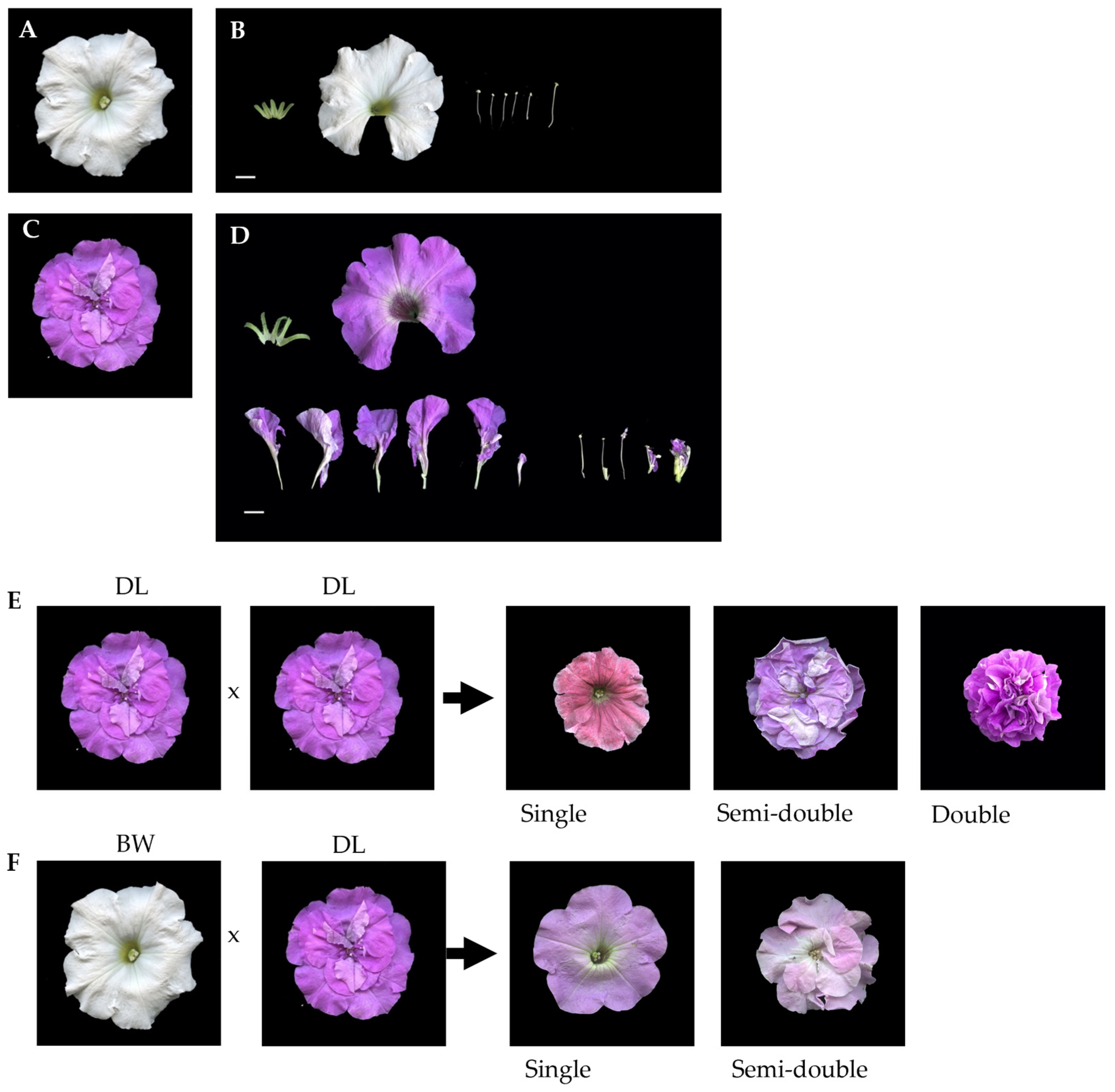
2.1. Inheritance of Double Flower Trait

To see the genetic characteristics of the double flower trait, crossing experiments were conducted between the single-flowered cultivar BW (Figure 1A,B) and the double-flowered cultivar DL (Figure 1C,D). Self-pollination of BW consistently yielded only single-flowered progeny, confirming its homozygous genotype in terms of the double-flowered trait. Self-pollination of DL was limited due to abnormal pistil development, leading to reduced seed production. In spite of the limited number of successfully obtained seeds, the selfed progeny showed phenotypic segregation of 4 (fully double):8 (semi-double):5 (single), which is not significantly different from 1:2:1 ratio (χ2 = 0.916, Figure 1E,F, Table 1), the expected segregation of Mendelian inheritance of a single and semi-dominant gene. Crossing between DL and BW yielded 196 progenies, comprising 94 single- and 102 semi-double-flowered plants, fitting a 1:1 segregation ratio (χ2 = 0.567, Table 1). An additional independent cross between DL and BW produced 142 progenies, consisting of 75 single- and 67 semi-double-flowered individuals, conforming to a ratio of 1:1 (χ2 = 0.502, Table 1). These results indicate that the double-flower trait in Petunia × hybrida is governed by a single gene with a semi-dominant inheritance pattern, where heterozygous individuals exhibit a semi-double phenotype, while homozygous recessive individuals remain single-flowered.

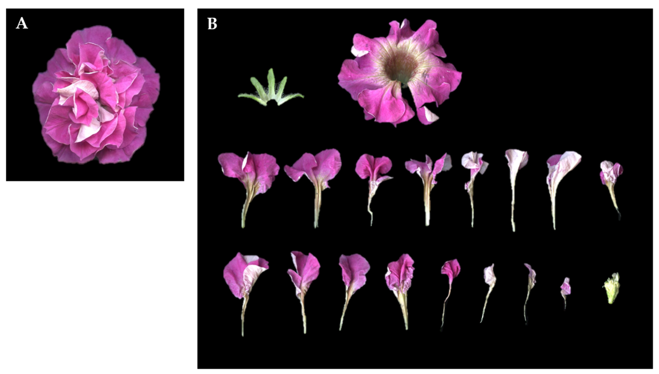
2.2. Morphology of Floral Organs and Their Surface Cell

Floral organ structures were compared among single-, semi-double-, and double-flowered petunias, with a focus on differences in petal number, stamen development, and floral organ morphology. In the single-flowered cultivar BW, floral organs followed a typical whorled arrangement, developing sequentially from the outermost layer as sepals, petals, stamens, and pistils (Figure 1A). In contrast, the semi-double-flowered cultivar DL exhibited an increased number of petals, with additional petaloid structures present between the outermost corolla and stamens. Some stamens in DL displayed intermediate petalization, with expanded filament bases and partially fused petal-like tissue at their tips (Figure 1D). In the progeny resulting from DL × DL crosses, a subset of fully double-flowered plants showed a significantly higher degree of petal multiplication than DL, forming a compact floral structure with layered petal whorls (Figure 2A). Floral dissections revealed that in these fully double-flowered plants, stamens were absent, and only sepals, petals, and morphologically altered pistils remained (Figure 2B).
Paraffin sections were prepared to examine the whorl structure, confirming that BW flowers consist of a single petal whorl, while DL flowers exhibit at least two distinct layers of petal whorls (Figure 3A,B). In addition, pistils in DL showed abnormal structure, including an increased ovary size and irregular carpel morphology, differing from the typical pistil structures observed in BW.
Abnormal structure of DL floral organs were also observed in the organ surface structure observed by scanning electron microscopy (SEM) observation (Figure 3C–L). In BW, ovary epidermal cells exhibited an irregular polygonal shape with clearly visible stomata (Figure 3C,D). Limb epidermal cells displayed uniform conical projections characteristic of petal surface texture (Figure 3E). Basal corolla cells were elongated (Figure 3F), similar to those typically observed in normal petal development.
In semi-double-flowered petunias DL, the stamen filaments and anthers exhibited surface features resembling those of petal epidermal cells (Figure 3I,J). The filaments showed elongated epidermal cells with ridged surfaces, while anthers displayed partial conical projections, differing from the smooth epidermis of typical stamens. Additionally, epidermal cells in the pistils exhibited a shift in morphology, with some regions displaying conical structures similar to petal epidermal cells (Figure 3G,H).
In fully double-flowered petunias, abnormalities in pistil development were even more pronounced. Although SEM analysis showed that pistil epidermal cells retained a morphology similar to that of normal single-flower petunias (Figure 3K,L), the overall floral structure was extensively altered, with additional stamen-like and pistil-like tissues appearing within the carpel. These structures formed layered floral components, resembling a secondary floral arrangement embedded within the primary flower (Figure 3K). These observations suggest that the homeotic transformation of stamens and pistils into petaloid structures is associated with genetic alterations affecting floral organ identity specification in double-flowered petunias.

2.3. Sequence Variation of Double-Flower-Related Genes in Petunia × hybrida

The whole-genome sequencing of ‘Baccarat White’ (BW) and ‘Duo Lavender’ (DL) generated a 1.5 GB scaffold database. To identify genetic variants associated with floral organ differentiation, sequences of genes related to the ABC model were examined. A total of four B-class genes (PhGLO1, PhGLO2, PhDEF (DEFICIENS), and PhTM6 (TOMATO MADS BOX GENE6)) and two C-class genes (pMADS3 (PETUNIA MADS BOX GENE3) and FBP6 (FLORAL BINDING PROTEIN6)) were identified in the assembled genome.
To further explore genetic factors involved in floral organ identity, full-length sequences of AP2 family genes were retrieved from publicly available genomic data of Arabidopsis thaliana (TAIR10) [36,37], Petunia axillaris (v1.6.2) [2] and Petunia inflata (v1.0.1) [2]. A comparison of these sequences led to the identification of eight euAP2 genes in Petunia axillaris: PeaxiROB1, PeaxiROB2, PeaxiROB3, PeaxiBEN, PeaxiBOB, and three additional unnamed AP2 family members (Peaxi162Scf01024g00326.1, Peaxi162Scf00072g00229.1, and Peaxi162Scf00389g00028.1).
Similarly, nine euAP2 genes were identified in Petunia inflata, including PeinfROB1, PeinfROB2, PeinfROB3, PeinfBEN, PeinfBOB, and four additional AP2 genes (Peinf101Scf12294g00004.1, Peinf101Scf01705g00017.1, Peinf101Scf01310g01017.1, and Peinf101Scf01034g02005.1). Homologous sequences of these AP2 family genes were also identified in the BW and DL scaffold databases, confirming their presence in both cultivars (Tables S1 and S4).
To investigate the phylogenetic relationships among these genes, their sequences were compared, and they were categorized into two major clades in Petunia axillaris: the AP2-type (PeaxiROB1, PeaxiROB2, PeaxiROB3) and the TOE-type (PeaxiBEN, PeaxiBOB, and three unnamed genes) (Figure 4). Notably, PeaxiBEN and PeaxiBOB exhibited high sequence similarity to ONI03164 from Prunus persica (peach), a gene previously reported to be associated with the double-flower trait. This finding suggests that AP2 genes may play an essential role in floral organ development in petunia (Figure 4).
A comparison of gene sequences between BW and DL revealed no differences in PhGLO1 and PhGLO2, PhDEF, and PhTM6 (Tables S2 and S5). Similarly, no notable sequence variations were detected in C-class genes, including pMADS3 and FBP6 (Tables S3 and S6). However, a structural variation was detected in PhBOB between BW and DL, particularly in the upstream region of the miR172 binding site. In BW, PhBOB exhibited an intact structure, whereas in DL, the gene was fragmented into two non-contiguous sequences. One fragment retained two EAR motifs, two AP2 domains, a linker region, and an NLS domain, while the other contained the region downstream of the miR172 binding site. No repetitive sequences were found between these two fragments, indicating that the structural variation in DL was caused by a large-scale insertion upstream of the miR172 binding site (Figure 5A and Figure S1).
Additionally, large deletions were identified in PhROB1 and PhROB3 in both BW and DL. However, these variations were restricted to intronic regions, suggesting that they may have a limited impact on gene expression (Tables S1 and S4).
The presence of this insertion in PhBOB indicates that structural modifications in AP2 family genes may contribute to floral organ identity changes. These findings are consistent with those of Gattolin et al. (2020) [17], further supporting the role of PhBOB in regulating petal number and the double-flower trait in petunia.

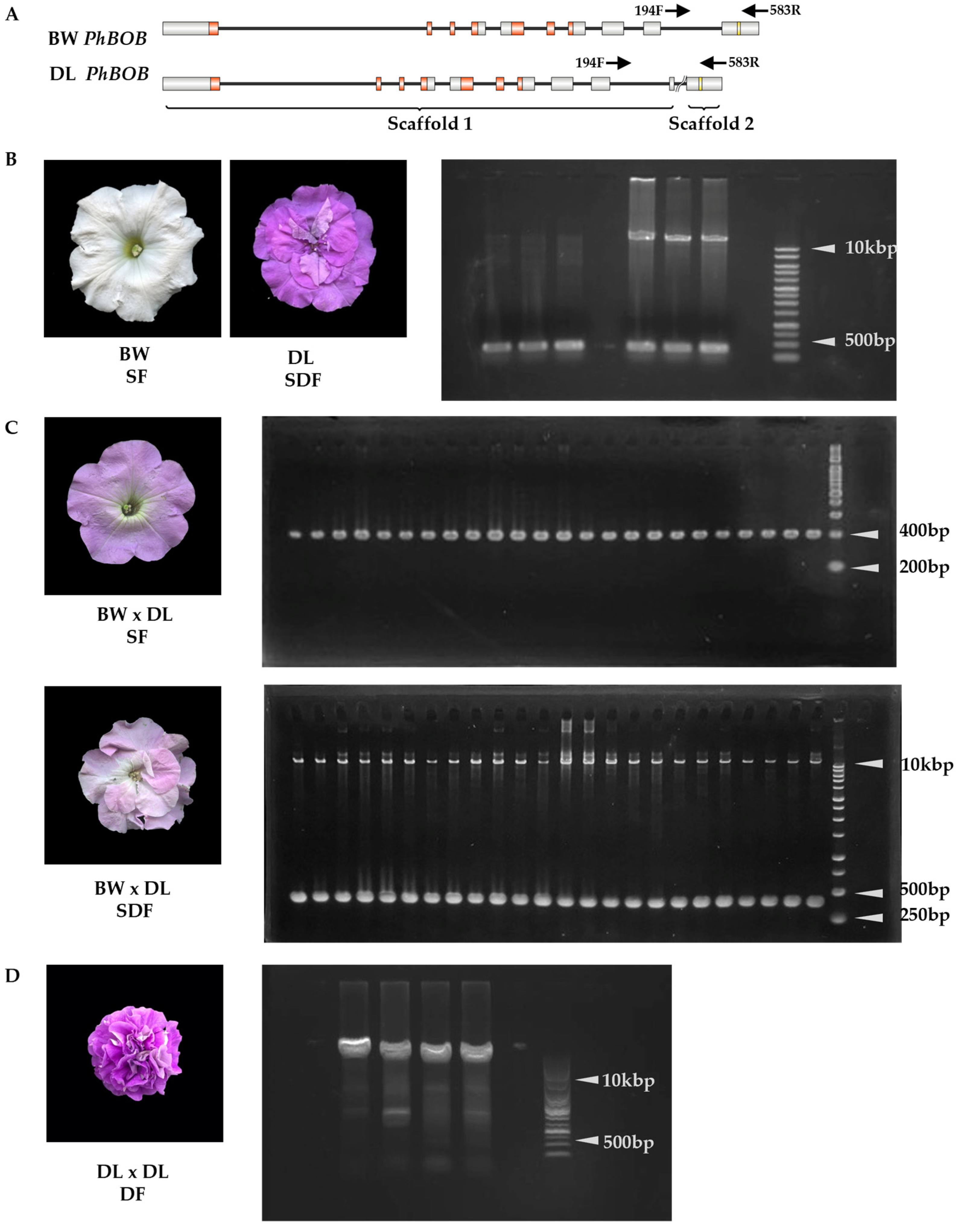
2.4. The 10 kbp Insertion in PhBOB Is Highly Linked to Double-Flowering in Petunias

Scaffold sequences upstream and downstream of PhBOB were compared between BW and DL. A forward primer was designed in the conserved upstream region and a reverse primer in the conserved downstream region (Figure 5A). These primers were used to genotype both the parental lines and the progeny derived from the BW × DL cross, as well as the selfed population of DL.
In BW, PCR amplification produced a single 390 bp fragment. In contrast, DL individuals showed two distinct bands: a 390 bp fragment and an additional ~10 kb fragment, which corresponds to a large insertion at the target site and reflects a heterozygous genotype (Figure 5B).
In the BW × DL F1 population, single-flowered plants exhibited only the 390 bp fragment, whereas semi-double-flowered plants displayed both the 390 bp and ~10 kb bands. PCR analysis of 96 single-flowered and 96 semi-double-flowered individuals confirmed this pattern (Figure 5C). All single-flowered plants were homozygous for the absence of the insertion (−/−), and all semi-double-flowered plants were heterozygous (+/−). No individuals homozygous for the insertion (+/+) were detected in the F1 population (Table 2).
To further clarify the relationship between the insertion and the floral phenotype, genotyping was performed on selfed progeny of DL. In this population, fully double-flowered individuals (DF) exhibited only the ~10 kb band, consistent with homozygosity for the insertion (+/+) (Figure 5D).
These results indicate that the 10 kbp insertion upstream of the miR172 target site in PhBOB is tightly linked to the double-flower trait and is likely involved in petal proliferation and floral organ transformation.

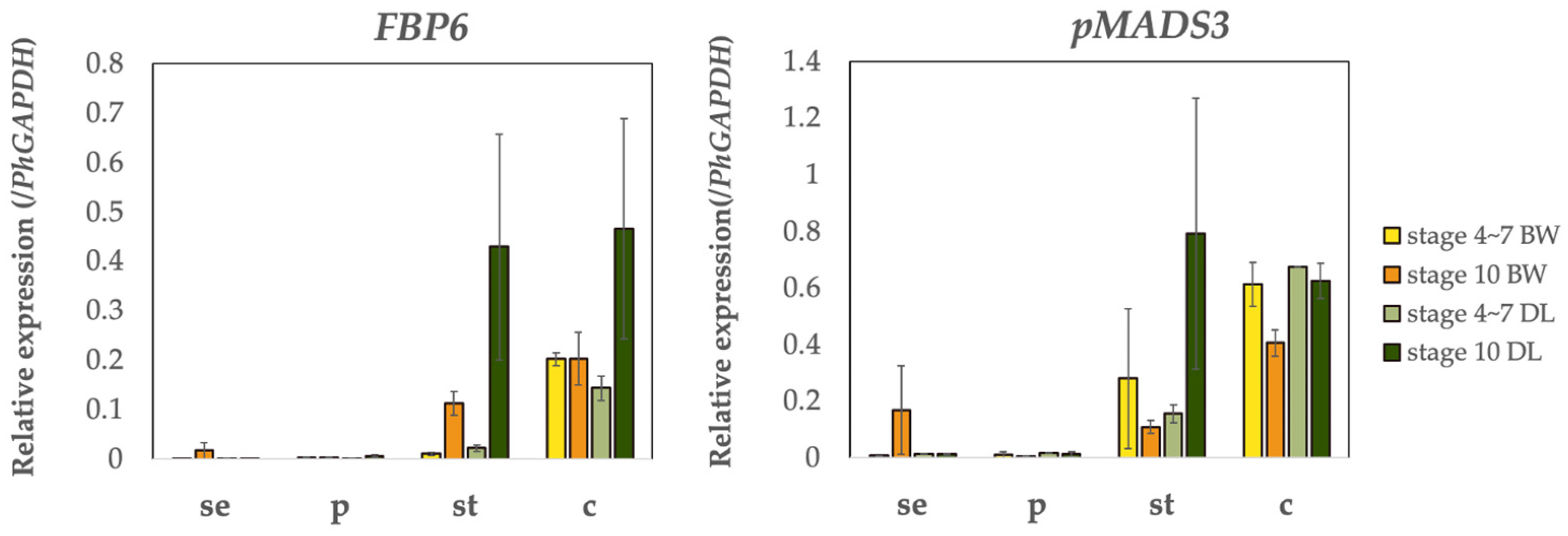
2.5. Expression of MADS-Box and AP2 Genes in BW and DL

Expression patterns of A-class (AP2 family genes) and C-class (pMADS3 and FBP6) genes were examined in different floral organs of BW and DL using qRT-PCR. Gene expression was quantified at ten developmental stages of flower buds (Figure 6A), with early floral development (stages 4–7) and late floral development (stage 10) selected as key time points for comparison.
Among A-class genes, BEN expression was consistently low (<0.01) across all floral organs, with no significant differences between BW and DL (Figure 6B). In contrast, BOB expression was higher in petal tissues. In DL, BOB expression was 6.8-fold higher than in BW during stages 4–7 and increased to 68.7-fold at stage 10. ROB3 showed a similar pattern, with a 3.1-fold increase in DL petals during early development and a 10.3-fold increase at stage 10.
ROB1 and ROB2 exhibited moderate expression in petals, particularly in stage 10 BW flowers, where transcript levels reached 0.1 and 0.05, respectively. Their expression in sepals, stamens, and pistils remained low with minimal variation.
C-class genes (pMADS3 and FBP6) were primarily expressed in stamens and pistils, with minimal levels detected in sepals and petals (Figure 7). FBP6 expression in stamens increased significantly at stage 10, with a 19.4-fold upregulation compared to stages 4–7. This increase was more pronounced in DL, where FBP6 transcript levels were 3.8-fold higher than in BW at stage 10. Similarly, FBP6 expression in DL pistils was 2.3-fold higher than in BW at stage 10. pMADS3 also showed significantly higher expression in DL stamens, with transcript levels 3.8-fold higher than in BW.

3. Discussion

3.1. PhBOB 10 kb Insertion Is Responsible for the Double-Flower Trait in Petunia

The double-flower trait in Petunia × hybrida shows a strong association with a 10 kb insertion located upstream of the miR172 target site in PhBOB, an AP2-like gene, suggesting a potential disruption of miR172-mediated post-transcriptional regulation. miR172 is known to play a crucial role in floral development by targeting AP2 genes, regulating their expression across diverse plant species. In Arabidopsis thaliana, miR172 directly suppresses APETALA2 (AP2), influencing floral organ identity [39], while in Brassica napus, a genome-wide analysis confirmed the regulatory interaction between miR172 and AP2 genes [40]. Similarly, in apple, the overexpression of miR172 significantly alters flowering time and floral organ identity, reinforcing its conserved function in floral patterning [41]. In tomato, miR172 has also been reported to regulate fruit development through its control of AP2 genes [42].
In ornamental and fruit-bearing species such as peach (P. persica), rose (R. chinensis), and Japanese apricot (P. mume), mutations in the miR172 binding site of AP2-like genes have been shown to prevent their degradation, resulting in elevated transcript levels and increased petal number [15,28,43,44]. These cases support the broader relevance of miR172-AP2 regulatory interactions in floral morphology.
In addition to directly regulating AP2 genes, miR172 is also involved in defining the spatial expression boundaries of B-class genes, which specify petal and stamen identities. In Arabidopsis, miR172 establishes the boundary for AP3 and PI expression in floral meristems, ensuring proper organ differentiation [45]. This spatial control is essential for preventing homeotic transformations between floral organs, highlighting the broader regulatory role of miR172 in flower development.
In petunia, the insertion identified near the miR172 binding site of PhBOB may interfere with this regulatory mechanism. Since AP2-like genes are known targets of miR172, the insertion could affect miRNA binding or stability, potentially resulting in altered gene expression. In support of this, PCR-based genotyping of F1 progenies revealed complete co-segregation between the 10 kb insertion and the double-flower phenotype. Single-flower individuals lacked the insertion, whereas all semi-double individuals were heterozygous, carrying both the wild-type and inserted alleles.
To assess the impact of this structural variation, gene expression analysis was performed. qRT-PCR results showed a substantial increase in PhBOB expression in petal tissues of double-flowered individuals, reaching levels approximately 69 times higher than those in single-flowered plants. This strong correlation between the presence of the insertion and elevated PhBOB transcript levels suggests a potential regulatory effect of the insertion on gene expression. As the overexpression of AP2-like genes has been linked to increased petal numbers in other species, PhBOB is likely to contribute significantly to double-flower formation in petunia.

3.2. PhBOB 10 kb Insertion Is Highly Conserved in Solanaceae and Contains CMC-EnSpm Transposon Sequences

BLAST analysis of the upstream 670 bp region of the 10 kb insertion in PhBOB (Table S9) revealed 98 significant matches across Solanoideae species, showing a high level of sequence similarity within this subfamily. In Capsicum species, only two matches were identified, reflecting a marked reduction in similarity compared to other Solanoideae members. This divergence highlights that while the upstream region is conserved in many Solanoideae species, it may have been lost or modified in certain lineages, reflecting different evolutionary pressures acting on this genomic region.
The downstream 652 bp region of the insertion (Table S9) was also found to be conserved across multiple Solanaceae species, including Solanum demissum, Solanum lycopersicum (tomato), Solanum tuberosum (potato), Lycium barbarum (goji berry), Nicotiana tomentosiformis, Nicotiana sylvestris (wood tobacco), and Capsicum annuum (chili pepper). This cross-genus presence within the Solanaceae family shows that the region is more broadly conserved. While its exact function remains unknown, its consistent presence across divergent species implies that it may have some regulatory significance, though experimental validation is still required.
Sequence analysis of the downstream region detected three segments with homology to CMC-EnSpm transposons, with sequence identities of 67% (6 × 10−39), 69% (3 × 10⁻²⁹), and 78% (4 × 10⁻²⁸), respectively. CMC-EnSpm transposons are one of the most abundant DNA transposon families in plant genomes and are known to contribute to genome evolution and structural variation. Ishimori et al. (2015) [14] reported that CMC-EnSpm elements are a major component of the Eustoma grandiflorum (lisianthus) genome, where RepeatMasker analysis showed that DNA transposons account for 92.0 Mb (12.8%) of the genome, with the CMC-EnSpm subfamily comprising 11.3 Mb, making it the most dominant DNA transposon subfamily. The widespread distribution of CMC-EnSpm elements in plant genomes supports their role as major contributors to genome structure and functional regulation.
Beyond their structural role, CMC-EnSpm transposons are known to influence gene expression through chromatin modifications and transcriptional regulation. Studies have shown that these transposons can modify chromatin structure, alter DNA methylation patterns, and provide novel promoter or enhancer sequences that regulate nearby genes [46,47]. In Populus trichocarpa, CMC-EnSpm transposons were found to regulate gene expression by modifying DNA methylation, leading to changes in gene activity in response to environmental stimuli [47]. Similarly, in Dioscorea alata, transcriptionally active CMC-EnSpm elements were associated with specific gene expression patterns, suggesting that they play a regulatory role in gene function. These studies highlight the functional significance of CMC-EnSpm transposons in modulating gene expression across plant species [48].
Given the strong evidence supporting the regulatory role of CMC-EnSpm transposons, it is likely that the 10 kb insertion in PhBOB plays a role in its overexpression in double-flower petunias. The presence of transposon sequences in the insertion suggests that chromatin modifications or alterations in transcriptional regulation may contribute to the upregulation of PhBOB in petal tissues. Further studies are needed to confirm whether the CMC-EnSpm insertion directly influences PhBOB expression through changes in DNA methylation, chromatin accessibility, or enhancer activity. Understanding the precise mechanism of how this transposon insertion affects PhBOB regulation will provide valuable insights into the genetic basis of the double-flower trait in petunia and could inform strategies for breeding novel floral morphologies in ornamental plants.

3.3. The Expression of C-Class Genes (FBP6 and pMADS3) Is Controlled by PhBOB

C-class genes play a critical role in defining reproductive organ identity in flowering plants, regulating the formation of stamens and carpels. While some species retain both PLE and AG homologs, others possess only one functional C-class gene [49,50]. In petunia, only two genes, pMADS3 and FBP6, function as C-class genes, both of which are homologs of AGAMOUS (AG), the key floral homeotic gene responsible for specifying stamens and carpels.
In single-flower petunias, FBP6 and pMADS3 are highly expressed in stamens and carpels, consistent with their role in reproductive organ identity. However, in semi-double-flower individuals, these genes remain highly expressed in stamens and carpels but are notably suppressed in additional petal regions. This spatial shift in gene expression indicates that PhBOB does not completely repress C-class genes but instead modifies their spatial expression domains, which is associated with the transformation of stamens into petal-like structures. This change in gene expression pattern may explain the floral phenotype of semi-double individuals, where extra petals develop while the reproductive organs are still partially retained.
A similar regulatory mechanism has been observed in other double-flower species, where AP2-like genes modulate, rather than completely repress, the spatial expression of C-class genes. In rose, peach, and lisianthus, mutations in AP2 genes have been shown to disrupt the spatial expression of C-class genes, leading to the petalization of reproductive organs while allowing some stamen and carpel development to persist [15,28,43,44]. This pattern is consistent with the role of PhBOB in petunia, which appears to act by altering the spatial domain of FBP6 and pMADS3 rather than fully silencing them. The ability of PhBOB to modulate C-class gene expression may be a key factor in the formation of semi-double flowers, where intermediate stamen–petal transformation occurs rather than a complete conversion of reproductive organs into petal tissues.

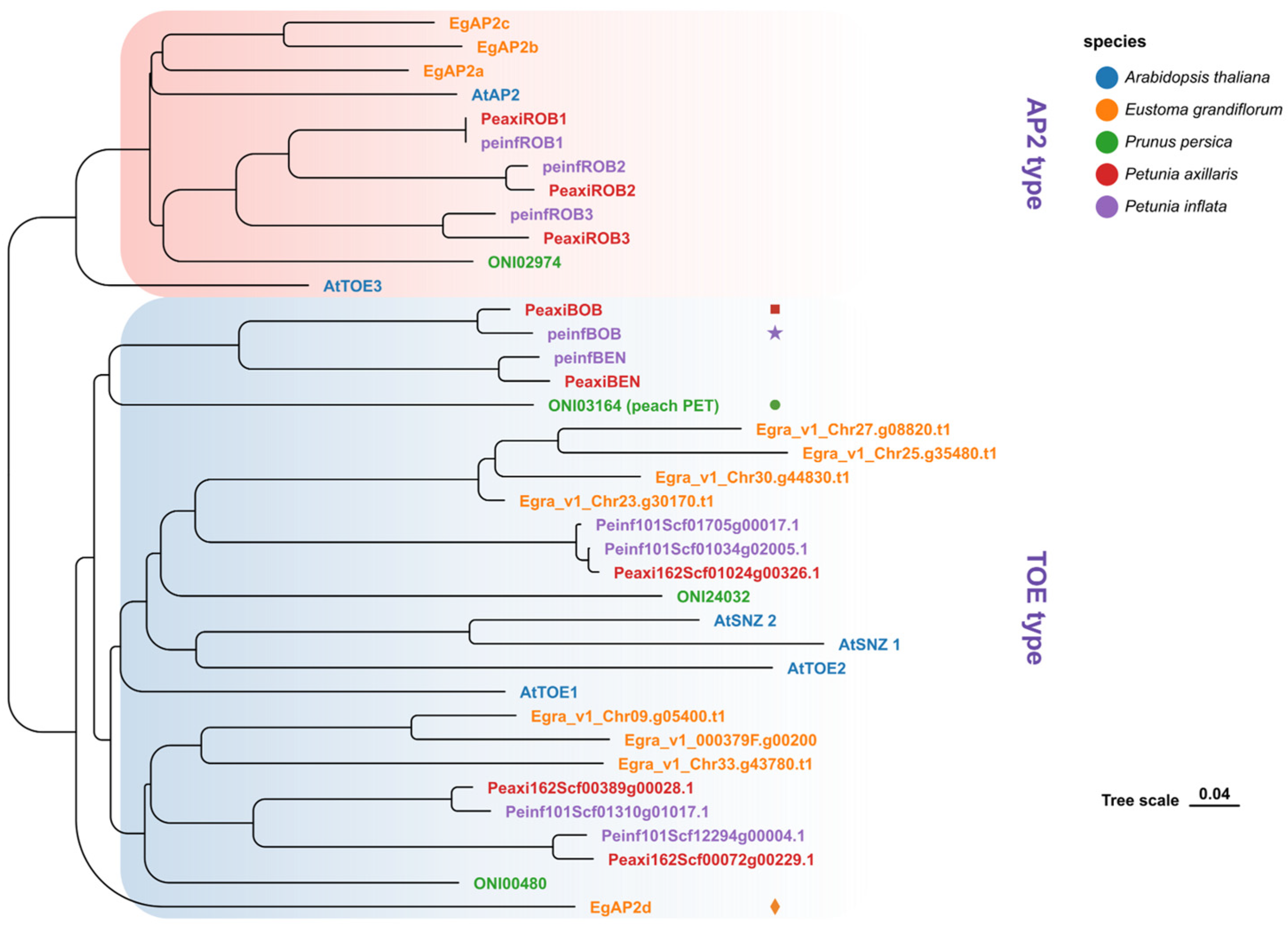
3.4. AP2 Genes in the TOE-Type Clade Are Associated with the Double-Flower Trait

The double-flower phenotype in Petunia × hybrida is strongly associated with PhBOB, a TOE-type AP2 gene. Phylogenetic analysis shows that PhBOB belongs to a clade known as PETALOSA, which includes genes such as Prupe.6G242400 in peach, Dca21030.1 in carnation, and XP_024186592 in rose [15,17]. In each case, the gene is expressed in floral organs, and its overexpression correlates with changes in floral identity.
In petunia, PhBOB shares not only phylogenetic similarity with these PETALOSA genes but also functional characteristics. The insertion found upstream of PhBOB is associated with its elevated expression in petal tissues, and semi-double flowers show altered spatial expression of floral identity genes consistent with AP2-related regulatory changes. These parallels in sequence, expression, and phenotype reinforce the classification of PhBOB as a functional member of the PETALOSA group.
While the PETALOSA lineage is clearly associated with dominant double-flower mutations, other TOE-type AP2 genes outside this clade have also been implicated in the regulation of petal proliferation. In Eustoma grandiflorum, the EgAP2d gene is strongly linked to the double-flower trait but is phylogenetically distant from the PETALOSA clade, suggesting independent evolutionary origins for this regulatory function [14,51]. Similarly, in Prunus mume (Japanese apricot), EgAP2L contributes to the double-flower phenotype but does not belong to the PETALOSA lineage [18], further supporting the hypothesis that multiple TOE-type AP2 genes, beyond those classified as PETALOSA, can regulate floral organ identity. These findings suggest that the ability of TOE-type AP2 genes to modulate double-flower formation has evolved independently in different plant lineages, indicating a broader functional role for this gene family in floral development.
Beyond the TOE-type group, AP2-type genes such as PhROB3 have also been identified in petunia. In semi-double flowers, PhROB3 exhibits differential expression between petals and stamens, and its involvement has been proposed in floral boundary regulation. Although phylogenetically distinct from TOE-type genes, its floral expression pattern and response to morphological changes suggest that PhROB3 may interact with PhBOB or respond to similar regulatory cues. This observation is consistent with findings in other species where different AP2 subfamilies co-regulate floral patterning.
Taken together, these findings demonstrate that while PETALOSA genes represent a distinct lineage of TOE-type AP2 genes responsible for dominant double-flower mutations, other TOE-type and AP2-type genes may also contribute to petal number regulation. The interaction between these gene families underlies the diversity of floral forms observed in ornamental plants, providing valuable insights into the molecular mechanisms driving double-flower evolution.

4. Materials and Methods

4.1. Plant Materials and Growth Conditions

This study utilized two commercial cultivars of Petunia × hybrida: ‘Baccarat White’ (BW, single-flower, white) and ‘Duo Lavender’ (DL, semi-double flower, purple). Plants were cultivated under controlled environmental conditions, with a 16 h photoperiod using white LED lights at a photosynthetic photon flux density (PPFD) of 200 μmol m−2s−1. Temperature conditions were maintained at 23 °C during the day and 27 °C at night to support optimal growth and flowering.
For crossing experiments, all stamens were removed from BW flowers before anthesis to prevent self-pollination. Pollen from DL flowers was manually transferred onto the stigma of emasculated BW flowers, and the pollinated flowers were immediately bagged to prevent cross-contamination from other pollen sources. Pollination was conducted in May 2019 and January 2020 to ensure seasonal reproducibility of the results.
To facilitate self-pollination in DL, high-temperature treatment was applied to induce pollen production. Each flower was individually bagged to ensure self-pollination without external pollen interference, allowing for controlled genetic analysis of the semi-double flower trait.

4.2. Paraffin Sectioning

For histological analysis, flower buds of Baccarat White (BW, single-flower) and Duo Lavender (DL, semi-double flower) petunias (Petunia × hybrida) were collected at different developmental stages and immediately fixed in FAA solution (5% formaldehyde, 5% acetic acid, 90% ethanol, v/v) at 4 °C overnight to preserve tissue integrity.
After fixation, samples were dehydrated through a graded ethanol series (50%, 70%, 85%, 90%, and 100%) and subsequently cleared using Histoclear to enhance wax infiltration. Following appropriate pre-processing, samples were embedded in paraffin to facilitate sectioning. A rotary microtome (Leica RM2235, Leica Biosystems, Nussloch, Germany) was used to obtain continuous sections (8 μm thickness), which were then mounted onto poly-L-lysine-coated slides to improve tissue adhesion and prevent detachment during staining and observation.
To remove paraffin residues, sections were deparaffinized using Histoclear, followed by rehydration through a descending ethanol gradient (100%, 95%, 90%, 80%, 70%, and 50%) to restore tissue hydration. Sections were then sealed using Entellan New mounting medium (Merck) for long-term preservation and microscopic examination.
For tissue staining, hematoxylin staining was applied to visualize floral organ structures, including sepals, petals, stamens, and carpels, to examine cell morphology and tissue differentiation. Finally, microscopic observation and imaging were conducted using a light microscope (Leica DM500, Leica Microsystems, Germany) to document tissue architecture and analyze morphological changes during different floral developmental stages.

4.3. Scanning Electron Microscopy (SEM) Observation

To examine the cellular morphology of leaf and floral organ surfaces in Petunia × hybrida, scanning electron microscopy (SEM) was conducted following standard sample preparation procedures to maintain tissue integrity and optimize imaging resolution.
Freshly collected leaves and floral organs were immersed in NanoSuit Solution I to form a protective layer, reducing electron beam-induced damage and enhancing sample conductivity. Excess solution was carefully blotted using filter paper or Kimwipes to remove residual liquid that could interfere with imaging.
Samples were then mounted onto SEM stubs using conductive double-sided adhesive tape to ensure secure adhesion and minimize movement or vibration during scanning. The specimens were subsequently transferred to a JCM-6000 scanning electron microscope (JEOL, Japan) for imaging. The accelerating voltage and working distance were adjusted to obtain clear microstructural images of the epidermal cells on petunia leaves and floral organs.

4.4. Whole-Genome Sequencing

Whole-genome sequencing (WGS) was performed on Petunia × hybrida cultivars ‘Baccarat White’ (BW, single-flower) and ‘Duo Lavender’ (DL, semi-double flower) using Next-Generation Sequencing (NGS) technology to obtain genome-wide sequence information and provide a foundation for subsequent gene identification and variant analysis.
Genomic DNA was extracted from fresh leaf tissues, and sequencing libraries were constructed using a random fragmentation approach with an insert size of 500 bp. High-throughput sequencing was carried out on the Illumina HiSeq X platform using 150 bp paired-end sequencing to ensure high coverage and sequencing depth. A total of 66 GB of raw sequencing data was generated for DL, and 75 GB for BW.
The raw sequencing data underwent quality control (QC) filtering, where low-quality reads, adapter sequences, and PCR duplicates were removed to ensure high-accuracy genome assembly. The filtered high-quality reads were then de novo assembled using ABySS [52], resulting in a 1.5 GB scaffold database, corresponding to the genome sequences of both BW and DL cultivars.

4.5. Identification and Screening of AP2 Family Genes in Petunia and Other Ornamental Plants

To comprehensively identify AP2 family genes in Petunia × hybrida and other ornamental plants, two strategies were employed to obtain high-quality AP2 candidate sequences. An annotation-based approach was first applied by searching for “AP2” in the Plant Transcription Factor Database (PlantTFDB, https://planttfdb.gao-lab.org/, accessed on 22 April 2025), selecting the target species to retrieve annotated AP2 gene sequences. This method leveraged an existing transcription factor database to facilitate the rapid identification of AP2 gene candidates.
To further refine the dataset, a Hidden Markov Model (HMM)-based screening approach was used. Genomic sequences, protein FASTA files, and GFF3 annotation files for Petunia were downloaded from publicly available genome databases. The HMM model for the AP2 domain was retrieved from the InterPro Pfam database (https://www.ebi.ac.uk/interpro/entry/pfam/PF00847/logo/, accessed on 22 April 2025), and an initial search was conducted using hmmsearch with an E-value threshold of 1.2 × 10⁻²⁸. To ensure the accuracy of the predicted AP2 domain sequences, further validation was performed using SMART (http://smart.embl-heidelberg.de/, accessed on 22 April 2025) and the NCBI Conserved Domain Database (CDD, http://www.ncbi.nlm.nih.gov/Structure/cdd/cdd.shtml, accessed on 22 April 2025). Verified AP2 domain sequences were aligned using ClustalW, and a petunia-specific HMM model was constructed with hmmbuild for a refined hmmsearch. A final selection threshold of E-value < 0.001 was applied to obtain high-confidence AP2 family genes. Given the presence of multiple transcript isoforms, GFF3 annotation files were utilized to match gene loci with corresponding mRNA sequences, and redundant sequences were removed to ensure the accuracy of the final AP2 gene dataset.
The genomic data used in this study were sourced from multiple databases: Arabidopsis thaliana data were obtained from Ensembl Plants (https://plants.ensembl.org/Arabidopsis_thaliana/Info/Annotation/, accessed on 08 December 2024), Eustoma grandiflorum from Plant Garden (https://plantgarden.jp/ja/download/t52518/t52518.G001/, accessed on 22 April 2025), Prunus persica from NCBI WGS Project AKXU02 (https://www.ncbi.nlm.nih.gov/datasets/genome/GCF_000346465.2/, accessed on 22 April 2025), while Petunia axillaris and Petunia inflata genomic data were accessed via the Sol Genomics Network (https://solgenomics.net/organism/Petunia_axillaris/genome, accessed on 8 December 2024) and (https://solgenomics.net/organism/Petunia_inflata/genome, accessed on 8 December 2024), respectively. The sequences used in phylogenetic analysis are listed in Supplementary Table S7. The phylogenetic tree was constructed using the Neighbor-Joining (NJ) method, with the Genetic Distance Model set to Jukes–Cantor. Tree visualization and annotation were performed using tvBOT [53].

4.6. Sequence Alignment and Homolog Identification of ABC-Class Genes in Petunia × hybrida

The scaffold database was imported into Geneious R10.2.3 for further analysis. The identified AP2 target gene sequences from Petunia axillaris were used as query sequences for BLAST searches against the scaffold databases of ‘Baccarat White’ (BW) and ‘Duo Lavender’ (DL). The sequences with the lowest E-value in the BLAST results were selected as candidate homologs. If the identified scaffold fragment did not contain the full-length sequence of the target gene, the scaffold was used as a new query sequence for an additional BLAST search in the BW and DL scaffold databases to extend the target sequence. To ensure the reliability of the sequence alignment results, the Dotplot function in Geneious was utilized to analyze the similarity between the query and candidate sequences, confirming high sequence homology.

4.7. Linkage Analysis

Genomic DNA was extracted from 200 mg of leaf tissue using the Maxwell RSC PureFood GMO and Authentication Kit (Catalog No. AS1600). Each PCR reaction was performed using 80 ng of genomic DNA as the template, with amplification carried out using KOD FX Neo (TOYOBO) and specific primer sets (Table S8). The PCR conditions were as follows: 94 °C for 2 min, followed by 30 cycles of 98 °C for 10 s, 60 °C for 30 s, and 68 °C for 7 min, with a final extension at 72 °C for 7 min before being held at 4 °C. PCR products were separated by electrophoresis on a 0.5% SeaKem GTG agarose gel (100 V, 30 min, Mupid-ex system) and visualized using a gel imaging system.

4.8. Gene Expression Analysis

Total RNA was extracted using the Maxwell RSC Plant RNA Kit, with tissue samples flash-frozen in liquid nitrogen prior to extraction. RNA reverse transcription was performed using the ReverTra Ace qPCR RT Master Mix with gDNA Remover Kit (TOYOBO). Quantitative real-time PCR (qRT-PCR) was conducted on a StepOne Real-Time PCR System (Applied Biosystems, Waltham, Massachusetts, USA) using KOD SYBR qPCR Mix in a 96-well plate with a final reaction volume of 10 μL, following the manufacturer’s instructions. The primers used in qRT-PCR are listed in Supplementary Table S8.
To analyze the dynamic changes in gene expression during flower development, the floral developmental process of ‘Baccarat White’ (BW, single-flower) and ‘Duo Lavender’ (DL, semi-double flower) was categorized into ten stages (Stage 1 to Stage 10) based on flower bud size. The corresponding bud lengths for each stage were as follows: Stage 1: 3.8 ± 0.7 mm, Stage 2: 7.8 ± 0.6 mm, Stage 3: 11.6 ± 2.8 mm, Stage 4: 15.9 ± 3.7 mm, Stage 5: 22.9 ± 2.3 mm, Stage 6: 26.1 ± 1.7 mm, Stage 7: 37.2 ± 1.2 mm, Stage 8: 42.9 ± 2.1 mm, Stage 9: 43.5 ± 2.8 mm, and Stage 10: fully opened flowers. To investigate the expression patterns of candidate genes at key developmental stages, floral tissues were collected from pre-flowering stages (Stages 4 to 7, bud length: 15.9 to 37.2 mm) and post-flowering Stage 10.
qRT-PCR was performed under the following thermal cycling conditions: Holding stage: 98 °C for 2 min; cycling stage: 50 cycles of 98 °C for 10 s, 60 °C for 10 s, and 68 °C for 30 s; melt curve stage: 95 °C for 15 s, 60 °C for 1 min, and 99 °C for 15 s. All experimental data were analyzed using Microsoft Excel, with three biological replicates and two technical replicates performed for each gene.

5. Conclusions

This study confirms that the double-flower trait in petunia follows a single-gene, semi-dominant inheritance pattern through genetic analysis. Morphological characterization using paraffin sectioning and scanning electron microscopy (SEM) revealed clear differences in floral organ structure among single-flowered (SF), semi-double-flowered (SDF), and fully double-flowered (DF) petunias. At the molecular level, we identified a 10 kb insertion in PhBOB that is strongly associated with the double-flower trait and exhibits complete co-segregation in F1 populations. Gene expression analysis further demonstrated that PhBOB is significantly upregulated in double-flowered petunias, reaching expression levels up to 69-fold higher than in single-flowered cultivars, suggesting that its overexpression is a key driver of petal proliferation.
In addition to PhBOB, another AP2 family gene, PhROB3, was also markedly upregulated in petals and stamens, particularly in semi-double and fully double flowers, where partial homeotic transformation of stamens into petal-like structures was observed. This suggests that PhROB3 may work in conjunction with PhBOB to regulate petal and stamen differentiation, adding another layer of complexity to petunia floral organ development.

Supplementary Materials

The following supporting information can be downloaded at: https://www.mdpi.com/article/10.3390/plants14091314/s1, Table S1. Nucleotide sequence of A-class genes from ‘Baccarat White’. Table S2. Nucleotide sequence of B-class genes from ‘Baccarat White’. Table S3. Nucleotide sequence of C-class genes from ‘Baccarat White’. Table S4. Nucleotide sequence of A-class genes from ‘Duo Lavender’. Table S5. Nucleotide sequence of B-class genes from ‘Duo Lavender’. Table S6. Nucleotide sequence of C-class genes from ‘Duo Lavender’. Table S7. Nucleotide sequence of phylogenetic analysis. Table S8. Oligo sequences used in this study. Table S9. Nucleotide sequence of the 10 kb insertion in PhBOB. Figure S1. Sequence alignment and schematic illustration of the putative insertion upstream of the miR172 binding site in PhBOB.

Author Contributions

T.X.: Conceptualization; methodology; investigation; data curation; formal analysis; visualization; writing—original draft. S.K.: Supervision; funding acquisition; project administration; writing—review and editing. All authors have read and agreed to the published version of the manuscript.

Funding

This research received no external funding.

Data Availability Statement

All data supporting the findings of this study are available within the paper and within its Supplementary Materials published online.

Conflicts of Interest

All authors have read and approved this version of the article, and due care has been taken to ensure the integrity of this work. The authors declare that they have no conflicts of interest.

References

  1. Souer, E.; Rebocho, A.B.; Bliek, M.; Kusters, E.; De Bruin, R.A.M.; Koes, R. Patterning of Inflorescences and Flowers by the F-Box Protein DOUBLE TOP and the LEAFY Homolog ABERRANT LEAF AND FLOWER of Petunia. Plant Cell 2008, 20, 2033–2048. [Google Scholar] [CrossRef] [PubMed]
  2. Bombarely, A.; Moser, M.; Amrad, A.; Bao, M.; Bapaume, L.; Barry, C.S.; Bliek, M.; Boersma, M.R.; Borghi, L.; Bruggmann, R.; et al. Insight into the Evolution of the Solanaceae from the Parental Genomes of Petunia Hybrida. Nat. Plants 2016, 2, 16074. [Google Scholar] [CrossRef] [PubMed]
  3. Gautam, P.; Terfa, M.T.; Olsen, J.E.; Torre, S. Red and Blue Light Effects on Morphology and Flowering of Petunia×hybrida. Sci. Hortic. 2015, 184, 171–178. [Google Scholar] [CrossRef]
  4. Chopy, M.; Cavallini-Speisser, Q.; Chambrier, P.; Morel, P.; Just, J.; Hugouvieux, V.; Rodrigues Bento, S.; Zubieta, C.; Vandenbussche, M.; Monniaux, M. Cell layer–specific expression of the homeotic MADS-box transcription factor PhDEF contributes to modular petal morphogenesis in petunia. Plant Cell 2024, 36, 324–345. [Google Scholar] [CrossRef]
  5. Geitmann, A. Petunia. Evolutionary, Developmental and Physiological Genetics. Ann. Bot. 2011, 107, vi. [Google Scholar] [CrossRef]
  6. Krizek, B.A.; Fletcher, J.C. Molecular Mechanisms of Flower Development: An Armchair Guide. Nat. Rev. Genet. 2005, 6, 688–698. [Google Scholar] [CrossRef]
  7. Schwarz-Sommer, Z.; Huijser, P.; Nacken, W.; Saedler, H.; Sommer, H. Genetic Control of Flower Development by Homeotic Genes in Antirrhinum majus. Science 1990, 250, 931–936. [Google Scholar] [CrossRef]
  8. Pelaz, S.; Ditta, G.S.; Baumann, E.; Wisman, E.; Yanofsky, M.F. B and C floral Organ Identity Functions Require SEPALLATA MADS-Box Genes. Nature 2000, 405, 200–203. [Google Scholar] [CrossRef]
  9. Pinyopich, A.; Ditta, G.S.; Savidge, B.; Liljegren, S.J.; Baumann, E.; Wisman, E.; Yanofsky, M.F. Assessing the Redundancy of MADS-Box Genes during Carpel and Ovule Development. Nature 2003, 424, 85–88. [Google Scholar] [CrossRef]
  10. Ehlers, K.; Bhide, A.S.; Tekleyohans, D.G.; Wittkop, B.; Snowdon, R.J.; Becker, A. The MADS Box Genes ABS, SHP1, and SHP2 Are Essential for the Coordination of Cell Divisions in Ovule and Seed Coat Development and for Endosperm Formation in Arabidopsis thaliana. PLoS ONE 2016, 11, e0165075. [Google Scholar] [CrossRef]
  11. Mizzotti, C.; Ezquer, I.; Paolo, D.; Rueda-Romero, P.; Guerra, R.F.; Battaglia, R.; Rogachev, I.; Aharoni, A.; Kater, M.M.; Caporali, E.; et al. SEEDSTICK Is a Master Regulator of Development and Metabolism in the Arabidopsis Seed Coat. PLoS Genet. 2014, 10, e1004856. [Google Scholar] [CrossRef] [PubMed]
  12. Bowman, J.L.; Smyth, D.R.; Meyerowitz, E.M. Genes Directing Flower Development in Arabidopsis. Plant Cell 1989, 1, 37–52. [Google Scholar] [CrossRef] [PubMed]
  13. Yao, J.; Xu, J.; Tomes, S.; Cui, W.; Luo, Z.; Deng, C.; Ireland, H.S.; Schaffer, R.J.; Gleave, A.P. Ectopic Expression of the PISTILLATAHomologous Md PI Inhibits Fruit Tissue Growth and Changes Fruit Shape in Apple. Plant Direct 2018, 2, e00051. [Google Scholar] [CrossRef] [PubMed]
  14. Ishimori, M. Genome Analysis of Eustoma Grandiflorum and Identification of the Causative Gene for the Double-Flower Trait. Ph.D. Thesis, The University of Tokyo, Tokyo, Japan, 2015. [Google Scholar]
  15. Gattolin, S.; Cirilli, M.; Pacheco, I.; Ciacciulli, A.; Da Silva Linge, C.; Mauroux, J.-B.; Lambert, P.; Cammarata, E.; Bassi, D.; Pascal, T.; et al. Deletion of the miR172 Target Site in a TOE-Type Gene Is a Strong Candidate Variant for Dominant Double-Flower Trait in Rosaceae. Plant J. 2018, 96, 358–371. [Google Scholar] [CrossRef]
  16. François, L.; Verdenaud, M.; Fu, X.; Ruleman, D.; Dubois, A.; Vandenbussche, M.; Bendahmane, A.; Raymond, O.; Just, J.; Bendahmane, M. A miR172 Target-Deficient AP2-like Gene Correlates with the Double Flower Phenotype in Roses. Sci. Rep. 2018, 8, 12912. [Google Scholar] [CrossRef]
  17. Gattolin, S.; Cirilli, M.; Chessa, S.; Stella, A.; Bassi, D.; Rossini, L. Mutations in Orthologous PETALOSA TOE-Type Genes Cause a Dominant Double-Flower Phenotype in Phylogenetically Distant Eudicots. J. Exp. Bot. 2020, 71, 2585–2595. [Google Scholar] [CrossRef]
  18. Liu, W.; Zheng, T.; Qiu, L.; Guo, X.; Li, P.; Yong, X.; Li, L.; Ahmad, S.; Wang, J.; Cheng, T.; et al. A 49-Bp Deletion of PmAP2L Results in a Double Flower Phenotype in Prunus Mume. Hortic. Res. 2024, 11, uhad278. [Google Scholar] [CrossRef]
  19. Magnani, E.; Sjölander, K.; Hake, S. From Endonucleases to Transcription Factors: Evolution of the AP2 DNA Binding Domain in Plants[W]. Plant Cell 2004, 16, 2265–2277. [Google Scholar] [CrossRef]
  20. Allen, M.D.; Yamasaki, K.; Ohme-Takagi, M.; Tateno, M.; Suzuki, M. A Novel Mode of DNA Recognition by a β-Sheet Revealed by the Solution Structure of the GCC-Box Binding Domain in Complex with DNA. EMBO J. 1998, 17, 5484–5496. [Google Scholar] [CrossRef]
  21. Cao, D.; Lin, Z.; Huang, L.; Damaris, R.N.; Yang, P. Genome-Wide Analysis of AP2/ERF Superfamily in Lotus (Nelumbo nucifera) and the Association between NnADAP and Rhizome Morphology. BMC Genomics 2021, 22, 171. [Google Scholar] [CrossRef]
  22. Nakano, T.; Suzuki, K.; Fujimura, T.; Shinshi, H. Genome-Wide Analysis of the ERF Gene Family in Arabidopsis and Rice. Plant Physiol. 2006, 140, 411–432. [Google Scholar] [CrossRef] [PubMed]
  23. Magar, M.M.; Liu, H.; Yan, G. Genome-Wide Analysis of AP2/ERF Superfamily Genes in Contrasting Wheat Genotypes Reveals Heat Stress-Related Candidate Genes. Front. Plant Sci. 2022, 13, 853086. [Google Scholar] [CrossRef]
  24. Licausi, F.; Ohme-Takagi, M.; Perata, P. APETALA 2/Ethylene Responsive Factor (AP 2/ERF) Transcription Factors: Mediators of Stress Responses and Developmental Programs. New Phytol. 2013, 199, 639–649. [Google Scholar] [CrossRef] [PubMed]
  25. Kagaya, Y.; Ohmiya, K.; Hattori, T. RAV1, a Novel DNA-Binding Protein, Binds to Bipartite Recognition Sequence through Two Distinct DNA-Binding Domains Uniquely Found in Higher Plants. Nucleic Acids Res. 1999, 27, 470–478. [Google Scholar] [CrossRef] [PubMed]
  26. Zhang, B.; Chen, X. Secrets of the MIR172 Family in Plant Development and Flowering Unveiled. PLOS Biol. 2021, 19, e3001099. [Google Scholar] [CrossRef]
  27. Díaz-Manzano, F.E.; Cabrera, J.; Ripoll, J.-J.; del Olmo, I.; Andrés, M.F.; Silva, A.C.; Barcala, M.; Sánchez, M.; Ruíz-Ferrer, V.; de Almeida-Engler, J.; et al. A Role for the Gene Regulatory Module microRNA172/TARGET OF EARLY ACTIVATION TAGGED 1/FLOWERING LOCUS T (miRNA172/TOE1/FT) in the Feeding Sites Induced by Meloidogyne Javanica in Arabidopsis thaliana. New Phytol. 2018, 217, 813–827. [Google Scholar] [CrossRef]
  28. Cirilli, M.; Rossini, L.; Chiozzotto, R.; Baccichet, I.; Florio, F.E.; Mazzaglia, A.; Turco, S.; Bassi, D.; Gattolin, S. Less Is More: Natural Variation Disrupting a miR172 Gene at the Di Locus Underlies the Recessive Double-Flower Trait in Peach (P. persica L. Batsch). BMC Plant Biol. 2022, 22, 318. [Google Scholar] [CrossRef]
  29. Wang, L.; Ma, H.; Lin, J. Angiosperm-Wide and Family-Level Analyses of AP2/ERF Genes Reveal Differential Retention and Sequence Divergence After Whole-Genome Duplication. Front. Plant Sci. 2019, 10, 196. [Google Scholar] [CrossRef]
  30. Yin, F.; Zeng, Y.; Ji, J.; Wang, P.; Zhang, Y.; Li, W. The Halophyte Halostachys Caspica AP2/ERF Transcription Factor HcTOE3 Positively Regulates Freezing Tolerance in Arabidopsis. Front. Plant Sci. 2021, 12, 638788. [Google Scholar] [CrossRef]
  31. Zhang, B.; Wang, L.; Zeng, L.; Zhang, C.; Ma, H. Arabidopsis TOE Proteins Convey a Photoperiodic Signal to Antagonize CONSTANS and Regulate Flowering Time. Genes Dev. 2015, 29, 975–987. [Google Scholar] [CrossRef]
  32. Morel, P.; Heijmans, K.; Ament, K.; Chopy, M.; Trehin, C.; Chambrier, P.; Rodrigues Bento, S.; Bimbo, A.; Vandenbussche, M. The Floral C-Lineage Genes Trigger Nectary Development in Petunia and Arabidopsis. Plant Cell 2018, 30, 2020–2037. [Google Scholar] [CrossRef] [PubMed]
  33. Morel, P.; Heijmans, K.; Rozier, F.; Zethof, J.; Chamot, S.; Bento, S.R.; Vialette-Guiraud, A.; Chambrier, P.; Trehin, C.; Vandenbussche, M. Divergence of the Floral A-Function between an Asterid and a Rosid Species. Plant Cell 2017, 29, 1605–1621. [Google Scholar] [CrossRef] [PubMed]
  34. Liu, X.; Kim, Y.J.; Müller, R.; Yumul, R.E.; Liu, C.; Pan, Y.; Cao, X.; Goodrich, J.; Chen, X. AGAMOUS Terminates Floral Stem Cell Maintenance in Arabidopsis by Directly Repressing WUSCHEL through Recruitment of Polycomb Group Proteins. Plant Cell 2011, 23, 3654–3670. [Google Scholar] [CrossRef] [PubMed]
  35. Nagahara, U. The Inheritance of the Double-Flower Trait in Petunia. J-STAGE 1930, 6, 205. [Google Scholar]
  36. Cheng, C.; Krishnakumar, V.; Chan, A.P.; Thibaud-Nissen, F.; Schobel, S.; Town, C.D. Araport11: A Complete Reannotation of the Arabidopsis Thaliana Reference Genome. Plant J. 2017, 89, 789–804. [Google Scholar] [CrossRef]
  37. Swarbreck, D.; Wilks, C.; Lamesch, P.; Berardini, T.Z.; Garcia-Hernandez, M.; Foerster, H.; Li, D.; Meyer, T.; Muller, R.; Ploetz, L.; et al. The Arabidopsis Information Resource (TAIR): Gene Structure and Function Annotation. Nucleic Acids Res. 2007, 36, D1009–D1014. [Google Scholar] [CrossRef]
  38. Xie, Y.; Li, H.; Luo, X.; Li, H.; Gao, Q.; Zhang, L.; Teng, Y.; Zhao, Q.; Zuo, Z.; Ren, J. IBS 2.0: An Upgraded Illustrator for the Visualization of Biological Sequences. Nucleic Acids Res. 2022, 50, W420–W426. [Google Scholar] [CrossRef]
  39. Aukerman, M.J.; Sakai, H. Regulation of Flowering Time and Floral Organ Identity by a MicroRNA and Its APETALA2-Like Target Genes. Plant Cell 2003, 15, 2730–2741. [Google Scholar] [CrossRef]
  40. Wang, T.; Ping, X.; Cao, Y.; Jian, H.; Gao, Y.; Wang, J.; Tan, Y.; Xu, X.; Lu, K.; Li, J.; et al. Genome-Wide Exploration and Characterization of miR172/euAP2 Genes in Brassica napus L. for Likely Role in Flower Organ Development. BMC Plant Biol. 2019, 19, 336. [Google Scholar] [CrossRef]
  41. Zhao, Q.; Sun, C.; Liu, D.-D.; Hao, Y.-J.; You, C.-X. Ectopic Expression of the Apple Md-miR172e Gene Alters Flowering Time and Floral Organ Identity in Arabidopsis. Plant Cell Tissue Organ Cult. PCTOC 2015, 123, 535–546. [Google Scholar] [CrossRef]
  42. Chung, M.-Y.; Nath, U.K.; Vrebalov, J.; Gapper, N.; Lee, J.M.; Lee, D.-J.; Kim, C.K.; Giovannoni, J. Ectopic Expression of miRNA172 in Tomato (Solanum lycopersicum) Reveals Novel Function in Fruit Development through Regulation of an AP2 Transcription Factor. BMC Plant Biol. 2020, 20, 283. [Google Scholar] [CrossRef] [PubMed]
  43. Pei, M.-S. Comparative Transcriptome Analyses of Fruit Development among Pears, Peaches, and Strawberries Provide New Insights into Single Sigmoid Patterns. BMC Plant Biol. 2020, 20, 108. [Google Scholar] [CrossRef] [PubMed]
  44. Li, J.; Li, F.; Qian, M.; Han, M.; Liu, H.; Zhang, D.; Ma, J.; Zhao, C. Characteristics and Regulatory Pathway of the PrupeSEP1 SEPALLATA Gene during Ripening and Softening in Peach Fruits. Plant Sci. 2017, 257, 63–73. [Google Scholar] [CrossRef] [PubMed]
  45. Zhao, L.; Kim, Y.; Dinh, T.T.; Chen, X. miR172 Regulates Stem Cell Fate and Defines the Inner Boundary of APETALA3 and PISTILLATA Expression Domain in Arabidopsis Floral Meristems: miR172 in Flower Development. Plant J. 2007, 51, 840–849. [Google Scholar] [CrossRef]
  46. Wei, B.; Liu, H.; Liu, X.; Xiao, Q.; Wang, Y.; Zhang, J.; Hu, Y.; Liu, Y.; Yu, G.; Huang, Y. Genome-Wide Characterization of Non-Reference Transposons in Crops Suggests Non-Random Insertion. BMC Genom. 2016, 17, 536. [Google Scholar] [CrossRef]
  47. Liang, L.; Chang, Y.; Lu, J.; Wu, X.; Liu, Q.; Zhang, W.; Su, X.; Zhang, B. Global Methylomic and Transcriptomic Analyses Reveal the Broad Participation of DNA Methylation in Daily Gene Expression Regulation of Populus Trichocarpa. Front. Plant Sci. 2019, 10, 243. [Google Scholar] [CrossRef]
  48. Panhwar, S.A.; Wang, D.; Lin, F.; Wang, Y.; Liu, M.; Chen, R.; Huang, Y.; Wu, W.; Huang, D.; Xiao, Y.; et al. Characterization of Active Transposable Elements and Their New Insertions in Tuber Propagated Greater Yam (Dioscorea alata). BMC Genom. 2024, 25, 864. [Google Scholar] [CrossRef]
  49. Dreni, L.; Kater, M.M. MADSReloaded: Evolution of the AGAMOUS Subfamily Genes. New Phytol. 2014, 201, 717–732. [Google Scholar] [CrossRef]
  50. Dreni, L.; Zhang, D. Flower Development: The Evolutionary History and Functions of the AGL6 Subfamily MADS-Box Genes. J. Exp. Bot. 2016, 67, 1625–1638. [Google Scholar] [CrossRef]
  51. Ishimori, M.; Kawabata, S. Conservation and Diversification of Floral Homeotic MADS-Box Genes in Eustoma Grandiflorum. J. Jpn. Soc. Hortic. Sci. 2014, 83, 172–180. [Google Scholar] [CrossRef]
  52. Simpson, J.T.; Wong, K.; Jackman, S.D.; Schein, J.E.; Jones, S.J.; Birol, I. ABySS: A Parallel Assembler for Short Read Sequence Data. Genome Res. 2009, 19, 1117–1123. [Google Scholar] [CrossRef] [PubMed]
  53. Xie, J.; Chen, Y.; Cai, G.; Cai, R.; Hu, Z.; Wang, H. Tree Visualization By One Table (tvBOT): A Web Application for Visualizing, Modifying and Annotating Phylogenetic Trees. Nucleic Acids Res. 2023, 51, W587–W592. [Google Scholar] [CrossRef]
Figure 1. Floral characteristics and segregation patterns in Petunia. (A) Top view of ‘Baccarat White’ (BW, white). (B) Floral dissection of ‘Baccarat White’ (BW), showing individual floral components. (C) Top view of ‘Duo Lavender’ (DL, purple). (D) Floral dissection of ‘Duo Lavender’ (DL), illustrating its structural components. (E) Segregation pattern of self-crossed progeny from ‘Duo Lavender’ (DL). The offspring exhibited three distinct floral forms: single, semi-double, and double flowers. (F) Segregation pattern of progeny derived from a cross between and ‘Duo Lavender’ (DL) and ‘Baccarat White’ (BW). The hybrid progeny segregated into single and semi-double flower types. Scale bars = 1 cm.
Plants 14 01314 g001
Figure 2. Floral characteristics and segregation patterns in petunia. (A) Top view of fully double-flower petunia. (B) Floral dissection of fully double-flower in F1 population, showing individual floral components.
Plants 14 01314 g002
Figure 3. Floral and epidermal characteristics of single, semi-double, and double flowers in Petunia × hybrida. (A,B) Paraffin section images of floral structures in single-flower ‘Baccarat White’ (BW) and semi-double-flower ‘Duo Lavender’ (DL). (CL) Scanning electron microscopy (SEM) images of floral organ epidermal tissues across different flower types. Floral organ types are labeled in yellow at the lower left of each panel, while flower types are indicated in the lower right. The cultivar name for each sample is indicated in the upper right corner of the respective figure. (CF) SEM images of floral organs in BW (SF), including the ovary (C,D) and petal structures (E,F). Ovary epidermal cells (C,D) display regular polygonal shapes with visible stomata. Petal epidermal cells exhibit typical conical projections in the limb region (E) and elongated cells at the petal base (F). Yellow asterisks (*) indicate corresponding anatomical regions used for cross-panel comparison. (G,H) SEM images of the ovary in DL (SDF). The red rectangles mark epidermal regions with petal-like features: conical cells in (G), similar to the petal limb, and elongated cells in (H), resembling the petal base (I,J). SEM images of petaloid stamens in DL (SDF). (K,L) SEM images of the ovary in fully double-flowered individuals (DF) from the F1 population. Red asterisks (*) indicate corresponding regions across panels. The ovary epidermis shows a similar morphology to that of single-flowered plants. Additional internal floral structures, including stamens and pistil-like organs, are observed.
Plants 14 01314 g003
Figure 4. Molecular characterization and evolutionary analysis of the petunia AP2 family. Phylogenetic relationships of AP2 family gene (AP2- and TOE-type) members among Arabidopsis thaliana and other species. The evolutionary tree includes sequences from Arabidopsis thaliana (At), Eustoma grandiflorum (Egra), Prunus persica (ONI), Petunia axillaris (Peaxi), and Petunia inflata (Peinf). The TOE-type and AP2-type clades are highlighted in red and blue, respectively. Different leaf colors indicate species, as shown in the legend on the right. Distinct symbols (square, star, circle, and diamond) represent genes associated with the double-flower phenotype in different species.
Plants 14 01314 g004
Figure 5. Linkage analysis of PhBOB and its association with the double-flower trait in Petunia × hybrida. (A) Gene structure of the TOE-type gene PhBOB in ‘Baccarat White’ (BW) and ‘Duo Lavender’ (DL). Solid boxes represent coding regions (CDS), with red boxes indicating the AP2 domain. The yellow box marks the miR172 binding site. A putative ~10 kb insertion is present in the DL allele, positioned upstream of the miR172 binding site. This insertion disrupts the continuity of the gene, resulting in two scaffolds in the DL genomic assembly. The locations of the primers 194F and 583R used for PCR genotyping are indicated by arrows. The diagram was generated using IBS 2.0 [38]. (B) PCR amplification of PhBOB in parental lines. The left three lanes represent three biological replicates of ‘Baccarat White’ (BW; single flower), each derived from independent seeds, showing a single 390 bp fragment. The right three lanes represent three independent individuals of ‘Duo Lavender’ (DL; semi-double flower), each showing both a 390 bp fragment and an additional ~10 kb fragment, indicating the presence of the insertion. (C) Genotyping results of F1 progeny derived from BW × DL. Single-flower (SF) individuals show only the 390 bp band, while semi-double-flower (SDF) individuals display both bands, consistent with heterozygosity for the insertion. (D) Genotyping of self-pollinated DL progeny. Individuals with fully double flowers (DF) exhibit homozygosity for the insertion (~10 kb). White arrowheads indicate the band size.
Plants 14 01314 g005
Figure 6. Expression of A-class genes in different floral organs. (A) Developmental stages of petunia flowers. Representative images of flower buds at 10 developmental stages from ‘Baccarat White’. The corresponding bud lengths for each stage were as follows: Stage 1: 3.8 ± 0.7 mm, Stage 2: 7.8 ± 0.6 mm, Stage 3: 11.6 ± 2.8 mm, Stage 4: 15.9 ± 3.7 mm, Stage 5: 22.9 ± 2.3 mm, Stage 6: 26.1 ± 1.7 mm, Stage 7: 37.2 ± 1.2 mm, Stage 8: 42.9 ± 2.1 mm, Stage 9: 43.5 ± 2.8 mm, and Stage 10: fully opened flowers. Scale bar = 10 mm. (B) Quantitative real-time PCR (qRT-PCR) analysis of A-class genes. Expression levels of PhBOB, PhBEN, PhROB1, PhROB2, and PhROB3 were analyzed in sepals (se), petals (p), stamens (st), and carpels (c) at early (stages 4–7) and late (stage 10) developmental stages in ‘Baccarat White’ (BW) and ‘Duo Lavender’ (DL). Gene expression is normalized to PhGAPDH. The results show distinct transcriptional patterns, suggesting their role in floral organ identity and differentiation. Error bars represent the standard error of the mean (n = 4).
Plants 14 01314 g006
Figure 7. Expression of C-class genes in different floral organs. Quantitative real-time PCR (qRT-PCR) analysis of C-class genes. Expression levels of FBP6 and pMADS3 were assessed in sepals (se), petals (p), stamens (st), and carpels (c) at early (stages 4–7) and late (stage 10) developmental stages in ‘Baccarat White’ (BW) and ‘Duo Lavender’ (DL). Gene expression is normalized to PhGAPDH. The results indicate their functional involvement in reproductive organ development, with notable expression in stamens and carpels. Error bars represent the standard error of the mean (n = 4).
Plants 14 01314 g007
Table 1. Segregation patterns of flower types in parental lines, F1 hybrids, and self-pollinated progenies of Petunia, with expected Mendelian ratios and chi-square analysis.
CrossTotalFlower TypeExp. RatioX2
SingleSemi-DoubleDouble
BW8800--
DL8080--
BW × BW88001:0:0-
DL × DL175841:2:10.916
BW × DL rep11969410201:1:00.567
BW × DL rep2142756701:1:00.502
Table 2. The number of the presence of insertion in PhBOB BW × DL hybrid offspring. Individuals were genotyped based on the presence or absence of the 10 kbp fragment insertion in PhBOB using PCR.
Flower
Phenotype
Genotype (−/−)
(No Insertion)
Genotype (+/−)
Heterozygous for Insertion
Total (n)
Single96096
Semi-double09696
Disclaimer/Publisher’s Note: The statements, opinions and data contained in all publications are solely those of the individual author(s) and contributor(s) and not of MDPI and/or the editor(s). MDPI and/or the editor(s) disclaim responsibility for any injury to people or property resulting from any ideas, methods, instructions or products referred to in the content.

