Article

The Development of Rapid Test Strips for Alexandrum tamarense

1
Key Laboratory of Ministry of Education for Coastal and Wetland Ecosystems, College of the Environment and Ecology, Xiamen University, Xiamen 361102, China
2
Xiamen Boson Biotech Co., Ltd., Xiamen 361021, China
*
Author to whom correspondence should be addressed.
Chemosensors 2025, 13(2), 53; https://doi.org/10.3390/chemosensors13020053
Submission received: 20 November 2024 / Revised: 21 January 2025 / Accepted: 28 January 2025 / Published: 5 February 2025

Abstract

:
Among algae that synthesize paralytic shellfish toxins (PSTs), Alexandrium tamarense is a widely distributed and highly dangerous species with significant impacts on the marine environment and human health. Therefore, establishing fast and reliable monitoring technology for Alexandrium tamarense is crucial. Developing effective detection and early warning systems for toxic red tides is of paramount importance. Conventional detection methods, such as microscopy and molecular biology, are complex and time-consuming, requiring specialized personnel and equipment, which makes them unsuitable for on-site rapid testing. In this study, we successfully developed polyclonal and monoclonal antibodies targeting Alexandrium tamarense using colloidal gold immunochromatography technology. Based on these antibodies, we created colloidal gold test strips capable of detecting Alexandrium tamarense in water samples. These test strips enable rapid detection of the target algae in aquatic environments and semi-quantitative estimation of algal concentrations using a colorimetric card. They can quickly determine whether the concentration of red tide algae has reached a critical level, allowing for timely preventive measures. This innovation holds significant practical value and broad application potential.

1. Introduction

Alexandrium tamarense, a representative species among algae, is a primary cause of harmful algal blooms (HABs) known for its high diversity and extensive distribution. Extensive research has been conducted on this species due to its ability to produce Paralytic Shellfish Poison (PSP) [1,2]. Alexandrium blooms are frequently identified as the primary cause of PSP outbreaks [3]. This dinoflagellate is globally distributed, with sightings reported in Europe, North America, Japan, South America, Africa, the Pacific, India, and the Mediterranean regions [4]. Since 2002, large-scale blooms of Alexandrium tamarense have been reported in Chinese waters, with persistent blooms documented in the Yangtze River Estuary [5]. This species is widely distributed along China’s coast, forming red tides in various areas including Qinhuangdao, Zhoushan, and Xiamen. The PSP produced by Alexandrium tamarense can accumulate in the bodies of important fisheries resources such as clams, mussels, and oysters, posing a threat to human health [6] and causing further damage to fisheries and the economy. Therefore, to effectively prevent HABs, rapid, convenient, and effective algal detection and early warning methods are essential.
Detection methods for algae capable of causing HABs include traditional observation methods such as microscopic counting and electron microscopy observation, as well as molecular biology-based methods such as Fluorescence in Situ Hybridization (FISH) and Quantitative Polymerase Chain Reaction (qPCR) [7,8]. The authors of a study [9] developed and evaluated the specificity and efficiency of qPCR assays for species-specific detection of Alexandrium catenella, Alexandrium pacificum, Alexandrium ostenfeldii, and for the first time, Alexandrium australiense, known to inhabit waters off the coasts of Australia and New Zealand. Chemical detection methods, such as Liquid Chromatography–Mass Spectrometry (LC-MS) and Fluorescence High-Performance Liquid Chromatography (HPLC), can quantitatively determine the toxic components produced by Alexandrium tamarense [10]. Immunological-based methods, including colloidal gold immunochromatographic assay, reverse dot blot hybridization (RDBH) [11], and Enzyme-Linked Immunosorbent Assay (ELISA), as well as methods utilizing flow cytometry [11], emerging biosensor methods [12], and so on, are also employed. Each of these methods comes with its own set of advantages and limitations. Identification and detection of algal cells using observational methods are prone to subjective and unexpected errors and are highly dependent on the skill and experience of the technician. While molecular and chemical detection methods offer superior accuracy, they necessitate advanced equipment and specialized technicians, rendering them unsuitable for rapid on-site testing. Emerging biosensors have high specificity, wide linearity, and low detection limits, but as a relatively new technology, they have not yet fully matured. Research on biosensors for toxic algae detection is scarce. Furthermore, all of these detection methods typically require a laboratory setting for testing, failing to accommodate the need for rapid on-site detection.
Among them, the colloidal gold immunochromatographic assay based on immunological methods, which the preparation of test strips, utilizes antibodies produced by mice against algal cells to detect algal cell antigens. It determines whether specific algal antigens that can bind specifically to these antibodies are present in water samples, allowing for a semi-quantitative assessment of algal concentration. Previous studies have developed Immunochromatographic Assay (ICA) tests for the detection of whole-cell Alexandrium minutum, and the developed test strips are capable of detecting algal cells in seawater with good selective specificity, without cross-reacting with other algal cells [13]. Additionally, in their studies, Zhou et al. and Cao et al. utilized immunochromatographic methods to develop test strips for the detection of brevetoxins and domoic acid in aquatic samples, which can support basic marine scientific research and play a significant role in the food safety testing of seafood products [14,15]. The development of tests for whole algal cells is challenging due to the average size of algal cells exceeding 10 μm and the low antigen abundance on the cell surface. As an important toxic red tide algae distributed worldwide, Alexandrium species require rapid qualitative and quantitative detection methods for seawater. The test strips prepared in this study for Alexandrium tamarense can serve as an effective and convenient detection method, playing a role in practical applications. This leads to a fundamental judgment on whether a red tide is occurring. The test strips prepared can be used for on-site detection at the sampling location. They have the advantages of fast detection speed, simple operation, and not requiring professional instruments or technicians, making them suitable for large-scale, high-frequency in situ water sample testing. This promotes their wider application and practical use, and they have promising prospects for application in HAB detection and early warning.

2. Materials and Methods

2.1. Alexandrium tamarense Cultivation Method

The Alexandrium tamarense (strain CCMA-260) was cultured in 2 L conical flasks filled with 1.5 L of f/2-enriched seawater medium (Figure 1). The culture conditions were maintained in a photoperiodic growth chamber (Laifu: PGX-330 A-3 H) located in Ningbo, China, under a 12 h light:12 h dark cycle illuminated by cool white fluorescent lamps providing approximately 3500 lux of light intensity. The temperature within the chamber was kept at 24 ± 0.5 degrees Celsius. The strain of Alexandrium tamarense used for the study was obtained from the Culture for Collections of Marine Bacteria and Phytoplankton (CCMBP) of Xiamen University. Cell enumeration of Alexandrium tamarense was conducted using a hemocytometer with a 0.5 mL chamber capacity, manufactured by Henan Taiheng Plastic Industry Co., Ltd., Xinxiang, China. The counting process was performed under a 5× magnification microscope (Nikon: E200 MV) located in Nanjing, China.

2.2. Preparation of Monoclonal and Polyclonal Antibodies

2.2.1. Immunization of BALB/c Mice

Upon reaching the exponential growth phase, Alexandrium tamarense cells were fixed overnight using 2% formaldehyde and centrifuged at 106× g for 10 min to collect the cells. These cells were then subjected to two washing cycles with PBS, each followed by 10 min centrifugation at 106× g. Following the washing steps, the collected cells were resuspended in 1 mL of PBS and combined with an equal volume of complete and incomplete Freund’s adjuvants (F5506-10 × 10 mL, Chondrex, Woodinville, WA, USA) [16]. The resulting mixture was used to immunize the Balb/c mice, with each immunization dose containing roughly 2 × 10⁶ cells/mL. The initial immunization uses cells emulsified with complete Freund’s adjuvant. Ten days after the initial immunization, the mice received three subsequent immunizations with cells emulsified in incomplete Freund’s adjuvant, spaced 7 days apart. One week following the final immunization, whole blood was collected from the mice, left to clot at room temperature for 2 h, and then centrifuged at 425× g for 10 min to separate and collect the serum.
The titer of the polyclonal antibodies within the serum was assessed using the indirect enzyme-linked immunosorbent assay (ELISA) technique. A quantity of 1 × 10⁵ cells/mL of Alexandrium tamarense was coated onto the wells of the ELISA plate and incubated overnight. The plate was subsequently washed 3–5 times with PBS containing 0.05% Tween-20 (PBS-T). (500 mL, Solarbio, Beijing, China) Post-washing, a 5% BSA blocking solution was applied to the plate, which was then incubated in a 37 °C water bath for 1 h to prevent non-specific binding. After another washing step, the primary antibody (serum from immunized mice) was added in varying dilutions and incubated at 37 °C for 1.5 h. Once the plate was washed again, the secondary antibody, conjugated with HRP (horseradish peroxidase) and specific to mouse IgG, was added, and the plate was incubated for 1.5 h at 37 °C. After the final wash, the substrate TMB was introduced to the wells and allowed to react in the dark for 10–15 min. The enzymatic reaction was then halted by the addition of 2 mol/L sulfuric acid (H2SO4). The optical density (OD) at 450 nm was measured for each well using a microplate reader. Upon achieving the required serum titer, the spleen cells from the mice were collected, and monoclonal antibodies were generated through the in vitro hybridoma technique [16,17].

2.2.2. Cell Fusion Assays and Monoclonal Screening

The procedure for the experiment was as follows: Combine splenocytes with myeloma cells in a fusion tube and adjust the volume with incomplete culture medium to 30 mL. Mix thoroughly and centrifuge at 1000 rpm for 10 min. Aspirate the supernatant completely using a pipette, ensuring minimal residual volume. Gently tap the tube to disperse the cell pellet. Add 1 mL of pre-warmed 50% polyethylene glycol (PEG) to the cell suspension while gently stirring. Allow the mixture to stand for 1 min and then sequentially add pre-warmed incomplete culture medium in increments of 1 mL, 2 mL, 3 mL, 4 mL, 5 mL, and 10 mL to neutralize the PEG. Centrifuge at 800 rpm for 5 min and discard the supernatant. Resuspend the pelleted cells in 5 mL of complete culture medium and adjust the volume to a final volume of 40 mL. Aliquot 100 μL of the cell suspension into each well of a 96-well cell culture plate and incubate at 37 °C with 5% CO2. After 6 h, supplement each well with 50 μL of selective culture medium. Replace half of the medium with fresh selective culture medium after 3 days. Monitor the growth of hybridoma cells daily and collect the supernatant for antibody testing when the cells cover more than 1/10 of the well bottom area. For antibody screening, employ an indirect enzyme-linked immunosorbent assay (ELISA) to select hybridoma cells. Positive wells were identified by a ratio of absorbance values P (hybridoma cell supernatant) to N (negative control) ≥ 2.1, and P > 0.1.
Clone hybridoma cells derived from wells that tested positive in the indirect ELISA. Begin by seeding the plates with 100 μL of prepared feeder cells per well. Dilute the positive hybridoma cells in HT medium containing 20% serum to a concentration of 1 × 10⁴ cells/mL. Perform a limiting dilution of the cells in a 96-well plate, starting with the first well and carrying out gradient dilutions both horizontally and vertically, with 100 μL per well. Culture the cells at 37 °C with 5% CO2 for 6 days; once visible clones are observed, test for antibodies by collecting the supernatant and using the indirect ELISA method for antibody detection. Select positive cells for subsequent cloning rounds. Complete three consecutive cloning cycles, ensuring all tests are positive to confirm stable proliferation and passage.
A large quantity of monoclonal antibodies was generated using the ascites method [18]. The experimental procedure was as follows: One week prior to inoculation with hybridoma cells, intraperitoneally inject 0.3 mL of liquid paraffin into the mice. Suspend hybridoma cells at a concentration of 2 × 10⁶ cells/mL in serum-free culture medium. Administer 0.5 mL of the cell suspension intraperitoneally to each mouse. Begin collecting ascites approximately 8 days post-injection and store at −80 °C. Use the Mouse Monoclonal Antibody Isotyping ELISA Kit (Proteintech, Rosemont, IL, USA) to determine the isotype of the monoclonal antibodies.
Purify the monoclonal antibodies using the Protein At Beads 4FF gravity column. Measure antibody efficacy using an indirect ELISA with the prepared monoclonal antibodies as the primary antibody at dilutions of 10, 1 × 10², 1 × 10³, 1 × 10⁴, 1 × 10⁵, and 1 × 10⁶. Use ascites from SP2/0 cells as the negative control.

2.3. Preparation of Test Strip Methods

2.3.1. Synthesis of Colloidal Gold Nano-Particles

In the experiment, the synthesis of nano-colloidal gold particles was carried out using the sodium citrate reduction method. First, 50 mL of 0.01% chloroauric acid solution was heated to 110 °C under magnetic stirring conditions, and 2 mL of 1% sodium citrate solution was rapidly added. Throughout the reaction, the solution was maintained at boiling point until the chloroauric acid solution turned red, indicative of the formation of gold nanoparticles. After heating for an additional 10 min, the solution was allowed to cool to room temperature and then stored at 4 °C in a refrigerator. Subsequently, monoclonal antibodies (MAbs) were conjugated with colloidal gold and purified [15]. Three sets of MAb at varying concentrations (0, 1, 2, 3, 4, and 5 μg/mL) were added to 500 μL of colloidal gold solution. All solutions were thoroughly mixed for 5 min at 25 °C. After mixing, 20 mL of 10% NaCl was added and color changes were observed after 10 min to determine the minimum MAb concentration required for the stabilization of colloidal gold [16]. 2 μg/mL MAb was then mixed with 1 mL of colloidal gold solution and gently mixed at 37 °C for 1 h to allow for conjugation. Next, 25 μL of a 10% BSA solution was added to block non-specific binding sites on the colloidal gold nanoparticles. The mixture was stirred for an additional 30 min to ensure proper blocking. Finally, the mixture was centrifuged at 10,000× g for 15 min at 4 °C to remove any unbound antibodies, leaving behind a purified conjugate of MAb and colloidal gold nanoparticles. We carefully decanted the supernatant, which contains unbound antibodies, and gently resuspend the conjugated precipitate in 30 μL of 10 mM phosphate-buffered saline (PBS) at a pH of 7.2, which also contains 0.25% bovine serum albumin (BSA). We stored the conjugate at 4 °C for later use [19,20]. Moving forward, monoclonal antibodies derived specifically from the organism Alexandrium tamarense in addition to antibodies acquired by immunizing sheep with the monoclonal antibodies were each meticulously dispensed onto nitrocellulose (NC) membranes. After dispensation, the membranes were allowed to dry, a process that facilitates the immobilization of the antibodies on the membrane surface, preparing them for their designated role in the assay.

2.3.2. Assembly of the Test Strip

The test strip comprises four sections: a glass fiber membrane sample pad, a glass fiber membrane conjugate pad with antibody labeling, a nitrocellulose membrane detection layer (NC membrane), and a glass fiber membrane absorbent pad. The monoclonal antibody conjugated with colloidal gold against Alexandrium tamarense was coated on the glass fiber membrane conjugate pad. These four components were sequentially stacked on the PVC backing plate, overlapping by 1–2 mm, and then 3.5 mm wide strips were cut using a strip cutter for further use. The test strips were then sealed in self-sealing plastic bags containing desiccant gel and stored at 37 °C [21,22,23].

2.4. Methods for Validation of Test Strip Performance

2.4.1. Effectiveness

Prepare solutions of Alexandrium tamarense at a concentration of approximately 1.2 × 10⁵ cells/mL, including algal standard solutions, algal cell culture medium, and algal cell extracts.
Preparation of Alexandrium tamarense Standard Solutions: Initially, collect algal cells by filtering through a 0.21 μm pore size membrane, wash once with distilled water and then wash twice with PBS buffer. Next, centrifuge at 4 °C at gravitational acceleration (8000 rpm) for 8 min to remove the supernatant and collect the algal cells, which are then resuspended in PBS buffer.
Preparation of Alexandrium tamarense Culture Medium: Obtain the culture medium from Alexandrium tamarense that is in a stable growth phase by centrifuging at 4 °C (8000 rpm, 8 min) and then filter the supernatant through a 0.21 μm pore size filter membrane, keeping the filtrate.
Preparation of Alexandrium tamarense Extracts: Disrupt the Alexandrium tamarense cells in an ice-cold water bath using ultrasonic homogenization (15 min, at 100 W power, pulse 15 s, stop 10 s) and then centrifuge at 4 °C for 10 min at 10,000 rpm [21]. Filter the supernatant through a 0.21 μm pore size filter membrane. Store the collected filtrate in cryopreservation tubes.
Finally, using the test strips, test the original algal solution and the three types of algal solutions derived from the processes mentioned above. Take different fractions of the prepared Alexandrium tamarense and dilute them into two series to obtain gradient solutions with dilution ratios of 1:1, 1:2, and 1:4. Dispense 60 μL of each diluted solution directly onto the sample pad of the colloidal gold test strip, incubate for 10–15 min, and observe the results when the color has stabilized.

2.4.2. Sensitivity

Employing PBS solution as a blank control, dilute the Alexandrium tamarense algal suspension, with an initial concentration of approximately 2.78 × 10⁵ cells/mL, into seven concentration gradients using PBS buffer: (a) 1:1 2.78 × 10⁵ cells/mL, (b) 1:2 1.39 × 10⁵ cells/mL, (c) 1:4 5.56 × 10⁴ cells/mL, (d) 1:8 2.78 × 10⁴ cells/mL, and (e) 1:80 2.78 × 10³ cells/mL. These dilutions are intended to establish the detection limit of the test strips. Dispense 60 to 80 μL of each diluted sample solution directly onto the test strips, incubate for 10 to 15 min. Conduct this procedure twice, ensuring consistency in the experimental setup. Observe and record the results once the coloration on the strips has stabilized.

2.4.3. Specificity

To authenticate the specificity of the fabricated test strips for Alexandrium tamarense, we selected seven red tide algal species that are prevalent in Fujian Province for specificity validation. These species encompass the following: Alexandrium tamarense (CCMA-260), Alexandrium catenella (CCMA-272), Gymnodinium catenatum (CCMA-167), Karenia mikimotoi (CCMA-83), Skeletonema costatum (CCMA-277), Prorocentrum lima (CCMA-109), and Prorocentrum donghaiense (CCMA-364). Each of the seven algal species was cultivated until reaching the plateau phase of growth. Subsequently, cell counts were performed. Based on the outcomes of these counts, the algal solutions were diluted to a uniform concentration using sterile seawater, prepared specifically for test strip detection. For each algal species, a volume of 60 μL to 80 μL of the algal solution was dispensed onto the test strips. These were then incubated for a duration of 10 to 15 min. Upon stabilization of the color reaction, the test outcomes were observed and documented photographically.
All algae were provided by the Center for Collections of Marine Bacteria and Phytoplankton (Xiamen University).

2.5. Field Application of Test Strips

To further validate the field application capabilities of this kind of test strip, we participated in the spring cruise of the Taiwan Strait monitoring area, Dongshan Bay, organized by Xiamen University in May 2023. The test strips were deployed on-site for sampling at seven locations in Dongshan Bay, as well as for two-week intensive monitoring at Pingtan and Lianjiang. The locations of the monitoring points are illustrated in Figure 2.
During the on-site detection, 500 mL of surface water were collected. The seawater was initially filtered using a 20-mesh sieve, followed by vacuum filtration using a filter with a pore size of 3 μm to further refine the sample. Upon completing filtration, the filter films were collected and placed into centrifuge tubes. Subsequently, 5 mL of PBS was added to the tubes and shaken to ensure a thorough mixture. A 60 μL aliquot of this solution was then extracted for testing. In May 2023, the Alexandrium tamarense test strips were utilized for on-site detection at the seven locations in Dongshan Bay.

3. Results

3.1. The Titer Results of the Polyclonal Antibodies and Monoclonal Antibodies

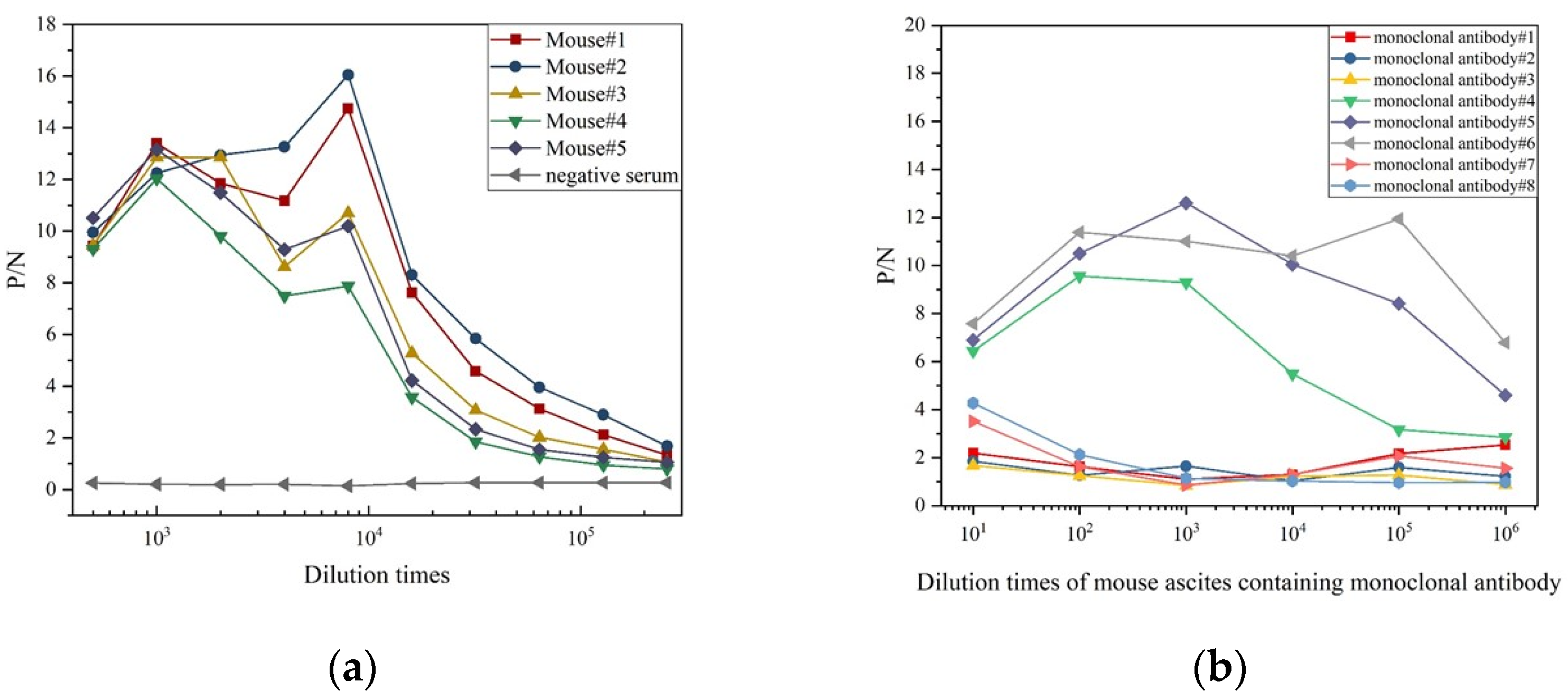
The graphical representation indicates a decline in P/N(positive/negative) values as the serum dilution factor rises (Figure 3a). Specifically, the sera from mice numbered 1 and 2 show the peak titers, maintaining P/N values above 2.1 even at a 128,000-fold dilution, suggesting a serum titer of approximately 128,000 for both mice. Conversely, mouse No. 4 showcases the lowest titer, approximated at 16,000. Overall, the five mice present with high serum titers, affirming the efficacy of the six immunization sessions. Consequently, the sera obtained from these mice are deemed appropriate for the generation of monoclonal antibodies specific to Alexandrium tamarense.
The titer assessment of eight monoclonal antibodies (MAbs) specifically targeting Alexandrium tamarense revealed that MAbs 1, 2, 3, 7, and 8 displayed P/N values below 2.1 at a 100-fold ascites dilution, signifying lower titer levels (Figure 3b). Conversely, MAbs 4, 5, and 6 maintained P/N values above 2.1 even at a 1,000,000-fold ascites dilution, demonstrating high titers. These findings affirm their suitability for the production of colloidal gold test strips.

3.2. The Structure of a Test Strip

As illustrated in Figure 4, the design of the test strip is anchored on a PVC support base. Adjacent to it is a glass fiber membrane serving as the sample pad, while the nitrocellulose (NC) membrane functions as the central detection zone. Sequentially adhered to the PVC base are the glass fiber sample pad and the conjugate pad, which contains monoclonal antibodies conjugated with colloidal gold specifically targeting Alexandrium tamarense. The NC membrane is meticulously coated at two distinct positions: the test line (T-line), harboring monoclonal antibodies specific to the algae, and the control line (C-line), coated with anti-mouse IgG antibodies. As shown in Figure 4a, upon adding a sample, it traverses the filter membrane through wicking action. If the sample contains the antigen, it will initially bind to the gold-labeled antibodies present in the glass fiber sample layer, forming a conjugate comprising the antigen, gold, and labeled antibody. This conjugate then migrates further and encounters the monoclonal antibodies specific to the target algae, forming a dual-antibody sandwich complex. The complex consists of a monoclonal antibody, an antigen, and a gold-labeled antibody, which triggers the staining of the T-line, thereby indicating a positive test result. Any excess gold-conjugated antibody will continue to diffuse and bind to the goat anti-mouse IgG antibody, resulting in C-line staining. Consequently, a positive test outcome is indicated. In contrast, as shown in Figure 4b, if the antigen is absent from the sample, the gold-labeled antibodies cannot bind to form a conjugate with the antigen and will not interact with the monoclonal antibodies specific to the algae at the T-line. In this scenario, the T-line remains colorless, whereas the C-line alone exhibits coloration due to the binding of the goat anti-mouse IgG antibodies with the gold-labeled antibodies, indicating a negative test result.
The presence or absence of coloration on the C-line can confirm the functionality of the test strip itself, ensuring that it is operational as intended. Whether the T-line develops coloration determines if the target antigen is present in the sample being tested, thereby indicating the presence or absence of the targeted algal species.

3.3. Performance Verification of Test Strips

3.3.1. Effectiveness of the Test Strips

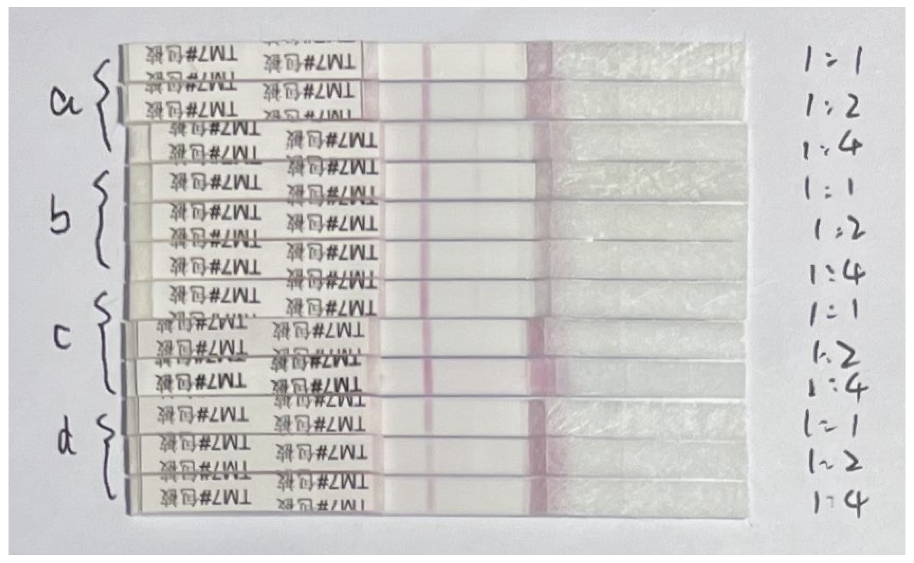
To ascertain the efficacy of the test strips, a series of experimental preparations were undertaken involving various fractions of the algae, encompassing the algal cell suspension (Figure 5a), standardized algal cell solutions (Figure 5b), algal cell extracts (Figure 5c), and the algal cell culture medium (Figure 5d). Gradient dilutions of the prepared Alexandrium tamarense fractions were performed in two sets, yielding solutions with dilution ratios of 1:1, 1:2, and 1:4. A volume of 60 μL from each of these solutions was directly applied to the sample pad of the colloidal gold strip. The strips were then subjected to an incubation period spanning from 3 to 15 min for the assessment of the results. Post stabilization of the chromogenic reaction, a qualitative evaluation was conducted to ascertain the presence and intensity of coloration in the test line (T-line) and control line (C-line). The outcomes of these test strip reactions are illustrated in Figure 5. Among the four algal components—namely, the algal cell solution, the algal cell standard solution, the algal cell culture medium, and the algal cell extract—the raw algal solution and the standard algal cell solution elicited a positive response on the test strip, while the algal cell extract provoked a subtle but discernible chromogenic reaction. The intensity of the coloration was notably augmented with increasing concentration (Figure 5). This substantiates that the test strip possesses the specificity to detect whole algal cells, exhibiting a diminished response to disrupted cells and the byproducts of their metabolism.
The algal cell solution from Alexandrium tamarense was sourced from pure algal cultures, encompassing algal cells along with the culture medium and antigens synthesized during the cultivation process. The standard algal cell solution consists exclusively of Alexandrium tamarense cells. The culture medium of algal cells predominantly harbors antigens secreted by these cells into the extracellular milieu. On the other hand, algal cell extracts are primarily composed of antigens derived from the inner and outer surfaces of Alexandrium tamarense cells. Notably, the test strip demonstrates the proficiency to selectively identify intact cells of Alexandrium tamarense. The algal cell suspension (Figure 5a) elicited a reaction on the test strip, characterized by the most vivid chromogenic effect. Subsequently, the algal cell standard solution (Figure 5b) produced a notably substantial chromogenic response on the test strip.
The experimental results of the replication experiments can be found in Supplementary Figures S1–S3.

3.3.2. Sensitivity of the Test Strips

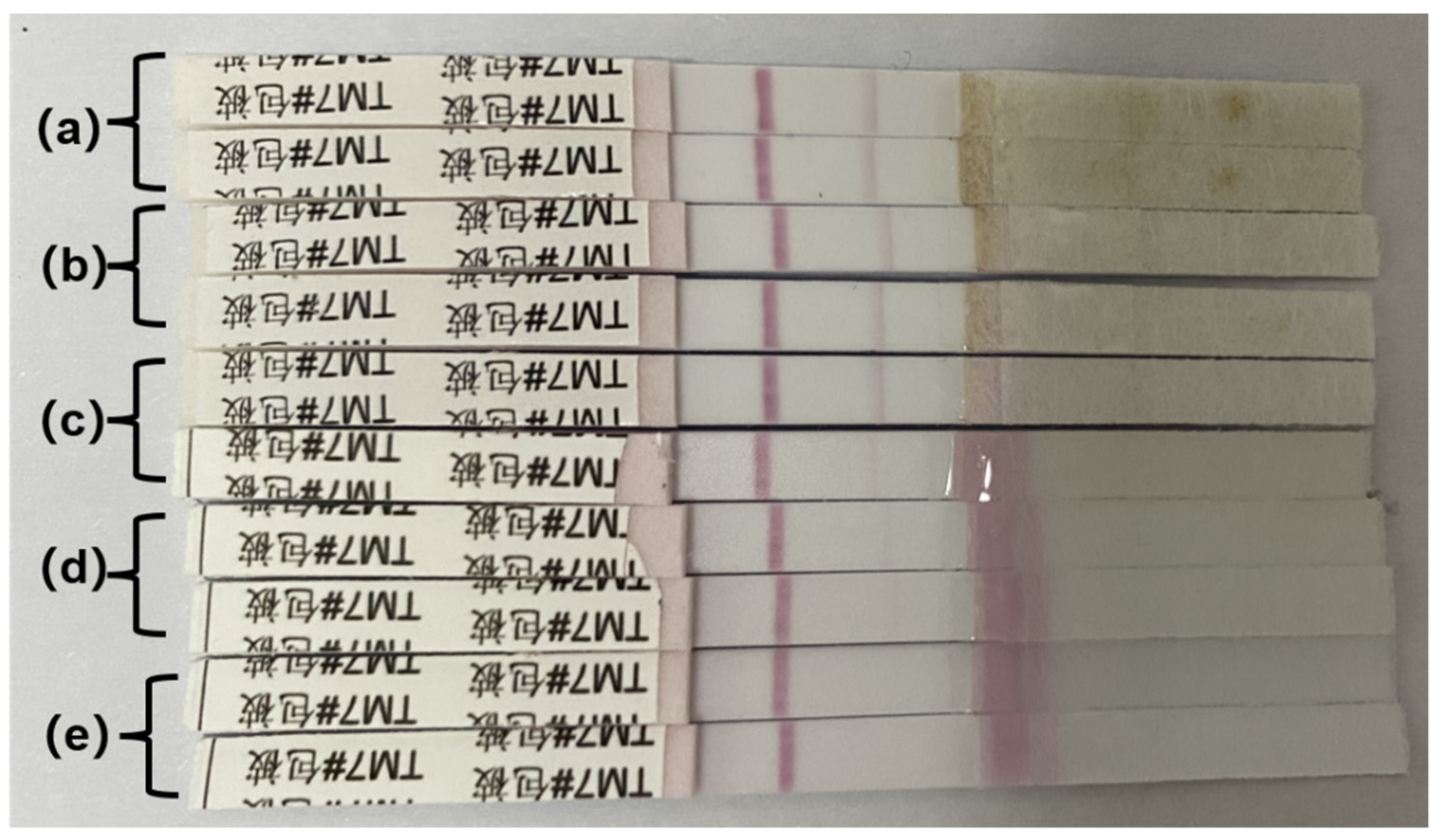
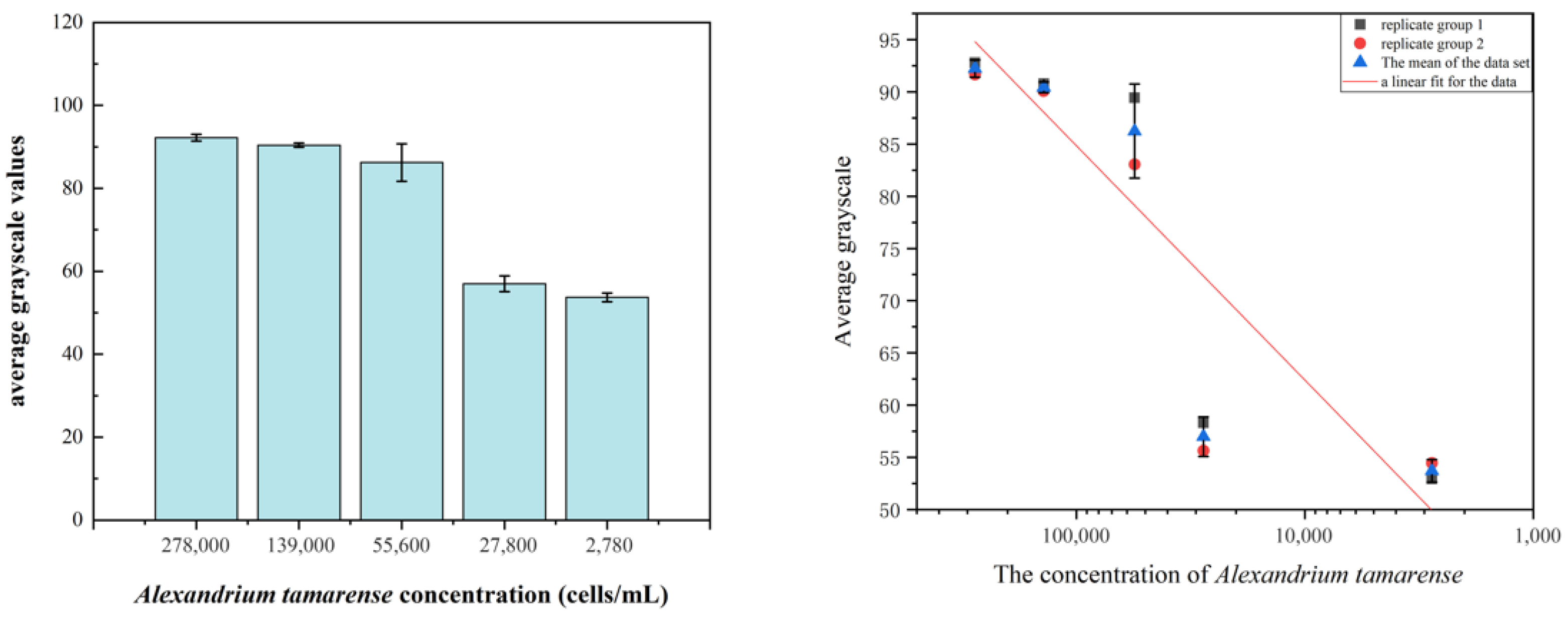
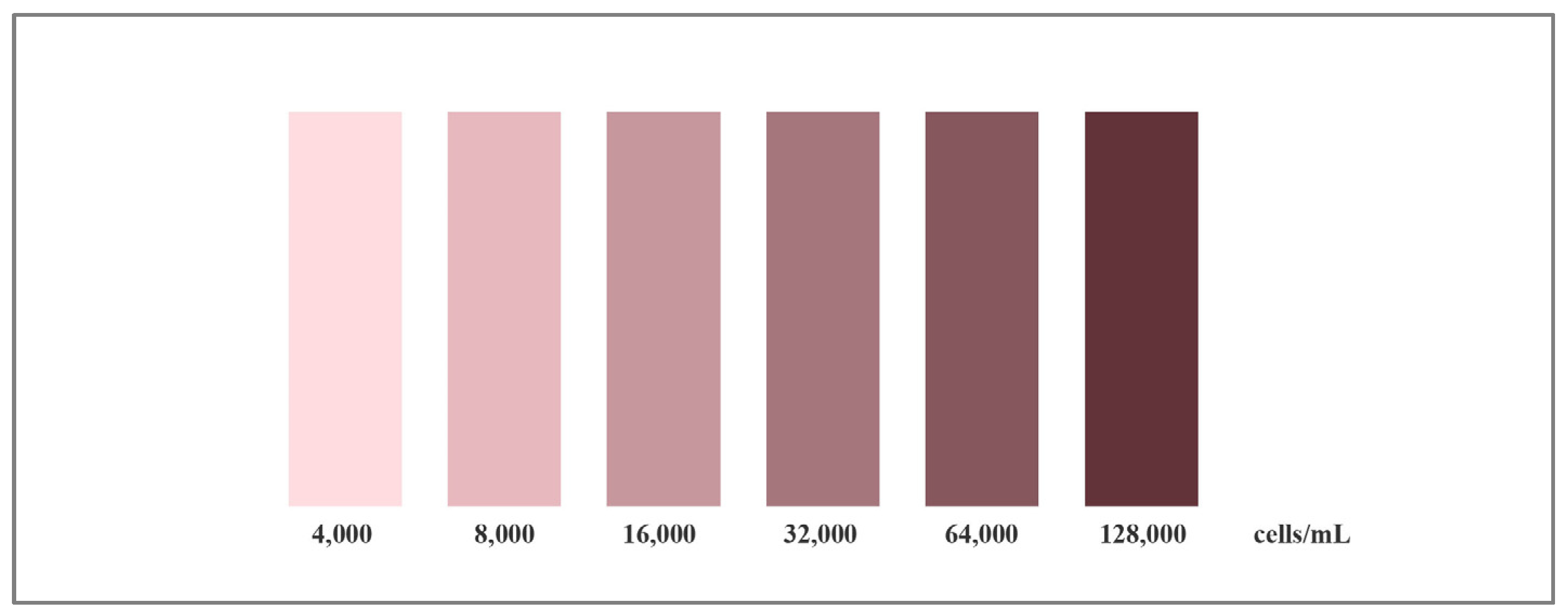
The detection results elucidating the sensitivity of the Alexandrium tamarense test strip are graphically represented as follows: Figure 6d at a detection concentration of 2.78 × 10⁴ cells/mL, the T-line of the test strip exhibits a subtle coloration. Conversely, at a detection concentration of 2.78 × 10³ cells/mL, the T-line of the test strip fails to manifest any coloration (Figure 6e). This observation implies that the detection limit for the Alexandrium tamarense test strip is approximately 2.78 × 10⁴ cells/mL. The grayscale scanning outcomes reveal that the intensity of T-line coloration diminishes as the concentration of Alexandrium tamarense decreases. When encountering higher concentrations, the variance in T-line coloration is notably less pronounced. Relying on the average grayscale data (Figure 7) derived from the scans, a standard curve delineating the relationship between the average grayscale values and algal concentration can be constructed. According to this standard curve, a standard colorimetric chart for Alexandrium tamarense (Figure 8) was developed. This facilitates the determination of the concentration range of the tested Alexandrium tamarense through a simple visual assessment of the test strip’s coloration.
The experimental results of the replication experiments can be found in Supplementary Figures S4–S6.

3.3.3. Specificity of the Test Strips

The validation of specificity for the Alexandrium tamarense test strip indicated favorable specificity for Alexandrium tamarense. And it also does not cross-react with other algae: Gymnodinium catenatum, Karenia mikimotoi, Skeletonema costatum, Prorocentrum lima, and Prorocentrum donghaiense. However, it exhibited a notable cross-reaction with Alexandrium catenella (Figure 9A(e)).
The experimental results of the replication experiments can be found in Supplementary Figures S7 and S8.
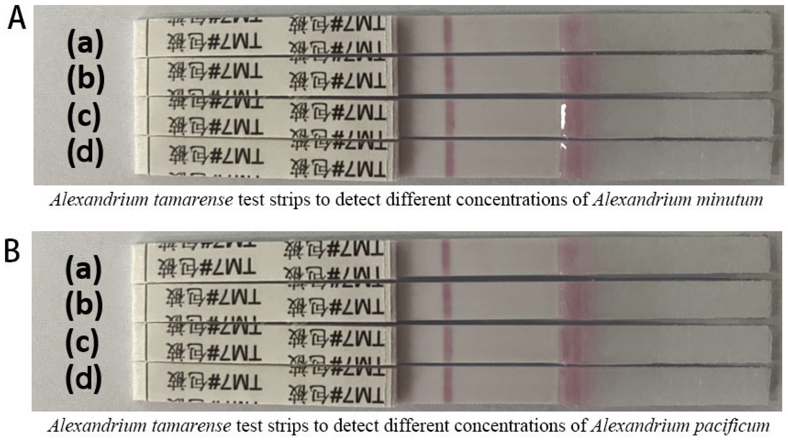
To determine the lower limit of cross-reactivity of the Alexandrium tamarense test strip with Alexandrium catenella, a dilution series was conducted on a suspension of Alexandrium catenella at a concentration of 2.22 × 10⁵ cells/mL, which was subsequently tested using the Alexandrium tamarense test strip. The detection outcomes (Figure 9B(d)) demonstrated that the lower limit of cross-reactivity was established at 2.22 × 10² cells/mL, signifying substantial cross-reactivity between the test strip and Alexandrium catenella. Given that both Alexandrium tamarense and Alexandrium catenella are from the Alexandrium genus of dinoflagellates, to determine whether the Alexandrium tamarense test strip shows cross-reactivity with other species within the Alexandrium genus, tests were performed using the strip on Alexandrium pacificum and Alexandrium minutum across different concentrations. The outcomes of these experiments, depicted in Figure 10A,B, reveal that neither A. pacificum nor A. minutum could produce a coloration response on the test strip’s T-line. This indicates that the A. tamarense test strip does not exhibit cross-reactivity with A. minutum and A. pacificum, demonstrating high specificity for A. tamarense. This facilitates the macroscopic differentiation between these species and Alexandrium tamarense.

3.3.4. Field Application of the Test Strips

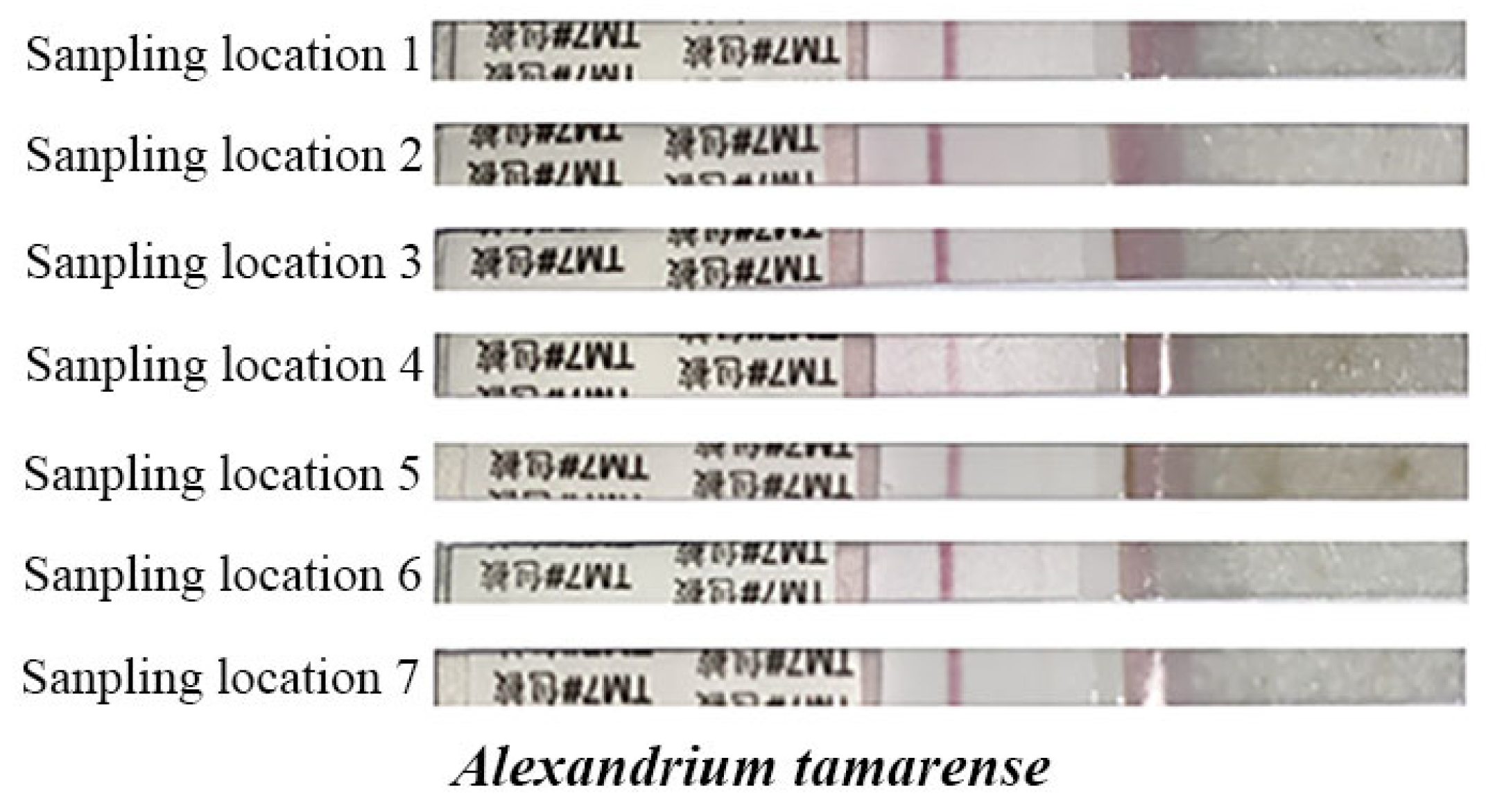
The detection results (Figure 11) showed that after enrichment of the seawater samples collected from Dongshan Bay, only the seawater from site 6 triggered the coloring of the T-line on the Alexandrium tamarense test strip. The detection results demonstrate the applicability of the test strip for on-site testing. They also confirm that the detection limit of algal concentration by the test strip can be further reduced through the process of concentrating and enriching the sample. This allows for obtaining detection results when the algal concentration in the water body is low, enabling the estimation of algal concentration in the water. This, in turn, facilitates the prediction and prevention of red tide occurrences.

4. Discussion

In this study, we employed hybridoma cell technology to develop monoclonal and polyclonal antibodies specifically targeting the toxin-producing Alexandrium tamarense. A colloidal gold-based rapid detection test strip was subsequently developed for the specific detection of this algal species. The effectiveness of the antibodies and the test strip was thoroughly investigated and validated. High-titer monoclonal antibodies were successfully produced, with the highest titer recorded at an impressive 128,000. The colloidal gold test strip, constructed using these antibodies, underwent experimental validation and exhibited a strong positive response to whole Alexandrium tamarense cells, confirming its specificity.
Sensitivity tests revealed that the test strip could detect Alexandrium tamarense at concentrations as low as 2.78 × 10⁴ cells/mL. Importantly, the test strip exhibited no cross-reactivity with other algal species, such as Gymnodinium catenatum, Karenia mikimotoi, Skeletonema costatum, Peridinium lima, and Prorocentrum donghaiense. These findings underscore the test strip’s high sensitivity and specificity, making it a promising tool for algal detection in aquatic environments.
Compared to the detection test strips prepared by Fabienne Gas et al. for Alexandrium minutum [13], the test strips developed in this study show slightly lower sensitivity but maintain strong selectivity for Alexandrium tamarense. The cross-reactivity with Alexandrium catenella was presumed to be due to their belonging to Alexandrium, and there seems to be some controversy regarding the nomenclature of Alexandrium catenella species [24]. The algae strains may share identical antigenic determinants. The monoclonal antibodies we prepared happened to bind with both, leading to cross-reactivity of the test strips with the non-target algal species Alexandrium catenella. Although the test strips developed in this study are somewhat lacking in sensitivity, this can be compensated for by simple filtration and concentration of seawater samples. Overall, the test strips prepared in this study provide a simple and convenient method for the on-site detection of Alexandrium tamarense in seawater, which has been proven feasible through our practical testing and holds significant practical value and broad application prospects.

5. Conclusions

The colloidal gold test strip enables a visible color change within 10–15 min, providing a simple and efficient detection process. Semi-quantitative estimations of algal concentrations can be achieved by comparing the results to a standardized colorimetric chart. While the test strip demonstrates certain limitations, these can be mitigated through specific approaches. For example, its sensitivity can be enhanced by pre-concentrating and enriching water samples, facilitating the detection of lower algal densities. Additionally, the test strip exhibits cross-reactivity with Alexandrium catenella but does not produce positive results with the Pacific and Minuscule Alexandrium species, allowing for differentiation. The cross-reactivity observed with Alexandrium catenella may result from shared epitopes targeted during monoclonal antibody preparation, with minimum cross-reaction thresholds identified at 2.78 × 10³ cells/mL.
By focusing on the toxic red tide-producing alga Alexandrium tamarense, This study involved immunizing mice with whole Alexandrium tamarense to prepare monoclonal antibodies specific to AT, with the highest titer ratio of P/N values reaching up to 12.6. We selected antibodies with higher titers and successfully prepared colloidal gold test strips. Furthermore, this study successfully developed rapid detection strips utilizing colloidal gold immunochromatography technology. The experiments confirmed the efficiency, sensitivity, and specificity of the test strips. These strips are rapid and user-friendly, requiring no specialized equipment, thus making them exceptionally suitable for on-site testing. This innovation provides an effective and reliable approach for monitoring and early warning of toxic red tides in coastal areas, offering substantial potential for practical applications.
The colloidal gold test strips developed in this study provide a novel and practical solution for the detection of Alexandrium tamarense in aquatic environments. By leveraging the high sensitivity and specificity of monoclonal and polyclonal antibodies, the test strips enable the rapid identification of toxic red tide algae without the need for complex equipment or procedures. This approach is particularly valuable for on-site applications, offering real-time monitoring capabilities that are essential for mitigating the environmental and economic impacts of red tide outbreaks. In addition to its current application, the test strip technology holds the potential to be adapted for the detection of other harmful algal species, contributing to broader efforts in marine ecosystem management.
This study underscores the importance of developing accessible and user-friendly detection tools to support sustainable coastal resource management. By utilizing test strips that are specifically designed for red tide species prevalent in various geographical regions, more targeted red tide prevention strategies can be deployed. This approach is highly promising for its application in marine environmental monitoring, aquaculture, and safeguarding public health, particularly in coastal regions.

Supplementary Materials

The following supporting information can be downloaded at: https://www.mdpi.com/article/10.3390/chemosensors13020053/s1, Figure S1. The effectiveness test results of Alexandrium tamarense; Figure S2. The effectiveness test results of Alexandrium tamarense (2025/1/16); Figure S3. The effectiveness test results of Alexandrium tamarense (2025/1/16); Figure S4. The sensitivity results of Alexandrium tamarense test strips (2024/8/10); Figure S5. The sensitivity results of Alexandrium tamarense test strips (2025/1/17); Figure S6. The sensitivity results of Alexandrium tamarense test strips (2025/1/17); Figure S7. Specific detection results of Alexandrium tamarense (2024/8/10); Figure S8. Specific detection results of Alexandrium tamarense (2025/1/17).

Author Contributions

All authors contributed to the study’s conception and design. Conceptualization, K.Z. and C.Z.; methodology, C.Z. and J.F.; validation, Z.K. and J.M.; formal analysis, J.F. and Z.K.; investigation, J.M.; resources, C.Z. and K.Z.; data curation, Z.K. and J.M.; writing—original draft preparation, Z.K. and J.F.; writing—review and editing, J.M. and Z.K.; visualization, C.Z. and Z.K.; supervision, K.Z.; project administration, K.Z.; funding acquisition, C.Z. and K.Z. All authors have read and agreed to the published version of the manuscript.

Funding

This study was supported by the National Key Research and Development Program of China (2017YFCO506100).

Institutional Review Board Statement

Animals were bred and maintained in the SPF-grade laboratory at Xiamen University Animal Center, under standard housing conditions with regular feed and water. During the experimental process, the rearing of animals and the conduct of experiments complied with five relevant laws and regulations, such as “Technical Specification for Ethical Review of Laboratory Animal Welfare” (DB11T1734-2020). Appropriate clean drinking water and food necessary to maintain health and vitality were provided to the reared animals; suitable habitats were offered to enable the animals to rest and sleep comfortably; it was ensured that they were in good condition and properly handled (including during the experimental process and euthanasia); sufficient space and appropriate facilities were provided; and they were grouped with their fellow animals so that the animals could freely express their normal behaviors. The ethical code of the animal experiment in this paper is XMULAC20220020.

Data Availability Statement

No new data were created.

Acknowledgments

This work was supported by the Fujian Fishery Resources Monitoring Center for offshore toxic (harmful) red tide algae. The authors gratefully acknowledge comments from Kefu Zhou that greatly improved the manuscript. The authors gratefully acknowledge the technical support provided by Xiamen Boson Biotech. The authors are grateful to the Center for Collections of Marine Bacteria and Phytoplankton (CCMBP) of Xiamen University for supplying the algae for our research.

Conflicts of Interest

The funders had no role in the design of the study; in the collection, analysis, or interpretation of the data; in the writing of the manuscript; or in the decision to publish the results. Author Changgong Zhang is employed by the Xiamen Boson Biotech Co., Ltd. The remaining authors declare that the research was conducted in the absence of any commercial or financial relationships that could be construed as a potential conflict of interest.

References

  1. Deeds, J.R.; Landsberg, J.H.; Etheridge, S.M.; Pitcher, G.C.; Longan, S.W. Non-Traditional Vectors for Paralytic Shellfish Poisoning. Mar. Drugs 2008, 6, 308–348. [Google Scholar] [CrossRef]
  2. Anderson, D.M.; Alpermann, T.J.; Cembella, A.D.; Collos, Y.; Masseret, E.; Montresor, M. The Globally Distributed Genus Alexandrium: Multifaceted Roles in Marine Ecosystems and Impacts on Human Health. Harmful Algae 2012, 14, 10–35. [Google Scholar] [CrossRef] [PubMed]
  3. Genovesi, B.; Shin-Grzebyk, M.-S.; Grzebyk, D.; Laabir, M.; Gagnaire, P.-A.; Vaquer, A.; Pastoureaud, A.; Lasserre, B.; Collos, Y.; Berrebi, P.; et al. Assessment of Cryptic Species Diversity within Blooms and Cyst Bank of the Alexandrium tamarense Complex (Dinophyceae) in a Mediterranean Lagoon Facilitated by Semi-Multiplex PCR. J. Plankton Res. 2011, 33, 405–414. [Google Scholar] [CrossRef]
  4. Lilly, E.L.; Halanych, K.M.; Anderson, D.M. Species Boundaries and Global Biogeography of the Alexandrium tamarense Complex (Dinophyceae). J. Phycol. 2007, 43, 1329–1338. [Google Scholar] [CrossRef]
  5. Song, H.W.; Lee, S.; Park, Y.K.; Woo, S.Y. Quantitative Sasang Constitution Diagnosis Method for Distinguishing between Tae-eumin and Soeumin Types Based on Elasticity Measurements of the Skin of the Human Hand. Evid.-Based Complement. Altern. Med. 2009, 6, 93–98. [Google Scholar] [CrossRef] [PubMed]
  6. Soto-Liebe, K.; Méndez, M.A.; Fuenzalida, L.; Krock, B.; Cembella, A.; Vásquez, M. PSP Toxin Release from the Cyanobacterium Raphidiopsis brookii D9 (Nostocales) Can Be Induced by Sodium and Potassium Ions. Toxicon 2012, 60, 1324–1334. [Google Scholar] [CrossRef]
  7. Hosoi-Tanabe, S.; Sako, Y. Species-Specific Detection and Quantification of Toxic Marine Dinoflagellates Alexandrium tamarense and A. catenella ByReal-Time PCR Assay. Mar. Biotechnol. 2005, 7, 506–514. [Google Scholar] [CrossRef]
  8. Nagai, S.; Itakura, S. Specific Detection of the Toxic Dinoflagellates Alexandrium tamarense and Alexandrium catenella from Single Vegetative Cells by a Loop-Mediated Isothermal Amplification Method. Mar. Genom. 2012, 7, 43–49. [Google Scholar] [CrossRef]
  9. Ruvindy, R.; Bolch, C.J.; MacKenzie, L.; Smith, K.F.; Murray, S.A. QPCR Assays for the Detection and Quantification of Multiple Paralytic Shellfish Toxin-Producing Species of Alexandrium. Front. Microbiol. 2018, 9, 3153. [Google Scholar] [CrossRef]
  10. Watanabe, R.; Matsushima, R.; Harada, T.; Oikawa, H.; Murata, M.; Suzuki, T. Quantitative Determination of Paralytic Shellfish Toxins in Cultured Toxic Algae by LC-MS/MS. Food Addit. Contam. Part A 2013, 30, 1351–1357. [Google Scholar] [CrossRef]
  11. Chen, G.F.; Zhang, C.Y.; Wang, Y.Y.; Chen, W. Application of Reverse Dot Blot Hybridization to Simultaneous Detection and Identification of Harmful Algae. Environ. Sci. Pollut. Res. 2015, 22, 10516–10528. [Google Scholar] [CrossRef] [PubMed]
  12. Chin Chwan Chuong, J.J.; Rahman, M.; Ibrahim, N.; Heng, L.Y.; Tan, L.L.; Ahmad, A. Harmful Microalgae Detection: Biosensors versus Some Conventional Methods. Sensors 2022, 22, 3144. [Google Scholar] [CrossRef] [PubMed]
  13. Gas, F.; Baus, B.; Pinto, L.; Compere, C.; Tanchou, V.; Quéméneur, E. One Step Immunochromatographic Assay for the Rapid Detection of Alexandrium minutum. Biosens. Bioelectron. 2010, 25, 1235–1239. [Google Scholar] [CrossRef] [PubMed]
  14. Tsao, Z.-J.; Liao, Y.-C.; Liu, B.-H.; Su, C.-C.; Yu, F.-Y. Development of a Monoclonal Antibody against Domoic Acid and Its Application in Enzyme-Linked Immunosorbent Assay and Colloidal Gold Immunostrip. J. Agric. Food Chem. 2007, 55, 4921–4927. [Google Scholar] [CrossRef] [PubMed]
  15. Zhou, Y.; Pan, F.-G.; Li, Y.-S.; Zhang, Y.-Y.; Zhang, J.-H.; Lu, S.-Y.; Ren, H.-L.; Liu, Z.-S. Colloidal Gold Probe-Based Immunochromatographic Assay for the Rapid Detection of Brevetoxins in Fishery Product Samples. Biosens. Bioelectron. 2009, 24, 2744–2747. [Google Scholar] [CrossRef] [PubMed]
  16. Li, H.; Meng, K.; Yu, Q.; Chen, C.; Chen, J.; Li, J.; Wang, X.; Wang, Y.; Li, M.; Chen, C.; et al. Rapid Detection Method of Skeletonema pseudocostatum and Preparation of Test Strip. Environ. Sci. Pollut. Res. 2022, 29, 70202–70208. [Google Scholar] [CrossRef]
  17. Hnasko, R. (Ed.) ELISA: Methods and Protocols; Springer: New York, NY, USA, 2015; Volume 1318, ISBN 978-1-4939-2741-8. [Google Scholar]
  18. Wu, Q.; Guo, X.; Huang, Q.; Xie, Y.; Guo, L.; Yang, X.; Sun, M.; Yin, D. Development of a Colloidal Gold Immunochromatographic Test Strip for Detecting the Smooth Brucella. Sci. Rep. 2024, 14, 25068. [Google Scholar] [CrossRef]
  19. Zhang, C. Hybridoma Technology for the Generation of Monoclonal Antibodies. In Antibody Methods and Protocols; Springer: Berlin/Heidelberg, Germany, 2012; pp. 117–135. [Google Scholar]
  20. Chuanlai, X.; Huting, W.; Chifang, P.; Zhengyu, J.; Liqiang, L. Colloidal Gold-Based Immumochromatographic Assay for Detection of Diethylstilbestrol Residues. Biomed. Chromatogr. 2006, 20, 1390–1394. [Google Scholar] [CrossRef]
  21. Omidfar, K.; Kia, S.; Larijani, B. Development of a Colloidal Gold-Based Immunochromatographic Test Strip for Screening of Microalbuminuria. Hybridoma 2011, 30, 117–124. [Google Scholar] [CrossRef]
  22. Guo, L.; Song, S.; Liu, L.; Peng, J.; Kuang, H.; Xu, C. Comparsion of an Immunochromatographic Strip with ELISA for Simultaneous Detection of Thiamphenicol, Florfenicol and Chloramphenicol in Food Samples. Biomed. Chromatogr. 2015, 29, 1432–1439. [Google Scholar] [CrossRef]
  23. Lin, L.; Song, S.; Wu, X.; Liu, L.; Kuang, H. A Colloidal Gold Immunochromatography Test Strip Based on a Monoclonal Antibody for the Rapid Detection of Triadimefon and Triadimenol in Foods. Food Agric. Immunol. 2020, 31, 447–462. [Google Scholar] [CrossRef]
  24. John, U.; Litaker, R.W.; Montresor, M.; Murray, S.; Brosnahan, M.L.; Anderson, D.M. Formal Revision of the Alexandrium tamarense Species Complex (Dinophyceae) Taxonomy: The Introduction of Five Species with Emphasis on Molecular-Based (RDNA) Classification. Protist 2014, 165, 779–804. [Google Scholar] [CrossRef]
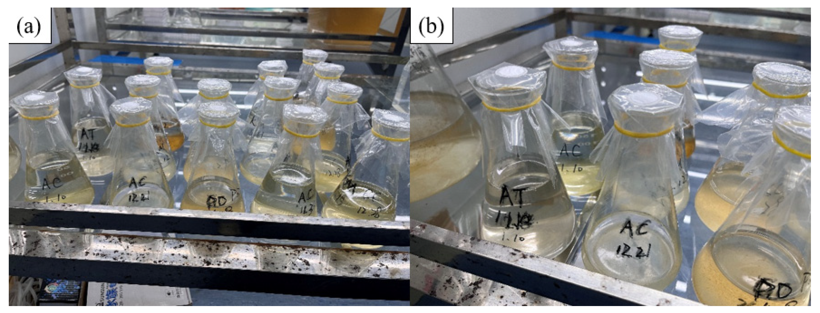
Figure 1. The cultivation of algae was conducted in the Algae Species Room of the College of Environment and Ecology, Xiamen University. (a) Full view of algae culture, (b) Partial view of algae culture.
Chemosensors 13 00053 g001
Figure 2. Distribution map of sampling and monitoring sites along the coast of Fujian Province.
Chemosensors 13 00053 g002
Figure 3. The titer of polyclonal antibodies and monoclonal antibodies against Alexandrium tamarense: (a) the titer of polyclonal antibodies against Alexandrium tamarense and (b) the titer of monoclonal antibodies against Alexandrium tamarense.
Chemosensors 13 00053 g003
Figure 4. Structure of the test strip and the principle of the reaction process: (a) positive reaction and (b) negative reaction.
Chemosensors 13 00053 g004
Figure 5. The effectiveness test results of Alexandrium tamarense: (a) the algal cell suspension with dilutions of 1:1, 1:2, and 1:4; (b) standardized algal cell solutions with dilutions of 1:1, 1:2, and 1:4; (c) algal cell extracts with dilutions of 1:1, 1:2, and 1:4; (d) the algal cell culture medium with dilutions of 1:1, 1:2, and 1:4.
Chemosensors 13 00053 g005
Figure 6. The sensitivity result of Alexandrium tamarense test strips. The concentration of Alexandrium tamarense (each group had two test strips set up for replication): (a) 2.78 × 10⁵ cells/mL; (b) 1.39 × 10⁵ cells /mL; (c) 5.56 × 10⁴ cells/mL; (d) 2.78 × 10⁴ cells/mL; (e) 2.78 × 10³ cells /mL.
Chemosensors 13 00053 g006
Figure 7. Sensitivity grayscale scanning results of Alexandrium tamarense test strips.
Chemosensors 13 00053 g007
Figure 8. Standard colorimetric chart for Alexandrium tamarense (Alexandrium tamarense of different concentrations will display bands of varying color intensity (T-Line) on the test strip).
Chemosensors 13 00053 g008
Figure 9. (A) Specific detection results of Alexandrium tamarense: (a) Karenia mikimotoi; (b) Gymnodinium catenatum; (c) Skeletonema costatum; (d) Prorocentrum donghaiense; (e) Alexandrium catenella. (B) Results of using Alexandrium tamarense test strips to detect different concentrations of Alexandrium catenella. The concentration of Alexandrium catenella: (a) 2.22 × 10⁵ cells/mL (b) 2.22 × 10⁴ cells/mL; (c) 2.22 × 10³ cells/mL; (d) 2.22 × 10² cells/mL; (e) 111 cells/mL.
Chemosensors 13 00053 g009
Figure 10. Results of using Alexandrium tamarense test strips to detect different concentrations of Alexandrium minutum and Alexandrium pacificum: (A) the concentration of Alexandrium minutum; (a) 6 × 10⁴ cells/mL; (b) 6 × 10³ cells/mL; (c) 6 × 10² cells/mL; (d) 60 cells/mL. (B) The concentration of Alexandrium pacificum: (a) 1.8 × 10⁴ cells/mL; (b) 1.8 × 10³ cells/mL; (c) 1.8 × 10² cells/mL; (d) 18 cells/mL.
Chemosensors 13 00053 g010
Figure 11. Detection results of Alexandrium tamarense in Dongshan Bay.
Chemosensors 13 00053 g011
Disclaimer/Publisher’s Note: The statements, opinions and data contained in all publications are solely those of the individual author(s) and contributor(s) and not of MDPI and/or the editor(s). MDPI and/or the editor(s) disclaim responsibility for any injury to people or property resulting from any ideas, methods, instructions or products referred to in the content.