Article

Exploration of the Potential Targets and Molecular Mechanism of Carthamus tinctorius L. for Liver Fibrosis Based on Network Pharmacology and Molecular Docking Strategy

1
School of Basic Medical Sciences, Chengdu University of Traditional Chinese Medicine, 1166 Liutai Avenue, Wenjiang District, Chengdu 611137, China
2
College of Traditional Chinese Medicine, Hainan Medical University, No. 3 Xueyuan Road, Longhua District, Haikou 571199, China
*
Author to whom correspondence should be addressed.
These authors contributed equally to this work.
Processes 2022, 10(9), 1735; https://doi.org/10.3390/pr10091735
Submission received: 8 August 2022 / Revised: 25 August 2022 / Accepted: 30 August 2022 / Published: 1 September 2022

Abstract

:
Carthamus tinctorius L. (Honghua, HH) is an herbal medicine and functional food widely used to treat chronic liver diseases, including liver fibrosis. By using network pharmacology and molecular docking experiments, the present study aims to determine the bioactive components, potential targets, and molecular mechanisms of HH for treating liver fibrosis. The components of HH were screened from the Traditional Chinese Medicine Systems Pharmacology Database and Analysis Platform and literature, and the SwissTargetPrediction database was used to predict the treatment targets of HH. Genecards and DisGeNET databases contained targets for liver fibrosis, and the STRING database provided networks of protein–protein interactions. Gene ontology and Kyoto Encyclopedia of Genes and Genomes pathway enrichment analyses were performed using the Database of Annotation, Visualization and Integrated Discovery. The protein–protein interactive network and drug–component–major target–pathway interactive network were visualized and analyzed by Cytoscape software. Finally, Autodock Vina and Discovery Studio software were used for molecular docking Validation. A total of 23 candidate bioactive compounds with 187 treatment targets of HH were acquired from the databases and literature. A total of 121 overlapping targets between HH and liver fibrosis were found to provide the molecular basis for HH on liver fibrosis. Quercetin, beta carotene, and lignan were identified as key components with targeting to ESR1, PIK3CA, and MTOR. HH is engaged in the intervention of various signaling cascades associated with liver fibrosis, such as PI3K/AKT/mTOR pathway, MAPK pathway, and PPAR pathway. In conclusion, HH treats liver fibrosis through multi-component, multi-target, and multi-pathway mechanisms.

1. Introduction

Liver fibrosis, caused by chronic liver injuries such as viral hepatitis, fatty liver disease, and cholestasis, is an abnormal response to repair the liver [1]. Liver fibrosis is characterized by excessive deposition of collagen and other extracellular matrix (ECM). With liver fibrosis processes, normal liver tissue is replaced by scar tissue, the structure is destroyed, and the function is impaired, which leads to further cirrhosis, liver failure, or even liver cancer, leading to death of patients [2]. As a result of non-alcoholic fatty liver disease, liver fibrosis is a major predictor of its mortality [3]. In addition, approximately one million people die from complications of cirrhosis every year, making it the 11th leading cause of death in the world [4]. Obviously, the end-stage liver disease related to liver fibrosis is becoming a tremendous public health challenge globally.
Liver fibrosis was considered to be an irreversible pathological event in the past. Remarkably, emerging evidence has suggested that the progression of fibrosis can be reversed after the removal of damaging factors [2]. Some basis of fibrosis resolution has been proven, including the interruption of harmful substances that cause chronic liver damage, elimination or inactivation of myofibroblasts, intervention of inflammatory responses, and the degradation of ECM [5]. Consequently, the early diagnosis and timely interventional treatment of liver fibrosis may be one of the important measures to resolve chronic liver diseases.
The activation and proliferation of hepatic stellate cells (HSCs) generate myofibroblasts, which is the main cellular source of ECM and the main driver of liver fibrosis [6]. In order to alleviate or reverse liver fibrosis, treatment strategies that inhibit the activation and proliferation of HSCs are necessary [1]. Advances have been reported in HSC based therapeutic approaches in recent years. Drugs including pirfenidone, sorafenib, and obeticholic acid have been shown to exhibit anti-fibrosis potential by inhibiting the activation of HSCs in clinical studies [7,8]. Some natural products have also exerted anti-fibrosis activities for liver fibrosis in preclinical studies, such as curcumin and ferulic acid [9,10]. However, specific drugs available for patients with liver fibrosis are still lacking in clinical practice, indicating an urgent demand to explore more effective and safe treatment strategies [11].
Herbal medicines containing natural products are receiving attention in the treatment of chronic liver diseases due to their multiple pharmacological activities [12]. Carthamus tinctorius L. (Honghua, HH) is a medicinal plant which has anti-inflammatory, antioxidant, and antitumor effects and is cultivated and applied in more than 60 countries around the world [13]. Moreover, HH has also a wide range of uses in the food fields, including as a natural colorant and food additive with potential benefits to human health [14,15]. In particular, HH is a traditional Chinese medicine used to stimulate blood circulation and remove stasis from the body, which has been used in cardiovascular diseases and liver diseases with a long history. Some traditional medicinal formulas containing HH have been applied for liver fibrosis with satisfactory results [16]. Moreover, hydroxysafflor yellow A in HH has exerted significant anti-HSC activation effect as a way to improve experimental liver fibrosis [17,18]. These finding provided evidence of the potential of HH for liver fibrosis; however, the molecular mechanisms still remain unclear.
Network pharmacology analyzes the association between drugs and diseases at the protein or systemic level, which is important to reveal the molecular mechanisms of multi-component and multi-target herbal medicines for the treatment of diseases [19]. In the present study, network pharmacology was used to understand the mechanisms of HH in liver fibrosis, and molecular docking experiments were applied to verify the affinity between ligands and receptors (Figure 1), with the hope of providing evidence for HH treatment of liver fibrosis and to provide a basis for further studies.

2. Materials and Methods

2.1. Prediction of the Bioactive Components and Treatment Targets of HH

Most of the bioactive components of HH were collected from the Traditional Chinese Medicine Systems Pharmacology Database and Analysis Platform (TCMSP, https://old.tcmsp-e.com/tcmsp.php (accessed on 1 June 2022)). The evaluation of absorption, distribution, metabolism, and excretion (ADME) has become an essential part of the drug discovery process, as drugs with good metabolic kinetic profiles are more likely to be potential treatment strategies [20]. Therefore, compounds with oral bioavailability (OB) greater than or equal to 30% and drug-likeness (DL) greater than or equal to 0.18 were identified as bioactive ingredients of HH [21,22]. In addition, hydroxysafflor yellow A, which have been demonstrated in the literature to have anti-fibrosis activity, was retained for further analysis. Subsequently, all the bioactive components were imported into SwissTargetsPrediction (http://swisstargetprediction.ch/ (accessed on 1 June 2022)) to predict the treatment targets of HH. All the targets were restricted to “Homo Sapiens”. Notably, the validation study of SwissTargetsPrediction showed that the first 15 predicted results have at least one target that has been experimentally validated for the majority of compounds, suggesting that the top 15 predictive genes are most likely to be the targets of molecules [23]. Therefore, the top 15 target genes predicted for each component were retained and identified as the treatment targets of HH.

2.2. Collection of the Targets of Liver Fibrosis

Two databases, Genecards (https://www.genecards.org/ (accessed on 2 June 2022)) and DisGeNET (https://www.disgenet.org/ (accessed on 2 June 2022)) were used to collect the disease targets. The targets obtained in Genecards were ranked according to the Relevance Score, and those that were larger than the median were retained. Next, the targets from the two databases were combined for subsequent analysis.

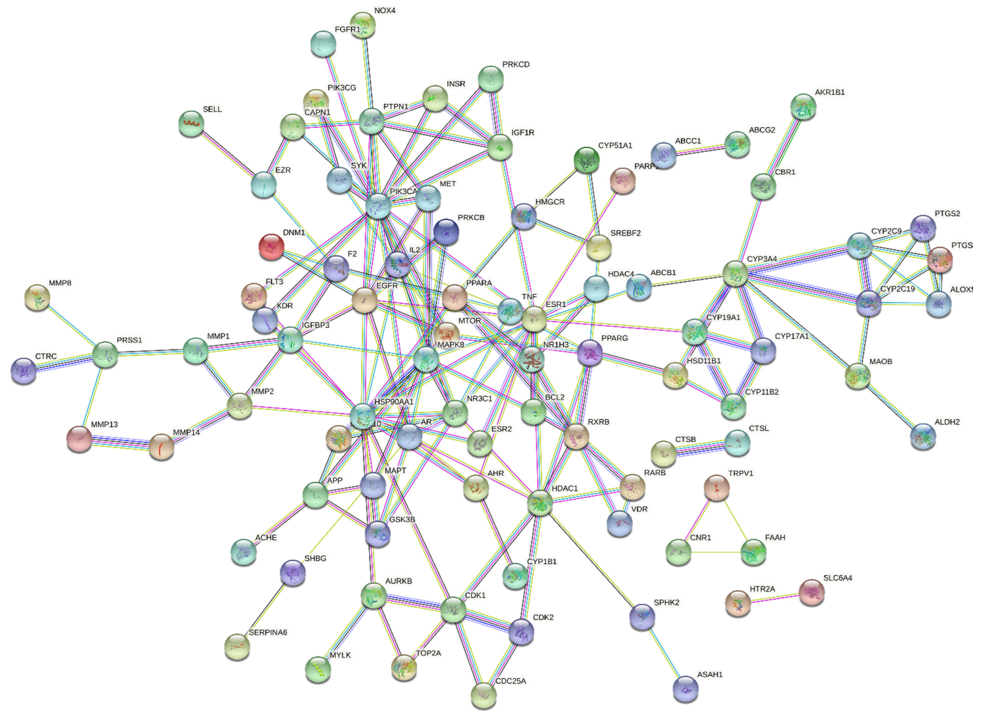
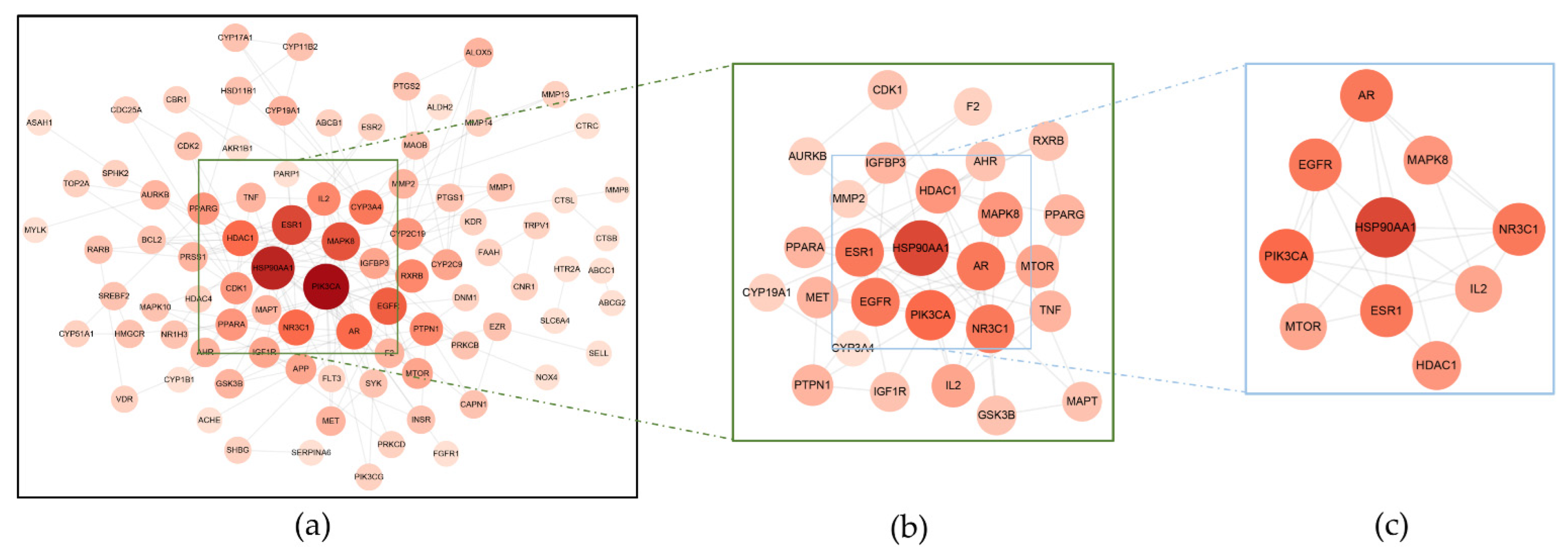
2.3. The Construction of the Protein–Protein Interactive (PPI) Network

The overlapping targets between HH and liver fibrosis were imported into the STRING database (https://cn.string-db.org/ (accessed on 5 June 2022)), which is dedicated to research of organism-wide protein association networks. In the STRING database, each predicted protein–protein interaction is assigned an association score between 0 and 1. The score is rated based on supporting evidence and reflects the degree of confidence that an interaction is biologically meaningful, specific, and reproducible [24]. Higher confidence score means less interaction and less false positives [25]. Therefore, the organisms were set to Homo sapiens; confidence was set to the highest (>0.900); and independent nodes were hidden to obtain the PPI network. The PPI network was transferred to Cytoscape (version 3.9.1, the National Institute of General Medical Sciences, Bethesda, MD, USA) software for visualization and consequent analysis.

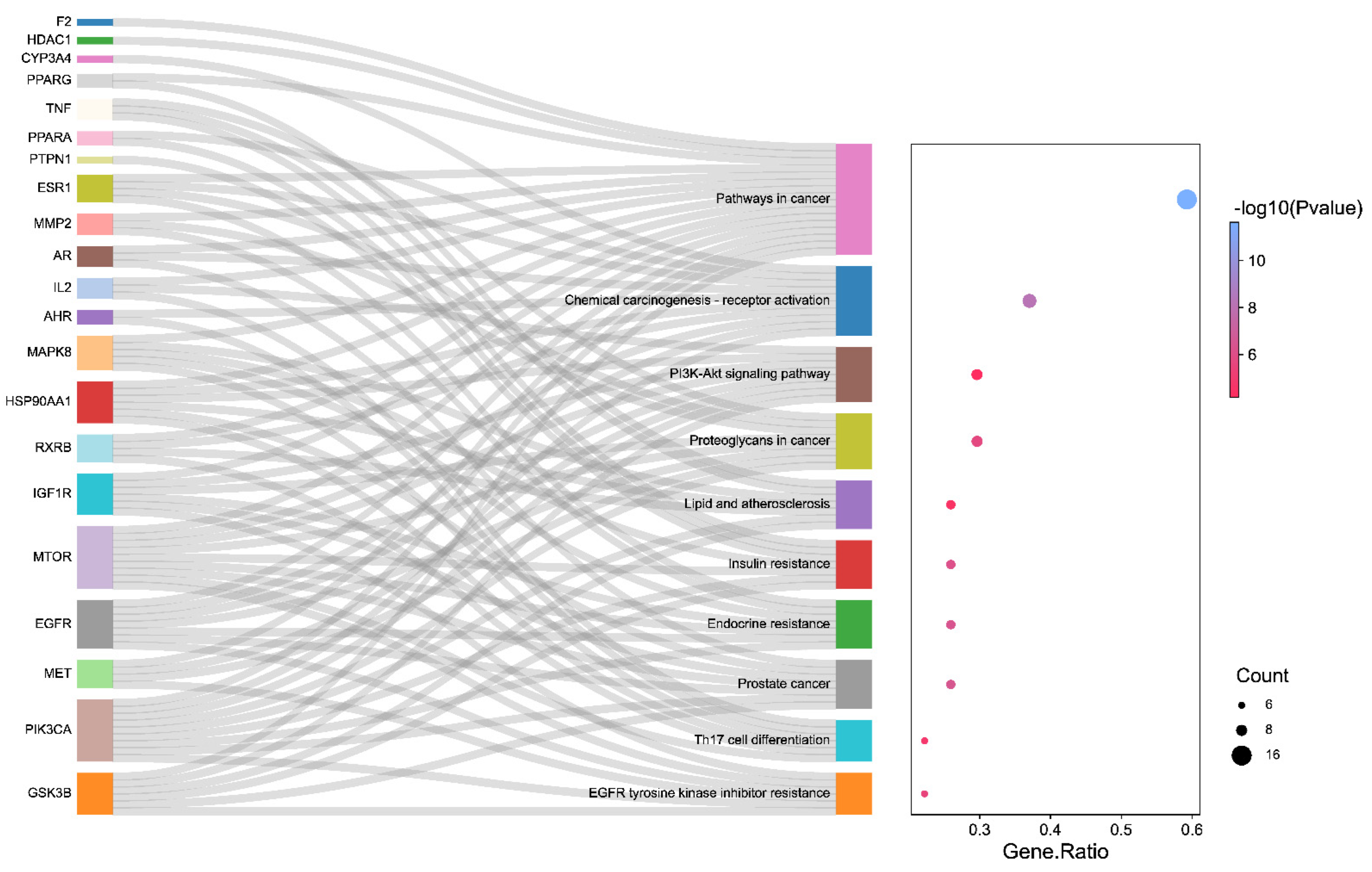
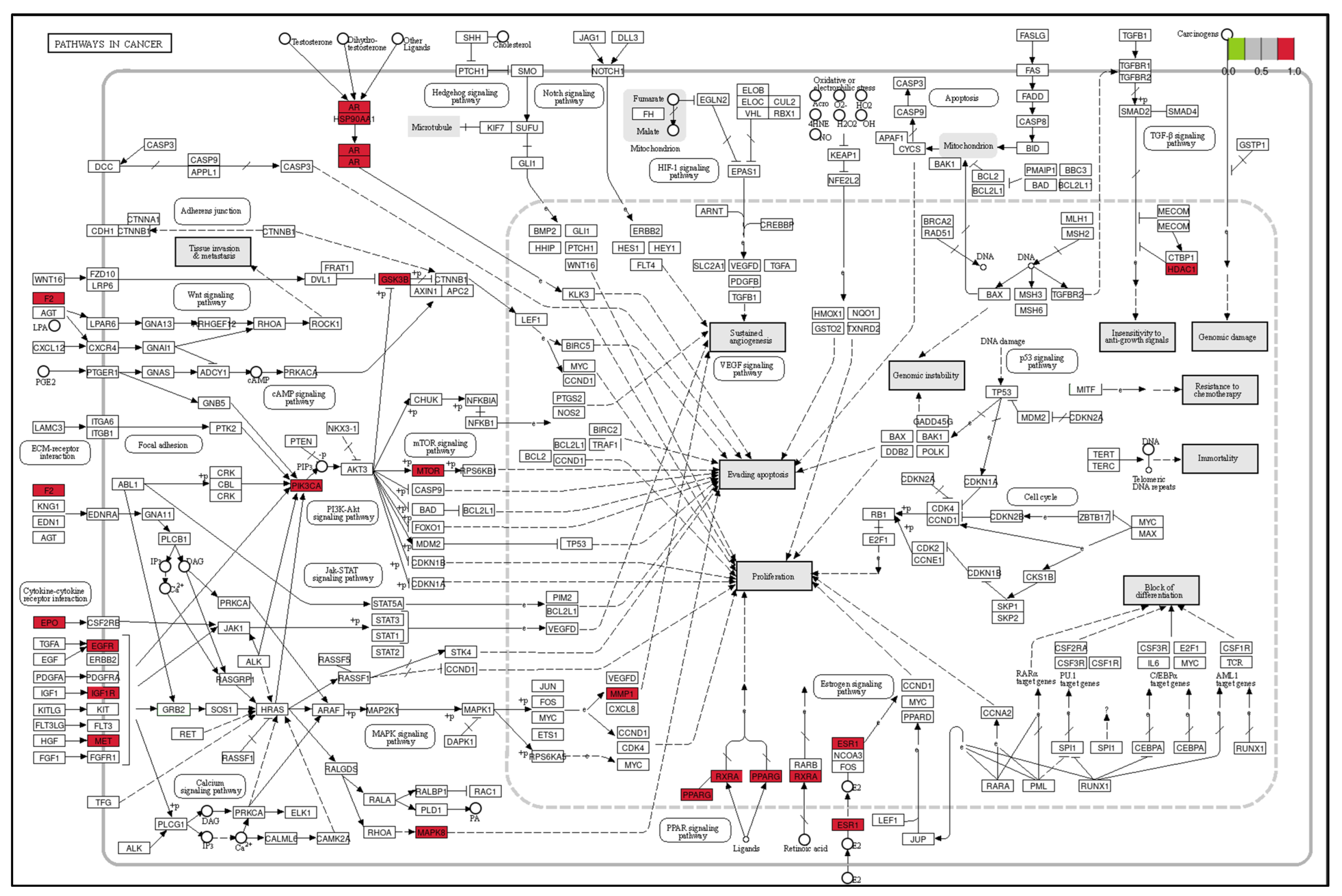
2.4. The analysis of Gene Ontology (GO) and Kyoto Encyclopedia of Genes and Genomes (KEGG) Pathway Enrichment for Major Targets

The PPI network was analyzed by “Analyze network” tool of Cytoscape. Briefly, targets with three topological values (Betweenness, Closeness, Degree) all greater than the median were identified as major targets. In order to explore the biological processes of HH on liver fibrosis, the major targets were imported into the Database for Annotation, Visualization and Integrated Discovery (DAVID, https://david.ncifcrf.gov/ (accessed on 10 June 2022)) to perform the GO and KEGG pathway enrichment analysis. The top ten terms with p values less than 0.05 were kept and visualized.

2.5. The Identifition of Key Componets and Targets of HH in the Treatment of Liver Fibrosis

For identifying the possible key components and corresponding targets of HH in the treatment of liver fibrosis, the interactive network of drug–component–target–pathway was constructed by Cytoscape. According to the results of “Analyze network”, components with top three-degree values were considered as the key components of HH. Additionally, the PPI network was further analyzed and the targets with three topological values all greater than the median were recognized as key targets.

2.6. The Validation by Molecular Docking Experiments

Molecular docking experiments were utilized to demonstrate the ligand–receptor interactions between HH and liver fibrosis. Three key ingredients were applied as ligands. Target proteins for molecular docking needed to meet two requirements: (1) protein-encoding genes must be key targets in the PPI network and (2) the top three targets in drug–component–major target–pathway network sorted by degree value.
The chemical constructions of ingredients were searched from Pubchem (https://pubchem.ncbi.nlm.nih.gov/ (accessed on 20 June 2022)), and the protein structures of targets were downloaded from the PDB database (https://www.rcsb.org/ (accessed on 20 June 2022)). Molecular docking experiments were performed by AutoDock Vina (version 1.2.0, the Center for Computational Structural Biology, La Jolla, CA, USA) and visualized by Discovery Studio (version 4.5, Dassault Systems, Paris, France).

3. Results

3.1. The Bioactive Components and Treating Targets of HH in the Treatment of Liver Fibrosis

A total of 189 components of HH were found by the TCMSP database. After the screening for OB and DL values, 22 components were kept and identified as bioactive compounds in HH. According to the literature, hydroxysafflor yellow A was retained due to its significant anti-fibrosis effect. Therefore, 23 compounds of HH were acquired (Table 1). Based on these 23 components, the SwissTargetPrediction was used to predict the treatment targets of HH (Supplementary Table S1). Finally, 187 targets of HH were acquired.
A total of 7711 targets of liver fibrosis were obtained from the Genecards database. Then, 3855 targets with a relevance score larger than the median were reserved. There were 40 targets acquired from the DisGeNET database. After removing the duplicates, 3859 genes were considered as targets of liver fibrosis. Finally, 121 overlapping targets were acquired between HH and liver fibrosis (Supplementary Table S2), suggesting the molecular basis of HH in the treatment of liver fibrosis (Figure 2).

3.2. The PPI Network Constructed by STRING

A total of 121 targets were uploaded to STRING to construct the PPI network (Figure 3). After the screening of organisms, confidence, and node association, the network containing 90 nodes and 165 edges was acquired and transferred to Cytoscape (Figure 4a). There were 27 nodes identified as major targets by calculating three topological parameters (Table 2 and Figure 4b). Finally, ten node targets comprising MAPK, AR, ESR1, HSP90AA1, NR3C1, PIK3CA, EGFR, HDAC1, MTOR, and IL2 were recognized as key targets of HH for treating liver fibrosis (Figure 4c).

3.3. The Results of GO and KEGG Pathway Enrichment Analysis

A total of 27 major targets were imported into the DAVID database to perform the GO and KEGG enrichment analysis. The results showed that the targets of HH for liver fibrosis were mainly concentrated on nucleus, cytoplasm, cytosol, nucleoplasm, plasma membrane, membrane, macromolecular complex, chromatin, extracellular region, and mitochondrion; the biological processes were positive regulation of transcription from the RNA polymerase II promoter, positive regulation of transcription, DNA-templated, negative regulation of gene expression, positive regulation of gene expression, negative regulation of apoptotic process, negative regulation of transcription from RNA polymerase II promoter, signal transduction, regulation of transcription from RNA polymerase II promoter, positive regulation of smooth muscle cell proliferation, and positive regulation of protein kinase B signaling; the molecular functions of HH on liver fibrosis were focused on protein binding, ATP binding, identical protein binding, enzyme binding, zinc ion binding, DNA binding, RNA polymerase II transcription factor activity, ligand-activated sequence-specific DNA binding, sequence-specific DNA binding, protein kinase binding, transcription factor activity, and sequence-specific DNA binding (Figure 5).
The KEGG pathway enrichment analysis suggested that the major targets of HH on liver fibrosis were involved in pathways in cancer, chemical carcinogenesis-receptor activation, proteoglycans in cancer, the phosphoinositide 3-kinase (PI3K)/protein kinase (AKT) signaling pathway, prostate cancer, endocrine resistance, insulin resistance, lipid and atherosclerosis, epidermal growth factor receptor (EGFR) tyrosine kinase inhibitor resistance, and Th17 cell differentiation (Figure 6). The most target gene-enriched pathway, pathways in cancer, was visualized by the KEGG mapper to visualize the regulatory approaches involved in HH (Figure 7). Among these, the PI3K/AKT signaling pathway, mechanistic target of rapamycin (mTOR) signaling pathway, mitogen-activated protein kinase (MAPK) signaling pathway, peroxisome proliferator-activated receptor (PPAR) signaling pathway, and estrogen signaling pathway, which are involved in the pathogenesis of liver fibrosis, are regulated by HH through targeting the key targets.

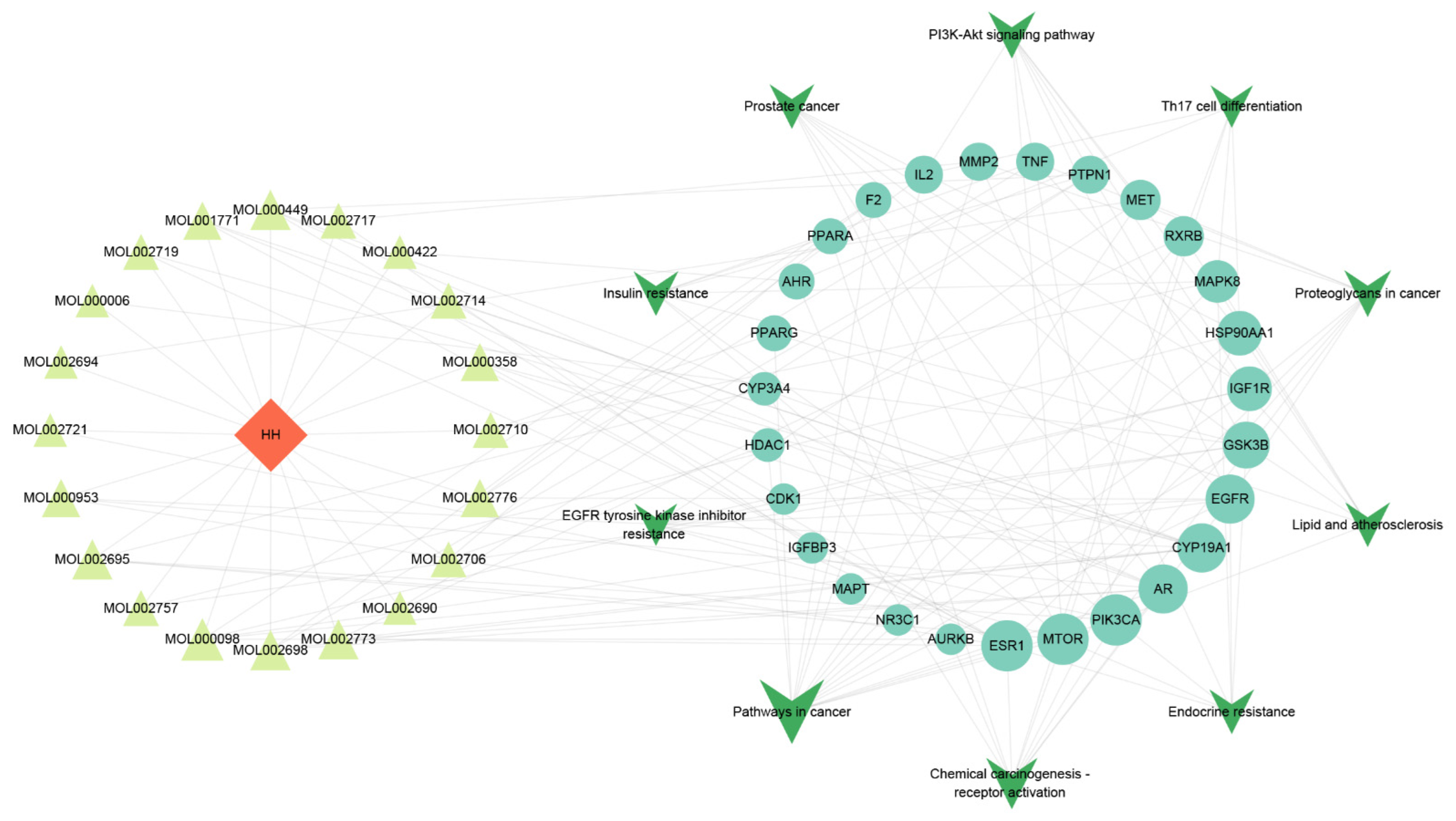
3.4. The Analysis Results of Drug–Component–Major Target–Pathway Interactive Network

The drug–component–major target–pathway interactive network with 58 nodes and 152 edges was constructed by Cytoscape, indicating the multi-component, multi-target, and multi-pathway characteristics of HH in the treatment of liver fibrosis (Figure 8). Three components with highest degree value, comprising quercetin (MOL000098), beta-carotene (MOL002773), and lignan (MOL002695) were considered as key compounds of HH. In addition, ESR1, MTOR, and PIK3CA were the potential critical targets of HH for treating liver fibrosis.

3.5. Molecular Docking Experiments Results

Three key components and critical targets were used for molecular docking experiments to predict their direct combined effects. Typically, the binding energy which is less than −7.0 kcal/mol indicates a strong binding activity between ligands and receptors [26]. The docking results have shown the strong binding activities with different binding forms between key components and critical targets (Figure 9). The 2D visualized diagram showed the connection between ligands and receptors by different forces (Figure 10).

4. Discussion

It is estimated that approximately two million deaths occur each year as a result of chronic liver diseases. It is common for chronic liver disease patients to undergo liver fibrosis as a pathological stage, which may progress to cirrhosis and liver cancer, causing death and accounting for 3.5% of global deaths [27]. The finding of liver fibrosis regression makes it possible to reverse liver fibrosis and even to prevent cirrhosis and liver cancer effectively [28]. Consequently, anti-fibrosis drugs and strategies are essential for the treatment of chronic liver diseases.
The theory of traditional Chinese medicine holds that blood stasis is conducive to some chronic liver diseases, such as fatty liver disease, cirrhosis, and liver cancer. Therefore, clinically, HH is widely used to treat chronic liver diseases because of its ability to activate blood circulation and remove stasis. There is, however, a lack of understanding about how HH contributes to ameliorate liver fibrosis. In light of the intricacy of herbal medicinal components, the network pharmacology characterized by a holistic and systematic approach was used to understand the possible molecular mechanisms of HH for liver fibrosis in the present study.
Based on databases and previous literature, we identified 23 bioactive ingredients in HH and obtained 187 possible targets. Most of the targets overlapped with those of liver fibrosis, suggesting the potential and molecular basis of HH for liver fibrosis. The consequent analysis has indicated that quercetin, beta carotene, and lignan may have critical roles in the anti-liver fibrosis activities of HH. Quercetin is commonly found in fruits, vegetables, and herbal medicines [29]. The anti-fibrosis potential of quercetin has gained recent attention. The benefits of quercetin-based treatment for liver fibrosis were demonstrated through the inhibition of HSC activation, the promotion of ECM degradation, and the intervention in autophagy [30,31]. Moreover, quercetin can exhibit hepatoprotective effects via anti-lipid accumulation, anti-inflammatory, antioxidant, and anti-apoptotic properties on hepatocytes [32]. Similarly, beta carotene is available from food and herbal medicines and has significant health-promoting effects on the human body [33]. The intake of beta carotene has been shown to prevent or improve diabetes and obesity, which are also risk factors for liver fibrosis [34]. Some clinical studies have found an inverse association between liver steatosis and fibrosis with beta carotene intakes, suggesting its potential to prevent chronic liver diseases [35,36]. In addition, the treatment effect of beta carotene for liver fibrosis has been proven by preclinical studies, and the mechanisms by which it works may be linked to the inhibition of oxidative stress and inflammation [37,38].
Lignan is a natural compound formed by the polymerization of two phenylpropanoid derivatives, such as schisandrin B, honokiol, and magnolol [39]. As a result, the pharmacological activities of lignan are usually derived from the compound prior to polymerization. Schisandrin B and honokiol have exerted anti-liver fibrosis effects by suppressing oxidative stress and mediating the transforming growth factor-β (TGF-β)/Smad pathway to inhibit the activation and proliferation of HSCs [40]. Furthermore, magnolol can attenuate liver fibrosis by inhibiting Th17 cell differentiation [41].
It is worth noting that hydroxysafflor yellow A was included in our analysis although it is not characterized by good pharmacokinetic and metabolic profiles. Hydroxysafflor yellow A is receiving extensive interests because of its potential anti-fibrosis activity. It is demonstrated that hydroxysafflor yellow A has extensive anti-fibrotic functions via reducing hepatocyte apoptosis through decreasing oxidative damage and inflammatory response, inhibiting HSCs activation, and accelerating ECM degradation [18,42,43]. However, it has not emerged as a key component of HH for liver fibrosis in the present study, which may due to the differences in therapeutic targets between animals and humans. Certainly, this cannot dismiss the great potential of hydroxysafflor yellow A against liver fibrosis. It has become possible to develop drugs based on molecular modifications. And such structural modifications will result in stronger activities and improvement of the pharmacokinetic properties of some natural products [44]. In summary, the identification and investigation of key components will provide strong preclinical and clinical evidence for HH for liver fibrosis.
The drug–component–major target–pathway interactive network has suggested that HH treats liver fibrosis through the multi-compound and multi-target characteristics. Based on the results of KEGG pathway enrichment, several signaling pathways linked to liver fibrosis are triggered by the major targets, such as pathways in cancer, PI3K-Akt signaling pathway, endocrine resistance, EGFR tyrosine kinase inhibitor resistance, and Th17 cell differentiation, with pathways in cancer being most enriched. Further, ESR1, PIK3CA, and MTOR were considered as critical target genes based on the PPI network and drug–component–major target–pathway interactive network. The strong binding activities between components with targets have demonstrated the regulatory effects of HH for liver fibrosis.
In the vitro hepatocyte model, the ability of quercetin on hepatic apolipoprotein AI regulation and high-density lipoprotein synthesis is associated with the induction of ESR1 mRNA expression [45]. Additionally, quercetin significantly reduced mTOR protein expression to regulate autophagy and exerted hepatoprotective effects in a high-fat diet-induced model of nonalcoholic fatty liver disease [46]. PIK3CA is an important gene involved in the PI3K/AKT pathway, which catalyzes the PIK3 enzyme by encoding p110α and stimulates the downstream AKT1 protein to activate the pathway. Quercetin exhibited significant inhibitory effects on the PI3K/AKT pathway, including inhibition of PI3K activation and reduced phosphorylation of AKT1, although there is no preclinical evidence for direct intervention of quercetin in PIK3CA mRNA expression [47]. Beta carotene is able to suppress PI3K and mTOR protein expression and regulate autophagy to relieve fibrosis in a lung fibrosis model, demonstrating its potential to regulate PIK3CA and MTOR target genes [48]. These studies provide clear evidence for the intervention of key components in HH on critical targets and can help to reveal their exact regulatory effects on target genes or encoding proteins. Nevertheless, in view of the different biological effects that may exist for target genes in different organs or cells, the regulatory effects of key components on critical targets in the treatment of liver fibrosis deserve in-depth exploration.
Pathways in cancer is a collection of various signaling pathways that play roles in cancer. The KEGG mapper have shown that PI3K/AKT signaling pathway, mTOR signaling pathway, MAPK signaling pathway, PPAR signaling pathway, and estrogen signaling pathway are all part of it and are regulated by HH.
Liver fibrosis is known to be driven by the activation of HSCs. Certain molecular signaling pathways have been engaged in the activation and proliferation of HSCs [49]. The PI3K/AKT signaling pathway is an upstream factor of mTOR and contributes to the proliferation, apoptosis, and autophagy of cells [50]. In liver, the activation of the PI3K/AKT pathway can stimulate HSC proliferation and α1 collagen transcription and translation, to promote liver fibrosis [51,52]. Meanwhile, the PI3K/AKT pathway can induce mTOR expression to inhibit autophagy [50]. Generally, the inhibition of the PI3K/AKT/mTOR cascade is thought to ameliorate liver fibrosis by activating autophagy to promote the apoptosis of HSCs [53]. However, it has also been found that increased autophagic activity in HSCs can provide energy to HSCs through degradation of lipid droplets, further promoting their activation and proliferation [54]. The reasons for these contradictory phenomena are still not elucidated but may be related to the effects caused by different degrees of autophagy in HSCs. Moderate autophagy may provide energy for HSCs, while excessive autophagic activation may cause the death of HSCs, which still needs further research [50].
The TGF-β signaling pathway is thought to be the crucial mediator in the fibrosis process, including liver fibrosis, due to its ability to induce myofibroblast differentiation [55]. Phosphorylation of Smad3 via TGF-β has been described as the major fibrotic pathway and the canonical TGF-β pathway [56]. In addition, some non-canonical pathways of TGF-β signal have been identified, including the MAPK pathway [57]. It is reported that all MAPK subfamilies, including ERK, JNK, p38 MAPK, and ERK5 can be activated by a TGF-β signal [57]. Remarkably, hydroxysafflor yellow A in HH was seen to exert an inhibiting role of the ERK, ERK5, and p38 MAPK signaling pathways to suppress the progression of liver fibrosis [18,58].
PPARs are a family of transcription factors which function as lipid transducers in tissues [59]. Moreover, PPARs are able to prevent liver fibrosis by modulating inflammation, regulating lipid storage, and keeping HSCs quiescent [60]. Pioglitazone and rosiglitazone have shown significant therapeutic effects in liver fibrosis by targeting PPARs, which also suggests that PPARs may be the potential targets for the treatment of liver fibrosis [61]. Estrogen is one of the most important hormones for women, which can exert its biological activity by binding to its receptor, estrogen receptor α and estrogen receptor β [62]. The epidemiological study has found a positive correlation between estrogen deficiency and liver fibrosis in women, suggesting an important role of the estrogen signaling pathways in hepatoprotection [63]. In view of the expression of estrogen receptor β in HSCs, the potential mechanism of estrogen treatment of liver fibrosis is thought to be related to estrogen receptor β interactions that promote inactivation of HSCs [64]. In summary, the contribution of estrogen in hepatoprotection should also be taken into account.
All along, the clinical use of traditional Chinese medicine has been impeded by possible adverse reactions such as renal toxicity and hepatotoxicity [65]. HH is considered as a safe herbal medicine according to the Pharmacopoeia of the People’s Republic of China (2020 edition). However, some conflicting findings were reported on the toxicity of HH. Namjoo et al. reported that the methanolic extract of HH at 20 mg/kg or 40 mg/kg doses on pregnant mice may cause liver tissue damage in newborn mice, which may be related to the deficiency of lysosomal acid lipase in newborn mice [66]. Mirhoseini et al. found that the aqueous extract of HH administered at a dose of 200 mg/kg caused adverse effects on testicular tissue and sperm production in mice, suggesting its possible reproductive toxicity [67]. The concerns of toxicity of HH have been raised by these studies. Contrastingly, a sub-chronic toxicity research showed that a dose of 100 mg/kg of ethanolic extract of HH did not cause any toxic reactions but reduce alanine aminotransferase (ALT) and aspartate aminotransferase (AST) levels in rats [68]. This phenomenon has suggested that HH does affect the liver, but it may not be hepatotoxic. In addition, a rigorous toxicity assessment of concentrated water solution of HH using a dose of 1000 mg/kg suggested that HH did not show any toxic effects on the reproduction and early development of maternal animals and their offspring [69]. Furthermore, HH at 476 mg/kg, 1430 mg/kg, or 4290 mg/kg doses was able to improve histopathological changes in the liver and reduce ALT and AST levels to exert hepatoprotective effects by modulating hepatic metabolic profiles, reducing inflammatory responses, and inhibiting oxidative stress in an alcohol-induced acute liver injury model in rats [70]. In light of numerous conflicting evidences available, it is difficult to state whether HH has significant hepatotoxicity. Moreover, the methods of HH extraction, the dose administered, and the duration of treatment are different in various studies, which may also be important factors affecting the experimental results. However, in any case, caution needs be taken when using HH with high doses for a long time, particularly for women in the peripartum period, infants and children. In addition, PIK3CA and MTOR target genes involved in the PI3K/AKT/mTOR pathway are predicted to be critical targets of HH in the treatment of liver fibrosis in our study. In view of the complex role of mTOR pathway-mediated autophagy, the exact influence of HH treatment on autophagy in HSCs remains to be answered, which may be one of the necessary ways to reveal the impact of HH on liver health. Therefore, further toxicological research is needed to provide more definitive evidence on the short-term and long-term safety of HH for clinical application, especially on the effects of liver health.
Taken together, we predicted the bioactive components, potential targets, and molecular mechanisms of HH in the treatment of liver fibrosis using a network pharmacology approach and validated them by using molecular docking experiments. However, there are still some limitations of the current study. Firstly, both the online databases and literature are time-sensitive, possibly there are also unknown active components in HH to be characterized. Likewise, therapeutic targets for liver fibrosis have space for renewal. Notably, the application of systemic pharmacology and high-throughput technologies such as transcriptomics and genomics may help to discover the directly targeted genes of HH in the treatment of liver fibrosis. Secondly, the screening based on OB and DL is not the only criterion to evaluate the clinical potential of a compound, as exemplified by hydroxysafflor yellow A. A large number of methods that can improve the bioavailability of promising drugs have been investigated and proven, such as structural modifications and nanomaterial binding, which may also be a future research direction. Finally, the value of network pharmacology lies in the prediction of key components and target genes of HH based on the known therapeutic potential of it for liver fibrosis and verifying the direct receptor–ligand interactions between key components and proteins encoded by target genes through molecular docking, which contributes to revealing the exact mechanisms of HH in treating liver fibrosis. However, such a research strategy can only provide information on the interventional effects of components on the disease targets. It is limited to suggesting whether the exact regulatory effects, including an improving or inhibiting role, and these need to be confirmed by other studies or explored in depth in the next step. In the present study, quercetin, beta carotene, and lignan are predicted to be key components of HH and may be used to treat liver fibrosis by targeting ESR1, PI3KCA, and MTOR. Although there is a certain amount of literature evidence supporting the modulatory effects of these components on corresponding targets, complete compositional characterization, in vitro and vivo validation studies are still needed to be designed and conducted to clarify the roles of these active components of HH in the treatment of liver fibrosis, which is our next research objective.

5. Conclusions

To the best of our knowledge, this is the first systematic exploration of the molecular mechanisms of HH in the treatment of liver fibrosis. HH treats liver fibrosis through multi-component, multi-target, and multi-pathway mechanisms in a holistic manner. Key components such as quercetin, beta carotene, and lignan may be involved in the PI3K/AKT/mTOR pathway, MAPK signaling pathway, PPAR pathway, and estrogen signaling pathway by targeting key proteins such as ESR1, PIK3CA, and MTOR, ultimately improving liver fibrosis by ameliorating inflammation, inhibiting oxidative damage, suppressing HSCs activation and proliferation, and promoting ECM degradation. Some key issues, including the influence of HH on liver health in clinical practice, and the exact regulatory effects of the bioactive ingredients on target genes, need to be explored in subsequent research. To sum up, our study may provide a basis for revealing the mechanisms of HH on liver fibrosis and provide knowledge for HH-based research in the future.

Supplementary Materials

The following supporting information can be downloaded at: https://www.mdpi.com/article/10.3390/pr10091735/s1, Supplementary Table S1: The bioactive components and treating targets of HH; Supplementary Table S2: The disease targets of liver fibrosis.

Author Contributions

Y.H.: Conceptualization, Methodology and Writing—Original Draft; Y.L.: Methodology, Software, and Visualization; Q.R.: Software and Validation; Q.G.: Investigation; S.T.: Writing—Review and Editing; W.H.: Writing—Review and Editing and Project administration. All authors have read and agreed to the published version of the manuscript.

Funding

This research was funded by the by the National Natural Science Foundation of China (grant number: 82074329).

Acknowledgments

Figure 5, Figure 6 and Figure 7 were plotted by https://www.bioinformatics.com.cn (accessed on 17 June 2022), a free online platform for data analysis and visualization.

Conflicts of Interest

The authors declare no conflict of interest.

References

  1. Tan, Z.; Sun, H.; Xue, T.; Gan, C.; Liu, H.; Xie, Y.; Yao, Y.; Ye, T. Liver Fibrosis: Therapeutic Targets and Advances in Drug Therapy. Front. Cell Dev. Biol. 2021, 9, 730176. [Google Scholar] [CrossRef] [PubMed]
  2. Zoubek, M.E.; Trautwein, C.; Strnad, P. Reversal of Liver Fibrosis: From Fiction to Reality. Best Pract. Res. Clin. Gastroenterol. 2017, 31, 129–141. [Google Scholar] [CrossRef] [PubMed]
  3. Vilar-Gomez, E.; Calzadilla-Bertot, L.; Wai-Sun Wong, V.; Castellanos, M.; Aller-de la Fuente, R.; Metwally, M.; Eslam, M.; Gonzalez-Fabian, L.; Alvarez-Quiñones Sanz, M.; Conde-Martin, A.F.; et al. Fibrosis Severity as a Determinant of Cause-Specific Mortality in Patients With Advanced Nonalcoholic Fatty Liver Disease: A Multi-National Cohort Study. Gastroenterology 2018, 155, 443–457.e17. [Google Scholar] [CrossRef]
  4. Liu, P.; Mao, Y.; Xie, Y.; Wei, J.; Yao, J. Stem Cells for Treatment of Liver Fibrosis/Cirrhosis: Clinical Progress and Therapeutic Potential. Stem Cell Res. Ther. 2022, 13, 356. [Google Scholar] [CrossRef] [PubMed]
  5. Caligiuri, A.; Gentilini, A.; Pastore, M.; Gitto, S.; Marra, F. Cellular and Molecular Mechanisms Underlying Liver Fibrosis Regression. Cells 2021, 10, 2759. [Google Scholar] [CrossRef]
  6. Cai, X.; Wang, J.; Wang, J.; Zhou, Q.; Yang, B.; He, Q.; Weng, Q. Intercellular Crosstalk of Hepatic Stellate Cells in Liver Fibrosis: New Insights into Therapy. Pharmacol. Res. 2020, 155, 104720. [Google Scholar] [CrossRef] [PubMed]
  7. Flores-Contreras, L.; Sandoval-Rodríguez, A.S.; Mena-Enriquez, M.G.; Lucano-Landeros, S.; Arellano-Olivera, I.; Alvarez-Álvarez, A.; Sanchez-Parada, M.G.; Armendáriz-Borunda, J. Treatment with Pirfenidone for Two Years Decreases Fibrosis, Cytokine Levels and Enhances CB2 Gene Expression in Patients with Chronic Hepatitis C. BMC Gastroenterol. 2014, 14, 131. [Google Scholar] [CrossRef] [PubMed]
  8. Neuschwander-Tetri, B.A.; Loomba, R.; Sanyal, A.J.; Lavine, J.E.; van Natta, M.L.; Abdelmalek, M.F.; Chalasani, N.; Dasarathy, S.; Diehl, A.M.; Hameed, B.; et al. Farnesoid X Nuclear Receptor Ligand Obeticholic Acid for Non-Cirrhotic, Non-Alcoholic Steatohepatitis (FLINT): A Multicentre, Randomised, Placebo-Controlled Trial. Lancet 2015, 385, 956–965. [Google Scholar] [CrossRef]
  9. Mu, M.; Zuo, S.; Wu, R.-M.; Deng, K.-S.; Lu, S.; Zhu, J.-J.; Zou, G.-L.; Yang, J.; Cheng, M.-L.; Zhao, X.-K. Ferulic Acid Attenuates Liver Fibrosis and Hepatic Stellate Cell Activation via Inhibition of TGF-β/Smad Signaling Pathway. Drug Des. Dev. Ther. 2018, 12, 4107–4115. [Google Scholar] [CrossRef]
  10. Chen, A.; Zheng, S. Curcumin Inhibits Connective Tissue Growth Factor Gene Expression in Activated Hepatic Stellate Cells in Vitro by Blocking NF-KappaB and ERK Signalling. Br. J. Pharmacol. 2008, 153, 557–567. [Google Scholar] [CrossRef] [Green Version]
  11. Nan, Y.; Su, H.; Lian, X.; Wu, J.; Liu, S.; Chen, P.; Liu, S. Pathogenesis of Liver Fibrosis and Its TCM Therapeutic Perspectives. Evid. Based Complement. Altern. Med. 2022, 2022, 5325431. [Google Scholar] [CrossRef] [PubMed]
  12. He, L.; Guo, C.; Peng, C.; Li, Y. Advances of Natural Activators for Nrf2 Signaling Pathway on Cholestatic Liver Injury Protection: A Review. Eur. J. Pharmacol. 2021, 910, 174447. [Google Scholar] [CrossRef]
  13. Wu, X.; Cai, X.; Ai, J.; Zhang, C.; Liu, N.; Gao, W. Extraction, Structures, Bioactivities and Structure-Function Analysis of the Polysaccharides From Safflower (Carthamus tinctorius L.). Front. Pharmacol. 2021, 12, 767947. [Google Scholar] [CrossRef] [PubMed]
  14. Waki, T.; Terashita, M.; Fujita, N.; Fukuda, K.; Kato, M.; Negishi, T.; Uchida, H.; Aoki, Y.; Takahashi, S.; Nakayama, T. Identification of the Genes Coding for Carthamin Synthase, Peroxidase Homologs That Catalyze the Final Enzymatic Step of Red Pigmentation in Safflower (Carthamus tinctorius L.). Plant Cell Physiol. 2021, 62, 1528–1541. [Google Scholar] [CrossRef]
  15. Hong, H.; Lim, J.M.; Kothari, D.; Kwon, S.H.; Kwon, H.C.; Han, S.-G.; Kim, S.-K. Antioxidant Properties and Diet-Related α-Glucosidase and Lipase Inhibitory Activities of Yogurt Supplemented with Safflower (Carthamus tinctorius L.) Petal Extract. Food Sci. Anim. Resour. 2021, 41, 122–134. [Google Scholar] [CrossRef]
  16. Xi, S.; Yue, L.; Shi, M.; Peng, Y.; Xu, Y.; Wang, X.; Li, Q.; Kang, Z.; Li, H.; Wang, Y. The Effects of Taoren-Honghua Herb Pair on Pathological Microvessel and Angiogenesis-Associated Signaling Pathway in Mice Model of CCl4-Induced Chronic Liver Disease. Evid. Based Complement. Altern. Med. 2016, 2016, 2974256. [Google Scholar] [CrossRef]
  17. Zhang, Y.-B.; Dong, H.-Y.; Zhao, X.-M.; Fan, L.; Zou, Y.; Zhang, C.; Li, G.; Liu, J.-C.; Niu, Y.-C. Hydroxysafflor Yellow A Attenuates Carbon Tetrachloride-Induced Hepatic Fibrosis in Rats by Inhibiting ERK5 Signaling. Am. J. Chin. Med. 2012, 40, 481–494. [Google Scholar] [CrossRef]
  18. Liu, Q.; Wang, C.Y.; Liu, Z.; Ma, X.S.; He, Y.H.; Chen, S.S.; Bai, X.Y. Hydroxysafflor Yellow A Suppresses Liver Fibrosis Induced by Carbon Tetrachloride with High-Fat Diet by Regulating PPAR-γ/P38 MAPK Signaling. Pharm. Biol. 2014, 52, 1085–1093. [Google Scholar] [CrossRef]
  19. Tao, Y.; Tian, K.; Chen, J.; Tan, D.; Liu, Y.; Xiong, Y.; Chen, Z.; Tian, Y. Network Pharmacology-Based Prediction of the Active Compounds, Potential Targets, and Signaling Pathways Involved in Danshiliuhao Granule for Treatment of Liver Fibrosis. Evid. Based Complement. Altern. Med. 2019, 2019, 2630357. [Google Scholar] [CrossRef]
  20. Fan, C.; Wu, F.R.; Zhang, J.F.; Jiang, H. A Network Pharmacology Approach to Explore the Mechanisms of Shugan Jianpi Formula in Liver Fibrosis. Evid Based Complement. Alternat. Med. 2020, 2020, 4780383. [Google Scholar] [CrossRef]
  21. Williams, J.; Siramshetty, V.; Nguyễn, Ð.-T.; Padilha, E.C.; Kabir, M.; Yu, K.-R.; Wang, A.Q.; Zhao, T.; Itkin, M.; Shinn, P.; et al. Using in Vitro ADME Data for Lead Compound Selection: An Emphasis on PAMPA PH 5 Permeability and Oral Bioavailability. Bioorg. Med. Chem. 2022, 56, 116588. [Google Scholar] [CrossRef] [PubMed]
  22. Bitew, M.; Desalegn, T.; Demissie, T.B.; Belayneh, A.; Endale, M.; Eswaramoorthy, R. Pharmacokinetics and Drug-Likeness of Antidiabetic Flavonoids: Molecular Docking and DFT Study. PLoS ONE 2021, 16, e0260853. [Google Scholar] [CrossRef] [PubMed]
  23. Daina, A.; Michielin, O.; Zoete, V. SwissTargetPrediction: Updated Data and New Features for Efficient Prediction of Protein Targets of Small Molecules. Nucleic Acids Res. 2019, 47, W357–W364. [Google Scholar] [CrossRef] [PubMed]
  24. Szklarczyk, D.; Morris, J.H.; Cook, H.; Kuhn, M.; Wyder, S.; Simonovic, M.; Santos, A.; Doncheva, N.T.; Roth, A.; Bork, P.; et al. The STRING Database in 2017: Quality-Controlled Protein-Protein Association Networks, Made Broadly Accessible. Nucleic Acids Res. 2017, 45, D362–D368. [Google Scholar] [CrossRef]
  25. Szklarczyk, D.; Gable, A.L.; Lyon, D.; Junge, A.; Wyder, S.; Huerta-Cepas, J.; Simonovic, M.; Doncheva, N.T.; Morris, J.H.; Bork, P.; et al. STRING V11: Protein-Protein Association Networks with Increased Coverage, Supporting Functional Discovery in Genome-Wide Experimental Datasets. Nucleic Acids Res. 2019, 47, D607–D613. [Google Scholar] [CrossRef]
  26. Liu, J.; Liu, J.; Tong, X.; Peng, W.; Wei, S.; Sun, T.; Wang, Y.; Zhang, B.; Li, W. Network Pharmacology Prediction and Molecular Docking-Based Strategy to Discover the Potential Pharmacological Mechanism of Huai Hua San Against Ulcerative Colitis. Drug Des. Dev. Ther. 2021, 15, 3255–3276. [Google Scholar] [CrossRef] [PubMed]
  27. Asrani, S.K.; Devarbhavi, H.; Eaton, J.; Kamath, P.S. Burden of Liver Diseases in the World. J. Hepatol. 2019, 70, 151–171. [Google Scholar] [CrossRef]
  28. Kisseleva, T.; Brenner, D. Molecular and Cellular Mechanisms of Liver Fibrosis and Its Regression. Nat. Rev. Gastroenterol. Hepatol. 2021, 18, 151–166. [Google Scholar] [CrossRef]
  29. Andres, S.; Pevny, S.; Ziegenhagen, R.; Bakhiya, N.; Schäfer, B.; Hirsch-Ernst, K.I.; Lampen, A. Safety Aspects of the Use of Quercetin as a Dietary Supplement. Mol. Nutr. Food Res. 2018, 62, 1700447. [Google Scholar] [CrossRef] [PubMed]
  30. Wang, R.; Zhang, H.; Wang, Y.; Song, F.; Yuan, Y. Inhibitory Effects of Quercetin on the Progression of Liver Fibrosis through the Regulation of NF-KB/IkBα, P38 MAPK, and Bcl-2/Bax Signaling. Int. Immunopharmacol. 2017, 47, 126–133. [Google Scholar] [CrossRef] [PubMed]
  31. Liu, N.; Feng, J.; Lu, X.; Yao, Z.; Liu, Q.; Lv, Y.; Han, Y.; Deng, J.; Zhou, Y. Isorhamnetin Inhibits Liver Fibrosis by Reducing Autophagy and Inhibiting Extracellular Matrix Formation via the TGF-Β1/Smad3 and TGF-Β1/P38 MAPK Pathways. Mediat. Inflamm. 2019, 2019, 6175091. [Google Scholar] [CrossRef] [PubMed]
  32. Zhao, X.; Wang, J.; Deng, Y.; Liao, L.; Zhou, M.; Peng, C.; Li, Y. Quercetin as a Protective Agent for Liver Diseases: A Comprehensive Descriptive Review of the Molecular Mechanism. Phytother. Res. 2021, 35, 4727–4747. [Google Scholar] [CrossRef]
  33. Bohn, T.; Desmarchelier, C.; El, S.N.; Keijer, J.; van Schothorst, E.; Rühl, R.; Borel, P. β-Carotene in the Human Body: Metabolic Bioactivation Pathways—From Digestion to Tissue Distribution and Excretion. Proc. Nutr. Soc. 2019, 78, 68–87. [Google Scholar] [CrossRef]
  34. Marcelino, G.; Machate, D.J.; Freitas, K.d.C.; Hiane, P.A.; Maldonade, I.R.; Pott, A.; Asato, M.A.; Candido, C.J.; Guimarães, R. de C.A. β-Carotene: Preventive Role for Type 2 Diabetes Mellitus and Obesity: A Review. Molecules 2020, 25, 5803. [Google Scholar] [CrossRef]
  35. Liu, X.; Shen, H.; Chen, M.; Shao, J. Clinical Relevance of Vitamins and Carotenoids With Liver Steatosis and Fibrosis Detected by Transient Elastography in Adults. Front. Nutr. 2021, 8, 760985. [Google Scholar] [CrossRef] [PubMed]
  36. Wang, L.; Ding, C.; Zeng, F.; Zhu, H. Low Levels of Serum β-Carotene and β-Carotene/Retinol Ratio Are Associated with Histological Severity in Nonalcoholic Fatty Liver Disease Patients. Ann. Nutr. Metab. 2019, 74, 156–164. [Google Scholar] [CrossRef]
  37. El-Baz, F.K.; Salama, A.; Ali, S.I.; Elgohary, R. Haematococcus Pluvialis Carotenoids Enrich Fractions Ameliorate Liver Fibrosis Induced by Thioacetamide in Rats: Modulation of Metalloproteinase and Its Inhibitor. BioMed Res. Int. 2021, 2021, 6631415. [Google Scholar] [CrossRef]
  38. Latief, U.; Ahmad, R. β-Carotene Inhibits NF-ΚB and Restrains Diethylnitrosamine-Induced Hepatic Inflammation in Wistar Rats. Int. J. Vitam. Nutr. Res. 2020, 20, 1–10. [Google Scholar] [CrossRef]
  39. Pan, X.; Ma, X.; Jiang, Y.; Wen, J.; Yang, L.; Chen, D.; Cao, X.; Peng, C. A Comprehensive Review of Natural Products against Liver Fibrosis: Flavonoids, Quinones, Lignans, Phenols, and Acids. Evid Based Complement. Altern. Med. 2020, 2020, 7171498. [Google Scholar] [CrossRef]
  40. Chen, Q.; Zhang, H.; Cao, Y.; Li, Y.; Sun, S.; Zhang, J.; Zhang, G. Schisandrin B Attenuates CCl4-Induced Liver Fibrosis in Rats by Regulation of Nrf2-ARE and TGF-β/Smad Signaling Pathways. Drug Des. Dev. Ther. 2017, 11, 2179–2191. [Google Scholar] [CrossRef] [Green Version]
  41. Zhang, H.; Ju, B.; Zhang, X.; Zhu, Y.; Nie, Y.; Xu, Y.; Lei, Q. Magnolol Attenuates Concanavalin A-Induced Hepatic Fibrosis, Inhibits CD4+ T Helper 17 (Th17) Cell Differentiation and Suppresses Hepatic Stellate Cell Activation: Blockade of Smad3/Smad4 Signalling. Basic Clin. Pharmacol. Toxicol. 2017, 120, 560–570. [Google Scholar] [CrossRef]
  42. Zhang, Y.; Guo, J.; Dong, H.; Zhao, X.; Zhou, L.; Li, X.; Liu, J.; Niu, Y. Hydroxysafflor Yellow A Protects against Chronic Carbon Tetrachloride-Induced Liver Fibrosis. Eur. J. Pharmacol. 2011, 660, 438–444. [Google Scholar] [CrossRef]
  43. Li, Y.; Shi, Y.; Sun, Y.; Liu, L.; Bai, X.; Wang, D.; Li, H. Restorative Effects of Hydroxysafflor Yellow A on Hepatic Function in an Experimental Regression Model of Hepatic Fibrosis Induced by Carbon Tetrachloride. Mol. Med. Rep. 2017, 15, 47–56. [Google Scholar] [CrossRef] [PubMed]
  44. Cai, X.-H.; Guo, H.; Xie, B. Structural Modifications of Matrine-Type Alkaloids. Mini Rev. Med. Chem. 2018, 18, 730–744. [Google Scholar] [CrossRef] [PubMed]
  45. Haas, M.J.; Onstead-Haas, L.M.; Szafran-Swietlik, A.; Kojanian, H.; Davis, T.; Armstrong, P.; Wong, N.C.W.; Mooradian, A.D. Induction of Hepatic Apolipoprotein A-I Gene Expression by the Isoflavones Quercetin and Isoquercetrin. Life Sci. 2014, 110, 8–14. [Google Scholar] [CrossRef] [PubMed]
  46. Liu, L.; Gao, C.; Yao, P.; Gong, Z. Quercetin Alleviates High-Fat Diet-Induced Oxidized Low-Density Lipoprotein Accumulation in the Liver: Implication for Autophagy Regulation. BioMed Res. Int. 2015, 2015, 607531. [Google Scholar] [CrossRef]
  47. Ghafouri-Fard, S.; Shoorei, H.; Khanbabapour Sasi, A.; Taheri, M.; Ayatollahi, S.A. The Impact of the Phytotherapeutic Agent Quercetin on Expression of Genes and Activity of Signaling Pathways. Biomed. Pharmacother. 2021, 141, 111847. [Google Scholar] [CrossRef]
  48. Lu, Y.; Zhang, Y.; Pan, Z.; Yang, C.; Chen, L.; Wang, Y.; Xu, D.; Xia, H.; Wang, S.; Chen, S.; et al. Potential “Therapeutic” Effects of Tocotrienol-Rich Fraction (TRF) and Carotene “Against” Bleomycin-Induced Pulmonary Fibrosis in Rats via TGF-β/Smad, PI3K/Akt/MTOR and NF-ΚB Signaling Pathways. Nutrients 2022, 14, 1904. [Google Scholar] [CrossRef]
  49. Roehlen, N.; Crouchet, E.; Baumert, T.F. Liver Fibrosis: Mechanistic Concepts and Therapeutic Perspectives. Cells 2020, 9, 875. [Google Scholar] [CrossRef]
  50. Wang, H.; Liu, Y.; Wang, D.; Xu, Y.; Dong, R.; Yang, Y.; Lv, Q.; Chen, X.; Zhang, Z. The Upstream Pathway of MTOR-Mediated Autophagy in Liver Diseases. Cells 2019, 8, 1597. [Google Scholar] [CrossRef]
  51. Reif, S.; Lang, A.; Lindquist, J.N.; Yata, Y.; Gabele, E.; Scanga, A.; Brenner, D.A.; Rippe, R.A. The Role of Focal Adhesion Kinase-Phosphatidylinositol 3-Kinase-Akt Signaling in Hepatic Stellate Cell Proliferation and Type I Collagen Expression. J. Biol. Chem. 2003, 278, 8083–8090. [Google Scholar] [CrossRef]
  52. Parsons, C.J.; Takashima, M.; Rippe, R.A. Molecular Mechanisms of Hepatic Fibrogenesis. J. Gastroenterol. Hepatol. 2007, 22 (Suppl. S1), S79–S84. [Google Scholar] [CrossRef] [PubMed]
  53. Lee, S.W.; Kim, S.M.; Hur, W.; Kang, B.-Y.; Lee, H.L.; Nam, H.; Yoo, S.H.; Sung, P.S.; Kwon, J.H.; Jang, J.W.; et al. Tenofovir Disoproxil Fumarate Directly Ameliorates Liver Fibrosis by Inducing Hepatic Stellate Cell Apoptosis via Downregulation of PI3K/Akt/MTOR Signaling Pathway. PLoS ONE 2021, 16, e0261067. [Google Scholar] [CrossRef] [PubMed]
  54. Thoen, L.F.R.; Guimarães, E.L.M.; Dollé, L.; Mannaerts, I.; Najimi, M.; Sokal, E.; van Grunsven, L.A. A Role for Autophagy during Hepatic Stellate Cell Activation. J. Hepatol. 2011, 55, 1353–1360. [Google Scholar] [CrossRef] [PubMed]
  55. Hu, H.-H.; Chen, D.-Q.; Wang, Y.-N.; Feng, Y.-L.; Cao, G.; Vaziri, N.D.; Zhao, Y.-Y. New Insights into TGF-β/Smad Signaling in Tissue Fibrosis. Chem. Biol. Interact. 2018, 292, 76–83. [Google Scholar] [CrossRef]
  56. Dewidar, B.; Meyer, C.; Dooley, S.; Meindl-Beinker, A.N. TGF-β in Hepatic Stellate Cell Activation and Liver Fibrogenesis-Updated. Cells 2019, 8, 1419. [Google Scholar] [CrossRef]
  57. Derynck, R.; Budi, E.H. Specificity, Versatility, and Control of TGF-β Family Signaling. Sci. Signal 2019, 12, 570. [Google Scholar] [CrossRef]
  58. Dong, H.; Liu, Y.; Zou, Y.; Li, C.; Li, L.; Li, X.; Zhao, X.; Zhou, L.; Liu, J.; Niu, Y. Alteration of the ERK5 Pathway by Hydroxysafflor Yellow A Blocks Expression of MEF2C in Activated Hepatic Stellate Cells in Vitro: Potential Treatment for Hepatic Fibrogenesis. Pharm. Biol. 2013, 54, 435–443. [Google Scholar] [CrossRef]
  59. Fougerat, A.; Montagner, A.; Loiseau, N.; Guillou, H.; Wahli, W. Peroxisome Proliferator-Activated Receptors and Their Novel Ligands as Candidates for the Treatment of Non-Alcoholic Fatty Liver Disease. Cells 2020, 9, 1638. [Google Scholar] [CrossRef]
  60. Choudhary, N.S.; Kumar, N.; Duseja, A. Peroxisome Proliferator-Activated Receptors and Their Agonists in Nonalcoholic Fatty Liver Disease. J. Clin. Exp. Hepatol. 2019, 9, 731–739. [Google Scholar] [CrossRef] [Green Version]
  61. Musso, G.; Cassader, M.; Paschetta, E.; Gambino, R. Thiazolidinediones and Advanced Liver Fibrosis in Nonalcoholic Steatohepatitis: A Meta-Analysis. JAMA Intern. Med. 2017, 177, 633–640. [Google Scholar] [CrossRef] [PubMed]
  62. Yaşar, P.; Ayaz, G.; User, S.D.; Güpür, G.; Muyan, M. Molecular Mechanism of Estrogen-Estrogen Receptor Signaling. Reprod. Med. Biol. 2017, 16, 4–20. [Google Scholar] [CrossRef] [PubMed]
  63. Klair, J.S.; Yang, J.D.; Abdelmalek, M.F.; Guy, C.D.; Gill, R.M.; Yates, K.; Unalp-Arida, A.; Lavine, J.E.; Clark, J.M.; Diehl, A.M.; et al. A Longer Duration of Estrogen Deficiency Increases Fibrosis Risk among Postmenopausal Women with Nonalcoholic Fatty Liver Disease. Hepatology 2016, 64, 85–91. [Google Scholar] [CrossRef]
  64. Zhang, B.; Zhang, C.-G.; Ji, L.-H.; Zhao, G.; Wu, Z.-Y. Estrogen Receptor β Selective Agonist Ameliorates Liver Cirrhosis in Rats by Inhibiting the Activation and Proliferation of Hepatic Stellate Cells. J. Gastroenterol. Hepatol. 2018, 33, 747–755. [Google Scholar] [CrossRef] [PubMed]
  65. Teschke, R.; Zhang, L.; Long, H.; Schwarzenboeck, A.; Schmidt-Taenzer, W.; Genthner, A.; Wolff, A.; Frenzel, C.; Schulze, J.; Eickhoff, A. Traditional Chinese Medicine and Herbal Hepatotoxicity: A Tabular Compilation of Reported Cases. Ann. Hepatol. 2015, 14, 7–19. [Google Scholar] [CrossRef]
  66. Namjoo, A.; Nasri, H.; Talebi-Juneghani, A.; Baradaran, A.; Rafieian-Kopaei, M. Safety Profile of Carthamus Tinctorius L. in Lactation: Brain, Renal and Hepatotoxicity. Pak. J. Med. Sci. 2013, 29, 378–383. [Google Scholar] [CrossRef]
  67. Mirhoseini, M.; Mohamadpour, M.; Khorsandi, L. Toxic Effects of Carthamus tinctorius L. (Safflower) Extract on Mouse Spermatogenesis. J. Assist. Reprod. Genet. 2012, 29, 457–461. [Google Scholar] [CrossRef]
  68. Zhang, Z.; Liu, R.; Pu, X.; Sun, Y.; Zhao, X. Evaluation of the Sub-Chronic Toxicity of a Standardized Flavonoid Extract of Safflower in Rats. Regul. Toxicol. Pharmacol. 2017, 85, 98–107. [Google Scholar] [CrossRef]
  69. Lewin, G.; Joel, M.; Peter, B.; Lützow, M. Food Grade Safflower Concentrate: No Evidence for Reproduction and Early Developmental Toxicity. Reprod. Toxicol. 2021, 104, 155–165. [Google Scholar] [CrossRef]
  70. Fan, X.; Wang, X.; Lian, J.; Pei, Z.; Jiang, M.; Bai, M. Flos Carthami Exerts Hepatoprotective Action in a Rat Model of Alcoholic Liver Injury via Modulating the Metabolomics Profile. Evid. Based Complement. Altern. Med. 2022, 2022, 8158699. [Google Scholar] [CrossRef]
Figure 1. Research strategies for the current study.
Processes 10 01735 g001
Figure 2. The Venn diagram of targets between HH and liver fibrosis.
Processes 10 01735 g002
Figure 3. The PPI network constructed by STRING database.
Processes 10 01735 g003
Figure 4. The PPI network visualized by Cytoscape. (a) the interaction between 90 overlapping targets; (b) the interaction between major targets; (c) the interaction between key targets. The larger the node degree value, the darker the color.
Processes 10 01735 g004
Figure 5. The GO enrichment analysis results of 27 major targets (top 10 were listed).
Processes 10 01735 g005
Figure 6. The Sankey diagram of major targets involved in the KEGG signaling pathways (top 10 were listed).
Processes 10 01735 g006
Figure 7. The map of pathways of cancer. Nodes with red color represent the major target genes of HH on liver fibrosis.
Processes 10 01735 g007
Figure 8. The schematic diagram of drug–component–major target–pathway interactive network (Red node is HH; light green nodes are components; blue nodes are target genes; dark green nodes are KEGG pathways).
Processes 10 01735 g008
Figure 9. The binding energy between key components and targets visualized by heat map.
Processes 10 01735 g009
Figure 10. The 2D schematic diagram of molecular dockings of key components and critical targets.
Processes 10 01735 g010
Table 1. The 23 bioactive components of HH found in databases and literature.
ComponentsMOLIDPubchem CIDComponentsMOLIDPubchem CID
Poriferast-5-en-3beta-olMOL0017714578016-HydroxynaringeninMOL002719188308
FlavoxanthinMOL0026805281238QuercetagetinMOL0027215281680
4-[(E)-4-(3,5-dimethoxy-4-oxo-1-cyclohexa-2,5-dienylidene)but-2-enylidene]-2,6-dimethoxycyclohexa-2,5-dien-1-oneMOL002694102370577,8-dimethyl-1H-pyrimido [5,6-g]quinoxaline-2,4-dioneMOL00275721786815
LignanMOL002695261166Beta-caroteneMOL0027735280489
Lupeol-palmitateMOL002698162847783BaicalinMOL00277664982
PhytoeneMOL0027065280784Beta-sitosterolMOL000358222284
PhytoflueneMOL0027076436722KaempferolMOL0004225280863
Pyrethrin IIMOL0027105281555StigmasterolMOL0004495280794
6-HydroxykaempferolMOL0027125281638LuteolinMOL0000065280445
BaicaleinMOL0027145281605CLRMOL0009535997
Qt_carthamoneMOL002717131833009QuercetinMOL0000985280343
Hydroxysafflor Yellow AMOL0026906443665
Table 2. The major targets and their uniport IDs.
Target GeneUniprot IDDegreeTarget GeneUniprot IDDegree
PIK3CAP4233617PPARGP372316
HSP90AA1P0790015IGFBP3P179365
ESR1P0337212MTORP423455
MAPK8P4598311IGF1RP080695
EGFRP0053310AHRP358694
ARP102759GSK3BP498414
NR3C1P041509MAPTP106364
HDAC1Q135479AURKBQ96GD44
CYP3A4P086848CYP19A1P115114
RXRBP287027F2P007344
PTPN1P180317METP085814
IL2P605687MMP2P082534
CDK1P064936TNFP013754
PPARAQ078696
Publisher’s Note: MDPI stays neutral with regard to jurisdictional claims in published maps and institutional affiliations.

