


Construction of a Novel Oxalic Acid Biosensor Based on the Combination of Tissue Enzyme and Peroxide Mimic Enzyme
1
School of Environment and Civil Engineering, Nanchang Institute of Science and Technology, Nanchang 330108, China
2
Institute of Environmental Science, Shanxi University, Taiyuan 030006, China
3
Key Laboratory of Biofuels, Qingdao Institute of Bioenergy and Bioprocess Technology, Qingdao 266101, China
*
Authors to whom correspondence should be addressed.
Processes 2023, 11(10), 3012; https://doi.org/10.3390/pr11103012
Submission received: 14 June 2023
/
Revised: 4 August 2023
/
Accepted: 8 August 2023
/
Published: 19 October 2023
(This article belongs to the Special Issue Innovations in Green Separation and Extraction Processes of Natural Products)
Abstract
:A biosensor is considered an integrated receptor transducer device, with the ability to convert a biological impulse into an electrical signal. The amendment of biosensors has been recognized for its great potential by many researchers, due to its numerous applications e.g., environmental management, disease diagnosis, agricultural aspects, food companies, health care, drug monitoring, and water treatment as it can be used in the detection of water quality. Moreover, technological development of the biosensor is integrated with several merits such as affordability and enhancement in medical fields in disease detection and body response; furthermore, it is easy to use, effective, and scalable. This article briefly reviews how to construct an oxalic acid (OA) biosensor by integration of tissue enzymes and peroxide simulated enzymes. OA is converted to peroxide (H2O2) and carbon dioxide (CO2) with the help of the oxalate oxidase (OxOx) present in spinach leaves as catalyst. Afterwards, with the presence of cobalt ferrite (CoFe2O4), nanoparticles (NPs) have a catalytic effect on concentrated H2O2 and chemiluminescence (CL) luminol (C8H7N3O2). Therefore, CL flow can be constructed under a biosensor to determine OA in the sample. The co-presence of tissue column and CoFe2O4, as well as a high level of relative CL intensity can be obtained. The biosensor based on H2O2 and involving inorganic nanomaterials has many advantages such as high efficiency, affordability, outstanding sensitivity, stability and selectivity, a fast response, and an extended range of linearity with a lower detection limit. In addition, optimization factors for the oxalate biosensor, limitations, and outlooks for the biosensor were also highlighted.
1. Introduction
It is well-known that natural enzymes act as a biological catalyst with extremely high reaction specificity and catalytic efficiency [1]. However, natural enzymes also have their own disadvantages, such as high price, inconvenient storage, extreme sensitivity to extreme environments (humidity, high temperature, strong acids, and strong bases), they are easy to inactivate, and their non-physiological chemicals. Therefore, finding natural enzyme alternatives is still a challenge and has gained great attention by many researchers. Mimetic enzymes can be classified into five categories such as isomerases, hydrolases, transferases, and induced and oxidoreductases enzymes [2]. In recent years, many materials such as cyclodextrins, porphyrins, cup aromatics, crown ethers, Schiff bases, molecular imprinted polymers, and micelles have been developed to simulate enzymes [3]. Recently, inorganic NPs such as Fe3O4 NPs, nano-structure FeS [4], graphene oxide, V2O5 nanowires, and ferritin–platinum (Pt-Ft) NPs [5] have been found to have peroxidase catalytic activity. CeO2 NPs [6] and PtPb alloy composite nanomaterials grown on gold nanorods [7] have been found to have oxidase catalytic activity. It also has superoxide dismutase activity and catalase catalytic activity. However, these simulated enzymes are highly resistant to temperature, chemical reagents, and solvents, and they are cheap and easy to prepare into industrial products. The catalytic activity of most simulated enzymes is not highly catalytic or selective enough to fully simulate the catalytic properties of natural enzymes.
Enzymes can regulate the intrinsic reaction ability of their substrates, and their selectivity is determined by the matching of the geometric configuration of the complex generated by the enzyme and the substrate. For example, in the biochemical synthesis of cholesterol, cytochrome P-450 enzymes catalyze the oxidation of only three methyl groups in the triterpene lanosterol, although they are on saturated carbon atoms and not near any group with special functions. Even if there are two more reactive C=C double bonds and a secondary alcohol hydroxyl group that is easily oxidized, the oxidation reaction does not occur on these active groups [8], and only the methyl group can reach the oxidation unit of the enzyme–substrate complex. In order to improve the selectivity of the simulated enzyme, researchers used cyclodextrins [9] and crown ethers [10] to functionalize the simulated enzymes. The cavity of these substances can provide a substrate specific binding site, which can improve the selectivity of the simulated enzyme. In this case, the reaction rate and selectivity have been significantly increased. In addition, the synthesis of polymer substances, such as the use of molecular imprinted polymers [11] to construct simulated enzymes, can also obtain high selectivity, because during the synthesis process, monomers with the required catalytic functional groups (such as imidazole, –OH, and –COOH) can be introduced into the polymer by the copolymerization method. However, the synthesis steps of these simulated enzymes are more cumbersome. Moreover, all artificial enzymes only simulate the activity center of natural enzymes, it lacks the three-dimensional structure of natural enzymes, and the selectivity has not been improved. In order to improve their selectivity, people try to modify polymers to simulate enzymes. For example, modifying the heat-sensitive polymer (ONTPAAm) with iron tetra-sulfonate phthalocyanine (FeTSPc) [8] can obtain good selectivity, but the selectivity of FeTSPc is improved and its catalytic activity is reduced.
In the past, a large number of tissue enzymes have been used to build tissue enzyme biosensors. In the advent of tissue enzyme electrode, it has received widespread attention [12,13,14]. Most tissue biosensors are based on tying tissue slices to the electrode surface or making tissue columns in combination with chemiluminescence detection methods. Another tissue biosensor model is to fix the tissue homogenizer on a solgel film [15], carbon paste [16], or gelatin and glutaraldehyde [17]. Compared with biosensors based on fixed pure enzymes; biosensors based on tissue enzymes have the characteristics of a simple preparation method, strong stability, cheap price, and high activity. However, in the field of simulating enzymes, inorganic NPs have been neglected for a long time because they are often considered biologically and chemically inert. The inherent enzyme-mimicking properties of NPs reflect that magnetic ferric tetroxide nanoparticles have peroxide mimic enzyme catalytic activity as does that of the fifth-grade nanoparticles with a biocatalytic activity [18]. Researchers used the simulated enzyme properties of ferric oxide peroxide to construct a new method for the determination of H2O2 and glucose by photometry [19].
This review focuses on the oxalic acid (OA) biosensor. The OA is a dicarboxylic acid found in spinach, rhubarb, beet greens, and other foods. Fungi, bacteria, and other creatures create it, and industrial processes and products can contain OA, which occurs naturally. Studying OA sensing is important for human health. OA and calcium can create insoluble crystals, causing kidney stones in vulnerable people. These stones can hurt and require medical treatment. Researchers could locate OA in biological materials and diagnose and treat kidney stone diseases early by developing good OA detection methods. OA in leafy greens and vegetables might block calcium absorption, causing dietary deficits. Researchers and regulators can ensure food safety and accurate consumer information by precisely sensing and quantifying OA levels, and prevents metal surface corrosion in several production processes [20]. However, excessive OA can harm equipment and cause corrosion. Industrial operators can monitor OA levels in real time to prevent corrosion and increase process efficiency by using sensitive OA sensing methods. Finally, OA sensing systems can help us comprehend ecological and plant pathogen interactions. OA helps plant infections invade and colonize host tissues.
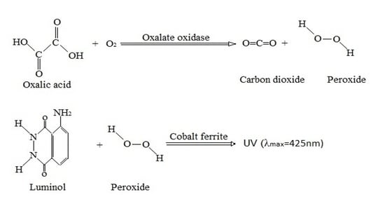
Based on the various applications of OA, it is worth investigating how organisms feel and respond to OA, researchers can learn about the molecular mechanisms behind these interactions and develop disease control measures for agriculture and forestry. Therefore, this review proposed the feasibility where spinach (spinacia oleracea) leaves can be fixed in glass tubes as a recognition element for OA. Afterwards, oxalate oxidase in spinach leaves catalyzes dissolved oxygen to oxidize OA to produce H2O2 (see the reaction formula in graphical abstract) and CoFe2O4 NPs can catalyze the H2O2-luminol luminescence system to produce high and strong chemiluminescence (CL). Based on this, OA can be selectively determined in samples. Compared with the electrochemical oxalate tissue sensor, the CL flow-through tissue sensor can offer more advantages of simple operation, rapidity, and high selectivity. Moreover, compared with other spinach leaf tissue CL oxalate sensors, the present sensor has higher sensitivity [21]. High efficiency, affordability, remarkable sensitivity, stability, selectivity, fast response, and an extended range of linearity with a reduced detection limit are the advantages of biosensors based on H2O2 and using inorganic NPs.
2. Fundamental Characteristics of Biosensors
A biosensor is a device which combines biological component, such as enzymes, antibodies, or microorganisms, with a physical transducer to detect and quantify a specific analyte. The biological component interacts with the analyte, producing a measurable signal that is converted into a digital or analog output by the transducer. Different scientists from numerous fields such as material science, chemistry, and physics have worked hand-in-hand to develop more complex, mature, and dependable biosensing devices. These devices can be applied in various fields, for instance in agriculture, medicine, biotechnology, and environmental monitoring. The fundamental models of biosensors process can be shown with the aid of Figure 1.
As shown in Figure 1, a specific biological component such as enzyme, tissue, living cells, nucleic acid, polysaccharide, or antibody, recognizes a specific analyte and sensor elements for instance, electric (potential, current, conductance, and impedance), intensity and phase of electromagnetic radiation, viscosity, mass, and temperature transduces the change in the biological component into electrical signal output. This explains that biological component must be highly specific to the analyte and does not recognize other substances that may be present in the sample. In addition, some basic properties of biosensors include: selectivity, sensitivity, specificity, speed, portability, and reusability. A detailed diagram and explained list of the biosensor’s different characteristics is shown in Figure 2.
Biosensors are regarded as powerful analytical tools that offer several advantages over traditional analytical methods. As presented in Figure 2, biosensors’ unique characteristics make them ideal for a wide range of applications in various fields. For instance, in healthcare, biosensors are being used for disease diagnosis, monitoring of drug efficacy, and personalized medicine. In environmental monitoring, they are used to detect pollutants and monitor water quality. Plus, in food safety, it is helpful in foodborne pathogens and contaminants detections, while in biodefense, they are used to detect biological threats. The development of OA sensors by immobilizing uricase and OxOx to form bio-thermochips was previously executed [22]. The study reported that the enthalpy change of OA oxidation was 143 kJ mol−1 and catalase catalyze the H2O2 reaction to generate an enthalpy change of 100 kJ mol−1, with best detection of OA between 0.2–0.8 mM. In another study, OA was determined in a urine sample by using an amperometric biosensor, which was made by immobilizing OxOX on the surface of a chromium (III) hexacyanoferrate-modified graphite electrode by using a bovine serum albumin and glutaraldehyde cross-linking procedure [23]. The biosensor showed great linearity in a concentration range of 2.5–100mM in an aqueous solution of pure OA, while in urine samples, a high correlation (R2 = 0.9949) was obtained, with long stability. Further, [24] constructed a electrochemical biosensor using nano-clay laponite and ionic liquid, 1-ethyl-3-methyl imidazolium chloride (C2mim)(Cl) film deposited on indium tin oxide glass electrode for the determination of OA. The result showed maximum sensitivity within the range of 1–20 mM; therefore, this fabricated biosensor can be applied for the detection of OA in urine samples. Moreover, various OA biosensors and sensors are well-described in the following table.
In Table 1, we highlighted some of the OA biosensors and sensors that have been developed. For instance, enzymatic biosensors can use OxOx to detect OA [25], OxOx immobilized on chemically reduced graphene oxide (CRGO) can suppress the aggregation of CRGO and exhibit significantly improved mass transport and can be used as biosensor with excellent sensitivity to OA. Their study has shown that the mass transport and electron transfer can be affected by the amount of CRGO. And also, the degree of mass diffusion and electron transfer have an interactive impact on biosensor electrochemical functioning. By development of the mass diffusion rate, it remarkably improved the electrochemical characteristics of a graphene-based biosensor; where it shows a low detection limit of 8 μM. In addition, because OA appears in many food stuffs such as vegetables; insoluble oxalates can be formed when OA combine with iron and calcium. Hence, it is paramount to examine OA quantity in food settings, [26] obtained platinum (Pt) NPs by an electrochemically induced alcohol-free solgel method. These Pt NPs were adhered with porous silica on glassy carbon electrode in a single process, and it can be used as an outstanding catalyst for OA electrochemical oxidation. The result achieved a large concentration range of 0–45 μM. Additionally, [27] demonstrated an electrochemical sensor for OA sensing, by using silver (Ag) NPs and nitrogen-doped graphene oxide nanocomposite. With the help of atmospheric pressure, microwave plasma led to the synthesis of that nanocomposite. This sensor performed with a high current response, and was stable and selective with a detection limit of 2 μM. Moreover, microbial biosensors use living microorganisms, such as bacteria or yeast, for OA detection. The microorganisms are genetically engineered to produce a fluorescent or luminescent signal in the presence of OA. These biosensors have the advantage of being able to detect OA in complex matrices, such as food or environmental samples, but may require special storage and handling conditions [28]. Biosensors can be differentiated based on the particular transducing mechanism used, which consists of many types such as: piezoelectric, DNA, surface plasmon, chemiluminescence-based, fluorescence-based, biomimetic, immuno, enzyme-based, cell-based, amperometric, and calorimetric biosensors [29].
3. Design and Performance Evaluation of Oxalic Acid Biosensor
3.1. Apparatus
Figure 3 shows a schematic diagram of a flow injection CL detection process, where pump 1 can be used to pump sample solution and current carrier, while pump 2 can help to deliver luminescent reagents (Luminol and CoFe2O4 NPs); the tube helps to connect the components of the flow path; the eight-way valve is the injection valve; which allows sufficient amounts of oxalic acid to be converted to H2O2 (in presence of oxalate oxidase) without prolonging the measurement time. In addition, flow cell works as a voltage generator and also signal can be recorded by a personal computer equipped with a data acquisition interface, and data acquisition and processing can be processed by specific software.
According to Figure 3, the pump 1 tube is inserted into the sample solution, the solution is buffered while luminol solution and CoFe2O4 NPs solution are connected to the pump 2, the roller pump can be turned on, and after the baseline is stabilized, the H2O2 generated in the tissue column is injected into the current carrying column and mixed with the luminescence reagent to produce CL, with peak and high quantitative. Therefore, the standard curve can be obtained by plotting the OA concentration with CL intensity.
3.2. Sensor Identification Mechanisms
The mechanism action of this biosensor involves the immobilization of an enzyme, such as OxOx, on the surface of the CoFe2O4 NPs. This enzyme catalyzes the oxidation of OA to produce CO2 and H2O2. Afterwards, H2O2 generated in this reaction can be detected by a transducer, for instance, a screen-printed electrode, which produces an electrical signal in response to the presence of the H2O2. Moreover, the CoFe2O4 NPs serve as a support matrix for the immobilization of the enzyme, providing a large surface area for enzyme binding and increasing the enzyme loading capacity. As well, presented in Figure 4, the typically used bio-component is the OxOx enzyme that works as a catalyst in the oxidation reaction of OA but remains unchanged at the end of the reaction. Moreover, magnetic properties of the CoFe2O4 NPs offer several advantages, including high sensitivity and selectivity, fast response time, and easy regeneration of the biosensor. Furthermore, it can also allow easy separation and recovery of the biosensor from the sample matrix, reducing the potential for interference from other compounds in the sample. In another study, under selected conditions for OA sensor, [39] reported the electrochemical oxidation of OA calculation; during the electrochemical parameters of the oxidation of OA, the oxidation peak currents increased with OA concentration in the range from 8.0 μM to 6.0 mM with a detection limit of 0.48 μM. Further studies are recommended in the evaluation of electrochemical sensing of OA to provide the CV or DPV either based on electrochemical oxidation or reduction, and its possible mechanism, notably from the biosensor involved in the immobilization of an enzyme, such as OxOx on the surface of the CoFe2O4 NPs. Based on [40], a plant tissue-based CL OA biosensor offers several advantages as OA products from the oxidation reaction catalyzed by OxOx to reach the inner detector surface, do not have to go through the membrane, thus this greatly accelerates the response time. In addition, it is highly sensitive owing to the use of the CL detection method, which can be improved by the comparatively large number of active ingredients loaded in the tissue column.
In addition, the biosensor operates by introducing a sample containing OA to the immobilized OxOx enzyme on the CoFe2O4 NPs. To clarify the role of tissue columns and CoFe2O4 NPs in this sensor, the response of this sensor in a different concentration of OA under various conditions can be examined. For example, if there is only a tissue column and no CoFe2O4 NPs, and only CoFe2O4 NPs and no tissue column, the luminescence signal can be ignored, and when the tissue column and CoFe2O4 NPs are present at the same time, CL can be obtained. This is due to the fact that OA can only produce H2O2 after being recognized by the tissue column by OxOx, and the H2O2 and luminol reactions can only produce strong CL when catalyzed by CoFe2O4 NPs. Because CoFe2O4 NPs have a peroxide mimetic effect, their catalytic principle is the same as that of hemoprotein horseradish peroxidase catalyzing H2O2 oxidation of luminol, which accelerates the decomposition of H2O2 into hydroxyl radicals.
3.3. Influencing and Optimization Conditions of Biosensor
The response characteristics of this sensor rely primarily on the effective identification of OA by the tissue column. Therefore, the main factors affecting the enzyme reaction, such as temperature, pH, and incubation time, have a significant impact on the response performance of the sensor, and here are some key optimization conditions that need to be considered.
3.3.1. Biosensor Recognition Conditions
Temperature is one of the most important factors affecting the response characteristics of a sensor. Thus, optimum level of temperature has to be critically examined for obtaining high and the strongest CL intensity. As a result, above the optimum temperature range, the response signal can drop sharply because of structural and metabolic damage of spinach leaves, resulting in the inactivation of the OxOx enzyme [41]. On another hand, the effect of pH and concentration of the buffer solution on the response characteristics of the sensor is also significant. Both must be scrutinized, to evaluate which pH range biosensor works best to obtain the strongest CL intensity. Additionally, residence time for the complete reaction of OA must be determined. However, the speed of analysis is limited. In order to obtain a higher CL intensity and faster analysis speed at the same time, the average time is proposed as the optimal reaction time. Measurement conditions of above-mentioned factors can affect the sensitivity and stability of the biosensor. Therefore, these conditions need to be optimized to ensure accurate and reproducible measurements.
In addition, surface modification of the transducer e.g., electrode and optical fiber can be modified with functional groups, coatings, or polymers to improve the selectivity of the biosensor. And the binding of the biological recognition element on the transducer surface can also affect the stability sensitivity and selectivity of the biosensor. Optimization of immobilization methods can include surface chemistry, cross-linking, and covalent binding techniques. Standardization and calibration are essential for accurate and reliable biosensor measurements. This involves determining the relationship between the biosensor output and the concentration of the analyte. Standardization involves ensuring consistent biosensor performance across different instruments and users. Furthermore, biosensors can be subject to interference from other compounds in the sample matrix, leading to false positive or negative results. Interference testing is necessary to identify potential interferences and optimize the biosensor design and measurement conditions to minimize their impact. Thus, optimizing the biosensor design, immobilization methods, measurement conditions, calibration, and interference testing are critical to achieving the desired sensitivity, selectivity, and stability of the biosensor for a particular application.
3.3.2. Illumination Conditions
The optimization of illumination conditions is important for biosensors that use optical detection methods such as fluorescence, luminescence, and absorbance. It should be optimized for maximum excitation of the fluorophore or chromophore, minimum background interference, and minimum photo bleaching or photochemical degradation. The mechanism of this biosensor is that H2O2 produces weak CL after oxidation of luminol from the tissue column, and CoFe2O4 NPs catalyze the system to obtain high and strong CL. Therefore, luminol pH, luminol concentration, and CoFe2O4 NPs concentration are also the key factors affecting the intensity of luminescence. At first, the luminol concentration and pH must be examined to determine luminous intensity and baseline. Therefore, the S/N (signal/noise) ratio is recommended as a criterion for evaluating the effect of luminol concentration and its pH on luminous intensity. To obtain the maximum S/N favorable range of luminol, pH and concentration have to be evaluated for the best condition for later experiments. The same implies that the pH and concentration of CoFe2O4 NPs on luminescence intensity must also be investigated.
Other optimization conditions of illumination for biosensors which have to be considered is the choice of the light wavelength, this depends on the properties of the biosensor and the analyte being detected. Particularly, the oxalic biosensor is said to be 425 nm [42], hence absorption or emission spectra of the biosensor and analyte need to be considered to optimize the wavelength for maximum sensitivity. And, its intensity and duration can affect the selectivity and stability of the biosensor. High-intensity illumination can cause photo bleaching or photo damage, while its low-intensity may not generate enough signal for accurate detection. Additionally, optimal duration and intensity depend on the biosensor and analyte properties, as well as the type of transducer being used. Moreover, the direction and angle of light can affect the S/N ratio of the biosensor; the direction should be perpendicular to the transducer surface to maximize the signal, while minimizing background noise [43]. The polarized illumination can improve the sensitivity of the biosensor by reducing background noise and enhancing the signal. But it depends on the biosensor and analyte properties, as well as the type of transducer being used. And also, the light source used for illumination should be stable, reliable, and provide consistent intensity and wavelength. In fact, the light source should be matched to the transducer and biosensor properties for optimal performance. Furthermore, the properties of the sample matrix can also affect the level of achievement of illumination. [44] examine how the presence of turbidity, colored compounds, or other interferents in the sample matrix can affect the absorption or emission spectra of the biosensor, requiring adjustments for a better performance of sensor.
4. Biosensors’ Limitations and Outlooks
Biosensors have wide range of applications in various fields and have the potential to revolutionize many areas of science and technology. Some of the future perspectives of biosensors include the development of multiplexed biosensors that can detect multiple analytes simultaneously, as the integration of biosensors with microfluidic devices for point-of-care testing, and the use of biosensors for real-time monitoring of environmental, industrial, and biological systems is also expected to grow in the future. In addition, one of the major trends in biosensors is the integration of nanotechnology, such as the use of NPs, nanotubes, and nanowires, into biosensor design. Based on the [45] report, these nanostructures offer several advantages, such as increased surface area, improved sensitivity, and enhanced biocompatibility, which can improve the performance of biosensors. Another trend in biosensors is the development of smart and wearable biosensors, which can be used for real-time monitoring of biomarkers in the body, such as glucose, lactate, and cholesterol. As such, they can be integrated into clothing or implanted devices, offering continuous monitoring and early detection of diseases. In addition, advances in artificial intelligence (AI) and machine learning (ML) are also recommended to have a significant Impact on biosensors. By integrating AI and ML algorithms into biosensors, it may be possible to improve the accuracy, reliability, and speed of detection, and even develop biosensors that can learn and adapt to new environments. The development of biosensors for remote sensing applications would benefit greatly from greater integration with microfluidic devices and wireless database technologies [46]. Therefore, to examine and increase biosensor shelf life, further research must be completed on the issue of biosensor stability and shell life.
5. Conclusions
This review has described the construction of the novel flow injection CL biosensor for the determination of oxalic acid. An OA biosensor can be constructed by the combination of tissue enzymes with inorganic NPs-based peroxide mimetic enzymes. Spinach leaves and CoFe2O4 magnetic NPs can be exploited as tissue enzymes and artificial peroxidase, respectively. In addition, oxalate oxidase in spinach leaves converts oxalic acid into H2O2 and also the CoFe2O4 NPs can effectively catalyze the strong CL of the H2O2-luminol weak luminescence system. The CL response of the oxalic acid biosensor can be remarkably enhanced by CoFe2O4 artificial enzyme, and the numerous associated advantages include high sensitivity, fast response, affordable, long-lived, stable, easy to construct, and assembly. Therefore, CL flow through a biosensor for the determination of oxalic acid in samples can be constructed without special pretreatment requirements for the sample. Thus, this review may aid future research in the construction of a novel oxalic acid biosensor based on tissue enzymes and peroxide mimic enzymes that is low-cost, allows micro-volume samples, and has high reproducibility, selectivity, and stability.
Author Contributions
Data curation, N.J.M.; Project administration, A.S.G.; Investigation, N.J.M.; Supervision: A.S.G.; Visualization: N.J.M. and A.S.G.; Resource, A.S.G.; Writing—original draft preparation, N.J.M.; Writing—review and editing: N.J.M., N.A., and A.S.G. All authors have read and agreed to the published version of the manuscript.
Funding
This work was supported by the start-up Funding for Research of Nanchang Institute of Science and Technology (NGRCZX-22-03), School of Environment and Civil Engineering, Nanchang, Jiangxi, China.
Data Availability Statement
All the data are contained within the manuscript.
Conflicts of Interest
The authors declare no conflict of interest.
References
- Alizadeh, T.; Nayeri, S.; Hamidi, N. Graphitic Carbon Nitride (g-C3N4)/Graphite Nanocomposite as an Extraordinarily Sensitive Sensor for Sub-Micromolar Detection of Oxalic Acid in Biological Samples. RSC Adv. 2019, 9, 13096–13103. [Google Scholar] [CrossRef] [PubMed]
- Arguello, J.; Magosso, H.A.; Ramos, R.R.; Canevari, T.C.; Landers, R.; Pimentel, V.L.; Gushikem, Y. Structural and Electrochemical Characterization of a Cobalt Phthalocyanine Bulk-Modified SiO2/SnO2 Carbon Ceramic Electrode. Electrochim. Acta 2009, 54, 1948–1953. [Google Scholar] [CrossRef]
- Ashrafi, A.M.; Bytesnikova, Z.; Barek, J.; Richtera, L.; Adam, V. A Critical Comparison of Natural Enzymes and Nanozymes in Biosensing and Bioassays. Biosens. Bioelectron. 2021, 192, 113494. [Google Scholar] [CrossRef] [PubMed]
- Basharzad, P.F.; Farhadi, K.; Forough, M.; Molaei, R. Silver Nanoparticles as a New Colorimetric Probe for Determination of Oxalic Acid in Urine. Sens. Lett. 2016, 14, 906–912. [Google Scholar] [CrossRef]
- Boss, E.; Pegau, W.S. Relationship of Light Scattering at an Angle in the Backward Direction to the Backscattering Coefficient. Appl. Opt. 2001, 40, 5503–5507. [Google Scholar] [CrossRef] [PubMed]
- Cardoso, A.R.; Frasco, M.F.; Serrano, V.; Fortunato, E.; Sales, M.G.F. Molecular Imprinting on Nanozymes for Sensing Applications. Biosensors 2021, 11, 152. [Google Scholar] [CrossRef]
- Chaibakhsh, N.; Moradi-Shoeili, Z. Enzyme Mimetic Activities of Spinel Substituted Nanoferrites (MFe2O4): A Review of Synthesis, Mechanism and Potential Applications. Mater. Sci. Eng. C 2019, 99, 1424–1447. [Google Scholar] [CrossRef]
- Dai, Z.; Liu, S.; Bao, J.; Ju, H. Nanostruetured FeS as a Mimic Peroxidase for Biocatalysis and Biosensing. Chem. A Eur. J. 2009, 15, 4321–4326. [Google Scholar] [CrossRef]
- Fan, J.; Yin, J.-J.; Ning, B.; Wu, X.; Hu, Y.; Ferrari, M.; Anderson, G.J.; Wei, J.; Zhao, Y.; Nie, G. Direct Evidence for Catalase and Peroxidase Activities of Ferritin-Platinum Nanoparticles. Biomaterials 2011, 32, 1611–1618. [Google Scholar] [CrossRef]
- Fang, Y.; Xu, X.; Guo, X.; Cui, B.; Wang, L. Simple and Ultrasensitive Electrochemical Sensor for Oxalic Acid Detection in Real Samples by One Step Co-Electrodeposition Strategy. Anal. Bioanal. Chem. 2020, 412, 5719–5727. [Google Scholar] [CrossRef]
- Fang, Y.; Cai, R.; Deng, J.; Deng, Z. Lettuce seed meal tissue-based membrane electrode with high biocatalytic activity for hydrogen peroxide. Electroanalysis 2004, 4, 819–822. [Google Scholar] [CrossRef]
- Fiorito, P.A.; de Torresi, S.I.C. Optimized Multilayer Oxalate Biosensor. Talanta 2004, 62, 649–654. [Google Scholar] [CrossRef]
- Gao, L.; Zhuang, J.; Nie, L.; Zhang, J.; Zhang, Y.; Gu, N.; Wang, T.; Feng, J.; Yang, D.; Perrett, S.; et al. Intrinsic Peroxidase-like Activity of Ferromagnetic Nanoparticles. Nat. Nanotechnol. 2007, 2, 577–583. [Google Scholar] [CrossRef] [PubMed]
- Hara, T.O.; Singh, B. Electrochemical Biosensors for Detection of Pesticides and Heavy Metal Toxicants in Water: Recent Trends and Progress. ACS Environ. Sci. Technol. Water 2021, 1, 462–478. [Google Scholar] [CrossRef]
- Hong, F.; Nilvebrant, N.-O.; Jönsson, L.J. Rapid and Convenient Determination of Oxalic Acid Employing a Novel Oxalate Biosensor Based on Oxalate Oxidase and SIRE Technology. Biosens. Bioelectron. 2003, 18, 1173–1181. [Google Scholar] [CrossRef] [PubMed]
- Income, K.; Ratnarathorn, N.; Themsirimongkon, S.; Dungchai, W. An Oxalic Acid Sensor Based on Platinum/Carbon Black-Nickelreduced Graphene Oxide Nanocomposites Modified Screenprinted Carbon Electrode. J. Electrochem. Sci. Technol. 2019, 10, 416–423. [Google Scholar] [CrossRef]
- Joshi, N.; Rawat, K.; Solanki, P.R.; Bohidar, H. Biocompatible Laponite Ionogels Based Non-Enzymatic Oxalic Acid Sensor. Sens. Bio-Sens. Res. 2015, 5, 105–111. [Google Scholar] [CrossRef]
- Kaur, H.; Bhosale, A.; Shrivastav, S. Biosensors: Classification, Fundamental Characterization and New Trends: A Review. Int. J. Health Sci. Res. 2018, 8, 313–333. Available online: www.ijhsr.org (accessed on 10 April 2023).
- Lathika, K.M.; Sharma, S.; Inamdar, K.V.; Raghavan, K.G. Oxalate Depletion from Leafy Vegetables Using Alginate Entrapped Banana Oxalate Oxidase. Biotechnol. Lett. 1995, 17, 407–410. [Google Scholar] [CrossRef]
- Law, A.L.; Abdulazeez, T.; Samuel, B.A. Progress and Recent Advances in Phosphate Sensors: A Review. Talanta 2013, 114, 191–203. [Google Scholar] [CrossRef]
- Malik, P.; Katyal, V.; Malik, V.; Asatkar, A.; Inwati, G.; Mukherjee, T.K. Nanobiosensors: Concepts and Variations. ISRN Nanomaterials 2013, 2013, 327435. [Google Scholar] [CrossRef]
- Marinescu, L.G.; Bols, M. Very High Rate Enhancement of Benzyl Alcohol Oxidation by an Artificial Enzyme. Angew. Chem. 2006, 118, 4706–4709. [Google Scholar] [CrossRef]
- Martínez-Periñán, E.; Gutiérrez-Sánchez, C.; García-Mendiola, T.; Lorenzo, E. Electrochemiluminescence Biosensors Using Screen-Printed Electrodes. Biosensors 2020, 10, 118. [Google Scholar] [CrossRef]
- Meunier, B. Metalloporphyrins as Versatile Catalysts for Oxidation Reactions and Oxidative DNA Cleavage. Chem. Rev. 1992, 92, 1411–1456. [Google Scholar] [CrossRef]
- Milardović, S.; Grabarić, Z.; Tkalčec, M.; Rumenjak, V. Determination of Oxalate in Urine, Using an Amperometric Biosensor with Oxalate Oxidase Immobilized on the Surface of a Chromium Hexacyanoferrate-Modified Graphite Electrode. J. AOAC Int. 2000, 83, 1212–1217. [Google Scholar] [CrossRef] [PubMed]
- Nejadmansouri, M.; Majdinasab, M.; Nunes, G.S.; Marty, J.L. An Overview of Optical and Electrochemical Sensors and Biosensors for Analysis of Antioxidants in Food during the Last 5 Years. Sensors 2021, 21, 1176. [Google Scholar] [CrossRef]
- Nelson, E.M. Scientific Developments Lead To New Control Problems. Nutr. Rev. 1956, 14, 97–98. [Google Scholar] [CrossRef]
- Peng, Y.Y.; Wei, Q.; Zhu, J.Z. Plant Tissue-Based Chemiluminescence Flow Biosensor for the Determination of Oxalate. Kao Teng Hsueh Hsiao Hua Heush Hsueh Pao/Chem. J. Chin. Univ. 2001, 22, 217. [Google Scholar]
- Piletsky, S.A.; Piletska, E.V.; Chen, B.; Karim, K.; Weston, D.; Barrett, G.; Lowe, P.; Turner, A.P.F. Chemical Grafting of Molecularly Imprinted Homopolymers to the Surface of Microplates. Application of Artificial Adrenergic Receptor in Enzyme-Linked Assay for β-Agonists Determination. Anal. Chem. 2000, 72, 4381–4385. [Google Scholar] [CrossRef]
- Pirmohamed, T.; Dowding, J.M.; Singh, S.; Wasserman, B.; Heckert, E.; Karakoti, A.S.; King, J.E.S.; Seal, S.; Self, W.T. Nanoceria Exhibit Redox State-Dependent Catalase Mimetic Activity. Chem. Commun. 2010, 46, 2736–2738. [Google Scholar] [CrossRef]
- Preda, G.; Bizerea, O.; Vlad-Oros, B. Sol-Gel Technology in Enzymatic Electrochemical Biosensors for Clinical Analysis. Biosens. Health Environ. Biosecurity 2011, 363–368. [Google Scholar] [CrossRef]
- Razzino, C.A.; Serafín, V.; Gamella, M.; Pedrero, M.; Montero-Calle, A.; Barderas, R.; Calero, M.; Lobo, A.O.; Yáñez-Sedeño, P.; Campuzano, S.; et al. An Electrochemical Immunosensor Using Gold Nanoparticles-PAMAM-Nanostructured Screen-Printed Carbon Electrodes for Tau Protein Determination in Plasma and Brain Tissues from Alzheimer Patients. Biosens. Bioelectron. 2020, 163, 112238. [Google Scholar] [CrossRef]
- Rodriguez, J.A.; Hernandez, P.; Salazar, V.; Castrillejo, Y.; Barrado, E. Amperometric Biosensor for Oxalate Determination in Urine Using Sequential Injection Analysis. Molecules 2012, 17, 8859–8871. [Google Scholar] [CrossRef] [PubMed]
- Shang, L.; Zhao, F.; Zeng, B. Electrodeposition of PdAu Alloy Nanoparticles on Ionic Liquid Functionalized Graphene Film for the Voltammetric Determination of Oxalic Acid. Electroanalysis 2013, 25, 453–459. [Google Scholar] [CrossRef]
- Shimohigoshi, M.; Karube, I. Development of Uric Acid and Oxalic Acid Sensors Using a Bio-Thermochip. Sens. Actuators B Chem. 1996, 30, 17–21. [Google Scholar] [CrossRef]
- Su, L.; Jia, W.; Hou, C.; Lei, Y. Microbial Biosensors: A Review. Biosens. Bioelectron. 2010, 26, 1788–1799. [Google Scholar] [CrossRef]
- Thakur, M.; Bhargava, A.; Pundir, C. Determination of Urinary Oxalate with Arylamine Glass-Bound Sorghum Oxalate Oxidase and Horseradish Peroxidase. Int. J. Appl. Sci. Biotechnol. 2016, 4, 346–351. [Google Scholar] [CrossRef]
- Topçu, S.; Sezgintürk, M.K.; Dinçkaya, E. Evaluation of a New Biosensor-Based Mushroom (Agaricus Bisporus) Tissue Homogenate: Investigation of Certain Phenolic Compounds and Some Inhibitor Effects. Biosens. Bioelectron. 2004, 20, 592–597. [Google Scholar] [CrossRef]
- Wang, A.J.; Rechnitz, G.A. Prototype Transgenic Biosensor Based on Genetically Modified Plant Tissue. Anal. Chem. 1993, 65, 3067–3070. [Google Scholar] [CrossRef]
- Wang, X.; Cheng, Y.; You, Z.; Sha, H.; Gong, S.; Liu, J.; Sun, W. Sensitive Electrochemical Determination of Oxalic Acid in Spinach Samples by a Graphene-Modified Carbon Ionic Liquid Electrode. Ionics 2015, 21, 877–884. [Google Scholar] [CrossRef]
- Wei, H.; Wang, E. Fe3O4 Magnetic Nanoparticles as Peroxidase Mimetics and Their Applications in H2O2 and Glucose Detection. Anal. Chem. 2008, 80, 2250–2254. [Google Scholar] [CrossRef] [PubMed]
- Zafar, M.A.; Liu, Y.; Allende, S.; Jacob, M.V. Electrochemical Sensing of Oxalic Acid Using Silver Nanoparticles Loaded Nitrogen-Doped Graphene Oxide. Carbon Trends 2022, 8, 100188. [Google Scholar] [CrossRef]
- Zeng, W.; Li, J.; Mao, Z.; Hong, Z.; Qin, S. Synthesis, Oxygenation and Catalytic Oxidation Performance of Crown Ether-Containing Schiff Base-Transition Metal Complexes. Adv. Synth. Catal. 2004, 346, 1385–1391. [Google Scholar] [CrossRef]
- Zhang, K.; Hu, X.; Liu, J.; Yin, J.-J.; Hou, S.; Wen, T.; He, W.; Ji, Y.; Guo, Y.; Wang, Q.; et al. Formation of PdPt Alloy Nanodots on Gold Nanorods: Tuning Oxidase-like Activities via Composition. Langmuir 2011, 27, 2796–2803. [Google Scholar] [CrossRef] [PubMed]
- Zhang, Y.; Wu, C.; Zhang, J.; Guo, S. Mass Transport Effect on Graphene Based Enzyme Electrochemical Biosensor for Oxalic Acid Detection. J. Electrochem. Soc. 2017, 164, B29–B33. [Google Scholar] [CrossRef]
- Zhu, L.; Li, Y.; Zhu, G. A Novel Renewable Plant Tissue-Based Electrochemiluminescent Biosensor for Glycolic Acid. Sens. Actuators B Chem. 2004, 98, 115–121. [Google Scholar] [CrossRef]
Figure 1.
Simple diagram of biosensors.

Figure 2.
Fundamental properties of biosensor.

Figure 3.
Diagram of a flow-through oxalic acid biosensor. (S) Sample; (A) Buffer solution; (B) Luminol; (C) CoFe2O4 nanoparticles; (P1) Pump 1; (P2) Pump 2; (TC) Tissue column; (I) Injection valve; (F) flow cell; (D) Detector; and (R) Recorder or personal computer.
Figure 3.
Diagram of a flow-through oxalic acid biosensor. (S) Sample; (A) Buffer solution; (B) Luminol; (C) CoFe2O4 nanoparticles; (P1) Pump 1; (P2) Pump 2; (TC) Tissue column; (I) Injection valve; (F) flow cell; (D) Detector; and (R) Recorder or personal computer.

Figure 4.
Working principle of oxalic acid biosensor.


Table 1.
Comparison of various Oxalic acid biosensor/sensors.
| Oxalate Oxidase Source | Buffer Solution | Binding Support | Low Detection Limit | Linear Range | Optimization Conditions of Biosensor | Reference | ||||
|---|---|---|---|---|---|---|---|---|---|---|
| Element | pH | Concentration | pH | Temp °C | Concentration | |||||
| Barley seedlings | Succinic acid | 4.0 | 50 mM | Sensors based on injection of the recognition element | 20 μM | 0–5 mM | 5.0 | 45 | 25 mM | [30] |
| Barley roots | Succinate buffer | 3.5 | 20 mM | Graphene | 8 μM | 10–300 mV/s | ND | ND | 1 mM | [25] |
| Barley seedlings, lyophilized powder, bovine serum albumin and glutaraldehyde | Succinic buffer | 3.8 | 14 mM | Chromium (III) hexacyanoferrate-modified graphite electrode | <1 µM | 2.5–100 mM | ND | ND | ND | [23] |
| Potassium hydroxide | 3.8 | 4 mM | ||||||||
| Potassium chloride | 3.8 | 0.1 M | ||||||||
| EDTA | 3.8 | 5.4 mM | ||||||||
| ND | Acetate buffer solution | 4.0 | 0.1 M | Electrode modified with Fe (III)-tris(2-thiopyridone)borate complex as mediator coupled with injection of magnetic solid | 1.0 mg·L−1 | ND | ND | ND | ND | [31] |
| Barley seedlings | Succinic buffer | 3.6 | ND | Ruthenium, nickel and iron hexacyanometallate modified graphite electrode | ND | 100 μM | 3.6 | ND | 70 μM | [32] |
| ND | ND | ND | ND | Silver nanoparticles | 3.3 μM | 10–40 μM | ND | ND | ND | [33] |
| Prussian Blue, Barley seeding and self-doped polyamiline film | Succinate buffer solution | 3.8 | ND | GC electrode modified with polyprrolel hexacyano metalate | 0.08 mmol L−1 | 0.08 to 0.45 mmol L−1 | 3.8 | 35 | ND | [34] |
| Sorghum leaf | Sodium phosphate buffer | 7.0 | 0.05 M | 4-aminophnazone, phenol and immobilized peroxidase as chromogen | 0.05 mmol L−1 | 0.10–1.0 mM | 5.5 | 45 | ND | [35] |
| Spinach leaves | Sulfuric acid | ND | 0.2 M | PdAu alloy nanoparticles on ionic liquid-functionalized graphene film | 2.7 μM | 5–100 μM | ND | ND | ND | [36] |
| ND | ND | ND | ND | Cobalt phthalocyanine bulk–modified carbon ceramic composite | 7.1 × 10⁻⁶ mol L−1. | 1.6 × 10⁻⁵ –1.5 × 10⁻³ mol L−1 | ND | MD | ND | [37] |
| ND | Ammonium acetate | 4.5 | 50.0 mmol L−1 | Nanosized graphitic carbon nitride | 7.5 ×10⁷ M | 1–1000 μM | ND | ND | ND | [38] |
| Spinach and tomatoes | ND | ND | ND | Platinum nanoparticles | 25 nM | 0–45 μM | ND | ND | ND | [26] |
ND: Not detected.
Disclaimer/Publisher’s Note: The statements, opinions and data contained in all publications are solely those of the individual author(s) and contributor(s) and not of MDPI and/or the editor(s). MDPI and/or the editor(s) disclaim responsibility for any injury to people or property resulting from any ideas, methods, instructions or products referred to in the content. |
© 2023 by the authors. Licensee MDPI, Basel, Switzerland. This article is an open access article distributed under the terms and conditions of the Creative Commons Attribution (CC BY) license (https://creativecommons.org/licenses/by/4.0/).
Share and Cite
MDPI and ACS Style
Giwa, A.S.; Maurice, N.J.; Ali, N. Construction of a Novel Oxalic Acid Biosensor Based on the Combination of Tissue Enzyme and Peroxide Mimic Enzyme. Processes 2023, 11, 3012. https://doi.org/10.3390/pr11103012
AMA Style
Giwa AS, Maurice NJ, Ali N. Construction of a Novel Oxalic Acid Biosensor Based on the Combination of Tissue Enzyme and Peroxide Mimic Enzyme. Processes. 2023; 11(10):3012. https://doi.org/10.3390/pr11103012
Chicago/Turabian StyleGiwa, Abdulmoseen Segun, Ndungutse Jean Maurice, and Nasir Ali. 2023. "Construction of a Novel Oxalic Acid Biosensor Based on the Combination of Tissue Enzyme and Peroxide Mimic Enzyme" Processes 11, no. 10: 3012. https://doi.org/10.3390/pr11103012
Note that from the first issue of 2016, this journal uses article numbers instead of page numbers. See further details here.