Article

Ultrasound-Mediated DNA Transformation of Bacteria

Guangxi Key Laboratory of Green Processing of Sugar Resources, Guangxi University of Science and Technology, Liuzhou 545006, China
*
Authors to whom correspondence should be addressed.
Processes 2023, 11(7), 2163; https://doi.org/10.3390/pr11072163
Submission received: 29 June 2023 / Revised: 13 July 2023 / Accepted: 16 July 2023 / Published: 20 July 2023
(This article belongs to the Special Issue Processes in 2023)

Abstract

:
Ultrasound-mediated transformation has become a feasible means for plasmid transformation of microorganisms due to its simple operation, low influence from external factors, and low equipment requirements. This study investigated the effects of ultrasonic power, ultrasonic application time, microorganism growth phase, wash buffer, and Mg2+/Ca2+ presence on transformation efficiency. Using stationary-phase Escherichia coli in 0.1 M phosphate-buffered saline, the optimal ultrasonic power was 130 W, the optimal ultrasonic time was 12 s (working, 1 s; pause, 2 s), and the maximum transformation efficiency reached 3.24 × 10⁵ CFU/µg in the presence of Mg2+. Based on scanning electron microscopy, the mechanism underlying ultrasound-mediated transformation of microorganisms with an ultrasonic homogenizer involved the cavitation phenomenon, with reversible pore formation accompanied by cell expansion. This method was less dependent on cell types in comparison to other transformation methods, and it also achieved good transformation effect in Saccharomyces cerevisiae. This is the first time that the phenomenon of ultrasound-mediated microbial (microbubble-free) transformation has been reported using scanning electron microscopy, which has important significance for the mechanism analysis of relevant subsequent studies.

1. Introduction

DNA transformation is a key strategy in the field of molecular biology, including genetic engineering. The natural ability to undergo genetic transformation is absent in many bacteria, mainly due to the negative charges of DNA and bacteria, as well as the low porosity of membranes and membranes [1]. As a result, DNA often has to be forced into cells. At present, chemical and electrical transformations are the most commonly used methods.
Chemical transformation is mostly based on the observations of Mandel and Higa [2] and Cohen et al. [3] that bacteria can absorb DNA by treating the bacteria with cold calcium chloride followed by a brief heat treatment. Subsequently, the efficiency of chemical transformation is optimized [4], taking into account the influence of the microorganism growth phase, the application of various ions (such as calcium chloride, magnesium chloride, and cobalt chloride), the possible influence of trehalose [5], and the influence of nano-silver [6] and other substances. Irrespective of these conditions, chemical transformation involves using microorganisms in certain growth phases, low-temperature operation, chemical reagent treatment (CaCl2/MgCl2), and brief heat treatment. The environmental requirements are relatively harsh. Moreover, conventional chemical methods are not practical for eukaryotic transformation.
Therefore, electrical transformation (electroporation), which is more universal and less stringent regarding environmental requirements, has been rapidly adopted [7]. Through the application of an external electric field, the potential difference across the cell membrane reaches a critical value, so that the cell membrane becomes permeable. It is important to note that the potential difference should not exceed the value that causes membrane rupture and leads to cell death [8]. As an electric field is needed to realize the potential difference across the cell membrane, in order to avoid the influence of ions, the cells must be washed with deionized water several times in the process of preparing the receptive cells. Moreover, the use of electrical transformation equipment is critical, but often expensive (a BioRad system can cost $4000). Therefore, it is necessary to develop more universal and efficient transformation methods.
Ultrasound-induced transformation to improve the delivery of drugs/genes into cells has been widely used in the medical field [9]. Ultrasound can be used alone or in association with gas microbubbles. When the cell membrane is exposed to ultrasound in the presence of microvesicles, this method instantly increases the natural permeability of the cell membrane. This process is often referred to as ultrasonic perforation or microbubble-assisted ultrasonic penetration [10]. Sonoporation by ultrasound-mediated cavitation was first reported by Bao et al. using Chinese hamster cells and 2.25 MHz ultrasound [11]. Under ultrasonic action, microfluxes can be formed and low-amplitude oscillating bubbles can be generated, resulting in dynamic vesicle deformation and dissolution. By adjusting the ultrasound intensity, optimal cavitation (or violently ruptured bubbles) can be used to mechanically rupture the cell membrane, thereby forming pores, allowing DNA and other macromolecules to enter the cell [12,13,14]. The application of this method greatly enhances the clinically available dose of cytosuppressants approved for cancer treatment and can reduce off-target effects while enhancing the activity of targeted drugs. Therefore, in recent years, the study of drug transport in cells has attracted much attention. As the ultrasound equipment required (DongZhi, $750) is also not very expensive (compared to the equipment required for electrical conversion), ultrasound-mediated transformation is increasingly used in microbe-related molecular manipulation experiments. Song et al. successfully transformed a pBBR1MCS2 plasmid into Pseudomonas puticosa UWC1, E. coli DH5a, and Pseudomonas fluorescens SBW25 with high efficiency using a standard low-frequency 40 kHz ultrasonic bath [15]. Hayer used ultrasound to transform a pBR322 plasmid into E. coli, studying its efficiency at 48 kHz for 10–1200 s [16]. Lin et al. used ultrasound to transform shuttle vectors pHL015 into Thermoanaerobacter sp. X514 with an efficiency of 6 × 10² transformants/µg of methylated DNA [17]. Jaun et al. studied the effect of plasmid size on the efficiency of shock wave-mediated bacterial transformation [18]. However, due to the lack of real-time monitoring of ultrasound-mediated transformation at the cellular level, whether ultrasound-mediated transformation of microorganisms involves the cavitation phenomenon (similar to drug/gene delivery into mammalian cells) has not been accurately verified.
Although ultrasound-mediated transformation is convenient, many factors influence its efficiency. In addition to ultrasonic power and application time, the microorganism growth phase is crucial. In addition, although ions theoretically do not interfere with ultrasound-mediated transformation, the presence of certain ions may increase efficiency. Therefore, in this study, the effects of ultrasonic power, ultrasonic application time, microorganism growth phase, wash buffer, and ion presence on transformation efficiency were studied (Figure 1). The mechanism underlying ultrasound-mediated transformation of microorganisms using an ultrasonic homogenizer was then explored using freeze-drying and electron microscopy.

2. Materials and Methods

2.1. Strain and Plasmids

The bacteria strains and plasmids used in the study are listed in Table 1. E. coli DH5α and S. cerevisiae NGT-F1 were used for ultrasound-mediated DNA transformation. pZEABP plasmid with Ampr was used in the screening of DNA-transformed E. coli. pUG6 plasmid with G418r was used in the screening of DNA-transformed S. cerevisiae.

2.2. Cell Culture and Preparation of Receptive Cells

For E. coli, a single colony was inoculated into 5 mL of LB medium (10 g/L peptone, 5 g/L yeast extract, 10 g/L NaCl), which was cultured overnight at 37 °C and 200 rpm. The overnight seed culture was then inoculated into 50 mL of LB medium with a starting OD600 of 0.1. Until the stationary phase was reached, cells were collected and washed once with 0.1 M ice-cold PBS and diluted with 500 µL 0.1 M ice-cold PBS. For S. cerevisiae, the process was the same as for E. coli, but the medium changed from LB to YPD (20 g/L peptone, 10 g/L yeast extract, 150 g/L sucrose).

2.3. Ultrasonic Conversion Process

The ultrasonic equipment used here was the SCIENTZ (JY92-IIN) ultrasonic homogenizer. A total of 5 µL of plasmids (about 250 ng) was added into an ice-cold environment. Then, at 132 w power, ultrasonic transformation was performed for 12 s (working, 1 s; pausing, 2 s). Subsequently, 500 µL SOC was added and transferred to a 37 °C shaker for resuscitation culture. After 1 h, 100 µL culture solution was removed for coating.

2.4. Transformant Identification

Ultrasound-mediated DNA transformation was performed three times per condition. Under different conversion conditions, the OD600 of the bacteria was first determined for quantification, and the next experiment was carried out when the unified OD600 reached 7.0. The survival was determined by the single colony actually grown. After colony growth, 64 colonies were randomly selected each time for colony PCR identification. For E. coli transformation, primer E-F (accaatgcttaatcagtgaggc) and primer E-R (gctcatgagacaataaccctg) were used. For S. cerevisiae transformation, primer S-F (tcttcggggcgaaaactctc) and primer S-R (cgcgagcccatttataccca) were used. Taq Mix reagents from Vazyme Biotech Co., Ltd, were used for colony PCR identification (The appropriate bacteria were selected with toothpicks and placed in the reaction solution for PCR reaction, 95 °C for 3 min, 95 °C for 15 s, 55 °C for 15 s, 72 °C for 15 s/Kb, 30 cycles and 72 °C for 5 min).

2.5. Electron Microscope Analysis

The scanning electron microscope (SEM) used here was the Phenom (G2 Pro). For unbroken normal E. coli, logarithmic phase cells were cultured and quickly frozen with liquid nitrogen. For ultrasound-mediated DNA transformation of E. coli, liquid nitrogen was quickly added to fix the form under optimal transformation conditions (the ultrasonic power was set at 20% of the rated power (650 W), the exposure time was set as 12 s (working, 1 s; pause, 2 s)). After 1 h of resuscitation culture, liquid nitrogen was quickly added to observe the morphology of E. coli after ultrasound-mediated DNA transformation. Finally, vacuum freeze-drying and gold spraying methods were carried out.
Vacuum freeze-drying was performed using the freeze-dryer CHRIST ALPHA 1-2/LD-Plus (MARTIN CHRIST, Osterode am Harz, Germany) according to the manufacturer’s instruction. Gold spraying was performed using a gold spray instrument (Zhongke JS-20019, Beijing, China). We adjusted appropriate samples into the lid of the ion sputtering instrument and adjusted the power input to 5–10 (mA).

2.6. Statistical Analysis

All experiments were conducted in triplicate, and data were averaged and presented as means ± standard deviation. One-way analysis of variance followed by Tukey’s test was used to determine significant differences using the OriginPro (version 9.1) package. Statistical significance was defined as p < 0.05.

3. Results and Discussion

3.1. Ultrasound-Mediated Plasmid Transformation of E. coli

The first issue regarding ultrasound-mediated transformation is power. Although ultrasound can instantly enhance cell membrane permeability and promote the entry of foreign genes/drugs into cells (in a biophysical process called the sonic effect), energy overload from sound waves can also cause cell damage, and this phenomenon was first recorded in a study by Harvery on Bacillus in 1929 [21]. Thus, the supply of energy needs to be controlled in subsequent studies [22,23]. Therefore, using an ultrasonic application time of 9 s (working, 1 s; pause, 2 s), the ultrasonic power was set at 1%, 5%, 10%, 15%, 20%, and 30% of the rated power (650 W) (Figure 2). As shown in Figure 2, 20% of the rated power was optimal. The possible reason for this phenomenon is that the cell aperture formed by too little power is not enough to allow the plasmid to enter the cell, while too much power leads to local energy overload and, thus, to cell damage. Not only that, but ultrasound may also have a heating effect, so, experimental operations performed by physical methods often require an ice bath environment. But even then, this still causes local overheating and cell death, and Song et al. realized this in simulated temperature and power equations when studying ultrasound-mediated bacterial DNA transformation [15].
The total energy input received by the cell is not only related to the power input, but also to the ultrasonic application time. In microbubble-assisted ultrasonic penetration, as Ferrara et al. reported [24], by controlling the ultrasonic application time, the microbubble pulsation (corresponding to stable cavitation) can be controlled in order to generate the mechanical force required to puncture the membrane while avoiding microbubble rupture (corresponding to inertial cavitation). In the microbubble-free ultrasonic-assisted penetration of bacteria, the mechanical pressure caused by ultrasound directly acts on the cell membrane. Therefore, under the optimal power of 20% of the rated power, ultrasonic application times of 3, 6, 9, 12, 15, and 18 s were assessed (Figure 3). As shown in Figure 3, 12 s was the optimal time. For application times shorter than 12 s, due to the existence of resistance screening (Ampr), the pores caused by ultrasound were not enough for a large number of plasmids to enter, and the survival rate of E. coli was not high. For application times longer than 12 s, possibly due to the mechanical pressure caused by the ultrasound, cells were overloaded and damaged. Therefore, a balanced critical point (12 s) was used to maximize the conversion efficiency.
The microorganism growth phase is also important for the preparation of receptive cells; for example, for chemical transformation, the receptive cells need to be at the logarithmic phase which may be more receptive to foreign DNA [4]. To determine whether different growth phases of E. coli have similar effects on ultrasound-mediated transformation, E. coli in four phases (lag, logarithmic, stationary, and decline phases) were assessed (Figure 4). As shown in Figure 4, the stationary phase was optimal. Zinser and Kolter reviewed the evolution of E. coli during the stationary phase, and pointed that evolution during stationary phase is a continuous process, as the populations are repeatedly taken over by mutants of increased fitness throughout the stationary phase period [25]. So, the reason for this phenomenon may be that cell growth in the stationary phase is relatively adaptable and stable, and the ultrasonic energy received is relatively average.
Although ions theoretically do not interfere with ultrasound-mediated transformation, the presence of certain ions may increase the efficiency. It was reported that cell membranes increased permeability to calcein for a prolonged period while subjected to a laser-induced stress wave [26]. Deng et al. reported that Ca2+ is essential for Xenopus oocyte membrane resealing [27]. Song et al. reported that Mg2+, Ca2+, and even wash buffer may have some impact on the conversion efficiency [15]. Loske et al. showed that CaCl2 has better transformation efficiency [28]. Additionally, during chemical transformation, DNA uptake is more efficient for E. coli soaked in ice-cold salt solution (such as CaCl2 or MgSO4) than untreated E. coli [3,29]. Therefore, 0.1 M PBS, H2O, and LB (1% tryptone, 0.5% yeast extract, 1% sodium chloride) culture solutions for washing and collecting bacteria were assessed (Figure 5A), as were CaCl2 and MgSO4 (Figure 5B). At the same time, CaCl2 and MgSO4 commonly used in chemical conversion processes were used for the experiment (Figure 5B). As shown in Figure 5A, PBS was optimal, which may be due to the similar structural composition of cell membranes (phospholipid bilayer is the basic scaffold of the cell membrane, and the use of PBS may increase the stability of the membrane structure). However, as shown in Figure 5B, when using PBS with Mg2+/Ca2+, only Mg2+ increased the transformation efficiency, while Ca2+ decreased the transformation efficiency. This result is not consistent with that of Song [15]. For this reason, we speculate that there are two possible reasons: (1) Both Mg2+ and Ca2+ can increase the permeability of the cell membrane, but the ultrasound-mediated DNA transformation itself is realized through the cavitation phenomenon, so if the addition of ions makes the cell membrane too permeable, it may be more prone to damage under the action of ultrasound; i.e., the possible reason that Mg2+ can play a catalytic role is that it is the activator of some genes or enzymes. Shu et al. reported that the PhoP/PhoQ system is the most prevalent signal transduction mechanism that governs bacterial responses to environmental stimuli, but is closely linked to the presence of Mg2+ [30]. Ultrasound-mediated DNA transformation can create extreme environments inside and outside the cell membrane, where the cell’s carrying capacity is exceeded and destroyed. The presence of Mg2+ can play a role in stress. (2) Perhaps, as what Song et al. reported, Ca2+ causes changes in the conformation of plasmid DNA or cellular membrane structures that promote transformation. However, this experiment was conducted on the basis of PBS, and the formation of calcium phosphate may cause some adverse effects (such as blockage of pores, etc.), which needs to be further monitored.

3.2. Electron Microscopy

Ultrasound-mediated drug/gene delivery into cells involves the cavitation phenomenon [23]. Newman and Bettinger showed that ultrasound exposure in the presence of microbubbles can form, for a short time, pores in the plasma membrane up to 100 nm, which is associated with acoustic cavitation [23]. However, it has not been accurately verified whether ultrasound-mediated transformation of microorganisms (microbubble-free) also involves this cavitation phenomenon. Thus, in the subsequent study of ultrasonic transformation, researchers are also completely in accordance with the mechanism of analysis. Song et al. reported that Ca2+ has a promoting effect on ultrasound-mediated transformation, so the team based their theory on the cavitation effect to speculate about the mechanism [15]. In order to compensate for and confirm the cavitation phenomenon of ultrasound-mediated transformation in microorganisms (microbubble-free), in this study, under the optimal ultrasound-mediated transformation conditions, the mechanism was observed by timely freezing of the microorganisms and employing electron microscopy (Figure 6A–D). As shown in Figure 6A, normal E. coli has an intact rod shape. After long-duration high-power ultrasound, the cells were completely broken; only cell fragments could be seen using electron microscopy (Figure 6B). When the power and time were reduced to their optimal values (stationary phase, 20% of the rated power, application time of 12 s (working, 1 s; pause, 2 s)), the cells’ integrity was maintained; in this case, a single pore appeared in each cell, through which plasmids could enter the cell (Figure 6C). But, unfortunately, we did not observe multiple pores on the surface of a cell, which may be because the appearance of multiple pores enables the ease of connection between pores, resulting in cell fragmentation. For this reason, the mechanism of sonophoresis with low-frequency ultrasound is actually due to cavitational effects. However, under the same scanning electron microscope magnification (3 µm, 20,000×), it could be seen that the volume of E. coli after ultrasound-mediated transformation was enlarged (Figure 6A,C). As shown in Figure 6A, the length of normal cells is 1–1.4 µm, and the width is 0.2–0.25 (µm). Under the influence of ultrasound, the length of cells is 1.5–1.6 (µm), the maximum reaches 2.2 µm, and the width becomes 0.4–0.5 (µm). Pores of different sizes appeared on the cell surface, most of which were between 0.1 µm and 0.3 µm. After resuscitation culture, the length and width of the cells returned to normal levels again. This phenomenon has not been previously reported in the literature and, of course, this is for E. coli (microbubble-free). This may be due to the effects of ultrasound; in the absence of microbubbles, the entire cell cavity is used as a receptor for ultrasound and causes the cell to expand. From this, it can be concluded that the ultrasound-mediated transformation is mainly due to the cavitation phenomenon, just as Juan et al. reported, i.e., that cavitation is responsible for gene transfer, because transformation efficiency increased as bubble collapse was enhanced by exposing bacteria to tandem shock waves [18]. Loske et al. also reported that a preceding shock wave delivered 750 µs prior to a second pulse produced intensified cavitation and higher membrane permeabilization [28]. However, in-depth investigation into ultrasound-mediated transformation of E. coli (microbubble-free) in this paper showed that there is a difference between cells with or without microbubbles. This is the first time that the phenomenon of ultrasound-mediated microbial (microbubble-free) transformation has been reported using scanning electron microscopy.
Additionally, the pores were reversible. After ultrasound-mediated transformation, medium was added and left for 1 h. At this point, the pores began to shrink until they were completely closed and the cell size returned to normal (Figure 6D). However, it is worth noting that in E. coli, the pores in the plasma membrane formed by ultrasound do not open and close instantaneously as expected. Through scanning electron microscope observation, it was found that some pores that were not fully closed could still be observed after resuscitation culture.
Thus, ultrasound-mediated transformation of microorganisms (microbubble-free) involves the cavitation phenomenon, which produces pores. There may be only one pore on each microorganism’s surface (too many pores may cause cell rupture) and the pores are reversible. The ultrasound treatment was accompanied by cell swelling (which may facilitate cavitation on the cell surface). Combined with the above results, the optimal results were obtained by ultrasound for 12 s (working, 1 s; pause, 2 s) at 20% rated power, possibly due to the equilibrium of the mechanical pressure brought about by ultrasound on the surface of the cell membrane. Cell membranes allow DNA to enter while maintaining integrity (or the maximum repairable range of the cell). This represents the first evidence of such findings.

3.3. Ultrasound-Mediated Plasmid Transformation of Saccharomyces cerevisiae

The universality of the ultrasound-mediated transformation method was further considered using S. cerevisiae. Using the abovementioned optimal conditions for ultrasound-mediated transformation of E. coli (ultrasonic application time, 12 s; wash buffer, PBS; and ion presence, Mg2+) and S. cerevisiae in the stationary growth phase, the ultrasonic power was set at 5%, 10%, 15%, 20%, 25%, 30%, 35%, and 40% of the rated power (650 W) (Figure 7). As shown in Figure 7, S. cerevisiae achieved the best transformation efficiency at 35% of the rated power. The results once again prove the feasibility of ultrasound-mediated transformation and reinforce the idea that ultrasound as a mechanical method is less dependent on cell types in comparison to other transformation methods. Gerdesmeyer et al. reported that high-energy shock waves have deleterious effects on different bacteria and require a minimum energy threshold [31]. The cell structure of S. cerevisiae and E. coli is different, and the energy input required is also different. Of course, the efficiency of ultrasound-mediated transformation of S. cerevisiae could be further optimized.

4. Conclusions

In this study, ultrasound-mediated bacterial DNA transformation has been studied. The effects of ultrasonic power, ultrasonic application time, microorganism growth phase, wash buffer, and ion presence on transformation efficiency were studied. Using stationary-phase E. coli in 0.1 M PBS, the optimal ultrasonic power was 130 W (20% of 650 W), the optimal ultrasonic application time was 12 s (working, 1 s; pause, 2 s), and the maximum transformation efficiency reached 3.24 × 10⁵ CFU/µg in the presence of Mg2+. The mechanism underlying ultrasound-mediated transformation of microorganisms (microbubble-free) using an ultrasonic homogenizer involved the cavitation phenomenon, with reversible pore formation accompanied by cell expansion. The results of this study fill in the defects in the mechanism study of ultrasound-mediated DNA transformation in microbial (microbubble-free) applications, and the universality of ultrasound-mediated transformation has been demonstrated again. This study has important significance for the mechanism analysis of relevant subsequent studies.

Author Contributions

B.-P.W. performed all of the experimental works. Y.-M.Y., S.Y. and Y.X. performed microbial transformation and counting. C.-Y.L. and F.-X.N. designed the study and wrote the manuscript. All authors have read and agreed to the published version of the manuscript.

Funding

This work was funded by the central government’s special fund for local science and technology development (No. ZY22096007), the Guangxi Science and technology base and talents special project (No. AD22080011), the National Natural Science Foundation of China (Grant No. 32260246), the Guangdong Basic and Applied Basic Research Foundation (No. 2019A1515110381), and the doctoral fund of Guangxi university of science and technology (No. 21Z50).

Institutional Review Board Statement

Not applicable.

Informed Consent Statement

Not applicable.

Data Availability Statement

Not applicable.

Conflicts of Interest

The authors declare that the research was conducted in the absence of any commercial or financial relationship that could be construed as a potential conflict of interest.

References

  1. Asif, A.; Mohsin, H.; Tanvir, R.; Rehman, Y. Revisiting the mechanisms involved in calcium chloride induced bacterial transformation. Front. Microbiol. 2017, 8, 2169. [Google Scholar] [CrossRef] [PubMed]
  2. Mandel, M.; Higa, A. Calcium-dependent bacteriophage DNA infection. J. Mol. Biol. 1970, 53, 159–162. [Google Scholar] [CrossRef]
  3. Cohen, S.N.; Chang, A.C.; Hsu, L. Nonchromosomal antibiotic resistance in bacteria: Genetic transformation of Escherichia coli by R-factor DNA. Proc. Natl. Acad. Sci. USA 1972, 69, 2110–2114. [Google Scholar] [CrossRef]
  4. Green, R.; Rogers, E.J. Transformation of chemically competent E. coli. Methods Enzymol. 2013, 529, 329–336. [Google Scholar] [CrossRef] [PubMed] [Green Version]
  5. Ahn, T.; Yun, C.H. Trehalose increases chemical-induced transformation efficiency of Escherichia coli. Anal. Biochem. 2004, 333, 199–200. [Google Scholar] [CrossRef] [PubMed]
  6. Nagamani, G.; Alex, S.; Soni, K.B.; Anith, K.N.; Viji, M.M.; Kiran, A.G. A novel approach for increasing transformation efficiency in E. coli DH5α cells using silver nanoparticles. 3 Biotech 2019, 9, 113. [Google Scholar] [CrossRef] [PubMed]
  7. Green, M.R.; Sambrook, J. Transformation of Escherichia coli by Electroporation. Cold Spring Harb. Protoc. 2020, 2020, 101220. [Google Scholar] [CrossRef]
  8. Neumann, E. The relaxation hysteresis of membrane electroporation. In Electroporation and Electrofusion in Cell Biology; Springer: Boston, MA, USA, 1989. [Google Scholar] [CrossRef]
  9. Przystupski, D.; Ussowicz, M. Landscape of cellular bioeffects triggered by ultrasound-induced sonoporation. Int. J. Mol. Sci. 2022, 23, 11222. [Google Scholar] [CrossRef]
  10. Escoffre, J.M.; Aya, Z.; Anthony, N.; Ayache, B. In-vivo gene delivery by sonoporation: Recent progress and prospects. Curr. Gene Ther. 2013, 1, 2–14. [Google Scholar] [CrossRef] [PubMed]
  11. Bao, S.; Thrall, B.D.; Miller, D.L. Transfection of a reporter plasmid into cultured cells by sonoporation in vitro. Ultrasound Med. Biol. 1997, 23, 953–959. [Google Scholar] [CrossRef]
  12. Marmottant, P.; Hilgenfeldt, S. Controlled vesicle deformation and lysis by single oscillating bubbles. Nature 2003, 423, 153–156. [Google Scholar] [CrossRef] [PubMed]
  13. Feril, L.B., Jr.; Kondo, T.; Zhao, Q.L.; Ogawa, R.; Tachibana, K.; Kudo, N.; Fujimoto, S.; Nakamura, S. Enhancement of ultrasound-induced apoptosis and cell lysis by echo-contrast agents. Ultrasound Med. Biol. 2003, 29, 331–337. [Google Scholar] [CrossRef]
  14. Kalli, C.; Teoh, W.C.; Leen, E. Introduction of genes via sonoporation and electroporation. Adv. Exp. Med. Biol. 2014, 818, 231–254. [Google Scholar] [CrossRef]
  15. Song, Y.; Hahn, T.; Thompson, I.P.; Mason, T.J.; Preston, G.M.; Li, G.; Paniwnyk, L.; Huang, W.E. Ultrasound-mediated DNA transfer for bacteria. Nucleic Acids Res. 2007, 35, e129. [Google Scholar] [CrossRef] [Green Version]
  16. Kimran, H. The effect of ultrasound exposure on the transformation efficiency of Escherichia coli HB101. Biosci. Horiz. 2010, 3, 2. [Google Scholar] [CrossRef]
  17. Lin, L.; Song, H.; Ji, Y.; He, Z.; Pu, Y.; Zhou, J.; Xu, J. Ultrasound-mediated plasmid transformation in thermophilic anaerobic bacteria. PLoS ONE 2010, 5, e12582. [Google Scholar] [CrossRef] [PubMed] [Green Version]
  18. Campos-Guillén, J.; Fernández, F.; Pastrana, X.; Loske, A.M. Relationship between plasmid size and shock wave-mediated bacterial transformation. Ultrasound Med. Biol. 2012, 38, 1078–1084. [Google Scholar] [CrossRef] [PubMed]
  19. Wang, W.Y.; Wu, S.H.; Xie, Y.H.; Zhong, M.; Wei, M.L.; Li, Z.Y.; Long, X.F.; Niu, X.F. A high-throughput Screening Procedure (Py-Fe3+) for Enhancing Ethanol Production by Saccharomyces cerevisiae Using ARTP Random Mutagenesis. Processes 2022, 10, 2186. [Google Scholar] [CrossRef]
  20. Li, X.R.; Tian, G.Q.; Shen, H.J.; Liu, J.Z. Metabolic engineering of Escherichia coli to produce zeaxanthin. J. Ind. Microbiol. Biotechnol. 2015, 42, 627–636. [Google Scholar] [CrossRef]
  21. Harvey, E.N.; Loomis, A.L. The destruction of luminous bacteria by high frequency sound waves. J. Bacteriol. 1929, 5, 373–376. [Google Scholar] [CrossRef] [Green Version]
  22. Forbes, M.M.; O’Brien, W.D., Jr. Development of a theoretical model describing sonoporation activity of cells exposed to ultrasound in the presence of contrast agents. J. Acoust. Soc. Am. 2012, 131, 2723–2729. [Google Scholar] [CrossRef] [PubMed]
  23. Newman, C.M.; Bettinger, T. Gene therapy progress and prospects: Ultrasound for gene transfer. Gene Ther. 2007, 14, 465–475. [Google Scholar] [CrossRef] [PubMed] [Green Version]
  24. Ferrara, K.; Pollard, R.; Borden, M. Ultrasound microbubble contrast agents: Fundamentals and application to gene and drug delivery. Annu. Rev. Biomed. Eng. 2007, 9, 415–447. [Google Scholar] [CrossRef] [Green Version]
  25. Zinser, E.R.; Kolter, R. Escherichia coli evolution during stationary phase. Res. Microbiol. 2004, 155, 328–336. [Google Scholar] [CrossRef] [PubMed]
  26. Lee, S.; Anderson, T.; Zhang, H.; Flotte, T.J.; Doukas, A.G. Alteration of cell membrane by stress waves in vitro. Ultrasound Med. Biol. 1996, 22, 1285–1293. [Google Scholar] [CrossRef]
  27. Deng, X.C.; Sieling, F.; Hua, C.; Jian, M. Ultrasound-induced cell membrane porosity. Ultrasound Med. Biol. 2004, 30, 519–526. [Google Scholar] [CrossRef]
  28. Loske, A.M.; Campos-Guillen, J.; Fernández, F.; Castaño-Tostado, E. Enhanced shock wave-assisted transformation of Escherichia coli. Ultrasound Med. Biol. 2011, 37, 502–510. [Google Scholar] [CrossRef]
  29. James, C.B. CHAPTER 19-Gene Cloning. In Guide to Biochemistry; Butterworth: Oxford, UK; Waltham, MA, USA, 1989; pp. 230–241. [Google Scholar] [CrossRef]
  30. Minagawa, S.; Ogasawara, H.; Kato, A.; Yoko, Y.; Eguchi, Y.; Oshima, T.; Mori, H.; Ishihama, A.; Utsumi, R. Identification and molecular characterization of the Mg2+ stimulon of Escherichia coli. J. Bacteriol. 2003, 185, 3696–3702. [Google Scholar] [CrossRef] [Green Version]
  31. Gerdesmeyer, L.; Eiff, C.V.; Horn, C.; Henne, M.; Gollwitzer, H. Antibacterial effects of extracorporeal shock waves. Ultrasound Med. Biol. 2005, 31, 115–119. [Google Scholar] [CrossRef]
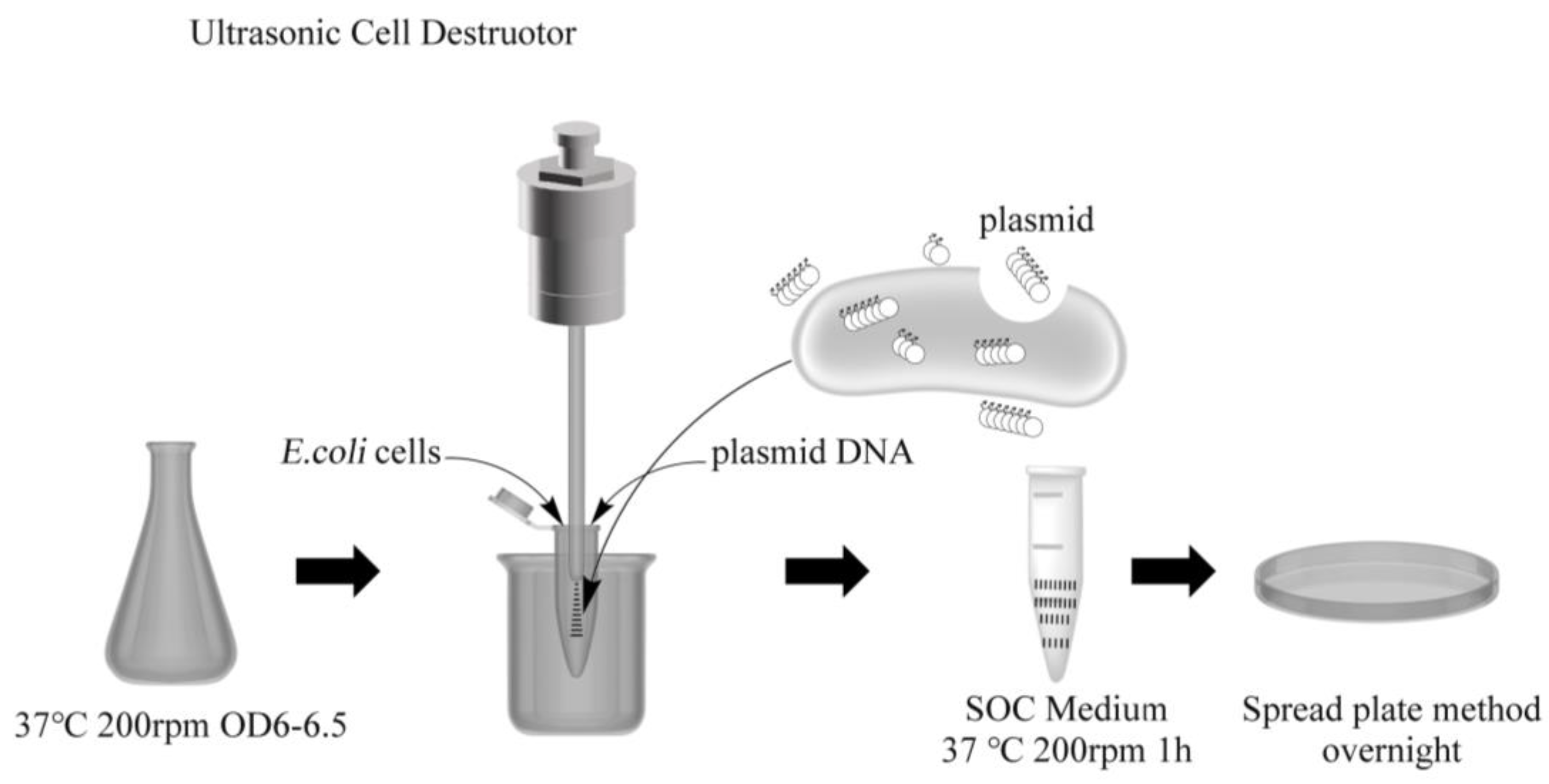
Figure 1. Ultrasound-mediated DNA transformation of bacteria.
Processes 11 02163 g001
Figure 2. Effect of input power on ultrasonic transformation of E. coli. Ultrasonic exposure time was set at 9 s (working, 1 s; pause, 2 s), 650 W rated power.
Processes 11 02163 g002
Figure 3. Effect of exposure time on ultrasonic transformation of E. coli. The ultrasonic power was set at 20% of the rated power (650 W), the exposure time was set as working, 1 s; pause, 2 s.
Processes 11 02163 g003
Figure 4. Effect of growth cycle on ultrasonic transformation of E. coli. The ultrasonic power was set at 20% of the rated power (650 W), the exposure time was set as 12 s (working, 1 s; pause, 2 s).
Processes 11 02163 g004
Figure 5. Effect of exposure circumstance on ultrasonic transformation of E. coli. The stationary phase cells were collected, the ultrasonic power was set at 20% of the rated power (650 W), the exposure time was set as 12 s (working, 1 s; pause, 2 s). (A): Effects of washing and collecting bacterial solutions; (B): Effects of Mg2+ and Ca2+ under the action of PBS solution.
Processes 11 02163 g005
Figure 6. Electron microscope analysis. (A): Normal E. coli cultured in LB medium at stationary phase; (B): Completely broken (stationary phase) E. coli; the ultrasonic power was set at 35% of the rated power (650 W), the exposure time was set as 120 s (working, 1 s; pause, 2 s); (C): E. coli during ultrasonic transformation, the stationary-phase cells were then collected, the ultrasonic power was set at 20% of the rated power (650 W), the exposure time was set as 12 s (working, 1 s; pause, 2 s); (D): E. coli resuscitated (500 µL SOC was added and transferred to a 37 °C shaker for resuscitation culture) after ultrasound transformation. For (A,C,D), the scanning electron microscope was set at 3 µm, 20,000×. In figure (C), the big arrow points to the hole created by the ultrasound; the volume of E. coli after ultrasound-mediated transformation was enlarged. In figure (D), the holes in the surface of the cell basically disappeared, and the arrows point to the holes that have not fully closed.
Processes 11 02163 g006aProcesses 11 02163 g006b
Figure 7. Ultrasound plasmid transfer of S. cerevisiae. The ultrasonic power was set at 20% of the rated power (650 W), the exposure time was set as working, 1 s; pause, 2 s.
Processes 11 02163 g007
Table 1. Strains and plasmids used in this study.
NameDescriptionReference/Sources
STRAIN
E. coli DH5αsupE44 Δ(lacZYA-argF) U169 (Φ80lacZ ΔM15) hsdR17 recA endA1Invitrogen
S. cerevisiae S288CMATα SUC2 gal2 mal2 mel flo1 flo8-1 hap1 ho bio1 bio6 ATCC 204508
S. cerevisiae NGT-F1derived from S. cerevisiae S288C, and tolerance to 381 g/L source[19]
PLASMID
pZEABPconstitute expression vector, pBR322 ori, P37 promoter, Ampr, BglBrick, ePathBrick
containing four isocaudomers (AvrII, NheI, SpeI, and XbaI)
[20]
pUG6Shuttle plasmid, Ampr in E. coli, G418r in S. cerevisiae Novagen VT1696
Disclaimer/Publisher’s Note: The statements, opinions and data contained in all publications are solely those of the individual author(s) and contributor(s) and not of MDPI and/or the editor(s). MDPI and/or the editor(s) disclaim responsibility for any injury to people or property resulting from any ideas, methods, instructions or products referred to in the content.