Article

Furfural Biodegradation in a Moving Bed Biofilm Reactor Using Native Bacteria and Agroforestry Waste as Supports

by
Alejandro Ruben Farias
1,*,
Maria Cecilia Panigatti
2 and
Diana Lia Vullo
3,4,*
1
Grupo de Investigación Sobre Temas Ambientales y Químicos (GISTAQ), Facultad Regional Resistencia, Universidad Tecnológica Nacional, French 414, H3500CHJ Resistencia, Chaco, Argentina
2
Facultad Regional Rafaela, Universidad Tecnológica Nacional, Int. de Acuña 49, S2300ADA Rafaela, Santa Fe, Argentina
3
Área Química, Instituto de Ciencias, Universidad Nacional de General Sarmiento, J.M. Gutiérrez 1150, B1613GSX Los Polvorines, Buenos Aires, Argentina
4
CONICET, Godoy Cruz 2290, C1425FQB Buenos Aires, Argentina
*
Authors to whom correspondence should be addressed.
Processes 2025, 13(5), 1337; https://doi.org/10.3390/pr13051337
Submission received: 13 March 2025 / Revised: 22 April 2025 / Accepted: 24 April 2025 / Published: 27 April 2025
(This article belongs to the Special Issue The Recycling Process of Agro-Industrial Waste)

Abstract

:
Furfural is a relevant industrial product, but its presence in water and soil generates contamination and health risks. Moving bed biofilm reactors (MBBRs) are an increasingly used alternative to eliminate contaminants with the advantage of occupying small spaces, despite their high dependence on support and the microorganisms involved in the process. This work proposes furfural elimination through a laboratory-scale MBBR using Bacillus licheniformis GTQ1, Microbacterium sp. GISTAQ2, and Brevundimonas sp. GISTAQ1 isolated from an industrial effluent and agroforestry waste (rice husks, pine sawdust, and quebracho chips) as supports. The biofilm development was tested with both axenic and mixed cultures, confirming high coverage by Scanning Electron Microscope (SEM) images, especially in triple-mixed cultures. Biodegradation tests were carried out in the MBBR with 15 g rice husks or quebracho chips as supports and a 4000 mg L−1 initial furfural concentration for 72 h. The mixed culture achieved almost a 100% furfural removal in three days with a rate of 3.97% per hour with rice husks and 2.61% per hour with quebracho chips. This laboratory-scale MBBR development is a promising first step ready for a scale-up for its implementation in industries to significantly reduce the environmental impact of the discharge of this type of effluent.

1. Introduction

Furfural (FUR), also known as furancarboxaldehyde, has great industrial importance due to its multiple uses: furfuryl alcohol (FOL) obtaining, refining of animal and vegetable oils, oil processing, synthesis of urea, formaldehyde, etc. FUR world production is approximately 280 kt, of which those from China (≈200 kt), the Dominican Republic, and South Africa represent 90% of the total [1]. However, at the same time, FUR is on the list of hazardous substances regulated by the Occupational Safety and Health Administration (OSHA) and, in the Scientific Committee on Consumer Safety (SCCS) of the European Union (EU), is classified as Carcinogen Category 2 (substances that cause concern because they can induce heritable mutations in human germ cells) [2]. FUR shows toxicity for various living organisms including a severe inhibition of many microbial metabolic pathways [3]. Concentrations between 7 and 25 mM (673 to 2402 mg L−1) negatively affect the microbial cell growth [4]. Likewise, FUR behaves as an inhibitor in the lignocellulosic biomass fermentation process, so its removal is important to achieve maximum process productivity [5,6,7]. Furfural concentrations in the hydrolysate obtained by thermochemical pretreatment of lignocellulosic biomass usually ranges from 0.5 to 3 g L−1 [8]. In addition, it also causes a decrease in intracellular ATP and NADPH levels, delaying cell growth by lengthening the lag phase of microorganisms involved in the production of biofuels [9,10]. Indigenous wild strains that can eliminate FUR have been isolated, identified, and characterised, and genetic modifications have been carried out in order to improve the degradation capacity [11,12,13,14,15,16,17].
Approximately 1.2 × 1030 bacterial and archaeal cells occupy different habitats on the planet, of which around 40−80% live in the form of attached communities or biofilms [18]. These communities are embedded in a polymeric matrix, which is secreted by themselves and is composed mostly of glycoproteins, glycolipids, mono- and polysaccharides, extracellular DNA, minerals, and water, usually named extracellular polymeric substances (EPSs). The EPSs act as an adhesive and protective support, favouring cell–cell interactions and nutrient acquisition, being responsible for the structure maintenance [19,20,21]. Biofilm communities are able to metabolise or retain organic contaminants and heavy metals by sorption mechanisms thanks to a properly regulated gene expression pattern mediated by quorum sensing [22]. Carrier roughness is an important characteristic. Tugba Kilic [23] highlighted investigations showing that the bacterial adherence rate increased with increasing surface roughness. Therefore, carrier roughness has a positive correlation with biofilm formation, since it is involved in the initial attachment and detachment of cells. In addition, roughness can increase the surface wettability. Consequently, selecting the appropriate roughness determines the optimal balance between the biofilm formation and detachment rate. Other important criteria include the matrix’s ability to be lightweight, flexible, cost-effective, mechanically and chemically stable, inert, non-biodegradable, non-polluting, non-toxic, and water-insoluble, among others. Biofilm formation therefore limits cell mobility in space, preserving viability and function, which allows for improved biotreatment of industrial effluents [24,25,26,27,28,29]. When compared to the use of planktonic microorganisms, among the different biological remediation techniques, biofilm-mediated remediation has been regarded as a safe, competent, and organised option for the removal/refinement of pollutants.
Several works have shown that membrane bioreactors (MBRs) as biological treatment processes can substantially enhance efficacy. The MBR offers benefits over traditional biological treatments, including a stable effluent, a smaller footprint, and reduced sludge generation [30].
The moving bed biofilm reactor (MBBR) is a wastewater biofilm-based technology that is currently implemented in more than 50 countries. To develop an MBBR, it is essential to determine the biofilm formation capacity as well as the generation time of the proper microorganisms for the wastewater biotreatment [31,32]. The key to the biofilm formation for a successful MBBR is related to the structure of the microbial community and the functionality of the organisms attached to the support surface [33]. However, it remains unknown how the microbial community evolves in the biofilm during the nutrient loading regime in an MBBR and how the community change is related to the contaminant biotransformation [34]. The selection of a biocarrier is a crucial step, because it impacts the cost-effectiveness of the process, the durability of the biofilm, and the treatment performance [35]. Several organic and inorganic natural materials have been employed as biofilm carriers in wastewater treatment. Zhang et al. showed that environmentally friendly Luffa sponges served as the feasible carriers and a better application prospect in the field of hydrolysed polyacrylamide-containing wastewater treatment [36]. This natural carrier could also promote the immobilisation of anaerobic microorganisms with a protecting effect [37]. Other organic materials frequently employed in wastewater treatments are bamboo and cactus fibre [35].
Rice is the most consumed cereal in the world with an estimated utilisation of 524.6 Mt in 2023/24 [38]. Approximately 20–22% of the total weight of the rice grain is husk, with its use currently very low [39]. The narrow application spectrum is associated with a high resistance to degradation and a high ash and lignin content [40]. Various lignocellulosic materials, such as pine sawdust and rice husk, have been successfully employed as biofilm carriers in microalgal cultivation [41]. On the other hand, pine and eucalyptus sawdust is one of the main wastes from the primary industrialisation of wood. It is estimated that 50% of the industrially processed wood in Argentina becomes 2 dry Mt per year of waste that is not properly used. Likewise, in northeastern Argentina, Paraguay, and southern Brazil, there are various industries for the extraction of tannins from quebracho colorado (Schinopsis balansae). These generated wastes represent a problem for industry and the environment. Therefore, agroforestry wastes can be utilised as sustainable substitutes for commercial carriers. Among them, wood husk, pine bark, cotton sticks, coconut husk fibre, sugarcane bagasse, cypress cones, date palm fibre, wheat straw, and others have been reported as environmentally friendly and inexpensive carriers with a potential application for microbial growth [35]. Zakaria et al. [42] used Acinetobacter haemolyticus immobilised on wood husk to detoxify Cr(VI) by its biotransformation to Cr(III). Pine bark and corn straw were used to prepare a high-efficiency and low-cost immobilised activated sludge for the treatment of phenol waste whilst lignocellulosic residues were used as bio-carriers for algal biofilm growth and pine sawdust as the optimal carrier for biomass production [43,44]. Rice husk was used as a biofilm carrier in the biological denitrification of wastewater, and Shao et al. [45] recommended it as an economical and effective external carbon source. Alitaleshi et al. [46] investigated the feasibility of using rice husk as a cost-effective and environmentally friendly bio-carrier for immobilising isolated microorganisms to treat synthetic aquaculture system wastewater. Zainab et al. [47] used Luffa sponge, coconut husk fibre, and wood chips as biofilm carriers to increase methane productivity and organic matter removal efficiency in anaerobic digestion. Al-Amshawee et al. [48] compared different materials as biofilm carriers for wastewater treatment in a fixed-bed biofilm reactor, achieving a 63% rate of COD removal in 7 days using Luffa sponge.
Only a few microbial metabolic pathways of FUR are known. Rodríguez et al. [16] reported FUR biodegradation mediated by a native strain of Bacillus cereus which selectively produced furfuryl alcohol from 30 mM of the substrate, while Bu et al. [49] achieved the bioconversion of 208 mM of FUR into furfuryl alcohol, using glucose and a biphasic system (aqueous/organic) to reduce the contact of FUR with Bacillus coagulans NL01. According to Wierckx et al. [50], FUR degradation proceeds via the 2-furoic acid pathway, which is then metabolised to the primary intermediate 2-oxoglutarate, showing that the enzymes of this degradative pathway are encoded in eight hmf genes, organised in two distinct clusters in bacteria such as Cupriavidus basilensis HMF14. Zheng et al. [17] found that all furfuryl alcohol would be oxidised to furoic acid (FA) with sufficient time, assuming that, as demonstrated, this redox reaction was reversible. Several researchers have reported the biotransformation of FUR into furfuryl alcohol [51,52], and some propose the degradation to furoic acid, with or without a prior passage through furfuryl alcohol [17,53]. However, other reports propose the route of direct oxidation to furoic acid to then be transformed into 2-oxoglutarate and enter the tricarboxylic acid cycle [54] or even the possibility that furfuryl alcohol can be transformed again into FUR and then definitively oxidised to furoic acid [15,19,55].
Regarding the environmental concern and the scarcity of reported studies on the development of innovative and ecofriendly biotechnologies to promote FUR removal from wastewaters, the aim of this work was to design a laboratory-scale MBBR by using agroforestry wastes as support material for bacterial biofilms involved in a biodegradation process. The central key is the design of proper biotreatment to be applied in the future by its scaling-up. The strains used were selected from soils, sediments, and effluents from industrial furfural production plants, able to efficiently degrade this compound. Likewise, the idea of using raw material such as agroforestry waste in a bioprocess mediated by native bacteria contributes to sustainable strategies in a circular economic context.

2. Materials and Methods

2.1. Microorganisms

Three indigenous bacterial strains were used in this work. Bacillus licheniformis (GTQ1); Brevundimonas sp. GISTAQ1, NCBI GenBank Accession Number MT787293; and Microbacterium sp. GISTAQ2, NCBI GenBank Accession Number OM540364 were previously isolated from the effluent treatment plant belonging to a furfural-producing industry and proved to be an efficient biodegradation process in planktonic state in both axenic and mixed cultures [56].

2.2. Biofilm Formation Assays

2.2.1. Inoculum Preparation

A first bacterial preinoculum was developed in duplicate in a broth containing the following: 49 mL of 1% w/v meat peptone as the main source of organic nitrogen [57], 0.5 mL of 1 M NH4Cl-assimilable nitrogen source [58] (J.T. Baker Inc. at 99.7% purity), and 0.5 mL of 1 M NaH2PO4 (Mallinckrodt Chemical Works, at 99.0% purity), for both pH regulation and a phosphorus source [59] at 30 °C and 200 rpm in a DLAB orbital shaker (SK-0330-PRO) for 24 h. Then, 5 mL of each culture were transferred to a 250 mL flask with 45 mL of a minimal mineral medium (M9-glucose) containing 34 g L−1 Na2HPO4 (Mallinckrodt Chemical Works 99.8%), 15 g L−1 KH2PO4 (Mallinckrodt Chemical Works 99.0%), 2.5 g L−1 NaCl (Alcor 99.9%), and 0.5 g L−1 NH4Cl (J.T. Baker Inc. 99.7%), supplemented with 10 µL of 1 M CaCl2, 0.2 mL of 1 M MgSO4, and 2.5 mL of 20% w/v glucose. Cultures were incubated at 30 °C and 200 rpm for 24 h. All solutions used were previously sterilised in an autoclave at 1.5 bar and 115 °C for a period of 15 min.

2.2.2. Plate Assay by Crystal Violet Dying Technique

Sterile 96-well microplates were used as a support, following the method recommended by Merrit et al. [60], with some adaptations [61]. The three bacteria were grown individually and in double- and triple-mixed cultures with a simultaneous cell-free control. Each assay was performed in sextuplicate. First, 180 µL of M9 broth (the same formulation as that used in the inoculum preparation) was added to each well and then inoculated with 20 µL of each single, double, and triple culture, previously adjusted to an Optical Density at 610 nm (OD610 nm) of 0.7. The plates were sealed with Parafilm® and incubated at 30 °C for 72 h. Then, the supernatants were removed and washed with Phosphate-Buffered Saline (PBS) solution (137 mM NaCl, 2.7 mM KCl, 10 mM Na2HPO4, and 1.8 mM KH2PO4) and rinsed with distilled water. Next, 200 µL of a 0.1 g mL−1 crystal violet (CV) (dissolved in ethanol-0.01 g mL−1 ammonium oxalate) was added. After a 12 min exposure, the CV was removed, eliminating the excess with distilled water. Then, 200 µL of 96% ethanol was added to dissolve the attached CV, and the dye concentration was estimated by measuring absorbance at 585 nm.

2.2.3. Biofilm Development on Wastes

The formation of biofilms on different supports was determined, both for individual strains and for mixed cultures. The selected materials were rice husk from a rice processing company, pine sawdust from local sawmills, discarded quebracho chips from a tannin factory, and commercial vegetable Luffa sponge. The finer grained material, such as pine sawdust and rice husk, was sieved using a 1.17 mm mesh, while the Luffa sponge and quebracho chips were separated with a 3.27 mm mesh, always taking the portion retained by the sieve. Then, 30 g of each support were immersed in distilled water at 80 °C for 40 min and then dried in an oven at 60 °C for 8 h. Once cold, 1 ± 1 × 10⁻⁴ g of each support were autoclaved in glass vials at 1.5 bar and 115 °C 15 min. Regarding bacteria, Brevundimonas sp. GISTAQ1, Microbacterium sp. GISTAQ2, and Bacillus licheniformis GTQ1 were tested individually or in double- and triple-mixed cultures. Tests were carried out in triplicate in Erlenmeyer flasks with 1 g of support, 34 mL of distilled water, 10 mL of M9 broth, and 1 mL of 20% w/v glucose. The flasks were inoculated with 5 mL of bacterial inoculum. Simultaneously, an abiotic control was included under the same culture conditions at 30 °C for 72 h.

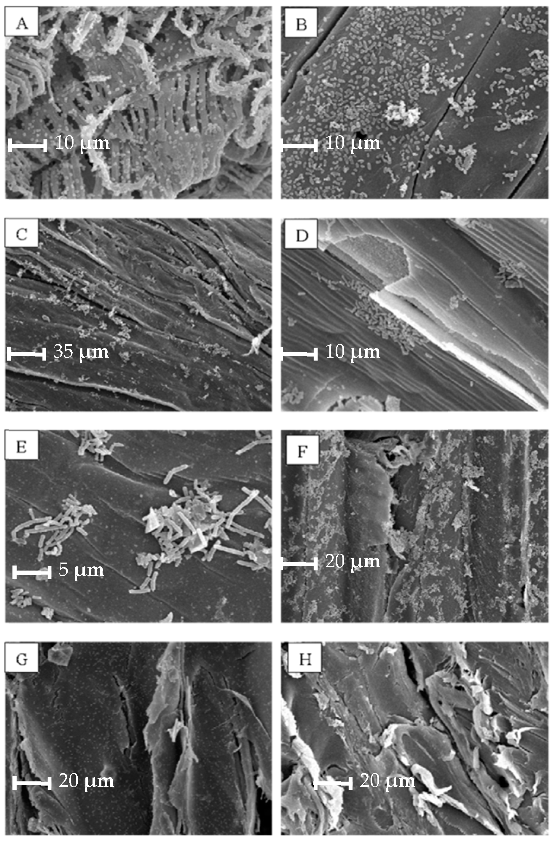
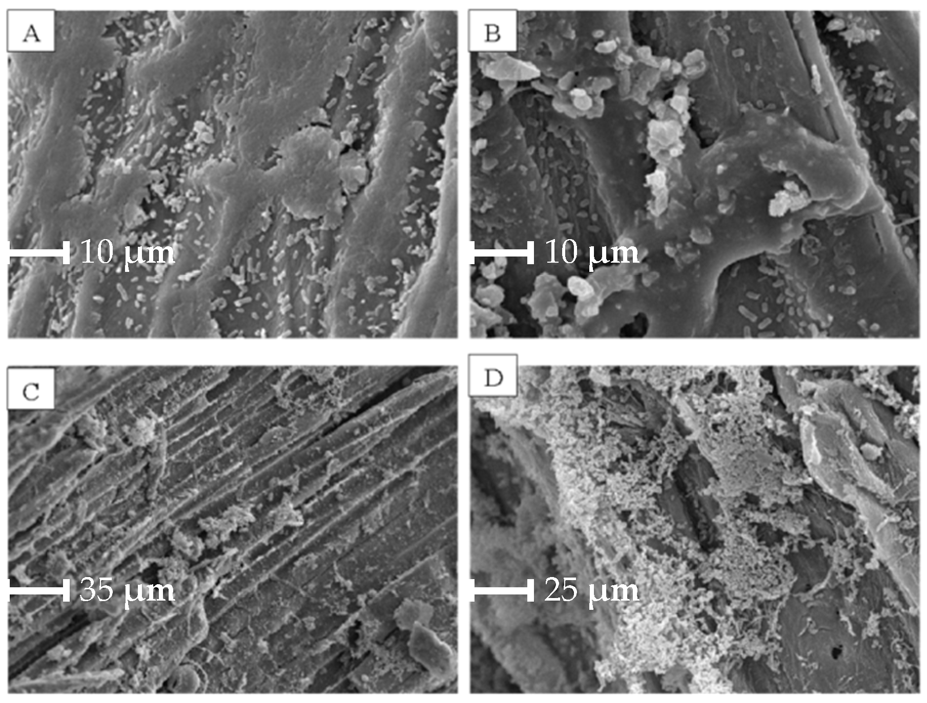
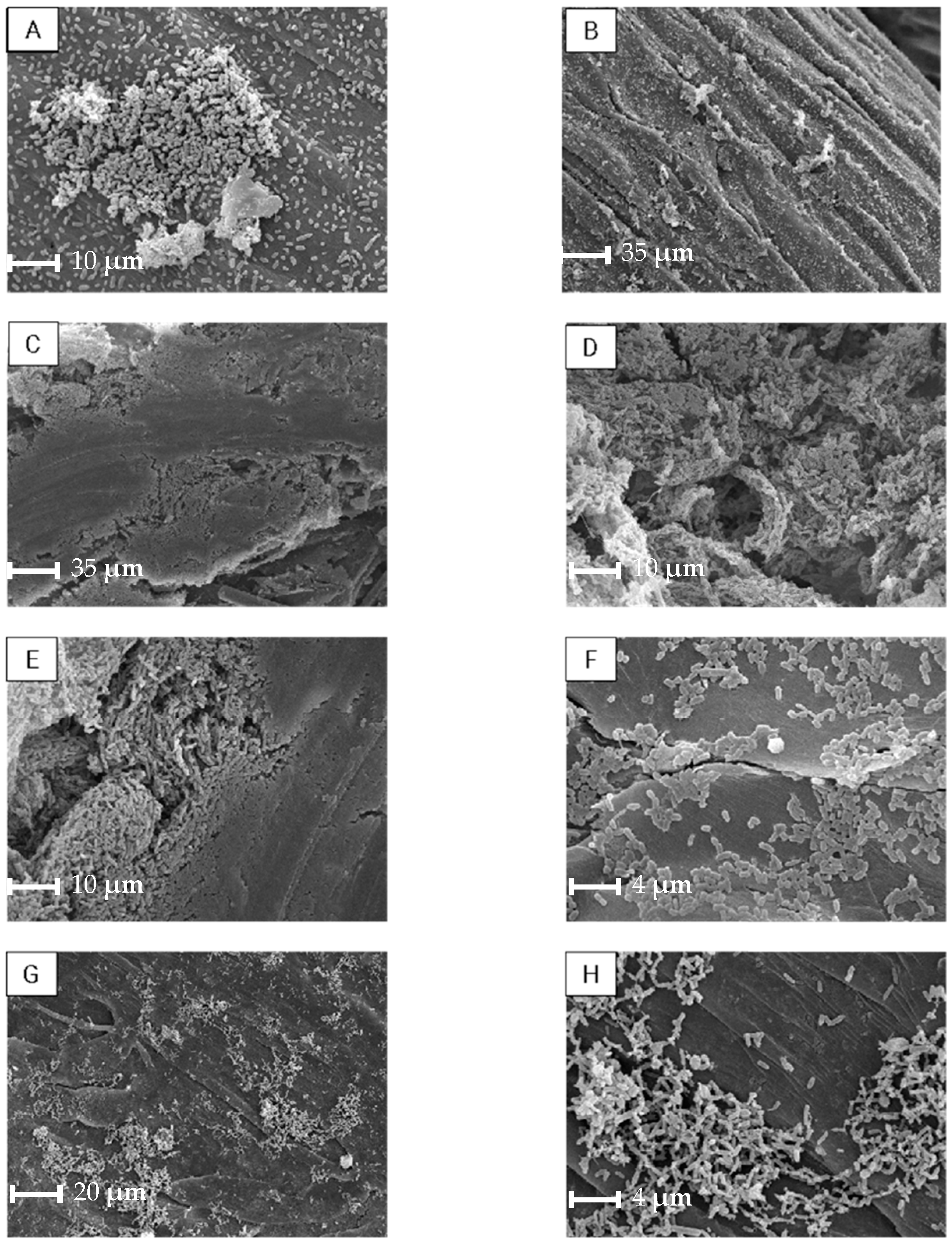
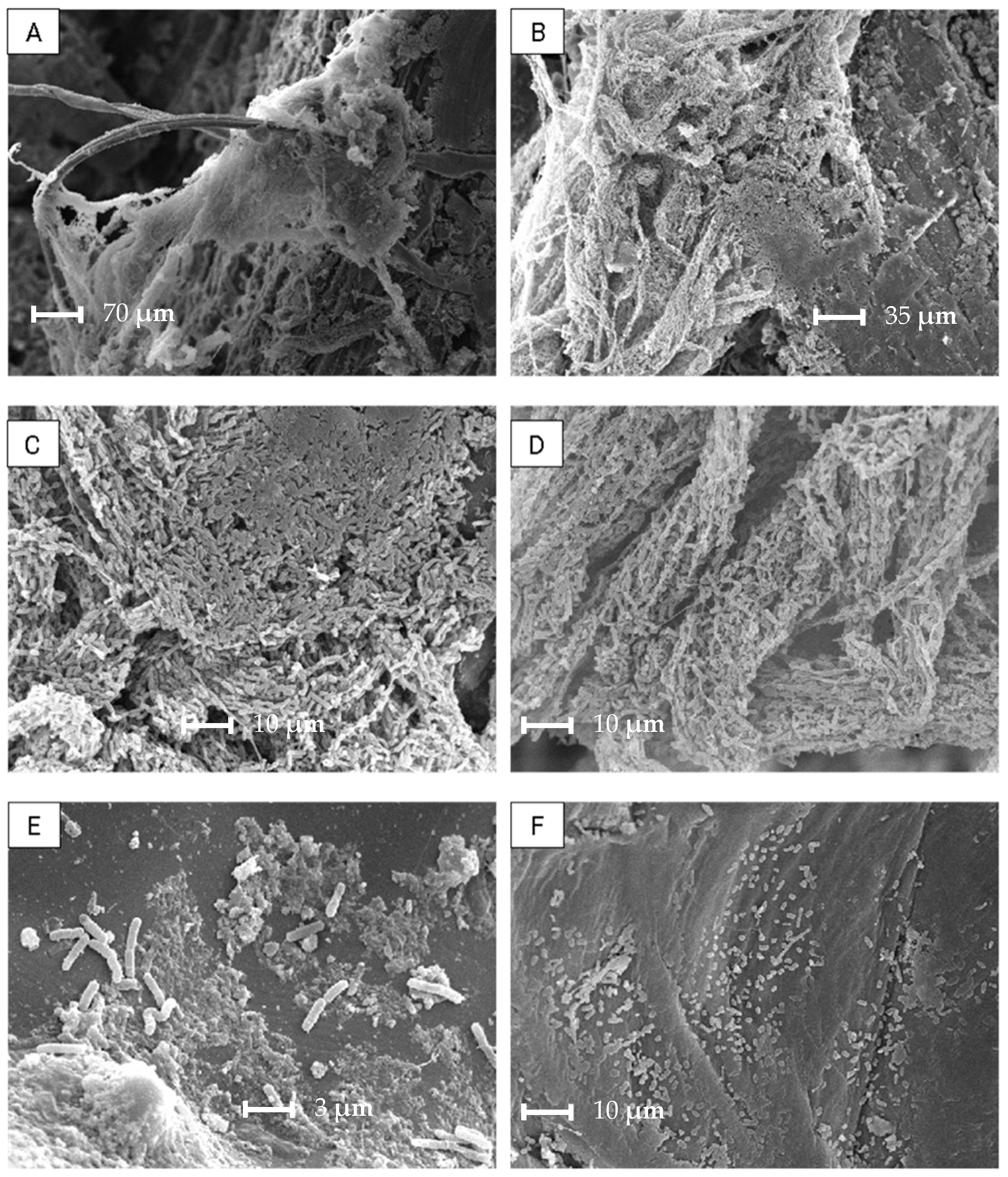
2.2.4. Biofilm Evaluation by Scanning Electron Microscopy

Part of the biofilm-developed materials was preserved in a Davidson-AFA fixing solution (33% absolute ethanol, 22% formaldehyde, 11.5% glacial acetic acid, and 33.5% distilled water). After a “critical point drying” and gold plating, the observation was carried out using a Scanning Electron Microscope (SEM-Jeol-5800 LV).

2.3. MBBR Setup

The FUR degradation was tested in a specially designed MBBR, built up with mixed cultures of Brevundimonas sp. GISTAQ1, Microbacterium sp. GISTAQ2, and Bacillus licheniformis GTQ1 immobilised on rice husks or exhausted quebracho chips. The MBBR experimental unit was built up on a laboratory scale (Figure 1) using a polyvinyl chloride
(PVC) column 300 mm long and 35 mm internal diameter, constituting a 275 mL reactor and a total working volume of 1.2 L. The prototype had a recirculation line consisting of a 1 L buffer tank and an APEMA PC25D peristaltic pump. A cotton mesh cloth filter was used at the inlet and outlet of the reactor to avoid fluidised bed losses [62]. The reactor and all accessories were previously sanitised with 7 g L−1 NaClO for 2 h and then rinsed repeatedly with sterile distilled water. A mass of 15 g of the corresponding pretreated support (clean, dry, and sterile) were weighed for each test. Once the support was loaded into the reactor, M9-glucose was added and inoculated with 40 mL of previously prepared cultures of each of the three strains. Since then, the pump was connected to feed the reactor. Once full, a flow rate was fixed at 1.5 L h−1. Under the conditions mentioned, the hydraulic retention time (HRT) is 10.8 min per cycle, with a recirculation of 47.4 min, so the “effective” HRT is 328 min in 24 h.
At the same time, a low aeration flux was connected using Atman AT-701 equipment. Approximately 70% of the fresh M9-glucose was renewed every 48/72 h to maintain constant the exponential growth state, along 30 days, at room temperature. A cell-free control, using the same conditions imposed for the bioreactor, was also tested.

2.4. MBBR Degradation Tests

Although the biofilm generation was carried out with a continuous fresh medium supply, the degradation test was operated in a batch mode. Once the biofilm generation phase ended, the whole medium volume was removed, and a simulated effluent, consisting of M9-glucose medium with the addition of FUR at a final concentration of 4000 mg L−1, was added to the tank. The system was closed, the pump and aeration were connected, and duplicate samples at 0 and 4 or 8 h along a 96 h period were collected for FUR quantification. The reactor was finally disassembled, and a sample of the fluidised bed was collected, rinsed with sterile distilled water, and preserved in Davidson-AFA fixing solution for the analysis of the developed biofilm by SEM.

2.5. Analytical Determination of FUR

For the identification and quantification of FUR, an HPLC method was developed as established by the ASTM Standard D 5837 [63] using Shimadzu CBM 20A equipment with an SPD 20 A UV (278 nm) detector. The method used an injection volume of 20 µL, a mobile phase acetonitrile/water (17.5:82.5), a flow rate of 1.0 mL min−1, 30 °C, and a Shimadzu column C18 reverse phase of 1.8 μm particle size, 100 Å pore size, 100 mm length, and 2.1 mm internal diameter. As a standard, commercial Merck® FUR (CAS No.:98-01-1) and another directly produced from the industrial plant that obtains 98% pure furfural were used. Figures of merit of the adapted HPLC method are detailed in the Supplementary Information file.

2.6. Statistical Evaluation

To assess the capacity for biofilm development on microtiter plates, a Q test was initially performed to identify the discordant values (outliers). Subsequently, to evaluate the differences among the group means, an analysis of variance (ANOVA) was conducted, followed by Tukey’s HSD test. A significance level of α = 0.05 was adopted.
In MBBR degradation tests, the standard error was calculated for each point based on the analytical method.

3. Results and Discussion

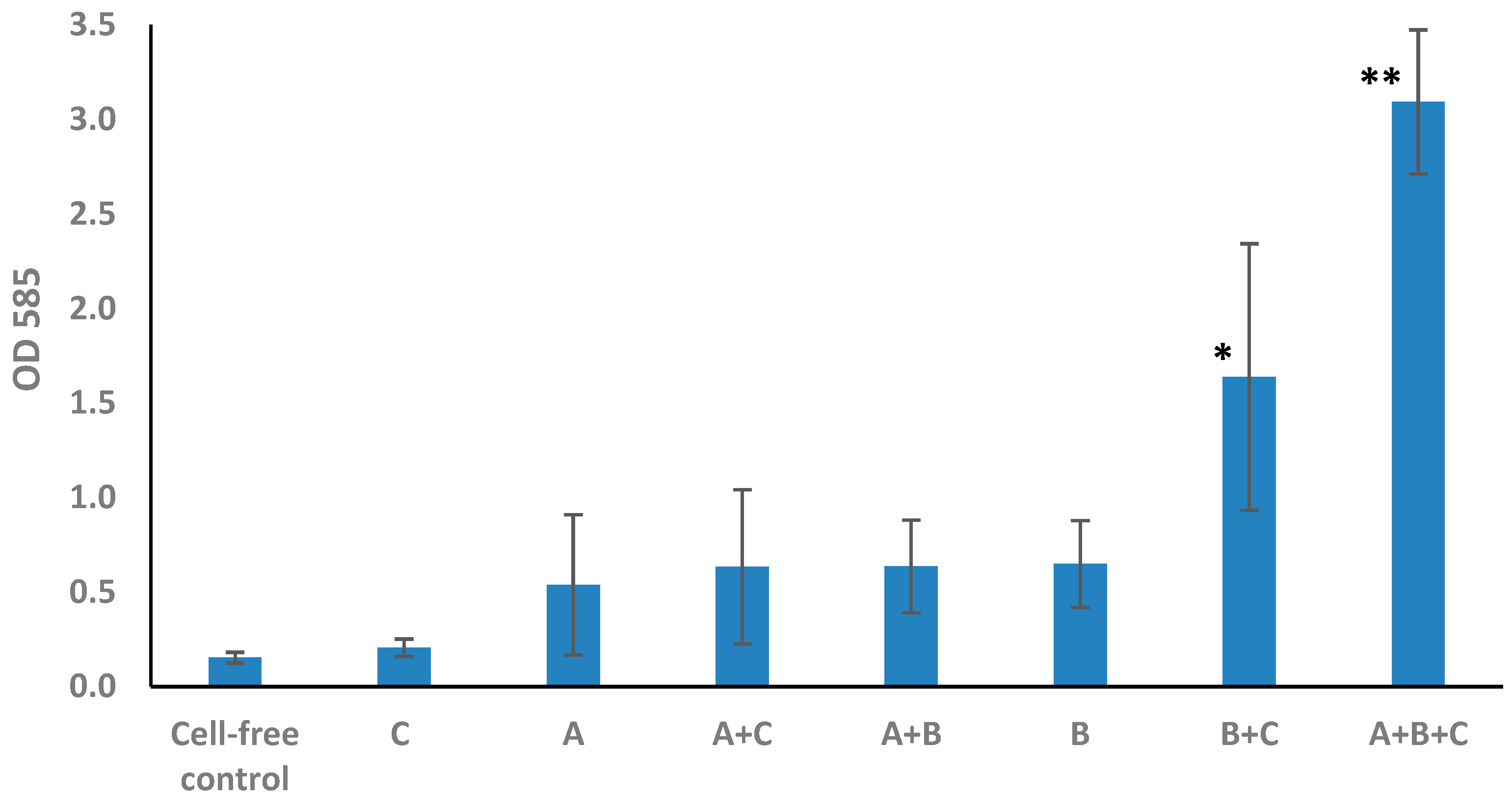
3.1. Determination of Biofilm Formation by Plate Assay

As described, the biofilm state represents a good habitat for forming syntrophic bacterial partnerships, because it offers a complete environment for two or more metabolically diverse bacterial species [22]. As a bar graph, Figure 2 shows the biofilm development of the assayed strains and their combinations, expressed as absorbance values at 585 nm. Populations that were statistically different (p < 0.05) are marked with asterisks. From the results obtained, the following can be observed:
  • The presence of biofilm in any of the individual cultures cannot be statistically confirmed.
  • Regarding double combinations, in those that contain Brevundimonas sp. GISTAQ1, the presence of biofilm cannot be confirmed.
  • The only double combination that could develop biofilm was Microbacterium sp. GISTAQ2 + Bacillus licheniformis GTQ1.
  • The highest biofilm production was observed with the triple combination Brevundimonas sp. GISTAQ1 + Microbacterium sp. GISTAQ2 + Bacillus licheniformis GTQ1.
No significant statistical differences were registered for the biofilm development of Brevundimonas sp. GISTAQ1 individually or in each double combination. These results could be related to the low contribution of Brevundimonas sp. GISTAQ1 to mixed biofilm generation. Likewise, no significant differences were observed between Brevundimonas sp. GISTAQ1 and Microbacterium sp. GISTAQ2 individually nor with the double combination Brevundimonas sp. GISTAQ1 + Microbacterium sp. GISTAQ2.
Zhu et al. [64] obtained similar results, demonstrating that biofilms composed by mixed cultures could be more stable, leading to a higher biomass compared to the corresponding biofilms of a single species due to the interactions between them. Laganenka and Sourjik [65] showed that Escherichia coli formed more structured biofilms with larger microcolonies when co-cultured with Enterococcus faecalis. The formation of mixed aggregates also promoted stress resistance of both species, which could be explained by the formation of larger E. coli aggregates.
The SEM images confirmed biofilm development in all samples; however, maximal biofilm formation was achieved with the mixed cultures. These results are consistent with the colorimetric assay performed and with the results obtained by Liu et al. [66] observing poor biofilm formation using axenic cultures of Microbacterium oxydans and Paenibacillus amylolyticus, with an improvement while associated with other four species. This fact strongly suggests that these low-abundance microorganisms could play a key role in significantly influencing the spatial organisation and, therefore, stabilising the function and composition of complex microbiomes.
Several randomly selected units (between 5 and 10) from each support were analysed. Figure 3 shows some images obtained via SEM. Although the thickness of the biofilm formed cannot be evaluated and despite the qualitative nature of the methodology, the bacterial cells colonizing the matrix can be clearly observed. In general, a consolidated biofilm was not observed in individual strains. This pattern was clearly appreciated in all the tested supports. The bacterial distribution was uniform over the surface without cellular aggregations or microcolonies, so the colonisation was not so evident accompanied by sporadic coverage. Figure 3H shows one of the cell-free samples processed as a blank, with no bacterial growth observed as expected.
SEM images of double combinations are shown in Figure 4. In this case, there are many sectors where the surface was clearly almost completely covered with biofilm (Figure 4A,B), together with a heterogeneous distribution (Figure 4C,D).
In the triple-bacterial combination, no significant differences were observed in biofilm production using rice husk, pine sawdust, or Luffa sponge. However, this triple combination produced the highest biofilm density on the quebracho chips (Figure 5). In the quebracho chips (Figure 5C–E), the extension of the biofilm developed by the triple-bacterial consortium was such that, in some sectors, the coverage was fully achieved. According to what was observed by Relucenti et al. [67], the EPS appears as a dense and shrunken mass with a rough surface dotted with holes rather than as a structure within which an intricate and microcanalicular system develops.
Regarding the obtained results, the mixed culture, consisting of Bacillus licheniformis GTQ1 + Microbacterium sp. GISTAQ2 + Brevundimonas sp. GISTAQ1 on quebracho chips and rice husks as supports, was selected for MBBR biodegradation tests.

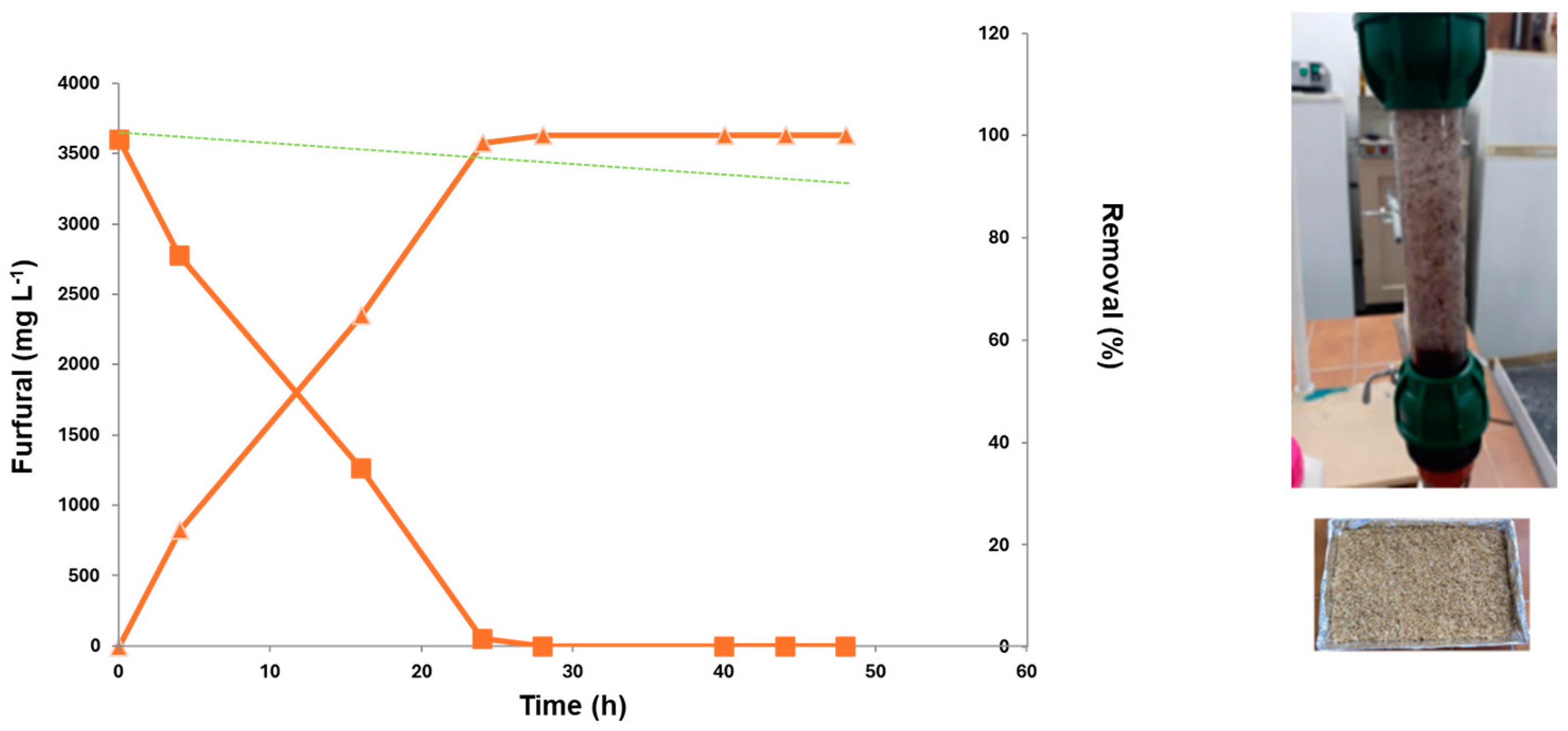
3.2. FUR Degradation in MBBR

To check the MBBR performance, the absence of losses was previously verified through a hydraulic and fluidisation test.
Figure 6 shows the results obtained for the FUR degradation in the MBBR reactor using the mixed culture immobilised on spent quebracho chips. A rapid and complete elimination of FUR was registered 40 h after starting the test. A complete removal percentage of 100% was confirmed, substantially improving the elimination rate compared to the efficiency achieved with planktonic cells. In fact, in a previous study, the bacterial consortium formed by Bacillus licheniformis GTQ1 + Microbacterium sp. GISTAQ2 + Brevundimonas sp. GISTAQ1 used furfural as a carbon source more effectively than the individual strains, removing 100% of furfural up to 2723 mg L−1 in 48 h when working in planktonic state [56].
Likewise, Figure 7 shows FUR biodegradation results with rice husks as a support. A rapid and total elimination in just 24 h was registered. In fact, at similar initial concentrations, the FUR removal rate using rice husks as a support was almost doubled compared to that obtained with quebracho chips (24 h vs. 40 h, respectively).
The degradation percentages in cell-free controls were 12.3% and 10.2% for rice husks and quebracho chips, respectively. In this way, it was confirmed that the removal occurred through a biodegradation mechanism and not through adsorption or any other chemical process involved.
Regarding the samples taken from the reactor, abundant biofilm generation was confirmed. In Figure 8, biofilm formation on quebracho chips was microscopically registered under different magnifications. The biofilm matrix, including cells, could be clearly observed. The structure of this biofilm seemed to be stable, robust, and with an extensive coverage, indicating that the presence of the contaminant at the tested concentration did not affect it. This confirmed the advantage of the bacterial biofilm generation in terms of providing cells the protection and hence resistance against aggressive contaminants such as FUR. In fact, with a mixed culture consisting of the same bacteria but under planktonic conditions, no growth or consumption of the contaminant was observed in 72 h when FUR concentrations among 4000 and 4500 mg L−1 were tested [56].
The results obtained were significant, since the mixed culture achieved a removal of almost 4000 mg L−1 of FUR with a removal rate of 3.97% per hour for rice husks and 2.61% per hour using quebracho chips as supports in the MBBR. The concentration and removal rates demonstrated a greater efficiency compared to most of the other reports with native strains [3,12,17,52,68,69,70,71,72,73,74,75] or even with chemical alternatives applying oxidative techniques [76].
The degradation efficiency of an MBBR depends on many factors. Piculell [77] demonstrated the importance of biofilm thickness control or the role of suspended biomass in the MBBR and its dependence on the operating conditions. In this way, by studying and optimising these factors on the designed MBBR, the FUR biodegradation could be improved, leading to a sustainable waste treatment.
The observation of a robust and well-formed bacterial biofilm in the experimental system developed in this work correlated directly with the high levels of contaminant degradation achieved. This complex biological structure not only acted as a biocatalyst, transforming the contaminant into less toxic compounds like furoic acid (FA) or furfuryl alcohol (FOL), but also promoted biomass retention, optimising the process. The obtained results suggest that the formation of stable biofilms is essential for the design of efficient bioreactors. In both aerobic and anaerobic microorganisms, the most common native mechanism observed for FUR detoxification involves cofactors (e.g., NADH or NAD+) to convert the carbonyl group into the corresponding alcohol or acid [78]. Researchers have reported the biotransformation of FUR into FOL [51,52], with some proposing degradation to FA, either directly or via FOL [17,53], and others suggesting the direct oxidation route to FA, followed by conversion to 2-oxoglutarate and entry into the tricarboxylic acid cycle. However, the production of FA and FOL from FUR exhibits significant variations, primarily due to these metabolic products also serving as substrates in the FUR degradation metabolic pathway [12]. Chromatographic analysis during FUR degradation revealed the simultaneous disappearance of the FUR peak with the appearance of a peak at tR 1.5–2 min. According to the literature, this latter peak could be assigned to FA or FOL, the initial biotransformation products [63]. Therefore, Bacillus licheniformis GTQ1, Microbacterium sp. GISTAQ2, and Brevundimonas sp. GISTAQ1, even in mixed cultures, potentially utilised the FA or FOL pathways for FUR biotransformation (data included in Supplementary Information).
Most of the reported research on FUR tolerance or biodegradation has been carried out at laboratory scale, applying planktonic cultures under batch conditions. Different approaches were developed, applying different microorganisms under other variables such as pH, temperature, cosubstrates, etc. Pseudomonas is one of the most used genera for FUR biodegradation studies. Maity et al. [69] achieved a complete degradation of 500 mg FUR L−1 in 12 h with Pseudomonas putida, while Crigler et al. [72] proved a degradation of 1000 mg FUR L−1, and Lee et al. [74] registered a bacterial tolerance up to 1700 mg FUR L−1, both with the same species. Pedrino et al. [79] contributed to the development of new strains within the Pseudomonas genus through tolerance adaptive laboratory evolution (TALE). In particular, KT2440 end strains exhibited tolerance to furfural (2000 mg L−1) and 5-hidroximetylfurfural (HMF) (1700 mg L−1). Igeño et al. [15] reported a mutated araC family gene (also a regulator of carbon source assimilation), enabling P. pseudoalcaligenes to utilise furfural as a carbon source. Zou et al. [80] unravelled the mechanism of furan aldehyde tolerance in P. putida, providing a new basis for engineering strains aimed at improving furfural tolerance.
Another widely used genus was Bacillus. Most of the literature described a limit in tolerance and/or degradation of around 2500 mg FUR L−1: Becerra et al. applied Bacillus toyonensis to degrade 2400 mg FUR L−1 in 5 days [12], and Ye et al. [75] and Bischoff et al. [71] worked with Bacillus coagulans, obtaining a tolerance of 2500 mg FUR L−1. On the other hand, Rodríguez et al. [17] evidenced a bioconversion of up to 3000 mg FUR L−1 with Bacillus cereus. Interestingly, Zheng et al. [68] observed a total inhibition of B. cereus development with FUR concentrations of 4000 mg L−1.
Other studies have used fungi such as Amorphoteca resinae, which degraded up to 1000 mg L−1 of FUR in 55 h [3]. Anaerobic bacteria have also been used. Belay et al. [70] reported a degradation up to 1000 mg FUR L−1 with Methanococcus delthae, but the bacterium was inhibited at concentrations between 2000 and 2500 mg L−1. Hunter et al. [73], however, achieved in 4 days a FUR degradation of 75% at initial concentrations of 4500 mg L−1 with Leuconostoc mesenteroides and L. pseudomesenteroides.
Ren et al. [7] managed to improve the FUR tolerance of S. cerevisiae after multiple rounds of progressive X-ray radiation combined with adaptive laboratory evolution; significantly, these mutant strains were able to grow in high concentrations of furfural (up to 4500 mg L−1) but not as the sole carbon source.
Similar results were obtained by Peng et al. [81], who worked with Bacillus sp., achieving a degradation of 70% in 72 h for a FUR concentration of 4000 mg L−1. While these studies demonstrated the ability to degrade the contaminant to similar final concentrations as those obtained here, the results evidenced a higher efficiency in terms of removal within a given timeframe. This difference could be attributed to various factors, including the unique composition of the microbial consortium and optimised operating conditions. The more favourable degradation kinetics observed in the developed system suggests a higher efficiency in contaminant degradation that could be translated into shorter treatment times or smaller reactor volumes. These results highlight the potential of this approach for treating FUR-contaminated wastewater.
Regarding the possible metabolic pathway, Liu et al. [82] determined FUR, furoic acid, and furfuryl alcohol under methodological conditions similar to those established in this work, determining a retention time (RT) for FUR at 4.3 min, 2.5 min for furfuryl alcohol, and 2.0 min for furoic acid. Based on the chromatographic peaks obtained and their specific retention times, it was also determined that the metabolic pathway followed by all isolates would be through 2-furoic acid production, one of the main pathways for FUR biodegradation already mentioned in various scientific publications.
Low space requirements, operational flexibility, resilience to changes in the environment, reduced hydraulic retention time, high active biomass concentration, enhanced ability to degrade recalcitrant compounds, and a slower microbial growth rate, which result in lower sludge production, are all advantages of wastewater treatment with biofilm systems [22]. The present work demonstrates that contaminant biodegradation can be achieved efficiently using aerobic bacterial consortium. This strategy has the advantage of being easier to implement on an industrial scale. The tolerance to varying environmental conditions further supports the advantages of this approach.

4. Conclusions

The native strains Bacillus licheniformis GTQ1, Microbacterium sp. GISTAQ2, and Brevundimonas sp. GISTAQ1 isolated from furfural contaminated sites resulted in good candidates to be used in biodegradation experiments because of their ability to develop biofilms individually and in mixed cultures. The synthetic consortium was able to form more structured biofilms than those obtained with monocultures. Both the Luffa sponge and the agroindustrial waste (rice husks, pine sawdust, and discarded quebracho chips) behaved as excellent matrices, promoting the biofilm formation. Considering that one of the most important parameters in the design and performance of MBBR is the biofilm area and, therefore, the specific area of the effective carrier, it could be concluded that an effluent treatment with high furfural loads could be successfully performed. The laboratory-scale reactor design is presented as the preliminary conceptualisation of the proposed treatment. Although the inherent limitations of the scale prevent the derivation of conclusive inferences concerning its field implementation, it does offer essential information for the planning and design of higher-capacity reactors. However, the designed laboratory-scale MBBR with Bacillus licheniformis GTQ1, Microbacterium sp. GISTAQ2, and Brevundimonas sp. GISTAQ1 immobilised on wastes from agroindustry proved high removal rates, giving a place to the future scaling-up process and implementation in sustainable biotreatments in a circular economy context.

Supplementary Materials

The following supporting information can be downloaded at: https://www.mdpi.com/article/10.3390/pr13051337/s1, File S1: Supplementary information Farias et al. V2 docx.

Author Contributions

A.R.F. participated in developing all stages of the process and carrying out the experimental procedures as well as in data curation and in writing the original draft. D.L.V. collaborated in the conceptualisation and experimental design, as well as in supervision, writing, review, and editing. M.C.P. participated in data visualisation as well as writing, reviewing, and editing the manuscript. All authors have read and agreed to the published version of the manuscript.

Funding

This work was funded by the Universidad Tecnológica Nacional, Facultad Regional Resistencia (UTI4023TC), and the Universidad Nacional de General Sarmiento, Consejo Nacional de Investigaciones Científicas y Tecnológicas (CONICET), Argentina.

Data Availability Statement

The data supporting this article have been included as part of the Supplementary Information.

Acknowledgments

A.R.F. and D.L.V. dedicate this work in memory of their dearest coauthor, Maria Cecilia Panigatti.

Conflicts of Interest

The authors declare no conflicts of interest. The funders had no role in the design of the study; in the collection, analyses, or interpretation of data; in the writing of the manuscript; or in the decision to publish the results.

Abbreviations

The following abbreviations are used in this manuscript:
FURFurfural
MBBRsMoving bed biofilm reactors
FOLFurfuryl alcohol
EPSsExtracellular polymeric substances
MBRsMembrane bioreactors
OD610 nmOptical Density at 610 nm
SEMScanning Electron Microscope
FAFuroic acid
HRTHydraulic retention time
HMF5-Hidroximetylfurfural

References

  1. Mariscal, R.; Maireles-Torres, P.; Ojeda, M.; Sádaba, I.; Granados, M.L. Furfural: A renewable and versatile platform molecule for the synthesis of chemicals and fuels. Energy Environ. Sci. 2016, 9, 1144–1189. [Google Scholar] [CrossRef]
  2. SCCS. Opinion on Furfural. Eur. Comm. 2012, 1–26. [Google Scholar]
  3. Ran, H.; Zhang, J.; Gao, Q.; Lin, Z.; Bao, J. Analysis of biodegradation performance of furfural and 5-hydroxymethylfurfural by Amorphotheca resinae ZN1. Biotechnol. Biofuels 2014, 7, 51. [Google Scholar] [CrossRef]
  4. Park, H.S.; Um, Y.; Sim, S.J.; Lee, S.Y.; Woo, H.M. Transcriptomic analysis of Corynebacterium glutamicum in the response to the toxicity of furfural present in lignocellulosic hydrolysates. Process Biochem. 2015, 50, 347–356. [Google Scholar] [CrossRef]
  5. Choi, S.Y.; Gong, G.; Park, H.-S.; Um, Y.; Sim, S.J.; Woo, H.M. Extreme furfural tolerance of a soil bacterium Enterobacter cloacae GGT036. J. Biotechnol. 2015, 193, 11–13. [Google Scholar] [CrossRef]
  6. Seo, H.M.; Jeon, J.M.; Lee, J.H.; Song, H.-S.; Joo, H.B.; Park, S.H.; Choi, K.Y.; Kim, Y.H.; Park, K.; Ahn, J.; et al. Combinatorial application of two aldehyde oxidoreductases on isobutanol production in the presence of furfural. J. Ind. Microbiol. Biotechnol. 2016, 43, 37–44. [Google Scholar] [CrossRef] [PubMed]
  7. Ren, J.; Zhang, M.; Guo, X.; Zhou, X.; Ding, N.; Lei, C.; Jia, C.; Wang, Y.; Zhao, J.; Dong, Z.; et al. Furfural tolerance of mutant Saccharomyces cerevisiae selected via ionizing radiation combined with adaptive laboratory evolution. Biotechnol. Biofuels Bioprod. 2024, 17, 117. [Google Scholar] [CrossRef] [PubMed]
  8. Li, B.; Liu, N.; Zhao, X. Response mechanisms of Saccharomyces cerevisiae to the stress factors present in lignocellulose hydrolysate and strategies for constructing robust strains. Biotechnol. Biofuels Bioprod. 2022, 15, 28. [Google Scholar] [CrossRef]
  9. Cho, H.Y.; Nam, M.S.; Hong, H.J.; Song, W.S.; Yoon, S.-I. Structural and Biochemical Analysis of the Furan Aldehyde Reductase YugJ from Bacillus subtilis. Int. J. Mol. Sci. 2022, 23, 1882. [Google Scholar] [CrossRef]
  10. Kim, J.; Son, H.F.; Hwang, S.; Gong, G.; Ko, J.K.; Um, Y.; Han, S.O.; Lee, S. Improving Lipid Production of Yarrowia lipolytica by the Aldehyde Dehydrogenase-Mediated Furfural Detoxification. Int. J. Mol. Sci. 2022, 23, 4761. [Google Scholar] [CrossRef]
  11. Becerra, M.L.; Lizarazo, L.M.; Rojas, H.A.; Prieto, G.A.; Martinez, J.J. Biotransformation of 5-hydroxymethylfurfural and furfural with bacteria of Bacillus genus. Biocatal. Agric. Biotechnol. 2022, 39, 102281. [Google Scholar] [CrossRef]
  12. Feldman, D.; Kowbel, D.J.; Cohen, A.; Glass, N.L.; Hadar, Y.; Yarden, O. Identification and manipulation of Neurospora crassa genes involved in sensitivity to furfural. Biotechnol. Biofuels 2019, 12, 210. [Google Scholar] [CrossRef]
  13. Sárvári Horváth, I.; Franzén, C.J.; Taherzadeh, M.J.; Niklasson, C.; Lidén, G. Effects of furfural on the respiratory metabolism of Saccharomyces cerevisiae in glucose-limited chemostats. Appl. Environ. Microbiol. 2003, 69, 4076–4086. [Google Scholar] [CrossRef]
  14. Igeño, M.I.; Macias, D.; Blasco, R. A case of adaptive laboratory evolution (ALE): Biodegradation of furfural by Pseudomonas pseudoalcaligenes CECT 5344. Genes 2019, 10, 499. [Google Scholar] [CrossRef] [PubMed]
  15. Ishii, J.; Yoshimura, K.; Hasunuma, T.; Kondo, A. Reduction of furan derivatives by overexpressing NADH-dependent Adh1 improves ethanol fermentation using xylose as sole carbon source with Saccharomyces cerevisiae harboring XR-XDH pathway. Appl. Microbiol. Biotechnol. 2013, 97, 2597–2607. [Google Scholar] [CrossRef] [PubMed]
  16. Rodríguez, A.; Rache, L.Y.; Brijaldo, M.H.; Romanelli, G.P.; Luque, R.; Martinez, J.J. Biocatalytic transformation of furfural into furfuryl alcohol using resting cells of Bacillus cereus. Catal. Today 2021, 372, 220–225. [Google Scholar] [CrossRef]
  17. Zheng, Z.; Xu, Q.; Tan, H.; Zhou, F.; Ouyang, J. Selective Biosynthesis of Furoic Acid From Furfural by Pseudomonas Putida and Identification of Molybdate Transporter Involvement in Furfural Oxidation. Front. Chem. 2020, 8, 587456. [Google Scholar] [CrossRef]
  18. Flemming, H.C.; Wuertz, S. Bacteria and archaea on Earth and their abundance in biofilms. Nat. Rev. Microbiol. 2019, 17, 247–260. [Google Scholar] [CrossRef]
  19. Dos Santos, A.L.S.; Galdino, A.C.M.; de Mello, T.P.; Ramos, L.d.S.; Branquinha, M.H.; Bolognese, A.M.; Neto, J.C.; Roudbary, M. What are the advantages of living in a community? A microbial biofilm perspective! Memórias Do Inst. Oswaldo Cruz 2018, 113, e180212. [Google Scholar] [CrossRef]
  20. Koo, H.; Allan, R.N.; Howlin, R.P.; Stoodley, P.; Hall-Stoodley, L. Targeting microbial biofilms: Current and prospective therapeutic strategies. Nat. Rev. Microbiol. 2017, 15, 740–755. [Google Scholar] [CrossRef]
  21. Rabin, N.; Zheng, Y.; Opoku-Temeng, C.; Du, Y.; Bonsu, E.; Sintim, H.O. Biofilm formation mechanisms and targets for developing antibiofilm agents. Future Med. Chem. 2015, 7, 493–512. [Google Scholar] [CrossRef]
  22. Saini, S.; Tewari, S.; Dwivedi, J.; Sharma, V. Biofilm-mediated wastewater treatment: A comprehensive review. Mater. Adv. 2023, 4, 1415–1443. [Google Scholar] [CrossRef]
  23. Kilic, T. Factors Affecting Biofilm Formation and the Effects of These Factors on Bacteria. In Exploring Bacterial Biofilms; Dincer, S., Ed.; IntechOpen: Rijeka, Croatia, 2025; p. 17. [Google Scholar]
  24. Mehrotra, T.; Dev, S.; Banerjee, A.; Chatterjee, A.; Singh, R.; Aggarwal, S. Use of immobilized bacteria for environmental bioremediation: A review. J. Environ. Chem. Eng. 2021, 9, 105920. [Google Scholar] [CrossRef]
  25. Dzionek, A.; Wojcieszyńska, D.; Guzik, U. Natural carriers in bioremediation: A review. Electron. J. Biotechnol. 2016, 23, 28–36. [Google Scholar] [CrossRef]
  26. Partovinia, A.; Rasekh, B. Review of the immobilized microbial cell systems for bioremediation of petroleum hydrocarbons polluted environments. Crit. Rev. Environ. Sci. Technol. 2018, 48, 1–38. [Google Scholar] [CrossRef]
  27. Asri, M.; Elabed, S.; Ibnsouda Koraichi, S.; El Ghachtouli, N. Biofilm-Based Systems for Industrial Wastewater Treatment. In Handbook of Environmental Materials Management; Hussain, C.M., Ed.; Springer: Berlin/Heidelberg, Germany, 2018; pp. 1–21. [Google Scholar]
  28. Mazioti, A.A.; Koutsokeras, L.E.; Constantinides, G.; Vyrides, I. Untapped potential of moving bed biofilm reactors with different biocarrier types for bilge water treatment: A laboratory-scale study. Water 2021, 13, 1810. [Google Scholar] [CrossRef]
  29. Rehman, F.; Thebo, K.H.; Aamir, M.; Akhtar, J. Nanomembranes for water treatment. In Nanotechnology in the Beverage Industry: Fundamentals and Applications; Amrane, A., Rajendran, S., Nguyen, T.A., Assadi, A.A., Sharoba, A.M., Eds.; Elsevier: Amsterdam, The Netherlands, 2020; pp. 207–240. [Google Scholar]
  30. Moktadir, M.A.; Maliha, M.; Tujjohra, F.; Munmun, S.A.; Alam, M.S.; Islam, M.A.; Rahman, M.M. Treatment of tannery wastewater by different membrane bioreactors: A critical review. Environ. Adv. 2024, 15, 100478. [Google Scholar] [CrossRef]
  31. Bhushan, S.; Gogoi, M.; Bora, A.; Ghosh, S.; Barman, S.; Biswas, T.; Sudarshan, M.; Thakur, A.R.; Mukherjee, I.; Dey, S.K.; et al. Understanding bacterial biofilm stimulation using different methods—A criterion for selecting epiphytes by plants. Microbiol. Biotechnol. Lett. 2019, 47, 303–309. [Google Scholar] [CrossRef]
  32. Gogoi, M.; Mukherjee, I.; Ray Chaudhuri, S. Characterization of ammonia remover Bacillus albus (ASSF01) in terms of biofilm-forming ability with application in aquaculture effluent treatment. Environ. Sci. Pollut. Res. 2022, 29, 61838–61855. [Google Scholar] [CrossRef]
  33. Biswas, K.; Taylor, M.W.; Turner, S.J. Successional development of biofilms in moving bed biofilm reactor (MBBR) systems treating municipal wastewater. Appl. Microbiol. Biotechnol. 2014, 98, 1429–1440. [Google Scholar] [CrossRef]
  34. Liang, C.; de Jonge, N.; Carvalho, P.N.; Nielsen, J.L.; Bester, K. Biodegradation kinetics of organic micropollutants and microbial community dynamics in a moving bed biofilm reactor. Chem. Eng. J. 2021, 415, 128963. [Google Scholar] [CrossRef]
  35. Lago, A.; Rocha, V.; Barros, O.; Silva, B.; Tavares, T. Bacterial biofilm attachment to sustainable carriers as a clean-up strategy for wastewater treatment: A review. J. Water Process Eng. 2024, 63, 105368. [Google Scholar] [CrossRef]
  36. Zhang, Y.; Zhao, L.; Song, T.; Cheng, Y.; Bao, M.; Li, Y. Simultaneous nitrification and denitrification in an aerobic biofilm biosystem with loofah sponges as carriers for biodegrading hydrolyzed polyacrylamide-containing wastewater. Bioprocess Biosyst. Eng. 2020, 43, 529–540. [Google Scholar] [CrossRef]
  37. Wang, W.; Wu, Y.; Zhang, C. High-density natural luffa sponge as anaerobic microorganisms carrier for degrading 1,1,1-TCA in groundwater. Bioprocess Biosyst. Eng. 2016, 40, 383–393. [Google Scholar] [CrossRef] [PubMed]
  38. FAO. Food and Agriculture Organization of the United Nations. Agricultural Market Information System (AMIS). Mark. Monit. 2024, 115, 1–19. [Google Scholar]
  39. Peanparkdee, M.; Iwamoto, S. Bioactive compounds from by-products of rice cultivation and rice processing: Extraction and application in the food and pharmaceutical industries. Trends Food Sci. Technol. 2019, 86, 109–117. [Google Scholar] [CrossRef]
  40. Nadaleti, W.C. Utilization of residues from rice parboiling industries in southern Brazil for biogas and hydrogen-syngas generation: Heat, electricity and energy planning. Renew. Energy 2019, 131, 55–72. [Google Scholar] [CrossRef]
  41. Rawindran, H.; Leong, W.H.; Suparmaniam, U.; Liew, C.S.; Raksasat, R.; Kiatkittipong, W.; Mohamad, M.; Ghani, N.A.; Abdelfattah, E.A.; Lam, M.K.; et al. Residual palm kernel expeller as the support material and alimentation provider in enhancing attached microalgal growth for quality biodiesel production. J. Environ. Manag. 2022, 316, 357–363. [Google Scholar] [CrossRef]
  42. Zakaria, Z.A.; Zakaria, Z.; Surif, S.; Ahmad, W.A. Biological detoxification of Cr(VI) using wood-husk immobilized Acinetobacter haemolyticus. J. Hazard. Mater. 2007, 148, 164–171. [Google Scholar] [CrossRef]
  43. Zhang, Q.; Yu, Z.; Jin, S.; Zhu, L.; Liu, C.; Zheng, H.; Zhou, T.; Liu, Y.; Ruan, R. Lignocellulosic residue as bio-carrier for algal biofilm growth: Effects of carrier physicochemical proprieties and toxicity on algal biomass production and composition. Bioresour. Technol. 2019, 293, 122091. [Google Scholar] [CrossRef]
  44. Yan, H.; Zhang, Q.; Wang, Y.; Cui, X.; Liu, Y.; Yu, Z.; Xu, S.; Ruan, R. Rice straw as microalgal biofilm bio-carrier: Effects of indigenous microorganisms on rice straw and microalgal biomass production. J. Environ. Manag. 2023, 341, 118075. [Google Scholar] [CrossRef] [PubMed]
  45. Shao, L.; Xu, Z.X.; Yin, H.L.; Chu, H. Rice husk as carbon source and biofilm carrier for water denitrification. J. Biotechnol. 2008, 18, S662. [Google Scholar] [CrossRef]
  46. Alitaleshi, F.; Daghbandan, A.; Alireza, P. Performance of rice husk biocarrier on ammonia nitrogen removal in the MBBR treating aquaculture wastewater using biological attached growth process: Performance and kinetic study. J. Environ. Chem. Eng. 2024, 12, 111446. [Google Scholar] [CrossRef]
  47. Zainab, A.; Meraj, S.; Liaquat, R. Study on Natural Organic Materials as Biofilm Carriers for the Optimization of Anaerobic Digestion. Waste Biomass Valorization 2020, 11, 2521–2531. [Google Scholar] [CrossRef]
  48. Al-Amshawee, S.K.A.; Yunus, M.Y.B.M.; Alalwan, H.A.; Lee, W.H.; Dai, F. Experimental investigation of biofilm carriers of varying shapes, sizes, and materials for wastewater treatment in fixed bed biofilm reactor: A qualitative study of biocarrier performance. J. Chem. Technol. Biotechnol. 2022, 97, 2592–2606. [Google Scholar] [CrossRef]
  49. Bu, C.Y.; Yan, Y.X.; Zou, L.H.; Zheng, Z.J.; Ouyang, J. One-pot biosynthesis of furfuryl alcohol and lactic acid via a glucose coupled biphasic system using single Bacillus coagulans NL01. Bioresour. Technol. 2020, 313, 123705. [Google Scholar] [CrossRef] [PubMed]
  50. Wierckx, N.; Koopman, F.; Ruijssenaars, H.J.; De Winde, J.H. Microbial degradation of furanic compounds: Biochemistry, genetics, and impact. Appl. Microbiol. Biotechnol. 2011, 92, 1095–1105. [Google Scholar] [CrossRef]
  51. Qin, L.Z.; He, Y.C. Chemoenzymatic Synthesis of Furfuryl Alcohol from Biomass in Tandem Reaction System. Appl. Biochem. Biotechnol. 2020, 190, 1289–1303. [Google Scholar] [CrossRef]
  52. Yan, Y.; Bu, C.; He, Q.; Zheng, Z.; Ouyang, J. Efficient bioconversion of furfural to furfuryl alcohol by Bacillus coagulans NL01. RSC Adv. 2018, 8, 26720–26727. [Google Scholar] [CrossRef]
  53. Wang, Z.W.; Gong, C.J.; He, Y.C. Improved biosynthesis of 5-hydroxymethyl-2-furancarboxylic acid and furoic acid from biomass-derived furans with high substrate tolerance of recombinant Escherichia coli HMFOMUT whole-cells. Bioresour. Technol. 2020, 303, 122930. [Google Scholar] [CrossRef]
  54. Koenig, K.; Andreesen, J.R. Xanthine dehydrogenase and 2-furoyl-coenzyme A dehydrogenase from Pseudomonas putida Fu1: Two molybdenum-containing dehydrogenases of novel structural composition. J. Bacteriol. 1990, 172, 5999–6600. [Google Scholar] [CrossRef]
  55. Wang, X.; Gao, Q.; Bao, J. Transcriptional analysis of Amorphotheca resinae ZN1 on biological degradation of furfural and 5-hydroxymethylfurfural derived from lignocellulose pretreatment. Biotechnol. Biofuels 2015, 8, 1–13. [Google Scholar] [CrossRef]
  56. Farías, A.; Echeverría, M.; Utgés, E.; Fontana, G.; Cuadra, P. Furfural Biodegradation in Consortium through Bacillus licheniformis, Microbacterium sp. and Brevundimonas sp. J. Sustain. Dev. Energy Water Environ. Syst. 2022, 10, 1–10. [Google Scholar] [CrossRef]
  57. Fernández Linares, L.C.; Rojas Avelizapa, N.G.; Roldán Carrillo, T.G.; Ramírez Islas, M.E.; Zegarra Martínez, H.G.; Uribe Hernández, R. Manual de Técnicas de Análisis de Suelos Aplicadas a la Remediación. 2006. Available online: https://es.scribd.com/doc/121876082/Manual-de-Analisis-de-Suelos (accessed on 15 April 2022).
  58. Boopathy, R.; Daniels, L. Isolation and characterization of a furfural degrading sulfate-reducing bacterium from an anaerobic digester. Curr. Microbiol. 1991, 23, 327–332. [Google Scholar] [CrossRef]
  59. Hareland, W.A.; Crawford, R.L.; Chapman, P.J.; Dagley, S. Metabolic function and properties of 4-hydroxyphenylacetic acid 1-hydroxylase from Pseudomonas acidovorans. J. Bacteriol. 1975, 121, 272–285. [Google Scholar] [CrossRef]
  60. Merritt, J.H.; Kadouri, D.E.; O’Toole, G.A. Growing and analyzing static biofilms. Curr. Protoc. Microbiol. 2011, 22, 1B-1.1–1B-1.18. [Google Scholar] [CrossRef]
  61. Burmølle, M.; Webb, J.S.; Rao, D.; Hansen, L.H.; Sørensen, S.J.; Kjelleberg, S. Enhanced biofilm formation and increased resistance to antimicrobial agents and bacterial invasion are caused by synergistic interactions in multispecies biofilms. Appl. Environ. Microbiol. 2006, 72, 3916–3923. [Google Scholar] [CrossRef] [PubMed]
  62. Sanabria Cubillos, A.; Pacheco Ojeda, J.D. Diseño y Evaluación de un Reactor Biológico de Lecho Móvil de Cargas Secuenciales Como Alternativa de Tratamiento Para un Vertimiento Procedente de una Industria Farmacéutica. 2019. Available online: https://ciencia.lasalle.edu.co/items/d4d2886a-661a-4a60-a769-7bfc58f70b3d/full (accessed on 23 September 2023).
  63. D5837-99; A Standard Test Method for Furanic Compounds in Electrical Insulating Liquids by High- Performance Liquid Chromatography (HPLC). In ASTM Volume 10.03: Electrical Insulating Liquids and Gases. ASTM: West Conshohocken, PA, USA, 2005.
  64. Zhu, Z.; Shan, L.; Li, X.; Hu, F.; Yuan, Y.; Zhong, D.; Zhang, J. Effects of interspecific interactions on biofilm formation potential and chlorine resistance: Evaluation of dual-species biofilm observed in drinking water distribution systems. J. Water Process. Eng. 2020, 38, 101564. [Google Scholar] [CrossRef]
  65. Laganenka, L.; Sourjik, V. Autoinducer 2-dependent Escherichia coli biofilm formation is enhanced in a dual-species coculture. Appl. Environ. Microbiol. 2018, 84, e02638-17. [Google Scholar] [CrossRef]
  66. Liu, W.; Russel, J.; Røder, H.; Madsen, J.; Burmølle, M.; Sørensen, S.J. Low-abundant species facilitates specific spatial organisation that promotes multispecies biofilm formation. Environ. Microbiol. 2017, 19, 2893–2905. [Google Scholar] [CrossRef]
  67. Relucenti, M.; Familiari, G.; Donfrancesco, O.; Taurino, M.; Li, X.; Chen, R.; Artini, M.; Papa, R.; Selan, L. Microscopy methods for biofilm imaging: Focus on sem and VP-SEM pros and cons. Biology 2021, 10, 51. [Google Scholar] [CrossRef]
  68. Zheng, D.; Bao, J.; Lu, J.; Gao, C. Isolation and characterization of a furfural-degrading bacterium Bacillus cereus sp. strain DS1. Curr. Microbiol. 2015, 70, 199–205. [Google Scholar] [CrossRef] [PubMed]
  69. Maity, J.P.; Chandra Samal, A.; Rajnish, K.; Singha, S.; Sahoo, T.R.; Chakraborty, S.; Bhattacharya, P.; Chakraborty, S.; Sarangi, B.S.; Dey, G.; et al. Furfural removal from water by bioremediation process by indigenous Pseudomonas putida (OSBH3) and Pseudomonas aeruginosa (OSBH4) using novel suphala media: An optimization for field application. Groundw. Sustain. Dev. 2023, 20, 100895. [Google Scholar] [CrossRef]
  70. Belay, N.; Boopathy, R.; Voskuilen, G. Anaerobic transformation of furfural by Methanococcus deltae ΔLH. Appl. Environ. Microbiol. 1997, 63, 2092–2094. [Google Scholar] [CrossRef] [PubMed]
  71. Bischoff, K.M.; Liu, S.; Hughes, S.R.; Rich, J.O. Fermentation of corn fiber hydrolysate to lactic acid by the moderate thermophile Bacillus coagulans. Biotechnol. Lett. 2010, 32, 823–828. [Google Scholar] [CrossRef]
  72. Crigler, J.; Eiteman, M.A.; Altman, E. Characterization of the Furfural and 5-Hydroxymethylfurfural (HMF) Metabolic Pathway in the Novel Isolate Pseudomonas putida ALS1267. Appl. Biochem. Biotechnol. 2020, 190, 918–930. [Google Scholar] [CrossRef] [PubMed]
  73. Hunter, W.J.; Manter, D.K. Pre-treatment step with Leuconostoc mesenteroides or L. pseudomesenteroides strains removes furfural from Zymomonas mobilis ethanolic fermentation broth. Bioresour. Technol. 2014, 169, 162–168. [Google Scholar] [CrossRef]
  74. Lee, S.A.; Wrona, L.J.; Cahoon, A.B.; Crigler, J.; Eiteman, M.A.; Altman, E. Isolation and Characterization of Bacteria That Use Furans as the Sole Carbon Source. Appl. Biochem. Biotechnol. 2016, 178, 76–90. [Google Scholar] [CrossRef]
  75. Ye, L.; Hudari, M.S.B.; Li, Z.; Wu, J.C. Simultaneous detoxification, saccharification and co-fermentation of oil palm empty fruit bunch hydrolysate for l-lactic acid production by Bacillus coagulans JI12. Biochem. Eng. J. 2014, 83, 16–21. [Google Scholar] [CrossRef]
  76. Rahmani, A.; Salari, M.; Tari, K.; Shabanloo, A.; Shabanloo, N.; Bajalan, S. Enhanced degradation of furfural by heat-activated persulfate/nZVI-rGO oxidation system: Degradation pathway and improving the biodegradability of oil refinery wastewater. J. Environ. Chem. Eng. 2020, 8, 104468. [Google Scholar] [CrossRef]
  77. Piculell, M. New Dimensions of Moving Bed Biofilm Carriers. Influence of Biofilm Thickness and Control Possibilities. 2016. Available online: https://portal.research.lu.se/en/publications/new-dimensions-of-moving-bed-biofilm-carriers-influence-of-biofil (accessed on 18 August 2023).
  78. Guarnieri, M.T.; Ann Franden, M.; Johnson, C.W.; Beckham, G.T. Conversion and assimilation of furfural and 5-(hydroxymethyl)furfural by Pseudomonas putida KT2440. Metab. Eng. Commun. 2017, 4, 22–28. [Google Scholar] [CrossRef] [PubMed]
  79. Pedrino, M.; Narcizo, J.P.; Aguiar, I.R.; Reginatto, V.; Guazzaroni, M.E. Upgrading Pseudomonas sp. toward Tolerance to a Synthetic Biomass Hydrolysate Enriched with Furfural and 5-Hydroxymethylfurfural. ACS Omega 2025, 10, 5449–5459. [Google Scholar] [CrossRef] [PubMed]
  80. Zou, L.; Jin, X.; Tao, Y.; Zheng, Z.; Ouyang, J. Unraveling the mechanism of furfural tolerance in engineered Pseudomonas putida by genomics. Front. Microbiol. 2022, 13, 1035263. [Google Scholar] [CrossRef]
  81. Peng, L.; Wang, L.; Che, C.; Yang, G.; Yu, B.; Ma, Y. Bacillus sp. strain P38: An efficient producer of l-lactate from cellulosic hydrolysate, with high tolerance for 2-furfural. Bioresour. Technol. 2013, 149, 169–176. [Google Scholar] [CrossRef]
  82. Liu, X.; Zhang, Y.; Shang, M.; Zhu, L.; Yu, M. The Effects of Furoic Acid and Furfuryl Alcohol on Detection of Furfural in Transformer Oil by High—Performance Liquid Chromatography. Chromatographia 2020, 83, 927–932. [Google Scholar] [CrossRef]
Figure 1. MBBR experimental unit.
Processes 13 01337 g001
Figure 2. Colorimetric assay for biofilm development determination of Bacillus licheniformis GTQ1, Brevundimonas sp. GISTAQ1, and Microbacterium sp. GISTAQ2 individually or combined in mixed cultures. Bar graph ordered by average absorbance at 585 nm with their respective standard deviation. The asterisks (* and **) represent the species and their combinations according to significant differences resulting from Tukey’s HSD test (n = 6; α = 0.05, p < 0.0001). A—Brevundimonas sp. GISTAQ1; B—Microbacterium sp. GISTAQ2; C—Bacillus licheniformis GTQ1.
Processes 13 01337 g002
Figure 3. SEM images for individual strains developed on matrices. (A) Brevundimonas sp. GISTAQ1 in Luffa ×2000. (B) Brevundimonas sp. GISTAQ1 in pine sawdust ×2000. (C) Microbacterium sp. GISTAQ2 in Luffa ×600. (D) Microbacterium sp. GISTAQ2 in rice husk ×2000. (E) Bacillus licheniformis GTQ1 in rice husk ×4000. (F) Microbacterium sp. GISTAQ2 in pine sawdust ×1000. (G) Brevundimonas sp. GISTAQ1 in pine sawdust ×1000. (H) Abiotic control in pine sawdust ×1000.
Processes 13 01337 g003
Figure 4. SEM images for bacterial double combinations developed on matrices. (A) Microbacterium sp. GISTAQ2 + Bacillus licheniformis GTQ1 in rice husk ×2000. (B) Brevundimonas sp. GISTAQ1 + Microbacterium sp. GISTAQ2 in pine sawdust ×2000. (C) Brevundimonas sp. GISTAQ1 + Microbacterium sp. GISTAQ2 in quebracho chips ×600. (D) Brevundimonas sp. GISTAQ1 + Bacillus licheniformis GTQ1 in Luffa ×1500. Images were obtained after 72 h of incubation at 30 °C.
Processes 13 01337 g004
Figure 5. Formation of biofilms on different supports by mixed cultures of Brevundimonas sp. GISTAQ1 + Microbacterium sp. GISTAQ2 + Bacillus licheniformis GTQ1. (A) In Luffa ×2000. (B) In pine sawdust ×600. (C) In quebracho chips ×600. (D) In quebracho chips ×2000. (E) Quebracho chips ×2000. (F) Pine sawdust ×4500. (G) Rice husks ×1000. (H) Rice husks ×4500. Images were obtained after 72 h of incubation at 30 °C.
Processes 13 01337 g005
Figure 6. FUR removal kinetics in MBBR with the mixed culture consisting of Bacillus licheniformis GTQ1 + Microbacterium sp. GISTAQ2 + Brevundimonas sp. GISTAQ1 immobilised on quebracho chips. The dotted line indicates the decrease in FUR concentration under control conditions (supports without biofilm development). The standard error is detailed in the Supplementary Information. Right and above: Laboratory-scale reactor. Right and bottom: Quebracho chip support. ■: FUR concentration (mg L−1); ▲: FUR removal (%).
Processes 13 01337 g006
Figure 7. Biodegradation curve and percentage of degradation of FUR as a function of time in MBBR using the mixed culture of Bacillus licheniformis GTQ1 + Microbacterium sp. GISTAQ2 + Brevundimonas sp. GISTAQ1 immobilised on rice husks. The dotted line indicates the decrease in FUR concentration under control conditions (supports without biofilm development). The standard error is detailed in the Supplementary Information. Right and above: MBBR rice husk assay. Right and bottom: Rice husk support. ■: FUR concentration (mg L−1); ▲: FUR removal (%).
Processes 13 01337 g007
Figure 8. SEM images of quebracho chips (AD) and rice husks (E,F) 30 days after the biofilm generation and biodegradation step using the mixed culture of Bacillus licheniformis GTQ1 + Microbacterium sp. GISTAQ2 + Brevundimonas sp. GISTAQ1. (A) Width 440 µm. Magnification ×300. (B) Width 220 µm. Magnification ×600. (C,D) Width 66 µm. Magnification ×2000. (E) Width 18 µm ×5000. (F) Width 66 µm ×2000.
Processes 13 01337 g008
Disclaimer/Publisher’s Note: The statements, opinions and data contained in all publications are solely those of the individual author(s) and contributor(s) and not of MDPI and/or the editor(s). MDPI and/or the editor(s) disclaim responsibility for any injury to people or property resulting from any ideas, methods, instructions or products referred to in the content.

