Article

Novel Lactic Acid Bacteria Strains from Regional Peppers with Health-Promoting Potential

by
Ivana Micaela Nuñez
1,
María Cecilia Verni
1,
Fernando Eloy Argañaraz Martinez
2,
Jaime Daniel Babot
3,
Victoria Terán
1,
Mariana Elizabeth Danilovich
1,2,
Elena Cartagena
1,2,*,
María Rosa Alberto
1,2,† and
Mario Eduardo Arena
1,2,†
1
Instituto de Biotecnología Farmacéutica y Alimentaria (INBIOFAL) CONICET–UNT, Avenida N Kirchner 1900, San Miguel de Tucumán CP 4000, Argentina
2
Facultad de Bioquímica, Química y Farmacia, Universidad Nacional de Tucumán (UNT), Ayacucho 471, San Miguel de Tucumán CP 4000, Argentina
3
Reference Centre for Lactobacilli (CERELA-CONICET), Chacabuco 145, San Miguel de Tucumán CP 4000, Argentina
*
Author to whom correspondence should be addressed.
These authors contributed equally to this work.
Fermentation 2024, 10(4), 209; https://doi.org/10.3390/fermentation10040209
Submission received: 1 March 2024 / Revised: 4 April 2024 / Accepted: 9 April 2024 / Published: 11 April 2024
(This article belongs to the Special Issue Recent Trends in Lactobacillus and Fermented Food, 2nd Edition)

Abstract

:
This study provides a comprehensive investigation of lactic acid bacteria (LAB) isolated from Argentinean Capsicum annum L. This research covers important aspects, including genotypic characterization, bacterial stress tolerance, adhesion ability, safety evaluation, and functional and technological properties. The predominant isolates were identified as Lactilactobacillus curvatus and Lactiplantibacillus plantarum. A Rep-PCR analysis grouped the isolates into 11 clonal groups. Lp. plantarum LVP 40 and LV 46, Levilactobacillus brevis LVP 41, Pediococcus pentosaceus LV P43, and Lt. curvatus LVP44 displayed both safety and resilience against adverse conditions such as a slow pH, bile, and simulated gastric and intestinal juices. Moreover, the LAB strains exhibited high hydrophobicity and auto-aggregation percentages, NaCl tolerance, and a substantial acidifying capacity. LAB supernatants demonstrated promising surfactant and emulsifying properties. Likewise, they differentially inhibited Staphylococcus aureus and Pseudomonas aeruginosa biofilms, showcasing their potential as antipathogenic agents. Noteworthily, some strains displayed considerable co-aggregation with these pathogens, and several isolates showed an effective antimutagenic and detoxifying power, further emphasizing their multifaceted capabilities. Five pepper bacterial strains showcased beneficial properties, suggesting their potential for gut health enhancement. In summary, these LAB strains hold promise as vegetable fermentation starters, contributing to food safety and versatile applications in food science.

1. Introduction

Among vegetables, peppers contribute significantly to the human diet due to their high content of antioxidants, vitamins (A, C, and E, mainly), minerals, polyphenols, and pigments. However, peppers have a comparatively short storage and shelf life than other vegetables, such as root vegetables. Therefore, developing methods to preserve the nutritional content and extend the shelf life of peppers could provide significant benefits for both the food industry and the consumers. Multiple ancient civilizations have used pickling as one of the oldest preservation methods for various food products, including vegetables and fruits [1]. Although, traditionally, fermented pickles were homemade products obtained by spontaneous fermentation, they are now evolving as a select entrée to improve quality and safety [2]. It is well known that using a preselected mixed starter culture for lactic acid (LA) fermentation leads to superior quality in different fermented vegetables or fruits [3].
Functional foods are defined as foods or ingredients with additional physiological benefits and nutritional value. In recent years, there has been an increase in interest in probiotics, and a significant body of research now recognises probiotic products and fermented foods as potential functional foods that may contribute to improved gut health, prevention, and the treatment of diseases.
Lactic acid bacteria (LAB) have traditionally been the main probiotics used in food processing as starter cultures, pharmaceuticals, and biological control agents. Currently, more than 62 different genera of LAB are widely used in commercial products as a safe fermentation culture. However, it is necessary to address tools of different complexity for the isolation, molecular characterization, identification, and evaluation of the probiotic properties of LAB before they can be considered [4].
Since probiotics are expected to provide beneficial health effects to the host, they must show resistance to the acidic conditions of the stomach and the presence of bile salts and pancreatin in the small intestine. It is also essential that probiotic strains have good patterns of susceptibility to antibiotics and cell surface properties, such as auto-aggregation, cell surface hydrophobicity, auto-aggregation, and co-aggregation, to facilitate LAB colonization in the intestinal wall. In addition, functional attributes like antimicrobial and antimutagenic activities are desirable for probiotic strains to be effective [5].
The search for new probiotic strains with unique properties continues to be an area of great interest despite the availability of numerous well-characterised probiotic strains worldwide. In this sense, there is a growing interest in exploring the probiotic and biotechnological potential of microorganisms already adapted to the food matrices in which they are found naturally. This is especially relevant in the case of bacteria isolated from red and green peppers (Capsicum annum L.), which have the advantage of being already adapted to this specific microbiome. By evaluating the functional and technological capabilities of regional pepper isolates, we identified new strains with unique properties useful in various applications, including the food and pharmaceutical industries.
The primary focus of this study was to examine the beneficial properties of lactic acid bacteria extracted from peppers in the northern region of Argentina. Given this vegetable’s crucial role in the local economy, exploring ways to increase its market value is imperative. Our main goal was to isolate and identify strains of lactic acid bacteria that can potentially promote human health by conducting extensive investigations of their functional and technological properties for the development of innovative and sustainable practices in regional agriculture.

2. Materials and Methods

2.1. Isolation and LAB Identification

2.1.1. Source

Lactic acid bacteria were isolated from fresh Capsicum annum L. (green and red peppers) obtained from three different sources in Tucumán, Argentine: home garden peppers which did not receive fungicides and peppers purchased at the local market.

2.1.2. Isolation of Peppers’ Bacterial Strains

Each sample aseptically collected was washed with 1 mL of sterile saline solution (0.85%). Serial dilutions were performed, of which 100 µL aliquots were plated out onto Man Rogosa and Sharpe (MRS) agar (MRS-agar) supplemented with 0.02% cycloheximide and 0.1 g/L sodium azide for lactobacilli isolation. Cultures were carried out in triplicate using the pour plate method. Plates were incubated at 37 °C for 48 h under microaerophilic and anaerobic conditions generated by Mitsubishi Gas Chemical envelopes, Tokyo, Japan. Then, the isolates were examined microscopically for Gram reaction and catalase production, and their morphology was also analysed under an optical microscope. All isolates were kept at −20 °C and −80 °C in MRS glycerol (20%). Likewise, each isolate was lyophilised for its adequate conservation.

2.1.3. Identification of LAB Isolates

For genotypic characterization, the total genomic DNA of the isolates was extracted with a commercial kit (PrestoTM Mini gDNA Bacteria Kit Quick Protocol, GeneaidBiotech Ltd., Taipai, Taiwan) according to the manufacturer’s instructions. Amplification of the 16s rDNA was carried out using a pair of primers 27F (5′-GTGCTGCAGAGAGTTTGATCCTGGCTCAG-3′) and 1492R (5′-CACGGATCCTACGGGTACCTTGTTACGACTT-3′) [6]. The polymerase chain reaction mixture consisted of 1.5 mmol/L MgCl 2.5 µL 10× reaction buffer, 100 µmol/L dNTPs, 0.5 µmol/L of each primer, 4 mL bacterium DNA, and 1.5 U Taq polymerase (Invitrogen, San Diego, CA, USA). Amplification consisted of initial denaturation at 94 °C for 5 min, followed by 30 cycles of denaturation at 94 °C for 1 m, hybridization at 52 °C for 2 ms, and extension at 72 °C for 2 ms, with a final extension step at 72 °C for 7 min. The polymerase chain reaction was conducted in a thermocycler My Cycler (Bio-Rad Laboratories, Hercules, CA, USA). The amplification products were separated by electrophoresis (80 v) on a 0.8% (w/v) agarose gel stained with SYBR Gel DNA Safe Stain (Invitrogen) in 1× TAE buffer (40 mmol/L Tris-acetate, 1 mmol/L EDTA, pH 8). The PCR products were purified with a PCR Purification AccuPrep Kit (Bioneer, Alameda, CA, USA) and then sequenced using a 3730 XL Sequencer (Applied Biosystems, Foster City, CA, USA) by MACROGEN Inc. (Seoul, Republic of Korea). The 16S rRNA gene sequences obtained were edited with the Chromas Pro software (1.5 version Technelysium Pty. Ltd. (South Brisbane, Australia), 2003–2009) and analysed with the DNAMAN software (2.6 version Lynnon-Biosoft). Sequence homologies were examined by comparing the obtained sequences with those of the GenBank/EMBL/DDBJ database using the BLAST software (http://blast.ncbi.nlm.nih.gov/Blast.cgi) and identified according to the closest relative. The 16S ribosomal DNA sequences of the isolated strains were deposited in the GenBank.

2.1.4. Bacterial Strain Identification

Subsequently, the isolates were differenced at the strain level by repetitive sequence-based (rep-PCR) fingerprinting using the primer (GTG)5 (5′-GTGGTGGTGGTGGTG-3′) described by Gevers et al. (2001) [7]. The PCR reactions were carried out as follows: 5 min of denaturation at 94 °C, followed by 30 cycles of denaturation at 94 °C for 1 min, annealing at 40 °C for 1 min, and extension at 65 °C for 8 min, with a final extension step at 65 °C for 16 min [7]. The mastermix (20 µL) contained 4 µL of 5× buffer (Inbio Highway, Buenos Aires, Argentina), 4 µL of 2.5 mM dNTPs (Inbio Highway, Buenos Aires, Argentina), 2 U of Taq polymerase (Inbio Highway, Buenos Aires, Argentina), 1 µL of DNA template (50 ng), 2 µL of 10 µM (GTG)5 primer (Genbiotech, Buenos Aires, Argentina), 4 µL of 25 mM MgCl2, and 4.8 µL of deionised water. PCR reactions were performed in a MyCycler device (Bio-Rad Laboratories, Hercules, CA, USA), and the amplification products were separated by electrophoresis at 40 V for 200 min on 1.5% (w/v) agarose stained with GelRedTM (Biotium, Hayward, CA, USA) in 1 × TAE Buffer (40 mM Tris-acetate, 1 mM EDTA). The Rep-PCR fingerprints were analysed as indicated by Versalovic et al. (1994) [8], using the GelJ software v.2.0 [9], using the Dice correlation coefficient, and dendrograms were constructed employing the UPGMA clustering method according to Ishii and Sadowsky (2009) [10].

2.2. Stress Tolerance

2.2.1. pH Resistance

Bacterial cells were incubated at 37 °C for 18 h (5% initial culture overnight). Cells were harvested by centrifugation (3500 rpm, 15 min at 4 °C, 2193× g) and washed once with phosphate-buffered saline (PBS, pH 7.2). The resistance of the isolates was evaluated as survival in PBS buffer at different pH values (3, 4, and 7) for 2 h with an initial inoculum adjusted to an optical density (OD) of 0.35 at 600 nm (107–108 CFU/mL) (Spectrophotometer Genesys 50, Thermo Fisher Scientific, Madison, WI, USA). The number of CFU/mL was determined after 0, 1, and 2 h of incubation at 37 °C at the different pH values in microaerophilic conditions. A decrease less than three logarithmic cycles in 2 h was considered resistant.

2.2.2. Bile Tolerance

In order to assess how the strains, respond to the presence of bile, 0.3% (w/v) bile (oxgall, Sigma-Aldrich, St. Louis, MO, USA) was added to the MRS medium. The active cultures of each strain were subjected to centrifugation (3500 rpm, 15 min at 4 °C, 2193× g), and the resulting pellets were resuspended in an appropriate volume of medium to achieve an initial inoculum of OD 600 nm = 1 (Spectrophotometer Genesys 50, Thermo Scientist, Waltham, MA, USA). The bacterial suspensions were used to inoculate (5%) MRS medium with or without oxgall (107 CFU/mL initial inoculum), and the cultures were then incubated at 37 °C under microaerophilic conditions. The growth of the cultures was monitored by observing changes in absorbance over 24 h (microplate reader, Multiskan Go, Thermo Fisher Scientific, Vantaa, Finland). To identify bacteria that are resistant to bile, the Gilliland growth retardation criterion was utilised. This criterion determines the delay time to reach an OD of 0.3 in the presence of bile relative to a control without bile [11]. Bacteria are classified as resistant if the delay time falls between 0 and 15 min, tolerant if the delay time is between 15 and 40 min, slightly tolerant if the delay time is between 40 and 60 min, and non-tolerant if the delay time exceeds 60 min.
Also, the percentage of bacterial survival was determined in the different growth phases (Lag, logarithmic, and stationary) by the following equation:
Survival (%) = Absorbance in the presence of bile/Absorbance in the absence of bile × 100.
The LAB strains were classified as resistant above 67%, tolerant between 34.0 and 66.9%, and sensitive below 33.9%, according to Vera-Mejía et al. (2018) [12].

2.2.3. Resistance to Sequential Exposition of Simulated Gastric and Intestinal Juices

To determine the bacterial resistance to gastrointestinal tract conditions [13], 100 µL of each bacterial cell suspension containing 109 CFU/mL was transferred to 900 µL of sterile peptone water, and the cell count was performed by the plate counting method on the agar culture medium suitable for each strain. The cells were washed twice with sterile saline water, harvested by centrifugation, resuspended in 1 mL of simulated gastric juice at pH levels of 3 and 4, and then incubated for 1 h at 37 °C in the shaker. Subsequently, the cells were washed twice, resuspended in 5 mL of simulated intestinal juice, and incubated for 90 min at 37 °C in a shaker. The cell count was performed before and after the gastric and intestinal treatments by plating onto MRS agar. The plates were incubated at 37 °C for 48 h under microaerophilic conditions. The simulated gastric juice composition was 125 mM NaCl, 7 mM KCl, 45 mM NaHCO3, and 3 g/L pepsin from porcine gastric mucosa (3412 Units/mg, Sigma P6887). The pH was adjusted to 3 and 4 with 100 mM HCl. The composition of simulated intestinal juice was 0.3% (w/v) oxgall (dehydrated fresh bile, Sigma, MO, USA) and 0.1% (w/v) pancreatin from porcine pancreas (≥3× USP activity, Sigma P1625). The pH adjusted to a value of 8 with a solution of 5 N NaOH.

2.3. Adhesion Capacity

2.3.1. Surface Hydrophobicity

Bacterial cells were incubated at 37 °C for 18 h (5% initial culture overnight). The cells were harvested by centrifugation (3500 rpm, 15 min at 4 °C, 2193× g), washed once with PBS, and resuspended in PBS. Cell density was adjusted to an OD of 0.8 (108 CFU/mL) at 600 nm (A0). A 3 mL volume of the cell suspension was added to 1 mL of an organic solvent (xylene, chloroform, and ethyl acetate) and stirred for 2 min. The phases were allowed to separate for 15 min at room temperature, and the OD at 600 nm of the aqueous phase was measured to determine the decrease in its absorbance, which was taken as an indicator of cell surface hydrophobicity (A1) [14]. The percentage of bacterial adhesion to the solvent was determined by the following equation:
Hydrophobicity (%) = (A0 − A1/A0) × 100,
where A0 is the absorbance at zero time, and A1 is the absorbance at 15 min.
The strains were classified as strongly hydrophobic above 50%, moderately hydrophobic between 20 and 50%, and hydrophilic below 20%, according to Tyfa et al. (2015) [15].

2.3.2. Auto-Aggregation

The auto-aggregation assay based on Hojjati et al. (2020)’s method [14] was conducted with slight modifications. In brief, formerly Lactobacillus strains were cultured in MRS broth at 37 °C for 18 h. The bacterial cells were collected by centrifugation (3500 rpm, 15 min at 4 °C, 2193× g), washed twice, and re-suspended in PBS to attain an optical density of 0.8 at 600 nm. The suspended solution was left to incubate at room temperature for 24 h, after which the optical absorption of the top portion (Abs final) was measured. A microplate reader (Multiskan Go, Thermo) monitored the absorbance at 600 nm of the cell suspensions in different time intervals. The results were expressed as a percentage using the following formula:
Auto-aggregation (%) = (A0 − At/A0) × 100,
where At represents the absorbance at times t = 2, 4, and 24 h, where A0 is the absorbance at zero time.

2.4. Safety Assessment

2.4.1. Haemolytic Activity

The bacterial cells were subjected to an overnight culture at 37 °C with an initial inoculum of 5%. After incubation, they were harvested by centrifugation (3500 rpm, 15 min at 4 °C, 2193× g), washed once with PBS, and resuspended to obtain an optical density of 0.8 (equivalent to 108 CFU/mL) at 600 nm. BHI agar–blood 5% medium, which comprised 100 mL of 2.5% w/v agar medium and 5 mL of complete human blood, was used for the experiment. The medium was vortexed and poured into small Petri dishes (10 mL) and allowed to solidify for 10 min, and a single study bacterium was inoculated per plate using a sterile loop for streaking (10 µL). The plates were then incubated for 48 h at 37 °C. The presence of a halo around the bacterial inoculum, indicating the lysis of erythrocytes, was used as a criterion to determine haemolytic activity. The strains were then classified according to their haemolytic ability as α-haemolysis (green), β-haemolysis (clear), and γ-haemolysis (no haemolysis).

2.4.2. Antibiotic Sensitivity Test

The antibiotic susceptibility test was performed using the disk diffusion method, employing ten commercially available antibiotic disks (Brizuela-Lab., Córdoba, Argentina). These discs were placed on the surface of an MRS agar culture medium (2.5%, w/v), previously covered with soft MRS agar (5 mL) inoculated with 250 µL of a standardised suspension of the indicator strain (108 CFU/mL). Incubation was carried out for 48 h at 37 °C under microaerophilic conditions. After incubation, zones of inhibition surrounding the disks were observed.

2.5. Technological Properties

2.5.1. Compatibility of Strains

The agar diffusion method was used to check the compatibility between the selected probiotic strains [16]. MRS agar plates (2.5%, w/v) were covered on the surface with soft MRS agar (15 mL) inoculated with 750 µL of the indicator strain (108 CFU/mL). The dishes were allowed to solidify for 1 h at 25 °C, and wells were made in the top agar layer of the plates. Then, 30 µL of the culture cell-free supernatant of each strain was added to study their compatibility with the grass strain in question (indicator). The plates were incubated at 37 °C for 48 h, and the presence or absence of inhibition halos around the wells was observed.

2.5.2. NaCl Resistance

An inoculation of 5% of a cell culture adjusted to 108 CFU/mL was carried out in MRS broth culture media with and without NaCl (2.5%, 5%, 7.5%, and 10%, w/v). All variants were incubated in microaerophilic conditions at 37 °C, and cell growth (OD 600 nm) was measured using a microplate reader (Multiskan Go, Thermo) after 24 h of incubation at 37 °C.

2.5.3. Acidification Capacity

The strains were cultivated in MRS broth (inoculated at 5% from a culture 108 CFU/mL) for 24 h at 37 °C under microaerophilic conditions. Subsequently, the pH of the supernatants of the different cultures obtained by centrifugation (3500 rpm, 15 min at 4 °C, 2193× g) was determined.

2.6. Biosurfactant Production

2.6.1. Surfactant Capacity

The oil dispersion assay is a rapid and susceptible method for detecting surfactants. Therefore, it is an excellent tool for exploring the air–liquid surface activities of LAB supernatants. After 24 h of incubation, the whole bacterial cells were removed by centrifugation (3500 rpm, 15 min at 4 °C, 2193× g), and the supernatants were filtered through a 0.22 µm pore size filter to obtain cell-free supernatants. For the bioassay, 20 µL of mineral oil was placed in a crystalliser, 250 mm in diameter, containing deionised water (100 mL), over millimetre paper according to a protocol described by Cartagena et al. (2021) [17]. Then, 10 µL of each cell-free supernatant was gently placed in the centre of the oil film. If biosurfactant was present, in the supernatant, the oil would be displaced, and a clearing zone would be formed. The diameter of this clearing zone on the surface of the oil correlated with the biosurfactant’s production and activity [18,19]. The diameters of the clear halos (mm) visualised under visible light were measured fivefold concerning the control supernatant. Tween 80 (polysorbate 80, Merck, Darmstadt, Germany) was used as a reference standard (positive control) [17]. The MRS medium (without bacteria) showed activity due to the presence of the sorbitan mono-oleate emulsifier in its composition, so this activity was discounted from each experiment.

2.6.2. Emulsifying Capacity

The emulsifying properties of the cell-free supernatants were evaluated using the emulsification activity (E24) test. This test was conducted in quintuplicate using a micro-method described by Verni et al. (2022) [20], which employs mineral oil as the oil phase and tween 80 as the reference standard. Briefly, a volume of the oil phase was added to an equal volume of cell-free supernatant, and the resulting mixture was vigorously vortexed for 2 min and left to stand for 24 h. Then, the height of the emulsion layer and the total height of both phases were measured. The liquid–liquid interfacial activity was calculated using the following formula:
Emulsification activity (%) = height of the emulsion layer (mm)/total height (mm) × 100

2.7. Antipathogenic Activity

2.7.1. Inhibition of Bacterial Pathogenic Biofilm Adhesion

The non-stick activity test was conducted to evaluate the effects of probiotic bacteria metabolites on the biofilm formation of pathogenic bacteria, using polystyrene microplates. In each well, the following were added: 160 µL of Mueller Hinton broth (for Staphylococcus aureus ATCC 6538P and HT1) or Luria Bertani broth (for Pseudomonas aeruginosa ATCC 27853 and PAO1), 20 µL of supernatants obtained after 24 h of incubation of the tested LAB, and 20 µL of each culture of pathogenic bacterium with a biofilm phenotype (final concentration of 108 CFU/mL). The microcultures were incubated at 37 °C for only 1 h. Biofilm quantification was performed as previously described [20] using a crystal violet solution. Positive controls for the inhibition of bacterial adhesion were included, which comprised azithromycin (5 µg/mL), a known quorum-sensing inhibitor in P. aeruginosa PAO1 [21], ciprofloxacin (5 µg/mL), an antibiofilm antibiotic [22], and the non-ionic surfactant, tween 80. Wells without LAB supernatants were also included as the negative control (100% biofilm formation). The results were expressed as the inhibition percentage concerning the negative control.

2.7.2. Co-Aggregation with Pathogens

Non-pathogenic and pathogenic bacteria were mixed in equal amounts and vortexed for 30 s. The mixture was left at room temperature for 24 h, and the absorbance was measured at 600 nm. The absorbance of each suspension was also determined individually. Co-aggregation was quantified by calculating the reduction percentage of the absorbance in the mixed suspension in comparison to the individual suspensions [14], using the following formula:
% Co-aggregation = [(ODX + ODY/2) − ODX+Y)/(ODX + ODY/2)] × 100,
where ODX = absorbance of the non-pathogenic bacterial suspension at zero incubation time, ODY = absorbance of the pathogenic bacterial suspension at zero incubation time, and ODX+Y = absorbance of the mixed bacterial suspension at 1, 4, or 24 h of incubation at 600 nm.

2.8. Antimutagenic Activity

The probiotics’ mutagen-binding ability was assessed by measuring the inhibition of the Salmonella typhimurium TA100 mutation by sodium azide, as previously described by Díaz et al. (2022) [23]. One hundred µL of the potential probiotic bacterial suspensions (adjusted to 0.1, 0.4, and 0.9 at 600 nm) was mixed with 100 µL of the mutagenic solution. A positive control (100% revertants) was prepared with sodium azide only (without probiotic bacteria). Each suspension was incubated at 37 °C for 2 h and then centrifuged at 5000 rpm at 4 °C. The supernatants containing the mutagenic substance not bound to lactic acid bacteria (residual mutagen) were then incubated with S. typhimurium TA 100 (109 CFU/mL). LAB with the ability to bind mutagens have antimutagenic activity and the detoxifying power of important mutagens.
The antimutagenic activity was expressed as the percentage of inhibition of S. typhimurium mutation, as Maron and Ames (1983) reported [24].
Inhibition (%) = [(A − B)/(A − C)] × 100%,
where A = number of His+ revertants induced by the mutagen (positive control), B = number of His+ revertants with bacteria and mutagen, and C = number of spontaneous His+ revertants (negative control) determined in PBS containing the LAB culture without mutagens.

2.9. Statistical Analysis

Data are presented as the mean ± SD from at least three independent experiments. Tukey’s test evaluated the statistical significance of differences between the mean values. A value of p < 0.05 was considered significant.

3. Results

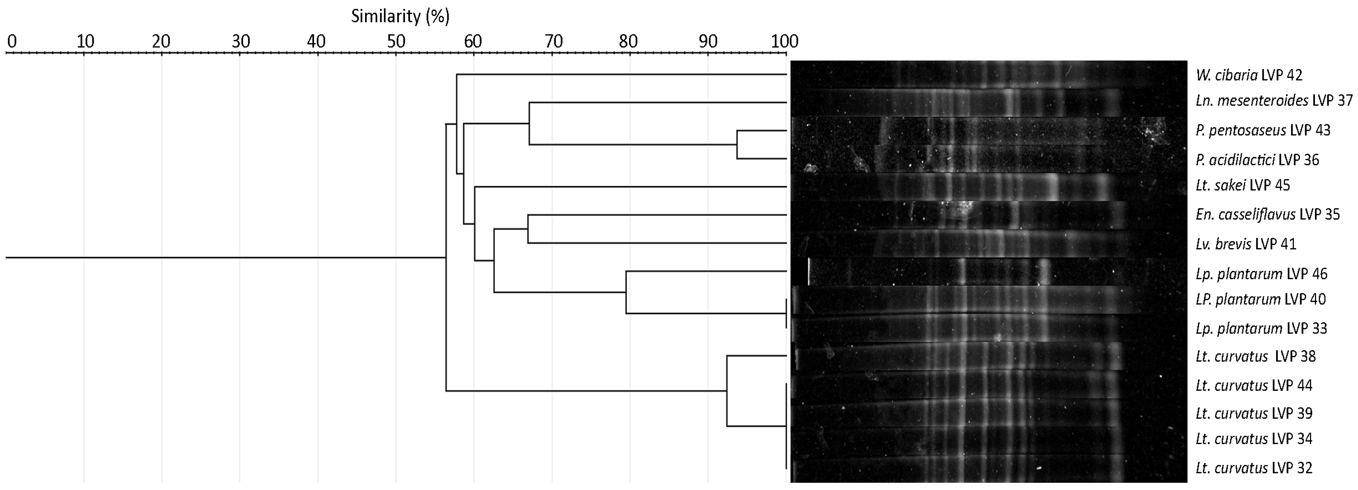
3.1. Isolation and Strains Identification

The comparative analysis of the sequences obtained from the isolated strains was conducted using different databases (BLAST, NCBI, and RDP). This study identified nine species with high identity scores ranging from 97.38% to 100% (Table 1). These sequences were deposited in GenBank, making them publicly available for further research.
The isolated strains were lyophilised and incorporated into the LIVAPRA strain collection with the LVP nomenclature followed by a number. The table below shows these microorganisms’ origin and the pepper colour from which they were isolated. Interestingly, the number of isolates obtained from market and orchard peppers was almost proportional. However, the colour of the pepper from which the strains had been obtained varied significantly, with the majority being derived from green peppers.
Most isolated strains were Lactilactobacillus curvatus (Lt. curvatus, 33%), Lactiplantibacillus plantarum (Lp. plantarum, 20%), and also from the genus Pediococci (13%), particularly Pediococcus acidilactici and Pediococcus pentosaceus. The other bacteria identified were Levilactobacillus brevis (Lv. brevis) and Lactilactobacillus sakei (Lt. sakei), Enterococcus casseliflavus (En. casseliflavus), Leuconostoc mesenteroides (Ln. mesenteroides), and Weisella cibaria (W. cibaria).
Overall, this comprehensive analysis provides essential insights into the microbial diversity of peppers and highlights the importance of understanding the microbial composition of food products to ensure their safety and quality.
Based on the 16S rRNA gene sequences, a phylogenetic relationship was established between the isolates to indicate the species and distribution of selected isolates.
The genetic relatedness of the isolated strains was assessed through Rep-PCR, and a dendrogram was constructed based on the calculated percentage of similarity (Figure 1). The power of discrimination, denoting the ability of the typing method to differentiate unrelated strains, was 97%, indicating that the isolates with a similarity percentage of less than 97% were considered distinct.
Conversely, the strains with similarly percentage greater than 97% were regarded as identical strains with indistinguishable profiles. Based on these criteria, a similarity coefficient of 0.97 derived from the dendrogram indicated that LVP 33 and 40 were the same strain, and our analysis revealed 100% genetic similarity. Likewise, LVP 32, 34, 39, and 44 were identified as being the same strain, with a 100% similarity score. The remaining strains resulted genetically distinct. Overall, the Rep-PCR analysis grouped the isolates into 11 clonal groups.

3.2. Bacterial Stress Tolerance

3.2.1. pH Resistance

All the strains survived under the different pH conditions tested, with variable survival rates, as shown in Table 2. Most isolated microorganisms exhibited good tolerance to a pH of 7.0 and a pH of 4.0. However, a notable sensitivity at a pH of 3 was observed in En. casseliflavus LVP 35, Ln. mesenteroides LVP 37, W. cibaria LVP 42, and Lt. curvatus LVP 38. In acidic conditions, these strains experienced a decrease in cell viability and growth, suggesting a higher susceptibility to acidic environments than other microorganisms.
In contrast, this study highlights the resilience of P. acidilactici LVP 36, Lp. plantarum LVP 40, LVP 44, and LVP 46, Lv. brevis LVP 41, P. pentosaceus LVP 43, and Lt. sakei LVP 45 at a pH of 3.0. Given their ability to tolerate a low pH, these strains were selected for additional parameter evaluation.

3.2.2. Bile Tolerance

According to Gilliland’s growth retardation criteria [11], the isolated strains of Lp. plantarum, Lt. curvatus, and P. pentosaceus demonstrated tolerance to bile. However, Lv. brevis LVP 41 exhibited resistance to bile. Conversely, the P. acidilactici LVP 36 and Lt. sakei LVP 45 strains were unable to tolerate bile (Table 3).
This study found that the isolated strains of Lp. plantarum exhibited tolerance to bile salts in different growth phases: Lag (3 h), logarithmic (7 h), and stationary (24 h) (Figure 2). However, the LVP 40 strain displayed the highest survival rate throughout all growth phases, as depicted in Figure 2. Notably, strains LVP 40, LVP 41, LVP 43, and LVP 44 consistently maintained a survival rate of 70–100% when exposed to bile salts during the lag, exponential, and stationary growth phases. This remarkable resilience and robustness in the face of gastrointestinal conditions highlighted their potential. Based on these promising results, further investigations have since been planned to explore the resistance of these four strains to the harsh environment of the gastrointestinal tract.
The results obtained regarding tolerance to bile and growth retardation in the presence of bile were in agreement, confirming the resistance of lactic acid bacteria to bile. This consistency between the two methodologies validated their use in assessing bile tolerance. In both cases, it was observed that the strains P. acidilactici LVP 36 and Lt. sakei LVP 45 did not exhibit tolerance to bile.

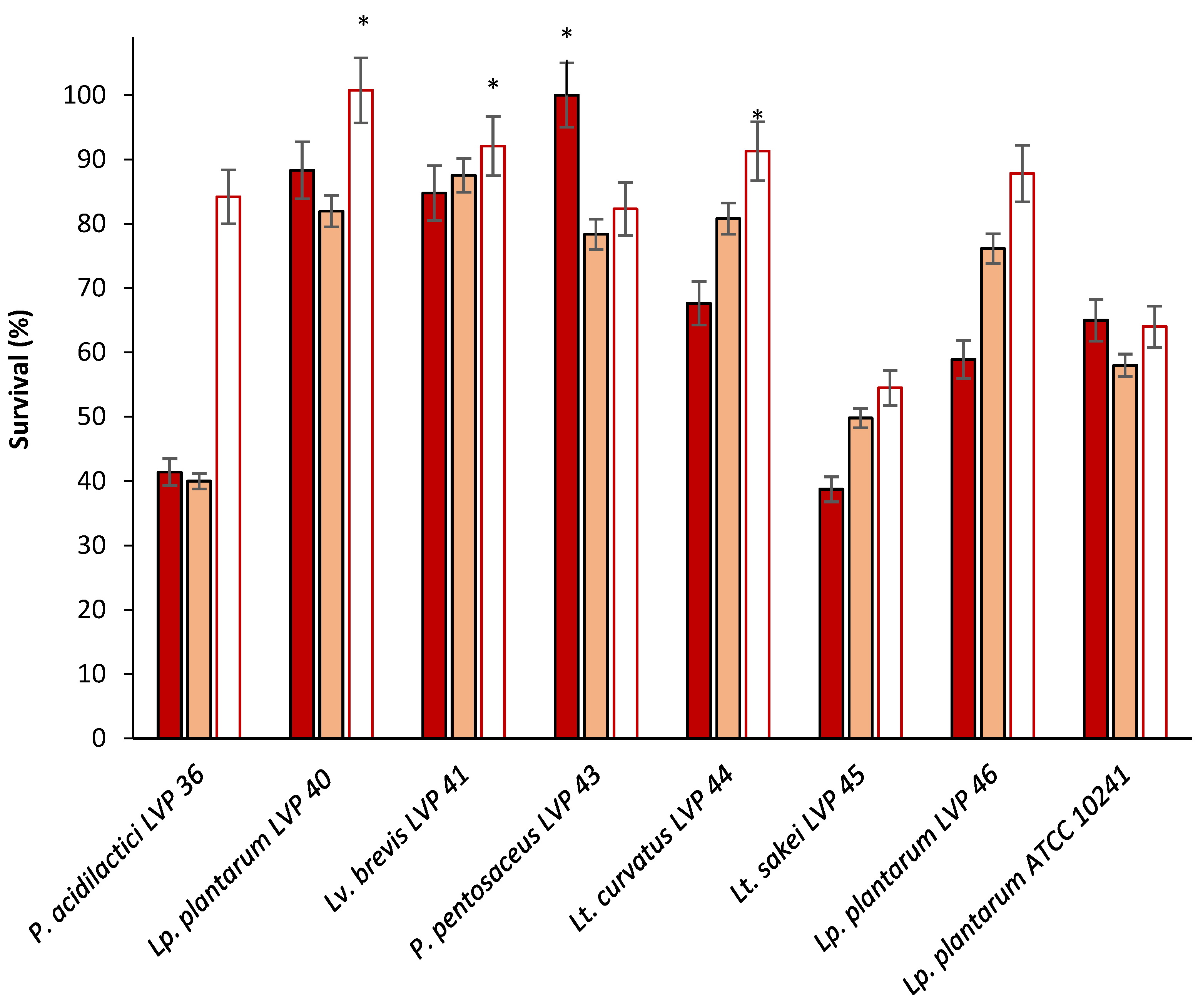
3.2.3. Resistance to Sequential Exposition to Stimulated Gastric and Intestinal Juice

The ability of lactic acid bacteria to survive the harsh conditions of the gastrointestinal tract is a crucial factor in determining their potential probiotic efficacy. In this study, lactic acid bacteria were exposed to artificial juices that simulated gastric and intestinal juice. The results indicated that all the tested bacteria strains exhibited reasonable survival rates under these conditions (Table 4). The maximum reduction in viability was observed for Lt. curvatus LVP 44, with a decrease of 2.78 log CFU/mL, which is an acceptable value for a probiotic strain. The strains that showed the best adaptation to the digestion process were Lp. plantarum LVP 40 and LVP 46, demonstrating decreases of 2.16 and 1.58 log CFU/mL for LVP 40 and 1.14 and 1.73 log CFU/mL for LVP 46 at pH levels of 3 and 4, respectively. These findings suggest that the selected isolates have the potential to be used as probiotics, as they can survive and adapt to harsh digestive system conditions.

3.3. Adhesion Capacity

3.3.1. Surface Hydrophobicity

The isolates evaluated showed varying levels in hydrophobicity across all solvents, with lower values than the reference strain, except for Lv. brevis. The most effective solvent for increasing the hydrophobicity of all the strains was ethylene acetate. It is important to note that hydrophobicity is a strain-dependent property. Thus, the differential behaviour observed between the two strains of Lp. plantarum can be attributed to this surface property. Lv. brevis resulted to be strongly hydrophobic, with the highest adherence observed in all the three solvents tested (84.45 in xylene, 98.92 in chloroform, and 96.92% in ethyl acetate). These hydrophobicity values were similar to or even higher than those obtained with the ATCC reference strain (Table 5).

3.3.2. Auto-Aggregation Ability

All the tested strains displayed a remarkable capability to undergo self-aggregation when incubated at 37 °C for 24 h in PBS. Notably, the strains Lp. plantarum LVP 40, P. pentosaceus LVP 43, Lt. curvatus LVP 44, and Lp. plantarum LVP 46 exhibited the highest degree of auto-aggregation. Following the 24 h incubation period, all the strains demonstrated auto-aggregation percentages surpassing 70%, except for Lv. brevis LVP 41, which exhibited a lower auto-aggregation rate of about 31% (Table 6).

3.4. Safety Assessment

3.4.1. Haemolytic Activity

Among the strains subjected to testing, none produced the formation of haemolysis halos on the blood–agar medium used in the experiments. The absence of clear/green halos indicated that the selected strains could not cause red blood cell haemolysis.

3.4.2. Antibiotic Sensitivity Test

The pattern of antibiotic resistance exhibited by the chosen strains was similar to that of the Lp. plantarum ATCC 10241 reference strain. Specifically, these strains demonstrated sensitivity to ampicillin, cephalothin, chloramphenicol, clindamycin, erythromycin, gentamicin, and rifampicin. On the contrary, they resisted the antibiotics vancomycin, ciprofloxacin, and norfloxacin. These findings provide a guarantee of safety for the use of these strains since their antibiotic resistance profiles conform to established standards (Table 7).

3.5. Technological Properties

3.5.1. Compatibility of Strains

The compatibility test on the five chosen strains revealed the absence of inhibition halos in all the experiments. That is, no supernatant from the selected bacteria prevented the growth of other bacteria (Figure S1, Supplementary Material). Consequently, the creation of mixtures of these lactic acid bacteria can be used effectively in food fermentation.

3.5.2. NaCl Resistance

The experimental evaluation of tolerance to high concentrations of NaCl showed that the LVP 40, LVP 43, LVP 46, and LVP 44 strains had a high tolerance level (2.5 and 5% NaCl). Conversely, the LVP 41 strain demonstrated a high tolerance to a 2.5% NaCl solution but a lower tolerance to a 5% solution.
It is worth highlighting that none of the strains exhibited tolerance to NaCl concentrations of 7% and 10%, as detailed in the Supplementary Materials. In all cases, the tolerance was more significant than the collection strain Lp. plantarum ATCC 10241, isolated from sauerkraut, which tolerates only 2.5% salt (Figure S2, Supplementary Material). The osmotic resistance of potential probiotic bacteria makes them suitable for fermenting vegetables in which concentrations close to 2% NaCl are generally used.

3.5.3. Acidification Capacity

The isolated microorganisms were cultured at 37 °C in MRS broth with an initial pH of 6.5 for 24 h. The pH was measured at the end of this period. Regarding the acidifying capacity, all the strains, except fir LVP 41, demonstrated the ability to lower the pH of the culture medium by more than two units after 24 h of incubation. The final pH values were 3.56, 3.72, 3.73, and 3.57 for Lp. plantarum LV P40, P. pentosaceus LVP 43, Lt. curvatus LVP 44, and Lp. plantarum LVP 46, respectively. In the reference strain Lp. plantarum ATCC 1024, isolated from fermented food, the final pH was 4.24. This characteristic is particularly advantageous from a technological perspective since it implies a quicker process to reach acidity levels that protect the product from contaminating microorganisms. The higher final pH in the culture with Lv. brevis LVP 41 (final pH 6.04) may be attributed to its obligatory heterofermentative nature, producing both lactic and acetic acid. Acetic acid is less acidic than lactic acid, contributing to the observed difference in acidity.

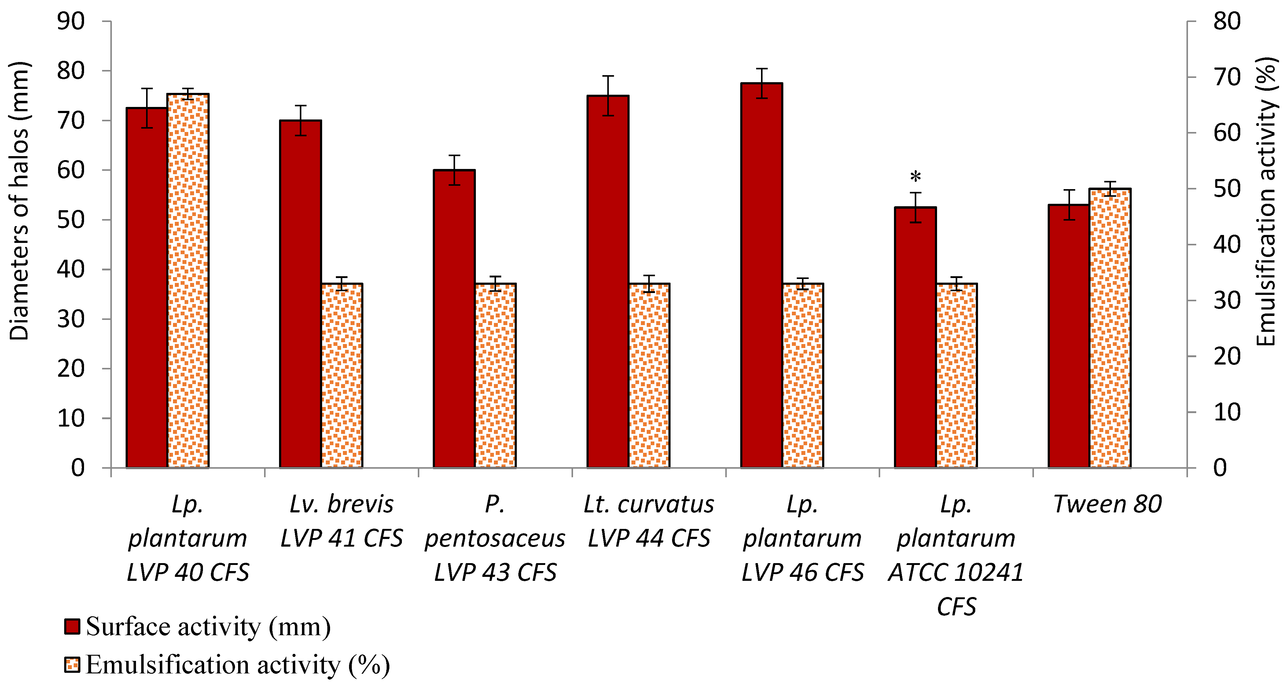
3.6. Surfactant and Emulsifying Properties

All LAB cell-free supernatants (CFS) showed oil-dispersing activities similar to those obtained with the commercial surfactant tween 80 (Figure 3). Lp. plantarum LVP 46, LVP 44, and LVP 40 CFS had the highest activity. Nevertheless, there were no significant differences between them. However, the reference surfactant had a lower oil dispersion halo than the Lactobacillus strains.
On the other hand, the emulsification activity of the bacterial supernatants was generally greater than 30% but lower than that of tween 80 (50%). Only Lp. plantarum LVP 40's supernatant exhibited an emulsification activity of 67%.

3.7. Antipathogenic Activity

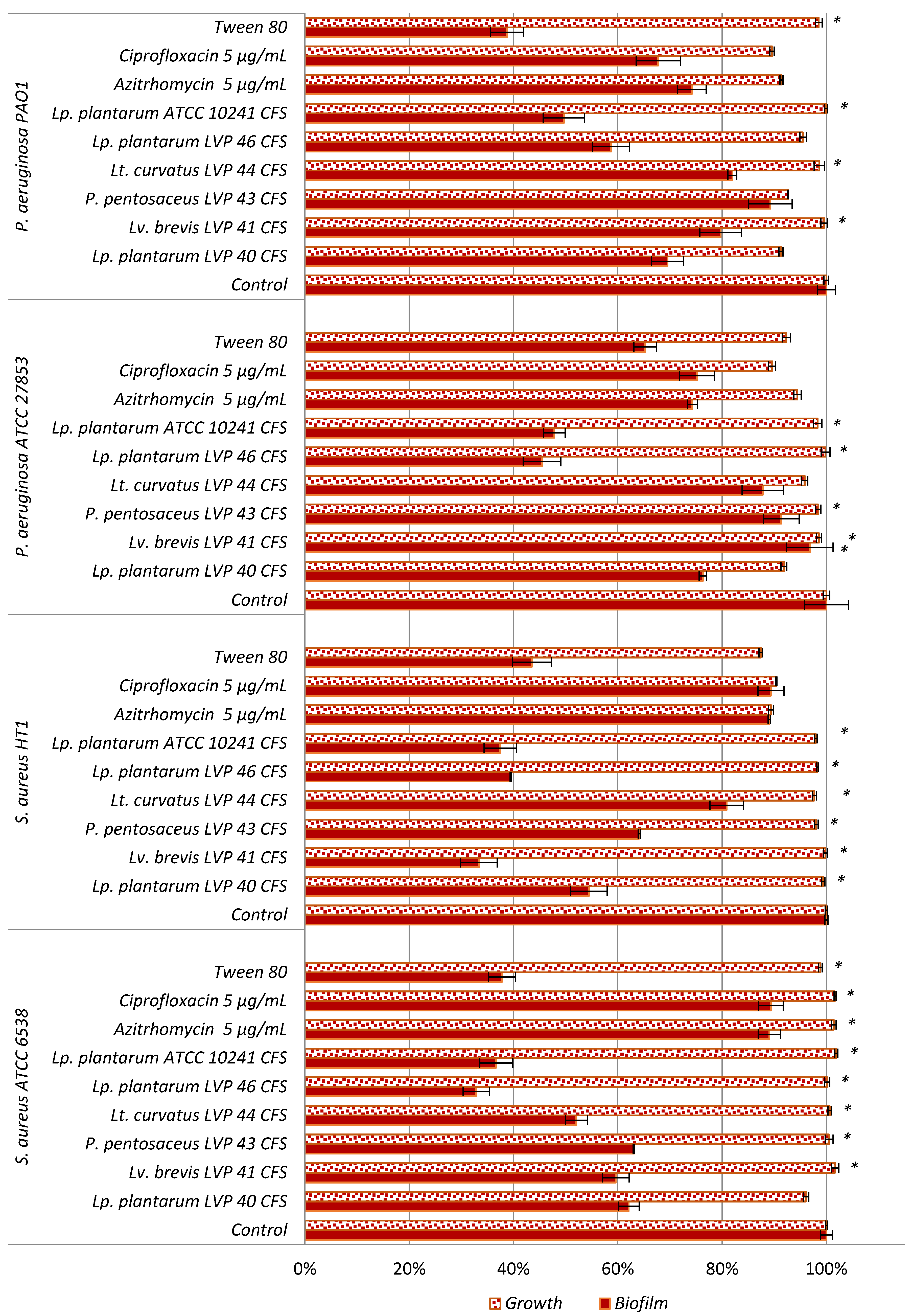
3.7.1. Inhibition of Bacterial Pathogenic Biofilm Adhesion

All LAB supernatants inhibited the formation of bacterial biofilms after 1 h of treatment (Figure 4). The biofilm of both S. aureus strains decreased in the presence of all CFS. For the ATCC 6538P strain, the most significant effect was observed with the supernatant of the Lp. plantarum LVP 46 strain (68%). Concerning the HT1 strain, the highest inhibition was noticed for the supernatant of Lv. brevis LVP 41 (67%).
For the P aeruginosa strains, all CFS inhibited biofilm formation in both strains assayed but with lower values than those presented for S. aureus. Among the strains, the most potent supernatant was derived from Lp. plantarum LVP46, showcasing 55% and 42% inhibition against P. aeruginosa ATCC 27853 and PAO1, respectively.
It is noteworthy that the inhibitory effects on biofilm adhesion surpassed those of commonly used antibiotics, ciprofloxacin and azithromycin, in all instances. Additionally, the observed inhibitions were comparable or even higher than those achieved with tween 80.
In contrast to what was observed in the biofilm formation by pathogenic bacteria, the supernatants of the potential probiotics did not significantly affect the growth of the pathogens under the conditions studied.

3.7.2. Co-Aggregation with Pathogens

Table 8 shows the results of co-aggregation between the isolated and selected strains and the four pathogenic bacteria after 1, 4, and 24 h of incubation. All the strains tested exhibited some degree of co-aggregation with the pathogens. The highest effect was observed in the bacterial strains Lv. brevis LVP 41 and Lp. plantarum LVP 40 and ATCC 10241 with both strains of P. aeruginosa (100 and 50%, respectively). The greatest co-aggregation with S. aureus was observed with P. pentosaceus LVP 43 and Lt. curvatus LVP 44, although the values varied depending on the strain.

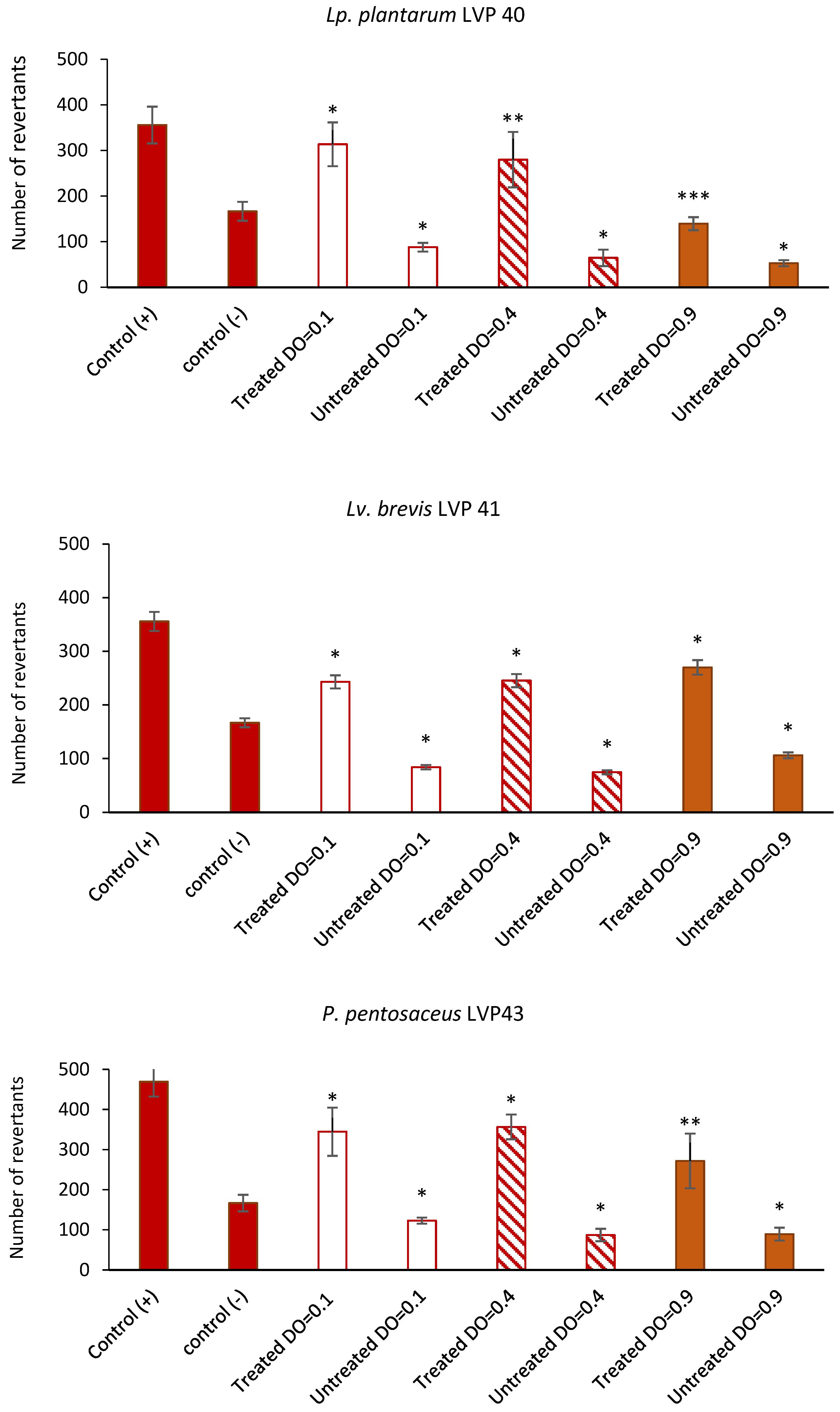
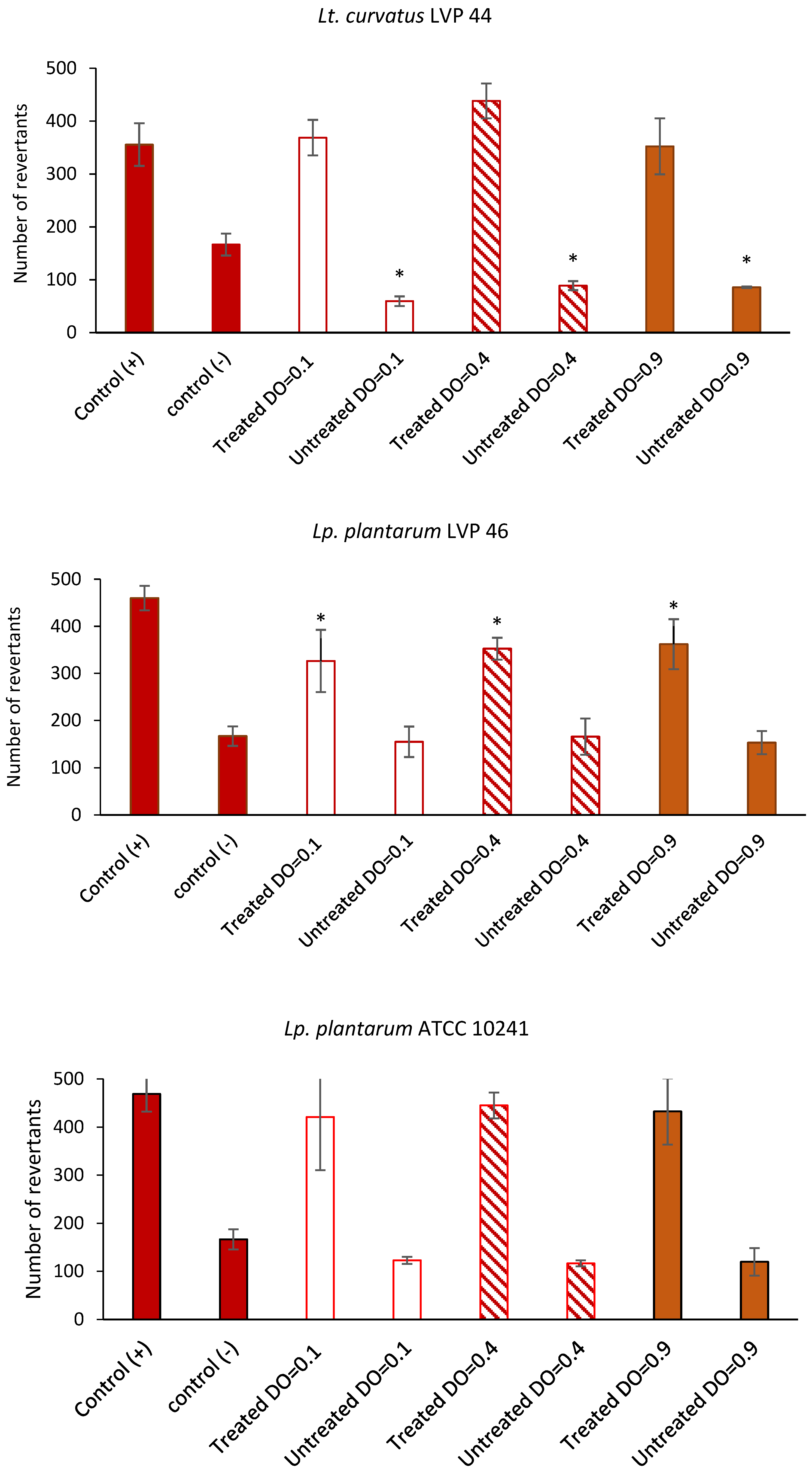
3.8. Antimutagenic Capacity of Lactic Acid Bacteria

The antimutagenic activity of the lactic acid bacteria against sodium azide is shown in Figure 5. S. typhimurium TA 100 was assessed against cell-free supernatants (CFS) derived from LAB cultures that had been treated (through the addition of a mutagenic substance) and untreated cultures (control). The LAB concentrations used were 4 × 10⁷ CFU/mL (OD600 nm = 0.1), 1.5 × 10⁸ CFU/mL (OD600 nm = 0.4), and 4 × 10⁸ CFU/mL (OD600 nm = 0.9). As seen in the figure below, the inhibition in the reversion of S. typhimurium by CFS was strain-dependent. The results revealed that the LAB strains Lv. brevis LVP 41, P. pentosaceus LVP 43, and Lp. plantarum LVP 46 had the ability to remove the mutagen, evidenced by the decrease in revertant colonies (34–38%, 30–52%, and 30–44%, respectively). Notably, in the case of strain Lp. plantarum LVP 40, the removal capacity increased as the used bacterial biomass increased (15–71%). The antimutagenic activity of these strains surpassed that of the reference strain Lp. plantarum ATCC 10241 (7–14%). On the contrary, Lt. curvatus LVP 44 did not show antimutagenic effects.

4. Discussion

Historically, pickling is one of the oldest preservation processes of several foodstuffs, such as vegetables, fruits, fish, and meat. Pickling imparts unique and desirable changes in flavour, texture, and colour that take place over time in fermented pickles. In many cultures, pickles have been associated with many nutritional benefits over time. In particular, LAB microorganisms contribute to this process and are increasingly linked to consumer health benefits. In fact, Lp. plantarum, Lv. brevis, Lactobacillus acidophilus, Limosilactobacillus fermentum; Leuconostoc fallax, and Ln. mesenteroides are the most studied [25].
Lactic acid bacteria dominate the fermented vegetable microbial community, providing several health-related properties. In concordance with our results, García-Burgos et al. (2020) stated that lactobacilli are the most prevalent microorganisms in the fermentation of fruit and vegetable juices and that species such as Lp. plantarum, Lactobacillus bavaricus, Lactobacillus xylosus, Lactobacillus bifidus, and Lv. brevis are frequently found [26]. In the same way, other authors reported that Lp. plantarum, Lv. brevis, Ln. mesenteroides, P. pentosaceus, Limosilactobacillus fermentum, and Lactococcus lactis were the main microorganisms isolated from vegetables [27,28].
It is important to note that the genus previously called Lactobacillus is one of the most widely used bacterial genera as probiotics, and its use as microbial food supplements has obtained the status of “Generally Recognized as Safe” (GRAS). Lactobacilli are found in the gastrointestinal tract of humans and animals, in plant- or animal-based fermented products, and in most commercially available fermented foods. In this study, bacterial isolates of peppers were evaluated for their stress tolerance, safety, technology, and probiotic properties.
Resilience to the challenging conditions of the gastrointestinal tract stands as a primary factor restricting the application of microorganisms as live probiotic agents. The capability to endure acidity and bile salts is widely acknowledged as crucial for the survival of LAB in the gut. Specifically, the ability to survive at a pH of 3.0 is deemed as the optimal acid tolerance for probiotic strains [29]. Consequently, this study assessed isolates for their capacity to withstand a pH of 3.0 and bile. Seven isolates were resistant to exposure to a pH of 3.0 and then were assessed for their bile tolerance. Only five strains supported the bile and were identified as lactobacilli and pediococci. This fact is in concordance with the results found in Lacticaseibacillus rhamnosus, Lactobacillus gasseri, and Limosilactobacillus, which exhibited a bile resistance of 3.0 g/L [30,31].
The adhesion capacity of LAB isolates that had been able to survive the conditions of the digestive system was evaluated. Hydrophobicity properties are strain-specific, in concordance with an earlier report [32]. So, the high hydrophobicity found in Lactobacillus strains agrees with that observed in Lactobacillus gasseri and Lacticaseibacillus rhamnosus, and the mean values depended on the origins and genera [31]. De Souza et al. (2019) also confirmed this by describing different strains of Lacticaseibacillus casei with hydrophobicity values ranging from 9.66 to 69.36% and, for Lm. fermentum strains, from 0.30 to 68.81% [33], and they stated that bacteria with a higher hydrophobicity can adhere better to epithelial cells and significantly influence the microbial composition in the intestine.
In addition, after 24 h of incubation, all LAB strains demonstrated self-aggregation percentages higher than 70%, except for Lv. brevis. In contrast, the auto-aggregation abilities of Lactobacillus gasseri, Lacticaseibacillus rhamnosus, and Limosilactobacillus reuteri were in a variable range between 5.8 and 28.5%, depending on the incubation time and strain [31]. Haemolytic effects were not observed in the tested isolates, and our results agreed with those reported by Damaceno et al. (2021) [34].
On the other hand, evaluating the antibiotic resistance of potential probiotic microorganisms is vital to limit the antibiotic resistance gene transfer between the typical microbiota and pathogens [35]. In the present study, all the strains were resistant to vancomycin, ciprofloxacin, and norfloxacin. In accordance with the present results, lactobacilli were previously reported as vancomycin- and ciprofloxacin-resistant [29,31]. In most Lactobacillus species, vancomycin resistance genes are chromosomally coded; therefore, there is no risk of transfer as in the case of plasmids [31]. Other authors noticed a variable resistance to gentamicin [36,37]. Nevertheless, in this work, neither of the strains was resistant to gentamicin.
The new isolates also showed a high tolerance to 2.5% of NaCl and a low tolerance to 5% of NaCl, while another study reported that several probiotics could tolerate 6.5% NaCl [38]. The stability of pickles during the fermentation process and storage is due to their content of LAB and its viability and metabolic activity (acidifying activity). The suitability and good selectivity of LAB starter cultures for large-scale production are essential for keeping the fermenting microflora stable for a long time [39].
The bacterial surfactant property has also been investigated in the present work. Biosurfactants are predominantly synthesised by bacteria and fungi and can reduce interfacial and surface tension between two immiscible liquids. These compounds exhibit various properties such as detergency, wettability, and foaming, making them suitable for biomedical and industrial applications [40].
LAB biosurfactants have shown promise as anti-adhesive agents to prevent the adhesion of pathogens to the host epithelium and solid surfaces, including biomedical instruments [41,42]. Exploring bacterial supernatants’ surface and interfacial activities, such as those produced by Lactobacillus strains, is essential for identifying new strategies to inhibit microbial adherence and control biofilm-forming pathogens [20,43]. Previous studies have identified glycoproteinaceous biosurfactants, such as surlactin, in various Lactobacillus species [17,44,45,46]. The findings found in this work are consistent with the results obtained by Verni et al. (2022), which revealed an emulsifying activity of the Lactobacillus paracasei subsp. paracasei CE75 supernatant similar to the Lactobacillus crispatus BC1′ biosurfactant [20,47].
Biofilm formation is initiated by the adhesion of individual bacteria to a surface. Surface-sensing creates bacteria’s awareness of their adhering state on the surface. It is essential to initiate the phenotypic and genotypic changes that characterise the transition from initial bacterial adhesion to a biofilm. This first stage is controlled by complex combinations of the physicochemical interactions between the cell membrane and the material surface [48,49]. In fact, the observed effects on the biofilm biomass after only one hour of incubation did not depend on bacterial growth inhibition but rather on non-stick effects, and these results agree with previous works that demonstrated antibiofilm effects exclusively due to Lactobacillus anti-adhesive properties [20,43,50,51]. Indeed, Gudiña et al. (2010) observed an anti-adhesive activity of the crude biosurfactant isolated from L. paracasei subsp. paracasei A20 higher against S. aureus (76.80%) than against P. aeruginosa (21.20%) at 50 mg/mL after four hours of treatment [50]. Meanwhile, Verni et al. (2022) demonstrated a potent inhibition of P. aeruginosa HT5 biofilm (72.01%) at one hour linked to the anti-adhesiveness effects of the L. paracasei biosurfactant [20]. Our results indicate that BAL supernatants are promising sources of biosurfactants with anti-adhesive properties. Specially, Lp. plantarum LVP46′s supernatant exerts a surface activity that correlates with strong antibiofilm effects against pathogenic bacteria Gram (+) and (−).
It is important to highlight that the antibiofilm and antipathogenic effects observed in LAB supernatants were consistent with previous studies [23,52,53,54]. These results are significant since biofilm formation, governed by quorum sensing (QS), constitutes a significant problem for the safety of several food products. Probiotics have multiple properties, and, although evidence is scarce, their involvement in the regulation of QS may bring new solutions in several areas, including food preservation. Several species of lactic acid bacteria (Lp. plantarum, Lm. fermentum, Lactobacillus acidophilus, Ls. casei, Lv. brevis, Lactobacillus reuteri, and Lt. curvatus) have already been reported at least once as quorum-quenching (QQ) agents [23,52,54,55]. For instance, a Lp. plantarum strain reduced the expression of some genes involved in the biofilm formation of S. aureus [56], Lv. brevis 3M004 inhibited biofilm formation of P. aeruginosa [57], and Lp. plantarum PA 100 inhibited the N-acyl-homoserine lactone (QS autoinducer) activity of P. aeruginosa by blocking their synthesis [53]. Recently, Diaz et al. demonstrated that Ls. casei probiotic strains inhibited the biofilm formation and critical virulence factors of S. aureus and P. aeruginosa [23,54].
Co-aggregation between pathogens and probiotic strains as well as auto-aggregation are regarded as one of the critical indicators for using probiotics [58]. The co-aggregation against S. aureus and P. aeruginosa was good and strain-dependent. A previous study found that Enterococcus faecium exhibited a significant co-aggregation effect with Salmonella enterica and S. aureus [32].
Finally, several studies suggest that fermented pickles containing probiotic strains may have a protective effect against colorectal cancer cells by potentially mitigating proliferative and mutagenic activity, suppressing the activity of the enzymes involved in the production of mutagens, carcinogens, and tumour promoters [59]. According to the present results, previous articles have reported antimutagenic properties of lactobacilli, with this effect being strain-dependent [5,23]. Nonetheless, additional research is imperative to validate the antimutagenic effects of fermented pickles that contain naturally occurring probiotics.

5. Conclusions

In summary, this study assessed the functional and technological properties of 15 strains of lactic acid bacteria isolated from bell peppers, identifying them phenotypically and genotypically. The results highlight that Lp. plantarum LVP 40, Lv. brevis LVP 41, P. pentosaceus LVP 43, Lt. curvatus LVP 44, and Lp. plantarum LVP 46 exhibit notable resistance to low pH values, bile salts, and in vitro gastrointestinal conditions. These strains also display surface properties, such as auto-aggregation, hydrophobicity, co-aggregation, and anti-adhesive capacity against pathogens. Furthermore, they demonstrate superior biosurfactant and antimutagenic properties compared to the reference strain.
The technological properties, including osmotic tolerance, acidifying capacity, and compatibility between these strains, suggest the potential of these bacteria to be used in mixed starter cultures for the production of fermented foods. Overall, these characteristics indicate that the five selected strains have the potential to be promising probiotics with functional merits for application in the fermentation of bell peppers. However, to qualify as probiotics, these strains require further studies.

Supplementary Materials

The following supporting information can be downloaded at https://www.mdpi.com/article/10.3390/fermentation10040209/s1: Figure S1: Strain compatibility assessment; Figure S2: Growth curves of lactic acid bacteria in the absence and presence of different NaCl concentrations.

Author Contributions

I.M.N.: Performed the experiments, analysed the data. M.C.V.: Performed the experiments, analysed the data. F.E.A.M.: Performed the experiments, analysed the data. J.D.B.: Performed the experiments, analysed the data. V.T.: Performed the experiments, analysed the data. M.E.D.: Performed the experiments, analysed the data. E.C.: Conceived and designed the experiments, analysed the data, wrote the paper. M.R.A.: Conceived and designed the experiments, analysed the data, wrote the paper. M.E.A.: Conceived and designed the experiments, analysed the data, wrote the paper. All authors have read and agreed to the published version of the manuscript.

Funding

The authors acknowledge the financial support from the SCAIT-UNT (PIUNT 26D-702, 26D-708, and 26D-715), the Agencia Nacional de Promoción Científica y Técnica ANPCyT (PICT 2018-02071 and 02514, PICT 2021-0060, PICT-2021-I-A-00439 I), and The Consejo Nacional de Investigaciones Científicas y Técnicas, CONICET (PUE 0021).

Institutional Review Board Statement

Not applicable.

Informed Consent Statement

Not applicable.

Data Availability Statement

Data will be made available on request.

Acknowledgments

The authors gratefully acknowledge the financial support of the UNT, ANPCyT, and CONICET that made this work possible.

Conflicts of Interest

The authors declare no conflicts of interest.

References

  1. Chakraborty, R.; Roy, S. Exploration of the diversity and associated health benefits of traditional pickles from the Himalayan and adjacent hilly regions of Indian subcontinent. J. Food Sci. Technol. 2018, 55, 1599–1613. [Google Scholar] [CrossRef]
  2. Zapaśnik, A.; Sokołowska, B.; Bryła, M. Role of lactic acid bacteria in food preservation and safety. Foods 2022, 11, 1283. [Google Scholar] [CrossRef] [PubMed]
  3. Behera, S.S.; El Sheikha, A.F.; Hammami, R.; Kumar, A. Traditionally fermented pickles: How the microbial diversity associated with their nutritional and health benefits? J. Funct. Foods 2020, 70, 103971. [Google Scholar] [CrossRef]
  4. Casarotti, S.N.; Borgonovi, T.F.; Batista, C.L.; Penna, A.L.B. Guava, orange and passion fruit by-products: Characterization and its impacts on kinetics of acidification and properties of probiotic fermented products. LWT-Food Sci. Technol. 2018, 98, 69–76. [Google Scholar] [CrossRef]
  5. Apás, A.L.; Gonzalez, S.N.; Arena, M.E. Potential of goat probiotic to bind mutagens. Anaerobe 2014, 28, 8–12. [Google Scholar] [CrossRef] [PubMed]
  6. Lane, D.J. 16S/23S rRNA sequencing. In Nucleic Acid Techniques in Bacterial Systematics; Stackebrandt, E., Goodfellow, M., Eds.; Wiley: New York, NY, USA, 1991; pp. 115–175. [Google Scholar]
  7. Maldonado, N.C.; Aristimuño Ficoseco, C.; Mansilla, F.I.; Melián, C.; Hébert, E.M.; Vignolo, G.M.; Nader-Macías, M.E.F. Identification, characterization and selection of autochthonous lactic acid bacteria as probiotic for feedlot cattle. Livest. Sci. 2018, 212, 99–110. [Google Scholar] [CrossRef]
  8. Versalovic, J. Genomic fingerprinting of bacteria using repetitive sequence-based polymerase chain reaction. Methods Mol. Cell Biol. 1994, 5, 25–40. [Google Scholar]
  9. Heras, J.; Domínguez, C.; Mata, E.; Pascual, V.; Lozano, C.; Torres, C.; Zarazaga, M. GelJ–a tool for analyzing DNA fingerprint gel images. BMC Bioinform. 2015, 16, 270. [Google Scholar] [CrossRef] [PubMed]
  10. Ishii, S.; Sadowsky, M.J. Applications of the rep-PCR DNA fingerprinting technique to study microbial diversity, ecology and evolution. Environ. Microbiol. 2009, 11, 733–740. [Google Scholar] [CrossRef] [PubMed]
  11. Gilliland, S.E.; Staley, T.E.; Bush, L.J. Importance of bile tolerance of Lactobacillus acidophilus used as dietary adjunct. J. Dairy Sci. 2004, 67, 3045–3051. [Google Scholar] [CrossRef] [PubMed]
  12. Vera-Mejía, R.; Ormaza-Donoso, J.; Muñoz-Cedeño, J.; Arteaga-Chávez, F.; Sánchez-Miranda, L. Cepas de Lactobacillus plantarum con potencialidades probióticas aisladas de cerdos criollos. Rev. Salud Anim. 2018, 40, 1–12. [Google Scholar]
  13. Zárate, G.; Chaia, A.P.; González, S.; Oliver, G. Viability and β-galactosidase activity of dairy propionibacteria subjected to digestion by artificial gastric and intestinal fluids. J. Food Prot. 2000, 63, 1214–1221. [Google Scholar] [CrossRef]
  14. Hojjati, M.; Behabahani, B.A.; Falah, F. Aggregation, adherence, anti-adhesion and antagonistic activity properties relating to surface charge of probiotic Lactobacillus brevis gp104 against Staphylococcus aureus. Microb. Pathog. 2020, 147, 104420. [Google Scholar] [CrossRef] [PubMed]
  15. Tyfa, A.; Kunicka-Styczyńska, A.; Zabielska, J. Evaluation of hydrophobicity and quantitative analysis of biofilm formation by Alicyclobacillus sp. Acta Biochim. Pol. 2015, 62, 785–790. [Google Scholar] [CrossRef] [PubMed]
  16. Parente, E.; Brienza, C.; Moles, M.; Ricciardi, A. A comparison of methods for the measurement of bacteriocin activity. J. Microbiol. Methods 1995, 22, 95–108. [Google Scholar] [CrossRef]
  17. Cartagena, E.; Orphèe, C.H.; Verni, M.C.; Arena, M.E.; González, S.N.; Argañaraz, M.I.; Bardón, A. Medio de Cultivo Promotor y Bacterias No Patógenas Detoxificantes de Compuestos Mutagénicos/Carcinogénicos. Instituto Nacional de la Propiedad Industrial-INPI. Patent No. 20190102418, 25 March 2021. [Google Scholar]
  18. Walter, V.; Syldatk, C.; Hausmann, R. Screening Concepts for the Isolation of Biosurfactant Producing Microorganisms. In Biosurfactants. Advances in Experimental Medicine and Biology; Sen, R., Ed.; Springer: New York, NY, USA, 2010; pp. 1–13. [Google Scholar]
  19. Sambanthamoorthy, K.; Feng, X.; Patel, R.; Patel, S.; Paranavitana, C. Antimicrobial and antibiofilm potential of biosurfactants isolated from lactobacilli against multi-drug-resistant pathogens. BMC Microbiol. 2014, 14, 197. [Google Scholar] [CrossRef] [PubMed]
  20. Verni, M.C.; Orphèe, C.H.; González, S.N.; Bardón, A.; Arena, M.E.; Cartagena, E. Flourensia fiebrigii SF Blake in combination with Lactobacillus paracasei subsp. paracasei CE75. A novel antipathogenic and detoxifying strategy. LWT 2022, 156, 113023. [Google Scholar]
  21. Tateda, K.; Comte, R.; Pechere, J.C.; K¨ohler, T.; Yamaguchi, K.; Van Delden, C. Azithromycin inhibits quorum sensing in Pseudomonas aeruginosa. Antimicrob. Agents Chemother. 2001, 45, 1930–1933. [Google Scholar] [CrossRef]
  22. Sandasi, M.; Leonard, C.M.; Van Vuuren, S.F.; Viljoen, A.M. Peppermint (Mentha piperita) inhibits microbial biofilms in vitro. S. Afr. J. Bot. 2011, 77, 80–85. [Google Scholar] [CrossRef]
  23. Díaz, M.A.; Alberto, M.R.; Vega-Hissi, E.G.; Gonzalez, S.N.; Arena, M.E. Interference in Staphylococcus aureus. Biofilm and virulence factors production by human probiotic bacteria with antimutagenic activity. Arab. J. Sci. Eng. 2022, 47, 241–253. [Google Scholar] [CrossRef]
  24. Maron, D.M.; Ames, B.N. Revised methods for the Salmonella mutagenicity test. Mutat. Res. Sect. Environ. Mutagen. Relat. Subj. 1983, 113, 173–215. [Google Scholar] [CrossRef] [PubMed]
  25. Şanlier, N.; Gökcen, B.B.; Sezgin, A.C. Health benefits of fermented foods. Crit. Rev. Food Sci. Nutr. 2019, 59, 506–527. [Google Scholar] [CrossRef]
  26. García-Burgos, M.; Moreno-Fernández, J.; Alférez, M.J.; Díaz-Castro, J.; López-Aliaga, I. New perspectives in fermented dairy products and their health relevance. J. Funct. Foods 2020, 72, 104059. [Google Scholar] [CrossRef]
  27. Sionek, B.; Szydłowska, A.; Kucukgoz, K.; Kołożyn-Krajewska, D. Traditional and new microorganisms in lactic acid fermentation of food. Fermentation 2023, 9, 1019. [Google Scholar] [CrossRef]
  28. Thierry, A.; Baty, C.; Marché, L.; Chuat, V.; Picard, O.; Lortal, S.; Valence, F. Lactofermentation of vegetables: An ancient method of preservation matching new trends. Trends Food Sci. Technol. 2023, 139, 104112. [Google Scholar] [CrossRef]
  29. Khalil, E.S.; Abd Manap, M.Y.; Mustafa, S.; Alhelli, A.M.; Shokryazdan, P. Probiotic properties of exopolysaccharide-producing Lactobacillus strains isolated from tempoyak. Molecules 2018, 23, 398. [Google Scholar] [CrossRef] [PubMed]
  30. Ali, M.S.; Lee, E.B.; Lim, S.K.; Suk, K.; Park, S.C. Isolation and identification of Limosilactobacillus reuteri PSC102 and evaluation of its potential probiotic, antioxidant and antibacterial properties. Antioxidants 2023, 12, 238. [Google Scholar] [CrossRef] [PubMed]
  31. Hyrslova, I.; Drab, V.; Cihlar, J.; Krausova, G.; Mrvikova, I.; Kana, A.; Stetina, J.; Musilova, S. Functional and probiotic characterization of newly isolated strains from infant feces and breast milk. Fermentation 2023, 9, 960. [Google Scholar] [CrossRef]
  32. Han, K.; Park, S.; Sathiyaseelan, A.; Wang, M.H. Isolation and characterization of Enterococcus faecium from fermented Korean soybean paste with antibacterial effects. Fermentation 2023, 9, 760. [Google Scholar] [CrossRef]
  33. de Souza, B.M.S.; Borgonovi, T.F.; Casarotti, S.N.; Todorov, S.D.; Penna, A.L.B. Lactobacillus casei and Lactobacillus fermentum strains isolated from mozzarella cheese: Probiotic potential, safety, acidifying kinetic parameters and viability under gastrointestinal tract condition. Probiotics Antimicrob. Proteins 2019, 11, 382–396. [Google Scholar] [CrossRef] [PubMed]
  34. Damaceno, Q.S.; Gallotti, B.; Reis, I.M.; Totte, Y.C.; Assis, G.B.; Figueiredo, H.C.; Silva, T.F.; Azevedo, V.; Nicoli, J.R.; Martins, F.S. Isolation and identification of potential probiotic bacteria from human milk. Probiotics Antimicrob. Proteins 2021, 15, 491–501. [Google Scholar] [CrossRef] [PubMed]
  35. Rodrigues da Cunha, L.; Ferreira, C.L.F.; Durmaz, E.; Goh, Y.J.; Sanozky-Dawes, R.; Klaenhammer, T. Characterization of Lactobacillus gasseri isolates from a breastfed infant. Gut Microbes 2012, 3, 15–24. [Google Scholar] [CrossRef] [PubMed]
  36. Georgieva, R.; Yocheva, L.; Tserovska, L.; Zhelezova, G.; Stefanova, N.; Atanasova, A.; Danguleva, A.; Ivanova, G.; Karapetkov, N.; Rumyan, N.; et al. Antimicrobial activity and antibiotic susceptibility of Lactobacillus and Bifidobacterium spp. intended for use as starter and probiotic cultures. Biotechnol. Biotechnol. Equip. 2015, 29, 84–91. [Google Scholar] [CrossRef] [PubMed]
  37. Campedelli, I.; Mathur, H.; Salvetti, E.; Clarke, S.; Rea, M.C.; Torriani, S.; Ross, R.P.; Hill, C.; O’Tool, P.W. Genus-wide assessment of antibiotic resistance in Lactobacillus spp. Appl. Environ. Microbiol. 2019, 85, e01738-18. [Google Scholar] [CrossRef] [PubMed]
  38. Jeyagowri, N.; Ranadheera, C.S.; Manap, M.Y.; Gamage, A.; Merah, O.; Madhujith, T. Phenotypic characterisation and molecular identification of potentially probiotic Lactobacillus sp. isolated from fermented rice. Fermentation 2023, 9, 807. [Google Scholar] [CrossRef]
  39. Yao, A.A.; Dortu, C.; Egounlety, M.; Pinto, C.; Vinodh, A.E.; Huch, M.; Franz, C.M.A.P.; Holzapfel, W.; Mbugua, S.; Mengu, M.; et al. Production of freeze-dried lactic acid bacteria starter culture for cassava fermentation into gari. Afr. J. Biotechnol. 2009, 8, 4996–5004. [Google Scholar]
  40. Markande, A.R.; Patel, D.; Varjani, S. A review on biosurfactants: Properties, applications and current developments. Bioresour. Technol. 2021, 330, 124963. [Google Scholar] [CrossRef]
  41. Vecino, X.; Rodríguez-López, L.; Ferreira, D.; Cruz, J.M.; Moldes, A.B.; Rodrigues, L.R. Bioactivity of glycolipopeptide cell-bound biosurfactants against skin pathogens. Int. J. Biol. Macromol. 2018, 109, 971–979. [Google Scholar] [CrossRef]
  42. Yan, X.; Gu, S.; Cui, X.; Shi, Y.; Wen, S.; Chen, H.; Ge, J. Antimicrobial, anti-adhesive and anti-biofilm potential of biosurfactants isolated from Pediococcus acidilactici and Lactobacillus plantarum against Staphylococcus aureus CMCC26003. Microb. Pathog. 2019, 127, 12–20. [Google Scholar] [CrossRef]
  43. Verni, M.C.; Garay, J.A.; Mendoza, L.; Bardón, A.; Borkosky, S.; Arena, M.E.; Cartagena, E. Lipophilic 9, 10-dehydrofukinone action on pathogenic and non-pathogenic bacterial biofilms. Why is this main volatile metabolite in Senecio? Chem. Biodivers. 2020, 17, e1900507. [Google Scholar] [CrossRef]
  44. Goŀek, P.; Bednarski, W.; Brzozowski, B.; Dziuba, B. The obtaining and properties of biosurfactants synthesized by bacteria of the genus Lactobacillus. Ann. Microbiol. 2009, 59, 119–126. [Google Scholar] [CrossRef]
  45. Fracchia, L.; Cavallo, M.; Allegrone, G.; Martinotti, M.G.A. Lactobacillus-derived biosurfactant inhibits biofilm formation of human pathogenic Candida albicans biofilm producers. Appl. Microbiol. Biotechnol. 2010, 2, 827–837. [Google Scholar]
  46. Ismaeel, M.C.; Ibrahim, K.M.; Al-Malikey, M.K. The effect of surlactin produced by Lactobacillus acidophilus on eye infectious bacteria in rabbits. Baghdad Sci. J. 2013, 10, 133–143. [Google Scholar] [CrossRef]
  47. De Gregorio, P.R.; Parolin, C.; Abruzzo, A.; Luppi, B.; Protti, M.; Mercolini, L.; Silva, J.A.; Giordani, B.; Marangoni, A.; Nader-Macías, M.E.F.; et al. Biosurfactant from vaginal Lactobacillus crispatus BC1 as a promising agent to interfere with Candida adhesion. Microb. Cell Factories 2020, 19, 133. [Google Scholar] [CrossRef]
  48. Bader, D.; Lee, D. Structure-properties of Soft Tissues Articular Cartilage. In Structural Biological Materials: Design and Structure Property Relationships; Elices, M., Ed.; Pergamon: Amsterdam, The Netherlands, 2000; pp. 73–104. [Google Scholar]
  49. Carniello, V.; Peterson, B.W.; Van der Mei, H.C.; Busscher, H.J. Physico-chemistry from initial bacterial adhesion to surface-programmed biofilm growth. Adv. Colloid Interface Sci. 2018, 261, 1–14. [Google Scholar] [CrossRef] [PubMed]
  50. Gudiña, E.J.; Rocha, V.; Teixeira, J.A.; Rodrigues, L.R. Antimicrobial and antiadhesive properties of a biosurfactant isolated from Lactobacillus paracasei ssp. paracasei A20. Lett. Appl. Microbiol. 2010, 50, 419–424. [Google Scholar] [CrossRef] [PubMed]
  51. Batoni, G.; Kaya, E.; Catelli, E.; Quinti, S.; Botti, M.; De Carli, A.; Bianchi, M.; Maisetta, G.; Esin, S. Lactobacillus probiotic strains differ in their ability to adhere to human lung epithelial cells and to prevent adhesion of clinical isolates of Pseudomonas aeruginosa from cystic fibrosis lung. Microorganisms 2023, 11, 1707. [Google Scholar] [CrossRef] [PubMed]
  52. Ramos, A.N.; Sesto Cabral, M.E.; Arena, M.E.; Arrighi, C.F.; Arroyo Aguilar, A.A.; Valdez, J.C. Compounds from Lactobacillus plantarum culture supernatants with potential pro-healing and antipathogenic properties in skin chronic wounds. Pharm. Biol. 2015, 53, 350–358. [Google Scholar] [CrossRef] [PubMed]
  53. Valdéz, J.C.; Peral, M.C.; Rachid, M.; Santana, M.; Perdigón, G. Interference of Lactobacillus plantarum with Pseudomonas aeruginosa in vitro and in infected burns: The potential use of probiotics in wound treatment. Clin. Microbiol. Infect. 2005, 11, 472–479. [Google Scholar] [CrossRef]
  54. Díaz, M.A.; González, S.N.; Alberto, M.R.; Arena, M.E. Human probiotic bacteria attenuate Pseudomonas aeruginosa biofilm and virulence by quorum-sensing inhibition. Biofouling 2020, 36, 597–609. [Google Scholar] [CrossRef] [PubMed]
  55. Davares, A.K.L.; Arsene, M.M.J.; Viktorovna, P.I.; Vyacheslavovna, Y.N.; Vladimirovna, Z.A.; Aleksandrovna, V.E.; Nikolayevich, S.A.; Nadezhda, S.; Anatolievna, G.O.; Nikolaevna, S.I.; et al. Quorum-sensing inhibitors from probiotics as a strategy to combat bacterial cell-to-cell communication involved in food spoilage and food safety. Fermentation 2022, 8, 711. [Google Scholar] [CrossRef]
  56. Kampouris, I.D.; Karayannakidis, P.D.; Banti, D.C.; Sakoula, D.; Konstantinidis, D.; Yiangou, M.; Samaras, P.E. Evaluation of a novel quorum quenching strain for MBR biofouling mitigation. Water Res. 2018, 143, 56–65. [Google Scholar] [CrossRef] [PubMed]
  57. Liang, Y.; Pan, Y.; Li, Q.; Wu, B.; Hu, M. RNA-seq-based transcriptomic analysis of AHL-induced biofilm and pyocyanin inhibition in Pseudomonas aeruginosa by Lactobacillus brevis. Int. Microbiol. 2022, 25, 447–456. [Google Scholar] [CrossRef] [PubMed]
  58. Collado, M.C.; Meriluot, J.; Salminen, S. Measurement of aggregation properties between probiotics and pathogens: In vitro evaluation of different methods. J. Microbiol. Methods 2007, 71, 71–74. [Google Scholar] [CrossRef] [PubMed]
  59. Kumar, M.; Kumar, A.; Nagpal, R.; Mohania, D.; Behare, P.; Verma, V.; Kumar, P.; Poddar, D.; Aggarwal, P.K.; Henry, C.J.K.; et al. Cancer-preventing attributes of probiotics: An update. Int. J. Food Sci. Nutr. 2010, 61, 473–496. [Google Scholar] [CrossRef] [PubMed]
Figure 1. Dendrogram generated from rep-PCR fingerprints using UPGMA analysis and Dice’s correlation coefficient.
Fermentation 10 00209 g001
Figure 2. Bile tolerance of lactic acid bacteria isolated from peppers. All experiments showed significant differences with respect to the control without bile (p < 0.05), except for the bars with asterisks.
Fermentation 10 00209 g002
Figure 3. Surface and interfacial activities of LAB cell-free supernatants. All the experiments showed significant differences with respect to the control tween 80 (p < 0.05), except for the bar with asterisk.
Fermentation 10 00209 g003
Figure 4. Effects of LAB supernatants on the growth and biofilm formation of pathogenic bacteria. All the experiments showed significant differences with respect to each control (p < 0.05), except for the bars with asterisks.
Fermentation 10 00209 g004
Figure 5. Revertants of S. typhimurium TA 100 versus CFS from different concentrations of treated (sodium azide) and untreated cultures. Control (+): the number of His+ revertants induced by the mutagen in the absence of CFS corresponds to a 100% reversion rate. Control (-): the number of spontaneous His+ revertants induced in the absence of both mutagen and CFS corresponds to the rate of spontaneous reversion. Treated: the number of His+ revertants induced by the mutagen in the presence of CFS. Untreated: the number of spontaneous His+ revertants induced in the absence of the mutagen but in the presence of CFS corresponds to the rate of spontaneous reversion. Data marked with asterisks indicate a significant difference (p < 0.05) compared to their respective control groups (positive or negative).
Fermentation 10 00209 g005aFermentation 10 00209 g005b
Table 1. Identification of the isolates using the 16S rRNA gene sequence.
BacteriaAccess Number GenBankBLASTPeppersOrigin
Lactilactobacillus curvatus LVP 32SUB5345111 LVP32 MK659876100%Red/GreenDG
Lactiplantibacillus plantarum LVP 33SUB5659046 LVP33 MK965193100%GreenM
Lactilactobacillus curvatus LVP 34 SUB5345224 LVP34 MK65988399.86%LeafDG
Enterococcus casseliflavus LVP 35SUB5345348 LVP35 MK65987799.93%GreenM
Pediococcus acidilactici LVP 36SUB5659133 LVP36 MK96510199.93%RedM
Leuconostoc mesenteroides LVP 37SUB5345695 LVP37 MK65987997.38%RedDG
Lactilactobacillus curvatus LVP 38SUB5345760 LVP38 MK65988099.93%GreenDG
Lactilactobacillus curvatus LVP 39SUB5349021 LVP39 MK676004100%GreenDG
Lactiplantibacillus plantarum LVP 40SUB5349099 LVP40 MK676008100%GreenM
Levilactobacillus brevis LVP 41 SUB5349336 LVP41 MK67600999.16%GreenM
Weisella cibaria LVP 42SUB5515270 LVP42 MK825577100%GreenM
Pediococcus pentosaceus LVP 43 SUB5349351 LVP43 MK676007100%RedDG
Lactilactobacillus curvatus LVP 44 SUB5349355 LVP44 MK67600699.79%GreenDG
Lactilactobacillus sakei LVP 45SUB5349726 LVP45 MK67600599.93%GreenDG
Lactiplantibacillus plantarum LVP 46SUB5515733 LVP46 MK825575100%GreenM
Domestic garden (DG). Market (M).
Table 2. Survival of LAB isolated from peppers during 2 h of exposure to different pH values.
BacteriapH 3pH 4pH 7
Enterococcus casseliflavus LVP 353.200.070.18
Pediococcus acidilactici LVP 362.970.140.04
Leuconostoc mesenteroides LVP 373.850.580.007
Lactilactobacillus curvatus LVP 384.110.130.11
Lactiplantibacillus plantarum LVP 40 2.3300.52
Levilactobacillus brevis LVP 41 1.460.260.07
Weisella cibaria LVP 427.081.230.30
Pediococcus pentosaceus LVP 431.570.110.11
Lactilactobacillus curvatus LVP 44 0.780.0030
Lactilactobacillus sakei LVP 452.740.390.25
Lactiplantibacillus plantarum LVP 462.6700.41
Lactiplantibacillus plantarum ATCC 102416.070.980.85
Results of log CFU/mL reduction after 2 h exposure to different pH values.
Table 3. Growth retardation due to the presence of bile.
BacteriaTime (h) to Reach OD = 0.3Δ Time
(h)
Δ Time (min)Growth Retardation Criteria
Without Bile (Control)With Bile
(Treated)
P. acidilactici LVP 364.66.21.481Not tolerant
Lp. plantarum LVP 403.640.424Tolerant
Lv. brevis LVP 413.23.30.16Resistant
P. pentosaceus LVP 434.44.90.530Tolerant
Lt. curvatus LVP 442.12.60.530Tolerant
Lt. sakei LVP 452.84.41.696Not tolerant
Lp. plantarum LVP 462.63.20.636Tolerant
Lp. plantarum ATCC 102413.84.81.060Little tolerant
Resistant: delay between 0 and 15 min; tolerant: delay between 15 and 40 min; little tolerant: delay between 40 and 60 min; and not tolerant: delay greater than 60 min.
Table 4. Resistance of lactic acid bacteria to the gastrointestinal tract.
BacteriaLog CFU/mL
InitialAfter Juice Exposition:
GastricIntestinal
Lp. plantarum LVP 40 pH 39.218.237.06
pH 49.408.297.83
Lv. brevis LVP 41 pH 39.807.887.09
pH 49.817.997.38
P. pentosaceus LVP 43pH 39.757.797.14
pH 49.887.927.17
Lt. curvatus LVP 44 pH 39.968.047.18
pH 49.898.107.30
Lp. plantarum LVP 46pH 39.148.308.00
pH 49.938.308.20
Lp. plantarum ATCC 10241pH 39.206.406.03
pH 49.847.807.01
Bacteria were incubated for 1 h in a simulated gastric juice at pH levels of 3 or 4 and then incubated in a simulated intestinal juice at a pH of 8.
Table 5. Hydrophobicity ability of the LAB strains.
BacteriaStrainsHydrophobicity (%)
XyleneChloroformEthyl Acetate
Lp. plantarumLVP 403.88 ± 0.02 a9.50 ± 0.00 c11.61 ± 0.09 a
Lv. brevisLVP 4184.45 ± 0.19 c98.92 ± 0.13 e96.92 ± 0.15 e
P. pentosaceusLVP 439.21 ± 0.09 c17.16 ± 0.12 b
Lt. curvatusLVP 445.61 ± 0.08 b24.55 ± 0.24 c
Lp. plantarumLVP 4611.35 ± 0.09 b1.78 ± 0.02 a18.06 ± 0.05 b
Lp. plantarumATCC 1024187.26 ± 0.45 c71.70 ± 0.77 d74.57 ± 0.73 d
Hydrophobicity in different organic solvents expressed in %. Different letters in the same column show significant differences between strains (p < 0.05).
Table 6. Auto-aggregation ability of the LAB strains.
BacteriaStrainsAuto-Aggregation (%)
1 h2 h3 h4 h24 h
Lp. plantarumLVP 4011.09 ± 1.50 b14.30 ± 0.64 b15.28 ± 1.16 b18.61 ± 0.04 c72.84 ± 7.54 b
Lv. brevisLVP 414.85 ± 1.54 a6.29 ± 0.91 a10.61 ± 0.07 a10.46 ± 0.61 a30.83 ± 2.06 a
P. pentosaceusLVP 4314.57 ± 0.27 b21.78 ± 0.53 c28.98 ± 1.59 d33.67 ± 1.10 d72.36 ± 1.59 b
Lt. curvatusLVP 4414.11 ± 1.90 b14.09 ± 2.19 b23.45 ± 2.00 c30.57 ± 4.67 d74.27 ± 0.47 b
Lp. plantarumLVP 4610.71 ± 0.93 b12.01 ± 0.33 b18.09 ± 0.84 b19.83 ± 1.64 c75.55 ± 1.59 b
Lp. plantarumATCC 102414.35 ± 1.92 a8.68 ± 0.46 a15.27 ± 2.05 b16.17 ± 0.35 b72.61 ± 3.91 b
Determination of bacterial auto-aggregation percentages after 1, 2, 3, 4, and 24 h of incubation in phosphate-buffered saline. Different letters in the same column show significant differences between strains (p < 0.05).
Table 7. Antimicrobial susceptibility.
GroupsATBAntimicrobial Susceptibility
Lp. plantarum LVP 40Lv. brevis LVP 41P. pentosaceus LVP 43Lt. curvatus LVP 44Lp. plantarum LVP 46Lp. plantarum ATCC 10241
Group 1 inhibitors of cell wall synthesisAmpicillinSSSSSS
CephalothinSSSSSS
VancomycinRRRRRR
Group 2 inhibitors of protein synthesisChloramphenicolSSSSSS
ClindamycinSSSSSS
ErythromycinSSSSSS
GentamicinSSSSSS
Group 3 inhibitors of nucleic acid synthesisCiprofloxacinRRRRRR
NorfloxacinRRRRRR
RifampicinSSSSSS
Concentration in the disk (µg): ampicillin 10; cephalothin 30; vancomycin 30; chloramphenicol 30; clindamycin 2; erythromycin 15; gentamicin 10; ciprofloxacin 5; norfloxacin 10; and rifampicin 5. R: resistant; S: sensitive.
Table 8. Co-aggregation ability of the lactic acid bacteria.
Lactic Acid BacteriaPathogenic Bacteria% Co-aggregation
1 h4 h24 h
Lp. plantarum LVP40P. aeruginosaATCC 278537.83 ± 1.59 c25.06 ± 2.83 c51.20 ± 3.33 d
PAO19.84 ± 1.64 c24.97 ± 1.04 c48.14 ± 2.70 d
S. aureusATCC 65384.31 ± 3.04 B4.31 ± 3.04 A4.31 ± 3.04 A
HT15.11 ± 0.74 B18.07 ± 2.12 C18.07 ± 2.12 B
Lv. brevis LVP41P. aeruginosaATCC 2785319.43 ± 4.60 e67.46 ± 4.83 d100 ± 0.12 e
PAO121.46 ± 1.69 e89.16 ± 0.74 d100 ± 1.14 e
S. aureusATCC 65381.04 ± 0.74 A1.04 ± 0.74 A1.04 ± 0.74 A
HT10.72 ± 0.51 A3.73 ± 1.49 A29.37 ± 6.67 C
P. pentosaceus LVP43P. aeruginosaATCC 278536.33 ± 0.11 c13.29 ± 3.05 a13.29 ± 3.05 a
PAO17.93 ± 1.84 c19.08 ± 4.78 b20.47 ± 0.99 b
S. aureusATCC 65389.00 ± 6.36 B,C,D15.42 ± 6.36 B,C36.99 ± 0.07 C
HT11.50 ± 0.70 A8.03 ± 2.56 B37.75 ±18.66 C
Lt. curvatus LVP44P. aeruginosaATCC 2785311.18 ± 4.66 d26.35 ± 1.74 c38.94 ± 0.46 c
PAO14.37 ± 0.27 b20.20 ± 1.00 b24.35 ± 2.94 b,c
S. aureusATCC 65387.47 ± 6.67 A,B,C68.29 ± 0.11 E76.89 ± 0.86 E
HT1-11.39 ± 1.51 B,C36.29 ± 7.61 C
Lp. plantarum LVP46P. aeruginosaATCC 278537.64 ± 0.15 c19.52 ± 0.40 b34.32 ± 10.47 c
PAO112.39 ± 0.57 d23.55 ± 3.13 b,c30.22 ± 2.69 c
S. aureusATCC 6538--34.32 ± 10.47 C
HT12.46 ± 0.50 A,B7.43 ± 1.64 B18.79 ± 14.39 A,B,C
Lp. plantarum ATCC10241P. aeruginosaATCC 278533.65 ± 0.86 b21.84 ± 5.55 b40.84 ± 24.42 b,c,d
PAO10.35 ± 0.24 a10.00 ± 0.00 a48.29 ± 5.03 d
S. aureusATCC 653814.26 ± 7.95 C,D14.26 ± 0.00 C14.26 ± 0.00 B
HT17.46 ± 3.79 B,C27.87 ± 6.45 D44.82 ± 5.42 C,D
Co-aggregation capacity of LAB strains with strains of Pseudomonas aeruginosa (ATCC 27853 and PAO1) and Staphylococcus aureus (ATCC 6538 and HT1) after 4 h of incubation in phosphate-buffered saline. Different letters in the same column show significant differences between the strains (p < 0.05).
Disclaimer/Publisher’s Note: The statements, opinions and data contained in all publications are solely those of the individual author(s) and contributor(s) and not of MDPI and/or the editor(s). MDPI and/or the editor(s) disclaim responsibility for any injury to people or property resulting from any ideas, methods, instructions or products referred to in the content.

