Article

Copper(II)-Promoted Reactions of α-Pyridoin Oxime: A Dodecanuclear Cluster and a 2D Coordination Polymer

by
Konstantina H. Baka
1,
Luís Cunha-Silva
2,
Catherine P. Raptopoulou
3,
Vassilis Psycharis
3,
Dionissios Papaioannou
1,
Mark M. Turnbull
4,
Zoi G. Lada
1,*,
Spyros P. Perlepes
1,* and
Theocharis C. Stamatatos
1,*
1
Department of Chemistry, University of Patras, 26504 Patras, Greece
2
LAQV/REQUIMTE & Department of Chemistry and Biochemistry, Faculty of Sciences, University of Porto, 4169-007 Porto, Portugal
3
Institute of Nanoscience and Nanotechnology, NCSR “Demokritos”, 15310 Aghia Paraskevi Attikis, Greece
4
Carlson School of Chemistry and Biochemistry, Clark University, 950 Main St., Worcester, MA 01610, USA
*
Authors to whom correspondence should be addressed.
Magnetochemistry 2025, 11(4), 35; https://doi.org/10.3390/magnetochemistry11040035
Submission received: 24 February 2025 / Revised: 12 April 2025 / Accepted: 15 April 2025 / Published: 18 April 2025
(This article belongs to the Special Issue Latest Research on the Magnetic Properties of Coordination Compounds)

Abstract

:
The reaction of CuCl2∙2H2O, (E)-2-hydroxy-1,2-di(pyridin-2-yl)ethanone oxime (α-pyroxH2) and Et3N in refluxing MeOH gave complex [Cu12Cl12(mpydol)4(pydox)2(MeOH)4] (1), where mpydol2− is the dianion of 1,2-dimethoxy-1,2-di(pyridin-2-yl)ethane-1,2-diol and pydox2− is the dianion of (E,E)-1,2-di(pyridin-2-yl)ethanedione dioxime. “Blind” experiments have proven that the transformation of α-pyroxH2 is copper(II)-assisted. By changing the solvent from MeOH to MeCN, the polymeric compound {[Cu4Cl4(pic)4]}n (2) was isolated; pic is the pyridine-2-carboxylato(-1) ligand. The observed α-pyroxH2 → pic transformation is also copper(II)-assisted. The topology of the metal ions in 1 can be described as consisting of four consecutive isosceles triangles in a zigzag configuration. Complex 2 is a 2D coordination polymer consisting of CuII4 squares. Complete mechanistic views for the α-pyroxH2 → mpydol2−, pydox2− and pic transformations are critically discussed. In 1, the six CuII ions of the “central” triangles seem to be strongly antiferromagnetically coupled, thus cancelling out their spins ( S Cu 6 = 0). The two local spins of S = 1/2 for each of the antiferromagnetically coupled “terminal” CuII3 triangles result in an overall S = 1 ground state spin value for 1. In 2, the four CuII ions within each tetrameric unit are practically isolated and ferromagnetic interactions occur between these units through CuII–(μ-Cl)–CuII bridges.

1. Introduction

Coordination of a ligand to a metal ion has been known to alter its chemical properties since the 19th century [1,2]. However, great progress in this field started in the second half of the 20th century, when it was generally revealed that the change in reactivity of coordinated ligands forms the basis for the use of metal complexes as stoichiometric reagents and homogeneous catalysts in important organic reactions [3,4,5,6,7]. The following reactivity patterns occur upon coordination: acid–base reactions, ligand coupling, internal redox reactions between the metal center and the ligand, template synthesis, metal-induced rearrangements and stabilization of unstable species and protection of functional groups by metal centers.
One of the promising functional groups for the metal ion-assisted/mediated/promoted reactivity is the oxime group (>C=N–OH) [8,9,10,11,12]. The real chemical history of oximes began in 1905 when Chugaev developed the famous method of the gravimetric determination of Ni(II) by its reaction with dimethylglyoxime, resulting in the red, highly insoluble bis(dimethylglyoximato)nickel(II) square planar complex [13]. The oxime group has been identified in a variety of natural products and compounds that exhibit biological activity. This group has garnered significant interest across diverse disciplines within both fundamental and applied sciences, including organic synthesis; coordination [14]; industrial [15], bioinorganic [16] and supramolecular [17] chemistry; molecular magnetism [18] and homogeneous catalysis [19].
For more than 110 years after the first synthesis of oximes, they were studied independently within the frameworks of inorganic and organic chemistry. Since ca. 1990, many cross-references between the two fields have appeared in the literature [8,9,10,11,12]. Today, the area of oxime reactions involving metal ions attracts the intense interest of many groups around the world, and an excellent review on this topic appeared in 2017 [12]. The primary activities of chemists encompass the following: metal ion-mediated preparation of various oxime derivatives, reactions resulting in a variety of carboxylic and heterocyclic compounds, functionalization of the oxime unit and coordination of an oxime group to a metal ion leading to transformation(s) of a side chain of oxime species. The present study focuses on the latter of these four research themes. Notably, we also make a contribution to the first one.
Being interested in the coordination chemistry [20,21] and metal ion-involving reactions [22,23] of oximes, we targeted α-pyridoin oxime (α-pyroxH2, Scheme 1) for such studies; the two hydrogen atoms in the abbreviation denote the number of potentially ionizable H atoms. The IUPAC name of this compound is (E)-2-hydroxy-1,2-di(pyridin-2-yl)ethanone oxime. This compound has not previously been documented in organic chemistry and its coordination chemistry remains unexplored; the only mononuclear complex [MnIII(α-pyroxH)2](ClO4) was structurally characterized nine years ago [24]. However, this complex was not prepared using α-pyroxH2, but using (Z)-1,2-di(pyridin-2-yl)ethanone oxime (dpeoH, Scheme 1) instead. Conversely, in the presence of CuII, a diverse array of coordination products was detected upon oxygenation [25]. Therefore, the initial goals of this work were as follows: (a) the synthesis and full characterization of α-pyroxH2; (b) the preparation and magnetic study of copper(II) complexes with this new ligand; and (c) the investigation of the copper(II)-assisted/mediated reactivity of coordinated α-pyroxH2. This molecule has three oxime atoms that would be involved in metal-involving reactivity, i.e., nucleophilic addition at the carbon atom and electrophilic attack on the nitrogen or/and oxygen atoms. Additionally, the alkoxy carbon and oxygen atoms can also undergo metal ion-assisted/promoted transformations. As we shall see, the first and third goals were successful, but the second one failed.
The study of copper(II)–oxime interactions is important for several reasons, including the following: (a) oximes are used for copper production [15]; (b) copper(II)–oximato complexes are used as efficient catalysts in catecholase activity [26]; (c) copper(II) complexes with ligands containing an oximate group are promising as antimicrobial agents against fungal and bacterial strains [27]; and (d) oximato-bridged dinuclear copper(II) complexes are good models for the development of magnetostructural correlations through advanced theoretical methods [28].
The IUPAC names and abbreviations of the main ligands used in this work are summarized in Table 1.

2. Results and Discussion

2.1. Synthesis of the Free Ligand

Compound α-pyroxH2 was synthesized by the 1:1.2:1.2 reaction between 2-hydroxy-1,2-di(pyridin-2-yl)ethanone (α-pyridoin, α-pyrH2), NH2OH∙HCl and NaO2CMe in MeOH/THF (1:1 v/v) under reflux (Scheme 2); the yield is slightly higher than 60%. The microanalytical data were excellent.
The reaction scheme illustrated in Scheme 2 deserves some comments. It has been well documented—based on IR, dipole moment, chemical reactivity and single-crystal X-ray crystallography—that α-pyridoin has a coplanar E-enediol configuration (Scheme 3) [29,30,31,32,33]. This form is stabilized by two strong intramolecular H bonds, with the hydroxyl oxygen atoms as donors and the neighboring 2-pyridyl nitrogen atoms as acceptors; thus, the α-hydroxy ketone form shown in Scheme 2 does not exist in the solid state. The enediol form is conjugated with the electron-withdrawing 2-pyridyl ring. This is also confirmed by the IR spectrum of the commercially available α-pyridoin (Figure S1), which clearly indicates the absence of the v(C=O) mode; the weak band at 1659 cm−1 might indicate a small percentage of the α-hydroxy ketone form in the solid. In solution, there exists a tautomeric equilibrium between the enediol and α-hydroxy ketone forms, which depends on the solvent [31]. In alcohols (such as MeOH, which was used in the synthesis of α-pyroxH2), the percentage of the α-hydroxyketone tautomer increases due to the partial breaking of the intramolecular H bonds, because of the formation of intermolecular H bonds with the MeOH hydroxyl group as donor and the 2-pyridyl nitrogen atom as acceptor. The α-hydroxy ketone form is presumably the species that reacts with H2NOH to form the oxime group, shifting the α-hydroxy ketone–enediol equilibrium towards the former. The existence of the tautomeric equilibrium is clearly reflected in the 1H-NMR spectrum of α-pyridoin in CD3OD (Figure S2), which suggests a mixture of species in solution.
The free ligand α-pyroxH2 was characterized by IR (Figure 1) and 1H-NMR (Figure S3) spectroscopies.
In the IR spectrum of α-pyroxH2, the strong and broad band at ~ 3450 cm−1 is assigned to the v(OH)oxime/v(OH)alcohol modes [34]. The v(C=N)oxime band is located at 1654 cm−1, probably overlapping with the δ(OH) modes, while the 2-pyridyl rings’ stretching vibrations are located in the 1595–1460 cm−1 region. The weak band at 1225 cm−1 (which is absent in the spectrum of α-pyridoin) can be assigned to the stretching vibration of the oxime group, v(N–O) [34]. In the 1H-NMR spectrum of the free ligand, the signals at δ 8.65–7.40, 5.70 and 3.37 ppm are due to the aromatic, hydroxyl (alcoholic) and C (aliphatic) protons, respectively [34]; the integration ratio is 8:1:1, respectively, as expected.

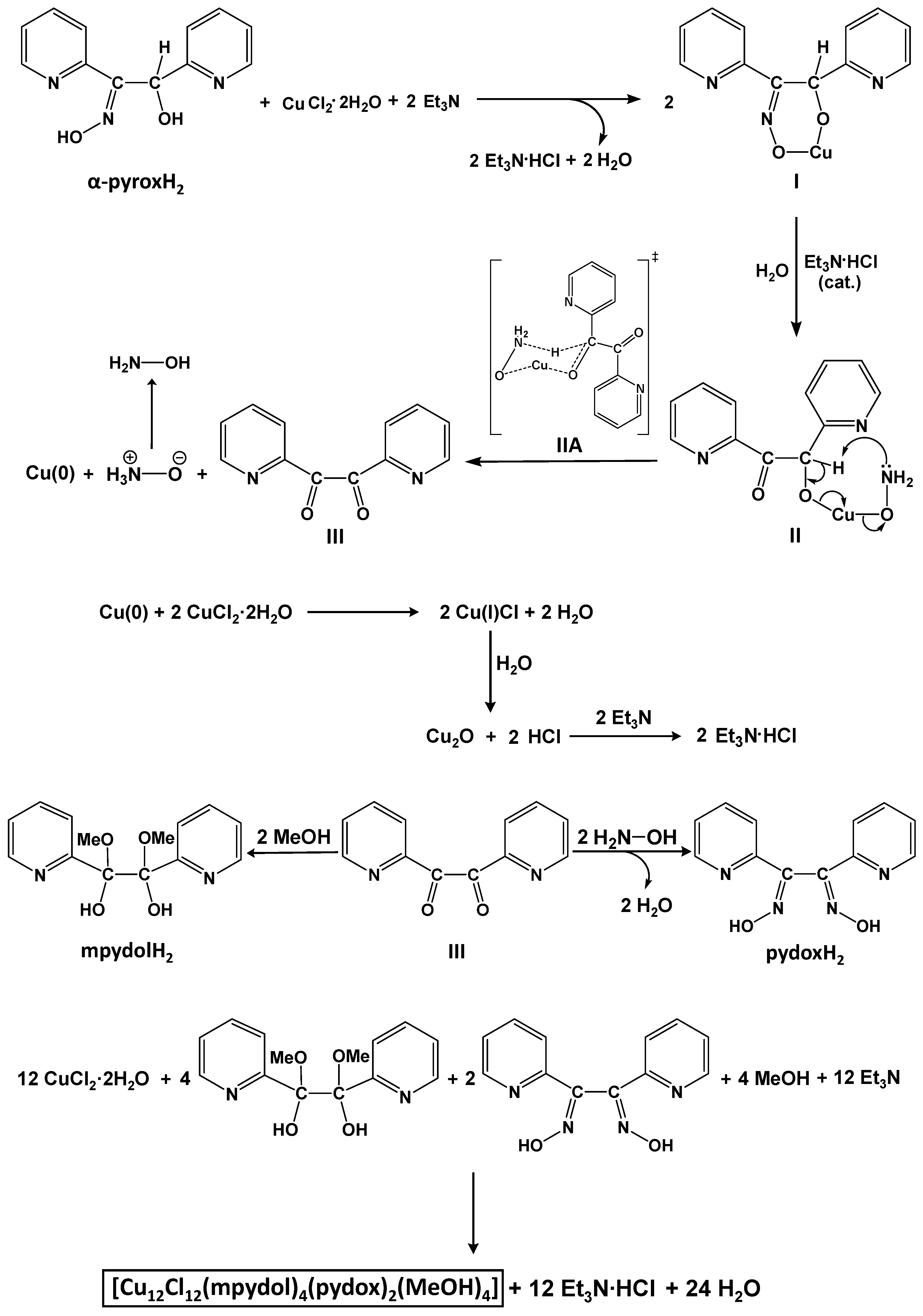
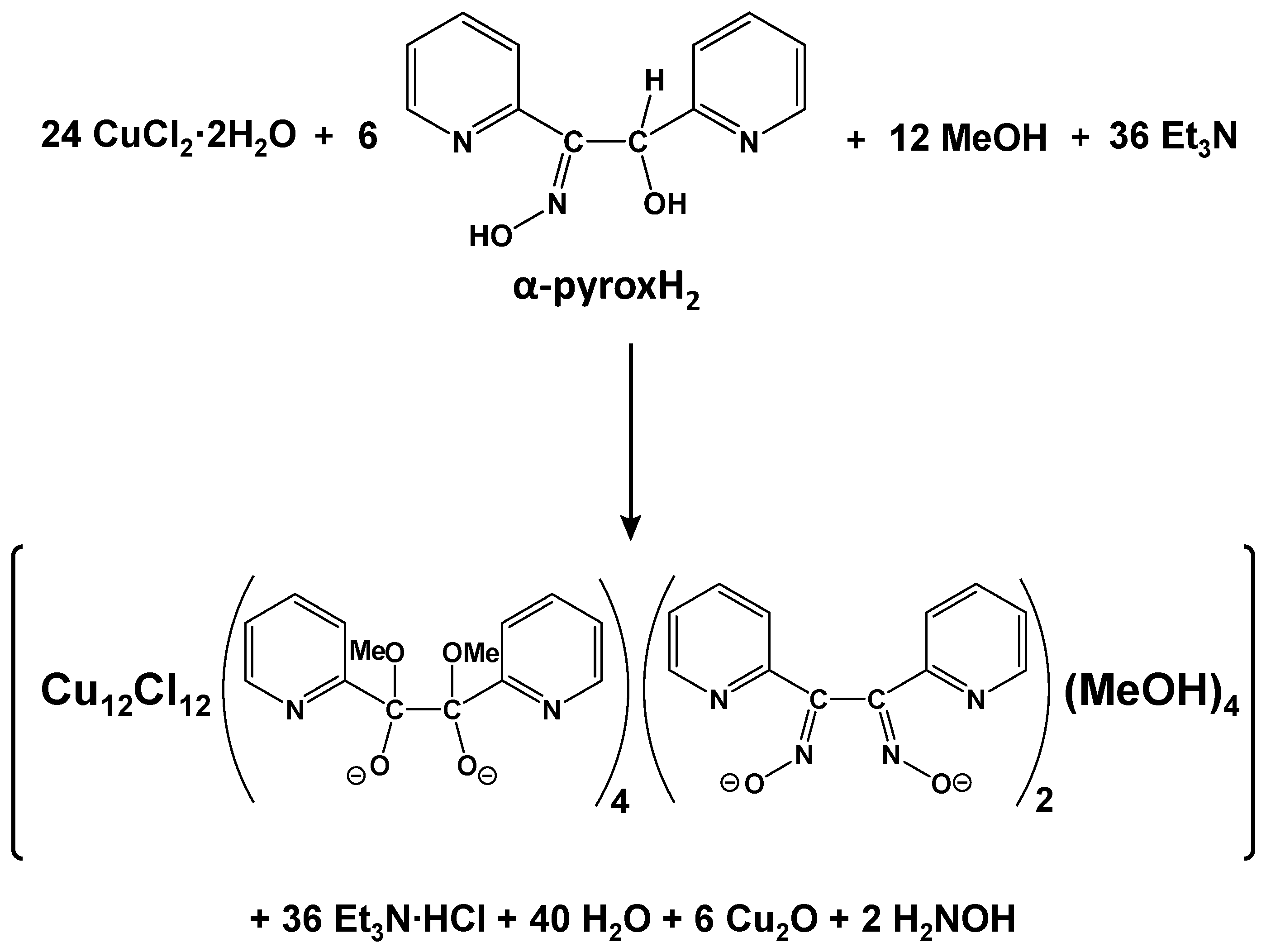
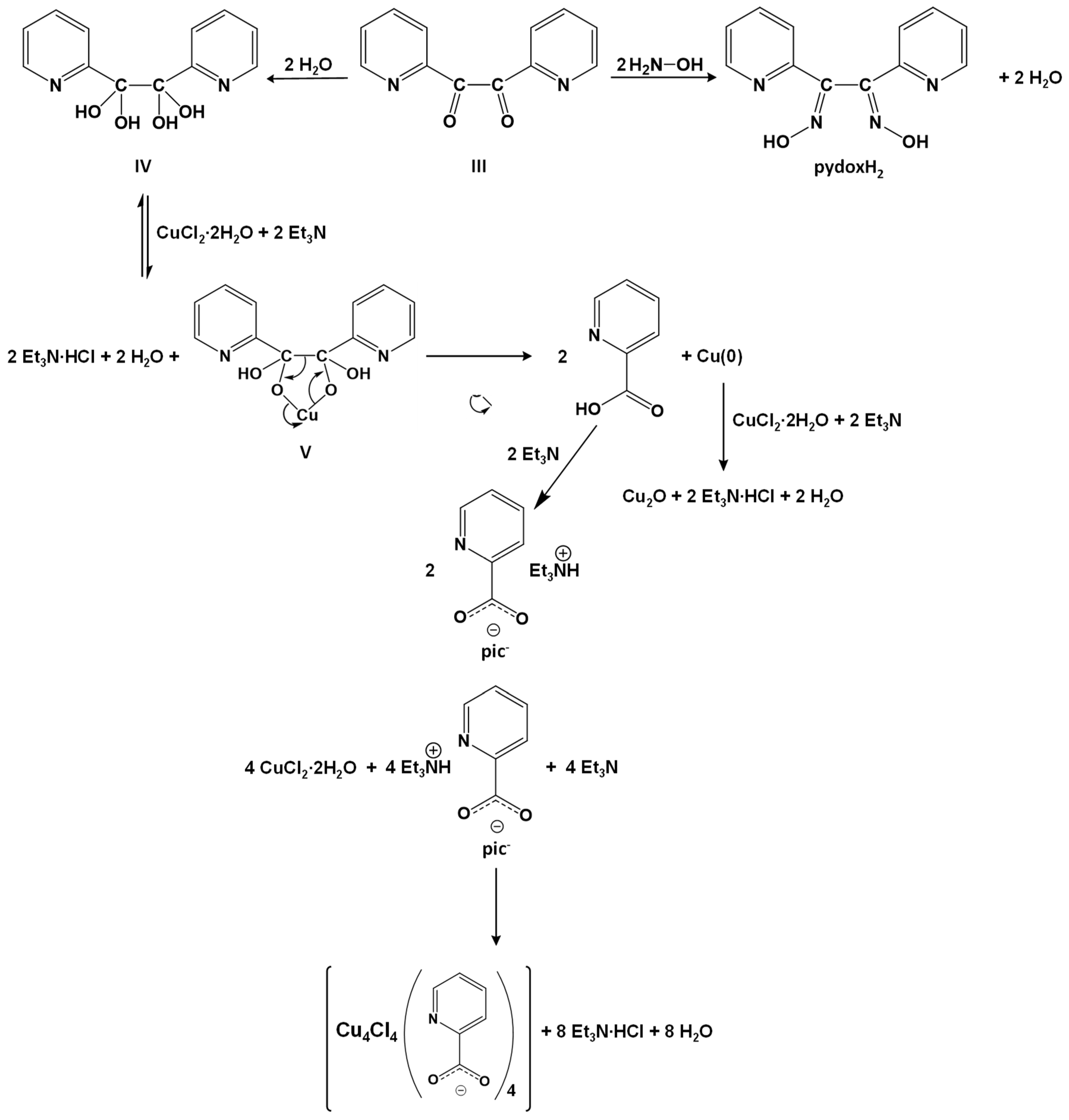
2.2. Comments for the Preparation of the Complexes—Mechanism Proposals

The Cu(II)/α-pyroxH2 reaction system is extremely complicated. We started our efforts using CuCl2∙2H2O as the starting material, and we were able to isolate and characterize two products. The reaction between 3 equivs of CuCl2∙2H2O and 1 equiv of α-pyroxH2, in the presence of 2 equivs of Et3N, in refluxing MeOH, gave a dark green solution, from which were subsequently isolated plate-like crystals of 1 in a 37% yield. Single-crystal X-ray crystallography (vide infra) proved 1 to be the dodecanuclear cluster [Cu12Cl12(mpydol)4(pydox)2(MeOH)4] (with lattice MeOH molecules), where mpydol2− is the dianion of 1,2-dimethoxy-1,2-di(pyridin-2-yl)ethane-1,2-diol and pydox2− is the dianion of (E,E)-1,2-di(pyridin-2-yl)ethanedione dioxime; the structural formulas of the transformed ligands (in their neutral forms) are shown in Scheme 4. We were both unsatisfied and satisfied with this result; unsatisfied because a complex containing α-pyroxH2 (or/and its dianionic forms) had not been prepared, but also satisfied because a copper(II)-assisted/mediated transformation of the original ligand had taken place. Note that 1 is the first structurally characterized metal complex with neutral or anionic forms of pydoxH2 and/or mpydolH2 as ligands (vide infra). To prove that the reaction is Cu(II)-promoted, we refluxed a methanolic solution of α-pyroxH2 and we slowly evaporated the solvent at room temperature until dryness; the obtained solid was washed with Et2O and dried in air overnight. The IR and 1H-NMR spectra of the obtained solid were identical with those of the authentic ligand prepared as illustrated in Scheme 2. An excess of Et3N (using constant the CuII:α-pyroxH2 reaction ratio and the solvent) gave again 1 (IR evidence) in a microcrystalline form with a ca. 40% yield. The use of n-Pr3N, n-Bu3N, Me4NOH, Et4NOH, n-Bu4NOH, NaOH or KOH as external bases (instead of Et3N) led again to 1 in comparable yields; replacement of these bases with the weaker ones Me3N and Me2NH (keeping all the other experimental conditions the same) also resulted in 1, but the crystallization process was slower (16 days until full precipitation instead of 10). Changing the CuCl2∙2H2O:α-pyroxH2 ratio from 3:1 to 3:2, 3:3 or even 3:4 (using a 2:1 base-to-ligand ratio) yielded, again, cluster 1. To investigate if the formation of the dodecanuclear cluster is exclusively the result of the presence of α-pyroxH2 in the reaction media, we performed the stoichiometric reaction, employing the synthesized organic compounds mpydolH2 [35] and pydoxH2 [36]. Thus, the 6:2:1:6 CuCl2∙2H2O/mpydolH2/pydoxH2/Et3N reaction system in refluxing MeOH led to the isolation of a green microcrystalline powder, whose IR spectrum is identical with that of 1, Equation (1); the yield was 58%. Thus, it seems that 1 is the thermodynamically stable product from the CuCl2∙2H2O/α-pyroxH2 and CuCl2∙2H2O/mpydolH2/pydoxH2 reaction systems in MeOH in the presence of an external base.
12   CuCl 2 · 2 H 2 O + 4   mpydolH 2 + 2   pydoxH 2 + 12   Et 3 N + 4   MeOH   M e O H   /   T   [ Cu 12 Cl 12 ( mpydol ) 4 ( pydox ) 2 ( MeOH ) 4 ]   +   12   ( Et 3 NH ) Cl   +   24   H 2 O
We continue with some mechanism ideas (Scheme 5) concerning the above-mentioned experimental observations. Taking into consideration the facts that (a) α-pyroxH2 is converted to the coordinated ligands mpydol2− and pydox2−, and (b) α-hydroxy ketones are converted to α-dicarbonyl compounds upon oxidation with various metal ion-containing species [37], including Cu(II) [38], it is reasonable to propose that the initially employed ligand, i.e., α-pyroxH2, is first converted to 2,2′-pyridyl (III) (IUPAC name: 1,2-di(pyridin-2-yl)ethane-1,2-dione). This transformation can be realized through the following three steps: (i) formation of a chelate Cu(II) complex (I) upon reaction of α-pyroxH2 with CuCl2∙2H2O and Et3N; (ii) hydrolysis of the oxime functionality under the catalytic activity of triethylammonium chloride; and (iii) redox-type decomposition of the as-obtained intermediate II, through the six-membered transition state IIA, giving rise to the key intermediate III, hydroxylamine and Cu(0). Cu(0) is then oxidized by CuCl2∙2H2O to Cu(I)Cl, which is further hydrolyzed to Cu2O and HCl [34]. On the other hand, the intermediate 1,2-di(pyridin-2-yl)ethane-1,2-dione (III) bears two highly activated ketone functionalities, being an α-diketone and at the same time substituted by the electron-withdrawing 2-pyridyl group. It can, therefore, react with the nucleophiles that are present in the reaction system, namely hydroxylamine, methanol and water. Although hydroxylamine is a strong nucleophile, much stronger than the other two, methanol, which is in large excess (as the reaction solvent), efficiently competes with hydroxylamine and, thus, two products are expected, i.e., the 1,2-diol mpydolH2 and the dioxime pydoxH2. The former is formed by the nucleophilic addition of methanol on the carbonyl groups and the latter by the condensation of the diketone with two molecules of hydroxylamine. Finally, the combination of CuCl2∙2H2O with mpydolH2 and pydoxH2 in the presence of Et3N leads to the formation of the isolated crystalline cluster 1. The overall reaction is depicted in Scheme 6. Accordingly, the stoichiometric (theoretical) α-pyroxH2:Et3N:CuCl2∙2H2O ratio is 1:6:4.
From the above discussion, it is evident that MeOH plays an important role in the preparation of 1, because it is primarily responsible for the formation of mpydol2− but also because it participates as a ligand in the complex. Keeping all the reaction parameters the same as in the synthesis of 1 (vide supra), but changing the solvent from MeOH to MeCN, the blue-green 2D polymeric complex {[Cu4Cl4(pic)4]}n (2) was isolated in a 46% yield, where pic is the picolinato(−1) ligand. We were again both satisfied and unsatisfied with this result. Unsatisfied because a complex containing α-pyroxH2 (or its anionic forms) had not been isolated and satisfied because another Cu(II)-mediated (as proven by “blind” experiments) transformation of the original ligand had taken place. An excess of Et3N or stronger bases (n-Pr3N, n-Bu3N, Me4NOH, n-Bu4NOH), but keeping constant the CuCl2∙2H2O-to-α-pyroxH2 ratio (3:1), leads again to 2 (IR evidence and unit-cell determination). The same product is also obtained using an excess of α-pyroxH2, i.e., CuCl2∙2H2O:α-pyroxH2 ratios of 3:2 and 3:3 (using a 2:1 base-to-ligand ratio). An increase in the CuCl2∙2H2O:α-pyroxH2 ratio from 3:1 (which initially gave 2) to 4:1 or 5:1 yielded a powder whose IR spectrum is different from that of 2; since we could not crystallize this product for structural determination, we did not pursue its characterization further. Again, to investigate if the isolation of the 2D polymer is exclusively the result of the presence of α-pyroxH2 in the reaction media, we performed the stoichiometric reaction (1:1:1) between CuCl2∙2H2O, picH (picolinic acid) and Et3N in refluxing MeCN, Equation (2), and confirmed (IR evidence, unit-cell determination) that the product is 2.
4 n   CuCl 2 · 2 H 2 O + 4 n   picH + 4 n Et 3 N   M e C N   / T     { [ Cu 4 Cl 4 ( pic ) 4 ] } n   +   4 n   ( Et 3 NH ) Cl   +   8 n   H 2 O
With the crystal structure of 2 at hand, we were rather disappointed to realize that the structure had been reported [39]. The compound had been prepared by the 1:2 reaction of CuCl2∙2H2O and picH in EtOH at room temperature. Since the supramolecular features and the magnetic properties of this complex had not been studied, we decided to incorporate a brief discussion about its structural and magnetic properties in the present article.
The proposed mechanism for the preparation of 2 is illustrated in Scheme 7. The reaction of CuCl2∙2H2O and α-pyroxH2 in the presence of Et3N leads to the key intermediate III, as already shown in Scheme 5. In the absence of MeOH, as MeCN is now the reaction solvent, the two nucleophiles present in the reaction medium are water and hydroxylamine. Hydroxylamine, being a much stronger nucleophile than water, first reacts with a half quantity of III, forming the dioxime pydoxH2, and the other half reacts with water, giving the tetraol IV. The latter reacts with CuCl2∙2H2O and Et3N, resulting in the chelate Cu(II) intermediate V. This intermediate decomposes to two molecules of picolinic acid and Cu(0). It should be noted that, in an analogous reaction, benzoic acid is usually a byproduct of the oxidation of benzoin to benzil, formed by the oxidation of the latter [37]. Picolinic acid is then deprotonated by Et3N to form pic, whereas Cu(0) oxidizes to Cu2O upon the action of CuCl2∙2H2O. Finally, CuCl2∙2H2O combines with pic, providing access to the polymeric product {[Cu4Cl4(pic)4]}n (2). The overall reaction is depicted in Scheme 8. Accordingly, the stoichiometric (theoretical) α-pyroxH2:Et3N:CuCl2∙2H2O ratio is 1:8:4.

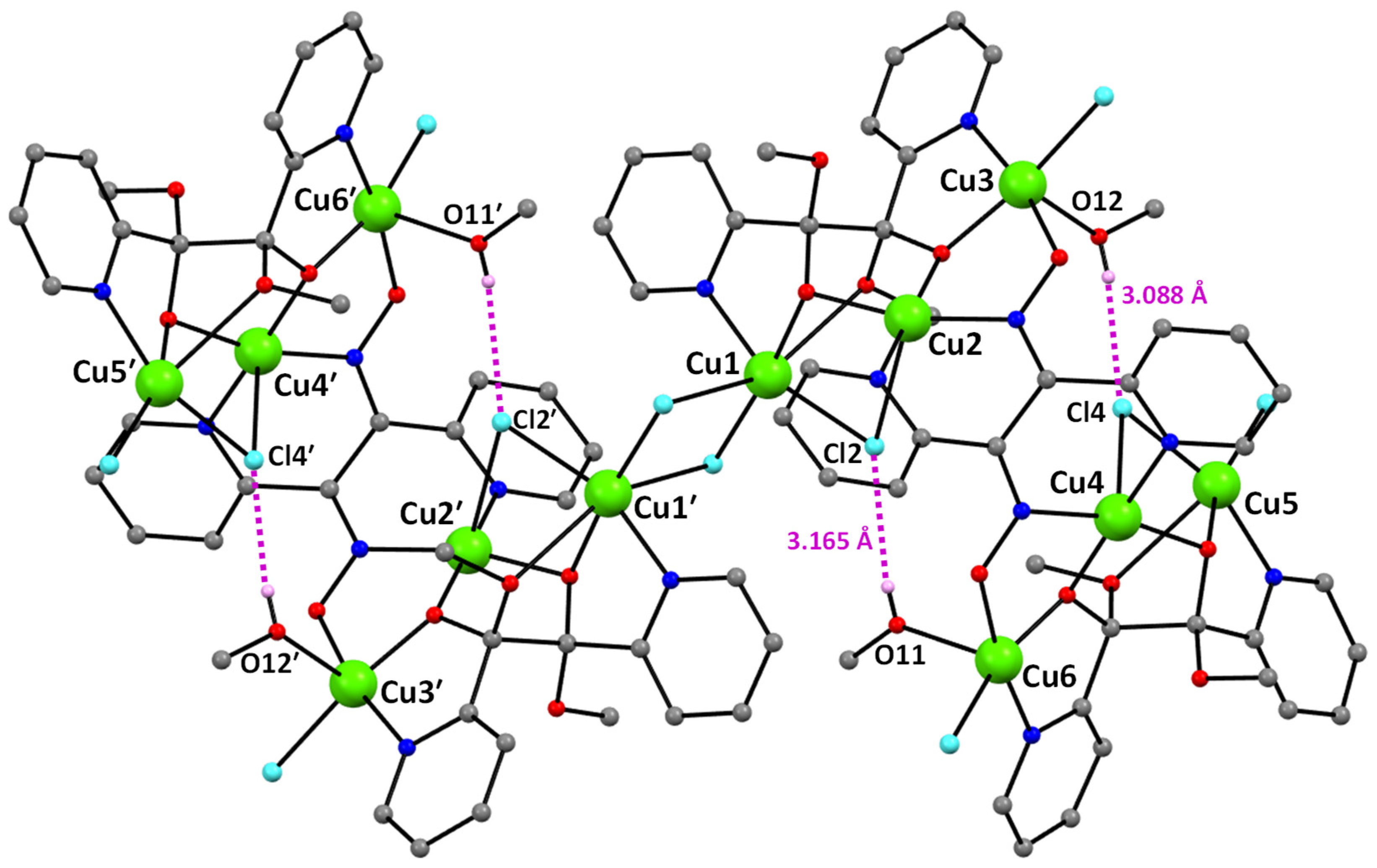
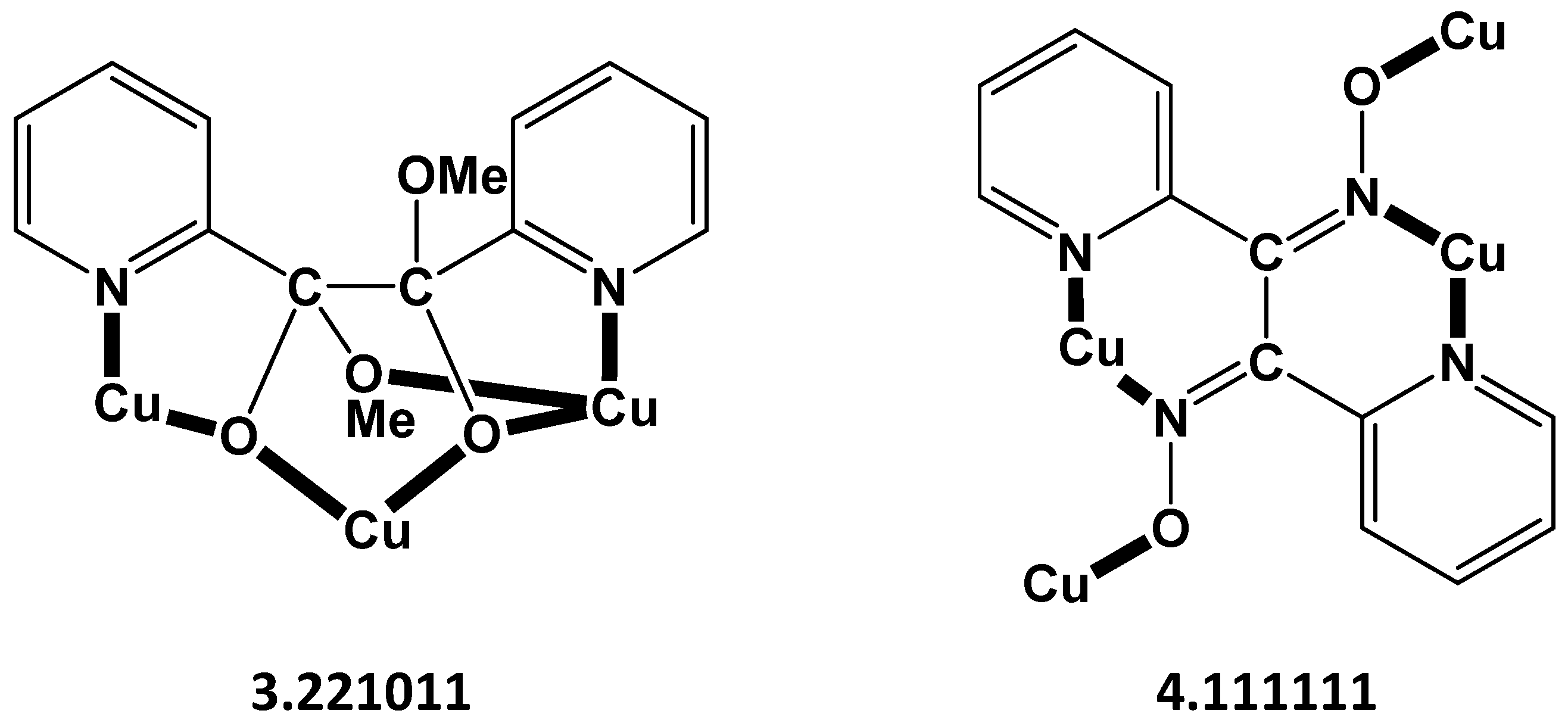
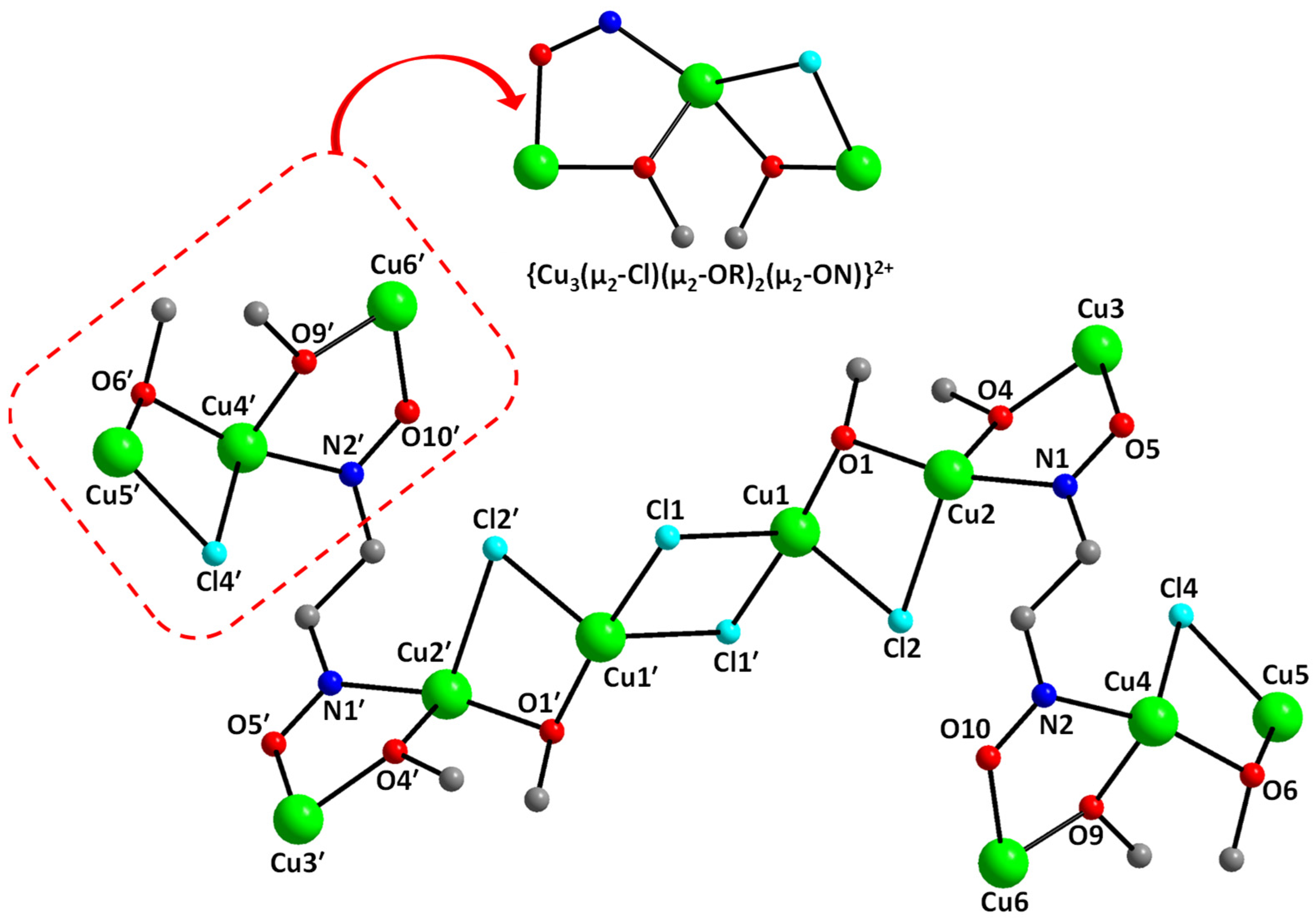
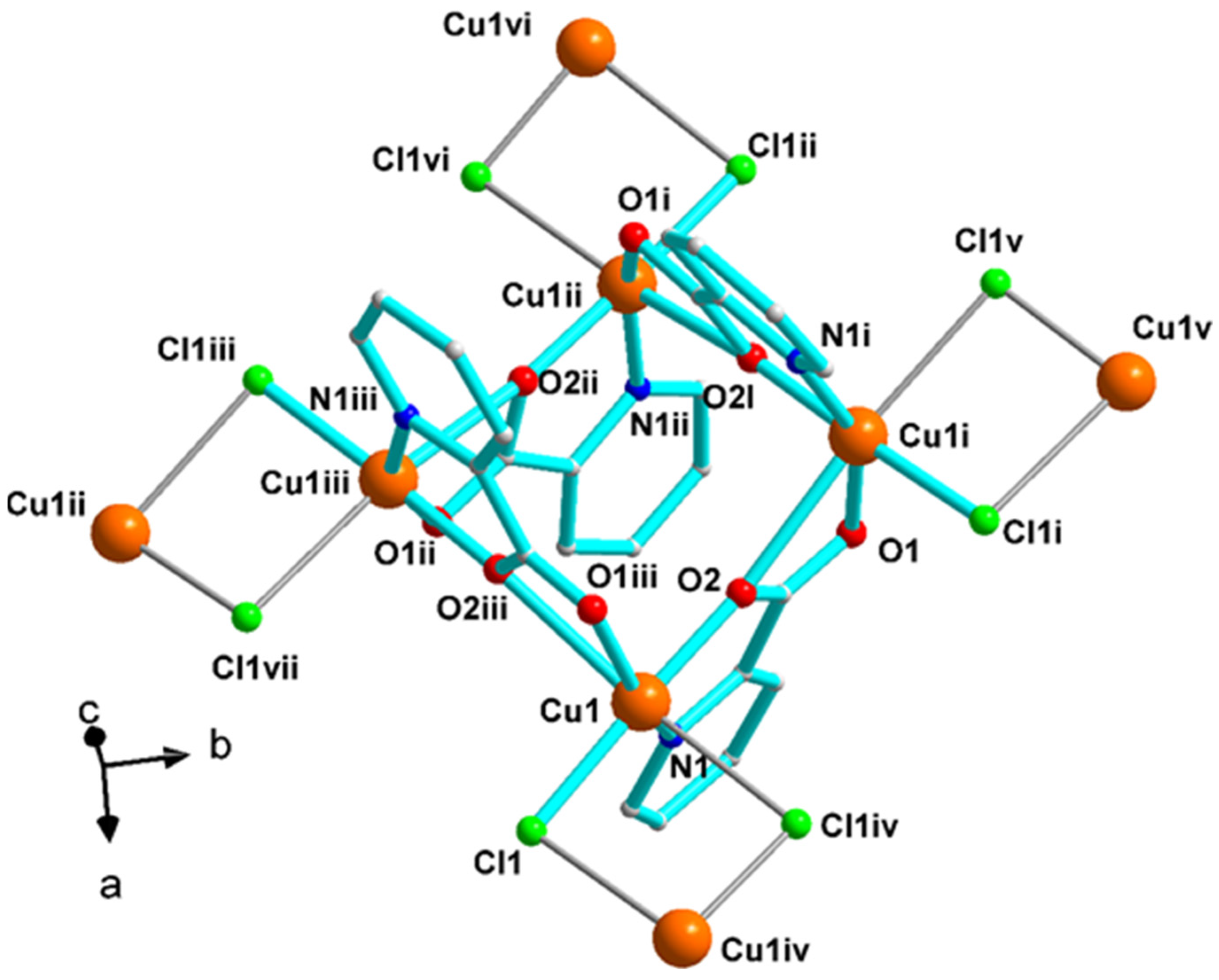
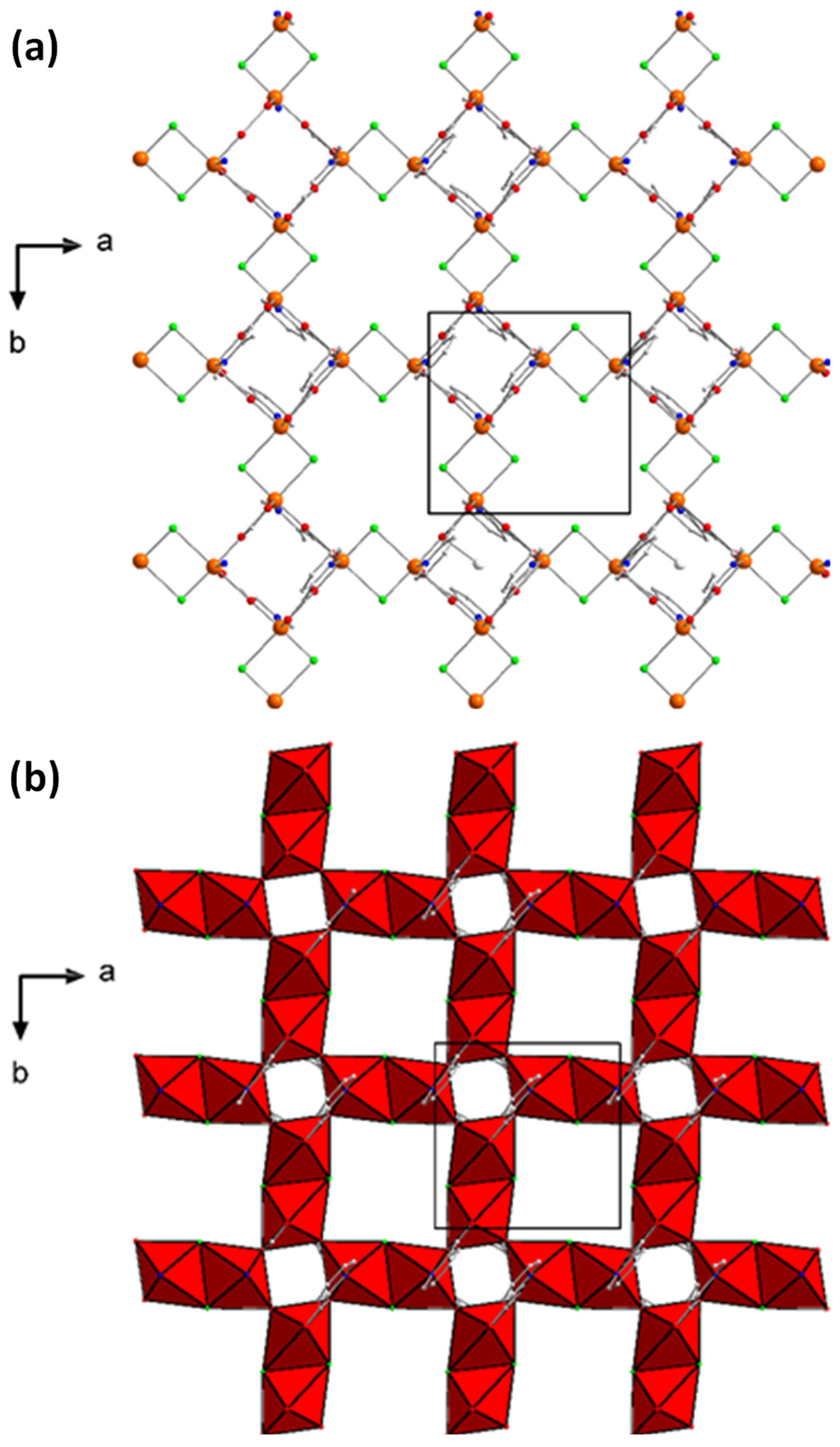
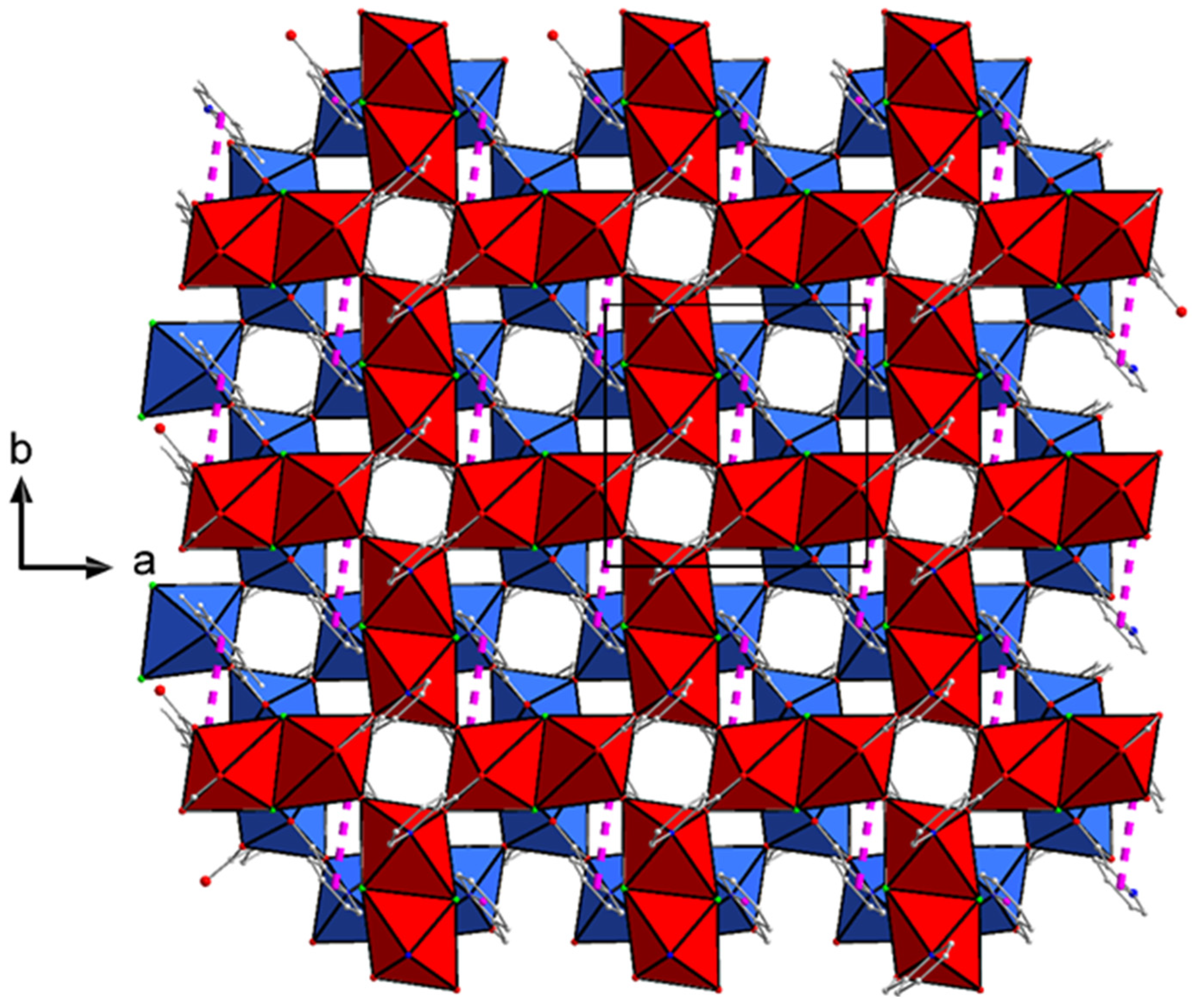
2.3. Description of Structures

The structures of 1∙12MeOH and 2 were solved by single-crystal, X-ray crystallography. Various structural plots are shown in Figure 2, Figure 3, Figure 4, Figure 5, Figure 6 and Figures S4–S8, and Scheme 9 and Scheme S1. Crystallographic data are presented in Table 2, while numerical data for interatomic distances, bond angles and H-bonding interactions can be found in the crystallographic cif files.
The crystal structure of 1∙12MeOH consists of the dodecanuclear cluster [Cu12Cl12(mpydol)4(pydox)2(MeOH)4] (Figure 2) and lattice MeOH molecules; the latter will not be further discussed. The twelve metal ions are held together by six μ2 chlorido, four mpydol2− and two pydox2− ligands; peripheral ligation is provided by six terminal chlorido ligands and four MeOH molecules. The cluster molecule has a crystallographically imposed inversion center at the midpoint of the Cu1∙∙∙Cu1′ vector.
Using Harris notation [40], the coordination modes of mpydol2− and pydox2− can be described as 3.221011 and 4.111111, respectively (Scheme 9); an alternative description is η1:η2:η1:η2:η1:μ3 (mpydol2−) and η1:η1:η1:η1:η1:η1:μ4 (pydox2−). The crystallographically independent pydox2− ligand bridges Cu2, Cu3, Cu4 and Cu6 (and symmetry equivalents). One of the two independent mpydol2− ligand bridges Cu1, Cu2 and Cu3, and the other bridges Cu4, Cu5 and Cu6. Cu1 and Cu1′ are doubly bridged by two chlorido ions (Cl1, Cl1′). The other two independent μ2-Cl ions, Cl2 and Cl4, bridge Cu1 and Cu2, and Cu4 and Cu5 (and symmetry equivalents), respectively. The four terminal MeOH molecules are coordinated to Cu3, Cu3′, Cu6 and Cu6′, while the six terminal chlorido groups form coordination bonds to Cu3, Cu3′, Cu5, Cu5′, Cu6 and Cu6′. Based on the above mentioned, the core (Figure 3) of the dodecanuclear molecule (assuming that the diatomic oximate groups, NO, are part of the core) is {Cu12(μ2-Cl)6(μ2-OR)8(μ2-NO)4}6+, where the oxygen atom of RO is an alkoxido oxygen from mpydol2−. This can be considered as consisting of four similar trinuclear subunits that create the {Cu3(μ2-Cl)(μ2-OR)2(μ2-NO)}2+ subcore, which are connected through the double chlorido bridge (Cl1, Cl1′) and two trans dioximato groups; this description is emphasized in Figure 3. Thus, the topology of the CuII ions is composed of four triangles (Cu2/Cu1/Cu3, Cu4/Cu5/Cu6 and their symmetry equivalents) in an open zigzag configuration; each “terminal” triangle is linked to its neighboring one through a dioximato group, while the connection between the two “central” triangles is achieved through two chlorido bridges. The triangles are nearly isosceles, the short sides being ca. 3.2 Å and the long one ca. 5.6 Å. The bond angles around the monoatomic bridges are as follows: Cu1–Cl1–Cu1′ = 89.8(1) ° , Cu1–Cl2–Cu2 = 79.8(1) ° , Cu4–Cl4–Cu5 = 80.5(1) ° , Cu1–O1–Cu2 = 109.6(1) ° , Cu2–O4–Cu3 = 116.5(1) ° , Cu4–O6–Cu5 = 109.5(1) ° and Cu4–O9–Cu6 = 116.2(2) ° .
Cu1/Cu1′ adopts an elongated, Jahn–Teller-distorted, octahedral geometry, with the two Jahn–Teller positions being occupied by a μ2 chlorido ligand and a methoxy atom from a 3.221011 mpydol2- ligand. Atoms Cu2, Cu3, Cu4, Cu5 and Cu6 are five-coordinate with square pyramidal coordination geometries; the corresponding trigonality indices (τ) are 0.13, 0.01, 0.15, 0.05 and 0.10 (τ is 0 for a perfect square pyramidal geometry and 1 for a perfect trigonal bipyramidal geometry). The coordination spheres of the six metal centers are {Cu1/Cu1′(μ2-Cl)3(μ2-Oalkoxido)(Omethoxy)(Npyridyl)}, {Cu2/Cu2′(μ2-Cl)(μ2-Oalkoxido)2(Npyridyl)(Noximato)}, {Cu3/Cu3′Cl(μ2-Oalkoxido)(Ooximato)(Omethanol)(Npyridyl)}, {Cu4/Cu4′(μ2-Cl)(μ2-Oalkoxido)2(Npyridyl)(Noximato)}, {Cu5/Cu5′Cl(μ2-Cl)(μ2-Oalkoxido)(Omethoxy)(Npyridyl)} and {Cu6/Cu6′Cl(μ2-Oalkoxido)(Ooximato)(Omethanol)(Npyridyl)}. A μ2-Cl, an Omethanol, a μ2-Cl, an Omethoxy and an Omethanol are the apical positions of Cu2, Cu3, Cu4, Cu5 and Cu6, respectively.
The intramolecular CuII∙∙∙CuII distances range from 3.546(1) (Cu1∙∙∙Cu1′) to 18.789(1) (Cu5∙∙∙Cu5′) Å. There are four weak intramolecular H bonds in 1 (Figure 2). Donors are the O atoms of the coordinated MeOH molecules, and acceptors are μ2 chlorido groups. The space-filling diagram of 1 is shown in Figure S5. This reveals a parallelogram arrangement in which the twelve “internal” CuII ions are surrounded by the bridging and terminal ligands. The longest intramolecular distance, as this is defined by the C atoms of the mpydol2− ligands, is ca. 2.5 nm, giving a nanoscale dimension in the molecular structure. The packing diagram of the compound provides evidence for a dense H-bonded assembly of molecules (Figure S6).
Compound 1 is the first structurally characterized complex of any metal containing mpydolH2 and/or pydoxH2 (or their anionic forms) as ligands. The dication of pydoxH2 in which the two 2-pyridyl N atoms are protonated, i.e., pydoxH42+, has been used as a counterion in (pydoxH4)[ReIVCl6] [41]. The dianion of the diethoxy analogue of mpydolH2, i.e., {(py)C(O)(OEt)C(O)(OEt)(py)}2− (py = 2-pyridyl), has been used as a μ3 ligand in pentanuclear copper(II) complexes; the dianionic ligand was derived from the Cu(II)-assisted/mediated ethanolysis of 2,2′-pyridil [42]. Compound 1 is a member of a relatively large family of dodecanuclear copper complexes, e.g., those described in references [43] and [44], only three of which contain chlorido ligands. These complexes are as follows: [CuII12Cl6(3,5-Me2Pz)4(3,5-Me2PzH)6(tBuPO3)6(tBuPO2OH)2] [45], where 3,5-Me2PzH is 3,5-dimethylpyrazole and tBuPO3H2 is tert-butylphosphonic acid; [CuII12(OH)4Cl8(ox)2(L)4(H2O)2] [46], where ox2− is oxalato(-2) and L2− is the dianion of a trinucleating ligand with pyridyl, pyrimidyl and amine groups; and the mixed-valence cluster [CuI10CuII2Cl2(mt)12] [47], where mt−1 is the methimazolato(−1) ligand. The former two have only chlorido bridging ligands, whereas the latter contains only terminal chlorido groups, each of them coordinated to a CuI ion. The metal topologies in the just-mentioned clusters are different from the topology observed in 1.
As mentioned in Section 2.2, the structure of the 2D coordination polymer {[Cu4Cl4(pic)4]}n (2) has been reported [39]. Since (a) the quality of the present structure is better and (b) the supramolecular features were not analyzed in detail, we present a short structural description, with a focus on the intralayer interactions between the tetrameric units and between the layers. Compound 2 crystallizes in the tetragonal space group P42/n, and the asymmetric unit consists of 1/4 of the tetrameric Cu4Cl4(pic)4 unit (Figure S7); these units constitute the nodes of the 2D layers. Figure 4 presents a node of the {[Cu4Cl4(pic)4]}n layers, which form the 2D polymer. The structure is described in origin setting 2, and the polymeric unit shown in Figure 4 possesses a −4 roto-inversion symmetry axis defined by the line that passes through point 1/4, 1/4, 0; this axis is parallel to c. The stereochemistry of CuII is Jahn–Teller distorted octahedral, with a picolinato O atom and a bridging Cl ion at the elongated axial positions. Atoms O2 and O2iii bridge Cu1 with two neighboring metal centers within the square tetrameric unit, and Cl1 and Cl1iv doubly bridge the tetrameric unit, with one metal center of a neighboring unit within the layer; the symmetry operations are listed in the caption of Figure 4. Thus, each tetrameric unit is connected with four such units in the same layer. The pic ligand adopts the 2.211 (or η1:η2:η1μ2) coordination mode (Scheme S1).
Figure 5a and Figure 5b illustrate the arrangement of tetrameric units within a layer extended parallel to the (001) planes in ball-and-stick and polyhedral modes, respectively. The octahedra within the tetrameric units share corners (O2 atoms) and those belonging to neighboring units share edges (pairs of Cl1 atoms). The −4 roto-inversion symmetry axis, possessed by the tetrameric units, results in the arrangement of the four pic ligands, belonging to the units, in pairs lying above and below the layers. This arrangement results in π–π interactions of 2-pyridyl rings that belong to pic ligands of neighboring layers, lying above and below each layer (Figure S8). The distance between centrosymmetrically related 2-pyridyl rings (symmetry operation: −x + 1, −y + 1, −z) is 3.284(2) Å. The relative arrangement of neighboring layers along the c axis is shown in Figure 6.
Compound 2 joins a handful of structurally characterized polymeric Cu(II) complexes containing, exclusively, the pic ligand or a combination of pic with other simple inorganic ligands. These complexes (with relevant information) are listed in Table 3; the structures of some complexes have been reported twice, i.e., {[Cu(pic)2]}n [48,49], {[Cu4(pic)6(H2O)2](ClO4)2}n [50,51] and {[Cu4Cl4(pic)4}n ([39], this work), or even three times, i.e., {[Cu(pic)2]∙2H2O}n [52,53,54]. Table 3 shows the following: (i) The pic ligand adopts three different coordination modes (1.101, 2.111, 2.211); the 2.211 mode appears only in {[Cu4Cl4(pic)4}n. (ii) The compounds are either 1D or 2D coordination polymers; and (iii) in most preparations, the pic ligand has been derived from a Cu(II)-assisted transformation of a more complicated ligand (see footnotes of Table 3).

2.4. Magnetic Properties

Direct current (dc) magnetic susceptibility data (χM) on dried and analytically pure samples of compounds 1 and 2 were collected in the 1.8–310 K range in an applied field of 1 kOe. Data are presented in Figure 7, Figure 8, Figures S9 and S10. In addition, the powder X-ray diffraction patterns of both compounds show good agreement with the theoretical ones, confirming the phase purity of the samples (Figures S11 and S12).
Cluster 1 is magnetically interesting. The magnetization (M) at 1.8 K seems to be “saturating” around 11,000 emu/mol (Figure S9), which is in agreement if there are only two S = 1/2 spin carriers. If that is correct, a first explanation is that ten of the CuII ions are antiferromagnetically coupled, leading to a singlet (S = 0) ground state at 1.8 K. This actually agrees with M vs. T data. The χMΤ vs. T plot (Figure 7) shows that the 310 K value is 1.24 emu K/Oe mol, much smaller than the 5.45 emu K/Oe mol value expected for twelve non-interacting CuII ions (with g = 2.2), suggesting the presence of strong antiferromagnetic exchange interactions. The χMΤ product shows a steadily decreasing value from 310 K down to ~ 100 K, where there is a long plateau at 0.9 emu K/Oe mol, which is in close agreement with two S = 1/2 spin carriers. Only at the very lowest temperatures is there a further downturn in χMΤ vs. T, indicating a very weak interaction between those in two magnetic moments, certainly too weak to model. No hysteresis was observed in the complex; M vs. H data were collected in both increasing and decreasing fields, with no difference in observed magnetization to obviate for ZFC-FC data, and that is the hysteresis test. A magnetic phase transition would likely show a discontinuity in the M vs. T data, which was not observed. Looking at the core of the dodecanuclear compound (Figure 3) and the connectivity of the central CuII ions, it is reasonable to assume that Cu1, Cu2, Cu3 and their symmetry equivalents are strongly antiferromagnetically coupled, yielding a local S = 0 value. Subsequently, the two outer CuII3 triangles, comprising Cu4, Cu5, Cu6 and their symmetry-related partners, are also antiferromagnetically coupled, a magnetic pathway that is mostly propagated by the double oximato/alkoxo bridge [50] between Cu4 and Cu6 atoms, thus leading to two isolated S = 1/2 local spins located at Cu5 and Cu5’. Hence, there are two effectively isolated CuII3 triangles giving a total ground state of Stotal = 1, in excellent agreement with the experimental data. Since four CuII2 pairs are partly bridged by diatomic oximato groups, the antiferromagnetic exchange interactions are reasonable [56]. The molecule 1 exhibits six different CuII sites which are unique; Cu1, Cu2 and Cu3 are similar to Cu4, Cu5 and Cu6, but not identical, so there are six different metal-ion sites with at least six different exchange pathways: Cu1···Cu1’, Cu1···Cu2, Cu2···Cu3 and the equivalent set for the Cu4, Cu5 and Cu6 ions. An interaction between Cu2/Cu3 and Cu4/Cu6 seems unlikely, or at least too weak to be significant. There is no model for this system. This is in accordance with literature data [43,44,45] where experimental data in complicated dodecanuclear Cu(II) complexes could not be fitted. Theoretical calculations might be useful, but with too many parameters to compare to the experimental data, this is a very difficult task.
The 310 K χMΤ value for 2 is 1.95 emu K/Oe mol per tetramer (Figure 8), close to the value expected for four non-interacting CuII ions (1.82 emu K/Oe mol for g = 2.2). This product is practically constant until ~ 70 K, suggesting four magnetically isolated CuII ions. The value of the product increases slowly in the 70–25 K range, and abruptly below 25 K, reaching a value of 11.70 emu K/Oe mol at 1.8 K. The χM vs. T data do not indicate saturation, and there are no signs of a maximum being reached, and this further suggests that the interactions are weak. This behavior is clearly consistent with weak ferromagnetic interactions. In addition, the magnetization is nearing saturation at ~ 23,000 emu/mol (Figure S10), just about the expected value for four CuII ions. The magnetization shows a rapid increase, even at the lowest applied magnetic fields, corroborating the overall ferromagnetic response of the compound. We tentatively propose that the four metal ions within each square unit (Cu1, Cu1i, Cu1ii, Cu1iii in Figure 4) are isolated. This is attributed to the very large CuII–(μ2-Opicolinato)–CuII angle [166.1(1) ° ] between neighboring metal ions, which precludes any interaction between the magnetic d x 2 y 2 orbitals. It has been established that a large Cu–O–Cu angle results in a poor overlap between the copper(II) d x 2 y 2 and oxygen px and py orbitals, and the interaction should not favor an antiferromagnetic exchange through a dominant sigma spin exchange pathway [44]. The ferromagnetic behavior arises from exchange interactions between the CuII ion of a square unit (e.g., Cu1 in Figure 4) and the CuII ion of a neighboring unit (e.g., Cu1iv in Figure 4), which are doubly bridged with asymmetric chlorido groups. Each bridging chlorido ion is in the equatorial plane of one metal center and in an axial position of the other. For such axial–equatorial bis(μ2-chloro)dicopper(II) units, a correlation has been established [57,58] between the singlet–triplet splitting and the quotient φ/R, where φ is the Cu–Cl–Cu bridging angle and R is the longest bridging bond distance. In our case, φ is 90.9 ° (e.g., the Cu1–Cl1iv–Cu1iv angle in Figure 4) and 2.731 Å (e.g., the Cu1–Cl1iv bond distance in the same figure). The value of the quotient in 2 (33.30) is well within the range predicted for ferromagnetic CuII∙∙∙CuII interactions.
One possibility of fitting the data, which could support the proposed magnetic lattice, would be to assume a spin dimer model (if the only significant interactions are between the doubly chlorido-bridged CuII ions, it is just a very complex dimer) and see the J value. Having strong evidence (vide supra) that there are no significant interactions in the central square unit, the system can be described as an array of “isolated” dimers. The fit of χMΤ vs. T data to a ferromagnetic dimer model with a Currie–Weiss correction for interdimer interactions is shown in Figure 8. The model [59] is based per mole of CuII ion, so we had to divide the formula weight by four to get any sensible results. Values are CC (Currie constant) = 0.417(2) emu K/mol Oe, JK = 21.1(3) K, fixed paramagnetic impurity = 0.1% and CW (Currie–Weiss constant) = 0.81(1) K; the latter value would indicate a ferromagnetic interdimer interaction, but the value is small enough, and it is probably within the error of the data.

3. Experimental Section

3.1. Materials and Instrumentation

All experimental work was performed under aerobic conditions using reagents and solvents (Alfa Aesar, Karlsruhe, Germany; Sigma-Aldrich, Tanfrichen, Germany) as received; α-pyridoin was purchased from Sigma-Aldrich. Deionized water was obtained from an in-house facility. The compounds mpydolH2 [35] and pydoxH2 [36] were synthesized as previously reported; their purity was checked by 1H NMR spectroscopy in d6-DMSO and microanalyses. The free ligand α-pyroxH2 was synthesized as detailed in Section 3.2 below. Analyses of C, H and N were conducted by the University of Patras Instrumental Analysis Service. FT-IR spectra (4000–400 cm−1) were recorded using a Perkin-Elmer 16 PC spectrometer (Perkin-Elmer, Waltham, MA, USA); the samples were in the form of KBr pellets obtained under pressure. 1H NMR spectra in CD3OD were recorded on a Bruker Avance DPX spectrometer (Bruker AVANCE, Billerica, MA, USA) at a resonance frequency of 600 MHz; tetramethylsilane was used as standard. Powder-XRD measurements were carried out on a Rigaku Miniflex 6G X-Ray Diffractometer (Rigaku, London, UK). For the magnetic measurements, powdered samples were mounted in gelatin capsules in clear plastic straws. The data were obtained using a Quantum Design MPMS-XL SQUID magnetometer (Quantum, San Diego, CA, USA). Magnetization data were collected as function of field in the 0–50 kOe range at 1.8 K. Some data points (every 10 kOe) were recollected as the field returned to zero to check for hysteresis effects; none were observed. Dc magnetic susceptibility data were collected in a 1 kOe field from 1.8 to 310 K. Susceptibility values were corrected for the gelatin capsule and straw (measured independently) and the diamagnetism of the constituent atoms as estimated from Pascal’s constants [60].

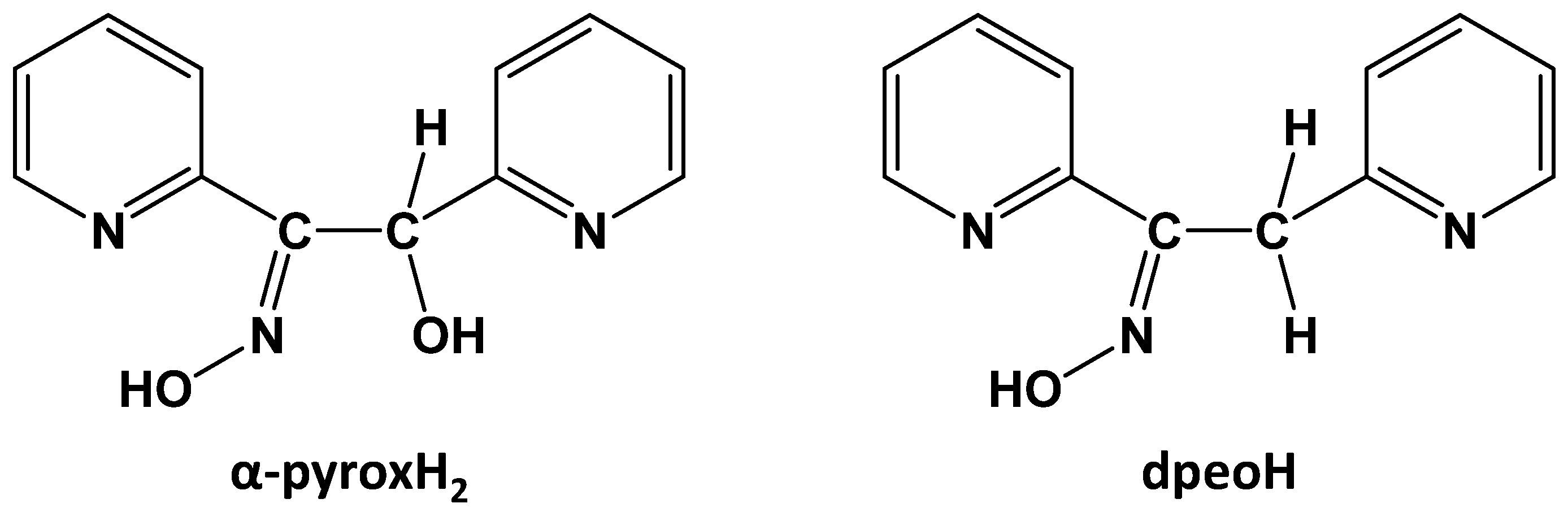
3.2. Synthesis of 2-Hydroxy-1,2-di(pyridin-2-yl)ethanone Oxime (α-pyroxH2)

To a colorless solution of NaO2CMe (0.273 g, 2.40 mmol) in MeOH (6 mL) was added NH2OH∙HCl (0.168 g, 2.40 mmol) without a noticeable change. To this solution was slowly added a brown-red solution of α-pyridoin (0.428 g, 2.00 mmol) in THF (6 mL). The two solutions were mixed under stirring, and a brown slurry was formed, which was refluxed for 2 h without any change. The flask was cooled to room temperature, and the resulting brown microcrystalline precipitate was collected by filtration, washed with Et2O (5 × 2 mL) and dried in air overnight. The yield was 61% based on the α-pyridoin available. Anal. Calcd. (%) for C12H11N3O2: C, 62.87; H, 4.84; N, 18.33. Found (%): C, 63.01; H, 4.89; N, 18.21. IR (KBr, cm−1): 3453 (sb), 3067 (w), 1713 (s), 1654 (s), 1595 (s), 1524 (w), 1463 (m), 1389 (s), 1364 (s), 1298 (w), 1272 (w), 1225 (w), 1181 (m), 1144 (s), 1093 (m), 1038 (w), 994 (m), 898 (w), 840 (m), 768 (m), 733 (m), 690 (m), 648 (w), 612 (w), 547 (w), 521 (w), 469 (w), 438 (w). 1H NMR (CD3OD, δ/ppm): 8.65 (s, 1H), 8.20–7.40 (8 signals, 8H), 5.70 (s, 1H), 3.37 (s, 1H).

3.3. Preparation of the Complexes

[Cu12Cl12(mpydol)4(pydox)2(MeOH)4]∙12MeOH (1∙12MeOH). Solid CuCl2∙2H2O (0.051 g, 0.30 mmol) was added to a dark orange solution of α-pyroxH2 (0.023 g, 0.10 mmol) and Et3N (28 μL, 0.20 mmol) in MeOH (10 mL). The resulting dark green solution was refluxed overnight under vigorous magnetic stirring, filtered and stored in an open flask. Slow evaporation at room temperature resulted in the precipitation of X-ray-quality green crystals after ∼10 days. The crystals were collected by filtration, washed with MeOH (2 × 3 mL) and Et2O (4 × 2 mL), and dried in a vacuum desiccator over silica gel. The yield was 37% based on the quantity of the α-pyroxH2 used. The sample was analyzed satisfactorily as lattice MeOH-free, i.e., as 1. Anal. Calcd. (%) for C84H88Cu12N16O24Cl12: C, 34.87; H, 3.07; N, 7.74. Found (%): C, 35.13; H, 3.15; N, 7.67. IR (KBr, cm−1, Figure S13): 3432 (wb), 3088 (w), 3073 (w), 1617 (m), 1589 (s), 1571 (sh), 1478 (m), 1443 (w), 1406 (m), 1288 (w), 1263 (w), 1236 (w), 1172 (w), 1145 (w), 1085 (w), 1038 (m), 978 (sh), 920 (sh), 873 (m), 825 (w), 762 (s), 717 (m), 692 (m), 646 (w), 556 (w), 470 (m), 412 (w).
{[Cu4Cl4(pic)4]}n (2). Solid CuCl2∙2H2O (0.051 g, 0.30 mmol) was slowly added to a dark orange solution of α-pyroxH2 (0.023 g, 0.10 mmol) and Et3N (28 μL, 0.20 mmol) in MeCN (10 mL). The resulting green-blue slurry was refluxed for 7 h under vigorous stirring, during which time the solid was dissolved. The solution was filtered and allowed to stand in an open vial. Slow evaporation at room temperature gave X-ray-quality, blue-green crystals of the product after 3 days. The crystals were collected by filtration, washed with MeCN (2 × 3 mL) and Et2O (2 × 3 mL), and dried in air overnight. The yield was 46% based on the quantity of the available α-pyroxH2. Anal. Calcd. (%) for C24H16Cu4N4O8Cl4: C, 32.59; H, 1.82; N, 6.34. Found (%): C, 32.88; H, 2.11; N, 6.21. IR (KBr, cm−1, Figure S14): 3432 (mb), 3078 (w), 2933 (w), 1635 (sh), 1603 (s), 1577 (sh), 1511 (w), 1473 (m), 1439 (m), 1375 (wb), 1278 (wb), 1244 (w), 1205 (w), 1160 (w), 1097 (s), 1071 (s), 1036 (m), 978 (m), 916 (m), 769 (s), 706 (w), 652 (w), 599 (wb), 534 (w), 504 (w), 469 (w), 430 (m).

3.4. Single-Crystal X-Ray Crystallography

A single crystal of complex 1∙12MeOH was collected from the crystallization vial, immediately immersed in highly viscous oil and mounted on a CryoLoop with the assistance of a stereomicroscope. Diffraction data were collected on a Bruker D8 diffractometer (Bruker AVANCE, Billerica, MA, USA), equipped with Mo Kα graphite-monochromated radiation. The crystal was positioned at 55 nm from the detector, and 60 s per frame of exposure was used, with the acquisition controlled by the APEX2 software package [61]. The temperature of acquisition [−123(2)   ] was set up with a cryosystem by the Oxford Cryosystems Series 700. Images were processed with the software SAINT+ [62], and absorption effects were corrected with the multi-scan method implemented in SADABS [63]. The structure was solved using SHELXTL incorporated in the Bruker APEX-III software package and refined using the SHELXL and PLATON programs [64,65,66]. The non-H atoms of the structure were successfully refined using anisotropic displacement parameters. Most H atoms were placed at geometrical positions using the suitable HFIX instructions in SHELXL and included in subsequent refinement cycles in riding-motion approximation with isotropic thermal displacement parameters fixed at 1.5 × Ueq of the carbon atoms to which they are attached. Various figures of the structure were created using the Diamond [67] and Mercury [68] packages.
For compound 2, a blue-green crystal was taken directly from the mother liquor and immediately cooled to −113   . Diffraction data were collected on a Rigaku R-AXIS SPIDER Image Plate Diffractometer (Rigaku Americas Corporation, The Woodlands, TX, USA) using graphite-monochromated Mo Kα radiation. Data collection (ω-scans) and processing (cell refinement, data reduction and empirical absorption correction) were performed using the CrystalClear program package [69]. The structure was solved by direct methods using SHELXS, ver. 2013/1 [69], and refined by full-matrix least-squares techniques on F2 using SHELXL, version 2014/6 [66]. The H atoms were introduced at calculated positions and refined as riding on their corresponding bonded atoms. All non-H atoms were refined anisotropically. Plots of the structure were drawn using the Diamond 3 [70] program package.
The X-ray crystallographic data for the two complexes were deposited with the Cambridge Crystallographic Data Centre (CCDC), Nos 2425425 (1∙12MeOH) and 2426248 (2).

4. Conclusions and Perspectives

It is difficult to conclude on a project that is still in its infancy. The important chemical messages of this work are as follows: (a) The compound α-pyridoin oxime has been synthesized for the first time. (b) The ligand α-pyridoin oxime (α-pyroxH2) is capable of undergoing Cu(II)-assisted/mediated reactivity, which is dependent on the reaction solvent. (c) The dodecanuclear coordination cluster 1 and the 2D coordination polymer 2 have interesting molecular and supramolecular structures. (d) Compound 1 is the first structurally characterized complex of any metal possessing mpydolH2 or pydoxH2 (or their anionic forms) as ligands; and (e) the magnetic properties of the two complexes are rather unusual, exhibiting non-zero magnetic states at low-temperature ranges due to their individual magnetic coupling schemes, as estimated by tentative magnetostructural correlations.
We believe that the research described herein is not exhausted of new results and that we have scratched only the surface of the coordination chemistry based on α-pyroxH2. Current efforts in our group are directed, among others, towards the following: (i) reactions of other 3d-metal and lanthanoid(III) ions with α-pyroxH2 in an attempt to obtain complexes of the intact ligand and to study their magnetic properties; (ii) discovery of other metal ion-mediated reactivity patterns of α-pyroxH2; and (iii) replacement of chlorides with other anions (Br, NO3, ClO4, MeCO2, acac, …) in the Cu(II) starting material that led to 1 and 2 in order to investigate their influence on the product identity and reactivity characteristics.

Supplementary Materials

The following are available online at https://www.mdpi.com/article/10.3390/magnetochemistry11040035/s1: Scheme S1: The coordination mode of the picolinato(−1) ligand in complex 2. Figures S1–S3: Spectroscopic (IR, 1H-NMR) data for α-pyridoin (starting material) and α-pyroxH2. Figures S4–S8: Various structural plots for complexes 1 and 2 (H bonds, space-filling diagram of 1, supramolecular characteristics). Figures S9 and S10: Magnetization vs. field plots for 1 and 2 at 1.8 K. Figures S11 and S12: Powder X-ray diffraction (p-XRD) patterns of 1 and 2. Figures S13 and S14: IR spectra of the complexes.

Author Contributions

K.H.B. contributed to the synthesis, crystallization and conventional spectroscopic characterization of α-pyroxH2, 1 and 2; in addition, she performed an exhaustive literature investigation, proposed mechanistic schemes for the various ligand transformations, and prepared some figures. L.C.-S., C.P.R. and V.P. collected single-crystal X-ray crystallographic data, solved the structures and did their refinement; V.P. also studied in depth the supramolecular characteristics of the crystal structure of 2. M.M.T. performed the variable-temperature and variable-field magnetic measurements and commented on them. D.P. provided valuable mechanistic ideas concerning the various transformations of the original ligand and wrote the relevant part of the paper. S.P.P. contributed to the interpretation of the results, studied the literature and wrote the paper (together with T.C.S. and Z.G.L.) based on the reports of his collaborators. T.C.S. and Z.G.L. coordinated the project, interpreted the results and wrote the paper (together with S.P.P.) based on the reports of the other members of their team. All the authors exchanged opinions concerning the interpretation and study of the results, and commented on the various drafts of the manuscript. All authors have read and agreed to the published version of the manuscript.

Funding

This paper has been financed by the funding program “MEDICUS” of the University of Patras (to Z.G.L.). L.C.-S. appreciates the financial support received from the PT national funds (FCT/MECI, Fundação para a Ciência e Tecnologia and Ministério da Educação, Ciência e Inovação) through the project UID/50006—Laboratório Associado para a Química Verde—Tecnologias e Processos Limpos.

Institutional Review Board Statement

Not applicable.

Informed Consent Statement

Not applicable.

Data Availability Statement

The original contributions presented in the study are included in the article and Supplementary Materials; further inquiries can be directed to the coordinator of the research.

Conflicts of Interest

The authors declare no conflicts of interest.

References

  1. Blackman, A. Reactions of Coordinated Ligands. Adv. Heterocycl. Chem. 1993, 58, 123–170. [Google Scholar]
  2. Pombeiro, A.J.L.; Kukushkin, V.Y. Ligand Reactivity: General Introduction. In Comprehensive Coordination Chemistry II; McCleverty, J.A., Meyer, T.J., Eds.; Elsevier: Amsterdam, The Netherlands, 2004; Volume 1, pp. 585–594. [Google Scholar]
  3. Constable, E.C. Metals and Ligand Reactivity; VCH: Weinheim, Germany, 1996. [Google Scholar]
  4. Jones, M.M. Activation of Small Molecules by Coordination. J. Chem. Educ. 1964, 41, 493–500. [Google Scholar] [CrossRef]
  5. Miessler, G.L.; Fischer, P.J.; Tarr, D.A. Inorganic Chemistry, 5th ed.; Pearson: Boston, MA, USA, 1994; pp. 468–470. [Google Scholar]
  6. Lippard, S.J.; Berg, J.M. Principles of Bioinorganic Chemistry; University Science Books: Mill Valley, CA, USA, 1994; pp. 35–37, 257, 258. [Google Scholar]
  7. Pombeiro, A.J.L.; Kukushkin, V.Y. Reactivity of Coordinated Nitriles. In Comprehensive Coordination Chemistry II; McCleverty, J.A., Meyer, T.J., Eds.; Elsevier: Amsterdam, The Netherlands, 2004; Volume 1, pp. 639–660. [Google Scholar]
  8. Kukushkin, V.Y.; Pombeiro, A.J.L. Oxime and oximate metal complexes: Unconventional synthesis and reactivity. Coord. Chem. Rev. 1999, 181, 147–175. [Google Scholar] [CrossRef]
  9. Kukushkin, V.Y.; Tudela, D.; Pombeiro, A.J.L. Metal-ion assisted reactions of oximes and reactivity of oxime-containing metal complexes. Coord. Chem. Rev. 1996, 156, 333–362. [Google Scholar] [CrossRef]
  10. Garnovskii, D.A.; Kukushkin, V.Y. Metal-mediated reactions of oximes. Russ. Chem. Rev. 2006, 75, 111–124. [Google Scholar] [CrossRef]
  11. Pombeiro, A.J.L.; Kukushkin, V.Y. Reactivity of Coordinated Oximes. In Comprehensive Coordination Chemistry II; McCleverty, J.A., Meyer, T.J., Eds.; Elsevier: Amsterdam, The Netherlands, 2004; Volume 1, pp. 631–637. [Google Scholar]
  12. Bolotin, D.S.; Bokach, N.A.; Demakova, M.Y.; Kukushkin, V.Y. Metal-Involving Synthesis and Reactions of Oximes. Chem. Rev. 2017, 117, 13039–13122. [Google Scholar] [CrossRef]
  13. Tschugaeff, L. Uber ein neues, empfindliches reagens auf nickel. Ber. Dtsch. Chem. Ges. 1905, 38, 2520–2522. [Google Scholar] [CrossRef]
  14. Smith, A.G.; Tasker, P.A.; White, D.J. The structures of phenolic oximes and their complexes. Coord. Chem. Rev. 2003, 241, 61–85. [Google Scholar] [CrossRef]
  15. Tasker, P.A.; Tong, C.C.; Westra, A.N. Co-extraction of cations and anions in base metal recovery. Coord. Chem. Rev. 2007, 251, 1868–1877. [Google Scholar] [CrossRef]
  16. Gerasimchuk, N.; Maher, T.; Durham, P.; Domasevitch, K.V.; Wilking, J.; Mokhir, A. Tin(IV) cyanoximates: Synthesis, Characterization, and Cytotoxicity. Inorg. Chem. 2007, 46, 7268–7284. [Google Scholar] [CrossRef]
  17. Tarai, A.; Nath, B. A review on oxime functionality: An ordinary functional group with significant impacts in supramolecular chemistry. Chem. Commun. 2024, 60, 7266–7287. [Google Scholar] [CrossRef] [PubMed]
  18. Yang, C.-J.; Zhang, Z.-Z.; Lin, S.-B. A review of manganese-based molecular magnets and supramolecular architectures from phenolic oximes. Coord. Chem. Rev. 2015, 289–290, 289–314. [Google Scholar] [CrossRef]
  19. Alonso, D.A.; Nájera, C. Oxime-derived palladacycles as source of palladium nanoparticles. Chem. Soc. Rev. 2010, 39, 2891–2902. [Google Scholar] [CrossRef] [PubMed]
  20. Lada, Z.G.; Polyzou, C.D.; Nika, V.; Stamatatos, T.C.; Konidaris, K.F.; Perlepes, S.P. Adventures in the coordination chemistry of 2-pyridyl oximes: On the way to 3d/4f-metal coordination clusters. Inorg. Chim. Acta 2022, 539, 120954. [Google Scholar] [CrossRef]
  21. Stamatatos, T.C.; Foguet-Albiol, D.; Lee, S.-C.; Stoumpos, C.C.; Raptopoulou, C.P.; Terzis, A.; Wernsdorfer, W.; Hill, S.O.; Perlepes, S.P.; Christou, G. “Switching on” the Properties of Single-Molecule Magnetism in Triangular Manganese(III) Complexes. J. Am. Chem. Soc. 2007, 129, 9484–9499. [Google Scholar] [CrossRef]
  22. Milios, C.J.; Stamatatos, T.C.; Perlepes, S.P. The coordination chemistry of pyridyl oximes. Polyhedron 2006, 25, 134–194. [Google Scholar] [CrossRef]
  23. Tsantis, S.T.; Bekiari, V.; Tzimopoulos, D.I.; Raptopoulou, C.P.; Psycharis, V.; Tsipis, A.; Perlepes, S.P. Reactivity of Coordinated 2-Pyridyl Oximes: Synthesis, Structure, Spectroscopic Characterization and Theoretical Studies of Dichlorodi{(2-pyridyl)Furoxan}Zinc(II) Obtained from the Reaction between Zinc(II) Nitrate and Pyridine-2-Chloroxime. Inorganics 2020, 8, 47. [Google Scholar] [CrossRef]
  24. Deville, C.; Padamati, S.K.; Sundberg, J.; McKee, V.; Browne, W.R.; McKenzie, C.J. O2 Activation and Double C–H Oxidation by a Mononuclear Manganese(II) Complex. Angew. Chem. 2015, 128, 555–559. [Google Scholar] [CrossRef]
  25. Deville, C.; McKee, V.; McKenzie, C.J. Copper-promoted methylene C-H oxidation to a ketone derivative by O2. Dalton Trans. 2017, 46, 709–719. [Google Scholar] [CrossRef]
  26. Dolai, M.; Saha, U. A simple Cu(II) complex of phenolic oxime: Synthesis, crystal structure, supramolecular interactions, DFT calculation and catecholase activity study. Heliyon 2020, 6, e04942. [Google Scholar] [CrossRef]
  27. Hassan, E.A.; Ebrahium, M.M.; Ebrahium, A.M. Metal complexes of hydrazone-oxime derivative as promising in vitro antimicrobial agents against some fungal and bacterial strains. Appl. Organomet. Chem. 2022, 36, e6663. [Google Scholar] [CrossRef]
  28. Luo, S.; Tu, T.; Chen, Z.; Zhong, Y.; Xu, J.; Lu, S.; Sun, X. Magneto-structural correlations of oximato-bridged dinuclear copper(II) complex: A theoretical perspective. Int. J. Quantum Chem. 2023, 123, e27142. [Google Scholar] [CrossRef]
  29. Hensel, H.R. Über eine Benzoin-Kondensation ohne Cyan-Ionen. Angew. Chem. 1953, 65, 491. [Google Scholar] [CrossRef]
  30. Bueher, C.A.; Addleburg, J.W.; Glem, D.M. The Influence of Chelation on the Stability of Enediols. I. 2-Pyridoin, 1-Phenyl-2-(2-pyridyl)-1,2-ethenediol and 1-Phenyl-2-(4-pyridyl)-1,2-ethenediol. J. Org. Chem. 1955, 20, 1350–1355. [Google Scholar] [CrossRef]
  31. Inoue, H.; Matsumoto, M.; Kiyoi, S.; Yamanaka, M. The Solvent Effect on the Stability of α-Pyridoin. Bull. Chem. Soc. Jpn. 1973, 46, 3900–3901. [Google Scholar] [CrossRef]
  32. Hatanaka, M.; Takahashi, K.; Nakamura, S.; Mashino, T. Preparation and antioxidant activity of α-pyridoin and its derivatives. Bioorg. Med. Chem. 2005, 13, 6763–6770. [Google Scholar] [CrossRef]
  33. Ashida, T.; Hirokawa, S.; Okaya, Y. The Crystal Structure of α-Pyridoin, 1,2-Di-2-pyridylethenediol-1,2. Acta Crystallogr. 1965, 18, 122–127. [Google Scholar] [CrossRef]
  34. Routzoumani, A.; Lada, Z.G.; Angelidou, V.; Raptopoulou, C.P.; Psycharis, V.; Konidaris, K.F.; Chasapis, C.T.; Perlepes, S.P. Confirming the Molecular Basis of the Solvent Extraction of Cadmium(II) Using 2-Pyridyl Oximes through a Synthetic Inorganic Chemistry Approach and a Proposal for More Efficient Extractions. Molecules 2022, 27, 1619. [Google Scholar] [CrossRef]
  35. Percino, M.J.; Chapela, V.M.; Romero, S.; Rodríguez-Barbarín, C.; Malendez-Bustamante, F.J. Synthesis of 1,2-dimethoxy-1,2-di(pyridin-2-yl)-1,2-ethanediol: Crystal and molecular structure determination. J. Chem. Crystallogr. 2006, 36, 303–308. [Google Scholar] [CrossRef]
  36. Sabaté, C.M.; Delalu, H. The Crystal Structure of (1E,2E)-2,2′-Dipyridylglyoxime. Heteratom. Chem. 2011, 23, 59–65. [Google Scholar] [CrossRef]
  37. Alsalim, T.A.; Hadi, J.S.; Ali, O.N.; Abbo, H.S.; Titinchi, S.J.J. Oxidation of Benzoin Catalyzed by Oxovanadium(IV) Schiff Base Complexes. Chem. Cent. J. 2013, 7, 3. [Google Scholar] [CrossRef] [PubMed]
  38. Depreux, P.; Bethegnies, G.; Marcimal-Lefeovre, A. Synthesis of Benzil from Benzoin with Copper(II) Acetate. J. Chem. Educ. 1988, 65, 553. [Google Scholar] [CrossRef]
  39. Goher, M.A.S.; Hafez, A.K.; Abu-Youssef, M.A.M.; Popitsch, A.; Fritzer, H.P.; Mautner, F.A. Synthesis and Spectral and Structural Characterization of a Bridging Chloropicolinatocopper(II) Complex, Cu(C5H4NCOO)Cl. Monatshefte Chem. 1994, 125, 833–840. [Google Scholar] [CrossRef]
  40. Coxall, R.A.; Harris, S.G.; Henderson, D.K.; Parsons, S.; Tasker, P.A.; Winpenny, R.E.P. Inter-ligand reactions: In situ formation of new polydentate ligands. J. Chem. Soc. Dalton Trans. 2000, 2349–2356. [Google Scholar] [CrossRef]
  41. Krawczyk, M.K.; Krawczyk, M.S.; Siczek, M.; Lis, T. 2,2′-[((1E,2E)-1,2-Bis(hydroxyimino)ethane-1,2-diyl]dipyridinium hexachloridorhenate(IV). Acta Crystallogr. 2012, E68, m1174–m1175. [Google Scholar] [CrossRef]
  42. Papaefstathiou, G.S.; Raptopoulou, C.P.; Tsohos, A.; Terzis, A.; Bakalbassis, E.G.; Perlepes, S.P. Alcoholysis of 2,2′-Pyridyl, (2-C5H4N)C(O)C(O)(2-C5H4N), in the Presence of Copper(II): A Family of Planar Pentanuclear Copper(II) Complexes Stabilized by [(2-C5H4N)C(O)(OR)C(O)(OR)(2-C5H4N)]2− and Carboxylate Ligands. Inorg. Chem. 2000, 39, 4658–4662. [Google Scholar] [CrossRef]
  43. Ali, B.; David, G.; Gendron, F.; Li, X.-L.; Cador, O.; Plass, W.; Le Guennic, B.; Tang, J. A Cu12 Metallacycle Assembled from Four C3-Symmetric Spin Frustrated Triangular Units. Magnetochemistry 2023, 9, 122. [Google Scholar] [CrossRef]
  44. Chiang, C.-H.; Tzeng, Y.-W.; Yang, C.-I.; Nakano, M.; Wan, W.-L.; Lai, L.-L.; Lee, G.-H. The synthesis of three new Cu5, Cu8 and Cu12 clusters via the use of a semi-flexible aminotriazine-based bis-methylpyridine ligand. Dalton Trans. 2017, 46, 1237–1248. [Google Scholar] [CrossRef]
  45. Chandrasekhar, V.; Kingsley, S. A Dodecanuclear Copper(II) Cage Containing Phosphonate and Pyrazole Ligands. Angew. Chem. Int. Ed. 2000, 39, 2320–2322. [Google Scholar] [CrossRef]
  46. Strotmeyer, K.P.; Fritsky, I.O.; Ott, R.; Pritzkow, H.; Krämer, R. Evaluating the Conformational Role of an Allosteric CuII Ion in Anion Recognition and Catalysis by a Tricopper Complex. Supramol. Chem. 2003, 15, 529–547. [Google Scholar] [CrossRef]
  47. Dodds, C.A.; Garner, M.; Reglinski, J.; Spicer, M.D. Coinage Metal Complexes of a Boron-Substituted Scorpionate Ligand. Inorg. Chem. 2006, 45, 2733–2741. [Google Scholar] [CrossRef]
  48. Shahbakhsh, M.; Saravani, H.; Dusek, M.; Poupon, M. Study of the one-step in situ growth synthesis of Cu-Pic coordination polymer and Cu-BTC MOF and their performance for detection of 4-Nitrophenol. Microchem. J. 2023, 189, 108557. [Google Scholar] [CrossRef]
  49. Żurowska, B.; Mroziński, J.; Ślepokura, K. Structure and magnetic properties of a double out-of-plane carboxylate-bridged Cu(II) compound with pyridine-2-carboxylate. Polyhedron 2007, 26, 3379–3387. [Google Scholar] [CrossRef]
  50. Hallett, A.J.; O’Brien, T.M.; Carter, E.; Kariuki, B.M.; Murphy, D.M.; Ward, B.D. Copper(II) complexes of pyridine-oxazoline (Pyox) ligands: Coordination chemistry, ligand stability, and catalysis. Inorg. Chim. Acta 2016, 441, 86–94. [Google Scholar] [CrossRef]
  51. Biswas, C.; Mukherjee, P.; Drew, M.G.B.; Gómez-Garcia, C.J.; Clemente-Juan, J.M.; Ghosh, A. Anion-Directed Synthesis of Metal-Organic Frameworks Based on 2-Picolinate Cu(II) Complexes: A Ferromagnetic Alternating Chain and Two Unprecedented Ferromagnetic Fish Backbone Chains. Inorg. Chem. 2007, 46, 10771–10780. [Google Scholar] [CrossRef] [PubMed]
  52. Siddiqui, K.A. 1-D Hydrogen bonded water in Cu(II)-picolinate coordination polymer: Synthesis, crystal structure, and thermogravimetric analysis. J. Coord. Chem. 2012, 65, 4168–4176. [Google Scholar] [CrossRef]
  53. Tessarolo, J.; Venzo, A.; Bottaro, G.; Armelao, L.; Rancan, M. Hampered Subcomponent Self-Assembly Leads to an Aminal Ligand: Reactivity with Silver(I) and Copper(II). Eur. J. Inorg. Chem. 2017, 2017, 30–34. [Google Scholar] [CrossRef]
  54. Ay, B.; Şahin, O.; Yildiz, E. One-pot hydrothermal synthesis of 1D copper(II) coordination polymers involving in-situ decarboxylation. Solid. State Sci. 2019, 96, 105958. [Google Scholar] [CrossRef]
  55. Sarkar, S.; Dey, S.; Mukherjee, T.; Zangrando, E.; Drew, M.G.B.; Chattopadhyay, P. Coordination behavior of pyridylmethylthioether system with cupric chloride and cupric bromide: C–S bond cleavage and crystal structures. J. Mol. Struct. 2010, 980, 94–100. [Google Scholar] [CrossRef]
  56. Koumousi, E.S.; Raptopoulou, C.P.; Perlepes, S.P.; Escuer, A.; Stamatatos, T.C. Strong antiferromagnetic coupling in doubly N,O oximato-bridged dinuclear copper(II) complexes. Polyhedron 2010, 29, 204–211. [Google Scholar] [CrossRef]
  57. Skorda, K.; Stamatatos, T.C.; Vafiadis, A.P.; Lithoxoidou, A.T.; Terzis, A.; Perlepes, S.P.; Mrozinski, J.; Raptopoulou, C.P.; Plakatouras, J.C.; Bakalbassis, E.G. Copper(II) chloride/1-methylbenzotriazole chemistry: Influence of various synthetic parameters on the product identity, structural and magnetic characterization, and quantum-chemical studies. Inorg. Chim. Acta 2005, 358, 565–582. [Google Scholar] [CrossRef]
  58. Marsh, W.E.; Patel, K.C.; Hatfield, W.F.; Hodgson, D.J. Magnetic Interactions in Chloro-Bridged Copper(II) Dimers. Structural and Magnetic Characterizations of Bis(μ-chloro)bis[chloro(N,N,N′-triethylethylenediamine)copper(II)], [Cu(Et3en)Cl2]2. Inorg. Chem. 1983, 23, 511–515. [Google Scholar] [CrossRef]
  59. Landee, C.P.; Turnbull, M.M. Review: A gentle introduction to magnetism: Units, fields, theory, and experiment. J. Coord. Chem. 2014, 67, 375–439. [Google Scholar] [CrossRef]
  60. Carlin, R.L. Magnetochemistry; Springer: Berlin/Heidelberg, Germany, 1986. [Google Scholar]
  61. APEX2. Data Collection Software, version 2012.4; Bruker AXS: Delft, The Netherlands, 2012.
  62. SAINT+. Data Integration Engine, version 7.23a; Bruker AXS: Madison, WI, USA, 2005.
  63. Sheldrick, G.M. SADABS, version 2.01; Bruker/Siemens Area Detector Absorption Correction Program; Bruker AXS: Madison, WI, USA, 1998.
  64. Sheldrick, G.M. SHELXL, version 2014; Program for Crystal Structure Refinement; University of Göttingen: Göttingen, Germany, 2014.
  65. Spek, A.L. PLATON, An integrated tool for the analysis of the results of a single crystal structure determination. Acta Crystallogr. 1990, A46, C34. [Google Scholar]
  66. Sheldrick, G.M. Crystal structure refinement with SHELXL. Acta Crystallogr. 2015, C71, 3–8. [Google Scholar]
  67. Bradenburg, K. DIAMOND, Release 3.1f; Crystal Impact GbR: Bonn, Germany, 2008.
  68. Bruno, I.J.; Cole, J.C.; Edgington, P.R.; Kessler, M.K.; Macrae, C.F.; McCabe, P.; Pearson, J.; Taylor, R. New software for searching the Cambridge structural database and visualizing crystal structures. Acta Crystallogr. 2002, B58, 389–397. [Google Scholar] [CrossRef]
  69. Sheldrick, G.M. A short history of SHELX. Acta Crystallogr. 2008, A64, 112–122. [Google Scholar] [CrossRef]
  70. Diamond, Crystal and Molecular Structure Visualization, version 3.2; Crystal Impact: Bonn, Germany, 2018.
Scheme 1. (Left) The structural formula and abbreviation of α-pyridoin oxime, the compound targeted for the study of its coordination chemistry and copper(II)-involving reactivity. (Right) The precursor molecule (Z)-1,2-di(pyridin-2-yl)ethanone oxime that resulted in the formation of α-pyroxH ligand in [MnIII(α-pyroxH)2](ClO4) [24].
Magnetochemistry 11 00035 sch001
Scheme 2. Synthesis of α-pyridoin oxime (α-pyroxH2).
Magnetochemistry 11 00035 sch002
Scheme 3. The actual form of α-pyridoin in the solid state.
Magnetochemistry 11 00035 sch003
Figure 1. The FT-IR spectrum (KBr/cm−1) of α-pyroxH2.
Magnetochemistry 11 00035 g001
Scheme 4. The neutral organic compounds 1,2-dimethoxy-1,2-di(pyridin-2-yl)ethane-1,2-diol (mpydolH2) and (E,E)-1,2-di(pyridin-2-yl)ethanedione dioxime (pydoxH2), which arise from the Cu(II)-assisted/promoted transformation of α-pyridoin oxime (α-pyroxH2) and whose dianions are incorporated as ligands in cluster 1.
Magnetochemistry 11 00035 sch004
Scheme 5. Mechanism proposal for the formation of cluster [Cu12Cl12(mpydol)4(pydox)2(MeOH)4] (1) from the Cu(II)-assisted/promoted transformation of α-pyroxH2 in MeOH.
Magnetochemistry 11 00035 sch005
Scheme 6. Schematic representation of the overall reaction that leads to cluster [Cu12Cl12(mpydol)4(pydox)2(MeOH)4] (1).
Magnetochemistry 11 00035 sch006
Scheme 7. Mechanism proposal for the formation of 2D polymeric complex {[Cu4Cl4(pic)4]}n (2) from the Cu(II)-assisted/promoted transformation of α-pyroxH2 in MeCN.
Magnetochemistry 11 00035 sch007
Scheme 8. Schematic representation of the overall reaction that leads to polymer {[Cu4Cl4(pic)4]}n (2).
Magnetochemistry 11 00035 sch008
Figure 2. Plot of the cluster molecule [Cu12Cl12(mpydol)4(pydox)2(MeOH)4] (1). The intramolecular H bonds are shown as mauve dashed lines. Only the H atoms that participate in these bonds are shown. Primes are used for atoms generated by the symmetry operation −x + 1, −y + 1, −z. Color codes: pink, H; cyan, Cl; red, O; dark blue, N; gray, C; dark green, Cu.
Magnetochemistry 11 00035 g002
Scheme 9. The coordination modes of mpydol2− (left) and pydox2− (right) that are present in molecule 1 and the Harris notation that describes these modes. The coordination bonds are drawn with bold lines.
Magnetochemistry 11 00035 sch009
Figure 3. Partially labeled plot of the complete {Cu12(μ2-Cl)6(μ2-OR)8(μ2-NO)4}6+ core that is present in 1. (Inset) A terminal trinuclear subunit that leads to the subcore {Cu3(μ2-Cl)(μ2-OR)2(μ2-NO)}2+. Primes are used for atoms generated by the symmetry operator −x + 1, −y + 1, −z. The color codes of the atoms are the same as in Figure 2.
Magnetochemistry 11 00035 g003
Figure 4. A tetrameric Cu4Cl4(pic)4 unit, which constitutes the node of a polymeric layer of 2. The unit is indicated with cyan-colored bonds; the other atoms belong to neighboring tetrameric units. Symmetry codes: i −y + 1/2, x, −z + 1/2; ii −x + 1/2, −y + 1/2, z; iii y, −x + 1/2, −z + 1/2; iv −x + 3/2, −y + 1/2, z; v y, −x + 3/2, −z + 1/2; vi x − 1, y, z; vii −y + 1/2, x−1, −z + 1/2.
Magnetochemistry 11 00035 g004
Figure 5. Arrangement of tetrameric Cu4Cl4(pic)4 units within a layer extended parallel to the (001) planes presented in (a) ball-and-stick and (b) polyhedral modes in the crystal structure of 2.
Magnetochemistry 11 00035 g005
Figure 6. Relative arrangement of layers along the c axis, with the layer on top presented with polyhedra in red and those lying below presented in blue, in the crystal structure of {[Cu4Cl4(pic)4]}n (2).
Magnetochemistry 11 00035 g006
Figure 7. The χMΤ vs. T plot for an analytically pure sample of compound [Cu12Cl12(mpydol)4(pydox)2(MeOH)4] (1) collected in a 1 kOe field.
Magnetochemistry 11 00035 g007
Figure 8. The χMΤ vs. T plot for an analytically pure sample of compound {[Cu4Cl4(pic)4]}n (2) collected in a 1 kOe field. The solid red line represents the fit of the data (see text).
Magnetochemistry 11 00035 g008
Table 1. IUPAC names and abbreviations (in their natural forms) used in the present work.
IUPAC NameAbbreviation
2-hydroxy-1,2-di(pyridin-2-yl)ethanone a α-pyrH2
(E)-2-hydroxy-1,2-di(pyridin-2-yl)ethanone oxime bα-pyroxH2
1,2-dimethoxy-1,2-di(pyridin-2-yl)ethane-1,2-diolmpydolH2
(E,E)-1,2-di(pyridin-2-yl)ethanedione dioxime cpydoxH2
(Z)-1,2-di(pyridin-2-yl)ethanone oximedpeoH
1,2-di(pyridin-2-yl)ethane-1,2-dione d(py)2COCO
pyridine-2-carboxylic acid epicH
Empirical names: a α-pyridoin; b α-pyridoin oxime; c 2,2’-pyridil dioxime; d 2,2’-pyridil; e picolinic acid.
Table 2. Crystallographic data for complexes [Cu12Cl12(mpydol)4(pydox)2(MeOH)4]∙12MeOH (1∙12MeOH) and {[Cu4Cl4(pic)4]}n (2).
Parameter1∙12MeOH2
Empirical formula C96H136Cl12Cu12N16O36C24H16Cl4Cu4N4O8
Formula weight/g mol−13278.08884.37
Temperature/°C−123−113
Crystal system triclinictetragonal
Space group P 1 - P42/n
a/Å 13.0613(6)9.7196(2)
b/Å 16.5801(5)9.7196(2)
c17.0534(7)14.9474(4)
α/° 101.523(3)90.0
β/° 94.103(4)90.0
γ112.393(4)90.0
Volume/Å3 3301.0(2)1412.09(7)
Z12
ρcalc/g cm−31.6492.080
μ/mm−12.213.41
2θmax52.854.0
Radiation (wavelength/Å)Mo Kα (λ = 0.71073)Mo Kα (λ = 0.71073)
Reflections collected 31743017269
Independent reflections (Rint)13464 (0.0547)1542 (0.025)
No. of parameters729116
Goodness of fit on F2 0.931.09
R1 a [I ≥ 2σ(I)]0.04660.0208
wR2 b [I ≥ 2σ(I)]0.12690.0543
Δρmaxρmin (e Å−3) 1.70/−1.260.38/−0.31
CCDC24254252426248
a R1 = ∑(||Fo| − |Fc||)/∑|Fo|. b wR2 = [∑[w(Fo2Fc2)2]/∑[w(Fo2)2]]1/2, w = 1/[σ2(Fo2) + (aP)2 + bP], where P = [max(Fo2, 0) + 2Fc2]/3.
Table 3. Structurally characterized polymeric complexes containing, exclusively, the picolinato(−1) ligand or a combination of this ligand with simple coordinated inorganic groups.
ComplexCoordination Mode(s) i of picDimensionalityReference
{[Cu(pic)2]}n a,b2.1111D[48,49]
{[Cu4(pic)6(H2O)2](ClO4)2}n c2.1112D[50,51]
{[Cu(pic)2]∙2H2O}n d,e,f2.1111D[52,53,54]
{[CuCl(pic)(H2O)]∙H2O }n g1.1011D[55]
{[Cu4(pic)6(H2O)2](BF4)2}n2.1112D[51]
{[Cu2(pic)3(H2O)2(NO3)]}n2.111, 1.1011D[51]
{[Cu4Cl4(pic)4}n h2.2112D[39], this work
a The complex described in ref. [48] was prepared by the oxidation of Cu0 (H2O2, or ammonium vanadate or periodic acid) in the presence of picolinic acid. b The complex described in ref. [49] was obtained by an oxidative P-dealkylation/dephosphorylation of diethyl 2-pyridylmethylphosphonate upon reaction with CuCl2. c The complex described in ref. [50] was prepared by the in situ hydrolysis of a pyridine–oxazoline ligand upon reaction with Cu(ClO4)2∙6H2O. d The complex described in ref. [52] was prepared by the in situ oxidation of picolinaldehyde in the presence of [Cu2(O2CMe)4(H2O)2]. e The complex described in ref. [53] was prepared by Cu(II)-mediated degradation of an aminal ligand. f The complex described in ref. [54] was prepared by the reaction of CuSO4∙5H2O, 2,6-pyridinecarboxylic acid and H3BO3 under hydrothermal conditions; such conditions are responsible for the decarboxylation of the dicarboxylic acid. g The complex described in ref. [55] was prepared by the CuCl2-assisted C-S cleavage of a pyridylmethylthioether-type organic ligand. h The compound of the present work was obtained by the reaction of CuCl2∙2H2O and α-pyridoin in the presence of Et3N; see text and Scheme 7 and Scheme 8. i Using the Harris notation.
Disclaimer/Publisher’s Note: The statements, opinions and data contained in all publications are solely those of the individual author(s) and contributor(s) and not of MDPI and/or the editor(s). MDPI and/or the editor(s) disclaim responsibility for any injury to people or property resulting from any ideas, methods, instructions or products referred to in the content.

