Article

Two Genotypes of Streptococcus iniae Are the Causative Agents of Diseased Ornamental Fish, Green Terror Cichlid (Aequidens rivulatus)

1
Tianjin Key Lab of Aqua-Ecology and Aquaculture, College of Fisheries, Tianjin Agricultural University, Tianjin 300384, China
2
Tianjin Fishery Research Institute, 442 Jiefangnan Road, Tianjin 300221, China
3
Tianjin Institute for Food Safety Inspection Technology, 104 Xishi Road, Tianjin 300384, China
4
Technology Innovation Center of Ecological Fishery Industrialization, College of Landscape and Life Science, Chongqing University of Arts and Sciences, Chongqing 402160, China
*
Author to whom correspondence should be addressed.
Fishes 2024, 9(4), 140; https://doi.org/10.3390/fishes9040140
Submission received: 8 February 2024 / Revised: 3 April 2024 / Accepted: 12 April 2024 / Published: 17 April 2024
(This article belongs to the Special Issue Advances in Aquatic Diseases and Immunity in Aquaculture)

Abstract

:
Green terror cichlid (Aequidens rivulatus) is a popular tropical freshwater ornamental fish. In 2021, an unknown disease was observed in cultured A. rivulatus in Tianjin, China, with a cumulative mortality rate of 25% within 7 days of onset. The main clinical signs were scale loss, skin ulceration, and slight bleeding. Histopathological observation revealed obvious damage to the liver, spleen, and kidney of diseased fish. In addition, abundant granulomas were observed in the spleen and head kidney of the diseased fish. To define the potential pathogens from A. rivulatus, bacteria were isolated from the visceral tissue of diseased fish with conventional methods. An artificial infection experiment was carried out to prove the pathogenicity of the isolated bacteria. The strains HG-2021-1 and HG-2021-3 were isolated from diseased fish and identified as being responsible for the disease. They were identified as Streptococcus iniae based on physiological and biochemical tests, lctO gene detection, and 16S rRNA gene sequence analysis. According to the result of multilocus sequence typing (MLST), HG-2021-1 and HG-2021-3 belong to different genotypes of S. iniae. Furthermore, they were found to contain the virulence genes pgmA, scpI, cpsD, and pdi, and the median lethal dose (LD50) for A. rivulatus was 1.8 × 10⁶ Colony-Forming Units (CFU)/mL and 6.6 × 10⁶ CFU/mL, respectively. To our knowledge, this is the first report of fish coinfected by two genotypes of S. iniae.
Key Contribution: This is the first report of fish coinfected by two genotypes of S. iniae, and of S. iniae-induced visceral nodules in fish.

1. Introduction

Ornamental fish production is a thriving industry that has witnessed significant growth resulting from the increasing demand driven by improved living standards. Compared with edible fish, ornamental fish offer higher economic value and profitability. China is a leading country in terms of ornamental fish farming, with a production value of CNY 9.486 billion in 2021 [1]. The green terror cichlid Aequidens rivulatus (Cichlidae) is an ornamental fish originating from South America. In 1999, it was introduced to China and quickly gained popularity among ornamental fish enthusiasts because of its vibrant colors and adaptability. It is now one of the primary species of tropical freshwater ornamental fish in China [2]. However, with the increase in aquaculture density and the deterioration of water environments, disease outbreaks have become a major bottleneck hindering its development. To date, there have been relatively few studies of diseases affecting A. rivulatus, with reports primarily focusing on the bacterial pathogens Streptococcus agalactiae, Citrobacter freundii, Aeromonas caviae, and Aeromonas veronii [3,4].
In July 2021, a disease outbreak occurred in A. rivulatus at a fish farm located in Tianjin Municipality, with a cumulative mortality rate that reached 25% within 7 days after the disease was first reported. The diseased A. rivulatus showed anorexia, lethargy, swimming abnormalities, loss of balance, and spinning around in the water. In the present study, we investigated the cause of this disease by sampling diseased fish for bacterial isolation, pathology, physiological and biochemical features, and 16s rDNA sequence analysis. Our results are significant for the development of guidance for the prevention and treatment of diseases affecting A. rivulatus.

2. Materials and Methods

2.1. Fish

In July 2021, an unknown disease was observed in cultured A. rivulatus in Jinnan district, Tianjin Municipality, with a cumulative mortality rate of 25% ~7 days after the disease was first reported. Thirty thousand fish (body weight 62.9 ± 16.2 g) were farmed in ten tanks. They were 45 M2 in area containing 54,000 L water, and the stocking density was 6 tails per 100 L water. No filter apparatus or sterilizing measures were used. One-fourth of the water was changed twice a day. The water temperature, dissolved oxygen, and pH recorded during the outbreaks were 28 °C, 5.6 mg L−1, and 8.2, respectively. Nine samples of fish with symptoms typical of the disease (body weight 43.3–89.5 g) were loaded into oxygen bags and quickly sent to the laboratory for diagnosis.

2.2. Bacterial Isolation and Parasite Check

After being euthanized by an overdose of tricaine methanesulphonate (MS222), each diseased A. rivulatus was wiped several times with 75% ethanol-saturated cotton. Each fish was then dissected under aseptic conditions and the liver, spleen, and kidney were scored onto Brain Heart Infusion (BHI) agar, thiosulfate–citrate–bile salts–sucrose agar (TCBS), and 7H10 agar supplemented with oleic acid, albumin, dextrose, and catalase (OADC) plates. The plates were cultured for 3 days at 28 °C. Dominant colonies were selected for bacterial purification. In addition, samples of mucus, gill, fins, and visceral tissue from three diseased fish were examined under a microscope for parasites.
Samples of spleen, liver, head kidney, and kidney from three diseased fish were cut into 1~2 mm3 portions and fixed in 2.5% glutaraldehyde. After ethanol dehydration, the samples were embedded in Epon812 resin, sectioned using a conventional approach, and stained with uranyl acetate. The samples were observed under a transmission electron microscope (TEM) and photographed.

2.3. Pathological Section

The liver, kidney, head kidney, spleen, and heart were removed from three diseased A. rivulatus and fixed in 10% neutral buffered formalin. Fixed tissues were processed by routine histological procedures; 5 μm thick tissue sections were stained with hematoxylin and eosin (H&E) and Ziehl–Neelsen staining (Z&N). Tissues from three healthy A. rivulatus were processed using the same methodology and served as controls.

2.4. Specific Pathogen Detection for Disease Fish

Samples of spleen, liver, and kidney were mixed from three diseased fish, and the genomic DNA was extracted using the Genomic DNA extraction kit (SBS, Shanghai, China) according to the manufacturer’s protocol. The genomic DNA was used to detect pathogens including Mycobacterium spp., Renibacterium salmoninarum, Rickettsia-like organisms, Nocardia seriolae, and Francisella spp. by PCR according to a previously published method (Table 1).

2.5. Physiological and Biochemical Features

Purified single colonies were streaked on Brain Heart Infusion agar (BHI) and tryptone soy agar (TSA) containing 5% sheep blood to enable observations of colony morphology. The isolated strains were then subjected to physiological and biochemical tests using the method developed by Dong and Cai, including Gram staining, motility, hemolysis, methyl red reaction, and so on [15].

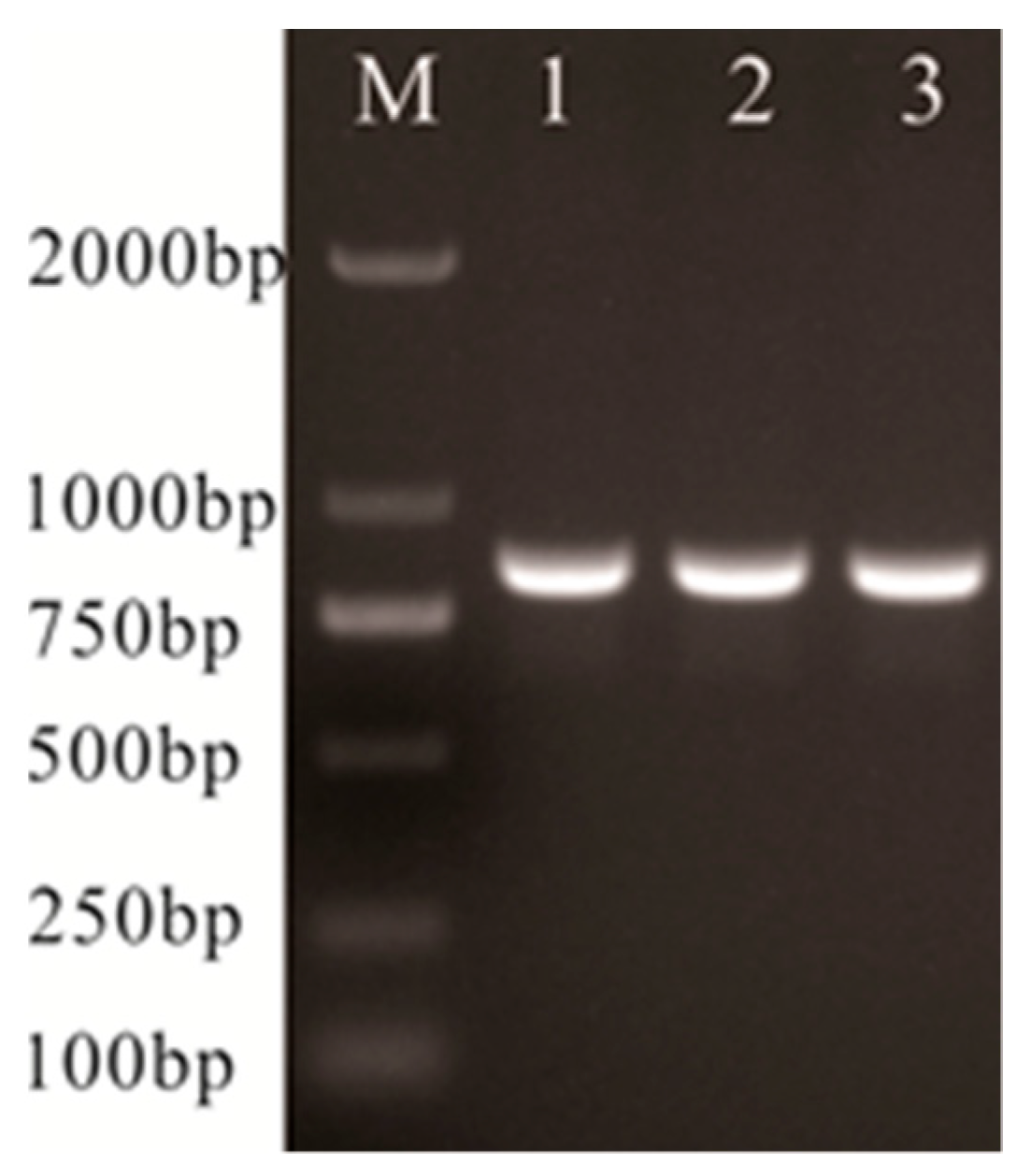
2.6. Lactate Oxidase-Encoding (lctO) and 16S rRNA Gene Sequence Analysis

The genomes of isolates HG-2021-1, HG-2021-3, and S. iniae ATCC 28179 were extracted, and PCR amplification of the lctO and 16S rRNA genes was performed using the primers in Table 1 in 50 µL PCR reaction mixture, containing 1 µL of each primer (10 µM), 2 µL template, 21 µL H2O, and 25 µL Premix Taq™ (TaKaRa, Dalian, China), in the C1000 ™ Thermal Cycler (Bio-Rad, Hercules, CA, USA), with an initial denaturation cycle of 95 °C for 5 min, then 35 cycles of 95 °C for 30 s, 55 °C for 30 s, 72 °C for 90 s, and then a final extension of 72 °C for 10 min. After being purified, the amplified products were sequenced by Sangon Biotech (Shanghai, China). The sequencing results of 16S rRNA genes were aligned and compared for homology with gene fragments in the National Center for Biotechnology Information (NCBI) GenBank using Basic Local Alignment Search Tool (BLAST) (http://www.ncbi.nlm.nih.gov/blast/ accessed on 3 February 2024). Multiple alignment of 16S rRNA sequences from related Streptococcus-type strains and isolates available on GenBank, construction of a phylogenetic tree by the neighbor-joining method, and 1000-replicate bootstrap analysis for the evaluation of phylogenetic tree topology were carried out with MEGA version 4.1 software [16,17]. Evolutionary distances were calculated using the Kimura two-parameter model [18].

2.7. Multilocus Sequence Typing (MLST)

Eight loci or housekeeping genes, including dnaN, rnhC, yfhQ, recD2, mutM, mutX, mutL, and mutS, were selected for MLST analysis using a previously published method [12,19]. By using the genomes of HG-2021-1 and HG-2021-3 as templates, the eight genes were amplified by PCR using specific primers (Table 1), and the amplified products were sequenced by Sangon Biotech (Shanghai). The gene sequences were analyzed using an online comparison tool (https://pubmlst.org/bigsdb?db=pubmlst_siniae_seqdef accessed on 1 April 2024) to determine the sequence numbers of the dnaN, rnhC, yfhQ, recD2, mutM, mutX, mutL, and mutS genes, and the corresponding sequence type (ST).

2.8. PCR Tests of Virulence Genes

By using the genome of HG-2021-1, HG-2021-3, and S. iniae ATCC 28179 as templates, PCR amplification was performed on C5a peptidase (scpI), SiM protein (simA), phosphoglucomutase (pgmA), capsular polysaccharide (cpsD), streptolysin S-associated protein (sagA), polysaccharide deacetylase (pdi), and CAMP factor (cfi) genes according to previous methods [13,14].

2.9. Artificial Infection

Strains HG-2021-1 and HG-2021-3 were selected for the infection test in which 270 healthy A. rivulatus (22.5 ± 3.6 g) were acclimated in the laboratory for 2 weeks. They were then arbitrarily divided into nine groups (n = 30) and placed in 45 L aquaria containing 35 L dechlorinated water. Water temperature, pH, dissolved oxygen, oxygen saturation value, and electric conductivity were 27–28 °C, 7.6–7.8, 6.1–8.3 mg/L, 76.3–103.9%, and 175 μS/cm–204 μS/cm, respectively. Group 1 was injected intraperitoneally with 0.1 mL PBS (pH 7.4) as the control group. Groups 2–5 were injected with 0.1 mL of 3.3 × 10⁸, 107, 106, and 105 CFU/mL of a tenfold serial dilution of the HG-2021-1 suspension, respectively, and groups 6–9 were injected with the same volume and the same concentration of the HG-2021-3 suspension. The test fish were continuously observed for 14 days; any morbid fish were removed quickly and used for re-isolation of the pathogens. The mortality rate was determined and LD50 was calculated. Furthermore, spleens and kidneys removed from three dying fish infected by different strains were used for pathological evaluation.

2.10. Antimicrobial Susceptibility Testing

The antimicrobial susceptibility patterns of the HG-2021-1 and HG-2021-3 isolates were tested using a previously published method [20]. Briefly, 0.1 mL of a 108 CFU/mL HG-2021-1 and HG-2021-3 suspension was plated onto Mueller Hinton agar supplemented with 5% fetal bovine serum (FBS). The plates were left for 5 min, and then drug-sensitive test papers including ampicillin, chloromycetin, streptomycin, norfloxacin, kanamycin, doxycycline, enoxacin, gentamycin, ciprofloxacin, erythromycin, roxithromycin, tetracycline, florfenicol, vancomycin, and tobramycin purchased from Hangzhou microbial reagent Co., Ltd. (Hangzhou, China) were added. The plates were then cultured for 48 h at 28 °C. The sensitivity of HG-2021-1 and HG-2021-3 to the test drugs was determined based on the diameter of the inhibition zone on each plate. According to the instructions provided by the manufacturer, the results were classified as sensitive (S), intermediate sensitive (I), and resistant (R).

3. Results

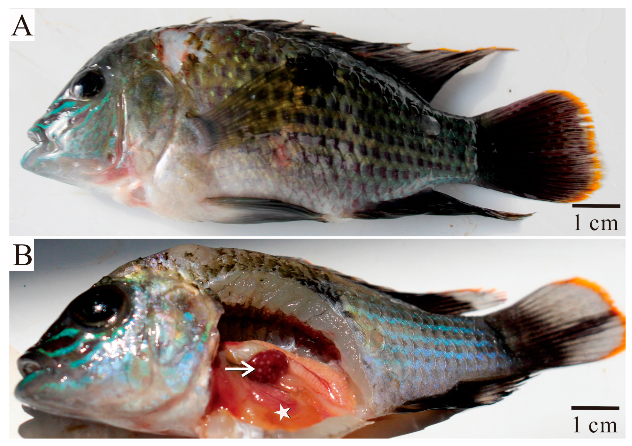
3.1. Clinical Signs and Pathological Features

Diseased fish showed a loss of scales, skin ulceration, and petechial hemorrhages (Figure 1A). Upon dissection, a small amount of fluid accumulation in the coelomic cavity and transparent intestinal walls were observed. The most notable feature was the presence of numerous nodules in the spleen (Figure 1B). Pathological examination revealed blood cell congestion in the diseased fish liver, accompanied by hepatocyte vacuolation, swelling, and necrosis (Figure 2B). Oval nodules were found in the spleen and head kidney, with a higher quantity in the spleen, some of which were interconnected (Figure 2D,E,H). The spleen exhibited a decreased red blood cell count and extensive cell necrosis (Figure 2D,E). Many macrophages were observed in the outer layer of the nodules in the head kidney (Figure 2H). Also, in the kidneys, granular degeneration and necrosis of the renal tubular epithelial cells were observed (Figure 2G). No pathological changes were found in the heart. Furthermore, small nodules were found in the spleen of the fish infected artificially (Figure 2I).

3.2. Specific Pathogen Detection in Diseased Fish and Isolation of Pathogenic Bacteria

Except for a few triehodinids in the gill filaments and mucus of the body surface, no other parasites were observed on other parts of the body with either the naked eye or a light microscope. In addition, no virus particles were observed in the gills, spleen, and kidney tissues during TEM.
PCR was used to detect the presence of Mycobacterium spp., Renibacterium salmoninarum, Rickettsia spp., Nocardia seriolae, and Francisella spp., but the results were negative (results not shown). No bacterium grew on TCBS or 7H10 plates; however, numerous colonies including two different colony morphologies were observed on BHI agar plates, both exhibiting gray circular colonies of varying sizes. One kind of colony was larger, with a diameter of 0.6–0.8 mm, whereas the other was smaller, with a diameter of 0.2–0.3 mm. These colonies were purified and named HG-2021-1 and HG-2021-3, respectively. HG-2021-1 exhibited uniform turbid growth, whereas HG-2021-3 exhibited sedimentary growth with transparent liquid and a white precipitate at the bottom (Figure 3).

3.3. Identification of Isolated Strains
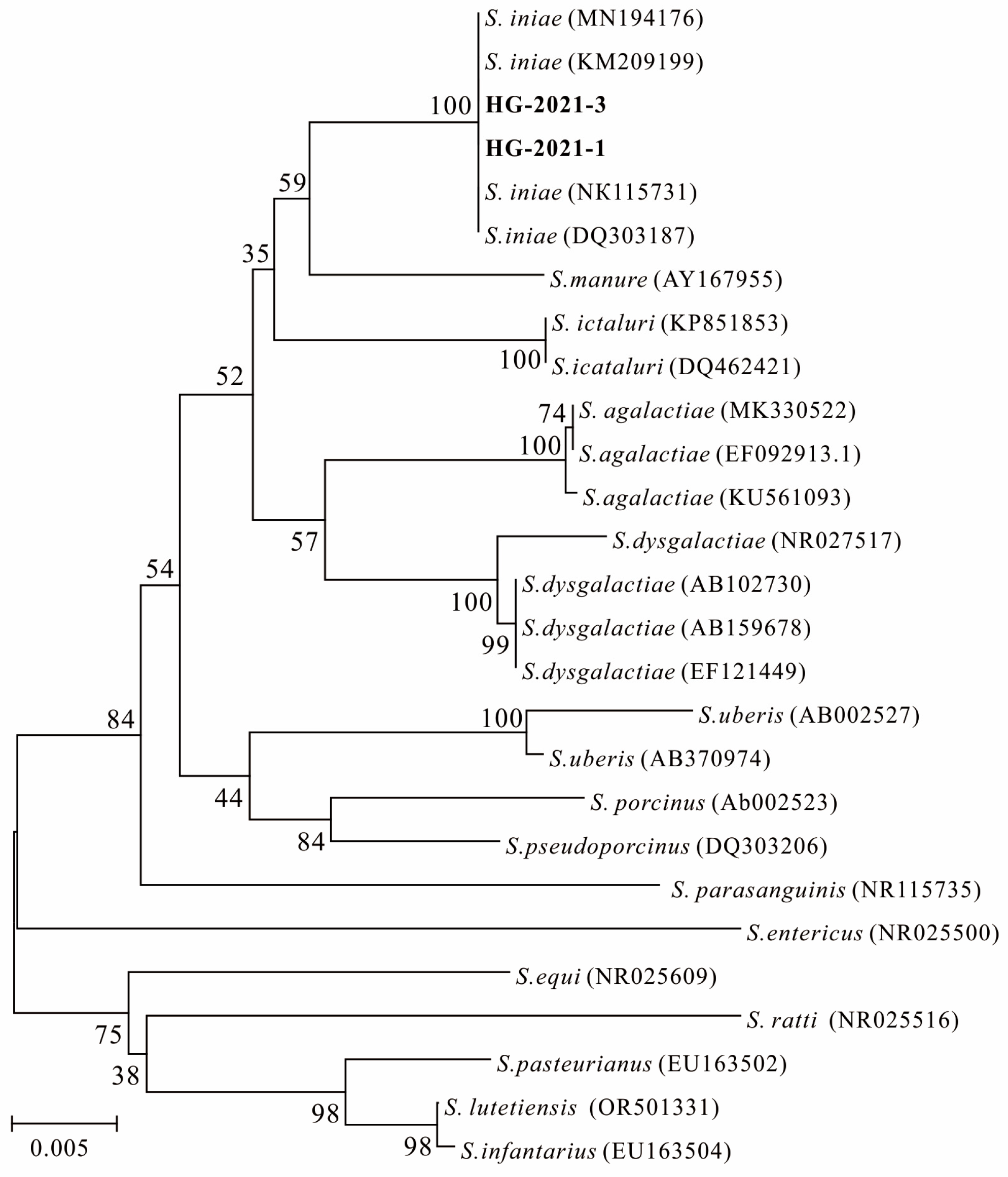
The physiological and biochemical characteristics of the colonies are summarized in Table 2. Both colonies showed Gram-positive cocci arranged in chains or pairs, with characteristics including no motility and α-type hemolysis. The strains were also capable of utilizing glucose, trehalose, maltose, and sucrose, but not mannitol or sorbitol. Furthermore, they showed positive results in methyl red tests, arginine dihydrolase, and esculin hydrolysis. Furthermore, the results showed little difference between strains HG-2021-1 and HG-2021-3. Strain HG-2021-1 showed positive results in phosphatase and negative results in amygdalin, xylose, and raffinose tests, while strain HG-2021-3 was the opposite (Table 2).
PCR amplification of partial lctO gene sequences of HG-2021-1, HG-2021-3, and Streptococcus iniae ATCC 28179 yielded fragments of 870 bp (Figure 4). Sequence assays showed that they displayed the same nucleotide sequence. PCR amplification of partial 16S rRNA gene sequences of HG-2021-1 (GenBank no. PP492817) and HG-2021-3 (GenBank no. PP495151) yielded fragments of ~1500 bp. Alignment analysis using the NCBI database showed high homology (99.7%) with S. iniae ATCC 29178 (GenBank no. NR115731). Phylogenetic analysis revealed that the 16S rRNA gene sequences of HG-2021-1 and HG-2021-3 also clustered with S. iniae (Figure 5). Based on the result of lctO gene detection, 16S rRNA gene sequence analysis, and physiological and biochemical reactions, both HG-2021-1 and HG-2021-3 were identified as S. iniae.

3.4. MLST

MLST analysis of the HG-2021-1 strain suggested that the corresponding sequence numbers of the dnaN, rnhC, yfhQ, recD2, mutM, mutX, mutL, and mutS genes were 1, 1, 1, 1, 1, 1, 1, and 1, respectively, with the corresponding sequence type (ST) being 4. Similarly, the sequence numbers of the eight genes in the HG-2021-3 strain were 4, 4, 2, 2, 3, 4, 2, and 3, respectively, with the corresponding sequence type (ST) being 1.

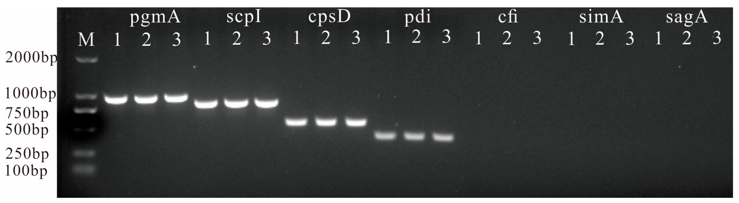
3.5. Detection of Virulence-Related Genes

Using HG-2021-1, HG-2021-3, and Streptococcus iniae ATCC 28179 as templates, PCR amplification was performed on the virus genes scpI, simA, pgmA, cpsD, sagA, pdi, and cfi. The amplification results showed that target bands of corresponding sizes were obtained for pgmA, scpI, cpsD, and pdi. The target fragment was not obtained for cfi, simA, or saga (Figure 6).

3.6. Artificial Infection

Two days after artificial infection, fish in the experimental group exhibited symptoms of anorexia and buoyancy disorders, whereas the control group showed no such signs. From the third day after artificial infection, fish began to die. After 14 days, the cumulative mortality of A. rivulatus in groups 2–5 was 100%, 93.3%, 60.0%, and 23.3%, respectively, and 90%, 70%, 43.3%, and 13.3% in groups 6–9, respectively. The LD50 of strain HG-2021-1 and HG-2021-3 was 1.8 × 10⁶ CFU/mL and 6.6 × 10⁶ CFU/mL, respectively (Table 3). The main symptoms observed in the dead fish included abdominal distension, the presence of ascites, and hemorrhagic spots on the body surface, similar to those observed in naturally diseased fish. The bacterial strains isolated from the affected fish internal organs exhibited the same physiological and biochemical characteristics, as well as 16S rRNA gene sequences, as HG-2021-1 and HG-2021-3.

3.7. Antibiotic Susceptibility Test

The antibiotic susceptibility test demonstrated that strain HG-2021-1 was consistent with HG-2021-3. Both were sensitive to streptomycin, ampicillin, florfenicol, roxithromycin, and erythromycin, but resistant to neomycin, polymyxin B, doxycycline, tobramycin, levofloxacin, norfloxacin, ciprofloxacin, vancomycin, and gentamycin (Table 4).

4. Discussion

In recent years, the formation of granulomas in visceral tissues has been reported in various marine and freshwater fish, such as turbot Scophthalmus maximus, half-smooth tongue sole Cynoglossus semilaevis Günther, seahorse Hippocampus erectus, and largemouth bass Micropterus salmoides [21,22,23,24,25]. Parasites, viruses, and bacteria have been identified as potential causes of nodules of internal organs in fish [26,27]. In our study, no parasites or viral particles were observed in the tissues of diseased fish exhibiting nodular symptoms, ruling out the possibility of parasitic or viral infections. It has been reported that bacteria such as Mycobacterium spp., Nocardia seriolae, Renibacterium salmoninarum, Rickettsia spp., Francisella spp., Photobacterium damselae, Aeromonas schubertii, and Aeromonas salmonicida can cause nodules of internal organs in fish [21,22,23,24,25,28,29,30,31]. PCR detection of the first five pathogens was used in the current study, but yielded negative results. These results ruled out the possibility of the diseased fish being infected by Mycobacterium spp., R. salmoninarum, Nocardia seriolae, Francisella spp., or Rickettsia spp.
Two different bacterial strains with distinct colony sizes were isolated from the internal organs of the affected fish using a BHI agar medium. Artificial infection experiments revealed that both strains were pathogenic to A. rivulatus, with experimentally infected fish exhibiting symptoms similar to those observed in naturally infected fish, indicating that they were the causative agents of the disease impacting A. rivulatus. Upon identification, both strains were identified as S. iniae.
Although S. iniae is phenotypically well characterized, laboratory identification can be difficult, especially when using commercial identification systems, because no currently available commercial system includes S. iniae in its database [10]. In this paper, lctO and 16S rRNA gene sequence analyses were performed. Based on these experimental results, strains HG-2021-1 and HG-2021-3 were identified as S. iniae.
Strains HG-2021-1 and HG-2021-3 exhibited significant differences in physiological and biochemical characteristics. This suggests that they may belong to different genotypes, and the hypothesis has been confirmed by MLST. Additionally, HG-2021-3 exhibited a higher LD50 in fish, indicating weaker virulence compared with HG-2021-1. This finding is consistent with that of Kim et al., who reported that two genotypes of S. iniae showed different virulence [32].
S. iniae is a major pathogen in many fish species, including freshwater fish, such as sturgeon Acipenser transmontanus L., tilapia Oreochromis spp., and channel catfish Ictalurus punctatus, and marine fish, as such red porgy Pagrus pagrus L. and small yellow croaker Larimichthys polyactis, causing severe economic losses [33,34,35,36,37,38]. We also isolated S. iniae from affected silver scat Selenotoca multifasciata in nearby fish farms, indicating that S. iniae is a common pathogen in the aquaculture of the region [39]. However, we did not observe nodular symptoms in the visceral tissues of silver scat. This might indicate that the formation of visceral nodules is not only pathogen- but also fish species-dependent. This finding is consistent with that of Luo et al., who reported that artificial infection of four different species of fish with Nocardia seriolae resulted in nodules that appeared on day 5 post-infection in Micropterus salmoides and Channa argus, whereas no nodules were observed in Ctenopharyngodon idella or Oreochromis niloticus even after 14 days of infection [40]. The identification of drugs that S. iniae was susceptible to indicates these could be used to treat infected fish. However, the formation of nodules can hinder drug penetration, thereby affecting the efficacy of drug treatment. To improve treatment outcomes and minimize economic losses caused by S. iniae. it is crucial for farmers to enhance their observation of cultured organisms, promptly detect diseases, and administer treatment with sensitive drugs before nodule formation.

5. Conclusions

In conclusion, two genotypes of S. iniae were isolated from diseased A. rivulatus and were believed to be the pathogenic agents; pathologic characteristics were observed, and five sensitive drugs were screened. The results provide a foundation for the development of guidance for the prevention and treatment of diseases affecting A. rivulatus.

Author Contributions

Z.L.: investigation, data curation, writing—review and editing. X.B.: formal analysis, writing—original draft preparation. S.H.: methodology, formal analysis. M.W.: software, literature comparative dialogue. Y.W.: software, literature comparative dialogue. H.S.: writing—review and editing, project administration, and funding acquisition. All authors have read and agreed to the published version of the manuscript.

Funding

This work was supported by the Project for Tianjin Fisheries Innovative Team (ITTFRS2021000, ITTFRS2021000-008), Projects for Chongqing technical innovation and application development (cstc2019jscx-gksbX0147), and the Chongqing Aquatic Science and Technology Innovation Key Project (CQFTIU2024-10).

Institutional Review Board Statement

All animal experiments were approved and conducted in compliance with all experimental practices and standards developed by the Animal Welfare and Research Ethics Committee of Tianjin Agricultural University.

Data Availability Statement

All the data generated or used during the study appear in the submitted article.

Acknowledgments

Thanks are due to the anonymous reviewers who provided detailed comments that helped improve the manuscript.

Conflicts of Interest

The authors declare that there are no conflicts of interest regarding the publication of the work described in this manuscript.

References

  1. Yu, X.J.; Hao, X.J.; Yang, L.K.; Dang, Z.Q. Development monitoring report of Chinese recreational fishery. China Fish. 2022, 12, 35–40. [Google Scholar]
  2. Wu, J.; Gao, Q.; Yang, H.; Yang, G.L. Artificial propagation and seed cultivation technology of green terror cichlid Aequidens rivulatus. Fish. Sci. Technol. Info. 2012, 39, 171–173. [Google Scholar]
  3. Yao, X.L.; Xu, X.L.; Li, H.M.; Zhong, W.H.; Zang, L.; Li, H.; Yang, C.J. Biological characteristics of pathogenic Streptococcus agalactiae isolated from Aequidens rivulatus. Prog. Fish. Sci. 2015, 36, 106–112. [Google Scholar]
  4. Hao, X.B.; Huang, Y.P.; Hu, Q.; Peng, Y.O.; Sun, L.M.; Gao, J.W.; Song, R. Pathogen isolation, identification and drug susceptibility analysis of Aequidens rivulatus hole-in-the-head disease. Curr. Fish. 2021, 6, 60–63. [Google Scholar]
  5. Talaat, A.M.; Reimschuessel, R.; Trucksis, M. Identification of mycobacteria infecting fish to the species level using polymerase chain reaction and restriction enzyme analysis. Vet. Microbiol. 1997, 58, 229–237. [Google Scholar] [CrossRef]
  6. Chase, D.M.; Pascho, R.J. Development of a nested polymerase chain reaction for amplification of a sequence of the p57 gene of Renibacterium salmoninarum that provides a highly sensitive method for detection of the bacterium in salmonid kidney. Dis. Aquat. Org. 1998, 3, 223–229. [Google Scholar] [CrossRef]
  7. Lei, Y.; Zhang, H.J.; Wang, J.; Zhang, W.E.; Tang, S.L.; Xiao, Y.; Wang, X.P. Development and Primary Application of a PCR Assay for Detection of Piscirickettsia-like Organisms. J. Guangdong Ocean Univ. 2015, 35, 30–34. [Google Scholar]
  8. Jiang, Y.Y.; Li, A.X. Establishment of a specific PCR assay to detect Nocardia seriolae. S. China Fish. Sci. 2011, 7, 47–51. [Google Scholar]
  9. Forsman, M.; Sandstrom, G.; Sjostedt, A. Analysis of 16s ribosomal DNA sequences of Francisella strains and utilization for determination of the phylogeny of the genus and for identification of strains by PCR. Int. J. Syst. Bacteriol. 1994, 44, 38–46. [Google Scholar] [CrossRef]
  10. Mata, A.I.; Blanco, M.M.; Dominguez, L.; Fernandez-Garayzabal, J.F.; Gibello, A. Development of a PCR assay for Streptococcus iniae based on the lactate oxidase (lctO) gene with potential diagnostic value. Vet. Microbial. 2004, 101, 109–116. [Google Scholar] [CrossRef]
  11. Lane, D.J. 16S/23S rRNA sequencing. In Nucleic Acid Techniques in Bacterial Systematics; Stackebrandt, E., Goodfellow, M., Eds.; Wiley: New York, NY, USA, 1991. [Google Scholar]
  12. Irion, S.; Rudenko, O.; Sweet, M.; Chabanet, P.; Barnes, A.C.; Tortosa, P.; Séré, M.G. Molecular investigation of recurrent Streptococcus iniae epizootics affecting coral reef fish on an oceanic island suggests at least two distinct emergence events. Front. Microbial. 2021, 12, 749734. [Google Scholar] [CrossRef]
  13. Xiong, X.Y.; Huang, G.Q.; Wang, Z.C.; Wen, X. Molecular typing, antibiogram type and detection of virulence genes of Stereptococcus iniae strains isolated from golden pompano (Tranchinotus ovatus) in Guangxi province. J. Fish. China 2018, 42, 586–595. [Google Scholar]
  14. Baums, C.G.; Hermeyer, K.; Leimbach, S.; Adamekk, M.; Czerny, C.P.; Horstgen-Schwark, G.; Valentin-Weigand, P.; Baumgartner, W.; Steinhagen, D. Establishment of a Model of Streptococcus iniae Meningoencephalitis in Nile Tilapia (Oreochromis niloticus). J. Comp. Path. 2013, 149, 94–102. [Google Scholar] [CrossRef] [PubMed]
  15. Dong, X.Z.; Cai, M.Y. Manual of Familiar Bacterium Identification; Science Press: Beijing, China, 2001. [Google Scholar]
  16. Saitou, N.; Nei, M. The neighbor-joining method: A new method for reconstructing phylogenetic trees. Mol. Biol. Evol. 1987, 4, 406–425. [Google Scholar] [PubMed]
  17. Tamura, K.; Battistuzzi, F.U.; Billing-Ross, P.; Murillo, O.; Filipski, A.; Kumar, S. Estimating divergence times in large molecular phylogenies. Proc. Natl. Acad. Sci. USA 2012, 109, 19333–19338. [Google Scholar] [CrossRef] [PubMed]
  18. Tamura, K.; Nei, M.; Kumar, S. Prospects for inferring very large phylogenies by using the neighbor-joining method. Proc. Natl. Acad. Sci. USA 2004, 101, 11030–11035. [Google Scholar] [CrossRef] [PubMed]
  19. Jolley, K.A.; James, E.B.; Maiden, M.C.J. Open-access bacterial population genomics: BIGSdb software, the PubMLST.org website and their applications [version 1; peer review: 2 approved]. Wellcome. Open. Res. 2018, 3, 124. [Google Scholar] [CrossRef]
  20. Bauer, A.W.; Kirby, W.M.; Sherris, J.C.; Turck, M. Antibiotic susceptibility testing by a standardized single disk method. Am. J. Clin. Pathol. 1966, 45, 493–496. [Google Scholar] [CrossRef] [PubMed]
  21. Luo, Z.; Zhang, Z.G.; Hao, S.; Bai, X.H.; Gao, Q.; Feng, S.M. Isolation and Identification of the Pathogen Causing granulomas in internal organs of turbot Scophthalmus maximus. J. Fish. Sci. China 2019, 26, 559–568. [Google Scholar] [CrossRef]
  22. Luo, Z.; Li, J.; Zhang, Z.G.; Hao, S.; Bai, X.H.; You, H.Z.; Mo, Z.L.; Feng, S.M. Mycobacterium marinum is the causative agent of splenic and renal granulomasin half-smooth tongue sole (Cynoglossus semilaevis Günther) in China. Aquaculture 2018, 490, 203–207. [Google Scholar] [CrossRef]
  23. Luo, Z.; Hao, S.; Bai, X.H.; Zhang, Z.G.; Ma, Y.F.; Feng, S.M.; Zhang, D.F. Isolation, identification, and genomic analysis of Mycobacterium ulcerans ecovar Liflandii from the farmed Chinese tongue sole, Cynoglossus semilaevis Günther. Aquaculture 2022, 548, 737614. [Google Scholar] [CrossRef]
  24. Bai, X.H.; Hao, S.; Fu, J.P.; Sun, H.C.; Luo, Z. Identification of Mycobacterium chelonae from Lined Seahorse Hippocampus erectus and Histopathological Analysis. Fishes 2023, 8, 225. [Google Scholar] [CrossRef]
  25. He, S.Y.; Wei, W.Y.; Liu, T.; Yang, Q.; Xie, H.; He, Q.Y.; Wang, K.Y. Isolation, identification and histopathological study on lethal sarcoidosis of Micropterus salmoides. J. Fish. China 2020, 44, 253–265. [Google Scholar]
  26. Zhen, C.; Pan, L.D. research progress on pathogen and histopathology of visceral sarcoidosis in fish. Sci. Fish. Farms. 2016, 7, 54–55. [Google Scholar]
  27. Xiao, P.; Jiang, M.; Liu, Y.; Sun, M.; Zhang, L.; Jie, L.; Li, G.; Mo, Z. Splenic necrosis signs and pathogen detection in cultured half-smooth tongue sole, Cynoglossus semilaevis Günther. J. Fish Dis. 2015, 38, 103–106. [Google Scholar] [CrossRef] [PubMed]
  28. Ge, M.X.; Fang, H. Renibacterium Salmoninarum and bacterial kidney disease of salmon (summary). J. Hebei. Vocat. Technical. Teach. Coll. 2003, 17, 51–55. [Google Scholar]
  29. Liu, C.; Chang, O.Q.; Zhang, D.F.; Li, K.B.; Wang, F.; Lin, M.H.; Shi, C.B.; Jiang, L.; Wang, Q.; Bergmann, S.M. Aeromonas schubertii as a cause of multi-organ necrosis in internal organs of Nile tilapia, Oreochromis niloticus. J. Fish Dis. 2012, 35, 335–342. [Google Scholar] [CrossRef]
  30. Qiu, Y.Y.; Zheng, L.; Mao, Z.J.; Chen, X.X. Isolation and identification of the causative agent and histopathology observation of white-spots disease in internal organs of Larimichthys crocea. Microbiol. China 2012, 39, 361–370. [Google Scholar]
  31. He, X.X.; Liu, K.M.; Wang, X.Y.; Hao, S.; Jia, L.; Shao, P.; Luo, Z. Isolation, identification and histopathology of pathogen of intussusception disease in half-smooth tongue sole Cynoglossus semilaevis. J. Dalian Ocean Univ. 2022, 37, 19–25. [Google Scholar]
  32. Kim, M.S.; Jin, J.W.; Han, H.J.; Choi, H.S.; Hong, S.; Cho, J.Y. Genotype and virulence of Streptococcus iniae isolated from diseased olive flounder Paralichthys olivaceus in Korea. Fish Sci. 2014, 80, 1277–1284. [Google Scholar] [CrossRef]
  33. Pierezan, F.; Shahin, K.; Hechman, T.l.; Ang, J.; Byrne, B.A.; Soto, E. Outbreaks of severe myositis in cultured white sturgeon (Acipenser transmontanus L.) associated with Streptococcus iniae. J. Fish Dis. 2020, 43, 485–490. [Google Scholar] [CrossRef] [PubMed]
  34. Chen, C.Y.; Chao, C.B.; Bowser, P.R. Comparative histopathology of Streptococcus iniae and Streptococcus agalactiae-infected tilapia. Bull. Eur. Ass. Fish Pathol. 2007, 27, 2–9. [Google Scholar]
  35. Yu, X.L.; Chen, M.; Li, C.; Li, P.P.; Lei, A.Y.; Zhong, M.N.; Liang, W.W. Channel catfish Ictalurus punctatus outbreak infected by bacterium Streptococcus iniae. J. Dalian Fish. Univ. 2008, 23, 185–191. [Google Scholar]
  36. Elaamri, F. First report of Streptococcus iniae in red porgy (Pagrus pagrus L.). J. Fish Dis. 2010, 33, 901–905. [Google Scholar] [CrossRef]
  37. Xu, W.; Shi, H.; Wang, W.; Zhang, D.Y.; Xu, W.J.; Chai, X.J. Isolation and identification of Streptococcus iniae in Larimichthys polyactis. Chin. J. Prev. Vet. Med. 2022, 44, 725–730. [Google Scholar]
  38. Pretto-Giordano, L.G.; Scarpassa, J.A.; Barbosa, A.R.; Altrão, C.S.; Ribeiro, C.G.G.; Vilas-Boas, L.A. Streptococcus iniae: An unusual important pathogen fish in Brazil. J. Aquac. Res. Dev. 2015, 6, 9. [Google Scholar] [CrossRef]
  39. Luo, Z.; Xu, J.; Han, J.G.; Xu, Y.X.; Ma, W.T.; Feng, S.M. Isolation and identification of Pathogenetic Streptococcus iniae from Selenotoca multifasciata. J. Huazhong Agric. Uni. 2012, 31, 95–99. [Google Scholar]
  40. Luo, Y.; Deng, Y.T.; Zhao, F.; Tan, A.P.; Zhang, M.C.; Jiang, L. Comparative on characteristics and pathogenicity of Nocardia seriolae isoalted from 9 fishes. Microbiol. China 2021, 48, 2733–2749. [Google Scholar]
Figure 1. Aequidens rivulatus showing typical symptoms of disease. (A) A loss of scales, skin ulceration, and slight bleeding; (B) abundant white granulomas in the spleen (→), and transparent intestinal walls (★).
Fishes 09 00140 g001
Figure 2. Histological sections of the organs of diseased Aequidens rivulatus. (A,C,F) Liver, spleen, and kidney of disease-free A. rivulatus for comparison. (B) Liver, showing hepatocyte vacuolation (→), swelling (Fishes 09 00140 i001), necrosis (Fishes 09 00140 i002), and accumulation of red blood cells (★). (D) Spleen, showing oval nodules interconnected by fibroblasts (Fishes 09 00140 i001). (E) Spleen, showing granulomas characterized by necrotic centers surrounded by a thin fibrous capsule (Fishes 09 00140 i002), a decreased number of red blood cells, and extensive cell necrosis (★). (G) Kidney, showing renal tubular epithelial cell granular degeneration (Fishes 09 00140 i001) and necrosis (★). (H) Anterior kidney, showing extensive cell necrosis (★), and many macrophages in the outer layer of the granuloma. (I) Spleen from the fish artificially infected with strain HG-2021-1, showing small circular nodules (★).
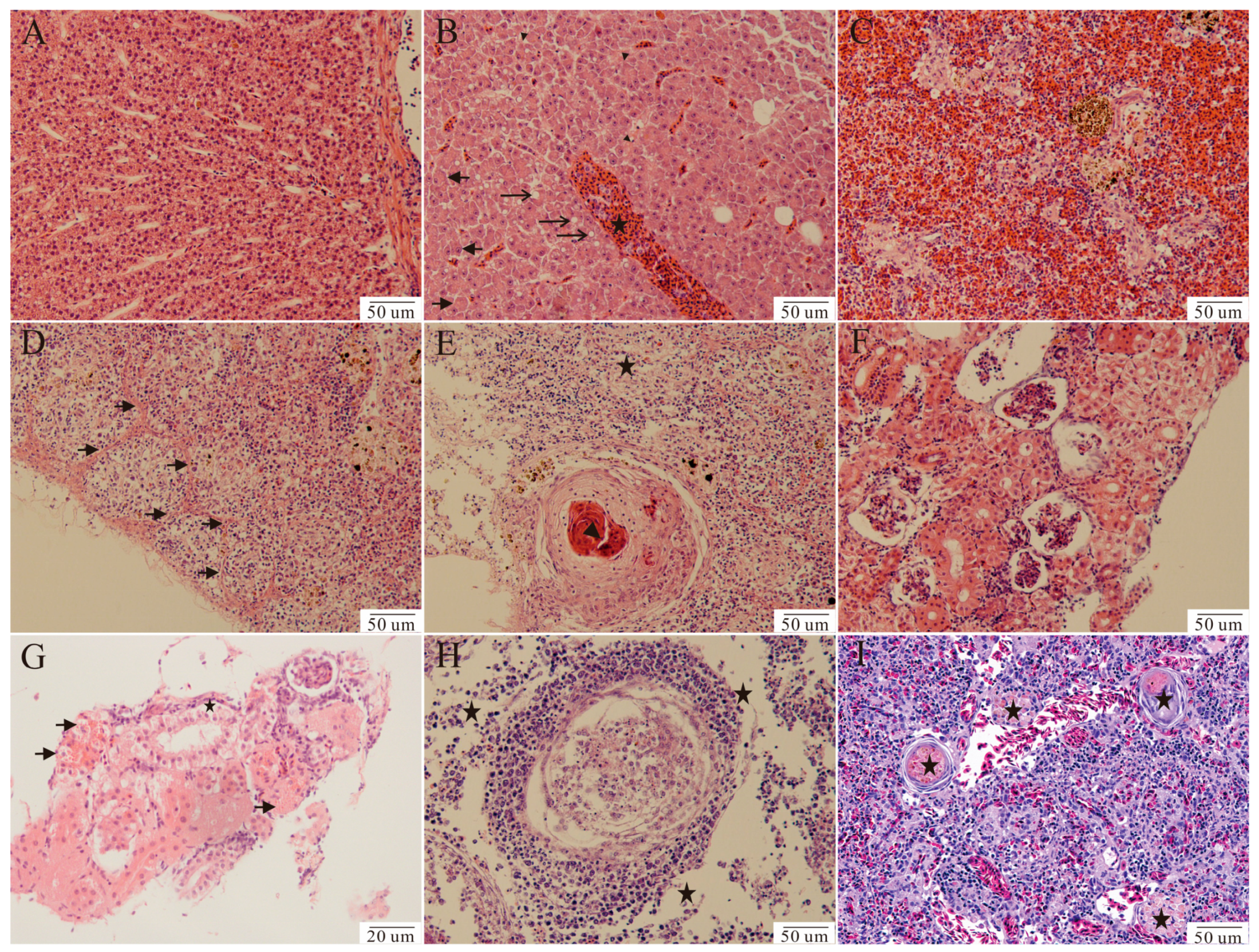
Fishes 09 00140 g002
Figure 3. The morphology of strains HG-2021-1 and HG-2021-3 cultured in the liquid culture medium BHI. (A) Strain HG-2021-1, showing uniform turbid growth. (B) Strain HG-2021-3, showing sedimentary growth with transparent liquid and a white precipitate at the bottom.
Fishes 09 00140 g003
Figure 4. PCR tests of lctO genes. M: DL2000 DNA marker; lanes 1, 2, and 3 indicate tests of isolates HG-2021-1, HG-2021-3, and Streptococcus iniae ATCC 28179.
Fishes 09 00140 g004
Figure 5. Phylogenetic tree constructed for strains HG-2021-1 and HG-2021-3 based on the 16S rRNA gene sequences of related Streptococcus spp. GenBank accession numbers are given in parentheses.
Fishes 09 00140 g005
Figure 6. PCR tests of virulence genes scpI, simA, pgmA, cpsD, sagA, pdi, and cfi. M: DL2000 DNA markers; lanes 1, 2, and 3 indicate isolates Streptococcus iniae ATCC 28179, HG-2021-1, and HG-2021-3.
Fishes 09 00140 g006
Table 1. PCR primers used in this study.
PrimerSequence (5′–3′)Length (bp)Application
T39GCGAACGGGTGAGTAACACG936Test Mycobacteria spp. by nested PCR [5]
T13TGCACACAGGCCACAAGGGA
preT43AATGGGCGCAAGCCTGATG300–312
T531ACCGCTACACCAGGAAT
P3AGCTTCGCAAGGTGAAGGG383Test R. salmoninarum by nested PCR [6]
M21GCAACAGGTTTATTTGCCGGG
P4ATTCTTCCACTTCAACAGTACAAGG320
M38CATTATCGTTACACCCGAAACC
PS-FCTAGGAGATGAGCCCGCGTTG390Test Piscirickettsia by PCR [7]
PS-RATTTCA CATCCAACTTAATCT
Noc-fCACCTACGAAAATCCCATTTGGT156Test Nocardia seriolae by PCR [8]
Noc-rCATCGGATTGGATTCAAGGACCTTGA
F5CCTTTTTGAGTTTCGCTCC1133Test Francisella spp. by PCR [9] (FORSMAN)
F11TACCAGTTGGAAACGACTGT
lctO-FAAGGGGAAATCGCAAGTGCC870Clone lctO gene [10]
lctO-RATATCTGATTGGGCCGTCTAA
27FAGAGTTTGATCCTGGCTCAG1480–1494Clone 16S rRNA gene [11]
1492RTACGGCTACCTTGTTACGCTT
dnaN-FGCACATGTTAATTCGCCAGAGG404Multilocus sequence typing [12]
dnaN-RCAGCACCAACTCTGATAATTTTCCA
mutL-FCCAACCAAGCAGGAAGTTCG545
mutL-RCGTTCTTGAGCTGCGTGTTG
mutM-FCAGAGTAGATGGTTTGACCC410
mutM-RTGCCCTGTATGATGCCTATC
mutS-FTTTAACTGGCGCATCCCCAT448
mutS-RTGGATCTTGCAACAGGTGAGT
mutX-FTGGCCATTGGTTTCATCAAGG547
mutX-RCGTAATCCCCTTCCCACGTT
recD2-FAGGGCTTCCTAGTGCTACCA563
recD2-RACTCGCTTTGCCCATCAAGA
rnhC-FGGAATCGCTGTTGTTGCAAGT582
rnhC-RTTGAGTGTTTGCGAAGTGGC
yfhQ-FAGGCCAGGTGATTTCAACCA511
yfhQ-RCAGGAGAAACCCAGGCCATT
pgmA-F AGACGGGGTCACAGACTACAT 949 Test virulence genes by PCR [13,14]
pgmA-R AGGAGCACTTTGACGGAATT
cfi-F GTGCCTCAACATCAAACA 328
cfi-RTAGCAAATCCCATATCAA
scpI-F GCAACGGGTTGTCAAAAATC 822
scpI-RGAGCAAAAGGAGTTGCTTGG
simA-FAATTCGCTCAGCAGGTCTTG 994
simA-R AACCATAACCGCGATAGCAC
pdi-F TTTCGACGACAGCATGATTG 381
pdi-RGCTAGCAAGGCCTTCATTTG
sagA-F AGGAGGTAAGCGTTATGTTAC 190
sagA-R AAGAAGTGAATTACTTTGG
cpsD-F TGGTGAAGGAAAGTCAACCAC 534
cpsD-R TCTCCGTAGGAACCGTAAGC
Table 2. Physiological and biochemical characteristics of isolated strains.
ItemsHG-2021-1 HG-2021-3ItemsHG-2021-1 HG-2021-3
Gram stain++Vogus–Proskauer
10 °C growthCatalase
45 °C growthMethyl red reaction++
Motility Esculin hydrolysis++
Mannitol Sucrose++
Glucose++6.5% NaCl growth
SorbitolGlucose gas production
OxidaseHemolysisαα
UreaseArginine dihydrolase++
D-trehalose++D-maltose++
a-galactosidaseβ-glucuronidase++
Phosphatase+D-raffinose+
D-amygdalin+D-xylose+
Note: “+” represents positive reaction results; “−” represents negative reaction results; “α” represents α-type hemolysis.
Table 3. Experimental infection with isolates HG-2021-1 and HG-2021-3 in Aequidens rivulatus.
IsolatesBacterial Concentration
(CFU/mL)
Amount of Death Post-Infection in Different DaysCumulative Mortality
(%)
LC50
(CFU/mL)
1234567891011121314
HG-2021-13.3 × 1080058121826303030303030301001.8 × 106
3.3 × 10700361118242728282828282893.3
3.3 × 1060016812151617181818181860.0
3.3 × 1050000124667777723.3
HG-2021-33.1 × 1080045814232727272727272790.06.6 × 106
3.1 × 107003377111214212121212170.0
3.1 × 1060012478810111313131343.3
3.1 × 1050000122333444413.3
PBS 0000000000000000
Table 4. Sensitivity of strains HG-2021-1 and HG-2021-3 to 16 different antibiotics.
AntibioticsSensitivityAntibioticsSensitivity
HG-2021-1HG-2021-3HG-2021-1HG-2021-3
TetracyclineIIAmpicillinSS
NeomycinRRNorfloxacinRR
StreptomycinSSCiprofloxacinRR
Polymyxin BRRFlorfenicolSS
DoxycyclineRRVancomycinRR
KanamycinIIRoxithromycinSS
TobramycinRRGentamycinRR
LevofloxacinRRErythromycinSS
Note: “S”: sensitive; “I”: intermediate sensitive; “R”: resistant.
Disclaimer/Publisher’s Note: The statements, opinions and data contained in all publications are solely those of the individual author(s) and contributor(s) and not of MDPI and/or the editor(s). MDPI and/or the editor(s) disclaim responsibility for any injury to people or property resulting from any ideas, methods, instructions or products referred to in the content.

