Article

The Establishment of the Multi-Visual Loop-Mediated Isothermal Amplification Method for the Rapid Detection of Vibrio harveyi, Vibrio parahaemolyticus, and Singapore grouper iridovirus

1
School of Breeding and Multiplication, Sanya Institute of Breeding and Multiplication, Hainan University, Sanya 572025, China
2
Collaborative Innovation Center of Marine Science and Technology, Hainan University, Haikou 570228, China
3
Hainan Provincial Key Laboratory for Tropical Hydrobiology and Biotechnology, School of Marine Biology and Fisheries, Hainan University, Haikou 570228, China
*
Authors to whom correspondence should be addressed.
Fishes 2024, 9(6), 225; https://doi.org/10.3390/fishes9060225
Submission received: 13 May 2024 / Revised: 7 June 2024 / Accepted: 11 June 2024 / Published: 13 June 2024
(This article belongs to the Special Issue Fish Diseases Diagnostics and Prevention in Aquaculture)

Abstract

:
Groupers are valuable economic fish in the southern sea area of China, but the threat of disease is becoming more and more serious. Vibrio harveyi, V. parahaemolyticus, and Singapore grouper iridovirus (SGIV) are three important pathogens that cause disease in groupers, and infection with either a single one or a mix of these pathogens poses a serious threat to the healthy development of grouper culture. To enhance the rapid diagnosis and screening in the early stages, it is necessary to develop rapid detection methods of these pathogens. To simultaneously and rapidly detect the three pathogens, in this study, we utilized the TolC of V. harveyi, DNAJ of V. parahaemolyticus, and RAD2 of SGIV as the target genes and established a triple visual loop-mediated isothermal amplification (LAMP) method. This LAMP method showed a detection time as fast as 30 min and a high sensitivity of 100 fg/μL. Moreover, this method exhibited strong specificity and no cross-reaction with seven types of Vibrio and Staphylococcus aureus, as well as five common viruses in aquatic animals. Then, the LAMP products were enzymically cut, and three characteristic strips were used to identify the pathogen species. The results of the clinical trials demonstrated that the method could accurately and specifically detect V. harveyi, V. parahaemolyticus, and SGIV in grouper tissues. In summary, this study successfully established a triple visual LAMP rapid detection method for V. harveyi, V. parahaemolyticus, and SGIV. The method offers several advantages including simple equipment, easy operation, rapid reaction, high specificity, high sensitivity, and visual results. It is suitable for the early and rapid diagnosis of groupers infected with V. harveyi, V. parahaemolyticus, and SGIV, thereby providing useful technical support for further application in the large-scale disease surveillance of aquaculture animals.
Key Contribution: This study established a rapid detection method based on LAMP technology that can simultaneously detect four critical pathogens in grouper culture. This method is characterized by the simple operation of its equipment, a short detection time, its visual results, and its ability to accurately identify the pathogen species by the products of the enzyme digestion, which is very suitable for large-scale preliminary screening operations in the early stages of farming.

1. Introduction

Groupers, belonging to the Perciformes order, Serranidae family, and Epinephelinae subfamily, are unique and valuable economic fish in the southern sea area of China. In recent years, the scale of grouper farming in China has been continuously expanding, and total production reached 205,000 tons in 2022 [1]. However, the outbreak of various diseases has inflicted substantial losses on juvenile and adult groupers, resulting in an economic loss of up to CNY 1.3 billion for grouper farming in 2022 [1,2]. Singapore grouper iridovirus (SGIV) is one of the major viral pathogens that infect groupers and causes a mortality rate of 60% to 100% in grouper populations. Groupers infected with SGIV commonly exhibit symptoms such as reduced vitality, loss of appetite, darkening of body color, and gill bleeding [3]. In addition, vibriosis is one of the main bacterial diseases in the growth process of groupers, which limits the healthy development of the grouper culture industry [4]. The pathogenic Vibrio bacteria that primarily cause vibriosis include V. harveyi, V. parahaemolyticus, V. alginolyticus, and others. Groupers infected with Vibrio show symptoms such as slow swimming, reduced food intake, decreased body balance, ascites, and redness and ulcerations on the body surface [5,6]. With the increasingly prominent problem of grouper disease, strengthening its early diagnosis and developing multi-target detection can effectively prevent grouper disease outbreaks.
Various molecular biological methods are commonly used in the rapid detection of the pathogens, including the traditional polymerase chain reaction (PCR), real-time quantitative reverse transcription PCR (qRT-PCR), as well as a range of temperature-independent isothermal amplification techniques [7]. Isothermal amplification has similar sensitivity and specificity to qRT-PCR, but has advantages in speed, cost, and portability, making it ideal for field testing in resource-limited environments. Loop-mediated isothermal amplification (LAMP) has gained significant popularity due to its numerous advantages such as high specificity, high sensitivity, minimal equipment requirements, and rapid response [8]. Notably, LAMP technology offers the visualization of the amplification products, allowing for direct observation of color changes using dyes such as calcein, SYBR Green I, and Hydroxynaphthol blue (HNB), thereby avoiding the phenomenon of being time-consuming as found with electrophoretic detection, turbidity meters, and other methods and breaking through the limitations of occasions for application [9,10]. Consequently, LAMP demonstrates significant potential for the rapid detection of pathogens.
In recent years, researchers have observed a growing trend of fish harboring multiple pathogens simultaneously. Therefore, a rapid multi-target pathogen detection method has been of wide concern. Expanding upon the foundations of LAMP technology, multiplex LAMP (multi-LAMP) incorporates multiple sets of primers targeting different genes, thus enabling the amplification of multiple target genes within a single reaction system [11,12]. This innovative technique not only retains the rapid amplification, simplicity, high sensitivity, specificity, and product visualization benefits of LAMP but also facilitates the simultaneous detection of multiple pathogens. As a result, it considerably reduces the time required for the detection of individual pathogens, making it a promising and efficient multi-target rapid detection technology. At present, multi-LAMP detection technology has been used in the nucleic acid detection of a variety of mammalian diseases. Fan et al. (2019) successfully established a dual LAMP detection method for bovine rotavirus and enterotoxigenic Escherichia coli [13]. The multi-LAMP detection method constructed by Wang et al. (2018) could simultaneously detect Ureaplasma urealyticum, Mycoplasma hominis, and M. genitalium and its sensitivity was determined to be 100 pg/μL for U. urealyticum, 100 pg/μL for M. hominis, and 1 ng/μL for M. genitalium, surpassing the sensitivity of a traditional PCR by a significant margin [14]. Siddique et al. (2019) established a dual LAMP method that could simultaneously detect two types of Vibrio (V. anguillarum and V. alginolyticus) [15]. However, a multi-LAMP technology that can simultaneously detect three important grouper pathogens (SGIV, V. harveyi, and V. parahaemolyticus) has never been reported.
The TolC gene in V. harveyi, which encodes an iron transport and anti-host immunity protein, serves as a conserved virulence marker, facilitating its differentiation from other Vibrios for rapid detection and diagnosis [16]. Likewise, the DNAJ gene of V. parahaemolyticus, functioning as a chaperone to stabilize proteins and regulate virulence, also acts as a pivotal marker for the rapid identification of the bacterium [17]. Furthermore, the SGIV RAD2 gene encodes a protein involved in nucleic acid repair, is integral to viral replication and assembly processes, and is characterized by high conservation [18].
In this study, we utilized the TolC of V. harveyi, DNAJ of V. parahaemolyticus, and RAD2 of SGIV as target genes; optimized the reaction system, reaction time, and reaction temperature; tested the reaction specificity and sensitivity; detected the reaction results using calcein as the indicator; and finally established a triple visual LAMP method for the detection of V. harveyi, V. parahaemolyticus, and SGIV. The technique is efficient, sensitive, simple, and quick to simultaneously detect the above three pathogens, which provides useful technical support for the healthy development of grouper culture.

2. Materials and Methods

2.1. Pathogens

The pathogens selected for the assays in this study comprised V. parahaemolyticus CICC 23924, V. cholerae CICC 23794, V. vulnificus CICC 21615, V. mimicus CICC 10474, and Staphylococcus aureus CICC 10384, along with various isolates originating from diseased aquatic organisms, which are maintained in our research collection, including V. harveyi QT520 [19], V. alginolyticus HN08155 [20], V. alfacsensis HN02358, V. campbellii HN02377, V. owensii HN01252, SGIV, infectious spleen and kidney necrosis virus (ISKNV), decapod iridescent virus 1 (DIV1), infectious hypodermal and hematopoietic necrosis virus (IHHNV), white spot syndrome virus (WSSV), and nervous necrosis virus (NNV).

2.2. Primer Design

Three target genes, the molecular chaperone DNAJ (GenBank No. AWA88525.1) of V. parahaemolyticus, the outer membrane channel protein gene TolC (GenBank No. APP06536.1) of V. harveyi, and the DNA repair protein RAD2 (GenBank No. YP_164192.1) of SGIV, were selected for this study. The primers were designed using Primer Explorer version 5.0, with the selection criteria centered on a Tm range of 60–65 °C, a negative delta Gibbs free energy (dG < −4 kcal/mol), and the absence of significant secondary structures. Their specificity was validated through the Basic Local Alignment Search Tool (BLAST), and the analysis and subsequent synthesis was carried out by Beijing Tsingke Biotechnology Co., Ltd., Beijing, China. Each primer set consisted of two external primers (F3 and B3), two internal primers (FIP and BIP), and two ring primers designed by the Primer Premier 5.0 (LF and LB) software. The theoretical calculations revealed that, by incorporating the BspT I (C/TTAAG) digestion site into the DNAJ inner primer, the Hind III (A/AGCTT) into the TolC inner primer, and the EcoR I (G/AATTC) into the RAD2 inner primer, the positive LAMP products of the three pathogens can be enzymatically distinguished, allowing for the identification of the species. All primers are shown in Table 1.

2.3. Preparation of LAMP Templates

V. harveyi, V. parahaemolyticus, V. alginolyticus, V. cholerae, V. vulnificus, V. mimicus, V. alfacsensis, V. campbellii, and V. owensii were cultured overnight at 30 °C in a lysogeny broth (LB) liquid medium until reaching an optical density at 600 nm (OD600) of approximately 0.8. S. aureus was cultured under the same conditions, except at a temperature of 37 °C. Then, genomic DNA from each strain was extracted using a bacterial genomic DNA extraction kit and employed as the template in the LAMP system.
The genomic DNA of SGIV, ISKNV, DIV1, IHHNV, and WSSV were extracted with a MiniBEST Viral RNA/DNA Extraction Kit (TaKaRa, Dalian, China). The total RNA and the first strand cDNA samples of NNV were extracted and synthesized using the MiniBEST Viral RNA/DNA Extraction Kit (TaKaRa, Dalian, China) and HiScript II 1st Strand cDNA Synthesis Kit (Vazyme, Nanjing, China), respectively. Then, the extracted genomic DNA and synthesized cDNA samples were used as templates in the LAMP system.

2.4. Establishment and Optimization of the Triple LAMP System

Based on the single LAMP system for V. harveyi, V. parahaemolyticus, and SGIV previously established in our laboratory, we prepared 25 μL of the triple LAMP basic reaction system according to Table 2.
On the basis of Table 2, to optimize the amplification conditions of the triple LAMP method and to visualize the products, different concentrations of the primer groups (100%, 80%, 60%, 40%, and 20% of the mixture of primers in the triple LAMP basic reaction system), various concentrations of the calcein mixture (20 μM, 50 μM, 100 μM, 150 μM, and 200 μM of calcein were mixed with 80 μM, 200 μM, 400 μM, 600 μM, and 800 μM of MnCl2, respectively), different Mg2+ concentrations (2 mM, 3 mM, 4 mM, 5 mM, 6 mM, 7 mM, and 8 mM), varied dNTPs concentrations (0.8 mM, 1 mM, 1.2 mM, 1.4 mM, 1.6 mM, and 1.8 mM), and diverse betaine concentrations (0 mM, 2 mM, 4 mM, 6 mM, 8 mM, 10 mM, and 12 mM) were optimized under the condition of ensuring only single factor variables. The reaction protocol was performed with a constant temperature water bath at 60 °C for 60 min, followed by heating at 80 °C for 5 min. The amplification products were assessed using calcein fluorescence combined with 2% gel electrophoresis. On the one hand, the color change of the reaction solution (bright green for positive, orange for negative) was observed to confirm the amplification of the target sequence. Additionally, 2% agarose gel electrophoresis was utilized to determine the optimal LAMP conditions. The experiment was independently repeated three times.

2.5. Optimization of Reaction Temperature and Time for the Triple LAMP

To improve the reaction efficiency of the triple LAMP and obtain the best reaction conditions, the reaction temperatures (57 °C, 58 °C, 59 °C, 60 °C, 61 °C, 62 °C, 63 °C, 64 °C, and 65 °C) of the triple LAMP were firstly optimized based on Section 2.4. Next, the reaction times (20 min, 25 min, 30 min, 35 min, 40 min, 45 min, 50 min, 55 min, and 60 min) were determined at the optimum temperature. The products were evaluated using both calcein fluorescence colorimetry and 2% gel electrophoresis. The experiment was independently repeated three times.

2.6. Construction of Standard Plasmids

The genomic DNA of V. harveyi, V. parahaemolyticus, and SGIV were used as templates, and the external primers F3 and B3 listed in Table 1 were used as the primers for the traditional PCR amplification. The PCR products were gelled, recovered, connected to the pEASY-T1-simple vectors, and transformed into E. coli DH5α. Then, a single colony was selected after blue–white spot screening. After the PCR detection, the positive single colony was sequenced by Shenzhen BGI Technology Co., Ltd., Shenzhen, China. Finally, the recombinant plasmids (named pTolC, pDNAJ, and pRAD2) were extracted from clones with the correct sequencing using the Plasmid Mini Kit I (Omega) and stored at −20 °C for further use.

2.7. Specificity Detection and Enzyme Digestion Identification of the Triple LAMP Reaction

The genomic DNA or cDNA mentioned in Section 2.3 were utilized as templates. Three plasmids (pTolC, pDNAJ, and pRAD2) mixed in equal proportions were used as the positive control, while ddH2O was used as the negative control. The amplification reaction was carried out based on the optimized LAMP reaction system and the reaction conditions described in Section 2.4 to Section 2.5. Subsequently, we conducted calcein fluorescence colorimetry and/or 2% gel electrophoresis to validate the specificity of the LAMP reaction system. The experiment was independently repeated three times.
Following digestion at 37 °C for 30 min, the LAMP products and their corresponding digestion products were electrophoresed on a 2% agarose gel. According to the size of the bands produced by the enzymolysis, the type of pathogen was determined.

2.8. Sensitivity Detection of the Triple LAMP Reaction

In this study, the genomic DNA prepared by the gradient dilution method served as the template for comparing the sensitivity of the optimized triple LAMP detection method with that of the conventional PCR and triple PCR methods.
The extracted genomic DNA of V. harveyi, V. parahaemolyticus, and SGIV were diluted to a series of concentrations (1 fg/μL, 10 fg/μL, 100 fg/μL, 1 pg/μL, 10 pg/μL, 100 pg/μL, 1 ng/μL, 10 ng/μL, and 100 ng/μL) with ddH2O as the template for the sensitivity detection. The optimized LAMP reaction system and the reaction conditions were applied to the LAMP reaction of V. harveyi, V. parahaemolyticus, and SGIV and their mixed templates. The sensitivity of the LAMP reaction system was evaluated with the calcein fluorescence colorimetry and/or 2% gel electrophoresis.
The genomic DNA from V. harveyi, V. parahaemolyticus, and SGIV, both individually and in combination, was diluted and utilized as templates. The conventional PCR reaction employed the outer primers F3 and B3. Additionally, triple PCR primers (Table 3) were employed for the triple PCR reaction. The PCR products were analyzed with a 2% agarose gel electrophoresis. All the experiments were independently repeated three times.

2.9. Application of the Triple LAMP Method in Groupers

The healthy hybrid groupers (E. lanceolatus ♂ × E. fuscoguttatus ♀) (average weight: 10.5 ± 1.2 g) were raised in the circulating water culture system before being randomly divided into five groups (Groups 1 to 5), each comprising 15 fish. The concentrations of V. harveyi and V. parahaemolyticus were adjusted to 1 × 10⁵ CFU/mL. Similarly, the concentration of SGIV was adjusted to 1 × 10⁵ copies/μL. Each fish in Group 1 was injected with 100 μL of V. harveyi; Group 2 with 100 μL of V. parahaemolyticus; Group 3 with 100 μL of SGIV suspension; Group 4 with 100 μL of a mixed suspension containing V. harveyi, V. parahaemolyticus, and SGIV; and Group 5 with 100 μL of PBS (control). Twenty hours post-injection, liver samples were collected, homogenized, and boiled with ddH2O at 100 °C for 10 min, then cooled on ice for 5 min, and finally centrifuged at 12,000 rpm for 3 min. The resulting supernatant served as the template for the LAMP reaction.
The established triple LAMP detection method, conventional PCR method, and triple PCR method were used to detect the grouper liver tissue samples mentioned above. Simultaneously, the mixtures of the standard plasmids pTolC, pDNAJ, pRAD2, and ddH2O were designated as the positive and negative controls, respectively. The detection results were validated using the calcein chromogenic method and 2% agarose gel electrophoresis; subsequently, the pathogen species were identified through enzyme digestion.

3. Results

3.1. Optimization of Triple LAMP Reaction System

The optimal LAMP system was determined by optimizing the concentration of the primer groups, calcein mixture, Mg2+, dNTPs, and betaine. The results demonstrated that the reaction system with 80% of the primers mixture (Figure 1A), 100 μM of the calcein mixture (Figure 1B), 4 mM of Mg2+ (Figure 1C), 1.6 mM of dNTPs (Figure 1D), and 2 mM of betaine (Figure 1E) yielded the highest clarity of the specific ladder band, along with a bright green-colored product.

3.2. Optimization of Reaction Temperature

The LAMP was conducted at different reaction temperatures. The results demonstrated that the specific ladder bands were amplified at all the selected temperatures (57 °C to 65 °C), and the products displayed a distinct and vibrant green color. Significantly, the clearest and most prominent specific ladder bands were observed at 62 °C; therefore, the optimal temperature for the triple LAMP was 62 °C (Figure 2).

3.3. Determination of Reaction Time

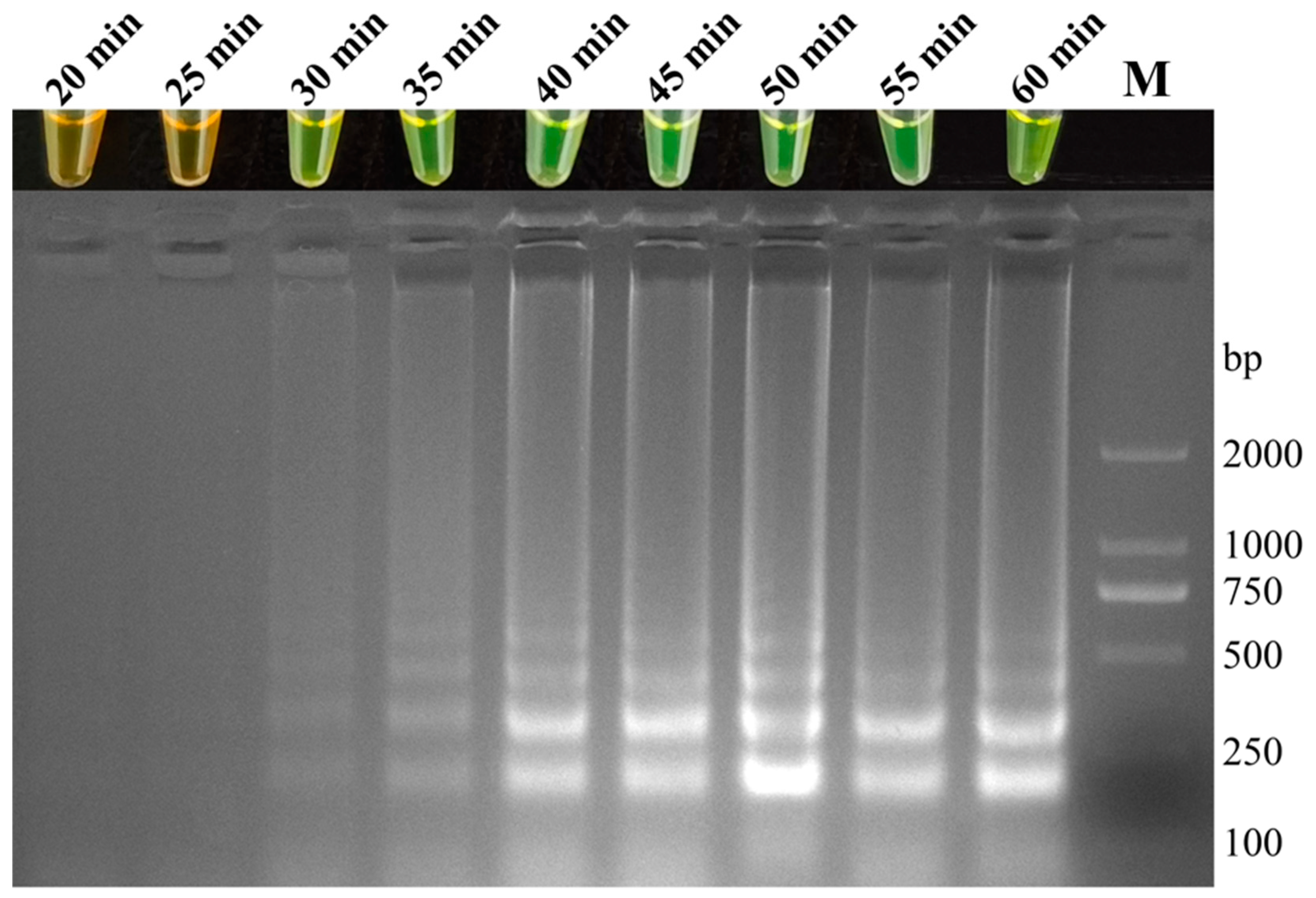
In order to determine the minimum effective time threshold of the triple LAMP reaction, various reaction time settings were tested in this study. The results indicated that when the LAMP reaction continued for 25 min, no ladder-like bands were observed. However, with an extension of the reaction time to 30–60 min, all the products displayed ladder-like bands and emitted a bright green fluorescence (Figure 3). Therefore, it was determined that the minimum threshold for the triple LAMP reaction time was 30 min.

3.4. Specificity Detection and Enzyme Digestion Identification

The genomic DNA and cDNA of the various pathogens extracted, as described in Section 2.3, were used as templates to amplify the reaction using the optimized triple LAMP detection method. The results showed that only V. harveyi, V. parahaemolyticus, SGIV, and the positive control displayed distinct ladder bands, whereas the remaining eight bacteria, five viruses, and negative controls showed no amplified bands. Notably, the products with ladder bands exhibited a vibrant green color (indicating a positive result), while those without ladder bands appeared orange–yellow (indicating a negative result). These findings highlighted the specificity of the triple LAMP detection method established in this study. No cross-reactivity was observed with the other seven Vibrio strains, S. aureus, or the other five viruses examined (Figure 4).
To identify the specific pathogen responsible for the positive results in the triple LAMP, we employed the enzyme digestion method for analysis. The results indicated that all positive LAMP products were digested by Hind III, BspT I, and EcoR I, yielding smaller bands. Each pathogen’s enzymatic fragments contained a band distinct from those of the other pathogens. The 318 bp band served as the indicator for V. harveyi, the 171 bp band for V. parahaemolyticus, and the 67 bp band for SGIV. The different indicator bands enable the accurate identification of each pathogen species (Figure 5).

3.5. Sensitivity Detection

The results indicated that the triple LAMP method’s minimum detection limit for V. harveyi, V. parahaemolyticus, and SGIV was 100 fg/μL, in contrast to the conventional PCR method’s limit of 1 pg/μL (Figure 6). Upon mixing the templates of V. harveyi, V. parahaemolyticus, and SGIV, the triple LAMP detection method showed three bands, including 146 bp of V. harveyi, 276 bp of V. parahaemolyticus, and 477 bp of SGIV. The minimum detection limits for V. harveyi, V. parahaemolyticus, and SGIV were 10 pg/μL, 1 pg/μL, and 1 pg/μL, respectively (Figure 7). Therefore, the sensitivity of the triple LAMP assay established in this study is 10 times higher than that of the conventional PCR assay, 10 times higher than that of the triple PCR assay in the detection of V. parahaemolyticus and SGIV, and 100 times higher than that of the triple PCR assay in the detection of V. harveyi.

3.6. Application of the Triple LAMP Detection Method

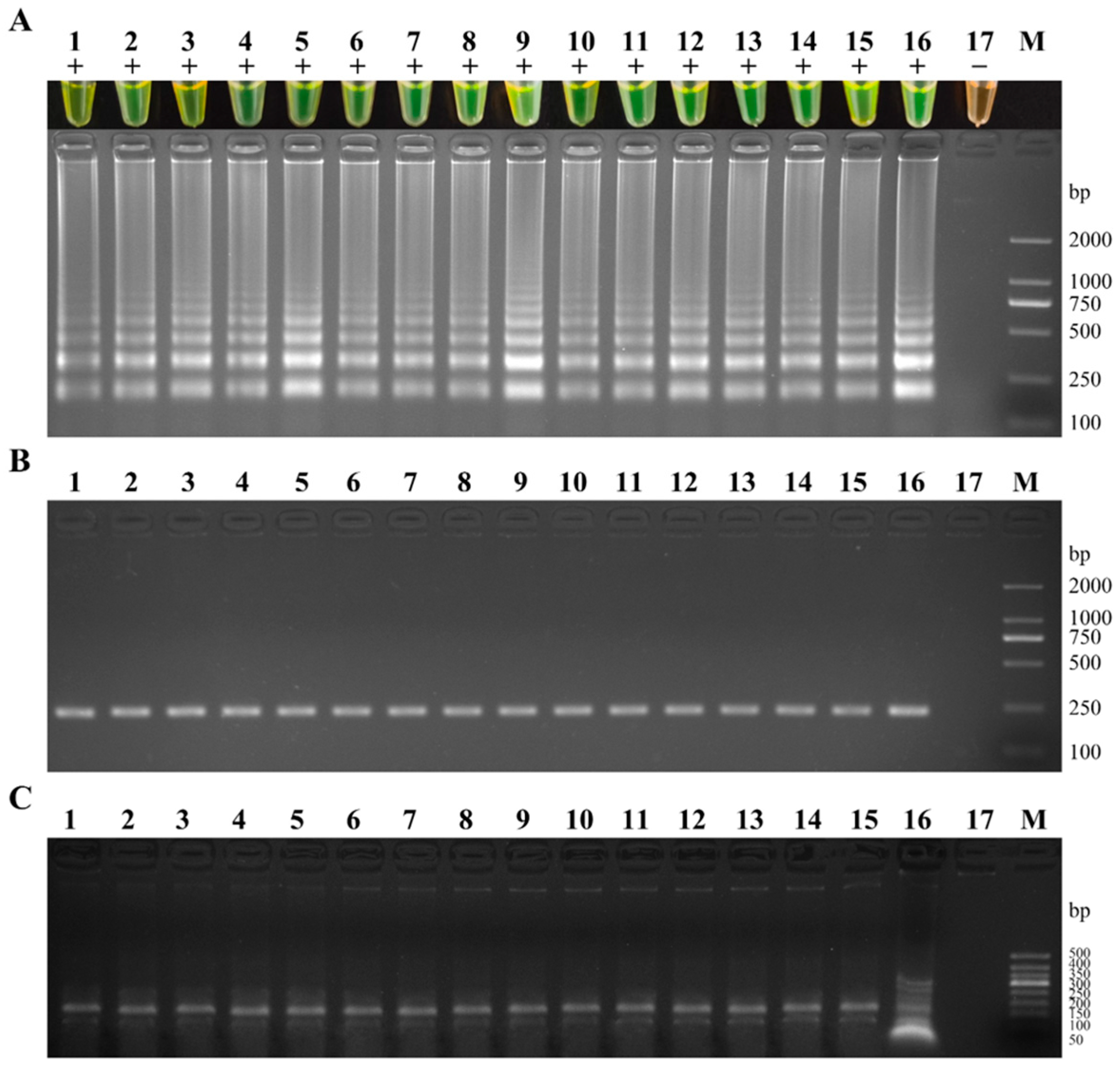
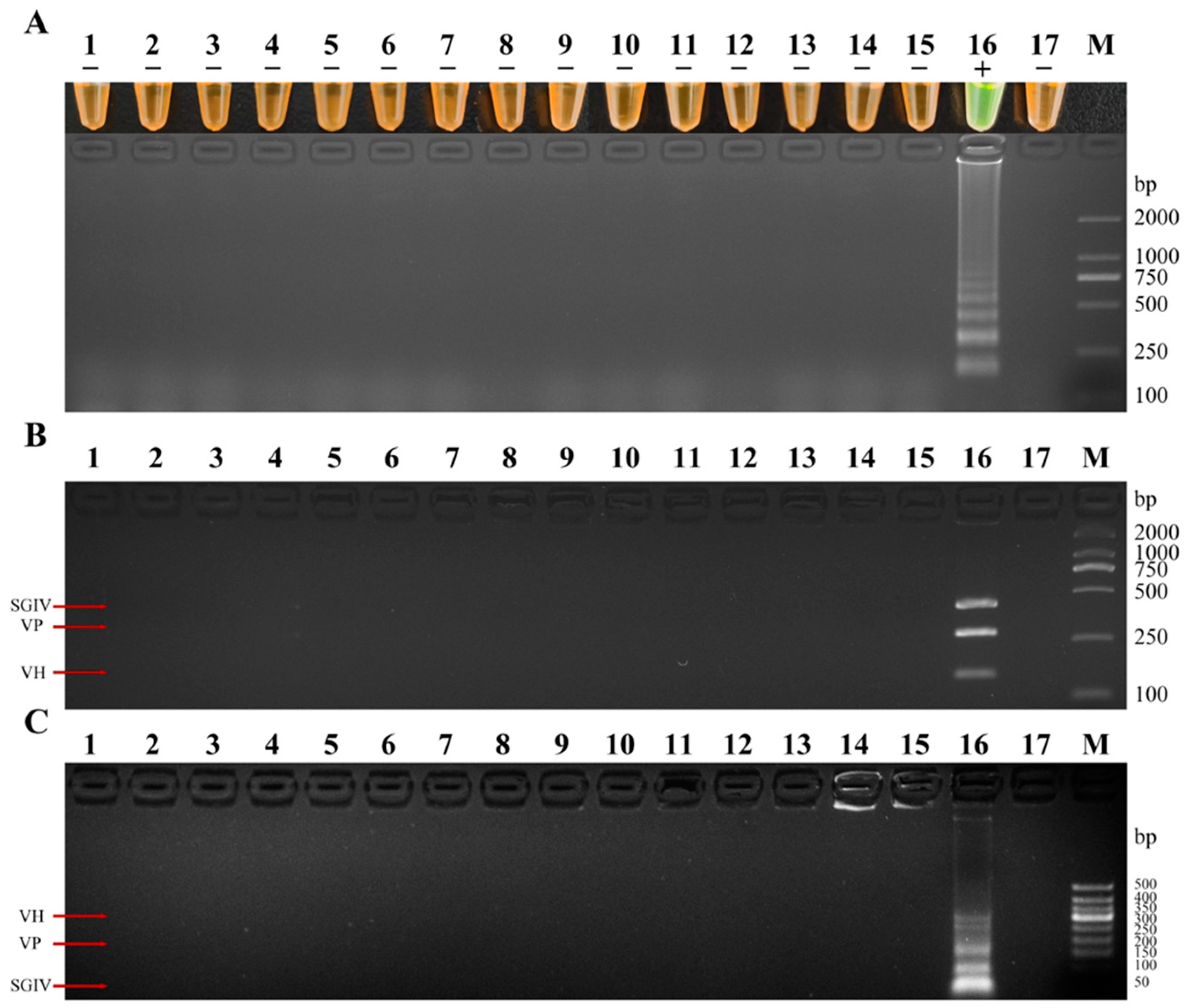
The results indicated that all 15 samples infected with V. harveyi exhibited positive amplification. The reaction color, a bright green, matched the electrophoresis detection results, and V. harveyi was identifiable by enzyme digestion (Figure 8). Similarly, the 15 samples infected with V. parahaemolyticus also showed positive amplification, with a bright green reaction color that aligned with the electrophoresis results, allowing for the identification of V. parahaemolyticus by enzymatic digestion (Figure 9). The 15 samples infected with SGIV similarly demonstrated positive amplification. The uniform bright green reaction color corresponded with the electrophoresis findings, and the SGIV identification was possible through enzyme digestion (Figure 10). The 15 samples with a mixed infection of V. harveyi, V. parahaemolyticus, and SGIV also yielded positive amplification results. The reaction color, bright green, was in agreement with the electrophoretic detection outcomes. Following enzymatic digestion, the samples enabled the identification of V. harveyi, V. parahaemolyticus, and SGIV (Figure 11). Moreover, the analysis of 15 healthy, uninfected grouper liver tissue samples revealed no positive amplification in any sample. The reaction color was orange–yellow, signifying that these samples were negative and uninfected by V. harveyi, V. parahaemolyticus, or SGIV (Figure 12).
Furthermore, the samples infected solely with V. harveyi, V. parahaemolyticus, and SGIV were detected using the conventional PCR method, while those co-infected with all three pathogens were detected using the triple PCR method. The detection results revealed that the 15 samples infected with either V. harveyi or V. parahaemolyticus amplified a single target band, confirming infection with one of these pathogens, with a detection coincidence rate of 100% (Figure 8 and Figure 9); of the 15 samples infected solely with SGIV, 13 exhibited amplification of a single target band, resulting in a detection coincidence rate of 86.7% (Figure 10). In cases of compound infection, both V. parahaemolyticus and SGIV amplified the target band, achieving a detection coincidence rate of 100%. However, only 14 samples infected with V. harveyi showed amplification of the target band, with a detection coincidence rate of 93.3% (Figure 11). In the control PBS group, no pathogens were detected (Figure 12).

4. Discussion

Aquaculture has become an important component of our agricultural economy at present. However, in recent years, with the increase in aquaculture density, fish diseases have been increasing, resulting in huge economic losses. Therefore, the establishment of effective, accurate, and rapid aquatic animal pathogen detection technology is an urgent problem to be solved at present and in the future. In this study, for the first time, we have successfully established a triple visual LAMP rapid detection method for three common and serious pathogens (V. harveyi, V. parahaemolyticus, and SGIV) in grouper culture and provided useful technical support for further application in the large-scale disease surveillance of aquaculture animals.
Currently, the detection methods for pathogens primarily involve a traditional medium culture, a colloidal gold-labeled chromatography strip, a PCR, and a real-time fluorescence quantitative PCR [21,22,23,24,25]. However, these methods have several limitations, including their complex operation, expensive equipment, need for professional technicians, and so on. For example, PCR offers strong specificity and high sensitivity, making it a more accurate diagnostic method [26]. However, it requires expensive instruments and specialized technicians, which limits its use in field conditions or resource-limited settings. In recent years, the LAMP technique has been widely used in the rapid detection of pathogens because of its high sensitivity, strong specificity, rapid detection, low cost, and the independent nature of its precision instruments. In contrast to the LAMP method, nucleic acid sequence-based amplification (NASBA) employs three enzymes, raising the reaction’s cost. Nevertheless, it provides a rapid inspection, detecting rotavirus, norovirus, and astrovirus in just 45 min [27]. Recombinase polymerase amplification (RPA), which also necessitates three enzymes, is similarly costly. Yet, combined with a lateral flow dipstick (LFD), it swiftly identifies SGIV in only 8 min [28]. Rolling circle amplification (RCA) requires a padlock probe of about 100 bp, resulting in elevated synthesis expenses. Despite this, it efficiently detects ISKNV within 40 min [29]. Aiming at the problem, Yan et al. (2024) developed and compared the four technologies of LAMP, SEA, CPA, and RPA. The results showed that the sensitivities of LAMP and RPA were the highest, reaching 1.2 pg/μL. However, the cost of RPA was 10 times that of LAMP, while the system and primers of LAMP were also complex [30]. In the context of large-scale aquaculture, while LAMP may not excel in speed over other isothermal amplification methods (such as RPA-LFD), its cost-effectiveness is a notable advantage. Despite these advantages, the LAMP technique has several limitations that require careful management. Designing LAMP primers requires careful consideration of factors such as length, GC content, and Tm value to ensure specificity and efficiency, while minimizing primer hybridization to avoid false positives. The high sensitivity of LAMP also makes contamination control critical. Strict laboratory practices are essential to prevent non-specific reactions and contamination [31]. Even so, Yu et al. (2022) developed a LAMP detection technique for SGIV based on the SGIV-VP61 gene, with a sensitivity of 5.66 copies/μL, which is 1000 times higher than a traditional PCR [32]. To further improve the detection efficiency, multi-LAMP technology has become a research hotspot in recent years. Zhou et al. (2016) established a dual LAMP technique for detecting V. vulnificus and V. parahaemolyticus, enabling the detection of both Vibrio species within 45 min at 62 °C, with a sensitivity of 8 × 10³ CFU/mL, which is a 100-fold increase compared to a PCR [33]. Due to the possible mutual interference between the primers, in general, under the same reaction system and the same primer concentration, the amplification rate of multiple primers is slower than under a single LAMP condition, and the more target pathogens to be detected at the same time, the more difficult the amplification is. Hence, the establishment of a multiple LAMP system with a high efficiency, a short reaction time, and high sensitivity is not easy to achieve.
In our study, we developed a fast triple visual LAMP method, operable in a simple 62 °C water bath with a 30 min reaction time. This indicates that our three primers work efficiently and specifically without cross-interference to detect the target pathogens. Compared to a PCR, the need for an expensive PCR instrument is eliminated, resulting in cost savings and a reduction in the detection time by approximately 1.5 h. In addition, the detection sensitivity of this method can reach 100 fg/μL, which is 10 times higher than that of a PCR. In terms of the observation of the amplified products, SYBR Green I color development is one of the commonly used methods [34]. However, this method needs the opening of the lid and the adding of dye after the reaction, and there is a risk of a false positive caused by aerosol pollution to a certain extent [35,36]. In contrast, this study introduced another dye, calcein, which was added to the reaction system at the time of preparation, avoiding possible aerosol contamination caused by opening the lid, greatly reducing the possibility of false positives and eliminating the need for agarose gel electrophoresis analysis. The purpose of establishing rapid pathogen detection technology is to better serve the aquaculture industry, and the practicability of this technology established by us was also tested in this study. In simulated sample tests, the triple LAMP method had a 100% agreement rate with the expected results for the detection of the three pathogens. The method in this study can detect all positive samples, especially for the detection of SGIV, while the PCR method can only detect part of the samples (with a positive rate of 86.7%), indicating that our method has a high detection sensitivity and excellent practicability.

5. Conclusions

In summary, in this study, the target genes of three pathogens were selected: the TolC of V. harveyi, the DNAJ of V. parahaemolyticus, and the RAD2 of SGIV. With calcein as the indicator of the detection results, a triple visual LAMP detection method for V. harveyi, V. parahaemolyticus, and SGIV was successfully established for the first time by optimizing the reaction system and reaction conditions. Overall, this method has the advantages of fast detection (up to 30 min), high sensitivity (100 fg/μL), strong specificity (no cross-reaction with other common pathogens), product visualization, and excellent practicability. As such, it is highly suitable for the rapid on-site detection of V. harveyi, V. parahaemolyticus, and SGIV in production settings.

Author Contributions

T.L.: conceptualization, data curation, formal analysis, and writing—original draft; R.D. and J.Z.: methodology; Y.Z. and C.L.: supervision and writing—review and editing; and Z.C. and Y.S.: conceptualization, funding acquisition, supervision, and writing—review and editing. All authors have read and agreed to the published version of the manuscript.

Funding

This work was supported by the Hainan Province Science and Technology Special Fund (ZDKJ2021016; ZDYF2022XDNY192; and ZDYF2023XDNY032), the Hainan University Collaborative Innovation Center Fund (XTCX2022HYB03), and the earmarked fund for HNARS-Grouper (HNARS-03-G03).

Institutional Review Board Statement

Not applicable.

Data Availability Statement

The authors declare that all the data supporting the findings of this study are available within the article.

Conflicts of Interest

The authors declare that they have no conflicts of interest.

References

  1. The People’s Republic of China Ministry of Agriculture. Fisheries Bureau. China Fishery Statistical Yearbook 2023; China Agriculture Press: Beijing, China, 2023. [Google Scholar]
  2. Luo, M.; Chen, F.X.; Liu, L.L.; Li, W.D.; Zeng, G.Q.; Tan, W.; Li, X.M. Progress in disease research of grouper aquaculture in China. Fish. Sci. 2013, 32, 549–554. [Google Scholar]
  3. Liu, Z.T.; Zhang, X.; Huang, X.H.; Huang, Y.H.; Qin, Q.W. Mechanism of oligochitosan improving non-specific immunity of Epinephelus fuscoguttatus (♀) × E. lanceolatu (♂). J. Fish. China 2022, 46, 85–94. [Google Scholar]
  4. He, L.G.; Liang, Y.S.; Yu, X.; Peng, W.; He, J.N.; Fu, L.J.; Lin, H.R.; Zhang, Y.; Lu, D.Q. Vibrio parahaemolyticus flagellin induces cytokines expression via toll-like receptor 5 pathway in orange-spotted grouper, Epinephelus coioides. Fish Shellfish Immunol. 2019, 87, 573–581. [Google Scholar] [CrossRef] [PubMed]
  5. Shen, G.M.; Shi, C.Y.; Fan, C.; Jia, D.; Wang, S.Q.; Xie, G.S.; Li, G.Y.; Mo, Z.L.; Huang, J. Isolation, identification and pathogenicity of Vibrio harveyi, the causal agent of skin ulcer disease in juvenile hybrid groupers Epinephelus fuscoguttatus × Epinephelus lanceolatus. J. Fish Dis. 2017, 40, 1351–1362. [Google Scholar] [CrossRef] [PubMed]
  6. Xu, H.; Zeng, Y.H.; Yin, W.L.; Lu, H.B.; Gong, X.X.; Zhang, N.; Zhang, X.; Long, H.; Ren, W.; Cai, X.N.; et al. Prevalence of bacterial coinfections with Vibrio harveyi in the industrialized flow-through aquaculture systems in Hainan province: A neglected high-risk lethal causative agent to hybrid grouper. Int. J. Mol. Sci. 2022, 23, 11628. [Google Scholar] [CrossRef] [PubMed]
  7. Qin, Z.W.; Xue, L.; Gao, J.S.; Hu, Y.D.; Zhang, J.M.; Wu, Q.P. Advances in nucleic acid isothermal detection technologies for foodborne viruses. Microbiol. China 2021, 48, 266–277. [Google Scholar]
  8. Yeh, H.Y.; Shoemaker, C.A.; Klesius, P.H. Sensitive and rapid detection of Flavobacterium columnare in channel catfish Ictalurus punctatus by a loop-mediated isothermal amplification method. J. Appl. Microbiol. 2006, 100, 919–925. [Google Scholar] [CrossRef] [PubMed]
  9. Cai, X.Q.; Xu, M.J.; Wang, Y.H.; Qiu, D.Y.; Liu, G.X.; Lin, A.; Tang, J.D.; Zhang, R.L.; Zhu, X.Q. Sensitive and rapid detection of Clonorchis sinensis infection in fish by loop-mediated isothermal amplification (LAMP). Parasitol. Res. 2010, 106, 1379–1383. [Google Scholar] [CrossRef] [PubMed]
  10. Suebsing, R.; Pradeep, P.J.; Jitrakorn, S.; Sirithammajak, S.; Kampeera, J.; Turner, W.A.; Saksmerprome, V.; Withyachumnarnkul, B.; Kiatpathomchai, W. Detection of natural infection of infectious spleen and kidney necrosis virus in farmed tilapia by hydroxynapthol blue-loop-mediated isothermal amplification assay. J. Appl. Microbiol. 2016, 121, 55–67. [Google Scholar] [CrossRef]
  11. Fan, Q.; Xie, Z.X.; Wei, Y.; Zhang, Y.F.; Xie, Z.Q.; Xie, L.J.; Huang, J.L.; Zeng, T.T.; Wang, S.; Luo, S.S.; et al. Development of a visual multiplex fluorescent LAMP assay for the detection of foot-and-mouth disease, vesicular stomatitis and bluetongue viruses. PLoS ONE 2022, 17, e0278451. [Google Scholar] [CrossRef]
  12. Li, S.; Wang, Y.S.; Yo, H.W.; Li, Y.; Xia, G.Q. Development of multiplex loop-mediated isothermal amplification assays to detect three kinds of food-borne pathogens. J. Nav. Univ. Eng. 2018, 30, 75–79+90. [Google Scholar]
  13. Fan, Q.; Xie, Z.X.; Xie, Z.Q.; Xie, L.J.; Huang, J.L.; Zhang, Y.F.; Zeng, T.T.; Wang, S.; Luo, S.S.; Deng, X.W.; et al. Development of multiplex fluorescence-based loop-mediated isothermal amplification assay for the detection of bovine rotavirus and enterotoxigenic E. coli. Chin. Vet. Sci. 2019, 49, 1119–1127. [Google Scholar]
  14. Wang, Y.; Cheng, Y.; Xie, J.; Liu, Y. Establishment of multiplex loop-mediated isothermal amplification for rapid detection of genitourinary mycoplasma. Clin. Lab. 2018, 64, 1217–1224. [Google Scholar] [CrossRef] [PubMed]
  15. Siddique, M.P.; Jang, W.J.; Lee, J.M.; Hasan, M.T.; Kim, C.H.; Kong, I.S. Detection of Vibrio anguillarum and Vibrio alginolyticus by singleplex and duplex loop-mediated isothermal amplification (LAMP) Assays Targeted to groEL and fklB Genes. Int. Microbiol. 2019, 22, 501–509. [Google Scholar] [CrossRef] [PubMed]
  16. Wang, Z.; Hervey, W.J.; Kim, S.; Lin, B.; Vora, G.J. Complete genome sequence of the bioluminescent marine bacterium Vibrio harveyi ATCC 33843 (392 [MAV]). Genome Announc. 2015, 3, e01493. [Google Scholar] [CrossRef] [PubMed]
  17. Nhung, P.H.; Ohkusu, K.; Miyasaka, J.; Sun, X.S.; Ezaki, T. Rapid and specific identification of 5 human pathogenic Vibrio species by multiplex polymerase chain reaction targeted to dnaJ gene. Diagn. Microbiol. Infect. Dis. 2007, 59, 271–275. [Google Scholar] [CrossRef] [PubMed]
  18. Lü, L.; Zhou, S.Y.; Chen, C.; Weng, S.P.; Chan, S.M.; He, J.G. Complete genome sequence analysis of an iridovirus isolated from the orange-spotted grouper, Epinephelus coioides. Virology 2005, 339, 81–100. [Google Scholar] [CrossRef] [PubMed]
  19. Tu, Z.; Li, H.; Zhang, X.; Sun, Y.; Zhou, Y. Complete genome sequence and comparative genomics of the golden pompano (Trachinotus ovatus) pathogen, Vibrio harveyi strain QT520. PeerJ 2017, 5, e4127. [Google Scholar] [CrossRef] [PubMed]
  20. Xu, X.D.; Xie, Z.Y.; Wang, S.F.; Xuan, X.Z.; Zhou, Y.C. Toxicity and immunogenicity of LPS from pathogenic Vibrio alginolyticus to grouper, Epinephelus malabaricus. Mar. Sci. 2010, 34, 47–51. [Google Scholar]
  21. Huang, Y.Y.; Yang, M.W.; Tan, H.L.; Liang, Z.; Zeng, L.; Wei, X.X.; Huang, G.H.; Tong, G.X. Development and application of duplex fluorescent quantitative PCR method for detecting Macrobrachium rosenbergii nodavirus and decapod iridescent virus 1. J. South. Agric. 2022, 53, 1474–1482. [Google Scholar]
  22. Liao, M.J.; Zhang, Z.; Rong, X.J.; Wang, Y.G.; Liu, Z.C.; Luan, J.; Li, B.; Wang, L.; Chen, G.P. Development of a SYBR Green I real-time PCR for detection of Vibrio harveyi based on the vhhP2 gene. J. Fish. Sci. China 2014, 21, 611–620. [Google Scholar]
  23. Pei, L.S.; Zhu, Y.L.; Mao, Q.; Chen, L.M.; Guo, Y.J.; Sun, J.F. Isolation, identification and antibiotic susceptibility analysis of thepathogenic Vibrio harveyi from Epinephelus coioides. J. Tianjin Agric. Univ. 2021, 28, 35–40. [Google Scholar]
  24. Sheng, X.Z.; Song, J.L.; Zhan, W.B. Development of a colloidal gold immunochromatographic test strip for detection of lymphocystis disease virus in fish. J. Appl. Microbiol. 2012, 113, 737–744. [Google Scholar] [CrossRef] [PubMed]
  25. Zhang, X.J.; Bai, X.S.; Bi, K.R.; Zhoi, B.Y.; Qin, L.; Yan, B.L.; Qin, G.M. Simultaneous detection of Vibrio parahaemolyticus and V. cholerae from aquatic animals by dulplex PCR. Fish. Sci. 2013, 32, 412–415. [Google Scholar]
  26. Zhu, J.L.; Wang, G.L.; Jin, S. Development and application of multiplex PCR of vibrios pathogenic to cultured large yellow croaker, Pseudnosciaena crocea (Richardson). J. Fish. Sci. China 2009, 16, 156–164. [Google Scholar]
  27. Mo, Q.H.; Wang, H.B.; Dai, H.R.; Lin, J.C.; Tan, H.; Wang, Q.; Yang, Z. Rapid and simultaneous detection of three major diarrhea-causing viruses by multiplex real-time nucleic acid sequence-based amplification. Arch. Virol. 2015, 160, 719–725. [Google Scholar] [CrossRef]
  28. Pan, Y.; Yang, J.H.; Peng, F.Y.; Huang, Y.H.; Qin, Q.W.; Huang, X.H. Rapid detection of Singapore grouper iridovirus by a recombinase polymerase amplification combined with lateral flow dipstick. J. Fish. China 2024, 48, 059411. [Google Scholar]
  29. Kong, C.J.; Shi, Y.H.; Li, H.Y.; Chen, J. Detection of infectious spleen and kidney necrosis virus (ISKNV) by HRCA approach. Fish. Sci. 2011, 30, 575–579. [Google Scholar]
  30. Yan, S.; Li, C.L.; Lan, H.Z.; Pan, D.D.; Wu, Y.C. Comparison of four isothermal amplification techniques: LAMP, SEA, CPA, and RPA for the identification of chicken adulteration. Food Control 2024, 159, 110302. [Google Scholar] [CrossRef]
  31. Wong, Y.P.; Othman, S.; Lau, Y.L.; Radu, S.; Chee, H.Y. Loop-mediated isothermal amplification (LAMP): A versatile technique for detection of micro-organisms. J. Appl. Microbiol. 2018, 124, 626–643. [Google Scholar] [CrossRef]
  32. Yu, Y.P.; Yang, Z.T.; Wang, L.; Sun, F.; Lee, M.; Wen, Y.F.; Qin, Q.W.; Yue, G.H. LAMP for the rapid diagnosis of iridovirus in aquaculture. Aquac. Fish. 2022, 7, 158–165. [Google Scholar] [CrossRef]
  33. Zhou, S.; Gao, Z.X.; Zhang, M. The development and application of a duplex loop-mediated isothermal amplification method for rapid detection of Vibrio vulnificus and Vibrio parahaemolyticus. Chin. J. Vet. Sci. 2016, 36, 1875–1881. [Google Scholar]
  34. Rahman, A.M.A.; Ransangan, J.; Subbiah, V.K. Improvements to the rapid detection of the marine pathogenic bacterium, Vibrio harveyi, using loop-mediated isothermal amplification (LAMP) in combination with SYBR Green. Microorganisms 2022, 10, 2346. [Google Scholar] [CrossRef] [PubMed]
  35. Kamra, E.; Singh, N.; Khan, A.; Singh, J.; Chauhan, M.; Kamal, H.; Mehta, P.K. Diagnosis of genitourinary tuberculosis by loop-mediated isothermal amplification based on SYBR Green I dye reaction. Biotechniques 2022, 73, 47–57. [Google Scholar] [CrossRef]
  36. Lai, M.Y.; Ooi, C.H.; Lau, Y.L. Validation of SYBR green I based closed-tube loop-mediated isothermal amplification (LAMP) assay for diagnosis of knowlesi malaria. Malar J. 2021, 20, 166. [Google Scholar] [CrossRef]
Figure 1. Optimization of the triple LAMP reaction system: (A) optimization of the primer group concentration; (B) optimization of the calcein mixture concentration; (C) optimization of the Mg2+ concentration; (D) optimization of the dNTPs concentration; and (E) optimization of the betaine concentration. * indicates the optimal result; and M indicates the DL2000 marker.
Fishes 09 00225 g001
Figure 2. Temperature optimization of the triple LAMP. * indicates the optimal result; and M indicates the DL2000 marker.
Fishes 09 00225 g002
Figure 3. Time optimization of the triple LAMP. M indicates the DL2000 marker.
Fishes 09 00225 g003
Figure 4. Specific detection of the triple LAMP. 1: Positive control (the template was the plasmids mixture of pTolC, pDNAJ, and pRAD2); 2: V. harveyi; 3: V. parahemolyticus; 4: SGIV; 5: V. alginolyticus; 6: V. cholerae; 7: V. vulnificus; 8: V. mimicus; 9: V. alfacsensis; 10: V. campbellii; 11: V. owensii; 12: S. aureus; 13: NNV; 14: ISKNV; 15: SHIV; 16: IHHNV; 17: WSSV; 18: negative control (with ddH2O as the template); and M: DL2000 marker. + indicates positive and − indicates negative.
Fishes 09 00225 g004
Figure 5. Enzyme digestion identification of the triple LAMP products. (A) V. harveyi; (B) V. parahaemolyticus; (C) SGIV; (D) V. harveyi and V. parahaemolyticus; (E) V. harveyi and SGIV; (F) V. parahaemolyticus and SGIV; and (G) V. harveyi, V. parahaemolyticus and SGIV. 1: LAMP positive samples; 2: corresponding digestion products; and M: 50 bp marker.
Fishes 09 00225 g005
Figure 6. The sensitivity detection of the triple LAMP and the common PCR. (A,B) The sensitivity detection of the triple LAMP and common PCR for V. harveyi; (C,D) the sensitivity detection of the triple LAMP and common PCR for V. parahaemolyticus; and (E,F) the sensitivity detection of the triple LAMP and common PCR for SGIV. 1: 10 fg/μL; 2: 100 fg/μL; 3: 1 pg/μL; 4: 10 pg/μL; 5: 100 pg/μL; 6: 1 ng/μL; 7: 10 ng/μL; 8: 100 ng/μL; 9: positive control (the mixture of the standard plasmids pTolC, pDNAJ, and pRAD2); and M: DL2000 marker (A,C,E)/DNA marker Ⅰ (B,D,F).
Fishes 09 00225 g006
Figure 7. Sensitivity detection of the triple LAMP and triple PCR for the V. harveyi, V. parahaemolyticus, and SGIV mixtures. (A) Sensitivity detection with the triple LAMP method; and (B) sensitivity detection with the triple PCR. 1: 1 fg/μL; 2: 10 fg/μL; 3: 100 fg/μL; 4: 1 pg/μL; 5: 10 pg/μL; 6: 100 pg/μL; 7: 1 ng/μL; 8: 10 ng/μL; 9: 100 ng/μL; 10: positive control (the mixture of the standard plasmids pTolC, pDNAJ, and pRAD2); and M: DL 2000 marker.
Fishes 09 00225 g007
Figure 8. Application of the triple LAMP method for the detection of V. harveyi. (A) Triple LAMP method; (B) conventional PCR method; and (C) enzyme digestion identification. 1–15: DNA of the liver tissue in the V. harveyi-infected group; 16: positive control (the mixture of the standard plasmids pTolC, pDNAJ, and pRAD2); 17: negative control (ddH2O); and M: DL2000 marker. + indicates positive and − indicates negative.
Fishes 09 00225 g008
Figure 9. Application of the triple LAMP method for the detection of V. parahaemolyticus. (A) Triple LAMP method; (B) conventional PCR method; and (C) enzyme digestion identification. 1–15: DNA of the liver tissue in the V. parahaemolyticus-infected group; 16: positive control (the mixture of the standard plasmids pTolC, pDNAJ, and pRAD2); 17: negative control (ddH2O); and M: DL2000 marker. + indicates positive and − indicates negative.
Fishes 09 00225 g009
Figure 10. Application of the triple LAMP method for the detection of SGIV. (A) Triple LAMP method; (B) conventional PCR method; and (C) enzyme digestion identification. 1–15: DNA of the liver tissue in the SGIV-infected group; 16: positive control (the mixture of the standard plasmids pTolC, pDNAJ, and pRAD2); 17: negative control (ddH2O); and M: DL2000 marker. + indicates positive and − indicates negative.
Fishes 09 00225 g010
Figure 11. Simulated application of the triple LAMP detection method for a triple infection. (A) Triple LAMP method; (B) triple PCR method; and (C) enzyme digestion identification. 1–15: DNA of the liver tissue in the compound infection group; 16: positive control (the mixture of the standard plasmids pTolC, pDNAJ, and pRAD2); 17: negative control (ddH2O); and M: DL 2000/50 bp marker. + indicates positive; − indicates negative; VH indicates V. harveyi; and VP indicates V. parahaemolyticus.
Fishes 09 00225 g011
Figure 12. Application of the triple LAMP detection method in healthy groupers. (A) Triple LAMP method; (B) triple PCR method; and (C) enzyme digestion identification. 1–15: DNA of the liver tissue in the PBS group; 16: positive control; 17: negative control; and M: DL 2000/50 bp marker. + indicates positive; − indicates negative; VH indicates V. harveyi; and VP indicates V. parahaemolyticus.
Fishes 09 00225 g012
Table 1. Primer sequences used for the LAMP assay.
PathogenPrime NamePrimer Sequence (5’−3’)
V. parahaemolyticusDNAJ-1-F3-1TTGCTATGGCTGCACTCG
DNAJ-1-B3-1TCTTTTTGGCGAGCGCTTAA
DNAJ-1-FIP-1GGCCAGTTTGTGTTTCTGACGG-CTTAAG-CGGCGAAGTTGAAGTTCCA (BspT I)
DNAJ-1-BIP-1GCGCGGTAAAGGTGTGAAAGGT-TTTT-AACTGGCGTTTCTACAACCA
DNAJ-1-LF-1TTTTAGGCTTACTCGTCCATCAA
DNAJ-1-LB-1CGTGGCGGTGGTATTGGT
V. harveyiTolC-2-F3-2TCCTTCGAGTTTCTCAAGCG
TolC-2-B3-2TCACGTAGCGATTCGTAGCT
TolC-2-FIP-2CTAGTTGGCGACCAACCGCT-AAGCTT-CGCGCACAAGACAACCTAG (Hind III)
TolC-2-BIP-2TCACTGACGTACACGATGCGC-TTTT-CGAGTTTTCCGCCAAGACT
TolC-2-LF-2GCTTTTTCTGCACGAACGAA
TolC-2-LB-2AAGCACAATACGATGCAGTACTTG
SGIVRAD2-3-F3-3CGTTGGAACTGAACGGAGAT
RAD2-3-B3-3GCGGTGATGTCTCTGCCTA
RAD2-3-FIP-3GCGGGACCCAACCTGTGAATTC-TTTT-ATGGATCTGTGCGTCATGTG (EcoR I)
RAD2-3-BIP-3GATGATACGAGCGCACGGAAGC-GAATTC-CTTTGTCCTTTCCGCGTCT (EcoR I)
RAD2-3-LF-3GCTGATTAAAATCGGTGCCG
RAD2-3-LB-3ACGACGTGCCCATTGAATCT
Underlined nucleotides indicate the restriction sites of the enzymes shown in parentheses or the linkers between the inner primers.
Table 2. The triple LAMP basic reaction system.
Reaction System ComponentsVolume (μL)
10 × Thermoplol buffer2.5
MgSO4 (100 mM)1.5
dNTPs (10 mM)3.5
DNAJ-1-FIP-1/BIP-1 (1:1, 50 M)1
DNAJ-1-F3-1/B3-1 (1:1, 10 M)1
DNAJ-1-LF-1/LB-1 (1:1, 25 M)1
TolC-2-FIP-2/BIP-2 (1:1, 50 M) 1
TolC-2-F3-2/B3-2 (1:1, 10 M)1
TolC-2-LF-2/LB-2 (1:1, 25 M)1
RAD2-3-FIP-3/BIP-3 (1:1, 50 M)1
RAD2-3-F3-3/B3-3 (1:1, 10 M)1
RAD2-3-LF-3/LB-3 (1:1, 25 M)1
Bst DNA polymerase (8 U)1
DNA template mixture (V. harveyi: V. parahaemolyticus: SGIV = 1:1:1)1
ddH2O6.5
Total25
Table 3. Multiplex PCR primer sequences.
PathogenPrime NamePrimer Sequence (5′−3′)Amplified Fragment
V. parahaemolyticusVP-FGTTCAGCAAACCTGTCCTACC146 bp
VP-RGCTTACTGGCACTTCACAATAGA
V. harveyiVH-FTGACGGTTTCAAAGTCGGTG276 bp
VH-RGCACTTCTTTCACAACACTACGG
SGIVRAD-FTCGTTGGATGCGTTCGG477 bp
RAD-RCGGTTCTGGCGGTGATGT
Disclaimer/Publisher’s Note: The statements, opinions and data contained in all publications are solely those of the individual author(s) and contributor(s) and not of MDPI and/or the editor(s). MDPI and/or the editor(s) disclaim responsibility for any injury to people or property resulting from any ideas, methods, instructions or products referred to in the content.