Article

Unveiling the Impact of LED Light on Growing Carrot Taproots: A Novel Hydroponic Cultivation System

Faculty of Biology-Oriented Science and Technology, Kindai University, Wakayama 649-6493, Japan
*
Author to whom correspondence should be addressed.
Submission received: 26 March 2025 / Revised: 23 April 2025 / Accepted: 24 April 2025 / Published: 25 April 2025
(This article belongs to the Special Issue Interdisciplinary Insights in Engineering Research)

Abstract

:
Root crops typically develop and enlarge their storage organs in the soil, where they are naturally shielded from light exposure. This characteristic influences their physiological development and presents challenges for hydroponic cultivation, as taproot enlargement is often inhibited when submerged in water. To overcome this limitation, this study introduced a novel hydroponic system that prevents direct submersion in the nutrient solution. By isolating the taproots from both soil and nutrient solution, this system allows precise control of the root-zone light environment using LED irradiation. Carrot taproots were cultivated under blue, green, and red LED light from 42 days after sowing to assess their specific responses to different wavelengths. The results revealed distinct pigment accumulation patterns influenced by light quality. Blue light induced anthocyanin accumulation in the epidermis and outer cortex within 2 days of exposure and also stimulated chlorophyll synthesis in these outer tissues. In contrast, green and red light treatments promoted chlorophyll accumulation primarily in the stele, with red light having the most pronounced effect. These findings suggest that carrot taproots exhibit specific physiological responses to light exposure, demonstrating their ability to adjust pigment biosynthesis depending on the wavelength. By integrating controlled lighting environments into hydroponic systems, this study provides new insights into root development mechanisms and presents a novel strategy for optimizing root crop cultivation.

1. Introduction

Light plays a fundamental role in plant development, influencing processes such as photosynthesis, photomorphogenesis, and secondary metabolism [1,2]. While chloroplasts are typically absent in roots, studies have shown that light exposure can induce chloroplast formation and root greening in several plant species [3,4]. In Arabidopsis thaliana, this process is regulated by auxin and cytokinin signaling pathways [3]. Photoreceptors such as cryptochrome and phytochrome have been implicated in root chloroplast formation [5]. Additional research has identified strong expressions of another photoreceptor, phototropin, in the upper root region, and UVR8 in the root apex [6,7]. UVR8 has also been shown to be involved in UV-B-induced root bending [8], further suggesting that roots possess photoreceptive capabilities. However, despite these findings, the precise mechanisms by which these photoreceptors regulate root physiology remain largely unexplored.
Hydroponic cultivation offers an effective alternative to soil-based agriculture by providing precise control over plant growth through nutrient solution management [9]. Among hydroponic techniques, the Deep Flow Technique (DFT) system and aeroponics are particularly advantageous as they allow root growth in enclosed spaces, facilitating environmental regulation [10,11]. Studies have demonstrated that modifying nutrient solution temperature can influence plant growth and composition, as observed in crops such as lettuce, carrots, and strawberries [12,13,14,15]. In hydroponically grown lettuce, lowering the nutrient solution temperature has been reported to increase anthocyanin accumulation in leaves due to oxidative stress [12]. Additionally, supplementing the nutrient solution with the antioxidant N-acetylcysteine (NAC) has been shown to mitigate oxidative stress and promote plant growth [16]. The ability to separate roots from soil in hydroponic systems has also enabled novel research into root exposure to light [17,18]. For instance, exposing hydroponic lettuce roots to ultraviolet, blue, green, red, and far-red LED light revealed that ultraviolet, blue, and far-red irradiation suppressed shoot growth [17]. Similarly, in medicinal plants such as Artemisia annua and Hypericum perforatum, root exposure to white, red, and blue LEDs enhanced the biosynthesis of phytochemicals in the aerial parts [18]. Moreover, studies on Brassica rapa have demonstrated that different light qualities alter growth and nutrient content in leaves, stems, and roots [19].
Despite these advancements, the application of hydroponic or aeroponic systems to root crops, particularly those that develop storage organs in the soil, presents significant challenges. Root enlargement is often inhibited when submerged in liquid nutrient solutions [20,21,22,23]. In carrots, for instance, submerging taproots in nutrient solutions has been reported to suppress their enlargement [24]. However, this inhibition can be alleviated by increasing the dissolved oxygen concentration in the solution [24,25]. To address this issue, novel cultivation methods that prevent direct submersion while maintaining optimal humidity have been explored [23,26,27,28]. Modified aeroponic systems have been successfully applied to crops such as potatoes, cassava, and burdock, allowing root enlargement without continuous contact with nutrient solutions [26,27,28]. In sweet potato cultivation, an alternative approach has been developed in which an air space is created above the nutrient solution within a DFT system, enabling storage roots to grow without being submerged [23]. Using this system, a study demonstrated that replacing the upper cover of the apparatus with a white plastic board allowed partial light penetration, which induced root chlorophyll synthesis and inhibited storage root enlargement in sweet potatoes [23].
Building upon this concept, we have developed a modified cultivation system specifically designed for carrots. This system, similar to the one used for sweet potatoes [23], features an air space for storage root enlargement and is equipped with LEDs to provide controlled artificial light exposure. This study aimed to investigate the effects of different light wavelengths on root growth and physiological responses, providing new insights into the role of light in root crop development and contributing to the advancement of hydroponic cultivation techniques.

2. Materials and Methods

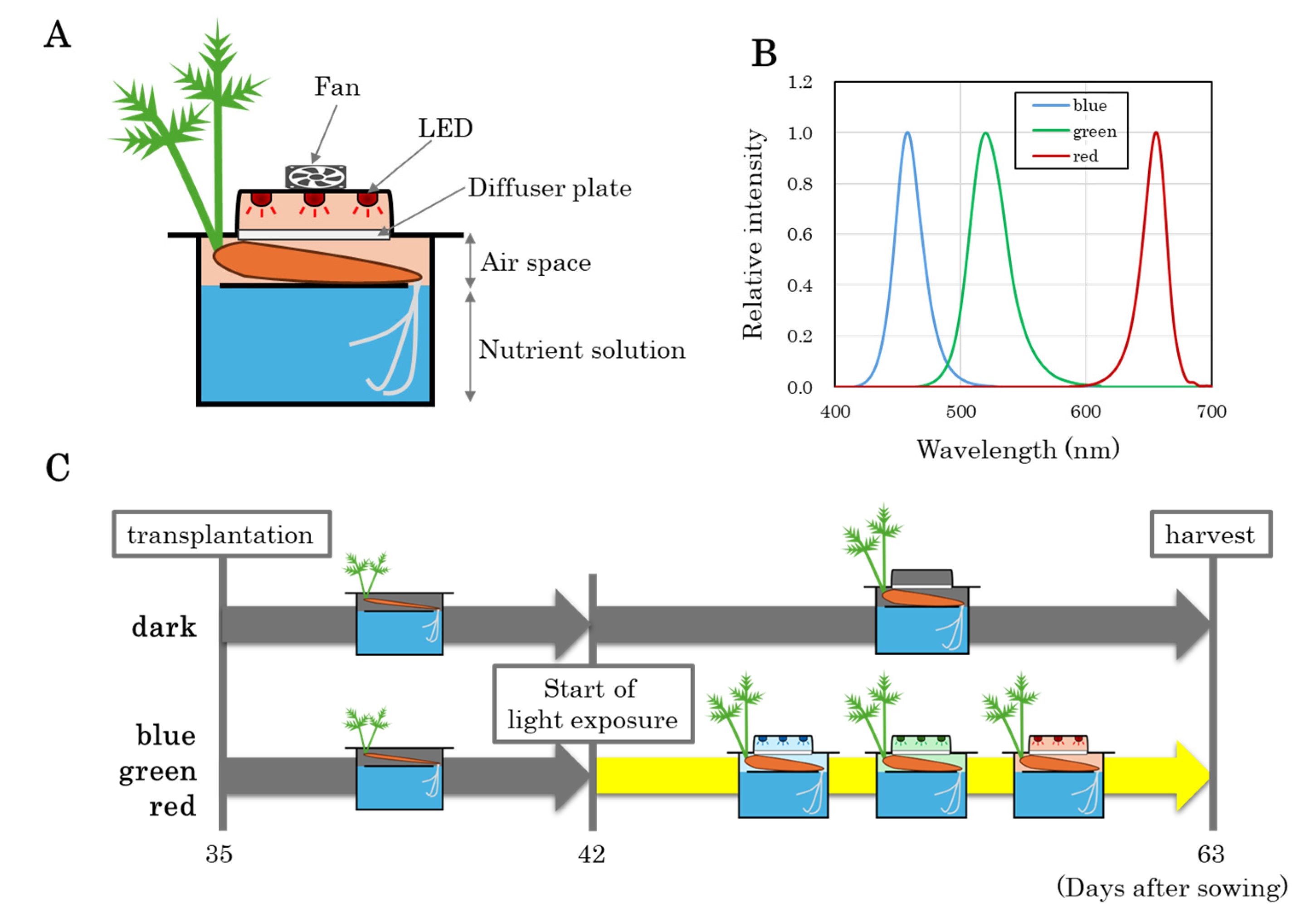
2.1. Experimental Conditions

Carrot seeds (Daucus carota L. cv. Orange Harmony, Marutane, Co., Ltd., Kyoto, Japan) were sown in black urethane foam cubes and germinated under controlled conditions at 20 °C with a photosynthetic photon flux (PPF) of 200 μmol m−2 s−1 for 16 h per day using fluorescent lamps (FL40SBR-A, NEC Co., Ltd., Tokyo, Japan). At 10 days after sowing, seedlings with a single main root extending from the bottom of the cubes were transferred to a DFT hydroponic system with continuous aeration. The nutrient solution consisted of half-strength OAT House recipe A [29,30]. The nutrient solution was prepared by combining OAT House 1 and OAT House 2 (OAT Agrio Co., Ltd., Tokyo, Japan) at a 3:2 ratio. After 35 days, plants with a single orange-colored taproot (approximately 1 mm in diameter) were relocated to a modified cultivation system allowing controlled light exposure to the taproot (Figure 1A).
The system consisted of a plastic box (32 cm × 19 cm × 17 cm) designed for DFT hydroponics and optimized for light exposure to carrot taproots. The subterranean section was divided into a dark, enclosed space containing both a submerged nutrient solution region and an air space devoid of solution. At transplantation, taproots were positioned within the air space, while fibrous roots remained immersed in the nutrient solution. The relative humidity within the air space was consistently maintained at 100%. Eight plants were placed per container. For the first week after transplantation, the taproots were maintained in darkness without LED exposure. On day 42 after sowing, blue, green, and red LEDs were installed above the air space and continuously illuminated at an intensity of 150 μmol m−2 s−1 for the remaining cultivation period. A diffusion plate was placed beneath the LEDs to ensure uniform light distribution to the taproots. Additionally, a cooling fan (4 cm × 4 cm) was installed above the LEDs to dissipate heat. The spectral characteristics of the LEDs are shown in Figure 1B. For the dark treatment, no LEDs were installed, and the taproots remained in darkness throughout the experiment. Morphological observations and color measurements were conducted after temporarily removing the LEDs and diffusion plates. At 63 days after sowing (21 days of light exposure), plants were harvested for further analyses. The experimental timeline is presented in Figure 1C.

2.2. Measurement of Taproot Color

The color of the taproots was assessed using a colorimeter (NR10QC, Guangdong Threenh Technology Co., Ltd., Guangzhou, China). Reflectance values of epidermis were recorded for L* (luminosity, black–white axis), a* (green–red axis), and b* (blue–yellow axis) at a central point on each taproot. At 63 days after sowing, harvested taproots were gently washed with water to remove surface debris before measurement. The epidermal color was evaluated separately for the upper side directly exposed to light and the lower side shielded from light. Additionally, the outer cortex color was determined after peeling off the epidermis, while stele color was assessed by cross-sectioning the taproots.

2.3. Measurement of Anthocyanin Content

Anthocyanin content was quantified using spectrophotometry following the method outlined in [31]. A 50 mg plant sample was homogenized in 1 mL of 90% methanol containing 1% hydrochloric acid and centrifuged at 10,000× g for 5 min. The absorbance of supernatant was measured at 533 nm, and total anthocyanin content was calculated using a gallic acidcyanidin-3-glucoside standard curve.

2.4. Measurement of Total Phenol Content

Total phenolic content was determined via the Folin–Ciocâlteu assay as described in [31]. A 50 mg plant sample was homogenized in 500 μL of 90% methanol and centrifuged at 10,000× g for 5 min. A 50 μL aliquot of the supernatant was diluted to 630 μL with distilled water and mixed with 50 μL of phenol reagent. After the addition of 300 μL of 5% sodium carbonate, the mixture was incubated at 25 °C for 30 min. Absorbance was recorded at 765 nm, and total phenolic content was calculated using a gallic acid standard curve.

2.5. Measurement of Chlorophyll Content

Total chlorophyll content was measured spectrophotometrically following the procedure in [23]. A 100 mg plant sample was homogenized in 500 μL of 80% acetone and centrifuged at 10,000× g for 5 min. The absorbance of supernatant was recorded at 647 nm and 664 nm. Total chlorophyll content was calculated based on the equation provided by Chazaux et al. [32].

2.6. Microscopic Observations

Thin sections of taproot tissue, including the epidermis and outer cortex, and the stele, were prepared using a laser knife. The sections were immediately placed on glass slides and observed under a fluorescent microscope (Ts2-F, Nikon, Co., Ltd., Tokyo, Japan). Chloroplast autofluorescence was excited at 488 nm using an LED, and a 550 nm long-pass filter was used for imaging.

2.7. Data Analysis

Statistical analyses were performed with R version 4.2.0. A one-way analysis of variance (ANOVA) was conducted to compare experimental groups, followed by Tukey’s honestly significant difference (HSD) test for pairwise comparisons. Statistical significance was defined as p < 0.05.

3. Results

3.1. Morphological Changes in Carrot Taproots Under Different Light Conditions

To investigate the effects of light exposure on the growth of carrot taproots, hydroponically grown carrot plants at 42 days after sowing were subjected to blue, green, and red LED irradiation to the taproots. Morphological observations over time revealed that taproot enlargement was induced in all treatment groups (Figure 2). In the dark-grown control group, no apparent changes in the color of the taproot epidermis were observed. However, in the blue light treatment, a reddish-purple coloration became visible on the epidermis as early as two days after exposure, with further intensification over time. In contrast, the initial orange coloration of the taproots in the green light treatment gradually diminished. The red light treatment did not cause significant visible color changes, but by 21 days of light exposure, the orange hue appeared slightly faded compared to the dark control.

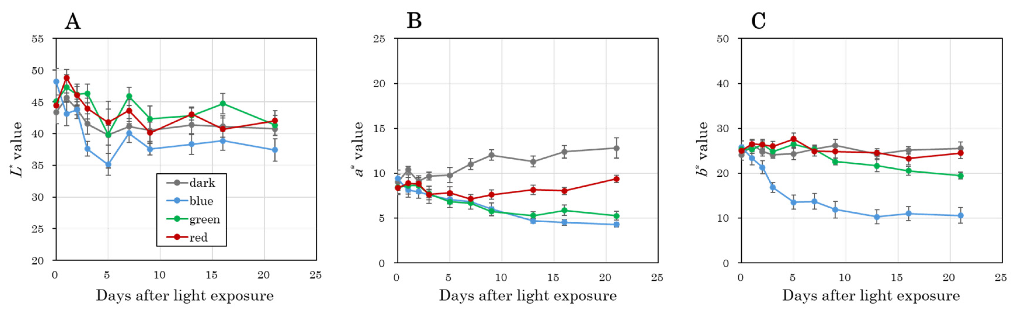
3.2. Time-Course Changes in Color Parameters of the Taproot Epidermis

To quantitatively analyze the changes in taproot epidermal color, L* (lightness), a* (red–green axis), and b* (yellow–blue axis) values were measured using a colorimeter. The L* value showed a decreasing trend in the blue light treatment from 3 days of light exposure, compared to the other groups (Figure 3A). The a* value, which indicates redness, showed an increasing trend from 7 days in the dark control group (Figure 3B). However, in all three light-exposed groups, a* values decreased significantly from 3 days of light exposure. This decrease was sustained in the blue and green light treatments until 14 days of light exposure. The b* value, representing yellowness, significantly decreased in the blue light treatment as early as 1 day of light exposure and continued declining until 5 days of light exposure (Figure 3C). In the green light treatment, a significant reduction in the b* value was observed from 7 days of light exposure.

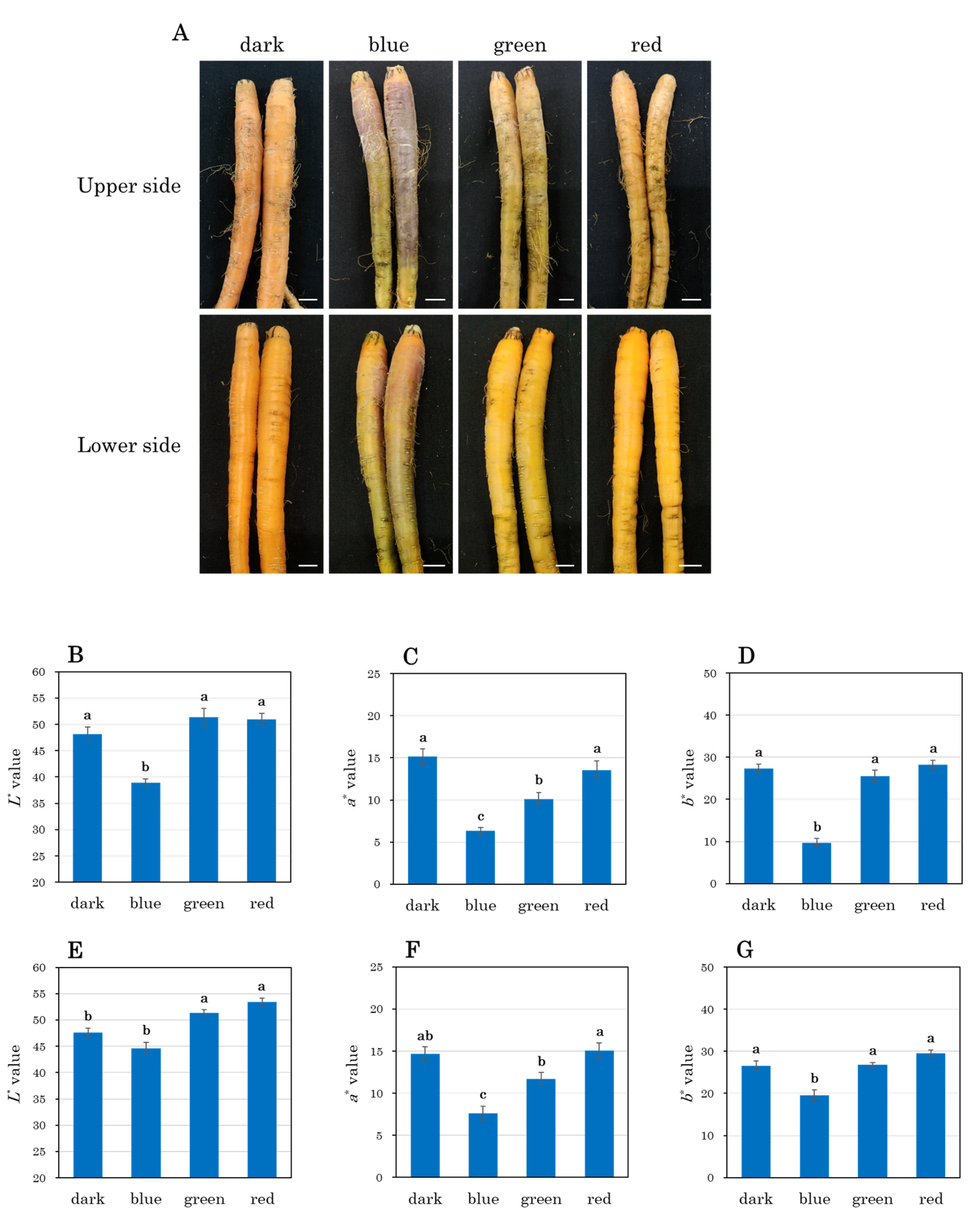
3.3. Visual and Quantitative Analysis of Taproots After 21 Days of Light Exposure

After 21 days of light exposure, taproots were harvested, gently washed to remove any adhered dirt, and analyzed for surface and internal characteristics. Visual observation revealed that light exposure induced more pronounced color changes on the upper side of the taproots (directly exposed to light) compared to the lower side (exposed to indirect and weaker light) (Figure 4A). The L* value on the upper side was significantly lower in the blue light treatment (Figure 4B). The a* value showed the most significant decrease in the blue light treatment, followed by the green light treatment, which also showed a significant decrease compared to the dark control and red light treatment (Figure 4C). Similarly, the b* value was significantly reduced in the blue light treatment (Figure 4D). Although similar trends were observed on the lower side of the taproots, the magnitude of the changes was less pronounced (Figure 4E–G).

3.4. Cross-Sectional Analysis of Taproots

Cross-sectional analysis of taproots revealed distinct pigmentation patterns in response to light treatments. In the blue light treatment, the outer region, including the epidermis and outer cortex, exhibited a reddish-purple hue, while the outer cortex displayed a faint green coloration (Figure 5A). In contrast, the green and red light treatments induced green pigmentation in the stele, with the red light treatment producing a more pronounced effect. After peeling the epidermis, color measurements of the outer cortex showed that the L* value was lowest in the blue light treatment, followed by the green, red, and dark treatments in ascending order (Figure 5B). The a* value was also lowest in the blue light treatment, while the green and red light treatments showed similar decreases compared to the dark control (Figure 5C). The b* value showed the most significant reduction in the blue light treatment, with a decreasing trend also observed in the green light treatment (Figure 5D). Color measurements in the stele showed that the L* value was lowest in the red light treatment, followed by the blue and green light treatments (Figure 5E). The a* value decreased significantly in the red light treatment, followed by the green light treatment, while the blue light treatment also showed a significant decrease compared to the dark control (Figure 5F). No significant differences in the b* value were observed among the treatment groups (Figure 5G).

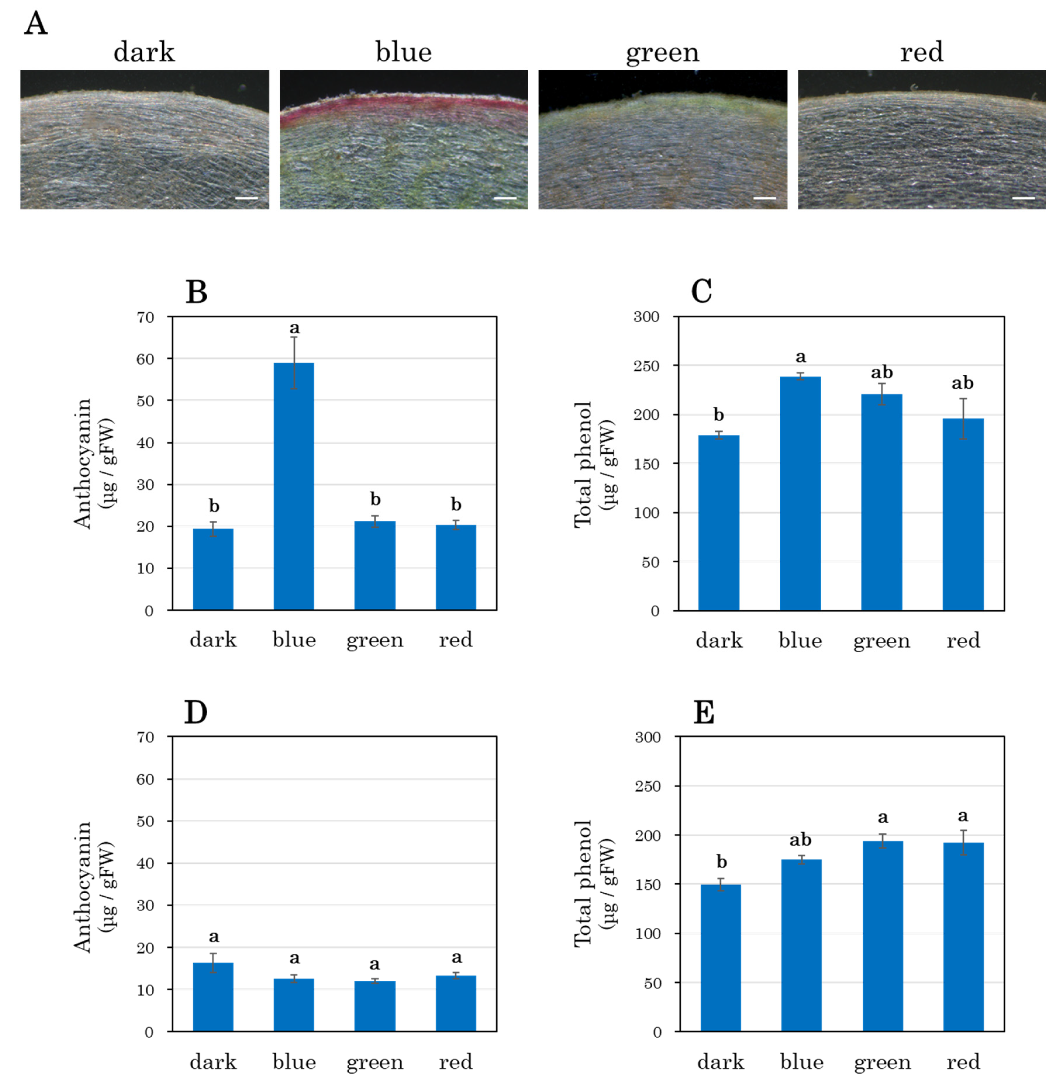
3.5. Anthocyanin and Total Phenol Accumulation

Microscopic observation of the taproot epidermis and outer cortex revealed strong reddish-purple pigmentation in the blue light treatment (Figure 6A). Quantification of anthocyanin content confirmed that anthocyanin accumulation was significantly higher in the blue light treatment than in other groups (Figure 6B). Given that anthocyanins are phenolic compounds synthesized in response to light [33], total phenol content was also analyzed. The results showed the highest accumulation in the blue light treatment, followed by the green and red light treatments (Figure 6C). In contrast, anthocyanin content in the stele did not differ significantly among treatment groups (Figure 6D). However, total phenol content in the stele was significantly higher in the green and red light treatments, while the blue light treatment showed an increasing trend compared to the dark control (Figure 6E).

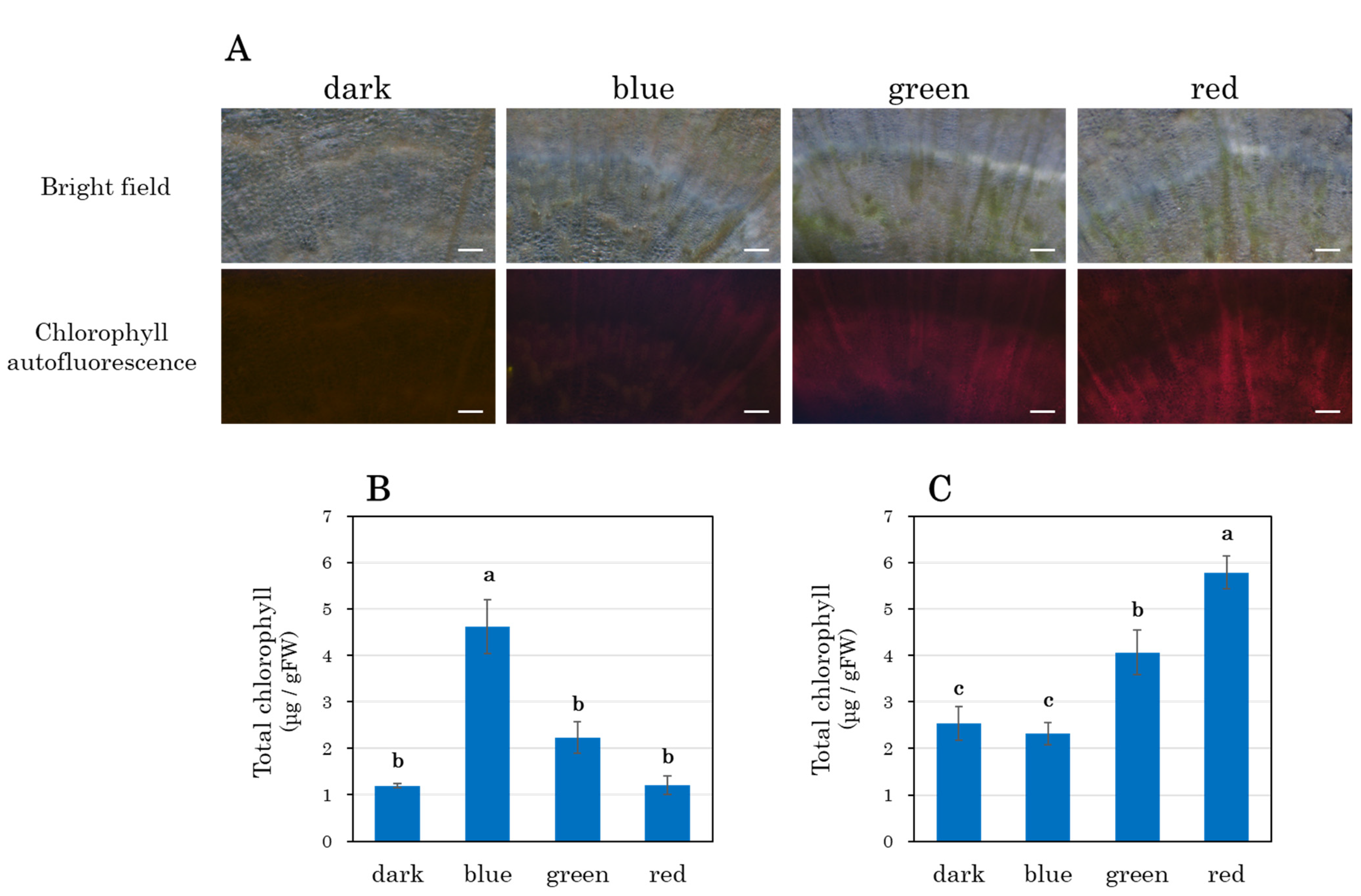
3.6. Chlorophyll Accumulation in the Stele

Since the green and red light treatments induced green pigmentation in the stele, microscopic observations were conducted. Green-colored areas in the stele were observed in both the red and green light treatments, with chlorophyll autofluorescence indicating chloroplast development (Figure 7A). Chlorophyll quantification showed that in the epidermis and outer cortex, chlorophyll content was significantly higher in the blue light treatment (Figure 7B). In contrast, in the stele, chlorophyll content was significantly increased in the red light treatment, followed by the green light treatment, compared to the dark control (Figure 7C).

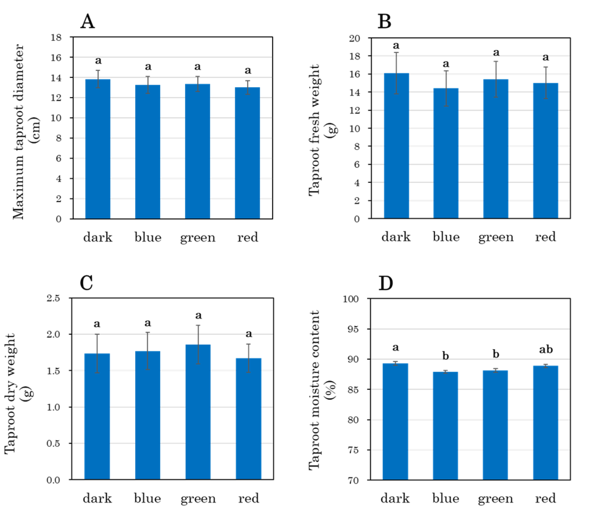
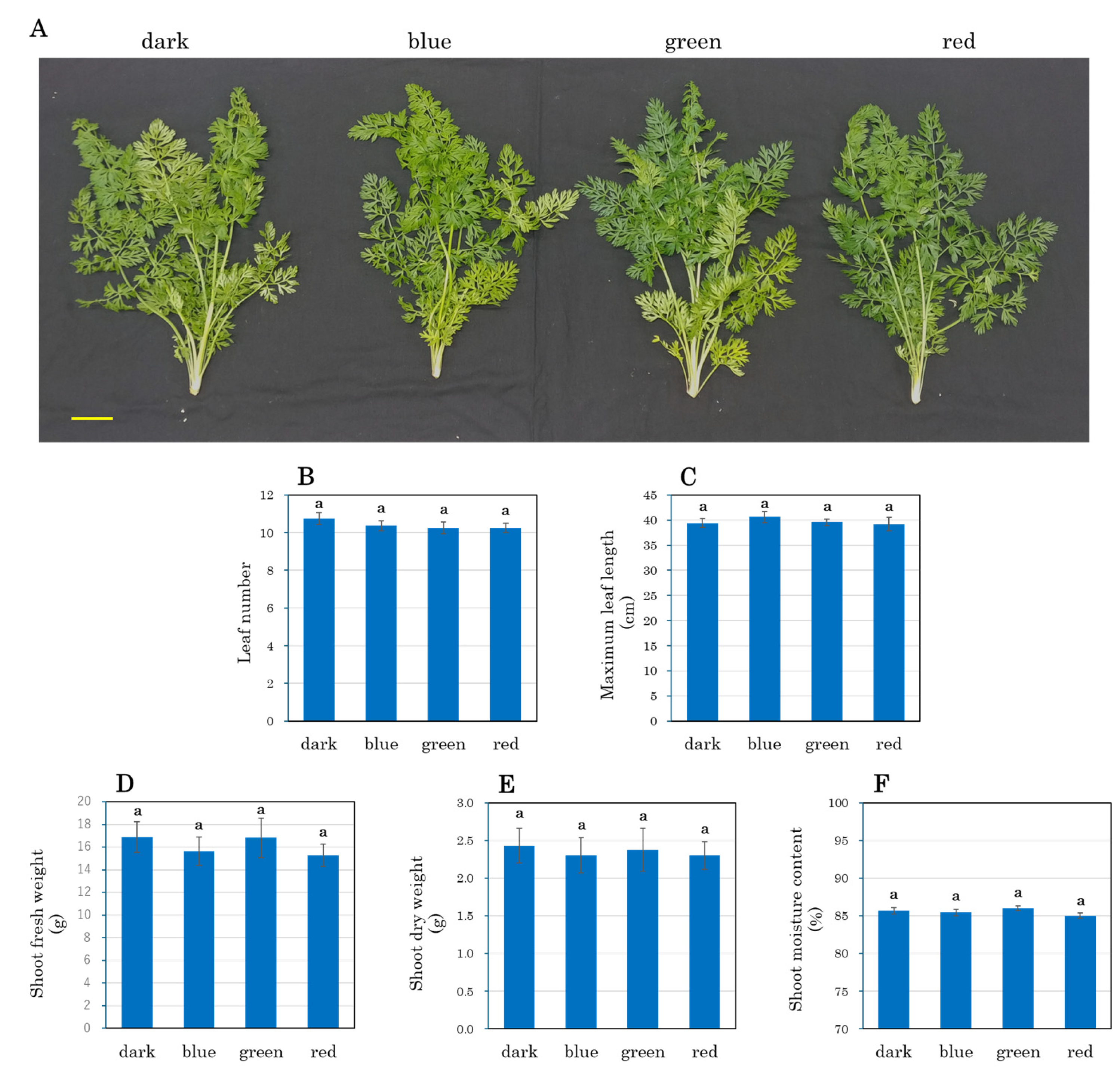
3.7. Growth Parameters of Carrot Taproots and Shoots

After 21 days of light exposure (63 days after sowing), the maximum taproot diameter, taproot fresh weight, and taproot dry weight showed no significant differences among treatments (Figure 8A–C). However, taproot moisture content was significantly lower in the blue and green light treatments compared to the dark control (Figure 8D). Shoot morphology was also evaluated, but no significant differences were observed among treatments in overall appearance (Figure 9A). Similarly, leaf number, maximum leaf length, shoot fresh weight, shoot dry weight, and shoot moisture content showed no differences among treatments (Figure 9B–F).

4. Discussion

4.1. Novel Hydroponic System and Root Development

The hydroponic system developed in this study successfully maintained carrot taproot enlargement. It achieved this by preventing submersion in the nutrient solution while sustaining a high-humidity environment. Previous studies have shown that carrot taproot enlargement is inhibited under submerged conditions due to hypoxia [24,25,34]. Submersion has also been reported to promote the formation of forked taproots [24]. By positioning the taproots in an air space rather than immersing them in the nutrient solution, our system eliminated these negative effects and ensured continued root development.
Root submersion in DFT hydroponic systems has been shown to inhibit the enlargement of various root crops [23,35]. Therefore, the approach presented in this study may be applicable to a wide range of root crops. In sweet potatoes, hydroponic systems incorporating air gaps above the nutrient solution have been developed to facilitate storage root enlargement [36]. Similarly, aeroponic systems have been utilized for root crop cultivation [26,27,28]. However, these systems require substantial vertical space to accommodate root elongation, which can be a limiting factor in certain cultivation settings. In contrast, the system presented in this study, as well as in previous research on sweet potato [23], orients the taproots horizontally, thereby reducing the need for vertical space. This design makes it particularly suitable for integration into vertical farming systems.
The response of root enlargement to water environment varies among species. In turnips, changes in soil moisture levels have been reported to affect shoot yield but not taproot yield [37]. In contrast, proper humidity is essential for storage root enlargement in sweet potatoes [38]. Studies have shown that exposing the upper portion of sweet potato storage roots to sunlight by removing the soil cover inhibits their enlargement, although the lower, soil-covered portion continues to expand [39]. A similar phenomenon has been observed in carrots, where soil removal leading to root exposure to light suppresses enlargement, but only in the illuminated regions [40]. These inhibitions may be linked to reduced humidity, as storage root expansion in carrots, sweet potatoes, and potatoes is known to be restricted under drought stress [41,42,43]. Given that soil moisture availability plays a critical role in carrot taproot enlargement [34,44], the ability of our system to maintain an optimal humidity environment likely played a key role in supporting root growth.

4.2. Anthocyanin Accumulation

In this study, blue light irradiation induced anthocyanin accumulation in the epidermis and outer cortex of carrot taproots. Previous research has demonstrated that blue light enhances the expression of anthocyanin biosynthesis genes in various plant species [45,46,47,48]. Anthocyanins function as antioxidants, and their synthesis is often triggered by oxidative stress [49,50]. For example, herbicide application induces superoxide and hydrogen peroxide accumulation, which in turn triggers anthocyanin synthesis [49]. In cotton seedlings, exposure to 100 μmol m−2 s−1 of blue light rapidly generates hydrogen peroxide in dark-grown hypocotyls [51]. Similarly, in our study, taproots that developed in darkness exhibited anthocyanin accumulation in response to 150 μmol m−2 s−1 of blue light, suggesting that oxidative stress induced by blue light contributed to the synthesis of anthocyanin.
Previous studies on carrots have reported that localized exposure to white light, which consists of multiple wavelengths, does not induce anthocyanin accumulation [40]. However, in lettuce, white LED lighting has been shown to enhance anthocyanin production in leaves [52]. Furthermore, increasing the proportion of blue light in mixed-light conditions has been found to promote anthocyanin accumulation in lettuce [53]. In our study, anthocyanin accumulation was observed only under blue light, while red and green light, as well as darkness, did not induce this response. This suggests that blue light plays a specific role in triggering anthocyanin biosynthesis in carrot taproots.
In pears, cryptochrome-mediated blue light signaling regulates anthocyanin accumulation in fruit epidermal cells [45]. Overexpression of CRY1 in Arabidopsis and poplar has been shown to increase anthocyanin accumulation in leaves and stems [46,54]. Given that photoreceptors are expressed throughout root tissues in Arabidopsis and carrots [4,55,56], it is likely that a cryptochrome-mediated signaling pathway also functions in carrot taproots.
In hairy root cultures, ultraviolet, blue, and red light exposures trigger various secondary metabolite productions [57,58,59,60,61]. In light-grown Echinacea Purpurea hairy roots, anthocyanins accumulate in the outer cortical cell layer, forming a distinct purple ring [60]. This pattern closely resembles our findings, suggesting that light-responsive anthocyanin biosynthesis pathways are conserved in root tissues. Carrot taproots contain high levels of carotenoids, which strongly absorb blue light [62]. This property may have limited blue light penetration, restricting anthocyanin accumulation to the epidermis and outer cortex.
Additionally, our study found that blue, green, and red light exposures increased total phenolic content in carrot taproots. In the epidermis and outer cortex, blue light resulted in the highest total phenol content, suggesting that anthocyanins contributed significantly to the overall phenolic accumulation. In contrast, in the stele, the highest phenolic content was observed under red light exposure. Previous studies on Arabidopsis roots have shown that white and blue light generate reactive oxygen species [62,63]. In carrot hairy root cultures, white light exposure enhances phenolic compound synthesis and antioxidant enzyme activity [64]. Considering that red light has also been reported to have the ability to induce phenolic compounds [65,66], it is likely that red light, which penetrates deeper into carotenoid-rich carrot taproots, triggered light-induced oxidative stress in the stele, thereby promoting phenolic accumulation.

4.3. Chlorophyll Accumulation

Chlorophyll accumulation in response to light exposure has been previously reported in carrot hairy root cultures [64,67]. In this study, red and green light irradiation, particularly red light, induced chlorophyll accumulation in the stele of carrot taproots. In Arabidopsis, light exposure to excised roots promotes chloroplast development in the stele through auxin and cytokinin signaling pathways [3,68]. The stele has high auxin concentrations transported from the shoots, while pericycle cells surrounding the stele exhibit elevated cytokinin levels [69,70]. Carotenoids, the dominant pigments in carrot taproots, have low absorption in the red-light spectrum [62]. Consequently, red light penetrates deeper into the taproot, reaching the stele, where it may induce chloroplast differentiation via hormone-mediated signaling.
In contrast, blue light irradiation induced chlorophyll accumulation in the epidermis and outer cortex of carrot taproots. Similar responses have been observed in sweet potatoes and potatoes, where light exposure promotes chloroplast development in the epidermis and subepidermal cells of storage roots [23,71,72]. In potatoes, the most effective wavelengths for tuber greening are blue (475 nm) and red (675 nm) [73]. Blue and red light are known to induce different types of chloroplast development [74,75,76]. Blue light promotes the formation of chloroplasts with high photosynthetic capacity, characteristic of sun-adapted leaves, whereas red light induces the development of shade-type chloroplasts [74]. These differences in chloroplast induction, along with variations in light penetration within carrot taproots, likely contributed to the distinct chlorophyll accumulation patterns observed in this study.
Although chlorophyll synthesis was induced in carrot taproots, it did not significantly impact overall plant growth. In developing sink organs such as fruits, early-stage chloroplast formation has little influence on biomass production [77]. Similarly, light exposure to sweet potato storage roots induces chlorophyll accumulation but does not affect shoot biomass [23]. In contrast, in hairy root cultures, light exposure enhances biomass production through photosynthesis [78]. These findings suggest that when primary photosynthetic organs (shoots) remain intact, chloroplast development in non-photosynthetic organs, such as storage roots, has minimal impact on overall plant growth.

5. Conclusions

This study highlights the significant influence of controlled light exposure on carrot taproots using a novel hydroponic system that prevents submersion while maintaining optimal humidity. The results demonstrate that blue light induces anthocyanin and chlorophyll accumulation in the epidermis and outer cortex, whereas red and green light promote chlorophyll accumulation in the stele. These findings suggest that taproots, which typically develop in darkness, exhibit spatially distinct physiological responses to light exposure. Despite these pigment changes, root enlargement remained unaffected, indicating that pigment biosynthesis in roots can be regulated independently of biomass accumulation.
Beyond conventional agriculture, this research has broader implications for controlled-environment farming, including applications in space agriculture. Notably, carrots have been identified as a key crop candidate for lunar and extraterrestrial farming [79,80,81,82]. The ability to regulate root growth and biochemical composition through light exposure could contribute to the development of sustainable crop production systems in environments where soil-based cultivation is impractical. Future studies should explore the molecular mechanisms underlying root photoreception and extend this research to other root crops to evaluate its broader applicability.

Author Contributions

Conceptualization, M.S.; methodology, M.S.; formal analysis, M.S., A.F., F.S. and T.K.; investigation, M.S., A.F., F.S. and T.K.; data curation, M.S.; writing—original draft preparation, M.S.; writing—review and editing, M.S. and T.S.; funding acquisition, M.S. All authors have read and agreed to the published version of the manuscript.

Funding

This research was supported by JSPS Grant-in-Aid for Scientific Research C, grant number 23K05474.

Institutional Review Board Statement

Not applicable.

Informed Consent Statement

Not applicable.

Data Availability Statement

The original contributions presented in this study are included in the article; further inquiries can be directed to the corresponding author.

Conflicts of Interest

The authors declare no conflicts of interest.

References

  1. Darko, E.; Heydarizadeh, P.; Schoefs, B.; Sabzalian, M.R. Photosynthesis under Artificial Light: The Shift in Primary and Secondary Metabolism. Philos. Trans. R. Soc. B Biol. Sci. 2014, 369, 20130243. [Google Scholar] [CrossRef] [PubMed]
  2. Paradiso, R.; Proietti, S. Light-Quality Manipulation to Control Plant Growth and Photomorphogenesis in Greenhouse Horticulture: The State of the Art and the Opportunities of Modern LED Systems. J. Plant Growth Regul. 2022, 41, 742–780. [Google Scholar] [CrossRef]
  3. Kobayashi, K.; Baba, S.; Obayashi, T.; Sato, M.; Toyooka, K.; Keränen, M.; Aro, E.-M.; Fukaki, H.; Ohta, H.; Sugimoto, K.; et al. Regulation of Root Greening by Light and Auxin/Cytokinin Signaling in Arabidopsis. Plant Cell 2012, 24, 1081–1095. [Google Scholar] [CrossRef] [PubMed]
  4. Quian-Ulloa, R.; Stange, C. Carotenoid Biosynthesis and Plastid Development in Plants: The Role of Light. Int. J. Mol. Sci. 2021, 22, 1184. [Google Scholar] [CrossRef]
  5. Usami, T.; Mochizuki, N.; Kondo, M.; Nishimura, M.; Nagatani, A. Cryptochromes and Phytochromes Synergistically Regulate Arabidopsis Root Greening under Blue Light. Plant Cell Physiol. 2004, 45, 1798–1808. [Google Scholar] [CrossRef]
  6. Galen, C.; Rabenold, J.J.; Liscum, E. Functional Ecology of a Blue Light Photoreceptor: Effects of Phototropin-1 on Root Growth Enhance Drought Tolerance in Arabidopsis thaliana. New Phytol. 2007, 173, 91–99. [Google Scholar] [CrossRef]
  7. Rizzini, L.; Favory, J.-J.; Cloix, C.; Faggionato, D.; O’Hara, A.; Kaiserli, E.; Baumeister, R.; Schäfer, E.; Nagy, F.; Jenkins, G.I.; et al. Perception of UV-B by the Arabidopsis UVR8 Protein. Science 2011, 332, 103–106. [Google Scholar] [CrossRef]
  8. Wan, J.; Zhang, P.; Wang, R.; Sun, L.; Wang, W.; Zhou, H.; Xu, J. UV-B Radiation Induces Root Bending Through the Flavonoid-Mediated Auxin Pathway in Arabidopsis. Front. Plant Sci. 2018, 9, 618. [Google Scholar] [CrossRef]
  9. Kozai, T. Sustanable Plant Factory: Closed Plant Production System with Articicial Light for High Resource Use Efficiencies and Quality Produce. Acta Hortic. 2013, 1004, 27–40. [Google Scholar] [CrossRef]
  10. Hirawan, D.; Nurhadiansyah, E.; Hadiana, A. Comparison NFT and DFT Hydroponic Method Based on Internet of Things. AIP Conf. Proc. 2023, 2510, 030030. [Google Scholar] [CrossRef]
  11. Garzón, J.; Montes, L.; Garzón, J.; Lampropoulos, G. Systematic Review of Technology in Aeroponics: Introducing the Technology Adoption and Integration in Sustainable Agriculture Model. Agronomy 2023, 13, 2517. [Google Scholar] [CrossRef]
  12. Sakamoto, M.; Suzuki, T. Effect of Root-Zone Temperature on Growth and Quality of Hydroponically Grown Red Leaf Lettuce (Lactuca sativa L. Cv. Red Wave). Am. J. Plant Sci. 2015, 06, 2350. [Google Scholar] [CrossRef]
  13. Sakamoto, M.; Suzuki, T. Elevated Root-Zone Temperature Modulates Growth and Quality of Hydroponically Grown Carrots. Agric. Sci. 2015, 6, 749–757. [Google Scholar] [CrossRef]
  14. Sakamoto, M.; Uenishi, M.; Miyamoto, K.; Suzuki, T. Effect of Root-Zone Temperature on the Growth and Fruit Quality of Hydroponically Grown Strawberry Plants. J. Agric. Sci. 2016, 8, 122. [Google Scholar] [CrossRef]
  15. Levine, C.P.; Hayashi, S.; Ohmori, Y.; Kusano, M.; Kobayashi, M.; Nishizawa, T.; Kurimoto, I.; Kawabata, S.; Yamori, W. Controlling Root Zone Temperature Improves Plant Growth and Pigments in Hydroponic Lettuce. Ann. Bot. 2023, 132, 455–470. [Google Scholar] [CrossRef]
  16. Sakamoto, M.; Suzuki, T. N-Acetylcysteine Mitigates Oxidative Stress Induced by Transplanting Lettuce Seedlings into a DFT Hydroponic System. Agronomy 2024, 14, 2112. [Google Scholar] [CrossRef]
  17. Fujiuchi, N.; Fujiwara, K. LED Dim Light Irradiation of the Root Zone Influences Growth and Development of Leaf Lettuce (Lactuca sativa) Plants under Nutrient Film Technique Hydroponics. Environ. Control Biol. 2012, 50, 101–106. [Google Scholar] [CrossRef]
  18. Paponov, M.; Ziegler, J.; Paponov, I.A. Light Exposure of Roots in Aeroponics Enhances the Accumulation of Phytochemicals in Aboveground Parts of the Medicinal Plants Artemisia annua and Hypericum perforatum. Front. Plant Sci. 2023, 14, 1079656. [Google Scholar] [CrossRef]
  19. Kon, S.; Toyofuku, K.; Muto, D.; Kimura, S.; Ogawa, A. Irradiating Roots of Komatsuna (Brassica napus) with Various Light Qualities Affects Growth and Nutrient Content in Leaves, Stems, and Roots. Sci. Hortic. 2024, 331, 113179. [Google Scholar] [CrossRef]
  20. Eguchi, T.; Yoshida, S. A Cultivation Method to Ensure Tuberous Root Formation in Sweetpotatoes (Ipomoea batatas (L.) Lam.). Environ. Control. Biol. 2004, 42, 259–266. [Google Scholar] [CrossRef]
  21. Kusakawa, T.; Inoue, M. Damage to Pot-cultured Carrot Growth due to a Temporarily Raised Groundwater Level and Flooding Period. Hort. Res. 2010, 9, 495–500. [Google Scholar] [CrossRef]
  22. Siqinbatu; Kitaya, Y.; Hirai, H.; Shibuya, T.; Endo, R. Effects of Soil Water Content on the Growth of Sweet Potato Grown in Sandy Soil. Eco-Eng 2014, 26, 75–80. [Google Scholar] [CrossRef]
  23. Sakamoto, M.; Suzuki, T. Impacts of Light Exposure and Soil Covering on Sweet Potato Storage Roots in a Novel Soilless Culture System. AgriEngineering 2024, 6, 3912–3930. [Google Scholar] [CrossRef]
  24. Terabayashi, S.; Harada, N.; Date, S.; Fujime, Y. Effects of Aeration and Root Immersion Level on the Development of Carrot Root in Hydroponics. Hortic. Res. 2008, 7, 439–444. [Google Scholar] [CrossRef]
  25. Que, F.; Wang, G.-L.; Feng, K.; Xu, Z.-S.; Wang, F.; Xiong, A.-S. Hypoxia Enhances Lignification and Affects the Anatomical Structure in Hydroponic Cultivation of Carrot Taproot. Plant Cell Rep. 2018, 37, 1021–1032. [Google Scholar] [CrossRef]
  26. Hayden, A. Aeroponic and Hydroponic Systems for Medicinal Herb, Rhizome, and Root Crops. HortScience 2006, 41, 536–538. [Google Scholar] [CrossRef]
  27. Selvaraj, M.G.; Montoya-P, M.E.; Atanbori, J.; French, A.P.; Pridmore, T. A Low-Cost Aeroponic Phenotyping System for Storage Root Development: Unravelling the below-Ground Secrets of Cassava (Manihot esculenta). Plant Methods 2019, 15, 131. [Google Scholar] [CrossRef]
  28. Tunio, M.H.; Gao, J.; Shaikh, S.A.; Lakhiar, I.A.; Qureshi, W.A.; Solangi, K.A.; Chandio, F.A. Potato Production in Aeroponics: An Emerging Food Growing System in Sustainable Agriculture for Food Security. Chil. J. Agric. Res. 2020, 80, 118–132. [Google Scholar] [CrossRef]
  29. Sakamoto, M.; Wada, M.; Suzuki, T. Effect of Partial Excision of Early Taproots on Growth and Components of Hydroponic Carrots. Horticulturae 2020, 6, 5. [Google Scholar] [CrossRef]
  30. Lee, J.-H.; Goto, E. Ozone Control as a Novel Method to Improve Health-Promoting Bioactive Compounds in Red Leaf Lettuce (Lactuca sativa L.). Front. Plant Sci. 2022, 13, 1045239. [Google Scholar] [CrossRef]
  31. Sakamoto, M.; Suzuki, T. Methyl Jasmonate and Salinity Increase Anthocyanin Accumulation in Radish Sprouts. Horticulturae 2019, 5, 62. [Google Scholar] [CrossRef]
  32. Chazaux, M.; Schiphorst, C.; Lazzari, G.; Caffarri, S. Precise Estimation of Chlorophyll a, b and Carotenoid Content by Deconvolution of the Absorption Spectrum and New Simultaneous Equations for Chl Determination. Plant J. 2022, 109, 1630–1648. [Google Scholar] [CrossRef] [PubMed]
  33. Yin, H.; Wang, L.; Wu, Y.; Xi, Z. Roles of Non-Visible Light and Temperature in the Regulation of Anthocyanin Synthesis in Fruits and Vegetables. Food Front. 2024, 5, 1968–1983. [Google Scholar] [CrossRef]
  34. White, J.M. Carrot Yield When Grown under Three Soil Water Concentrations. HortScience 1992, 27, 105–106. [Google Scholar] [CrossRef]
  35. Terabayashi, S.; Yomo, T.; Namiki, T. Root Development of Root Crops Grown in Deep Flow and Ebb & Flood Culture. Environ. Control Biol. 1997, 35, 99–105. [Google Scholar] [CrossRef]
  36. Uewada, T. The solution culture of sweet potatoes. Environ. Control. Biol. 1990, 28, 135–140. [Google Scholar] [CrossRef]
  37. Zhang, H.; Schonhof, I.; Krumbein, A.; Gutezeit, B.; Li, L.; Stützel, H.; Schreiner, M. Water Supply and Growing Season Influence Glucosinolate Concentration and Composition in Turnip Root (Brassica rapa ssp. Rapifera L.). J. Plant Nutr. Soil Sci. 2008, 171, 255–265. [Google Scholar] [CrossRef]
  38. Eguchi, T.; Kitano, M.; Eguchi, H. Growth of Tuberous Root as Affected by the Ambient Humidity in Sweetpotato (Ipomoea batatas Lam.). Environ. Control. Biol. 1999, 37, 197–201. [Google Scholar] [CrossRef]
  39. Hozyo, Y.; Kato, S. Thickening Growth and Re-thickening Growth of Tuberous Roots of Sweet Potato Pants (Ipomoea batatas Poiret). Jpn. J. Crop Sci. 1976, 45, 131–138. [Google Scholar] [CrossRef]
  40. Fuentes, P.; Pizarro, L.; Moreno, J.C.; Handford, M.; Rodriguez-Concepcion, M.; Stange, C. Light-Dependent Changes in Plastid Differentiation Influence Carotenoid Gene Expression and Accumulation in Carrot Roots. Plant Mol. Biol. 2012, 79, 47–59. [Google Scholar] [CrossRef]
  41. Mbinda, W.; Dixelius, C.; Oduor, R. Induced Expression of Xerophyta viscosa XvSap1 Gene Enhances Drought Tolerance in Transgenic Sweet Potato. Front. Plant Sci. 2019, 10, 1119. [Google Scholar] [CrossRef] [PubMed]
  42. Boguszewska-Mańkowska, D.; Zarzyńska, K.; Nosalewicz, A. Drought Differentially Affects Root System Size and Architecture of Potato Cultivars with Differing Drought Tolerance. Am. J. Potato Res. 2020, 97, 54–62. [Google Scholar] [CrossRef]
  43. Zhang, R.-R.; Wang, Y.-H.; Li, T.; Tan, G.-F.; Tao, J.-P.; Su, X.-J.; Xu, Z.-S.; Tian, Y.-S.; Xiong, A.-S. Effects of Simulated Drought Stress on Carotenoid Contents and Expression of Related Genes in Carrot Taproots. Protoplasma 2021, 258, 379–390. [Google Scholar] [CrossRef] [PubMed]
  44. Asaduzzaman, M.; Kobayashi, Y.; Mondal, M.F.; Ban, T.; Matsubara, H.; Adachi, F.; Asao, T. Growing Carrots Hydroponically Using Perlite Substrates. Sci. Hortic. 2013, 159, 113–121. [Google Scholar] [CrossRef]
  45. Tao, R.; Bai, S.; Ni, J.; Yang, Q.; Zhao, Y.; Teng, Y. The Blue Light Signal Transduction Pathway Is Involved in Anthocyanin Accumulation in ‘Red Zaosu’ Pear. Planta 2018, 248, 37–48. [Google Scholar] [CrossRef]
  46. Hong, G.-J.; Hu, W.-L.; Li, J.-X.; Chen, X.-Y.; Wang, L.-J. Increased Accumulation of Artemisinin and Anthocyanins in Artemisia annua Expressing the Arabidopsis Blue Light Receptor CRY1. Plant Mol. Biol. Rep. 2009, 27, 334–341. [Google Scholar] [CrossRef]
  47. Ma, Y.; Ma, X.; Gao, X.; Wu, W.; Zhou, B. Light Induced Regulation Pathway of Anthocyanin Biosynthesis in Plants. Int. J. Mol. Sci. 2021, 22, 11116. [Google Scholar] [CrossRef]
  48. Araguirang, G.E.; Richter, A.S. Activation of Anthocyanin Biosynthesis in High Light—What Is the Initial Signal? New Phytol. 2022, 236, 2037–2043. [Google Scholar] [CrossRef]
  49. Xu, Z.; Mahmood, K.; Rothstein, S.J. ROS Induces Anthocyanin Production Via Late Biosynthetic Genes and Anthocyanin Deficiency Confers the Hypersensitivity to ROS-Generating Stresses in Arabidopsis. Plant Cell Physiol. 2017, 58, 1364–1377. [Google Scholar] [CrossRef]
  50. Zheng, X.-T.; Yu, Z.-C.; Tang, J.-W.; Cai, M.-L.; Chen, Y.-L.; Yang, C.-W.; Chow, W.S.; Peng, C.-L. The Major Photoprotective Role of Anthocyanins in Leaves of Arabidopsis thaliana under Long-Term High Light Treatment: Antioxidant or Light Attenuator? Photosynth. Res. 2021, 149, 25–40. [Google Scholar] [CrossRef]
  51. Lv, Q.; Zhao, Q.; Zhu, C.; Ding, M.; Chu, F.; Li, X.; Cheng, K.; Zhao, X. Hydrogen Peroxide Mediates High-Intensity Blue Light-Induced Hypocotyl Phototropism of Cotton Seedlings. Stress Biol. 2023, 3, 27. [Google Scholar] [CrossRef] [PubMed]
  52. Sawatdee, S.; Jarunglumlert, T.; Pavasant, P.; Sakihama, Y.; Flood, A.E.; Prommuak, C. Effect of Mixed Light Emitting Diode Spectrum on Antioxidants Content and Antioxidant Activity of Red Lettuce Grown in a Closed Soilless System. BMC Plant Biol. 2023, 23, 351. [Google Scholar] [CrossRef] [PubMed]
  53. Chung, H.-Y.; Chang, M.-Y.; Wu, C.-C.; Fang, W. Quantitative Evaluation of Electric Light Recipes for Red Leaf Lettuce Cultivation in Plant Factories. HortTechnology 2018, 28, 755–763. [Google Scholar] [CrossRef]
  54. Chen, X.; Fan, Y.; Guo, Y.; Li, S.; Zhang, B.; Li, H.; Liu, L.-J. Blue Light Photoreceptor Cryptochrome 1 Promotes Wood Formation and Anthocyanin Biosynthesis in Populus. Plant Cell Environ. 2024, 47, 2044–2057. [Google Scholar] [CrossRef]
  55. Liu, S.; Zhang, L.; Gao, L.; Chen, Z.; Bie, Y.; Zhao, Q.; Zhang, S.; Hu, X.; Liu, Q.; Wang, X.; et al. Differential Photoregulation of the Nuclear and Cytoplasmic CRY1 in Arabidopsis. New Phytol. 2022, 234, 1332–1346. [Google Scholar] [CrossRef]
  56. Salisbury, F.J.; Hall, A.; Grierson, C.S.; Halliday, K.J. Phytochrome Coordinates Arabidopsis Shoot and Root Development. Plant J. 2007, 50, 429–438. [Google Scholar] [CrossRef]
  57. Binder, B.Y.K.; Peebles, C.A.M.; Shanks, J.V.; San, K.-Y. The effects of UV-B stress on the production of terpenoid indole alkaloids in Catharanthus roseus hairy roots. Biotechnol. Prog. 2009, 25, 861–865. [Google Scholar] [CrossRef]
  58. Chen, I.-G.J.; Lee, M.-S.; Lin, M.-K.; Ko, C.-Y.; Chang, W.-T. Blue Light Decreases Tanshinone IIA Content in Salvia miltiorrhiza Hairy Roots via Genes Regulation. J. Photochem. Photobiol. B Biol. 2018, 183, 164–171. [Google Scholar] [CrossRef]
  59. Wang, Y.; Zhang, H.; Zhao, B.; Yuan, X. Improved Growth of Artemisia annua L Hairy Roots and Artemisinin Production under Red Light Conditions. Biotechnol. Lett. 2001, 23, 1971–1973. [Google Scholar] [CrossRef]
  60. Abbasi, B.H.; Tian, C.-L.; Murch, S.J.; Saxena, P.K.; Liu, C.-Z. Light-Enhanced Caffeic Acid Derivatives Biosynthesis in Hairy Root Cultures of Echinacea purpurea. Plant Cell Rep. 2007, 26, 1367–1372. [Google Scholar] [CrossRef]
  61. Yokawa, K.; Kagenishi, T.; Kawano, T.; Mancuso, S.; Baluška, F. Illumination of Arabidopsis Roots Induces Immediate Burst of ROS Production. Plant Signal Behav. 2011, 6, 1460–1464. [Google Scholar] [CrossRef] [PubMed]
  62. Ashenafi, E.L.; Nyman, M.C.; Shelley, J.T.; Mattson, N.S. Spectral Properties and Stability of Selected Carotenoid and Chlorophyll Compounds in Different Solvent Systems. Food Chem. Adv. 2023, 2, 100178. [Google Scholar] [CrossRef]
  63. Yokawa, K.; Fasano, R.; Kagenishi, T.; Baluška, F. Light as Stress Factor to Plant Roots—Case of Root Halotropism. Front. Plant Sci. 2014, 5, 718. [Google Scholar] [CrossRef] [PubMed]
  64. Mukherjee, C.; Sircar, D.; Chatterjee, M.; Das, S.; Mitra, A. Combating Photooxidative Stress in Green Hairy Roots of Daucus Carota Cultivated under Light Irradiation. J. Plant Physiol. 2014, 171, 179–187. [Google Scholar] [CrossRef]
  65. Reis, A.; Kleinowski, A.M.; Klein, F.R.S.; Telles, R.T.; do Amarante, L.; Braga, E.J.B. Light Quality on the in Vitro Growth and Production of Pigments in the Genus alternanthera. J. Crop Sci. Biotechnol. 2015, 18, 349–357. [Google Scholar] [CrossRef]
  66. Ye, S.; Shao, Q.; Xu, M.; Li, S.; Wu, M.; Tan, X.; Su, L. Effects of Light Quality on Morphology, Enzyme Activities, and Bioactive Compound Contents in Anoectochilus roxburghii. Front. Plant Sci. 2017, 8, 857. [Google Scholar] [CrossRef]
  67. Mukherjee, C.; Samanta, T.; Mitra, A. Redirection of Metabolite Biosynthesis from Hydroxybenzoates to Volatile Terpenoids in Green Hairy Roots of Daucus carota. Planta 2016, 243, 305–320. [Google Scholar] [CrossRef]
  68. Kobayashi, K.; Ohnishi, A.; Sasaki, D.; Fujii, S.; Iwase, A.; Sugimoto, K.; Masuda, T.; Wada, H. Shoot Removal Induces Chloroplast Development in Roots via Cytokinin Signaling. Plant Physiol. 2017, 173, 2340–2355. [Google Scholar] [CrossRef]
  69. Bielach, A.; Podlesáková, K.; Marhavy, P.; Duclercq, J.; Cuesta, C.; Müller, B.; Grunewald, W.; Tarkowski, P.; Benková, E. Spatiotemporal Regulation of Lateral Root Organogenesis in Arabidopsis by Cytokinin. Plant Cell 2012, 24, 3967–3981. [Google Scholar] [CrossRef]
  70. Moore, S.; Jervis, G.; Topping, J.F.; Chen, C.; Liu, J.; Lindsey, K. A Predictive Model for Ethylene-Mediated Auxin and Cytokinin Patterning in the Arabidopsis Root. Plant Commun. 2024, 5, 100886. [Google Scholar] [CrossRef]
  71. Tanios, S.; Eyles, A.; Tegg, R.; Wilson, C. Potato Tuber Greening: A Review of Predisposing Factors, Management and Future Challenges. Am. J. Potato Res. 2018, 95, 248–257. [Google Scholar] [CrossRef]
  72. Wang, J.; Li, J.; Chen, W.; Yang, Z.; Li, X.; Wang, L.; Cao, S.; Shi, L. The Changes in Chlorophyll, Solanine, and Phytohormones during Light-Induced Greening in Postharvest Potatoes. Postharvest Biol. Technol. 2025, 219, 113291. [Google Scholar] [CrossRef]
  73. Petermann, J.B.; Morris, S.C. The Spectral Responses of Chlorophyll and Glycoalkaloid Synthesis in Potato Tubers (Solanum tuberosum). Plant Sci. 1985, 39, 105–110. [Google Scholar] [CrossRef]
  74. Buschmann, C.; Meier, D.; Kleudgen, H.K.; Lichtenthaler, H.K. Regulation of Chloroplast Development by Red and Blue Light. In Annual European Symposium on Photomorphogenesis; Song, P.-S., Ed.; Pergamon: Oxford, UK, 1978; pp. 195–198. ISBN 978-0-08-022677-4. [Google Scholar]
  75. Miao, Y.; Chen, Q.; Qu, M.; Gao, L.; Hou, L. Blue Light Alleviates ‘Red Light Syndrome’ by Regulating Chloroplast Ultrastructure, Photosynthetic Traits and Nutrient Accumulation in Cucumber Plants. Sci. Hortic. 2019, 257, 108680. [Google Scholar] [CrossRef]
  76. Li, Z.; Chen, Q.; Xin, Y.; Mei, Z.; Gao, A.; Liu, W.; Yu, L.; Chen, X.; Chen, Z.; Wang, N. Analyses of the Photosynthetic Characteristics, Chloroplast Ultrastructure, and Transcriptome of Apple (Malus domestica) Grown under Red and Blue Lights. BMC Plant Biol. 2021, 21, 483. [Google Scholar] [CrossRef]
  77. Garrido, A.; Conde, A.; Serôdio, J.; De Vos, R.C.H.; Cunha, A. Fruit Photosynthesis: More to Know about Where, How and Why. Plants 2023, 12, 2393. [Google Scholar] [CrossRef]
  78. Jiao, J.; Fu, J.-X.; Yao, L.; Gai, Q.-Y.; He, X.-J.; Feng, X.; Fu, Y.-J. The Growth, Adventitious Bud Formation, Bioactive Flavonoid Production, Antioxidant Response, and Cryptochrome-Mediated Light Signal Transduction in Isatis tinctoria L. Hairy Root Cultures Exposed to LED Lights. Ind. Crops Prod. 2023, 195, 116496. [Google Scholar] [CrossRef]
  79. Berkovich, Y.A.; Smolyanina, S.O.; Krivobok, N.M.; Erokhin, A.N.; Agureev, A.N.; Shanturin, N.A. Vegetable Production Facility as a Part of a Closed Life Support System in a Russian Martian Space Flight Scenario. Adv. Space Res. 2009, 44, 170–176. [Google Scholar] [CrossRef]
  80. Finetto, C.; Lobascio, C.; Rapisarda, A. Concept of a Lunar FARM: Food and Revitalization Module. Acta Astronaut. 2010, 66, 1329–1340. [Google Scholar] [CrossRef]
  81. Kamran, M.; Auroux, L.; Johnson, K.; Lewsey, M.G. Optimising Plant Form and Function for Controlled Environment Agriculture in Space and on Earth. Mod. Agric. 2023, 1, 86–97. [Google Scholar] [CrossRef]
  82. Ellery, A. Supplementing Closed Ecological Life Support Systems with In-Situ Resources on the Moon. Life 2021, 11, 770. [Google Scholar] [CrossRef] [PubMed]
Figure 1. Overview of the study design. (A) Hydroponic system utilized in this study. (B) Light spectrums of blue, green, and red LEDs used in this study. (C) Detailed timeline of this study.
Eng 06 00087 g001
Figure 2. Time-course changes in the morphology of the carrot taproots after light exposure. Scale bars represent 2.5 cm.
Eng 06 00087 g002
Figure 3. Time-course changes in the color parameters of the carrot taproots after light exposure. Time-course changes in the L* (A), a* (B), and b* (C) values of the taproot epidermis. Vertical bars represent ± SE (n = 8).
Eng 06 00087 g003
Figure 4. Visual comparison of the carrot taproots exposed to light for 21 days. (A) Taproots were observed from the upper and lower sides. Scale bars represent 1 cm. L* (B), a* (C), and b* (D) values of upper sides of taproots. L* (E), a* (F), and b* (G) values of lower sides of taproots. Vertical bars represent ± SE (n = 8). Different letters indicate significant differences between treatment groups (p < 0.05, Tukey–Kramer test).
Eng 06 00087 g004
Figure 5. Visual comparison of the carrot taproot sections exposed to light for 21 days. (A) Taproot sections were observed from horizontal and vertical slices. Scale bars represent 1 cm. L* (B), a* (C), and b* (D) values of the taproot outer cortex. L* (E), a* (F), and b* (G) values of the taproot steles. Vertical bars represent ± SE (n = 8). Different letters indicate significant differences between treatment groups (p < 0.05, Tukey–Kramer test).
Eng 06 00087 g005
Figure 6. Anthocyanin and total phenol accumulation of carrot taproots exposed to light for 21 days. (A) Microscopic images of the taproot cross-sections in the epidermis and cortex (scale bars: 200 μm). Anthocyanin content of epidermis and outer cortex (B), and of stele (D). Total phenol content of epidermis and outer cortex (C), and of stele (E). Vertical bars represent ± SE (n = 6). Different letters indicate significant differences between treatment groups (p < 0.05, Tukey–Kramer test).
Eng 06 00087 g006
Figure 7. Chlorophyll content of carrot taproots exposed to light for 21 days. (A) Microscopic images of the taproot cross-sections in the stele (scale bars: 200 μm). Total chlorophyll content of epidermis and outer cortex (B), and of stele (C). Vertical bars represent ± SE (n = 6). Different letters indicate significant differences between treatment groups (p < 0.05, Tukey–Kramer test).
Eng 06 00087 g007
Figure 8. Maximum diameter (A), fresh weight (B), dry weight (C), and moisture content (D) of carrot taproots exposed to light for 21 days. Vertical bars represent ± SE (n = 8). Different letters indicate significant differences between treatment groups (p < 0.05, Tukey–Kramer test).
Eng 06 00087 g008
Figure 9. Pictures of shoots (A), leaf number (B), maximum leaf length (C), shoot fresh weight (D), shoot dry weight (E), and shoot moisture content (F) of carrot taproots exposed to light for 21 days. Scale bar represents 5 cm. Vertical bars represent ± SE (n = 8). Different letters indicate significant differences between treatment groups (p < 0.05, Tukey–Kramer test).
Eng 06 00087 g009
Disclaimer/Publisher’s Note: The statements, opinions and data contained in all publications are solely those of the individual author(s) and contributor(s) and not of MDPI and/or the editor(s). MDPI and/or the editor(s) disclaim responsibility for any injury to people or property resulting from any ideas, methods, instructions or products referred to in the content.

