Article

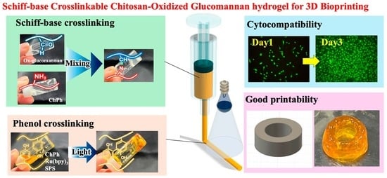
Photo- and Schiff Base-Crosslinkable Chitosan/Oxidized Glucomannan Composite Hydrogel for 3D Bioprinting

Graduate School of Engineering Science, Osaka University, 1-3 Machikaneyama, Toyonaka 560-8531, Japan
*
Authors to whom correspondence should be addressed.
Polysaccharides 2025, 6(1), 19; https://doi.org/10.3390/polysaccharides6010019
Submission received: 16 December 2024 / Revised: 28 January 2025 / Accepted: 28 February 2025 / Published: 4 March 2025

Abstract

:
Chitosan is an attractive material for developing inks for extrusion-based bioprinting of 3D structures owing to its excellent properties, including its mechanical properties and antimicrobial activity when used in wound dressings. A key challenge in formulating chitosan-based inks is to improve its gelation property to ensure reliable printing and the mechanical stability of the printed structures. To address these challenges, this article presents a novel chitosan/oxidized glucomannan composite hydrogel obtained through the combination of Schiff base and phenol crosslinking reactions. The proposed biomaterial forms soft hydrogels through Schiff base crosslinking, which can be further stabilized via visible light-induced phenol crosslinking. This dual-crosslinking approach enhances the printability and robustness of chitosan-based ink materials. The proposed chitosan/oxidized glucomannan hydrogel exhibits excellent extrudability and improved shape retention after extrusion, along with antimicrobial properties against Escherichia coli. Moreover, good cytocompatibility was confirmed in animal cell studies using mouse fibroblast 10T1/2 cells. These favorable features make this hydrogel highly promising for the extrusion-based bioprinting of complex 3D structures, such as tubes and nose-like structures, at a low crosslinker concentration and can expand the prospects of chitosan in bioprinting, providing a safer and more efficient alternative for tissue engineering and other biomedical applications.

Graphical Abstract

1. Introduction

Three-dimensional (3D) bioprinting is a cutting-edge technology for constructing 3D structures of cells and functional materials for biomedical applications [1,2]. Bioprinting techniques can be classified into three main types: extrusion-, inkjet-, and vat-polymerization-based bioprinting [3,4]. Among these, extrusion-based printing is widely used owing to its simplicity and scalability.
The development of new printable materials is a major challenge in extrusion-based bioprinting. The material used in bioprinting should form a hydrogel with sufficient mechanical strength to ensure that the printed structure maintains its form [5]. In addition, properties that promote cell adhesion and differentiation are highly desirable for hydrogels obtained from such printable materials. To date, various types of polymers, including synthetic ones (e.g., poly (vinyl alcohol), polyethylene glycol) and natural ones (e.g., gelatin, silk fibroin), have been studied for the materials of 3D bioprinting [6,7]. Polysaccharides, a class of natural polymers composed of sugar molecules, have gained significant attention due to their favorable biocompatibility and tunable mechanical properties [8,9].
Chitosan, a deacetylated chitin polysaccharide primarily derived from crustaceans, is recognized as a highly biocompatible material with significant potential for bioprinting applications [10,11,12]. Unlike other polysaccharides such as alginic acid and hyaluronic acid, chitosan is cationic, which is an excellent functionality for biomedical applications, including wound dressings and antimicrobial materials [13,14]. In addition, its physicochemical properties are easily tunable through chemical modifications [15]. This functional material also has great potential for 3D bioprinting applications. However, despite the development of various chitosan-based materials, their applications in 3D bioprinting remain limited because of the insufficient mechanical properties of the resultant hydrogels [16,17].
Crosslinking methods are crucial for obtaining stable 3D structures via extrusion-based 3D bioprinting. Typically, ionic and physical crosslinking methods have been used in the application of chitosan in 3D bioprinting [11,18,19]. Although chitosan-based materials crosslinked using these methods have excellent biocompatibility and mechanical properties, the bioprinting process invariably involves a layer-by-layer post-hydrogel process. For example, Jun et al. reported a chitosan ink reinforced with silk particles that was physically crosslinked using an aqueous NaOH solution [19]. Although this ink showed excellent biocompatibility and mechanical properties, the addition of NaOH was necessary after layer-by-layer extrusion to achieve a stable 3D printed structure, which prolongs the printing time.
To address the aforementioned limitations, our group previously developed a phenol-derivatized chitosan (ChPh) that could be cured via visible-light irradiation, resulting in covalent crosslinking (Figure 1a) [20]. Specifically, this ink could be crosslinked within 10 s under visible light (λmax = 452 nm) in the presence of sodium persulfate (SPS) and Ru(bpy)3 through phenol crosslinking to obtain a hydrogel, as illustrated in Figure 1b. In addition, the ink exhibited good antimicrobial activity. However, owing to its fluidity, the ink spread on the printing platform soon after extrusion, resulting in poor shape retention. In addition, although a high SPS concentration or intense light irradiation enabled higher printability with a rapid gelation time, such conditions potentially damage the cells included in the bioink and/or the bioink could degrade due to the formation of excessive amounts of SPS radicals [21]. To overcome this issue, an ink that retains its shape after extrusion while requiring a low crosslinker concentration is highly desirable.
One of the effective strategies for achieving high printability is to use a hydrogel, because unlike liquid materials, it maintains its shape after the extrusion process owing to its solid-like property [22,23,24]. In particular, a self-healing material is desirable for extrusion-based bioprinting because such a material recovers its gel state after the extrusion process, during which the ink deforms under the high shear stress applied for its extrusion [24,25,26]. Schiff base crosslinking by the reaction of amino groups with aldehyde groups is known to generate a soft hydrogel with self-healing capability [27,28,29]. In addition, this reaction occurs at room temperature without requiring toxic crosslinkers, which makes it ideal for extrusion-based bioprinting. Hydrogels obtained through Schiff base crosslinking have been previously used as injectable hydrogels for wound-dressing applications [28,30,31].
In this study, we developed a photo- and Schiff base-crosslinkable hydrogel composed of ChPh and oxidized glucomannan (Ox-glucomannan) to enhance the printability of chitosan-based materials for extrusion-based bioprinting. Ox-glucomannan was easily obtained by oxidizing glucomannan, a biocompatible and biodegradable polymer (Figure 1c) [32,33,34]. Upon mixing Ox-glucomannan with ChPh, a soft hydrogel was formed via a Schiff base reaction between the aldehyde groups of Ox-glucomannan and the amino groups of ChPh (Figure 1d). This hydrogel was further crosslinked by a photo-crosslinking reaction of the phenol moieties in ChPh to improve its mechanical robustness.
In the 3D printing process, the self-healing Schiff base hydrogel of the ChPh/Ox-glucomannan composite was initially extruded from the extruder of an extrusion-based 3D printer (Figure 1d). Then, the hydrogel was further stabilized through visible light-induced phenol crosslinking (Figure 1e). The mechanical properties of the developed hydrogel, including its extrudability and rheological behavior, were evaluated. This hydrogel exhibited extrudability and self-healing properties, enabling excellent printability. In addition, the hydrogel exhibited antimicrobial activity against Escherichia coli (E. coli) and excellent cytocompatibility in animal cells. The proposed chitosan/Ox glucomannan hydrogel is expected to broaden the application of chitosan in extrusion-based 3D bioprinting.

2. Materials and Methods

2.1. Materials

Chitosan (Chitosan LL; deacetylation: 80%, weight-average molecular weight: 75 kDa) was obtained from Yaizu Suisankagaku Industry (Shizuoka, Japan). Ru(bpy)3·Cl2·6H2O, SPS, 3-(4-hydroxyphenyl) propionic acid (HPP), 1-ethyl-3-(3-dimethylaminopropyl)carbodi-imide hydrochloride (EDC·HCl), and N,N,N′,N′-tetramethylethylenediamine (TEMED) were obtained from Sigma Aldrich (Burlington, MA, USA). Glucomannan and sodium periodate were purchased from Wako Pure Chemical Industries (Osaka, Japan). E. coli (OP50) was cultured in Luria Broth (LB) medium containing 0.5% (w/v) NaCl, 1% (w/v) Bacto tryptone (Becton Dickinson and Company, Franklin Lakes, NJ, USA), and Bacto yeast extract (Becton Dickinson and Company). Mouse fibroblast 10T1/2 cells were purchased from Riken Cell Bank (Ibaraki, Japan). The cells were cultured with 10 vol% fetal bovine serum (FBS) in Dulbecco’s modified Eagle’s medium (DMEM; Nissui, Tokyo, Japan) in an incubator with 5% CO2.

2.2. Synthesis of the Phenol Derivative of Chitosan (ChPh)

ChPh was synthesized according to a previously reported method [35] (Figure 1a). In brief, chitosan was dissolved in 20 mM HCl to a concentration of 7.0 wt%. TEMED was added at a concentration of 2.0 wt%, and the pH was adjusted to 5 using NaOH and HCl. Lactobionic acid, HPP, and EDC·HCl were subsequently added to the solution at 0.04, 1.5, and 1.0 wt%, respectively. After 20 h of stirring at room temperature, excess ethanol was added to precipitate the product. The precipitate was washed several times with an 80 wt% ethanol/water solution, dehydrated using ethanol, and dried under vacuum.

2.3. Synthesis of Oxidized Glucomannan (Ox-Glucomannan)

Ox-glucomannan was synthesized according to a previously reported method [32] (Figure 1c). Glucomannan was dissolved in deionized water at a concentration of 1.0 wt%. Sodium iodate was then added to the solution at a 1.0 wt% concentration. The resulting solution was stirred for 5 h at room temperature, after which 10 vol% ethylene glycol was added to it to reduce any unreacted periodate, and the mixture was stirred for an additional 2 h. The mixture was finally dialyzed to remove residual iodate, as confirmed by a silver nitrate test. The resulting solution was freeze-dried to obtain a solid product.

2.4. Fourier-Transform Infrared (FTIR) Spectroscopy

The FTIR spectra of glucomannan and Ox-glucomannan were obtained using an FT/IR-4100 instrument (JASCO, Tokyo, Japan). Each sample was dried in an oven prior to measurement. Ninety scans at a resolution of 2 cm−1 were conducted on each sample.

2.5. Preparation of Schiff Base Hydrogels

Schiff base hydrogels (Figure 1d) composed of Ox-glucomannan and ChPh were prepared as follows: The ChPh solution was mixed with Ox-glucomannan solutions of varying concentrations in phosphate-buffered saline (PBS, pH 7). The final ChPh concentration was fixed at 1.0 wt%. Six different samples were prepared, as listed in Table 1. Hydrogels used in every experiment described below were prepared using this method.

2.6. Extrudability

The extrudability of ChPh, ChPh-Ox25, ChPh-Ox50, ChPh-Ox100, ChPh-Ox200, and ChPh-Ox300 was evaluated using a syringe. Each sample was incorporated with 2.0 mM SPS and 1.0 mM Ru(bpy)3 for inducing the phenol crosslinking reaction. The samples were loaded into syringes and manually extruded through a tube of 1.5 mm diameter to assess their extrudability.

2.7. Rheological Properties

The self-healing properties of ChPh, ChPh-Ox25, ChPh-Ox50, and ChPh-Ox100 were evaluated based on a reported method [36,37]. A stainless steel plate with a diameter of 25 mm was used as the probe, and the bottom platform was also made of steel. The sample (0.65 mL) was poured onto the platform, and the gap between the probe and platform was set as 0.5 mm. Time-course measurements were conducted at 25 °C under 5–1000% strain at 1.6 Hz frequency for 4 min. For comparison, a 0.5 wt% glucomannan solution, a mixture of 0.5 wt% glucomannan and 1.0 wt% ChPh solutions, and a 1.0 wt% Ox-glucomannan solution were tested using the method described above.
In addition, the rheological properties of ChPh, ChPh-Ox25, ChPh-Ox50, and ChPh-Ox100, each containing 2.0 mM SPS and 1.0 mM Ru(bpy)3, were evaluated under visible-light exposure using a rheometer equipped with parallel plate geometry (HAAKE MARS III, Thermo Fisher Scientific, Waltham, MA, USA), as reported earlier [38]. A stainless steel plate with a diameter of 25 mm was used as the probe, and the bottom platform was made of transparent glass to allow visible-light irradiation. The sample (0.65 mL) was poured onto the platform, and a gap of 0.5 mm was set between the probe and platform. Time-course measurements were conducted at 25 °C under 5% strain with 0.3 Hz frequency. After 120 s of the measurement, the sample was exposed to visible light (0.2 W/m2 @ 452 nm) from the bottom of the platform.

2.8. Swelling

The swelling properties of the dual-crosslinked ChPh, ChPh-Ox25, ChPh-Ox50, and ChPh-Ox100 hydrogels were evaluated in PBS (pH 7). A 150 µL volume of the hydrogel sample with 2.0 mM SPS and 1.0 mM Ru(bpy)3 was loaded into a mold of 12 mm diameter and exposed to visible light (0.2 W/m2 @ 452 nm) for 10 min for phenol crosslinking. The sample was then placed in 30 mL of PBS at room temperature, and its diameter was measured after 72 h. Swelling was evaluated as follows:
Swelling   [ - ] = A r e a   o f   t h e   h y d r o g e l   a t   x   h o u r I n i t i a l   a r e a   o f   t h e   h y d r o g e l
The stability of the Schiff base hydrogels was also tested using the same swelling test. For this, ChPh-Ox50 hydrogels without SPS and Ru(bpy)3 were prepared and placed in 30 mL of PBS and acetate buffer (pH 4).

2.9. Cell Viabilities

For cytotoxicity evaluation, the viabilities of mouse fibroblast 10T1/2cells on the ChPh, ChPh-Ox25, ChPh-Ox50, and ChPh-Ox100 hydrogels were assessed. A 100 µL volume of each Schiff base hydrogel added with 2.0 mM SPS and 1.0 mM Ru(bpy)3 was placed in a well of a 48-well plate and exposed to visible light (0.2 W/m2 @ 452 nm) for 10 min to form phenol crosslinks. The samples were then washed several times with PBS. Thereafter, 200 µL of the cell suspension (2.0 × 10⁴ cells/mL) was added to the sample, and the plate was incubated at 37 °C in 5% CO2. After 1 and 3 d of incubation, the cells were stained with calcein-AM and propidium iodide to evaluate cell viability. The cells cultured on a standard cell culture dish were used as the control.

2.10. Antimicrobial Activities

To evaluate the antimicrobial activities of the ChPh, ChPh-Ox25, ChPh-Ox50, and ChPh-Ox100 hydrogels, a 150 µL gel sample containing 2.0 mM SPS and 1.0 mM Ru(bpy)3 was prepared in a mold of 12 mm diameter and exposed to visible light for 10 min. Each sample was then placed in 2 mL of an LB medium containing 2.5 × 10 CFU/mL of E. coli. The samples were incubated at 37 °C in a shaking incubator, and the bacterial concentration was measured as described previously [39].

2.11. 3D Printing

The 3D structures of the ChPh, ChPh-Ox25, ChPh-Ox50, and ChPh-Ox100 hydrogels were printed using a modified 3D printer [20]. Each sample, containing 2.0 mM SPS and 1.0 mM Ru(bpy)3, was loaded into a syringe and extruded through a 20-gauge needle at a flow rate of 2.0 mm/s using a syringe pump (KDS 220, KD Scientific Inc., Holliston, MA, USA) and exposed to visible light (0.2 W/m2 @ 452 nm) for phenol crosslinking to construct a tubular structure.

2.12. Statistical Analysis

Statistical analysis was performed using Student’s t-test. A p-value < 0.05 was considered statistically significant. Data analysis was conducted using Microsoft Excel (ver. 16.79, Microsoft, Redmond, WA, USA).

3. Results and Discussion

3.1. FTIR Spectra

The oxidation of glucomannan to Ox-glucomannan was confirmed by FTIR spectroscopy (Figure 2). For glucomannan, a broad peak was observed at around 3390 cm−1, corresponding to the O-H stretching vibration [40]. The characteristic peak at around 2900 cm−1 was assigned to the -CH2 stretching vibration [40]. The characteristic stretching vibrational peaks of C=O and C-O were observed at 1699 and 1248 cm−1, indicating the presence of acetyl groups [40]. The peak at 1635 cm−1 could be attributed to intramolecular hydrogen bonding [41,42]. The peaks at 1150 and 1022 cm−1 could be attributed to C-O-C stretching from the other groups of pyranose and C-OH bending vibrations [40,43], respectively. The peaks at 873 and 805 cm−1 were assigned to the stretching vibrations of glycosidic acid and mannose, respectively [40,43]. Similar vibrational bands were observed for Ox-glucomannan, except the disappearance of the peak at 805 cm−1, indicating the cleavage of mannose [28,40]. This result indicated the oxidation of glucomannan. However, the peak at around 1715 cm−1, attributed to the aldehyde group, was not observed [44,45,46], probably due to the low resolution of FTIR equipment or the overlapping of the acetyl group peak.

3.2. Extrudability

Next, we investigated the extrudability of the Schiff base hydrogels of the ChPh/Ox-glucomannan composites with different Ox-glucomannan contents. The samples were loaded into syringes and manually extruded (Figure 3). The ChPh solution without Ox-glucomannan exhibited fluid-like properties, and the sample injected from the syringe formed a liquid pool, as illustrated in Figure 3. In contrast, the ChPh/Ox-glucomannan samples could be extruded from the syringe in the form of a string owing to their viscoelastic properties. In particular, the ChPh-Ox25, ChPh-Ox50, and ChPh-Ox100 samples containing 0.25, 0.5, and 1.0 wt% of Ox-glucomannan, respectively, formed hydrogels that could be extruded from the syringe. This result indicates that the chitosan-based hydrogels could be prepared simply by mixing ChPh and Ox-glucomannan through Schiff base crosslinking. However, the samples with higher concentrations of Ox-glucomannan (2 and 3 wt%) could not be filled into the syringe because of their rigidity. Note that soft gels are desirable for extrusion-based 3D bioprinting because they retain their shape after extrusion from a nozzle, which is critical for good printability [47,48]. The hydrogels retained their shape and hydrogel state after the extrusion process.

3.3. Rheological Properties of the Hydrogels with Schiff Base and Phenol Crosslinks

To investigate the rheological properties of the Schiff base hydrogels, time sweep measurements were conducted to monitor the changes in the storage and loss moduli (G′ and G″, respectively) at different applied strains (5 and 1000%). For the ChPh-Ox25, ChPh-Ox50, and ChPh-Ox100 samples, the storage moduli (G′) significantly exceeded the loss moduli (G″) under low strain (5%), corresponding to the state shown in Figure 3. G′ increased with the increase in the Ox-glucomannan concentration in the hydrogel, indicating enhanced Schiff base crosslinking. On the other hand, the G″ exceeded the G′ under high strain (1000%), revealing an increase in their liquid-like property, indicating the shear-thinning behavior of the samples (Figure 4a–d). Furthermore, these properties of the hydrogels were retained after multiple cycles of switching between low and high strain values, indicating their self-recovery capability. In Schiff base hydrogelation, an imine bond is formed through a dehydration–condensation reaction between the amino and aldehyde groups of the hydrogel components, and the system is in an equilibrium state [49]. Under high strain, water movement inside the hydrogel is activated, promoting the hydrolysis of the imine bonds, resulting in increased G″. The hydrogels exhibited self-healing properties typical of Schiff base hydrogels [49,50]. They could maintain their gel state after being loaded into a syringe or extruded through a needle in the bioprinting process, during which they experience a high shear stress. This property can help repair the printed structure during the printing process, leading to improved printability [26,51].
Furthermore, the rheological properties of glucomannan, Ox-glucomannan, and a mixture of ChPh and glucomannan were also evaluated for comparison. The storage moduli of pristine glucomannan and the ChPh/glucomannan mixture did not exceed the loss moduli under both high and low strain values, indicating their fluidic properties (Figure 4e,f). In contrast, the storage modulus of Ox-glucomannan exceeded the loss modulus at the high strain; however, the difference was negligible (approximately 0.09 Pa, Figure 4g) and is probably due to the measurement limit of the rheometer. These results demonstrate that a high storage modulus is achieved only when ChPh and Ox-glucomannan are mixed.
Moreover, the storage and loss moduli of the samples were monitored under the exposure of visible light to verify the effect of phenol crosslinking on the mechanical properties of the hydrogels. The samples were exposed to visible light after 120 s of measurements (Figure 5a–d). Upon exposure to visible light, G’ continued to increase over time, reflecting the covalent crosslinking of the phenol groups anchored to chitosan under visible light [20,52]. The final G’ increased only slightly with increasing Ox-glucomannan concentration, and no significant difference was observed among the samples. These results suggest that a higher concentration of Ox-glucomannan affects the mechanical properties of the hydrogel by increasing the extent of Schiff base crosslinking before the phenol crosslinking process, as demonstrated by the injectability test.

3.4. Swelling

The swelling properties of the chitosan/Ox-glucomannan hydrogels were evaluated to understand their physicochemical stability over a 72 h duration (Figure 6a,b). Stability tests were conducted in PBS (pH 7) to mimic the nearly neutral physiological environment [53,54]. The hydrogels did not degrade for at least 72 h, indicating good stability, which is critical for their long-term use in biomedical applications [55,56]. However, all samples shrank over time (Figure 6b), with no significant difference in shrinkage observed among the samples (p > 0.05). The molecular weight or crosslinking network of the polymer plays a key role in the shrinkage of hydrogels [57]. In this study, we used low-molecular-weight chitosan (75 kDa), which may have caused shrinkage for the long-time stability test due to the lower degree of freedom. A polymer with a higher molecular weight (e.g., >500 kDa) could hold more water in its hydrogel network structure owing to its longer chains and its high degree of freedom [58,59]. In addition, the shrinkage of the hydrogel might be due to the hydrophobicity of the phenol groups anchored to chitosan. Phenol groups are hydrophobic owing to their aromatic hydrocarbon structure [60]. Furthermore, the crosslinking of these phenol groups by the light irradiation could increase the hydrophobicity of the hydrogel, leading to water expulsion and shrinkage. To explain the mechanisms underlying the shrinkage, additional investigations would be necessary to understand the dynamic equilibrium relationships between these contributing factors.

3.5. Effect of pH on Swelling and Stability

Furthermore, the effect of pH on the swelling of the chitosan-based hydrogels was investigated to determine the stability of the hydrogels with and without phenol crosslinking. Hydrogel samples of ChPh-Ox50 with and without phenol crosslinking were immersed in PBS (pH 7) or acetate buffer solution (pH 4) for 72 h. In the acetate buffer (acidic condition, pH 4), the hydrogel without phenol crosslinking degraded within 20 min of incubation (Figure 7a). This is because the amino moieties of chitosan interact with the protons in the acidic solution [58], causing the degradation of the imine bonds. In contrast, the hydrogels obtained through both Schiff base and phenol crosslinking did not degrade for at least 72 h. Phenol crosslinking is stable because an irreversible covalent bond is formed between phenol moieties. Thus, dual crosslinking of the gel components via phenol and Schiff base crosslinking could be effective for stabilizing the hydrogel under acidic physiological conditions, such as the stomach and skin [53]. Further, under neutral conditions (pH 7), both the hydrogels with and without phenol crosslinking did not degrade over 72 h, although both hydrogels shrank (Figure 7b). The formation of Schiff base crosslinks is promoted at a high pH (>4) [61]; therefore, the hydrogel without phenol crosslinking remained stable in PBS.
To further understand the effect of pH on swelling, the swelling behaviors of the ChPh-Ox50 hydrogels in PBS (pH 7) and acetate buffer (pH 4) solutions were compared for 72 h. The hydrogel shrank in PBS, as mentioned in Section 3.4, but swelled in the acetate buffer solution (Figure 7c). In acidic conditions, more amino groups are protonated, and they interact with more water molecules via hydrogen bonding, which probably caused swelling. Such a pH effect should be considered while designing 3D data of the targets in 3D bioprinting applications. For instance, the expansion of the hydrogel needs to be considered when the hydrogel is applied to wound dressing, as the pH of skin is mildly acidic (pH 4–7) [54,62].

3.6. Cell Viabilities

The cytocompatibility of the dual-crosslinked chitosan-based hydrogels was evaluated by assessing the viability of mouse fibroblast cells from mouse embryos on the ChPh, ChPh-Ox25, ChPh-Ox50, and ChPh-Ox100 hydrogels (Figure 8). A cell suspension was applied to the hydrogels, and the cells were cultured for 3 d. On day 1, cell attachment and elongation were observed on all hydrogel samples. On day 3, the cells on the ChPh hydrogel did not grow, although they were alive. Chitosan is a cationic polymer consisting of protonated amino groups in its molecular structure [8], which might allow cell extension or growth. On the other hand, cell growth was observed on the ChPh-Ox25, ChPh-Ox50, and ChPh-Ox100 hydrogels with Ox-glucomannan. The cell densities were, however, lower than those observed in the control group (cell culture dish). The improved cell growth on the composite hydrogels containing Ox-glucomannan compared with that on the ChPh hydrogel is likely due to the reduced cationic content owing to the bonding of the amino groups of chitosan with the aldehyde groups of Ox-glucomannan. In addition, the aldehyde groups of Ox-glucomannan in the hydrogel could interact with the external solution. In this study, DMEM containing various growth factors, including amino acids and FBS, was used as the cell culture medium. These components contain amino groups that likely interacted with the aldehyde groups of Ox-glucomannan, promoting cell attachment and growth on the hydrogel. Several studies have reported the cytocompatibility of chitosan hydrogels [15,63]. The properties of our hydrogels are consistent with previously reported results. For further functionalization, modification with additional cell growth factors, such as RGD-motif-containing peptide, may be necessary [64,65]. Furthermore, controlling the porosity of the hydrogels could enhance and regulate both cell proliferation and differentiation [66].

3.7. Antimicrobial Activities

Next, the antimicrobial activities of the ChPh, ChPh-Ox25, ChPh-Ox50, and ChPh-Ox100 hydrogels were evaluated (Figure 9a). When E. coli was cultured in an LB medium in the presence of each hydrogel, bacterial growth was inhibited in all samples for at least 24 h, whereas significant bacterial growth occurred in the LB medium without a hydrogel (Figure 9b). The antimicrobial activity of chitosan is primarily due to its cationic amino groups, which inhibit bacterial growth [67,68]. These amino groups are protonated in an aqueous medium, making chitosan positively charged. As bacterial cell surfaces are negatively charged, the positively charged chitosan interacts with and adsorbs bacteria, thereby inhibiting nutrient uptake from the surrounding environment [67]. Although no significant differences were observed between the results of ChPh and ChPh-Ox25, -Ox50, and -Ox100, the antimicrobial activity of the ChPh-Ox glucomannan sample slightly decreased compared to that of ChPh. This might be due to the interaction between the aldehyde group of Ox-glucomannan and the amino group of ChPh, inhibiting the interaction between ChPh and bacteria. Antimicrobial activity is an important factor for preventing microbial contamination when cells are cultured on scaffolds [69,70]. Moreover, it plays a critical role in preventing infections in wound dressing applications.

3.8. 3D Printing

To evaluate the printability of the ChPh, ChPh-Ox25, ChPh-Ox50, and ChPh-Ox100 hydrogel samples, tubular structures were printed using a 3D printer (Figure 10a,b). The tube structure printed using the ChPh hydrogel collapsed owing to the low SPS concentration (2.0 mM). In a previous study, good printability of ChPh was observed at 4.0 mM of SPS because of the rapid gelation of the sample under visible light [20]. In contrast, ChPh-Ox25 and ChPh-Ox50 exhibited good printability at the same low SPS concentration (2.0 mM). However, in the case of the ChPh-Ox100 hydrogel, the nozzle and the tube between the nozzle and syringe pump were clogged because of its stiffness. To use ChPh-Ox100 effectively, a syringe pump with a higher discharge capacity or a nozzle with a larger diameter would be required. Furthermore, nose-shaped structures were successfully printed with ChPh-Ox25 and ChPh-Ox50 hydrogels (Figure 10b). The structure printed with ChPh-Ox25 showed lower printability compared to ChPh-Ox50, likely due to its lower storage modulus prior to the light irradiation (Figure 5b,c). Among the samples, ChPh-Ox50 showed the optimal printability in this study. To obtain the 3D-printed structure with better printability, further investigation would be required, including the effects of ChPh or Ox-glucomannan concentration and printing parameters such as nozzle diameter and light intensity. Several groups have demonstrated the extrusion-based bioprinting of chitosan materials [7,14]. Although those samples showed great functionalities, including cell compatibility and physical stability, a post-hydrogelation process was necessary for stabilizing the 3D structures printed in a layer-by-layer manner, resulting in a long printing time [13,14]. Our results suggest that the ChPh/Ox-glucomannan hydrogels are effective for building a variety of 3D structures via extrusion-based 3D bioprinting without a post-crosslinking process. For bioprinting, a high cell viability is essential for encapsulating or casting cells. In addition, high printability is also critical for producing 3D structures with a high resolution, depending on the targeted application [71,72]. The soft chitosan-based hydrogel developed in this study retained its shape after extrusion, and its stiffness increased upon exposure to visible light. These results indicate that the chitosan/Ox-glucomannan hydrogel demonstrates good printability in extrusion-based 3D printing at a low SPS concentration. This material may have great potential for the application of wound dressing due to the printability along with the results of cell compatibility and antimicrobial activity.

4. Conclusions

In this study, we developed a novel hydrogel comprising chitosan and oxidized glucomannan. The implementation of Schiff base crosslinking enabled the formation of extrudable soft hydrogels, while phenol crosslinking triggered by visible light provided additional mechanical stability. The hydrogel showed self-healing properties, enabling extrudability from the syringe. Therefore, the hydrogel demonstrated excellent printability at a low SPS concentration of 2.0 mM used for phenol crosslinking, which is suitable for bioprinting. Especially, ChPh-Ox50 showed excellent printability. In addition, the hydrogel crosslinked with Schiff base and phenol crosslinking showed high stability under acidic and neutral conditions. Furthermore, the hydrogel exhibited strong antimicrobial activity and good cytocompatibility, which render it suitable for biomedical applications, such as tissue engineering. The enhanced mechanical properties and cytocompatibility of our chitosan/oxidized glucomannan hydrogels demonstrate the significant potential for expanding the use of chitosan-based hydrogels in 3D bioprinting, offering a safer and more efficient solution for fabricating complex biological structures. Further parameter optimization would be necessary for higher printability and practical application.

Author Contributions

M.H.: Conceptualization, investigation, analysis, visualization, and writing—original draft; S.S.: writing—review and editing and project administration; and writing. All authors have read and agreed to the published version of the manuscript.

Funding

This research was supported by a scholarship from JSPS KAKENHI (Grant No. 24KJ1594).

Institutional Review Board Statement

Not applicable.

Data Availability Statement

The raw data supporting the conclusions of this article will be made available by the authors on request.

Acknowledgments

M.H. thanks the members of the Sakai laboratory for kind support in his research activity.

Conflicts of Interest

The authors declare no conflicts of interest.

References

  1. Gungor-Ozkerim, P.S.; Inci, I.; Zhang, Y.S.; Khademhosseini, A.; Dokmeci, M.R. Bioinks for 3D Bioprinting: An Overview. Biomat. Sci. 2018, 6, 915–946. [Google Scholar] [CrossRef] [PubMed]
  2. Pedroza-González, S.C.; Rodriguez-Salvador, M.; Pérez-Benítez, B.E.; Moisés Alvarez, M.; Santiago, G.T. De Bioinks for 3D Bioprinting: A Scientometric Analysis of Two Decades of Progress. Int. J. Bioprint. 2021, 7, 68–91. [Google Scholar] [CrossRef]
  3. Santoni, S.; Gugliandolo, S.G.; Sponchioni, M.; Moscatelli, D.; Colosimo, B.M. 3D Bioprinting: Current Status and Trends—A Guide to the Literature and Industrial Practice. Bio-Des. Manuf. 2022, 5, 14–42. [Google Scholar] [CrossRef]
  4. Li, X.; Liu, B.; Pei, B.; Chen, J.; Zhou, D.; Peng, J.; Zhang, X.; Jia, W.; Xu, T. Inkjet Bioprinting of Biomaterials. Chem. Rev. 2020, 120, 10793–10833. [Google Scholar] [CrossRef]
  5. Jessop, Z.M.; Al-Sabah, A.; Gao, N.; Kyle, S.; Thomas, B.; Badiei, N.; Hawkins, K.; Whitaker, I.S. Printability of Pulp Derived Crystal, Fibril and Blend Nanocellulose-Alginate Bioinks for Extrusion 3D Bioprinting. Biofabrication 2019, 11, 045006. [Google Scholar] [CrossRef]
  6. Liu, F.; Wang, X. Synthetic Polymers for Organ 3D Printing. Polymers 2020, 12, 1765. [Google Scholar] [CrossRef]
  7. Khoeini, R.; Nosrati, H.; Akbarzadeh, A.; Eftekhari, A.; Kavetskyy, T.; Khalilov, R.; Ahmadian, E.; Nasibova, A.; Datta, P.; Roshangar, L.; et al. Natural and Synthetic Bioinks for 3D Bioprinting. Adv. Nanobiomed. Res. 2021, 1, 2000097. [Google Scholar] [CrossRef]
  8. Tai, C.; Bouissil, S.; Gantumur, E.; Carranza, M.S.; Yoshii, A.; Sakai, S.; Pierre, G.; Michaud, P.; Delattre, C. Use of Anionic Polysaccharides in the Development of 3D Bioprinting Technology. Appl. Sci. 2019, 9, 2596. [Google Scholar] [CrossRef]
  9. Yang, Q.; Peng, J.; Xiao, H.; Xu, X.; Qian, Z. Polysaccharide Hydrogels: Functionalization, Construction and Served as Scaffold for Tissue Engineering. Carbohydr. Polym. 2022, 278, 118952. [Google Scholar] [CrossRef]
  10. Shahidi, F.; Arachchi, J.K.V.; Jeon, Y.J. Food Applications of Chitin and Chitosans. Trends Food Sci. Technol. 1999, 10, 37–51. [Google Scholar] [CrossRef]
  11. Ramirez Caballero, S.S.; Saiz, E.; Montembault, A.; Tadier, S.; Maire, E.; David, L.; Delair, T.; Grémillard, L. 3-D Printing of Chitosan-Calcium Phosphate Inks: Rheology, Interactions and Characterization. J. Mater. Sci. Mater. Med. 2019, 30, 6. [Google Scholar] [CrossRef] [PubMed]
  12. Tian, B.; Liu, Y. Chitosan-Based Biomaterials: From Discovery to Food Application. Polym. Adv. Technol. 2020, 31, 2408–2421. [Google Scholar] [CrossRef]
  13. Matica, M.A.; Aachmann, F.L.; Tøndervik, A.; Sletta, H.; Ostafe, V. Chitosan as a Wound Dressing Starting Material: Antimicrobial Properties and Mode of Action. Int. J. Mol. Sci. 2019, 20, 5889. [Google Scholar] [CrossRef]
  14. Hu, Y.; Zhang, Z.; Li, Y.; Ding, X.; Li, D.; Shen, C.; Xu, F.J. Dual-Crosslinked Amorphous Polysaccharide Hydrogels Based on Chitosan/Alginate for Wound Healing Applications. Macromol. Rapid Commun. 2018, 39, e1800069. [Google Scholar] [CrossRef]
  15. El-Araby, A.; Janati, W.; Ullah, R.; Ercisli, S.; Errachidi, F. Chitosan, Chitosan Derivatives, and Chitosan-Based Nanocomposites: Eco-Friendly Materials for Advanced Applications (a Review). Front. Chem. 2023, 11, 1327426. [Google Scholar] [CrossRef] [PubMed]
  16. Zhang, X.; Cheng, F.; Islam, M.R.; Li, H. The Fabrication of the Chitosan-Based Bioink for in Vitro Tissue Repair and Regeneration: A Review. Int. J. Biol. Macromol. 2024, 257, 128504. [Google Scholar] [CrossRef]
  17. Fatimi, A.; Okoro, O.V.; Podstawczyk, D.; Siminska-Stanny, J.; Shavandi, A. Natural Hydrogel-Based Bio-Inks for 3D Bioprinting in Tissue Engineering: A Review. Gels 2022, 8, 179. [Google Scholar] [CrossRef] [PubMed]
  18. Fischetti, T.; Celikkin, N.; Contessi Negrini, N.; Farè, S.; Swieszkowski, W. Tripolyphosphate-Crosslinked Chitosan/Gelatin Biocomposite Ink for 3D Printing of Uniaxial Scaffolds. Front. Bioeng. Biotechnol. 2020, 8, 400. [Google Scholar] [CrossRef]
  19. Zhang, J.; Allardyce, B.J.; Rajkhowa, R.; Zhao, Y.; Dilley, R.J.; Redmond, S.L.; Wang, X.; Liu, X. 3D Printing of Silk Particle-Reinforced Chitosan Hydrogel Structures and Their Properties. ACS Biomater. Sci. Eng. 2018, 4, 3036–3046. [Google Scholar] [CrossRef]
  20. Hidaka, M.; Kojima, M.; Nakahata, M.; Sakai, S. Visible Light-Curable Chitosan Ink for Extrusion-Based and Vat Polymerization-Based 3d Bioprintings. Polymers 2021, 13, 1382. [Google Scholar] [CrossRef]
  21. Hong, B.M.; Park, S.A.; Park, W.H. Effect of Photoinitiator on Chain Degradation of Hyaluronic Acid. Biomater. Res. 2019, 23, 19–26. [Google Scholar] [CrossRef] [PubMed]
  22. Kurisawa, M.; Chung, J.E.; Yang, Y.Y.; Gao, S.J.; Uyama, H. Injectable Biodegradable Hydrogels Composed of Hyaluronic Acid-Tyramine Conjugates for Drug Delivery and Tissue Engineering. Chem. Commun. 2005, 34, 4312–4314. [Google Scholar] [CrossRef] [PubMed]
  23. Loebel, C.; Rodell, C.B.; Chen, M.H.; Burdick, J.A. Shear-Thinning and Self-Healing Hydrogels as Injectable Therapeutics and for 3D-Printing. Nat. Protoc. 2017, 12, 1521–1541. [Google Scholar] [CrossRef] [PubMed]
  24. Zhang, Y.; Jing, X.; Zou, J.; Feng, P.; Wang, G.; Zeng, J.; Lin, L.; Liu, Y.; Mi, H.Y.; Nie, S. Mechanically Robust and Anti-Swelling Anisotropic Conductive Hydrogel with Fluorescence for Multifunctional Sensing. Adv. Funct. Mater. 2024, 34, 2410698. [Google Scholar] [CrossRef]
  25. Gogoi, D.; Kumar, M.; Singh, J. A Comprehensive Review on Hydrogel-Based Bio-Ink Development for Tissue Engineering Scaffolds Using 3D Printing. Ann. 3D Print. Med. 2024, 15, 100159. [Google Scholar] [CrossRef]
  26. Saha, D.; Talukdar, D.; Pal, I.; Majumdar, S.; Lepcha, G.; Sadhu, S.; Yatirajula, S.K.; Das, G.; Dey, B. Mechanically Flexible Self-Healing Mg(II)-Metallogel: Approach of Triggering the ROS-Induced Apoptosis in Human Breast Cancer Cells. Langmuir 2024, 40, 19816–19829. [Google Scholar] [CrossRef]
  27. Heinrich, M.A.; Liu, W.; Jimenez, A.; Yang, J.; Akpek, A.; Liu, X.; Pi, Q.; Mu, X.; Hu, N.; Schiffelers, R.M.; et al. 3D Bioprinting: From Benches to Translational Applications. Small 2019, 15, 1–47. [Google Scholar] [CrossRef]
  28. Qin, D.; Zhang, A.; Wang, N.; Yao, Y.; Chen, X.; Liu, Y. Hydroxybutyl Chitosan/Oxidized Glucomannan Self-Healing Hydrogels as BMSCs-Derived Exosomes Carriers for Advanced Stretchable Wounds. Appl. Mater. Today 2022, 26, 101342. [Google Scholar] [CrossRef]
  29. Luo, H.; Xiong, G.; Hu, D.; Ren, K.; Yao, F.; Zhu, Y.; Gao, C.; Wan, Y. Characterization of TEMPO-Oxidized Bacterial Cellulose Scaffolds for Tissue Engineering Applications. Mater. Chem. Phys. 2013, 143, 373–379. [Google Scholar] [CrossRef]
  30. Heo, D.N.; Alioglu, M.A.; Wu, Y.; Ozbolat, V.; Ayan, B.; Dey, M.; Kang, Y.; Ozbolat, I.T. 3D Bioprinting of Carbohydrazide-Modified Gelatin into Microparticle-Suspended Oxidized Alginate for the Fabrication of Complex-Shaped Tissue Constructs. ACS Appl. Mater. Interfaces 2020, 12, 20295–20306. [Google Scholar] [CrossRef]
  31. Xu, J.; Liu, Y.; Hsu, S.-H. Hydrogels Based on Schiff Base Linkages for Biomedical Applications. Molecules 2019, 24, 3005. [Google Scholar] [CrossRef] [PubMed]
  32. Korkiatithaweechai, S.; Umsarika, P.; Praphairaksit, N.; Muangsin, N. Controlled Release of Diclofenac from Matrix Polymer of Chitosan and Oxidized Konjac Glucomannan. Mar. Drugs 2011, 9, 1649–1663. [Google Scholar] [CrossRef]
  33. Wang, M.; He, W.; Jin, X.; Song, X. Oxidized Konjac Glucomannan as a Paper Strength Agent. BioResources 2015, 10, 8089–8097. [Google Scholar] [CrossRef]
  34. Wang, Y.; Xie, R.; Li, Q.; Dai, F.; Lan, G.; Shang, S.; Lu, F. A Self-Adapting Hydrogel Based on Chitosan/Oxidized Konjac Glucomannan/AgNPs for Repairing Irregular Wounds. Biomater. Sci. 2020, 8, 1910–1922. [Google Scholar] [CrossRef]
  35. Sakai, S.; Yamada, Y.; Zenke, T.; Kawakami, K. Novel Chitosan Derivative Soluble at Neutral PH and In-Situ Gellable via Peroxidase-Catalyzed Enzymatic Reaction. J. Mater. Chem. 2009, 19, 230–235. [Google Scholar] [CrossRef]
  36. Wang, H.J.; Chu, Y.Z.; Chen, C.K.; Liao, Y.S.; Yeh, M.Y. Preparation of Conductive Self-Healing Hydrogelsviaan Interpenetrating Polymer Network Method. RSC Adv. 2021, 11, 6620–6627. [Google Scholar] [CrossRef]
  37. Choi, J.; Yoon, J.; Ahn, K.H.; Choi, S.H.; Char, K. Injectable Hydrogels with Improved Mechanical Property Based on Electrostatic Associations. Colloid Polym. Sci. 2021, 299, 575–584. [Google Scholar] [CrossRef]
  38. Sakai, S.; Ohi, H.; Hotta, T.; Kamei, H.; Taya, M. Differentiation Potential of Human Adipose Stem Cells Bioprinted with Hyaluronic Acid/Gelatin-Based Bioink through Microextrusion and Visible Light-Initiated Crosslinking. Biopolymers 2018, 109, e23080. [Google Scholar] [CrossRef]
  39. Wahid, M.H.; Eroglu, E.; LaVars, S.M.; Newton, K.; Gibson, C.T.; Stroeher, U.H.; Chen, X.; Boulos, R.A.; Raston, C.L.; Harmer, S.L. Microencapsulation of Bacterial Strains in Graphene Oxide Nano-Sheets Using Vortex Fluidics. RSC Adv. 2015, 5, 37424–37430. [Google Scholar] [CrossRef]
  40. Li, Z.; Zhang, L.; Mao, C.; Song, Z.; Li, X.; Liu, C. Preparation and Characterization of Konjac Glucomannan and Gum Arabic Composite Gel. Int. J. Biol. Macromol. 2021, 183, 2121–2130. [Google Scholar] [CrossRef]
  41. Fan, J.; Wang, K.; Liu, M.; He, Z. In Vitro Evaluations of Konjac Glucomannan and Xanthan Gum Mixture as the Sustained Release Material of Matrix Tablet. Carbohydr. Polym. 2008, 73, 241–247. [Google Scholar] [CrossRef]
  42. Jiang, Y.; Reddy, C.K.; Huang, K.; Chen, L.; Xu, B. Hydrocolloidal Properties of Flaxseed Gum/Konjac Glucomannan Compound Gel. Int. J. Biol. Macromol. 2019, 133, 1156–1163. [Google Scholar] [CrossRef]
  43. Chua, M.; Chan, K.; Hocking, T.J.; Williams, P.A.; Perry, C.J.; Baldwin, T.C. Methodologies for the Extraction and Analysis of Konjac Glucomannan from Corms of Amorphophallus Konjac K. Koch. Carbohydr. Polym. 2012, 87, 2202–2210. [Google Scholar] [CrossRef]
  44. Muhammad, M.; Willems, C.; Rodríguez-fernández, J.; Gallego- Ferrer, G.; Groth, T. Synthesis and Characterization of Oxidized Polysaccharides for in Situ Forming Hydrogels. Biomolecules 2020, 10, 1185. [Google Scholar] [CrossRef]
  45. Liu, C.; Li, J.; Li, K.; Xie, C.; Liu, J. Oxidized Konjac Glucomannan-Cassava Starch and Sucrose Esters as Novel Excipients for Sustained-Release Matrix Tablets. Int. J. Biol. Macromol. 2020, 156, 1045–1052. [Google Scholar] [CrossRef] [PubMed]
  46. Peng, X.; Li, L.; Xing, J.; Cheng, C.; Hu, M.; Luo, Y.; Shi, S.; Liu, Y.; Cui, Z.; Yu, X. Cross-Linking Porcine Peritoneum by Oxidized Konjac Glucomannan: A Novel Method to Improve the Properties of Cardiovascular Substitute Material. Collagen Leather 2023, 5, 5. [Google Scholar] [CrossRef]
  47. Zhou, Y.; Liao, S.; Chu, Y.; Yuan, B.; Tao, X.; Hu, X.; Wang, Y. An Injectable Bioink with Rapid Prototyping in the Air and In-Situ Mild Polymerization for 3D Bioprinting. Biofabrication 2021, 13, 045026. [Google Scholar] [CrossRef] [PubMed]
  48. Birman, T.; Seliktar, D. Injectability of Biosynthetic Hydrogels: Consideration for Minimally Invasive Surgical Procedures and 3D Bioprinting. Adv. Funct. Mater. 2021, 31, 29. [Google Scholar] [CrossRef]
  49. Li, S.; Pei, M.; Wan, T.; Yang, H.; Gu, S.; Tao, Y.; Liu, X.; Zhou, Y.; Xu, W.; Xiao, P. Self-Healing Hyaluronic Acid Hydrogels Based on Dynamic Schiff Base Linkages as Biomaterials. Carbohydr. Polym. 2020, 250, 116922. [Google Scholar] [CrossRef]
  50. Wu, H.; Bu, N.; Chen, J.; Chen, Y.; Sun, R.; Wu, C.; Pang, J. Construction of Konjac Glucomannan/Oxidized Hyaluronic Acid Hydrogels for Controlled Drug Release. Polymers 2022, 14, 927. [Google Scholar] [CrossRef]
  51. Chen, H.; Fei, F.; Li, X.; Nie, Z.; Zhou, D.; Liu, L.; Zhang, J.; Zhang, H.; Fei, Z.; Xu, T. A Facile, Versatile Hydrogel Bioink for 3D Bioprinting Benefits Long-Term Subaqueous Fidelity, Cell Viability and Proliferation. Regen. Biomater. 2021, 8, rbab026. [Google Scholar] [CrossRef]
  52. Sakai, S.; Kamei, H.; Mori, T.; Hotta, T.; Ohi, H.; Nakahata, M.; Taya, M. Visible Light-Induced Hydrogelation of an Alginate Derivative and Application to Stereolithographic Bioprinting Using a Visible Light Projector and Acid Red. Biomacromolecules 2018, 19, 672–679. [Google Scholar] [CrossRef] [PubMed]
  53. Gaohua, L.; Miao, X.; Dou, L. Crosstalk of Physiological PH and Chemical PKa under the Umbrella of Physiologically Based Pharmacokinetic Modeling of Drug Absorption, Distribution, Metabolism, Excretion, and Toxicity. Expert Opin. Drug Metab. Toxicol. 2021, 17, 1103–1124. [Google Scholar] [CrossRef] [PubMed]
  54. Szymańska, E.; Winnicka, K. Stability of Chitosan—A Challenge for Pharmaceutical and Biomedical Applications. Mar. Drugs 2015, 13, 1819–1846. [Google Scholar] [CrossRef]
  55. Filimonova, E.; Bergmann, T.; Zhao, S.; Dyatlov, V.A.; Malfait, W.J.; Wu, T. Effect of Polymer Concentration and Cross-Linking Density on the Microstructure and Properties of Polyimide Aerogels. J. Sol-Gel Sci. Technol. 2024, 110, 747–759. [Google Scholar] [CrossRef]
  56. Ahmed, E.M. Hydrogel: Preparation, Characterization, and Applications: A Review. J. Adv. Res. 2015, 6, 105–121. [Google Scholar] [CrossRef] [PubMed]
  57. Rolannd, B.; Agnese, M.; Marco, C. Swelling Behavior of Carboxymethylcellulose Hydrogels in Relation to Cross-Linking, pH, and Charge Density. Macromolucules 2000, 33, 7454–7480. [Google Scholar] [CrossRef]
  58. Xu, H.; Matysiak, S. Effect of PH on Chitosan Hydrogel Polymer Network Structure. Chem. Commun. 2017, 53, 7373–7376. [Google Scholar] [CrossRef]
  59. Hidaka, M.; Kojima, M.; Sakai, S. Characterization of Chitosan Hydrogels Obtained through Phenol and Tripolyphosphate Anionic Crosslinking. Polymers 2024, 16, 1274. [Google Scholar] [CrossRef]
  60. Tang, H.R.; Covington, A.D.; Hancock, R.A. Structure-Activity Relationships in the Hydrophobic Interactions of Polyphenols with Cellulose and Collagen. Biopolymers 2003, 70, 403–413. [Google Scholar] [CrossRef]
  61. Rumon, M.M.H.; Akib, A.A.; Sultana, F.; Moniruzzaman, M.; Niloy, M.S.; Shakil, M.S.; Roy, C.K. Self-Healing Hydrogels: Development, Biomedical Applications, and Challenges. Polymers 2022, 14, 4539. [Google Scholar] [CrossRef] [PubMed]
  62. Schmid-Wendtner, M.H.; Korting, H.C. The PH of the Skin Surface and Its Impact on the Barrier Function. Skin. Pharmacol. Physiol. 2006, 19, 296–302. [Google Scholar] [CrossRef] [PubMed]
  63. Lambers, H.; Piessens, S.; Bloem, A.; Pronk, H.; Finkel, P. Natural Skin Surface PH Is on Average below 5, Which Is Beneficial for Its Resident Flora. Int. J. Cosmet. Sci. 2006, 28, 359–370. [Google Scholar] [CrossRef] [PubMed]
  64. Mansur, H.S.; Costa, E.d.S.; Mansur, A.A.; Barbosa-Stancioli, E.F. Cytocompatibility Evaluation in Cell-Culture Systems of Chemically Crosslinked Chitosan/PVA Hydrogels. Mater. Sci. Eng. C 2009, 29, 1574–1583. [Google Scholar] [CrossRef]
  65. Dettin, M.; Zamuner, A.; Roso, M.; Iucci, G.; Samouillan, V.; Danesin, R.; Modesti, M.; Conconi, M.T. Facile and Selective Covalent Grafting of an RGD-Peptide to Electrospun Scaffolds Improves HUVEC Adhesion. J. Pept. Sci. 2015, 21, 786–795. [Google Scholar] [CrossRef]
  66. Sivaraman, K.; Muthukumar, K.; Shanthi, C. Adhesion and Proliferation Properties of Type I Collagen-Derived Peptide for Possible Use in Skin Tissue Engineering Application. Cell Biol. Int. 2022, 46, 391–402. [Google Scholar] [CrossRef]
  67. Ji, C.; Khademhosseini, A.; Dehghani, F. Enhancing Cell Penetration and Proliferation in Chitosan Hydrogels for Tissue Engineering Applications. Biomaterials 2011, 32, 9719–9729. [Google Scholar] [CrossRef]
  68. Ke, C.L.; Deng, F.S.; Chuang, C.Y.; Lin, C.H. Antimicrobial Actions and Applications of Chitosan. Polymers 2021, 13, 904. [Google Scholar] [CrossRef]
  69. Zheng, K.; Xiao, S.; Li, W.; Wang, W.; Chen, H.; Yang, F.; Qin, C. Chitosan-Acorn Starch-Eugenol Edible Film: Physico-Chemical, Barrier, Antimicrobial, Antioxidant and Structural Properties. Int. J. Biol. Macromol. 2019, 135, 344–352. [Google Scholar] [CrossRef]
  70. Singh, R.P.; Kumari, P.; Reddy, C.R.K. Antimicrobial Compounds from Seaweeds-Associated Bacteria and Fungi. Appl. Microbiol. Biotechnol. 2015, 99, 1571–1586. [Google Scholar] [CrossRef]
  71. McCarthy, R.R.; Ullah, M.W.; Pei, E.; Yang, G. Antimicrobial Inks: The Anti-Infective Applications of Bioprinted Bacterial Polysaccharides. Trends Biotechnol. 2019, 37, 1155–1159. [Google Scholar] [CrossRef] [PubMed]
  72. Liu, Z.; Zhang, M.; Bhandari, B.; Wang, Y. 3D Printing: Printing Precision and Application in Food Sector. Trends Food Sci. Technol. 2017, 69, 83–94. [Google Scholar] [CrossRef]
Figure 1. (a) Derivatization of chitosan with phenol moieties; (b) phenol crosslinking of ChPh; (c) oxidation of glucomannan; (d) Schiff base (imine) crosslinking of ChPh and Ox-glucomannan; (e) Concept of this study.
Polysaccharides 06 00019 g001
Figure 2. FTIR spectra of glucomannan and Ox-glucomannan.
Polysaccharides 06 00019 g002
Figure 3. Extrudabilities of ChPh (upper left), ChPh-Ox25 (upper middle), ChPh-Ox50 (upper right), ChPh-Ox100 (lower left), ChPh-Ox200 (lower middle), and ChPh-Ox300 (lower right) hydrogels containing 2.0 mM SPS and 1.0 mM Ru(bpy)3. The samples were extruded from a syringe.
Polysaccharides 06 00019 g003
Figure 4. Rheological behaviors of (a) ChPh, (b) ChPh-Ox25, (c) ChPh-Ox50, (d) ChPh-Ox100, (e) glucomannan, (f) ChPh and glucomannan, and (g) Ox-glucomannan under varied strain (5–1000%) at 1.6 kHz frequency. Solid circles (G′) and open circles (G″).
Polysaccharides 06 00019 g004
Figure 5. Changes in G′ and G″ over time under visible-light exposure (0.2 W/m2 @ 452 nm) for (a) ChPh, (b) ChPh-Ox25, (c) ChPh-Ox50, and (d) ChPh-Ox100 containing 2.0 mM SPS and 1.0 mM Ru(bpy)3 under 5% strain at 0.3 kHz frequency.
Polysaccharides 06 00019 g005
Figure 6. (a) Swelling behaviors of the ChPh, ChPh-Ox25, ChPh-Ox50, and ChPh-Ox100 hydrogels over 72 h. Data: mean ± standard deviation (n = 3–4, n.s.: p > 0.05). Each hydrogel contained 2.0 mM SPS and 1.0 mM Ru(bpy)3 and was exposed to visible light (0.2 W/m2 @ 452 nm) for 10 min. (b) Photographs of the ChPh, ChPh-Ox25, ChPh-Ox50, and ChPh-Ox100 hydrogels before and after immersion in PBS. Scale bars: 5 mm.
Polysaccharides 06 00019 g006
Figure 7. Swelling behaviors of ChPh-Ox50 with and without phenol crosslinking in (a) acidic and (b) neutral conditions (scale bars: 5 mm). (c) Effect of pH on the swelling of ChPh-Ox50 hydrogels obtained through Schiff base and phenol crosslinking. For phenol crosslinking, the hydrogel samples containing 2.0 mM SPS and 1.0 mM Ru(bpy)3 were exposed to the visible light (0.2 W/m2 @ 452 nm) for 10 min. Data: mean ± standard deviation (n = 4).
Polysaccharides 06 00019 g007
Figure 8. Cell behaviors on a cell culture dish and ChPh, ChPh-Ox25, ChPh-Ox50, and ChPh-Ox100 hydrogel specimens during 3 days of incubation (Scale bars: 250 µm). For phenol crosslinking, the hydrogel samples containing 2.0 mM SPS and 1.0 mM Ru(bpy)3 were exposed to the visible light (0.2 W/m2 @ 452 nm) for 10 min.
Polysaccharides 06 00019 g008
Figure 9. Antimicrobial activities of ChPh, ChPh-Ox25, ChPh-Ox50, and ChPh-Ox100 hydrogels containing 2.0 mM SPS and 1.0 mM Ru(bpy)3. (a) Photographs of the bacterial suspensions incubated with hydrogel samples for 24 h. (b) CFU values of the suspensions after 24 h of incubation. Data: mean ± standard deviation (n = 3).
Polysaccharides 06 00019 g009
Figure 10. (a) Image of extrusion-based 3D printing using ChPh-Ox glucomannan ink. (b) Comparison of the printability of the ChPh, ChPh-Ox25, ChPh-Ox50, and ChPh-Ox100 hydrogels in printing 3D structures using an extrusion-based 3D printer: (upper) tubular structure, (middle and bottom) top and side views of a nose structure, respectively (scale bars: 5 mm). Each ink contained 2.0 mM SPS and 1.0 mM Ru(bpy)3.
Polysaccharides 06 00019 g010
Table 1. Compositional ratios of chitosan and oxidized glucomannan for hydrogel preparation.
ChPh [wt%]Ox-glucomannan [wt%]The abbreviated symbol
1.0 -ChPh
1.00.25ChPh-Ox25
1.00.50ChPh-Ox50
1.01.0ChPh-Ox100
1.02.0ChPh-Ox200
1.03.0ChPh-Ox300
Disclaimer/Publisher’s Note: The statements, opinions and data contained in all publications are solely those of the individual author(s) and contributor(s) and not of MDPI and/or the editor(s). MDPI and/or the editor(s) disclaim responsibility for any injury to people or property resulting from any ideas, methods, instructions or products referred to in the content.

