Article

Inter-Species Competition of Mono- or Dual Species Biofilms- of MDR-Staphylococcus aureus and Pseudomonas aeruginosa Promotes the Killing Efficacy of Phage or Phage Cocktail

by
Pallavali RojaRani
1,2,3,*,
Guda Dinneswara Reddy
3,4,
Degati Vijayalakshmi
2,
Durbaka Vijaya Raghava Prasad
2 and
Jeong Dong Choi
1
1
Department of Environmental Engineering, Korean National University of Transportation, Chungju 27469, Republic of Korea
2
Department of Microbiology, Yogi Vemana University, Kadapa 516005, AP, India
3
Brane Enterprises Pvt Ltd., Hyderabad 500081, India
4
Department of Chemistry, Sogang University, Seoul 04107, Republic of Korea
*
Author to whom correspondence should be addressed.
Appl. Microbiol. 2024, 4(3), 1247-1256; https://doi.org/10.3390/applmicrobiol4030085
Submission received: 24 July 2024 / Revised: 11 August 2024 / Accepted: 15 August 2024 / Published: 20 August 2024

Abstract

:
Pseudomonas aeruginosa and Staphylococcus aureus are opportunistic bacteria frequently linked to burn wound infections. These bacteria can grow as biofilms, which increases their level of drug resistance to current antibiotics. The purpose of the present study is to analyze the effect of biofilm formation, phage and phage cocktail action on single species and dual species biofilms I, e the coexistence of Gram positive (S. aureus) and Gram negative (P. aeruginosa). To this scenario, we employed multi-drug resistant bacteria (P. aeruginosa and S. aureus at 109 CFU/µL) biofilm as single and in combination of both Gram-positive and Gram-negative bacterial biofilms of 24 h grown with respective phage (109 PFU/µL) and phage cocktail (109 PFU/µL) at 4 h of incubation under static conditions. The bacteriolytic activity of phages vB_SAnS_SADP1 and vB_PAnP_PADP4 on 24-h-old biofilms of P. aeruginosa (0.761 ± 0.031) and S. aureus (0.856 ± 0.055), both alone and in combination (0.67 ± 0.02), was the focus of this investigation. The structural organization of biofilms in single- or dual-species combinations under in vitro conditions was validated by scanning and confocal laser scanning microscopy investigations. After 24 h of incubation, single-species biofilms are denser and more resilient whereas dual species biofilms are more loosely associated. Loose association of dual-species biofilm under scanning electron microscopic images at the same conditions, indicated the interspecies -competition of the Gram-positive and Gram-negative bacteria and dual-species biofilms (0.67 ± 0.02) have weak associations and are readily impacted by phage and a phage cocktail (0.16 ± 0.02). Dual-species biofilms were more readily impacted in in vitro settings.

1. Introduction

Bacterial pathogens that are multidrug-resistant and polymicrobial are typically linked to burn wound infections. The most common bacteria isolated from burn wound infections were Escherichia coli, P. aeruginosa, S. aureus, and Klebsiella pneumoniae. The worst result for the patient is caused by the co-infection of these multidrug-resistant bacteria. High death rates are a result of bacterial virulence, immunological evasion, and a high degree of treatment resistance, as well as in vivo biofilm formation. Microorganisms grouped in close quarters, known as biofilms, develop on both biotic and abiotic surfaces or envelop themselves with extracellular polymers [1].
Awuor 2023 et al. reported that the predominant bacterial isolates from wound infections are S. aureus (20.7%), Klebsiella species (14.8%), P. aeruginosa (14.8%) and E. coli (4.4%) [2]. Baral 2024 et al. reported that 141 bacterial isolates from 75 diabetic septic wound subjects and predominant bacterial isolates were K. pneumoniae, E. coli, Proteus spp., P. aeruginosa and S. aureus which is almost 82.97% Gram-negative and 17.02% are Gram-positive multidrug resistant bacterial isolates. Among them, E. coli showed the highest drug resistance (81.8%), were K. pneumoniae (97.56%), P. aeruginosa (95.24%), E. coli (81.82%), and Proteus spp. (80%) [3]. We reported, A total of 115 bacterial isolates were isolated from 130 subjects, 70 (60.8%) of which were Gram-negative, while 45 (39.1%) were Gram-positive. The predominant bacterial isolates were P. aeruginosa was the most predominant 26 (22.6%), followed by S. aureus in 22 (19.1%), K. pneumoniae in 20 (17.3%), E. coli in 19 (16.5%), S. pyogenes in 9 (7.8%), Coagulase-negative Staphylococci spp. 8 (6.9%), Enterococcus spp. 6 (5.2%), Enterobacter spp. 3 (2.6%) and Proteus spp. 2 (1.7%) [4,5,6]. Most of the studies proposed and employed Bacteriophages as alternatives to multidrug resistant bacterial isolates [7].
The bacteria in a biofilm interact with one another by releasing pheromones or chemotactic factors. Quorum sensing is the term for this phenomenon [8]. Through chemotaxis, bacteria migrate toward surfactants; surface adhesions and the presence of surfactants are what cause biofilms to form. Through chemotaxis, bacteria migrate in the direction of surfactants; the formation of biofilms, one of the essential survival mechanisms of pathogens, is facilitated by surface adhesions and the presence of surfactants [9,10]. When the bacteria detect unfavorable environmental circumstances, they will start to develop biofilms and switch to living on those surfaces. An idea imposed on coordinated and cooperative groupings and analogous to multicellular animals has resulted from the structural and physiological complexity of biofilms [11,12]. Many diseases that affect humans are caused by biofilms, and many of these disorders are connected to medical devices. Biofilms pose significant challenges because of their innate resistance to defense mechanisms and antimicrobial therapy. Consequently, it is imperative to develop alternate strategies for the prevention or management of illnesses linked to biofilms [13,14].
Compared to planktonic cells, microorganisms in a biofilm have an inherent resistance to antimicrobial drugs [15,16]. It takes a high dose of antimicrobial medicines to deactivate the formation of biofilms. As per a report by the National Institute of Health (NIH), dental plaques, urogenital tract infections, peritonitis, and urogenital infections account for almost 80% of infections linked to biofilms [12,17]. Gram-positive and Gram-negative bacteria, such as Proteus species, P. aeruginosa, E. coli, K. pneumoniae, and S. aureus, can form biofilms. Since pathogenic bacterial biofilms develop resistance to current antimicrobial treatment mechanisms and frequently serve as a source of a high number of bacterial communities, they have been linked to infection and are frequently difficult to treat with available antibiotics. The bacteria contained within the biofilm exhibited a high level of treatment resistance and posed challenges for the host immune system to eradicate [18,19].
Now, several tactics are employed to prevent biofilm formation and reduce the amount of microorganisms present at infectious locations. Bacteriophage therapy is one of the most used techniques for biofilm treatment [20]. Humans, animals, and plants with bacterial infections were treated with bacteriophages [21]. Because of its advantages, using bacteriophages to control biofilms is one of the best approaches. At the site of infection, phages can reproduce, leading to an increase in progenies where the bacterial burden is higher and biofilm forms.
Furthermore, a solitary virion can generate hundreds of offspring phages, and most of the bacteriophages possess the ability to generate enzymes that degrade the extracellular polysaccharides found in bacteria. Because of this, bacteriophages have special qualities and great potential for managing biofilms. These applications, however, are still in the development stage or are still evolving in large-scale production [22,23,24]. Identification is, therefore, the most practical method or necessary and speculative way to arrive at the best practices for proper use. Bacterial communities associate to form biofilms on solid surfaces; the extracellular polymeric matrix of the bacteria enables the cells to adhere to the surface. In nature, biofilms typically consist of many species. Biofilms can cause infections and financial hardship by adhering to a wide range of biotic or abiotic surfaces, including plastic apparatus, human tissues, and medical gadgets. Phage or phage mixtures are suggested as possible alternatives for biofilm eradication because the effectiveness of current antibiotics and disinfectants on biofilms is limited.
In this present study, we are mainly attentive to showing the significant comparative lytic effect of phage and a phage cocktail on the single-species biofilms of P. aeruginosa and S. aureus and interspecies -competition of a dual-species (P. aeruginosa + S. aureus) combination isolated from burn wound infections by employing scanning electron and confocal laser scanning electron microscopic studies.

2. Methods and Materials

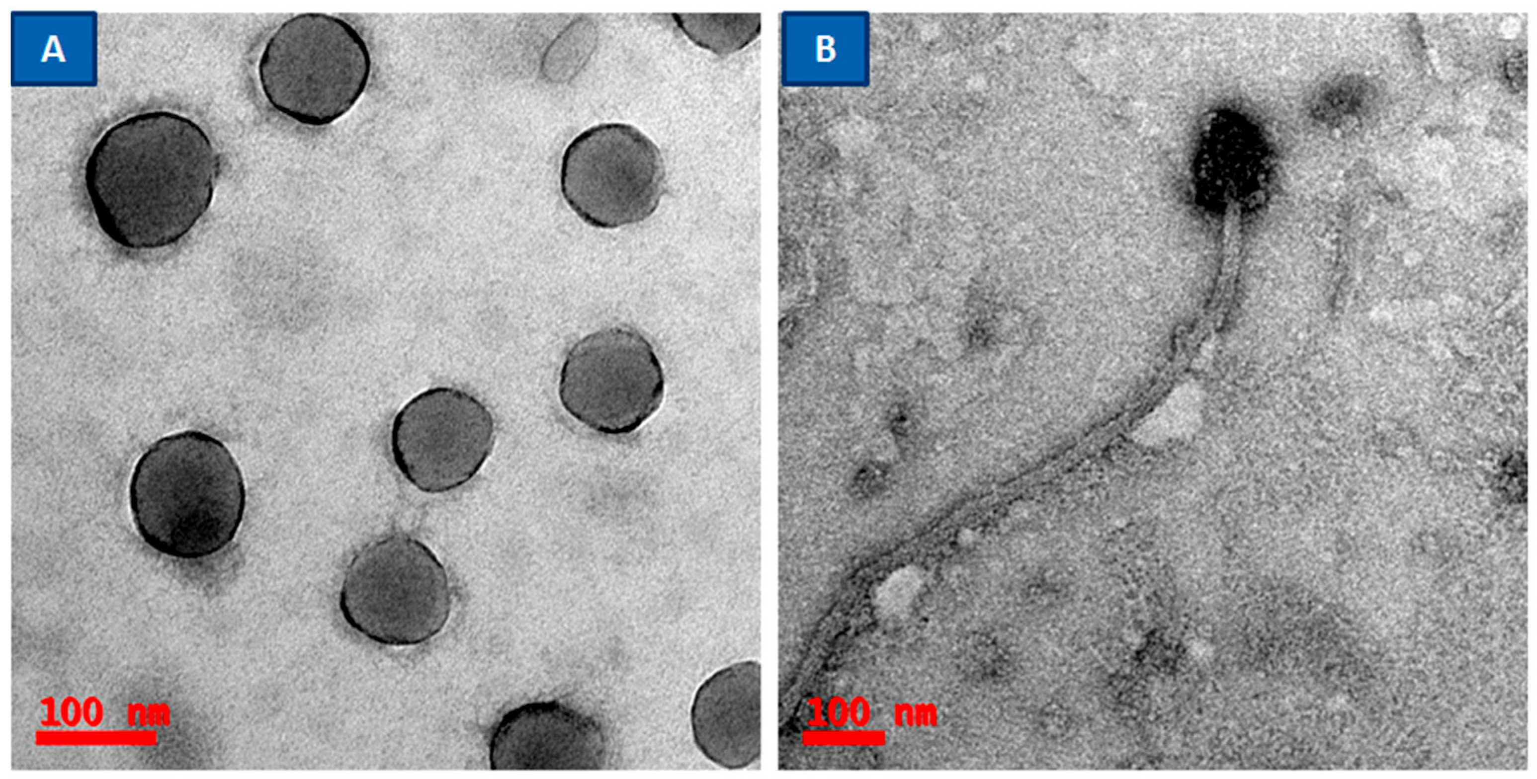
2.1. Bacterial Strains, Bacteriophages, and Growth Conditions

MDR bacterial isolates were isolated from patients with burn wound infections and were previously reported to be used for this study [5,6]. The experiments performed in this article were approved by the Institutional Review Board (IEC). P. aeruginosa and S. aureus were selected and grown on Luria agar (Himedia, Mumbai) at 37 °C for 24 h. Bacteriophages vB_PAnP_PADP4 for (P. aeruginosa) and vB_SAnS_SADP1 for (S. aureus) were isolated as described previously [5,6]. The bacteriophages were stored in salts of magnesium (SM) buffer (5.8 g/L NaCl, 2 g/L MgSO4·7H2O, 50 mL/L 1M-Tris-Hydrochloric acid pH 7.5) at 4 °C. The biofilm staining was performed using Film Tracer™ LIVE/DEAD® Biofilm viability kit (Molecular Probes, Life Technologies Ltd., Carlsbad, CA, USA) according to the instructions provided by the manufacturer. The experiments performed in this article were approved (Resolution Number: 002/2013-2014/ii/b/IEC/YVU/DVRPdT11/10/2014) by the Institutional Review Board (IEC), Yogi Vemana University, India.

2.2. Determination of Biofilm Biomass of Single or Dual Species

To determine the bacteriophage inhibitory effect on the single- or dual-species biofilms, 100 μL of bacterial culture (S. aureus/P. aeruginosa) and 100 μL of respective bacteriophages (109 PFU Plaque forming units) were added to 94-well culture plate for the single-species biofilm assay. For the dual-species study, the following combinations of bacterial cultures and bacteriophages were used as mentioned; 100 μL of S. aureus + 100 μL of P. aeruginosa (100 μL of SM buffer, 100 μL of phage SADP1 + 100 μL of Phage PADP4) and SM buffer were as used as a control instead of bacteriophages. The above sets of cultures were incubated for 24 h at 37 °C, and other groups were incubated with respective phage and the phage cocktail for four hours to determine the phage effect on biofilm biomass of single or dual species. After incubation, the planktonic bacteria were removed by washing twice with PBS (Phosphate Buffer Saline), buffer. A crystal violet assay measured the biomass attached to each well in 94 well tissue culture plates. The wells were washed four times with PBS (pH 7.4), and then biofilms were fixed with 200 µL of methanol (Qualigens, Mumbai, India) for 15 min. Methanol was removed, and to each well was added 200 µL of crystal violet (1% v/v, Qualigens, Mumbai, India) and incubated for 15 min. The wells were then washed with water and dried for two hours at room temperature, and 300 µL of ethanol (95%) (Qualigens, Mumbai, India) was added to dissolve the stain. The absorbance of the eluted stain was measured at 570 nm with a benchmark plus microplate spectrophotometer (Bio-Rad Laboratories, Hercules, CA, USA), and triplicates were maintained [6,25,26].

2.3. Scanning Electron Microscopy

Biofilms were grown on borosilicate glass coverslips and placed into the wells of a 24-well microtiter plate. Single- and dual-species biofilms formed on the coverslips were incubated with 100 µL of respective bacteriophages (109 PFU/µL) for four hours. After treatment, the coverslips were washed twice with PBS and dried in an incubator for 20 h at 37 °C. The biofilms coated on glass slides were fixed with glutaraldehyde (2.5%) and dehydrated through a series of graded ethanol (30–100%) for five minutes. Further, the glass slides were sputtered with gold after critical point drying, and the aggregated biofilms were examined using scanning electron microscopy (FEI, Tecnai G-2S Twin, Hillsboro, OR, USA) [6,25,26].

2.4. Confocal Laser Scanning Microscopy

The biofilms of MDR-bacterial isolates were grown for Twenty-four-hour-old, and their respective bacteriophages were added. After bacteriophage-treated (4 h) of incubation, the slides were stained with Syto ®9 stain and propidium iodide nucleic acid dyes. Briefly, a working solution of fluorescent stains was prepared by adding 3 µL of Syto ®9 stains and 3 µL of propidium iodide (PI) stain to 1 mL of filter-sterilized water. Then, 200 µL of staining solution was deposited on a glass coverslip surface coated with biofilms and treated with respective phages. After 15 min incubation at room temperature in the dark, samples were washed with sterile saline to remove the excess dye and rinsed with water from the base of the support material.
MDR bacterial biofilms and respective phage-treated coverslips were subjected to CLSM to detect the effect of bacteriophages on the MDR bacterial biofilms. The staining with FilmTracer™ LIVE/DEAD® Biofilm viability kit (Molecular Probes, Life Technologies Ltd.) was performed according to the instructions provided by the manufacturer [6,25,26].

3. Results

Lytic phages can lyse the bacteria, release progeny, and gradually spread around them, inhibiting bacterial growth and cell number. Indirectly, phage lytic action on bacteria decreases the biomass of biofilms. Since biofilms incubated for four hours with phage and a phage cocktail showed decreased biofilm biomass, we used SEM and CLSM to gather more evidence on the morphological changes during this process.

3.1. Phage or Phage Cocktail Action on Biofilm Biomass

S. aureus produced a higher amount of biomass (0.856) than P. aeruginosa (0.761). In contrast, in dual-species, combinations of S. aureus + P. aeruginosa (0.67) produced lower concentrations of biomass than the individual states. Interspecies competition between these Gram-positive and Gram-negative bacteria may be responsible for decreased OD values at 37 °C at 24 h of incubation. The phage lytic action was measured on single- and dual-species at 37 °C for four hours of incubation. The phage cocktail showed efficient bacteriolytic activity; its OD value (0.16 ± 0.02) is much less than in the individual state documented in Table 1.

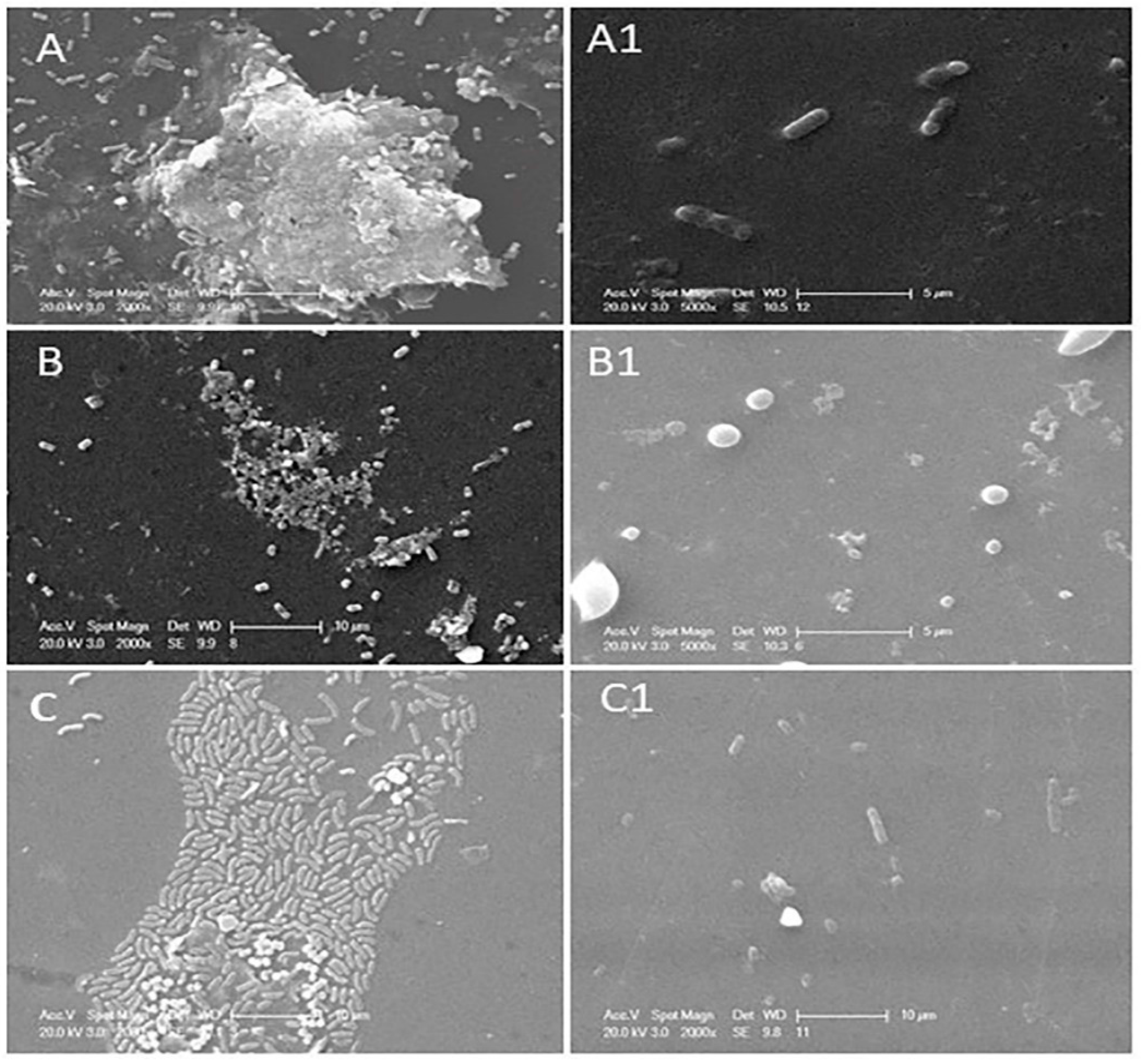
3.2. Determination of Phage or Phage Cocktail Lytic Action on Single- or Dual-Species Biofilm by Using SEM

The structural architecture of single- or dual-species biofilms of P. aeruginosa and S. aureus was determined by employing scanning electron and confocal laser scanning electron microscopic images. Scanning electron microscopic images of single- or dual-species biofilms are represented in Figure 1. P. aeruginosa (Figure 1A) forms a multi-layered complex biofilm after 24 h of incubation, whereas S. aureus (Figure 1B) forms a less complex biofilm than P. aeruginosa. In the case of the dual-species biofilm (Figure 1C), i.e., the combinational biofilm of P. aeruginosa and S. aureus, it formed a single-layered structure even after 24 h of incubation at 37 °C. Dual species biofilm is initiated with the same concentration (109 CFU/mL) of these bacteria, even though a more significant number of P. aeruginosa were found than S. aureus when observed with SEM; this study proved that P. aeruginosa inhibits S. aureus proliferation in dual-species biofilms.
Exoproducts of P. aeruginosa cause toxic effects on S. aureus, leading to a lower population density. These bacteria’ polysaccharide matrixes help prevent antibiotic regime action in biological conditions. Incubation with phage and the phage cocktail for four hours showed reduced biomass and biofilm of bacteria in both single- and dual-species biofilm combinations. The biofilm inhibitory action of phage or the phage cocktail is shown in Figure 1, where Figure 1A1 shows phage (vB_PAnP_PADP4) treatment with four hours of incubation, showed ands a trace amount of biomass and deformed rod-shaped bacteria. Phage vB_SAnS_SADP1 destructed the biofilm integrity, and single coccus were noticed with a limited biofilm matrix (Figure 1B1).
Dual species biofilm is treated with a phage cocktail consisting of phages vB_SAnS_SADP1 + vB_PAnP_PADP4, which showed excellent lytic action against their host bacteria, as shown in Figure 1C1. Interspecies- competition between this Gram-positive and Gram-negative bacterium leads to the formation of a single-layered biofilm. The phage cocktail effectively destroyed the formed biofilm within four hours of incubation at 37 °C.

3.3. Phage or Phage Cocktail Lytic Action on Single- or Dual-Species Biofilm by Using CLSM

Confocal laser scanning electron microscopic images of phage or the phage cocktail on single- or dual-species biofilms are shown in Figure 2. Figure 2A–C shows 24 h old native biofilms formed (Control) on a coverslip that appear in green color (stained with Syto®9) by P. aeruginosa, S. aureus, and a combinational biofilm of these two bacteria, respectively. The appearance of the green color in controls, due to the nucleic acid dye, i.e., Syto®9, stains only live bacterial cells. The single species formed densely packed biofilms compared to the dual-species biofilm. After treatment with the respective phage or the phage cocktails, single- or dual-biofilms are shown in Figure 2A1–C1 (Test). Propidium iodide (PI) stains the nucleic acids of dead cells. The red-colored spots illustrated in Figure 2A1–C1 are caused by dead bacterial cells. This is because of the bacteriolytic action of the phage and the phage cocktail.
Our observational studies of SEM and CLSM disclosed that single-species bacteria could form densely packed biofilms, whereas dual-species biofilms were less dense because of their interspecies- competition. Phage or the phage cocktail effectively lyse biofilms and provide a path for therapeutic applications of phages as antibiofilm agents. Generally, antibiotics cannot penetrate the biofilms because of the polysaccharide matrix, but phage progeny disturbs the matrix and lyse the bacteria. This is one of the premier advantages of phages, and there is renewed interest in alternatives to antibiotics. The phages that were isolated and employed against both single- and dual-species belonged to the Podoviridae (no or very short tail) family against P. aeruginosa and the Siphoviridae (non-contractile long tail) family against S. aureus and were observed under TEM studies (Figure 3).

4. Discussion

Biofilms are formed by the aggregation of prokaryotic or eukaryotic cells, surrounded by a matrix of extracellular polymeric substances (EPSs) consisting of long polysaccharide chains, DNA, and biological macromolecules. Biofilm formation is one of the essential characteristic features of pathogenic bacteria and a dangerous threat to human healthcare. The bacteria encased in a polysaccharide matrix form a complex multicellular structure and are more resistant to antimicrobial agents than planktonic cells. It is complicated to destroy multidrug-resistant bacteria if they form biofilms or are encased in biofilms. Therefore, there is an urgent need to find alternative strategies to combat biofilm-forming bacteria. In this scenario, phage-based antimicrobials are becoming a promising alternative to treat biofilms of pathogenic bacterial infections. Phages can lyse bacterial biofilm by producing lytic enzymes. A single dose of phage administration is efficient for lysing entire bacterial communities [6].
Biofilms are thought to underlie much of the resistance reported to antibiotics. As an outline of the life cycles of bacterial biofilms, it is exemplified that P. aeruginosa is a motile bacterium that can produce more complex biofilms than the non-motile except S. aureus, which forms extensive biofilms. Bacterial communities in the extracellular matrix showed special features that deviated from the planktonic bacterial cells, such as (a) intercellular signals between the community (Quorum sensing), which usually regulate the maturation and detachment of the biofilms to objects; (b) activation of secondary messengers, which play a role in forming biofilms, flagellar movements, and production of extracellular polysaccharides; (c) bap protein 12 plays a role in the matrix formation with the help of matrix scaffold proteins and creates a suitable environment for the bacteria to live in the biofilm [15,27,28]. The formation of biofilms depends on many internal and external factors, such as moist surfaces, energy sources on the site of the wound, type of bacterial association, availability of receptors for bacterial attachment, temperature, and pH.
Most of the studies reported that predominant bacterial isolates of septic wound infections are P. aeruginosa, S. aureus, K. pneumoniae, E. coli, Streptococcus pyogenes, Acinetobacter spp., Citrobacter, Proteus, and Enterobacter, which are almost multidrug-resistant and biofilm-forming bacterial isolates [29,30,31,32,33], consistent with our study. P. aeruginosa and S. aureus are opportunistic pathogens commonly associated with polymicrobial diseases. These bacteria form biofilms and contribute to increased tolerance to antibiotics. Alarming levels of drug resistance and biofilm-forming capabilities have led to the search for alternative strategies. Phage and phage cocktails have shown promising antibiofilm activity due to their mode of action. In this study, 24 h old mono- and dual-species biofilms were treated with phages (vB_SAnS_SADP1, vB_PAnP_PADP4) for four hours alone and in combination. Within four hours of incubation, both single- and dual-species biofilms were eradicated; this study is consistent with a study reported by Ergun Akturk et al., 2019, where single- or dual-species biofilms were reduced by employing both phages and various antibiotic combinations at six hours of incubation [34].
Dual-species biofilms of P. aeruginosa and S. aureus are less densely arranged than single-species biofilms; this loose arrangement of biofilm is because of an inhibitory effect of these two species due to their inter-species competition. Our scanning electron microscopic graphs clearly show the arrangement of bacterial biofilms in single- and dual-species biofilms. Our observations were consistent with other reports [35,36]. Mixed-species biofilms are easily treated using phage or cocktails. The phage cocktail effectively lysed the dual-species biofilm of P. aeruginosa and S. aureus, as shown in Figure 1C,C1, after four hours of incubation. Our study proved that only phage or a phage cocktail is sufficient to remove single- or dual-species biofilms; our study is consistent with other research by Tkhilaishvili et al., 2020, who reported that dual-species biofilms of P. aeruginosa and MRSA-S. aureus were killed by phage or a phage cocktail. In contrast, Ciprofloxacin is active against only the planktonic stage, but biofilms were eradicated at high concentrations ranging from 256 to 512 mg/L [37].
Ana Catarina Duarte et al. 2021 reported that 24 h old biofilms were treated with protein CHAPSH3b and phage phiLPLA-RODI alone and in combination; after incubation, the biofilm thickness was reduced in combinational treatment, and results were visualized by employing confocal microscopy. The biofilm showed a higher number of dead or compromised cells, which appeared red in color due to staining with propidium iodide, and live cells appeared in green color, consistent with our reports. This study proved that combinational therapy plays a vital role in eradicating S. aureus biofilms [16]. Mateusz Szymczak et al., 2024 demonstrated a novel strategy to control bacterial infection, based on the combined use of engineered T7 phages and phage-presented along with AgNPs by employing CLSM methodology. The results proved that the T7Ag-XII phages armed with AgNPs were more effective in terms of eradicating E. coli biofilm, compared with phages alone or AgNPs alone [38]. Melo LDR et al., 2020 developed a probe to detect the location of the biofilm cells of S. epidermidis that are infected by phage SEP1. Biofilms were formed directly on Thermanox™ coverslips and infected with SEP1 for 24 h and subjected for CLSM. After 6 h and 24 h of infection, it was possible to observe areas with infected and non-infected cells along with the biofilm. The intense red signal of the SEP1p probe, which allowed the detection of phages replicating within S. epidermidis biofilms [39]. In our previous study also proved that SEM and CLSM analyses indicated physical changes that occur to the biofilm due to the action of bacteriophage-mediated lysis. These findings support the possible use of bacteriophages as against MDR-bacterial biofilms [6].

5. Conclusions

Alarming levels of multidrug resistance and biofilm formation in bacteria have become a sweltering problem for human healthcare systems. The use of bacteriophages or phage cocktails in treating various bacterial pathogens is increasing because of their potential action against bacteria, irrespective of their multidrug resistance. The isolated phages and the phage cocktail showed excellent lytic activity toward single- or dual-species biofilms. Inter-species competition between Gram-positive and gram G-negative bacteria promoted the phage activity against biofilms.

Author Contributions

P.R. and D.V.R.P. conceived and designed the experiments. P.R. performed the experiments, data collection, and analysis. P.R. drafted the manuscript. P.R., G.D.R., D.V.R.P., D.V. and J.D.C. performed the analysis and interpretation of findings. P.R., G.D.R., D.V., D.V.R.P. and J.D.C. read and revised the manuscript. All authors were involved in reviewing the manuscript and approval for publication. All authors have read and agreed to the published version of the manuscript.

Funding

This research received no external funding.

Data Availability Statement

No new data were created or analyzed in this study. Data sharing is not applicable to this article.

Acknowledgments

Authors highly acknowledge the central facilities of Yogi Vemana University, Department of Nanotechnology, and University of Hyderabad for CLSM, SEM, and TEM services. P.R. gratefully acknowledges the University Grants Commission for financial support in the form of JRF and SRF fellowships.

Conflicts of Interest

Authors Pallavali RojaRani and Dinneswara Reddy were employed by the company Brane Enterprises Pvt Ltd. The remaining authors declare that this research was conducted in the absence of any commercial or financial relationships that could be construed as a potential conflicts of interest.

References

  1. Tian, F.; Li, J.; Nazir, A.; Tong, Y. Bacteriophage—A promising alternative measure for bacterial biofilm control. Infect. Drug Resist. 2021, 14, 205–217. [Google Scholar] [CrossRef] [PubMed]
  2. Awuor, S.O.; Omwenga, E.O.; Mariita, R.M.; Musila, J.M.; Musyoki, S. Monitoring the battleground: Exploring antimicrobial resistance and virulence factors in wound bacterial isolates. Access Microbiol. 2023, 5, 000613.v6. [Google Scholar] [CrossRef] [PubMed] [PubMed Central]
  3. Baral, P.; Afnan, N.; Ahmad Zahra, M.; Akter, B.; Rabia Prapti, S.; Muazzam Hossan, M.; Haque, F.K.M. Bacteriological analysis and antibiotic resistance in patients with diabetic foot ulcers in Dhaka. PLoS ONE 2024, 19, e0301767. [Google Scholar] [CrossRef] [PubMed] [PubMed Central]
  4. Pallavali, R.R.; Avula, S.; Degati, V.L.; Penubala, M.; Damu, A.G.; Durbaka, V.R.P. Data of antibacterial activity of plant leaves crude extract on bacterial isolates of wound infections. Data Brief 2019, 24, 103896. [Google Scholar] [CrossRef]
  5. Pallavali, R.R.; Degati, V.L.; Lomada, D.; Reddy, M.C.; Durbaka, V.R.P. Isolation and in vitro evaluation of bacteriophages against MDR-bacterial isolates from septic wound infections. PLoS ONE 2017, 12, e0179245. [Google Scholar] [CrossRef] [PubMed]
  6. Pallavali, R.R.; Degati, V.L.; Narala, V.R.; Velpula, K.K.; Yenugu, S.; Durbaka, V.R.P. Lytic Bacteriophages Against Bacterial Biofilms Formed by Multidrug-Resistant Pseudomonas aeruginosa, Escherichia coli, Klebsiella pneumoniae, and Staphylococcus aureus Isolated from Burn Wounds. Phage 2021, 2, 120–130. [Google Scholar] [CrossRef]
  7. Pallavali, R.; Shin, D.; Choi, J. Phage-based Biocontrol of antibiotic-resistant bacterium isolated from Livestock Wastewater Treatment Plant. Water 2023, 15, 1616. [Google Scholar] [CrossRef]
  8. Sagar, S.S.; Kumar, R.; Kaistha, S.D. Efficacy of Phage and Ciprofloxacin Co-therapy on the Formation and Eradication of Pseudomonas aeruginosa Biofilms. Arab. J. Sci. Eng. 2017, 42, 95–103. [Google Scholar] [CrossRef]
  9. Liu, S.; Lu, H.; Zhang, S.; Shi, Y.; Chen, Q. Phages against Pathogenic Bacterial Biofilms and Biofilm-Based Infections: A Review. Pharmaceutics 2022, 14, 427. [Google Scholar] [CrossRef]
  10. Zhu, J.; Miller, M.B.; Vance, R.E.; Dziejman, M.; Bassler, B.L.; Mekalanos, J.J. Quorum-sensing regulators control virulence gene expression in Vibrio cholerae. Proc. Natl. Acad. Sci. USA 2002, 99, 3129–3134. [Google Scholar] [CrossRef]
  11. Gutiérrez, D.; Fernández, L.; Rodríguez, A.; García, P. Are phage lytic proteins the secret weapon to kill staphylococcus aureus? mBio 2018, 9, e01923-17. [Google Scholar] [CrossRef]
  12. Fernández, L.; González, S.; Campelo, A.B.; Martínez, B.; Rodríguez, A.; García, P. Downregulation of autolysin-encoding genes by phage-derived lytic proteins inhibits biofilm formation in Staphylococcus aureus. Antimicrob. Agents Chemother. 2017, 61, 1–13. [Google Scholar] [CrossRef] [PubMed]
  13. Schulze, A.; Mitterer, F.; Pombo, J.P.; Schild, S. Biofilms by bacterial human pathogens: Clinical relevance—Development, composition and regulation—Therapeutical strategies. Microb. Cell 2021, 8, 28–56. [Google Scholar] [CrossRef]
  14. Vestby, L.K.; Grønseth, T.; Simm, R.; Nesse, L.L. Bacterial biofilm and its role in the pathogenesis of disease. Antibiotics 2020, 9, 59. [Google Scholar] [CrossRef]
  15. Hall, C.W.; Mah, T.F. Molecular mechanisms of biofilm-based antibiotic resistance and tolerance in pathogenic bacteria. FEMS Microbiol. Rev. 2017, 41, 276–301. [Google Scholar] [CrossRef] [PubMed]
  16. Duarte, A.C.; Fernández, L.; de Maesschalck, V.; Gutiérrez, D.; Campelo, A.B.; Briers, Y.; Lavigne, R.; Rodríguez, A.; García, P. Synergistic action of phage phiIPLA-RODI and lytic protein CHAPSH3b: A combination strategy to target Staphylococcus aureus biofilms. NPJ Biofilms Microbiomes 2021, 7, 39. [Google Scholar] [CrossRef]
  17. Pinto, A.M.; Cerqueira, M.A.; Bañobre-Lópes, M.; Pastrana, L.M.; Sillankorva, S. Bacteriophages for chronic wound treatment: From traditional to novel delivery systems. Viruses 2020, 12, 235. [Google Scholar] [CrossRef]
  18. Gutiérrez, D.; Rodríguez-Rubio, L.; Martínez, B.; Rodríguez, A.; García, P. Bacteriophages as weapons against bacterial biofilms in the food industry. Front. Microbiol. 2016, 7, 825. [Google Scholar] [CrossRef]
  19. Chen, L.; Fan, J.; Yan, T.; Liu, Q.; Zhang, S.Y.H.; Yang, J.; Deng, D.; Huang, S.; Ma, Y. Isolation and Characterization of Specific Phages to Prepare a Cocktail Preventing Vibrio sp. Va-F3 Infections in Shrimp (Litopenaeus vannamei). Front. Microbiol. 2019, 10, 2337. [Google Scholar] [CrossRef]
  20. Motlagh, A.M.; Bhattacharjee, A.S.; Goel, R. Biofilm control with natural and genetically modified phages. World J. Microbiol. Biotechnol. 2016, 32, 67. [Google Scholar] [CrossRef] [PubMed]
  21. Al-Razem, F.; Al-Aloul, H.; Ishnaiwer, M.; Qadi, R. Isolation and partial characterization of Salmonella Gallinarum bacteriophage. Saudi J. Biol. Sci. 2022, 29, 3308–3312. [Google Scholar] [CrossRef]
  22. Bragg, R.R.; Boucher, C.E.; van der Westhuizen, W.A.; Lee, J.-Y. The Potential Use of Bacteriophage Therapy as a Treatment Option in a Post-Antibiotic Era. Antibiot. Resist. Mech. New Antimicrob. Approaches 2016, 15, 309–328. [Google Scholar]
  23. Moghadam, M.T.; Amirmozafari, N.; Shariati, A.; Hallajzadeh, M.; Mirkalantari, S.; Khoshbayan, A.; Jazi, F.M. How phages overcome the challenges of drug resistant bacteria in clinical infections. Infect. Drug Resist. 2020, 13, 45–61. [Google Scholar] [CrossRef]
  24. Viertel, T.M.; Ritter, K.; Horz, H.P. Viruses versus bacteria-novel approaches to phage therapy as a tool against multidrug-resistant pathogens. J. Antimicrob. Chemother. 2014, 69, 2326–2336. [Google Scholar] [CrossRef]
  25. Abedon, S.T.; Danis-Wlodarczyk, K.M.; Wozniak, D.J.; Sullivan, M.B. Improving phage-biofilm in vitro experimentation. Viruses 2021, 13, 1175. [Google Scholar] [CrossRef] [PubMed]
  26. Wagner, E.M.; Fischel, K.; Rammer, N.; Beer, C.; Palmetzhofer, A.L.; Conrady, B.; Roch, F.-F.; Hanson, B.T.; Wagner, M.; Rychli, K. Bacteria of eleven different species isolated from biofilms in a meat processing environment have diverse biofilm forming abilities. Int. J. Food Microbiol. 2021, 349, 109232. [Google Scholar] [CrossRef]
  27. Muhammad, M.H.; Idris, A.L.; Fan, X.; Guo, Y.; Yu, Y.; Jin, X.; Qiu, J.; Guan, X.; Huang, T. Beyond Risk: Bacterial Biofilms and Their Regulating Approaches. Front. Microbiol. 2020, 11, 928. [Google Scholar] [CrossRef] [PubMed]
  28. O’Neill, E.; Pozzi, C.; Houston, P.; Humphreys, H.; Robinson, D.A.; Loughman, A.; Foster, T.J.; O’Gara, J.P. A novel Staphylococcus aureus biofilm phenotype mediated by the fibronectin-binding proteins, FnBPA and FnBPB. J. Bacteriol. 2008, 190, 3835–3850. [Google Scholar] [CrossRef]
  29. González, S.; Fernández, L.; Campelo, A.B.; Gutiérrez, D.; Martínez, B.; Rodríguez, A.; García, P. The behavior of Staphylococcus aureus dual-species biofilms treated with bacteriophage phiIPLA-RODI depends on the accompanying microorganism. Appl. Environ. Microbiol. 2017, 83, e02821-16. [Google Scholar] [CrossRef]
  30. Drago, L.; Agrappi, S.; Bortolin, M.; Toscano, M.; Romanò, C.L.; de Vecchi, E. How to study biofilms after microbial colonization of materials used in orthopaedic implants. Int. J. Mol. Sci. 2016, 17, 293. [Google Scholar] [CrossRef]
  31. Ayyaru, S.; Choi, J.; Ahn, Y.H. Biofouling reduction in a MBR by the application of a lytic phage on a modified nanocomposite membrane. Environ. Sci. 2018, 4, 1624–1638. [Google Scholar] [CrossRef]
  32. Baudin, M.; Cinquin, B.; Sclavi, B.; Pareau, D.; Lopes, F. Understanding the fundamental mechanisms of biofilms development and dispersal: BIAM (Biofilm Intensity and Architecture Measurement), a new tool for studying biofilms as a function of their architecture and fluorescence intensity. J. Microbiol. Methods 2017, 140, 47–57. [Google Scholar] [CrossRef]
  33. Liu, J.; Gao, S.; Dong, Y.; Lu, C.; Liu, Y. Isolation and characterization of bacteriophages against virulent Aeromonas hydrophila. BMC Microbiol. 2020, 20, 141. [Google Scholar] [CrossRef] [PubMed]
  34. Akturk, E.; Oliveira, H.; Santos, S.B.; Costa, S.; Kuyumcu, S.; Melo, L.D.R.; Azeredo, J. Synergistic action of phage and antibiotics: Parameters to enhance the killing efficacy against mono and dual-species biofilms. Antibiotics 2019, 8, 103. [Google Scholar] [CrossRef] [PubMed]
  35. Radlinski, L.; Rowe, S.E.; Kartchner, L.B.; Maile, R.; Cairns, B.A.; Vitko, N.P.; Gode, C.J.; Lachiewicz, A.M.; Wolfgang, M.C.; Conlon, B.P. Pseudomonas aeruginosa exoproducts determine antibiotic efficacy against Staphylococcus aureus. PLoS Biol. 2017, 15, e2003981. [Google Scholar] [CrossRef] [PubMed]
  36. Smith, A.C.; Rice, A.; Sutton, B.; Gabrilska, R.; Wessel, A.K.; Whiteley, M.; Rumbaugh, K.P. Albumin inhibits Pseudomonas aeruginosa quorum sensing and alters polymicrobial interactions. Infect. Immun. 2017, 85, e00116-17. [Google Scholar]
  37. Tkhilaishvili, T.; Wang, L.; Perka, C.; Trampuz, A.; Moreno, M.G. Using Bacteriophages as a Trojan Horse to the Killing of Dual-Species Biofilm Formed by Pseudomonas aeruginosa and Methicillin Resistant Staphylococcus aureus. Front. Microbiol. 2020, 11, 695. [Google Scholar]
  38. Szymczak, M.; Pankowski, K.A.; Kwiatek, A.; Grygorcewicz, B.; Karczewska-Golec, J.; Sadowska, K.; Golec, P. An effective antibiofilm strategy based on bacteriophages armed with silver nanoparticles. Sci. Rep. 2024, 14, 9088. [Google Scholar] [CrossRef]
  39. Melo, L.D.R.; Pinto, G.; Oliveira, F.; Vilas-Boas, D.; Almeida, C.; Sillankorva, S.; Cerca, N.; Azeredo, J. The Protective Effect of Staphylococcus epidermidis Biofilm Matrix against Phage Predation. Viruses 2020, 12, 1076. [Google Scholar] [CrossRef] [PubMed] [PubMed Central]
Figure 1. SEM images of 4 h phage- or phage cocktail-treated single- or dual-species biofilms of MDR bacterial isolates on the coverslip at 24 h of incubation. (A) P. aeruginosa (A1) Treated with phage vB_PAnP_PADP4); (B) S. aureus (B1) treated with phage vB_SAnS_SADP1), (C) S. aureus and P. aeruginosa, (C1) treated with phage cocktail (phage vB_SAnS_SADP1 + vB_PAnP_PADP4).
Applmicrobiol 04 00085 g001
Figure 2. Confocal laser scanning microscopic analysis of lytic phages or the phage cocktail on single- or dual-species biofilms. The biofilms of P. aeruginosa and S. aureus were stained with SYTO ® 9 (green color indicates live cells) and propidium iodide (red color indicates dead cells); (AC) biofilms were treated with only SM buffer and (A1C1) treated with phage vB_PAnP_PADP4, vB_SAnS_SADP1and, combination of these two phages respectively (Scale bars represent 20–100 µm).
Applmicrobiol 04 00085 g002
Figure 3. Phages are negatively stained with 0.5% Uranyl acetate and visualized with scale bars. (A) vB_PAnP_PADP4 (0.2 µm), (B) vB_SAnS_SADP1 (0.2 µm) at 80,000× magnification with transmission electron microscopy.
Applmicrobiol 04 00085 g003
Table 1. Data on optical density values of single and dual-species biofilm biomass at 24 h time interval in the presence of respective lytic phage or phage cocktail at 4 h incubation.
S.No.BacteriaBiofilm at 24 hPhage with 4 h
1.P. aeruginosa0.761 ± 0.0310.18 ± 0.016
2.S. aureus0.856 ± 0.0550.205 ± 0.018
3.P. aeruginosa + S. aureus0.67 ± 0.0200.16 ± 0.020
Disclaimer/Publisher’s Note: The statements, opinions and data contained in all publications are solely those of the individual author(s) and contributor(s) and not of MDPI and/or the editor(s). MDPI and/or the editor(s) disclaim responsibility for any injury to people or property resulting from any ideas, methods, instructions or products referred to in the content.

