Article

Degradation of a Micro-Hybrid Dental Composite Reinforced with Polyaramide Fiber under the Influence of Cyclic Loads

1
Chair of Integrated Dentistry, Medical University of Lublin, 20-093 Lublin, Poland
2
Independent Laboratory of Dental Techniques, 20-093 Lublin, Poland
3
Chair and Department of Jaw Orthopedics, Medical University of Lublin, 20-093 Lublin, Poland
4
Institute of Biological Basis of Animal Production, University of Life Sciences in Lublin, 20-950 Lublin, Poland
5
Faculty of Mechanical Engineering, Lublin University of Technology city, 20-618 Lublin, Poland
6
Department of Dental Prosthetics, Medical University of Lublin, 20-093 Lublin, Poland
*
Author to whom correspondence should be addressed.
Leszek Szalewski, Aneta Kamińska—these authors contributed equally to this work.
Appl. Sci. 2020, 10(20), 7296; https://doi.org/10.3390/app10207296
Submission received: 8 September 2020 / Revised: 11 October 2020 / Accepted: 12 October 2020 / Published: 19 October 2020
(This article belongs to the Special Issue Mechanical Properties of Materials)

Abstract

:

Featured Application

The article can help dentists in deciding whether to choose fiber-reinforced composite adhesive bridges as a treatment option. The results indicate that the choice of another therapeutic method should be considered in the case of increased occlusal forces (parafunction, bruxism, occlusal obstructions). The obtained results may become a starting point for research on new materials/fibers to increase the mechanical properties of adhesive composite bridges to obtain a material resistant to high occlusal forces.

Abstract

Dental composites reinforced with glass fibers have a low tensile modulus and relatively low fatigue resistance. The aim of the study was to analyze the fatigue properties of a dental composite reinforced with polyaramide fibers under the influence of a cyclic, vertical load. For this purpose, we designed a thermoformable template, corresponding to the construction of adhesive bridges in the side section of the jaw. Fifty-four composite samples were made for the study. They were divided into three groups—control (K) and two experimental groups (R1 and R2). The experimental samples were subjected to cyclic fatigue using 75 N load. The number of cycles was 4690 and 20,100. The study used a three-point bending test. Statistical analysis showed a change in elasticity in groups related to the number of load cycles. The study showed that the samples from the control group required the greatest force to break in relation to those subjected to the work cycles. The maximum force in control (K) group was 738.1 N, R1—487.8 N, and R2—451.4 N. The determined algorithm showed a change in deflection associated with the increase of force value. The study did not show any relationship between the type of sample fracture and the number of load cycles.

1. Introduction

Increasing aesthetic requirements of patients has led to the development of composite dental resins. Usually dental composites are made up from the matrix and fillers that are connected with each other by so-called silanes. Modifications of these two components in the last 20 years have increased the use of dental composites. They are a material commonly used to reconstruct lost tooth tissues using aesthetic restorations [1,2].
Composite materials are based on methacrylate compounds. Their matrix is an organic photopolymerizating resin, which consists mostly of bisphenol A-glycidyl methacrylate (Bis-GMA), triethylene glycol dimethacrylate (TEGMA), and urethane dimethacrylate (UDMA). The inorganic phase is macro-, micro-, or nanofillers, based mostly on silicon compounds [3,4]. Additionally, the composite contains photo initiators and proadhesive agents—silanes. The micro-hybrid composites contain a mixture of at least two types of “glass” or quartz molecules, irregular in shape and similar in diameter (from 0.2 to 3 μm) and from 5 to 15% of small particles (0.04 μm). In these materials, the filler constitutes 60–70% by volume, i.e., about 77–84% by weight of the composite part [5].
In order to increase the field of application, as well as to broaden the indications for the use of composite materials, their combinations with fibers such as carbon, polyaramide, polyethylene, and glass are used [6,7]. Currently, such solutions are used in periodontology [8], endodontics [9], prosthetics [10], and orthodontics [11].
Both wholly aromatic polyamides or the shorter aromatic polyamids form aramids and stand for synthetic polyamides comprising >85% amide groups (–CO–NH–) bound directly to two aromatic rings. Such polymers are characterized as high-performance materials due to their very good mechanical strength and exceptional high thermal resistance. They are spun into fibers and used in advanced fabrics, such as sport and work protective clothing, bullet-proof armor, advanced composites in armament and aerospace industries, composites such as asbestos substitutes, and high-temperature insulation paper.
The aramid structure is based on rigid aromatic amide linkage. It is responsible for the exceptional properties of these materials. Highly directional and efficient interchain hydrogen bonds are established, giving basis to materials with a high tendency to crystallize and with extremely high cohesive energy density. At the same time, they are also responsible for the insolubility of the wholly aromatic polyamides, a disadvantage that prevents the expansion of the application field of these materials, whereas the improvement of the solubility is a topic of the present research interest [12]. Aromatic polyamides are obtained in reactions that form amide bonds between aromatic rings of high thermal stability and high strength. The outstanding rigid molecular chain structure, good orientation, and organization of the crystalline structure provides high strength and low elongation of the orientation of the molecular chains. This provides high tensile strength, impact, and differentiated thermal stability for various temperature ranges for an extended time [13]. The covalent bonds in the polymer are responsible for the high strength. However, man-made polymers generally do not exhibit the corresponding potential high modulus. High modulus and strength may result from structural perfection, crystallinity, and crystalline and amorphous orientation. It is well known that the highest elastic moduli reported from linear polymers are generally much smaller than theoretical values [14].
The bending strength of a prosthetic restoration is an extremely important feature whose assessment is necessary in specific cases of prosthesis use, for example, when it is necessity to increase its resistance to fracture during a long period of use or when a prosthesis with an elongated structure is made [15]. Chewing is the main factor that causes mechanical degradation of the composite resin. While chewing, the mandible moves repeatedly in the vertical and horizontal planes [16,17]. The mechanics of the chewing process adversely affect composite resins and lead to stress within the material structure [18]. During chewing, prosthetic restorations are subjected to loads with forces whose direction is consistent with the long axis of the tooth. The size of chewing force in the mouth is from 3 to 36—49 N on average—but it can also reach 1000 N [19,20]. During this process, the opposing teeth remain in mutual point contact, and the highest loads occur in the mouth only sporadically [21].
The number of cyclic contacts during chewing and swallowing per day depend on the number of meals, their consistency, the person’s age and sex, the number of natural teeth, the type of prosthetic restorations, and the functional disorders of the masticatory apparatus that may significantly increase the number of contacts [22,23]. On average, single contacts last 0.3 s, and their number is variable, ranging from 658 to 2300 contacts a day [21,24,25].
The available literature has not yet explained all aspects of the degradation of composites nor, consequently, the problem related to the maximum time of the restoration use in real conditions. In general, tests have been carried out on standardized samples, reproducing cyclic loads and finite elements that were usually subjected to static loads [26,27,28,29].
The aim of the research was to determine and compare the effect of cyclic vertical loads on the strength of composite resins reinforced with high molecular weight polyaramide fiber.

2. Materials and Methods

2.1. Preparation of the Test

The study used a gypsum model of the jaw, from which a fragment covering the area of three teeth was dissected, between the second premolar and the second molar on the left side. The obtained model (model A) was duplicated in a silicone mass, and the obtained form was filled with a chemo-hardening, burnt-out acrylic resin, which was changed into a chromium–nickel alloy in the casting process. On the basis of the obtained model, we produced standard templates (Figure 1) that were used to make samples. Plates that were 1.5 mm thick were used to make the templates, which were placed in a pressure thermoforming device. The tiles were heated for 55 s, after which they were stretched on the prepared models. Next, in the gypsum model A, the first molar tooth was removed, and in the remaining two teeth, limiting this lack, a preparation was made within the crowns to obtain a model that was reproduced and turned into a chromium–nickel alloy (model B). Model B was used as a handle in which the samples were fixed during the test. In order to obtain the model on which the samples were made (C-model), before replacing with the chromium–nickel alloy, we blocked a 2-mm space for the future restored tooth (bridge span) with the use of a chemically curing resin. The obtained distance prevented the sample from leaning against the model during its possible deflection during the test (Figure 1). A chromium–nickel model (D) (Figure 1) of the opposed tooth (the first left mandible molar) was also made for model A, which allowed us to keep the proper contact of the opposing teeth. This model was used in the study as a contact element with the test samples.
The study used a technical composite in A2 color and a polyaramide fiber with a width of 3 mm, which were used to made adhesive bridges.

2.2. Specimen Preparation

On model C, we pressed thin thermoformable films to insulate the material from the model walls. Then, the first layer of 1 mm thick composite was applied to the prepared place, on which a 17 mm polyaramide fiber was placed, corresponding to the range between the prepared areas in crowns of the teeth defining the gap (sample length). The template was then applied and polymerized using a 470 nm wavelength tube (Clear Blue LED 1200 mW/cm2) for 40 s for every 5 mm section. Subsequent layers of 0.5 mm thick composite were applied and condensed using standard dental applicators and polymerized in the same way until the template was filled. The excess material was removed. After polymerization and obtaining anatomical shapes, we released the samples from the models, and any excess of the composite material was removed using a composite milling cutter. The value of the width and height of connectors in the samples was 4 × 3 mm, respectively.
The research material consisted of anatomical samples (n = 54), which were divided into three groups: the control group (K, 21 samples) and two experimental groups (R1, R2), divided according to the number of cycles: 18 and 15 samples, respectively.

2.3. Fatigue Strength Test

The Zwick/Roell Z 2.5 (ZwickRoell GmBh & Co. KG, Ulm, Germany) testing machine was used in the test. During strength tests, we placed all samples in the holder, which was model C, with a distance of 11.5 mm between two end supports. When testing the control samples, we set the initial force to 5 N, and the traverse speed was 5 mm/min. The test lasted until the sample was destroyed, which was defined as a 20% drop in strength in relation to the achieved maximum force for a given sample.
Samples from R1 and R2 were subjected to cyclic loads of 75 N at a 90° angle using the tooth’s opposed model. During this process, the traverse speed was 10 mm/min, and the sample holding time was 0.3 s. After this time, the tested object was relieved to zero force. Then, the cycle was repeated. The number of cycles in the R1 group was 4690, which is the average value corresponding to the number of weekly opposing teeth contact, and in the R2 group, we used 20,100 cycles, the number of which corresponds to monthly dental contacts [21]. After fatigue loads, the samples were subjected to a bending strength test in the same way as in group K. The obtained parameters were maximum force [N], deflection at maximum force [mm], deflection at yielding point [mm], and post-yield displacement [mm].

2.4. Statistical Analysis

The data were analyzed using the SPSS 20.0 PL statistical package (IBM, New York, NY, USA) [30], the Kolmogorov–Smirnov test (normality of distribution), and a one-way analysis of variance were used with Tukey’s multiple comparison test. Spearman’s nonparametric correlation coefficients and selected linear regression coefficients were also estimated. The distribution of the crack types recorded in individual samples was verified by a non-parametric χ2 test.

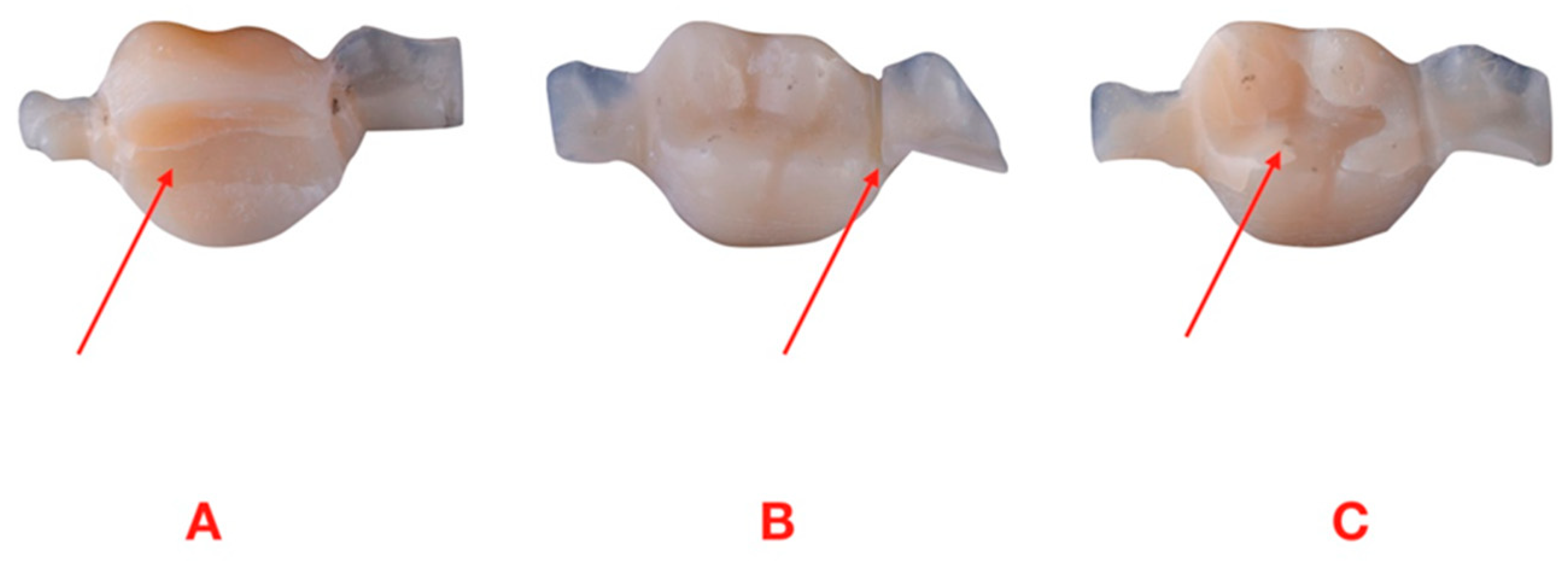
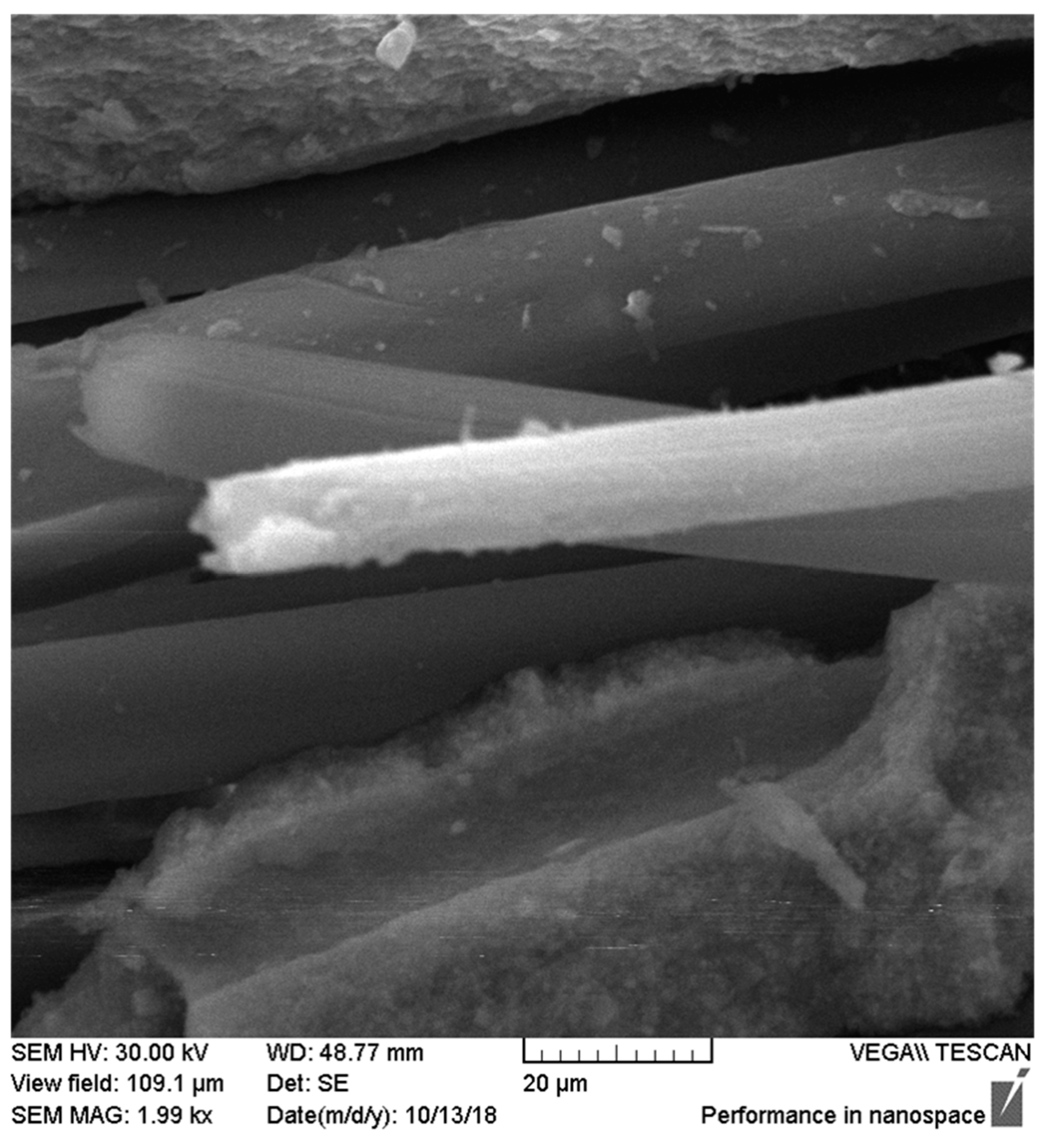
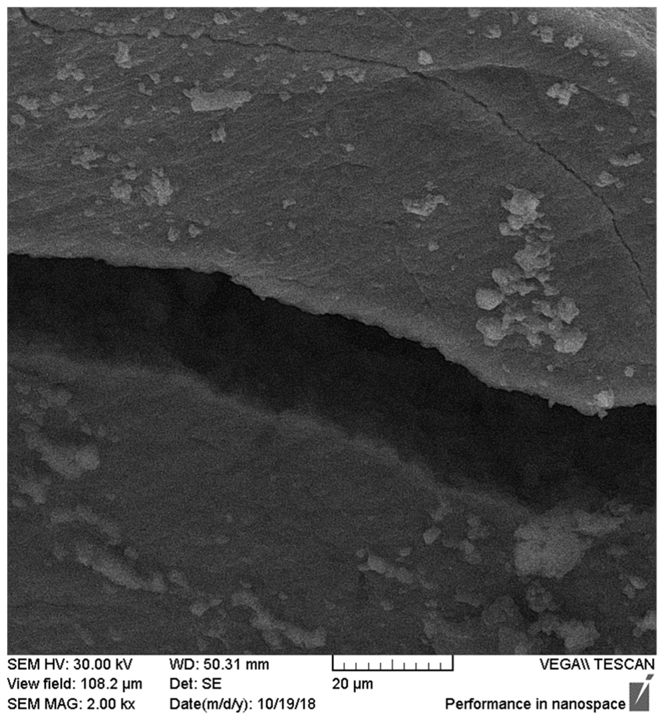
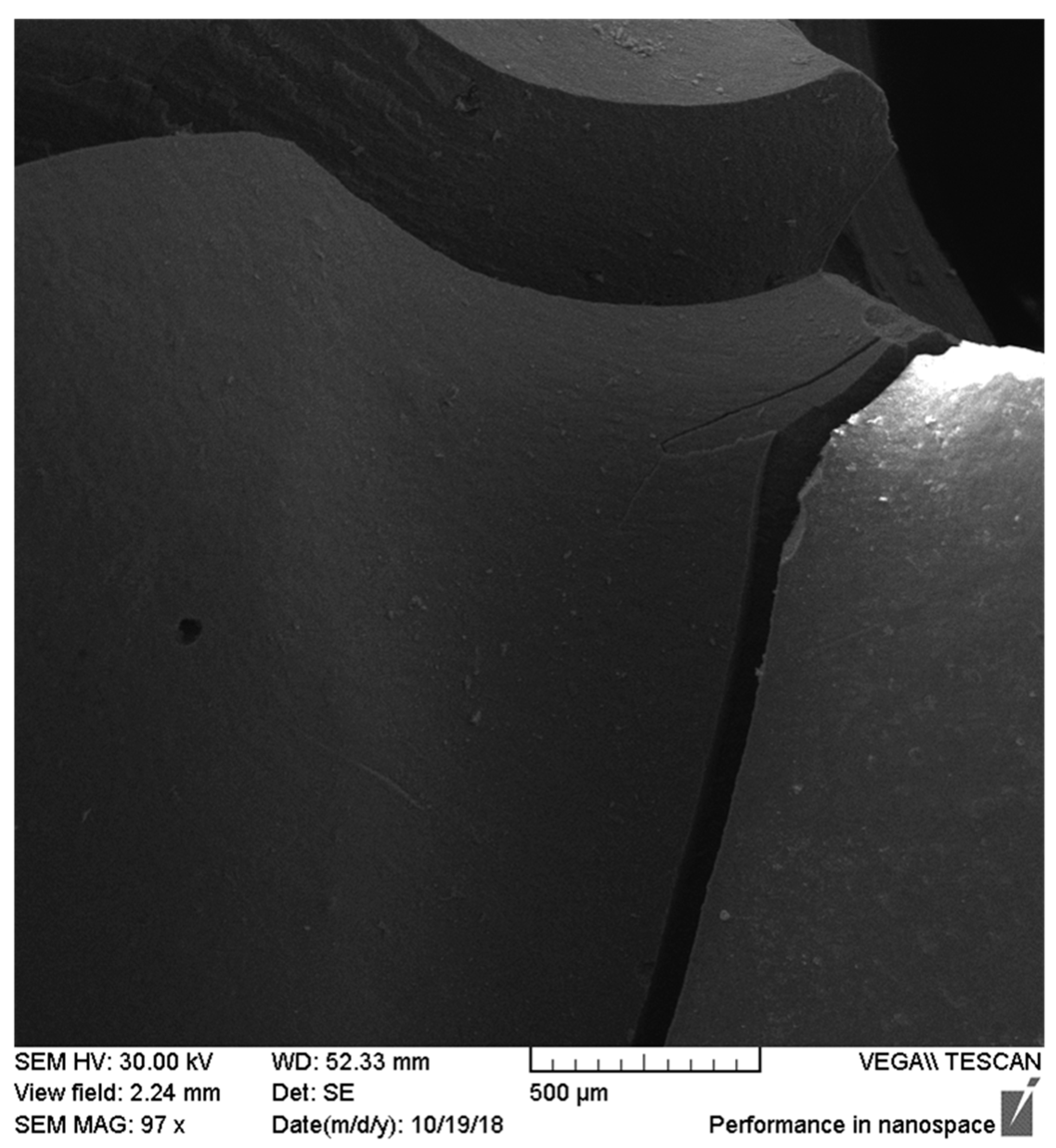
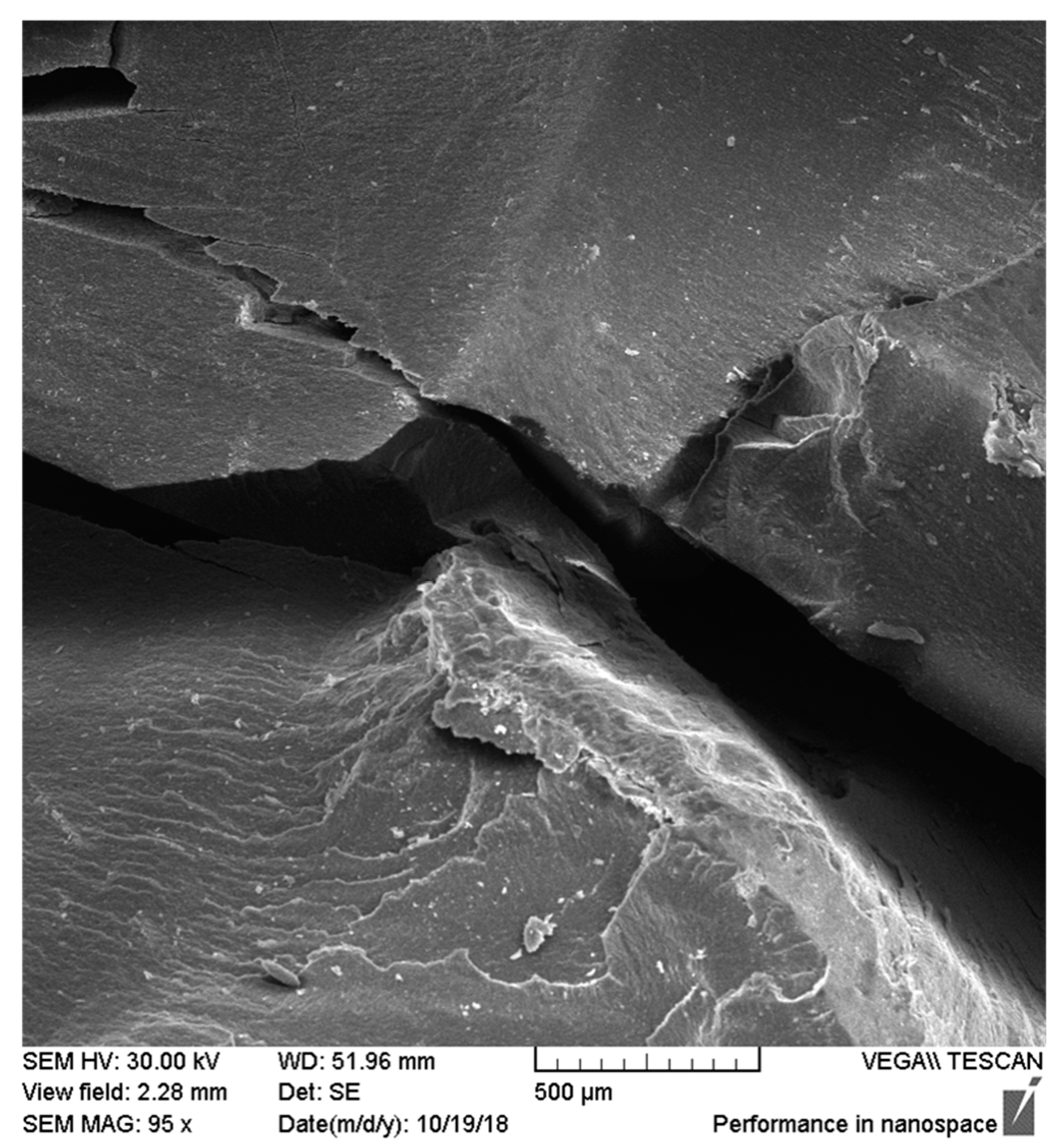
2.5. Microscope Observations
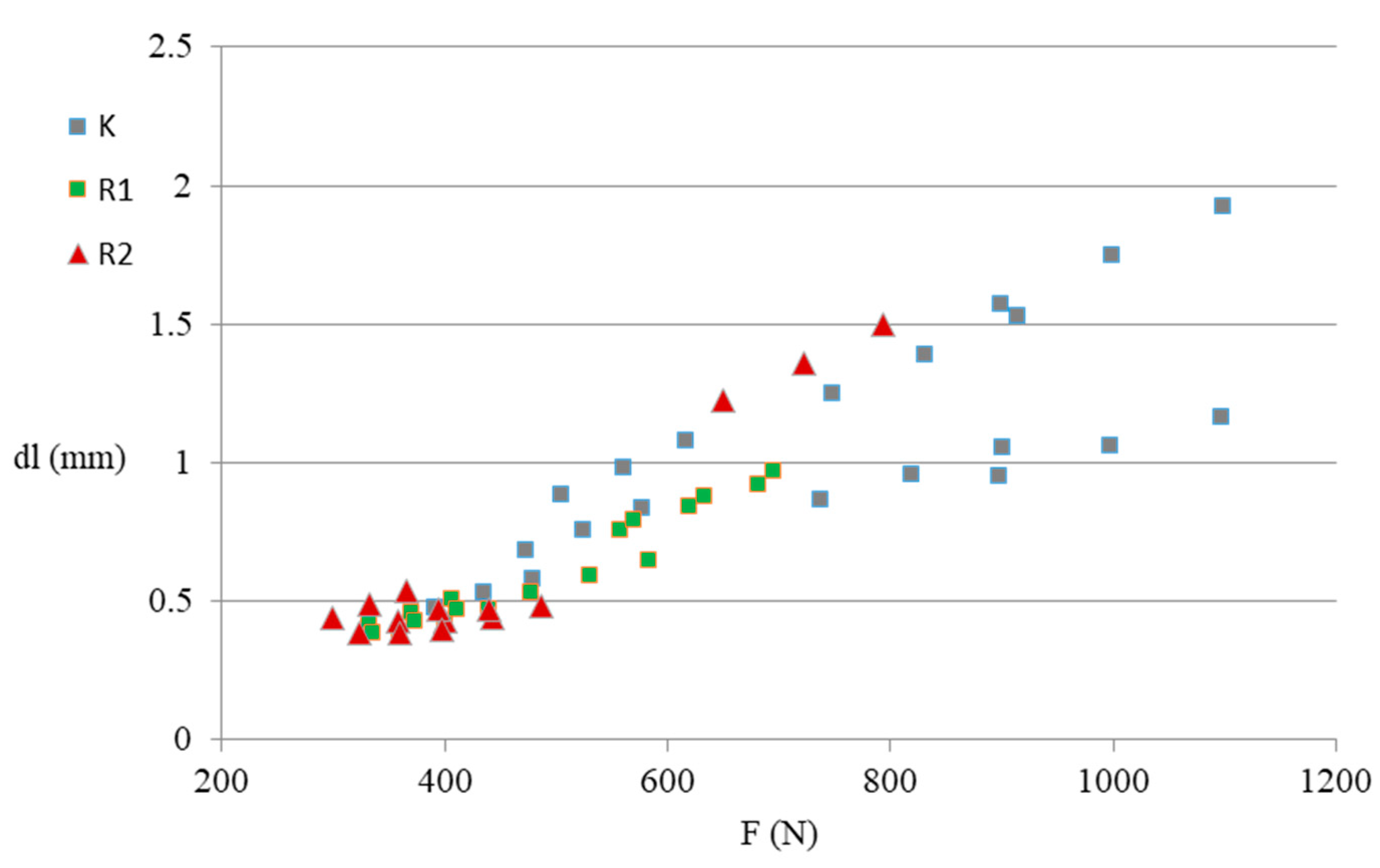
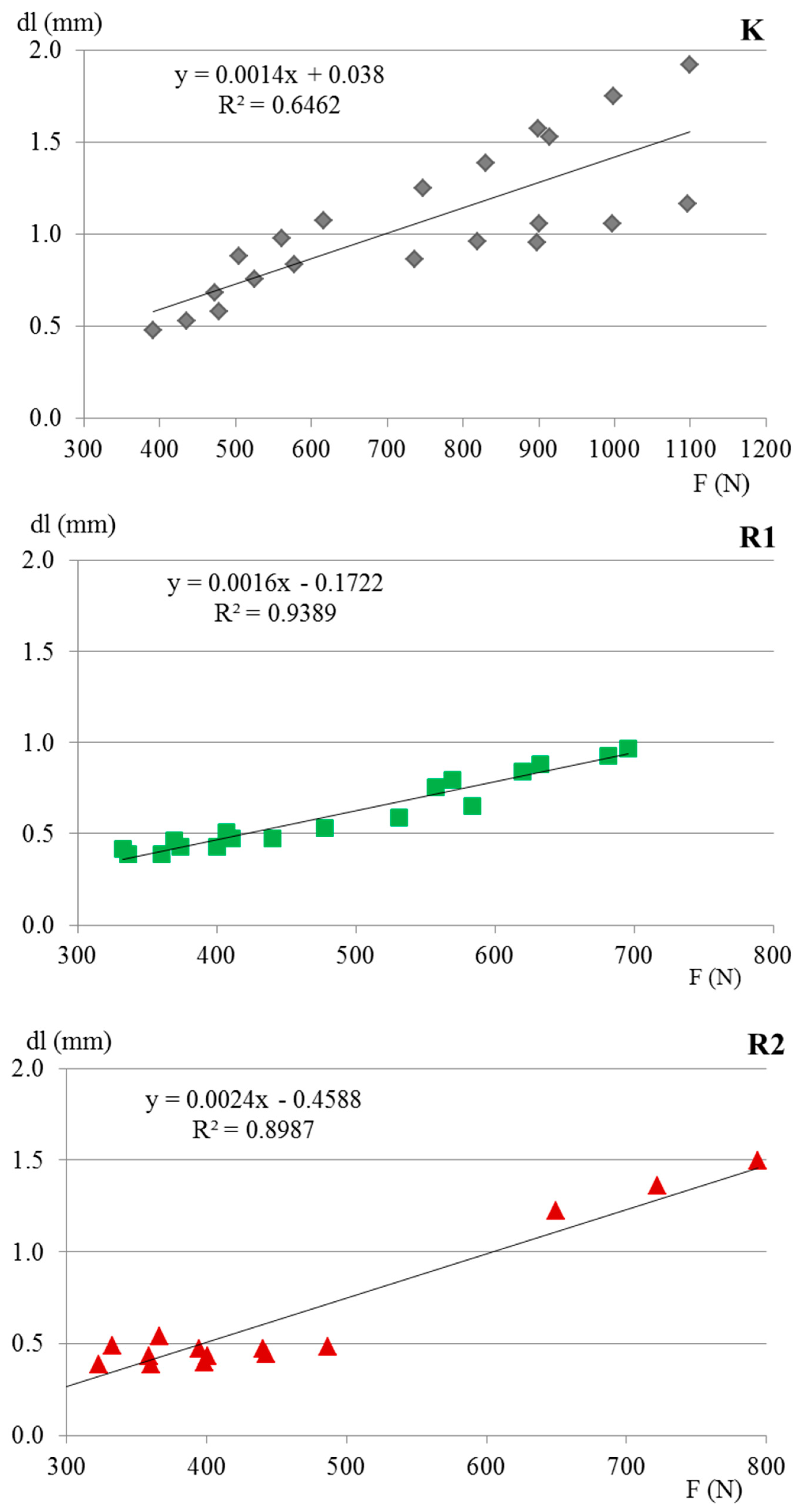
After durability tests, microscopic observations of the work surfaces of fractures of samples were made to check for possible damage. Observations were made using a VEGA//LMU scanning electron microscope with 100 and 2000 magnification.

3. Results

The study showed that the samples from the control group required the greatest force to break in relation to those subjected to the work cycles. The study did not show statistically significant differences between the destructive force in the case of groups R1 and R2, despite the differences in the number of load cycles; however, the numerical differences were found in favor of group treated by smaller number of cycles. Moreover, tests from experimental groups (R1 and R2) were characterized by significantly lower elasticity (deflection) under the maximum force action. Samples from experimental groups were definitely more inflexible compared to the control samples. The reduction of deflection after cyclic loads may result from changes in the internal structure of the samples. Detailed results regarding strength parameters for particular groups are presented in Table 1.
During the strength tests, we found three types of cracks: longitudinal, transversal, and defragmentation (Figure 2). It was not demonstrated that the method of sample fracture depended on the number of performed fatigue cycles (χ2, p = 0.132). Cracks in external polyaramide fibers were observed only in one sample from group R2 (Figure 3). Photographs from the electron microscope show the fiber structure, which is characterized by an empty space inside. Effective forces are focused on the outer surface of the fiber. The presence of fine cracks in the area of the main crack was also noticed. The vertical application of force causes the emergence of cutting forces, leading to side cracks in various directions. It has been observed that with the increase in the number of fatigue cycles, the number of these cracks is much higher, which is related to the increase in the hardness of the composite material with the simultaneous increase in brittleness (Figure 4, Figure 5 and Figure 6). Such a situation may result from the fact that the contact points occur on the unevenness of the teeth chewing surfaces and may be a consequence of the lack of perfect stiffness of the test sample system.
Table 2 presents correlation coefficients between the analyzed strength parameters. There was a statistically significant dependence (p ≤ 0.01) between the number of cycles and individual measured characteristics, whose values decreased considerably with the increase of the number of destructive cycles. It has been noted that with the increase of the maximum force, the flexibility of the sample characterized by material deflection increased significantly. Similarly, highly significant, positive relationships were found for all three plastic deformations of the analyzed samples.
Figure 7 illustrates the dependence of deflection at maximum force on the value of this force depending on the group treated by various numbers of working cycles. It is visible that each group demonstrated different reactions on breaking force. The most resistant was the control group not subjected to cyclic loads. The durability of other groups also varied according to number of cycles, with better resistance characterized by R1 group in comparison to the R2 group.
Figure 8 shows that the dependence of the deflection on the load value in groups K, R1, and R2 were distinguished on the basis of the fatigue cycle number. It is possible that materials in certain samples could behave the other way; however, we did not find any differences using standard error of the mean (SEM). All determined regression coefficients were statistically significant (p ≤ 0.01). Their properties resulted in the possibility of estimating the expected value of a variable. Therefore, on this basis, expected values of deflection were estimated under the influence of a specific load depending on the number of cycles (Table 3). Increasing the value of the acting force may lead to an increase in sample deflection in each of the studied groups.

4. Discussion

Fiber-reinforced composite materials are used as alternative materials for metal restorations. Modifications of composite resins, through various types of fibers improving the mechanical properties of restorations, allow for their use not only in prosthetic crowns, but also in solid partial dentures (FPD)—e.g., in adhesive bridges [31]. These additions differ from the total permanent dentures by the replacement of the crowns, on which the prosthetic bridge structure is based, with crown inserts, i.e., onlay and inlay. This construction of prosthetic bridges is an alternative for patients who do not agree to a complete permanent replacement due to the need of significant tooth tissue reduction, or when minimal tooth reduction is possible [32,33]. In the construction of bridges, the connectors between the individual elements of the bridge are most vulnerable to destruction. Solid partial dentures of fiber-reinforced composite resins are made by placing the fiber in the structure and surrounding it with a composite resin. During their fatigue cyclic tests, Lobhauer et al. [34] observed the slow spread of cracks in brittle materials, such as composite resins. The literature lacks studies describing polyaramid fiber due to the fact that this is a new material in dentistry. Research results derived from the study on composites reinforced with polyaramid fibers provided by Selvaraj et al. acknowledge that the addition of polyaramid into composite impact/increases composite strength [35].
Previous studies on dental composites used to make prosthetic bridges were mainly based on standardized samples. Meanwhile, such samples subjected to cyclic loads show lower values of bending strength (30–50%) than those obtained in static studies and are considered more sensitive in assessing the effectiveness of clinical materials [29]. When testing the strength of dental composites, Papadogiannis et al. [28] stated that fatigue strength is associated not only with the type of filler, but also with the resin matrix. Kuroda et al. [27] used standardized samples with dimensions of 3.0 × 4.0 × 40 mm in their tests, which underwent cyclic fatigue loads with a force of 100 N. They noted that the strength of fiber-reinforced dental composites increases the bending strength. They noted better strength of composites reinforced with glass fiber under the influence of increasing force compared to the unreinforced control sample. However, these studies did not take into account the physiological interactions between the opposing teeth during chewing. In a study by Nobuhisa et al. [26], who analyzed the FEM model of a three-point conventional bridge reinforced with fiber glass, the authors applied a load of 629 N in the rebuilding the lack of the first molar in the mandible. The significant improvement in connector rigidity under vertical load conditions causing twisting and bending movement was stated. As a consequence, the stiffness of connections between individual elements of the sample structure improved, which significantly reduced the deflection of the span because the stresses generated by the vertical load were transferred to the reinforcing fiber. However, no relationship was found between the number of fatigue cycles and bending strength. Similar dependences on the better strength of samples reinforced with polyaramide fiber were recorded in the present research, whereby the actual strength of the manufactured bridges was tested with a strictly established number of loading cycles. Although the experimental groups required significantly lower force in order to break, the destructive force was still in the reported range of habitual and maximal biting forces in molar teeth, which are 300 N and 500 N, respectively [36,37].
The authors are aware that the tests were carried out in laboratory conditions and do not perfectly reflect the conditions in the oral cavity (the authors did not use any bonding system between sample and the chromium–nickel model); nevertheless, the demonstrated differentiation indicates the validity of further research in this direction.

5. Conclusions

Insertion of numerous and fine polyamide fiber fragments into the composite resin may contribute to the elimination of cracks in the material itself, limiting their propagation in restorations. Such a reinforcement could contribute to extending the life of these additions. The improvement of mechanical properties of dental composites, including flexural strength, is of particular importance in the rehabilitation of patients with functional disorders of the masticatory apparatus, where chewing forces are very high.
Studies based on the analysis of anatomically reversible elements require the introduction of laboratory validation to allow comparing test results. The conditions for dental materials in the oral cavity are difficult. In addition, the large anatomical variability of elements reconstructed with the use of dental materials excludes the possibility of developing a conclusion on the basis of standard solutions.
The deflection of adhesive bridges predicted in the study indicates the need for further research in this field with the use of finite element modelling. The polyaramide fiber used in the research can replace the previously used glass fiber to strengthen the construction of prosthetic bridges.

Author Contributions

L.S.: conceptualization, data curation, formal analysis, methodology, writing—original draft; A.K.: conceptualization, data curation, formal analysis, methodology, writing—original draft; E.W.: data curation, methodology, resources, writing—original draft; J.B. (Justyna Batkowska): data curation, formal analysis, visualization, writing—original draft; T.W.: conceptualization, data curation, methodology, visualization, writing—review and editing; D.W.: data curation, formal analysis, methodology, visualization, writing—original draft; J.B. (Janusz Borowicz): conceptualization, formal analysis, supervision, writing—review and editing. All authors have read and agreed to the published version of the manuscript.

Funding

This research received no external funding.

Conflicts of Interest

The authors declare no conflict of interest.

References

  1. Cramer, N.B.; Stansbury, J.W.; Bowman, C.N. Recent advances and developments in composite dental restorative materials. J. Dent. Res. 2011, 90, 402–416. [Google Scholar] [CrossRef] [PubMed] [Green Version]
  2. Khurshid, Z.; Zafar, M.; Qasim, S.; Shahab, S.; Naseem, M.; Abu Reqaiba, A. Advances in nanotechnology for restorative dentistry. Materials 2015, 8, 717–731. [Google Scholar] [CrossRef] [PubMed] [Green Version]
  3. Wacławczyk, A.; Postek-Stefańska, L.; Pietraszewska, D.; Birkner, E.; Zalejska-Fiolka, J.; Wysoczańska-Jankowicz, I. TEGDMA and UDMA monomers released from composite dental material polymerized with diode and halogen lamps. Adv. Clin. Exp. Med. 2018, 27, 469–476. [Google Scholar] [CrossRef]
  4. Manojlovic, D.; Radisic, M.; Vasiljevic, T.; Zivkovic, S.; Lausevic, M.; Miletic, V. Monomer elution from nanohybrid and or mocer-based composites cured with different light sources. Dent. Mater. 2011, 27, 371–378. [Google Scholar] [CrossRef] [PubMed]
  5. LeSage, B.P. Aesthetic anterior composite restorations: A guide to direct placement. Dent. Clin. North. Am. 2007, 51, 359–378. [Google Scholar] [CrossRef] [PubMed]
  6. Sfondrini, M.F.; Massironi, S.; Pieraccini, G.; Scribante, A.; Vallittu, P.K.; Lassila, L.V.; Gandini, P. Flexural strengths of conventional and nanofilled fiber-reinforced composites: A three-point bending test. Dent. Traumatol. 2014, 30, 32–35. [Google Scholar] [CrossRef] [PubMed]
  7. Acharya, P.P.; Bhat, R. The physiochemical properties of dental resin composites reinforced with milled E-glass fibers. Silicon 2018, 10, 1999–2007. [Google Scholar]
  8. Akcalı, A.; Gümüş, P.; Özcan, M. Clinical comparison of fiber-reinforced composite and stainless-steelwire for splinting periodontally treated mobile teeth. Braz. Dent. Sci. 2014, 17, 39–49. [Google Scholar] [CrossRef]
  9. Albashaireh, Z.S.; Ghazal, M.; Kern, M. Effects of endodontic post surface treatment, dentin conditioning, and artificial aging on the retention of glass fiber-reinforced composite resin posts. J. Prosthet. Dent. 2010, 103, 31–39. [Google Scholar] [CrossRef]
  10. Bijelic-Donova, J.; Garoush, S.; Lassila, L.V.J.; Keulemans, F.; Vallittu, P.K. Mechanical and structural characterization of discontinuous fiber-reinforced dental resin composite. J. Dent. 2016, 52, 70–78. [Google Scholar] [CrossRef]
  11. Vallittu, P.K. An overview of development and status of fiber-reinforced composites as dental and medical biomaterials. Acta Biomater. Odontol. Scand. 2018, 4, 44–55. [Google Scholar] [CrossRef] [PubMed]
  12. Reglero Ruiz, J.A.; Trigo-López, M.; García, F.C.; García, J.M. Functional aromatic polyamides. Polymers 2017, 9, 414. [Google Scholar] [CrossRef] [PubMed] [Green Version]
  13. Konarzewski, V.H.C.; Spiekemann, F.L.; Santana, R.M.C. Natural ageing of polyaramide fiber from ballistic armor. Polímeros 2019, 29, 1–7. [Google Scholar] [CrossRef]
  14. Vasanthan, N. Polyamide fiber formation: Structure, properties and characterization. In Handbook of Textile Fibre Structure; Woodhead Publishing: Sawston, UK, 2009; pp. 232–256. [Google Scholar]
  15. Digholkar, S.; Madhav, V.N.V.; Palaskar, J. Evaluation of the flexural strength and microhardness of provisional crown and bridge materials fabricated by different methods. J. Indian Prosthodont. Soc. 2016, 16, 328–334. [Google Scholar]
  16. Pieniak, D.; Niewczas, A. Phenomenological evaluation of fatigue cracking of dental restorations under conditions of cyclic mechanical loads. Acta. Bioeng. Biomech. 2012, 14, 9–17. [Google Scholar]
  17. Hunicz, J.; Niewczas, A.; Kordos, P.; Pieniak, D. Experimental test stand for analysis of composite dental fillings degradation. Eksploat. Niezawodn. Maint. Reliab. 2007, 37, 37–43. [Google Scholar]
  18. Sarveshkumar, J.; Jayalakshmi, S. Bond failure and its prevention in composite restoration—A review. J. Pharm. Sci. Res. 2016, 8, 627–631. [Google Scholar]
  19. Ramalho, A.; Antunes, P.V. Reciprocating wear test of dental composites against human teeth and glass. Wear 2007, 263, 1095–1104. [Google Scholar] [CrossRef] [Green Version]
  20. Koc, D.; Dogan, A.; Bek, B. Bite force and influential factors on bite force measurement a literature review. Eur. J. Dent. 2010, 4, 223–232. [Google Scholar] [CrossRef] [Green Version]
  21. Chitmongkolsuk, S.; Heydecke, G.; Stappert, C.; Strub, J.R. Fracture strength of all-ceramic lithium disilicate and porcelain-fused-to-metal bridges for molar replacement after dynamic loading. Eur. J. Prosthodont. Restor. Dent. 2002, 10, 15–22. [Google Scholar]
  22. Pereira, L.J.; Gaviao, M.B.D.; Bonjardim, L.R.; Castelo, P.M.; Van der Bilt, A. Muscle thickness, bite force, and craniofacial dimensions in adolescents with signs and symptoms of temporomandibular dysfunction. Eur. J. Orthod. 2007, 29, 72–78. [Google Scholar] [CrossRef]
  23. Rosa, L.B.; Bataglion, C.; Siéssere, S.; Palinkas, M.; Mestriner, W., Jr.; De Freitas, O.; de Rossi, M.; de Oliveira, L.F.; Regalo, S.C. Bite force and masticatory efficiency in individuals with different oral rehabilitations. Open J. Stomatol. 2012, 2, 21–22. [Google Scholar] [CrossRef] [Green Version]
  24. Bakke, M. Bite force and occlusion. Semin. Orthod. 2006, 12, 120–126. [Google Scholar] [CrossRef]
  25. Eng, C.M.; Lieberman, D.E.; Zink, K.D.; Peters, M.A. Bite force and occlusal stress production in hominin evolution. Am. J. Phys. Anthr. 2013, 151, 544–557. [Google Scholar] [CrossRef] [PubMed]
  26. Aida, N.; Shinya, A.; Yokoyama, D.; Lassila, L.V.J.; Gomi, H.; Vallittu, P.K.; Shinya, A. Three-dimensional finite element analysis of posterior fiber-reinforced composite fixed partial denture Part 2: Influence of fiber reinforcement on mesial and distal connectors. Dent. Mater. J. 2011, 30, 29–37. [Google Scholar] [CrossRef]
  27. Kuroda, S.; Shinya, A.; Vallittu, P.K.; Nakasone, Y.; Shinya, A. Effect of water temperature on cyclic fatigue properties of glass-fiber-reinforced hybrid composite resin and its fracture pattern after flexural testing. J. Adhes. Dent. 2013, 15, 19–26. [Google Scholar]
  28. Papadogiannis, Y.; Lakes, R.S.; Palaghias, G.; Helvatjoglu-Antoniades, M.; Papadogiannis, D. Fatigue of packable dental composites. Dent. Mater. 2007, 23, 235–242. [Google Scholar] [CrossRef]
  29. Lohbauer, U.; Frankenberger, R.; Krämer, N.; Petschelt, A. Strength and fatigue performance versus filler fraction of different types of direct dental restoratives. J. Biomed. Mater. Res. B Appl. Biomater. 2006, 76, 114–120. [Google Scholar] [CrossRef]
  30. IBM Corp. Released, IBM SPSS Statistics for Windows, Version 20.0; IBM Corp.: Armonk, NY, USA, 2011. [Google Scholar]
  31. Yanagida, H.; Tanoue, N.; Minesaki, Y.; Kamasaki, Y.; Fujiwara, T.; Minami, H. Effects of polymerization method on flexural and shear bond strengths of a fiber-reinforced composite resin. J. Oral. Sci. 2017, 59, 13–21. [Google Scholar] [CrossRef] [PubMed] [Green Version]
  32. Garoushi, S.; Shinya, A. Fiber-reinforced onlay composite resin restoration: A case report. J. Contemp. Dent. Pr. 2009, 10, 104–110. [Google Scholar] [CrossRef]
  33. Garoushi, S.; Yokoyama, D.; Shinya, A.; Vallittu, P.K. Fiber-reinforced composite resin prosthesis to restore missing posterior teeth: A case report. Libyan. J. Med. 2007, 2, 139–141. [Google Scholar] [CrossRef] [PubMed]
  34. Lohbauer, U.; Frankenberger, R.; Krämer, N.; Petschelt, A. Time-dependent strength and fatigue resistance of dental direct restorative materials. J. Mater. Sci. Mater. Med. 2003, 14, 1047–1053. [Google Scholar] [CrossRef] [PubMed]
  35. Selvaraj, A.; Nachimuthu, L.; Manickam, M. Effect of polyaramid reinforced sisal epoxy composites: Tensile, impact, flexural and morphological properties. J. Mater. Res. Technol. 2020, 9, 7947–7954. [Google Scholar]
  36. Röhrle, O.; Saini, H.; Ackland, D.C. Occlusal loading during biting from an experimental and simulation point of view. Dent. Mater. 2018, 34, 58–68. [Google Scholar] [CrossRef]
  37. Umesh, S.; Padma, S.; Asokan, S.; Srinivas, T. Fiber Bragg Grating based bite force measurement. J. Biomech. 2016, 49, 2877–2881. [Google Scholar] [CrossRef] [PubMed]
Figure 1. The schema of sample preparation.
Applsci 10 07296 g001
Figure 2. The types of ruptures found during the experiment ((A) longitudinal, (B) transverse, (C) defragmentation).
Applsci 10 07296 g002
Figure 3. View of sample R2 (magnification 1.99 k×).
Applsci 10 07296 g003
Figure 4. View of control group sample (magnification 2.00 k×).
Applsci 10 07296 g004
Figure 5. View of sample R1 (magnification 97×).
Applsci 10 07296 g005
Figure 6. View of sample R2 (magnification 95×).
Applsci 10 07296 g006
Figure 7. The relationship between deflection at maximum force and the maximum force value.
Applsci 10 07296 g007
Figure 8. Relationship of deflection from the load values in particular groups: K, R1, R2.
Applsci 10 07296 g008
Table 1. Strength parameters of samples included in the tests (mean ± standard error of the mean (SEM)).
ParameterKR1R2
Maximum force [N]738.1 a ± 50.3487.8 b ± 28.8451.4 b ± 40.5
Deflection at maximum force [mm]1.204 a ± 0.1150.605 b ± 0.0490.630 b ± 0.106
Deflection at yielding point [mm]0.907 a ± 0.0750.483 b ± 0.0430.436 b ± 0.018
Post-yield displacement [mm]1.210 a ± 0.1120.568 b ± 0.0400.614 ab ± 0.107
a, b—differ significantly at ≤0.05; SEM—standard error of the mean.
Table 2. The correlation coefficients between measured samples’ characteristics.
No. of CyclesMaximum ForceDeflection at Maximum ForceDeflection at Yielding Point
Maximum force−0.577 **
Deflection at maximum force−0.608 **0.853 **
Deflection at yielding point−0.703 **0.747 **0.759 **
Post-yield displacement−0.666 **0.822 **0.979 **0.780 **
** correlation is significant at p ≤ 0.01 (one-sided).
Table 3. Expected values of deflection depending on the specific load (mm).
GroupF (N)
5006008001000
K0.6590.7991.0791.359
R10.6060.7661.0861.406
R20.761.011.512.01
Publisher’s Note: MDPI stays neutral with regard to jurisdictional claims in published maps and institutional affiliations.

