
- 2.5Impact Factor
- 5.5CiteScore
- 17 daysTime to First Decision
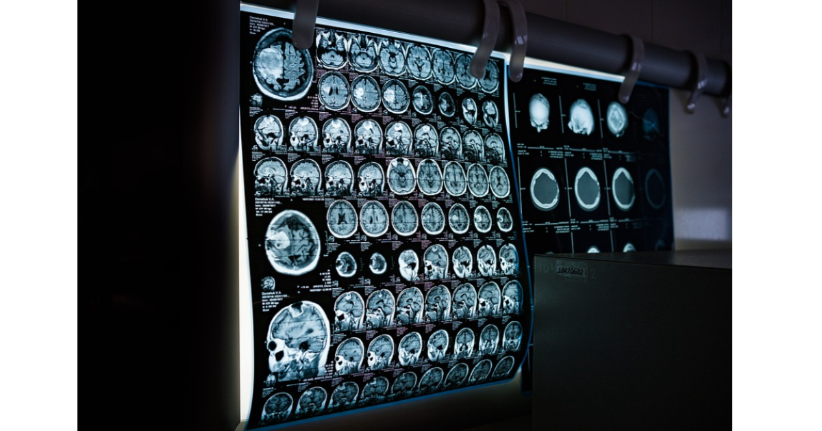
Medical Imaging Using Machine Learning and Deep Learning
This special issue belongs to the section “Computing and Artificial Intelligence“.
Special Issue Information
Dear Colleagues,
Recent years have witnessed a rapid growth of interest in the development of intelligent imaging systems for medical purposes. Intelligent medical imaging is attractive for its high speed, super-resolution, and low cost. In particular, machine learning (ML) and deep learning (DL) techniques that seamlessly integrate big data and high-performance computing have largely facilitated the study of advanced medical imaging systems and their applications. So far, a wide range of work has shown the merit of ML/DL-based imaging systems as compared to conventional ones. Still, a considerable amount of challenges remain to be addressed in this field, concerning not only fundamental theory but also its clinical applications.
This Special Issue will be dedicated to intelligent medical imaging pipelines, including but not limited to the learning theory, smart system design, imaging methods, algorithms, signal and image processing techniques, with their applications to electromagnetic imaging/computed tomography (CT)/magnetic resonance imaging (MRI)/positron emission tomography (PET)/ultrasound (US), as well as multimodalities joint imaging.
Dr. Rui Guo
Dr. He-Ming Yao
Dr. Francesco Zardi
Dr. Mengchu Wang
Guest Editors
Manuscript Submission Information
Manuscripts should be submitted online at www.mdpi.com by registering and logging in to this website. Once you are registered, click here to go to the submission form. Manuscripts can be submitted until the deadline. All submissions that pass pre-check are peer-reviewed. Accepted papers will be published continuously in the journal (as soon as accepted) and will be listed together on the special issue website. Research articles, review articles as well as short communications are invited. For planned papers, a title and short abstract (about 250 words) can be sent to the Editorial Office for assessment.
Submitted manuscripts should not have been published previously, nor be under consideration for publication elsewhere (except conference proceedings papers). All manuscripts are thoroughly refereed through a single-blind peer-review process. A guide for authors and other relevant information for submission of manuscripts is available on the Instructions for Authors page. Applied Sciences is an international peer-reviewed open access semimonthly journal published by MDPI.
Please visit the Instructions for Authors page before submitting a manuscript. The Article Processing Charge (APC) for publication in this open access journal is 2400 CHF (Swiss Francs). Submitted papers should be well formatted and use good English. Authors may use MDPI's English editing service prior to publication or during author revisions.
Keywords
- machine learning
- deep learning
- medical imaging
- imaging methods
- smart systems
Benefits of Publishing in a Special Issue
- Ease of navigation: Grouping papers by topic helps scholars navigate broad scope journals more efficiently.
- Greater discoverability: Special Issues support the reach and impact of scientific research. Articles in Special Issues are more discoverable and cited more frequently.
- Expansion of research network: Special Issues facilitate connections among authors, fostering scientific collaborations.
- External promotion: Articles in Special Issues are often promoted through the journal's social media, increasing their visibility.
- Reprint: MDPI Books provides the opportunity to republish successful Special Issues in book format, both online and in print.
Published Papers


Appl. Sci. - ISSN 2076-3417