MRI in Prostate Cancer

A special issue of Cancers (ISSN 2072-6694). This special issue belongs to the section "Cancer Causes, Screening and Diagnosis".

Deadline for manuscript submissions: 20 July 2024 | Viewed by 5432

Special Issue Editors


E-Mail Website
Guest Editor
Radiology-Basic Sciences, The University of Chicago, Chicago, IL, USA
Interests: quantitative MRI methods; diffusion weighted imaging (DWI); dynamic contrast-enhanced MRI (DCE-MRI); breast; prostate; urogenital; cancer imaging; body MRI; MRI physics

E-Mail Website
Guest Editor
Radiology-Basic Sciences, The University of Chicago, Chicago, IL, USA
Interests: prostate MRI; hybrid multi-dimensional MRI; rad-path correlation; quantitative MRI; non-invasive tissue estimation

Special Issue Information

Dear Colleagues,

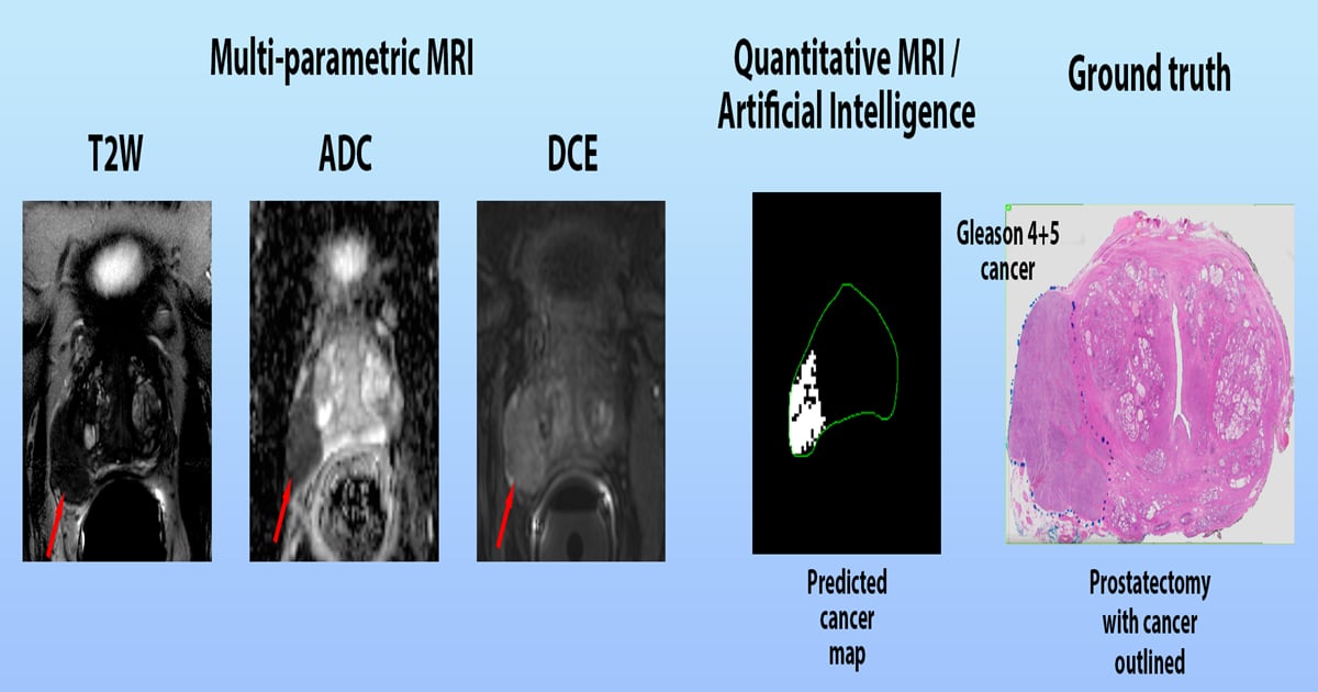
Prostate cancer is one of the most common types of cancer in men, representing a major public health concern. It is estimated that one in nine men will be diagnosed with prostate cancer in their lifetime. Magnetic resonance imaging (MRI) is a powerful imaging technique that has been used to diagnose and monitor prostate cancer. It uses a combination of a strong magnetic field and radio waves to create detailed images of the prostate. MRI can detect the size, shape, and location of a tumor, as well as its relationship to other organs and structures. It can also be used to monitor the progression of the disease and assess the effectiveness of treatment.

This Special Issue focuses on the use of MRI in the diagnosis and management of prostate cancer. It covers topics such as the use of MRI for prostate cancer screening, the role of MRI in the staging and treatment of prostate cancer, and the use of MRI to monitor the response to treatment. This Special Issue also includes reviews of the latest research and clinical applications of MRI in prostate cancer. It provides an invaluable resource for researchers, clinicians, and patients interested in the use of MRI in the diagnosis and management of prostate cancer.

Dr. Milica Medved
Dr. Aritrick Chatterjee
Guest Editors

Manuscript Submission Information

Manuscripts should be submitted online at www.mdpi.com by registering and logging in to this website. Once you are registered, click here to go to the submission form. Manuscripts can be submitted until the deadline. All submissions that pass pre-check are peer-reviewed. Accepted papers will be published continuously in the journal (as soon as accepted) and will be listed together on the special issue website. Research articles, review articles as well as short communications are invited. For planned papers, a title and short abstract (about 100 words) can be sent to the Editorial Office for announcement on this website.

Submitted manuscripts should not have been published previously, nor be under consideration for publication elsewhere (except conference proceedings papers). All manuscripts are thoroughly refereed through a single-blind peer-review process. A guide for authors and other relevant information for submission of manuscripts is available on the Instructions for Authors page. Cancers is an international peer-reviewed open access semimonthly journal published by MDPI.

Please visit the Instructions for Authors page before submitting a manuscript. The Article Processing Charge (APC) for publication in this open access journal is 2900 CHF (Swiss Francs). Submitted papers should be well formatted and use good English. Authors may use MDPI's English editing service prior to publication or during author revisions.

Keywords

  • MRI
  • prostate cancer
  • screening
  • diagnosis
  • treatment

Published Papers (4 papers)

Order results
Result details
Select all
Export citation of selected articles as:

Research

13 pages, 4255 KiB  
Article
Development and Validation of an Explainable Radiomics Model to Predict High-Aggressive Prostate Cancer: A Multicenter Radiomics Study Based on Biparametric MRI
by Giulia Nicoletti, Simone Mazzetti, Giovanni Maimone, Valentina Cignini, Renato Cuocolo, Riccardo Faletti, Marco Gatti, Massimo Imbriaco, Nicola Longo, Andrea Ponsiglione, Filippo Russo, Alessandro Serafini, Arnaldo Stanzione, Daniele Regge and Valentina Giannini
Cancers 2024, 16(1), 203; https://doi.org/10.3390/cancers16010203 - 1 Jan 2024
Cited by 1 | Viewed by 1491
Abstract
In the last years, several studies demonstrated that low-aggressive (Grade Group (GG) ≤ 2) and high-aggressive (GG ≥ 3) prostate cancers (PCas) have different prognoses and mortality. Therefore, the aim of this study was to develop and externally validate a radiomic model to [...] Read more.
In the last years, several studies demonstrated that low-aggressive (Grade Group (GG) ≤ 2) and high-aggressive (GG ≥ 3) prostate cancers (PCas) have different prognoses and mortality. Therefore, the aim of this study was to develop and externally validate a radiomic model to noninvasively classify low-aggressive and high-aggressive PCas based on biparametric magnetic resonance imaging (bpMRI). To this end, 283 patients were retrospectively enrolled from four centers. Features were extracted from apparent diffusion coefficient (ADC) maps and T2-weighted (T2w) sequences. A cross-validation (CV) strategy was adopted to assess the robustness of several classifiers using two out of the four centers. Then, the best classifier was externally validated using the other two centers. An explanation for the final radiomics signature was provided through Shapley additive explanation (SHAP) values and partial dependence plots (PDP). The best combination was a naïve Bayes classifier trained with ten features that reached promising results, i.e., an area under the receiver operating characteristic (ROC) curve (AUC) of 0.75 and 0.73 in the construction and external validation set, respectively. The findings of our work suggest that our radiomics model could help distinguish between low- and high-aggressive PCa. This noninvasive approach, if further validated and integrated into a clinical decision support system able to automatically detect PCa, could help clinicians managing men with suspicion of PCa. Full article
(This article belongs to the Special Issue MRI in Prostate Cancer)
Show Figures

Figure 1

16 pages, 3248 KiB  
Article
Prostate Cancers Invisible on Multiparametric MRI: Pathologic Features in Correlation with Whole-Mount Prostatectomy
by Aritrick Chatterjee, Alexander Gallan, Xiaobing Fan, Milica Medved, Pranadeep Akurati, Roger M. Bourne, Tatjana Antic, Gregory S. Karczmar and Aytekin Oto
Cancers 2023, 15(24), 5825; https://doi.org/10.3390/cancers15245825 - 13 Dec 2023
Cited by 1 | Viewed by 943
Abstract
We investigated why some prostate cancers (PCas) are not identified on multiparametric MRI (mpMRI) by using ground truth reference from whole-mount prostatectomy specimens. A total of 61 patients with biopsy-confirmed PCa underwent 3T mpMRI followed by prostatectomy. Lesions visible on MRI prospectively or [...] Read more.
We investigated why some prostate cancers (PCas) are not identified on multiparametric MRI (mpMRI) by using ground truth reference from whole-mount prostatectomy specimens. A total of 61 patients with biopsy-confirmed PCa underwent 3T mpMRI followed by prostatectomy. Lesions visible on MRI prospectively or retrospectively identified after correlating with histology were considered “identified cancers” (ICs). Lesions that could not be identified on mpMRI were considered “unidentified cancers” (UCs). Pathologists marked the Gleason score, stage, size, and density of the cancer glands and performed quantitative histology to calculate the tissue composition. Out of 115 cancers, 19 were unidentified on MRI. The UCs were significantly smaller and had lower Gleason scores and clinical stage lesions compared with the ICs. The UCs had significantly (p < 0.05) higher ADC (1.34 ± 0.38 vs. 1.02 ± 0.30 μm2/ms) and T2 (117.0 ± 31.1 vs. 97.1 ± 25.1 ms) compared with the ICs. The density of the cancer glands was significantly (p = 0.04) lower in the UCs. The percentage of the Gleason 4 component in Gleason 3 + 4 lesions was nominally (p = 0.15) higher in the ICs (20 ± 12%) compared with the UCs (15 ± 8%). The UCs had a significantly lower epithelium (32.9 ± 21.5 vs. 47.6 ± 13.1%, p = 0.034) and higher lumen volume (20.4 ± 10.0 vs. 13.3 ± 4.1%, p = 0.021) compared with the ICs. Independent from size and Gleason score, the tissue composition differences, specifically, the higher lumen and lower epithelium in UCs, can explain why some of the prostate cancers cannot be identified on mpMRI. Full article
(This article belongs to the Special Issue MRI in Prostate Cancer)
Show Figures

Figure 1

16 pages, 5145 KiB  
Article
Clinical-Genomic Risk Group Classification of Suspicious Lesions on Prostate Multiparametric-MRI
by Radka Stoyanova, Olmo Zavala-Romero, Deukwoo Kwon, Adrian L. Breto, Isaac R. Xu, Ahmad Algohary, Mohammad Alhusseini, Sandra M. Gaston, Patricia Castillo, Oleksandr N. Kryvenko, Elai Davicioni, Bruno Nahar, Benjamin Spieler, Matthew C. Abramowitz, Alan Dal Pra, Dipen J. Parekh, Sanoj Punnen and Alan Pollack
Cancers 2023, 15(21), 5240; https://doi.org/10.3390/cancers15215240 - 31 Oct 2023
Cited by 1 | Viewed by 995
Abstract
The utilization of multi-parametric MRI (mpMRI) in clinical decisions regarding prostate cancer patients’ management has recently increased. After biopsy, clinicians can assess risk using National Comprehensive Cancer Network (NCCN) risk stratification schema and commercially available genomic classifiers, such as Decipher. We built radiomics-based [...] Read more.
The utilization of multi-parametric MRI (mpMRI) in clinical decisions regarding prostate cancer patients’ management has recently increased. After biopsy, clinicians can assess risk using National Comprehensive Cancer Network (NCCN) risk stratification schema and commercially available genomic classifiers, such as Decipher. We built radiomics-based models to predict lesions/patients at low risk prior to biopsy based on an established three-tier clinical-genomic classification system. Radiomic features were extracted from regions of positive biopsies and Normally Appearing Tissues (NAT) on T2-weighted and Diffusion-weighted Imaging. Using only clinical information available prior to biopsy, five models for predicting low-risk lesions/patients were evaluated, based on: 1: Clinical variables; 2: Lesion-based radiomic features; 3: Lesion and NAT radiomics; 4: Clinical and lesion-based radiomics; and 5: Clinical, lesion and NAT radiomic features. Eighty-three mpMRI exams from 78 men were analyzed. Models 1 and 2 performed similarly (Area under the receiver operating characteristic curve were 0.835 and 0.838, respectively), but radiomics significantly improved the lesion-based performance of the model in a subset analysis of patients with a negative Digital Rectal Exam (DRE). Adding normal tissue radiomics significantly improved the performance in all cases. Similar patterns were observed on patient-level models. To the best of our knowledge, this is the first study to demonstrate that machine learning radiomics-based models can predict patients’ risk using combined clinical-genomic classification. Full article
(This article belongs to the Special Issue MRI in Prostate Cancer)
Show Figures

Figure 1

15 pages, 7515 KiB  
Article
MRI Radiomics-Based Machine Learning Models for Ki67 Expression and Gleason Grade Group Prediction in Prostate Cancer
by Xiaofeng Qiao, Xiling Gu, Yunfan Liu, Xin Shu, Guangyong Ai, Shuang Qian, Li Liu, Xiaojing He and Jingjing Zhang
Cancers 2023, 15(18), 4536; https://doi.org/10.3390/cancers15184536 - 13 Sep 2023
Cited by 5 | Viewed by 1379
Abstract
Purpose: The Ki67 index and the Gleason grade group (GGG) are vital prognostic indicators of prostate cancer (PCa). This study investigated the value of biparametric magnetic resonance imaging (bpMRI) radiomics feature-based machine learning (ML) models in predicting the Ki67 index and GGG of [...] Read more.
Purpose: The Ki67 index and the Gleason grade group (GGG) are vital prognostic indicators of prostate cancer (PCa). This study investigated the value of biparametric magnetic resonance imaging (bpMRI) radiomics feature-based machine learning (ML) models in predicting the Ki67 index and GGG of PCa. Methods: A total of 122 patients with pathologically proven PCa who had undergone preoperative MRI were retrospectively included. Radiomics features were extracted from T2-weighted imaging (T2WI), diffusion-weighted imaging (DWI), and apparent diffusion coefficient (ADC) maps. Then, recursive feature elimination (RFE) was applied to remove redundant features. ML models for predicting Ki67 expression and GGG were constructed based on bpMRI and different algorithms, including logistic regression (LR), support vector machine (SVM), random forest (RF), and K-nearest neighbor (KNN). The performances of different models were evaluated with receiver operating characteristic (ROC) analysis. In addition, a joint analysis of Ki67 expression and GGG was performed by assessing their Spearman correlation and calculating the diagnostic accuracy for both indices. Results: The ML model based on LR and ADC + T2 (LR_ADC + T2, AUC = 0.8882) performed best in predicting Ki67 expression, and ADC_wavelet-LHH_firstorder_Maximum had the highest feature weighting. The SVM_DWI + T2 (AUC = 0.9248) performed best in predicting GGG, and DWI_wavelet HLL_glcm_SumAverage had the highest feature weighting. The Ki67 and GGG exhibited a weak positive correlation (r = 0.382, p < 0.001), and LR_ADC + DWI had the highest diagnostic accuracy in predicting both (0.6230). Conclusion: The proposed ML models are suitable for predicting both Ki67 expression and GGG in PCa. This algorithm could be used to identify indolent or invasive PCa with a noninvasive, repeatable, and accurate diagnostic method. Full article
(This article belongs to the Special Issue MRI in Prostate Cancer)
Show Figures

Figure 1

Back to TopTop