Molecular and Cellular Mechanisms of Cardiovascular and Metabolic Diseases

A special issue of Cells (ISSN 2073-4409). This special issue belongs to the section "Cells of the Cardiovascular System".

Deadline for manuscript submissions: 31 January 2025 | Viewed by 3555

Special Issue Editor


E-Mail Website
Guest Editor
1. Environmental Influences on Health and Disease Group, Sanford Research, Sioux Falls, SD 57104, USA
2. Department of Pediatrics, Sanford School of Medicine, University of South Dakota, Sioux Falls, SD, USA
Interests: mitochondrial dysfunction; endoplasmic reticulum (ER) stress; oxidative/nitrosative stress; atherosclerosis; metabolomics; cell death

Special Issue Information

Dear Colleagues,

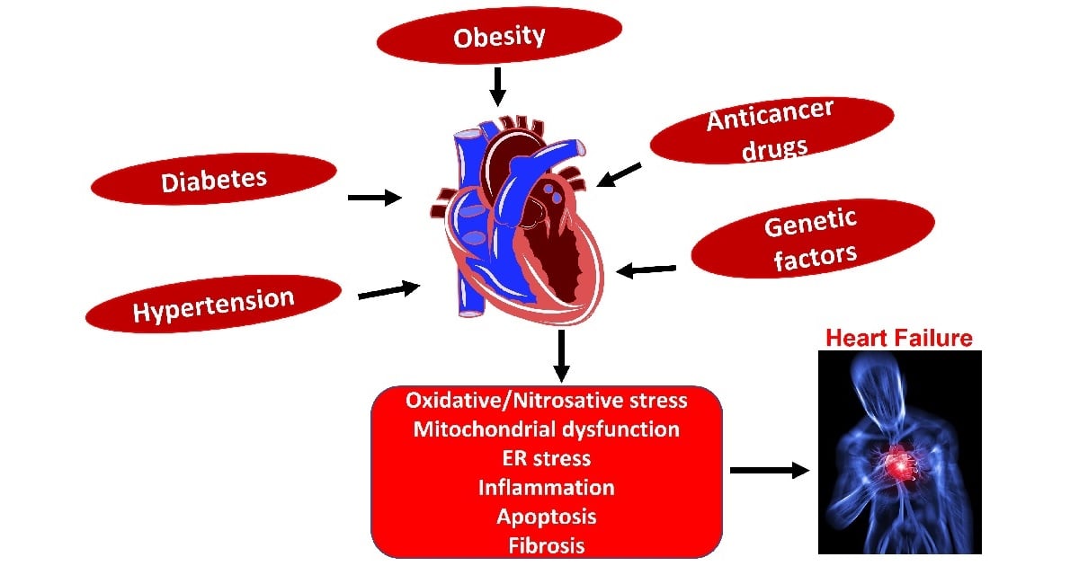
Globally, cardiovascular disease (CVD) is the leading cause of death, the primary contributor to disability, and causes about one third of all deaths. The prevalence of and disability due to CVD has doubled and is affecting more than 500 million people. CVD is influenced by both environmental and genetic triggers. Even though there is significant progress in identifying the multifactorial mechanisms associated with cardiometabolic complications; studies are still needed to unravel novel molecular mechanisms that may open new therapeutic horizons and will significantly reduce the prevalence and disability associated with this disease. This Special Issue focuses on the pathophysiology and novel molecular mechanism associated with cardiometabolic syndrome with a special emphasis on mitochondrial dysfunction, ER stress, oxidative stress, obesity, diabetes, anticancer drug-mediated cardiomyopathy, atherosclerosis, and nutrition. Identifying novel molecular mechanisms may help to implement cost effective, population-based early prevention.

Dr. Prathapan Ayyappan
Guest Editor

Manuscript Submission Information

Manuscripts should be submitted online at www.mdpi.com by registering and logging in to this website. Once you are registered, click here to go to the submission form. Manuscripts can be submitted until the deadline. All submissions that pass pre-check are peer-reviewed. Accepted papers will be published continuously in the journal (as soon as accepted) and will be listed together on the special issue website. Research articles, review articles as well as short communications are invited. For planned papers, a title and short abstract (about 100 words) can be sent to the Editorial Office for announcement on this website.

Submitted manuscripts should not have been published previously, nor be under consideration for publication elsewhere (except conference proceedings papers). All manuscripts are thoroughly refereed through a single-blind peer-review process. A guide for authors and other relevant information for submission of manuscripts is available on the Instructions for Authors page. Cells is an international peer-reviewed open access semimonthly journal published by MDPI.

Please visit the Instructions for Authors page before submitting a manuscript. The Article Processing Charge (APC) for publication in this open access journal is 2700 CHF (Swiss Francs). Submitted papers should be well formatted and use good English. Authors may use MDPI's English editing service prior to publication or during author revisions.

Keywords

  • mitochondrial dysfunction
  • endoplasmic reticulum (ER) stress
  • oxidative/nitrosative stress
  • diabetes
  • obesity
  • diet
  • atherosclerosis
  • hypertension
  • metabolomics
  • cell death
  • nutraceuticals/functional foods

Published Papers (3 papers)

Order results
Result details
Select all
Export citation of selected articles as:

Research

22 pages, 27479 KiB  
Article
CTRP13-Mediated Effects on Endothelial Cell Function and Their Potential Role in Obesity
by Muhammad Aslam, Ling Li, Sina Nürnberger, Bernd Niemann and Susanne Rohrbach
Cells 2024, 13(15), 1291; https://doi.org/10.3390/cells13151291 - 31 Jul 2024
Viewed by 255
Abstract
Background: Obesity, a major component of cardiometabolic syndrome, contributes to the imbalance between pro- and anti-atherosclerotic factors via dysregulation of adipocytokine secretion. Among these adipocytokines, the C1q/TNF-related proteins (CTRPs) play a role in the modulation of atherosclerosis development and progression. Here, we investigated [...] Read more.
Background: Obesity, a major component of cardiometabolic syndrome, contributes to the imbalance between pro- and anti-atherosclerotic factors via dysregulation of adipocytokine secretion. Among these adipocytokines, the C1q/TNF-related proteins (CTRPs) play a role in the modulation of atherosclerosis development and progression. Here, we investigated the vascular effects of CTRP13. Results: CTRP13 is not only expressed in adipose tissue but also in vessels/endothelial cells (ECs) of mice, rats, and humans. Obese individuals (mice, rats, and humans) showed higher vascular CTRP13 expression. Human Umbilical Vein Endothelial Cells (HUVECs), cultured in the presence of serum from obese mice, mimicked this obesity-associated effect on CTRP13 protein expression. Similarly, high glucose conditions and TNF-alpha, but not insulin, resulted in a strong increase in CTRP13 in these cells. Recombinant CTRP13 induced a reduction in EC proliferation via AMPK. In addition, CTRP13 reduced cell cycle progression and increased p53 phosphorylation and p21 protein expression, but reduced Rb phosphorylation, with the effects largely depending on alpha-2 AMPK as suggested by adenoviral overexpression of dominant-negative (DN) or wild-type (WT) alpha 1/alpha 2 AMPK. Conclusion: The present study demonstrates that CTRP13 expression is induced in ECs under diabetic conditions and that CTRP13 possesses significant vaso-modulatory properties which may have an impact on vascular disease progression in patients. Full article
Show Figures

Figure 1

17 pages, 2010 KiB  
Article
Cardiac Development Long Non-Coding RNA (CARDEL) Is Activated during Human Heart Development and Contributes to Cardiac Specification and Homeostasis
by Isabela T. Pereira, Rubens Gomes-Júnior, Aruana Hansel-Frose, Rhaíza S. V. França, Man Liu, Hossam A. N. Soliman, Sunny S. K. Chan, Samuel C. Dudley, Jr., Michael Kyba and Bruno Dallagiovanna
Cells 2024, 13(12), 1050; https://doi.org/10.3390/cells13121050 - 18 Jun 2024
Viewed by 727
Abstract
Successful heart development depends on the careful orchestration of a network of transcription factors and signaling pathways. In recent years, in vitro cardiac differentiation using human pluripotent stem cells (hPSCs) has been used to uncover the intricate gene-network regulation involved in the proper [...] Read more.
Successful heart development depends on the careful orchestration of a network of transcription factors and signaling pathways. In recent years, in vitro cardiac differentiation using human pluripotent stem cells (hPSCs) has been used to uncover the intricate gene-network regulation involved in the proper formation and function of the human heart. Here, we searched for uncharacterized cardiac-development genes by combining a temporal evaluation of human cardiac specification in vitro with an analysis of gene expression in fetal and adult heart tissue. We discovered that CARDEL (CARdiac DEvelopment Long non-coding RNA; LINC00890; SERTM2) expression coincides with the commitment to the cardiac lineage. CARDEL knockout hPSCs differentiated poorly into cardiac cells, and hPSC-derived cardiomyocytes showed faster beating rates after controlled overexpression of CARDEL during differentiation. Altogether, we provide physiological and molecular evidence that CARDEL expression contributes to sculpting the cardiac program during cell-fate commitment. Full article
Show Figures

Figure 1

17 pages, 3391 KiB  
Article
Dose-Dependent Effects of Radiation on Mitochondrial Morphology and Clonogenic Cell Survival in Human Microvascular Endothelial Cells
by Li Wang, Rafael Rivas, Angelo Wilson, Yu Min Park, Shannon Walls, Tianzheng Yu and Alexandra C. Miller
Cells 2024, 13(1), 39; https://doi.org/10.3390/cells13010039 - 23 Dec 2023
Cited by 1 | Viewed by 1737
Abstract
To better understand radiation-induced organ dysfunction at both high and low doses, it is critical to understand how endothelial cells (ECs) respond to radiation. The impact of irradiation (IR) on ECs varies depending on the dose administered. High doses can directly damage ECs, [...] Read more.
To better understand radiation-induced organ dysfunction at both high and low doses, it is critical to understand how endothelial cells (ECs) respond to radiation. The impact of irradiation (IR) on ECs varies depending on the dose administered. High doses can directly damage ECs, leading to EC impairment. In contrast, the effects of low doses on ECs are subtle but more complex. Low doses in this study refer to radiation exposure levels that are below those that cause immediate and necrotic damage. Mitochondria are the primary cellular components affected by IR, and this study explored their role in determining the effect of radiation on microvascular endothelial cells. Human dermal microvascular ECs (HMEC-1) were exposed to varying IR doses ranging from 0.1 Gy to 8 Gy (~0.4 Gy/min) in the AFRRI 60-Cobalt facility. Results indicated that high doses led to a dose-dependent reduction in cell survival, which can be attributed to factors such as DNA damage, oxidative stress, cell senescence, and mitochondrial dysfunction. However, low doses induced a small but significant increase in cell survival, and this was achieved without detectable DNA damage, oxidative stress, cell senescence, or mitochondrial dysfunction in HMEC-1. Moreover, the mitochondrial morphology was assessed, revealing that all doses increased the percentage of elongated mitochondria, with low doses (0.25 Gy and 0.5 Gy) having a greater effect than high doses. However, only high doses caused an increase in mitochondrial fragmentation/swelling. The study further revealed that low doses induced mitochondrial elongation, likely via an increase in mitochondrial fusion protein 1 (Mfn1), while high doses caused mitochondrial fragmentation via a decrease in optic atrophy protein 1 (Opa1). In conclusion, the study suggests, for the first time, that changes in mitochondrial morphology are likely involved in the mechanism for the radiation dose-dependent effect on the survival of microvascular endothelial cells. This research, by delineating the specific mechanisms through which radiation affects endothelial cells, offers invaluable insights into the potential impact of radiation exposure on cardiovascular health. Full article
Show Figures

Figure 1

Back to TopTop