Skeletal Muscle Differentiation and Epigenetics (Closed)

A topical collection in Cells (ISSN 2073-4409). This collection belongs to the section "Cell and Gene Therapy".

Viewed by 36447

Editor


E-Mail Website
Collection Editor
Department of Molecular Medicine, Sapienza University of Rome, Viale Regina Elena 324, 00161 Rome, Italy
Interests: skeletal muscle differentiation; cell cycle inhibitors; MyoD; gene expression; transcriptional control; chromatin architecture; chromatin dynamics; epigenetics; long noncoding RNAs

Topical Collection Information

Dear Colleagues,

Skeletal myogenesis is a well-characterized process, both as regards the developmental phases of muscle formation and the adult phase of muscle regeneration. The commitment of mesodermal precursors to the myogenic lineage and the terminal differentiation of myoblasts into myofibers are regulated at multiple levels, ranging from pre-transcriptional to post-translational mechanisms. Special attention is being paid to the diverse epigenetic strategies by which muscle-specific patterns of gene expression are generated and maintained. Considerable evidence has been accumulated, showing that myogenic transcription factors, such as the prototypical pioneer factor MyoD, work in concert with chromatin modifiers in order to establish an open chromatin environment permissive for the transcriptional activation of muscle-specific genes. More recently, genome-wide studies correlating transcription factor binding, 3D chromatin dynamics, and gene expression have provided further insight into the molecular events underlying the coordinate activation or repression of entire sets of genes during myogenesis. Despite the advances in our understanding of these complex processes, many aspects of the epigenetics of skeletal muscle differentiation remain to be elucidated.

This Topical Collection will present a collection of recent original research papers and review articles in all areas of this field. Potential subjects include, but are not limited to, the identification and characterization of novel epigenetic players as well as of novel functional interactions of myogenic factors with chromatin-modifying enzymes, chromatin remodelers, regulatory noncoding RNAs, and chromatin architectural proteins. Additional topics of interest are the roles of extracellular and intracellular signaling in the modulation of chromatin function and the dysregulation of epigenetic networks in skeletal muscle pathologies, with a view to developing new therapeutic approaches based on the manipulation of specific regulatory pathways.

Prof. Dr. Rossella Maione
Collection Editor

Manuscript Submission Information

Manuscripts should be submitted online at www.mdpi.com by registering and logging in to this website. Once you are registered, click here to go to the submission form. Manuscripts can be submitted until the deadline. All submissions that pass pre-check are peer-reviewed. Accepted papers will be published continuously in the journal (as soon as accepted) and will be listed together on the collection website. Research articles, review articles as well as short communications are invited. For planned papers, a title and short abstract (about 100 words) can be sent to the Editorial Office for announcement on this website.

Submitted manuscripts should not have been published previously, nor be under consideration for publication elsewhere (except conference proceedings papers). All manuscripts are thoroughly refereed through a single-blind peer-review process. A guide for authors and other relevant information for submission of manuscripts is available on the Instructions for Authors page. Cells is an international peer-reviewed open access semimonthly journal published by MDPI.

Please visit the Instructions for Authors page before submitting a manuscript. The Article Processing Charge (APC) for publication in this open access journal is 2700 CHF (Swiss Francs). Submitted papers should be well formatted and use good English. Authors may use MDPI's English editing service prior to publication or during author revisions.

Keywords

  • skeletal muscle differentiation and regeneration
  • epigenetic control of gene expression
  • chromatin stucture and architecture
  • DNA methylation
  • histone modifications
  • nucleosome remodeling
  • noncoding RNAs
  • muscle regulatory factors
  • signal transduction

Related Special Issue

Published Papers (10 papers)

2023

Jump to: 2022, 2021

13 pages, 1979 KiB  
Article
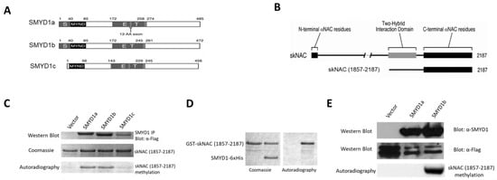
Lysine Methyltransferase SMYD1 Regulates Myogenesis via skNAC Methylation
by Li Zhu, Mark A. Brown, Robert J. Sims, Gayatri R. Tiwari, Hui Nie, R. Dayne Mayfield and Haley O. Tucker
Cells 2023, 12(13), 1695; https://doi.org/10.3390/cells12131695 - 22 Jun 2023
Cited by 3 | Viewed by 2187
Abstract
The SMYD family is a unique class of lysine methyltransferases (KMTases) whose catalytic SET domain is split by a MYND domain. Among these, Smyd1 was identified as a heart- and skeletal muscle-specific KMTase and is essential for cardiogenesis and skeletal muscle development. SMYD1 [...] Read more.
The SMYD family is a unique class of lysine methyltransferases (KMTases) whose catalytic SET domain is split by a MYND domain. Among these, Smyd1 was identified as a heart- and skeletal muscle-specific KMTase and is essential for cardiogenesis and skeletal muscle development. SMYD1 has been characterized as a histone methyltransferase (HMTase). Here we demonstrated that SMYD1 methylates Skeletal muscle-specific splice variant of the Nascent polypeptide-Associated Complex (skNAC) transcription factor. SMYD1-mediated methylation of skNAC targets K1975 within the carboxy-terminus region of skNAC. Catalysis requires physical interaction of SMYD1 and skNAC via the conserved MYND domain of SMYD1 and the PXLXP motif of skNAC. Our data indicated that skNAC methylation is required for the direct transcriptional activation of myoglobin (Mb), a heart- and skeletal muscle-specific hemoprotein that facilitates oxygen transport. Our study revealed skNAC as a methylation target of SMYD1, illuminates the molecular mechanism by which SMYD1 cooperates with skNAC to regulate transcriptional activation of genes crucial for muscle functions and implicates the MYND domain of the SMYD-family KMTases as an adaptor to target substrates for methylation. Full article
Show Figures

Figure 1

21 pages, 4481 KiB  
Article
Bta-miR-206 and a Novel lncRNA-lncA2B1 Promote Myogenesis of Skeletal Muscle Satellite Cells via Common Binding Protein HNRNPA2B1
by Junxing Zhang, Hui Sheng, Linlin Zhang, Xin Li, Yiwen Guo, Yimin Wang, Hong Guo and Xiangbin Ding
Cells 2023, 12(7), 1028; https://doi.org/10.3390/cells12071028 - 27 Mar 2023
Cited by 3 | Viewed by 1920
Abstract
Skeletal muscle satellite cells (MuSCs) can proliferate, differentiate, and self-renew, and can also participate in muscle formation and muscle injury repair. Long noncoding RNAs (lncRNAs) can play an important role with the RNA binding protein and microRNAs (miRNAs) to regulate the myogenesis of [...] Read more.
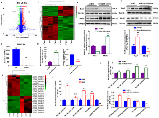
Skeletal muscle satellite cells (MuSCs) can proliferate, differentiate, and self-renew, and can also participate in muscle formation and muscle injury repair. Long noncoding RNAs (lncRNAs) can play an important role with the RNA binding protein and microRNAs (miRNAs) to regulate the myogenesis of bovine MuSCs, however, its molecular mechanism is still being explored. In this study, differentially expressed 301 lncRNAs were identified during the myogenic differentiation of cells based on an in vitro model of induced differentiation of bovine MuSCs using RNA sequencing (RNA-seq). Based on the ability of miR-206 to regulate myogenic cell differentiation, a new kind of lncRNA-lncA2B1 without protein-coding ability was found, which is expressed in the nucleus and cytoplasm. Subsequently, lncA2B1 inhibited cell proliferation by downregulating the expression of the proliferation marker Pax7 and promoted myogenic differentiation by upregulating the expression of the differentiation marker MyHC, whose regulatory function is closely related to miR-206. By RNA pulldown/LC-MS experiments, heterogeneous ribonucleoprotein A2/B1 (HNRNPA2B1), and DExH-Box Helicase 9 (DHX9) were identified as common binding proteins of lncA2B1 and miR-206. Overexpression of lncA2B1 and miR-206 significantly upregulated the expression level of HNRNPA2B1. Downregulation of HNRNPA2B1 expression significantly decreased the expression level of the differentiation marker MyHC, which indicates that miR-206 and lncA2B1 regulate myogenic differentiation of bovine MuSCs by acting on HNRNPA2B1. This study screened and identified a novel lncRNA-lncA2B1, which functions with miR-206 to regulate myogenesis via the common binding proteins HNRNPA2B1. The results of this study provide a new way to explore the molecular mechanisms by which lncRNAs and miRNAs regulate muscle growth and development. Full article
Show Figures

Figure 1

2022

Jump to: 2023, 2021

19 pages, 1194 KiB  
Review
MyoD-Induced Trans-Differentiation: A Paradigm for Dissecting the Molecular Mechanisms of Cell Commitment, Differentiation and Reprogramming
by Cecilia Battistelli, Sabrina Garbo and Rossella Maione
Cells 2022, 11(21), 3435; https://doi.org/10.3390/cells11213435 - 31 Oct 2022
Cited by 9 | Viewed by 4432
Abstract
The discovery of the skeletal muscle-specific transcription factor MyoD represents a milestone in the field of transcriptional regulation during differentiation and cell-fate reprogramming. MyoD was the first tissue-specific factor found capable of converting non-muscle somatic cells into skeletal muscle cells. A unique feature [...] Read more.
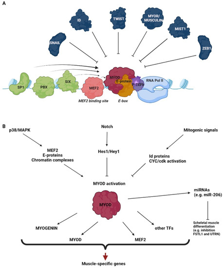
The discovery of the skeletal muscle-specific transcription factor MyoD represents a milestone in the field of transcriptional regulation during differentiation and cell-fate reprogramming. MyoD was the first tissue-specific factor found capable of converting non-muscle somatic cells into skeletal muscle cells. A unique feature of MyoD, with respect to other lineage-specific factors able to drive trans-differentiation processes, is its ability to dramatically change the cell fate even when expressed alone. The present review will outline the molecular strategies by which MyoD reprograms the transcriptional regulation of the cell of origin during the myogenic conversion, focusing on the activation and coordination of a complex network of co-factors and epigenetic mechanisms. Some molecular roadblocks, found to restrain MyoD-dependent trans-differentiation, and the possible ways for overcoming these barriers, will also be discussed. Indeed, they are of critical importance not only to expand our knowledge of basic muscle biology but also to improve the generation skeletal muscle cells for translational research. Full article
Show Figures

Figure 1

13 pages, 3273 KiB  
Article
A Temporary Pause in the Replication Licensing Restriction Leads to Rereplication during Early Human Cell Differentiation
by Marie Minet, Masood Abu-Halima, Yiqing Du, Julia Doerr, Christina Isted, Nicole Ludwig, Andreas Keller, Eckart Meese and Ulrike Fischer
Cells 2022, 11(6), 1060; https://doi.org/10.3390/cells11061060 - 21 Mar 2022
Cited by 1 | Viewed by 2500
Abstract
Gene amplifications in amphibians and flies are known to occur during development and have been well characterized, unlike in mammalian cells, where they are predominantly investigated as an attribute of tumors. Recently, we first described gene amplifications in human and mouse neural stem [...] Read more.
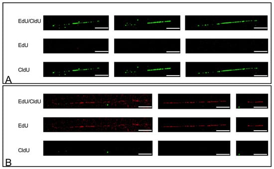
Gene amplifications in amphibians and flies are known to occur during development and have been well characterized, unlike in mammalian cells, where they are predominantly investigated as an attribute of tumors. Recently, we first described gene amplifications in human and mouse neural stem cells, myoblasts, and mesenchymal stem cells during differentiation. The mechanism leading to gene amplifications in amphibians and flies depends on endocycles and multiple origin-firings. So far, there is no knowledge about a comparable mechanism in normal human cells. Here, we describe rereplication during the early myotube differentiation of human skeletal myoblast cells, using fiber combing and pulse-treatment with EdU (5′-Ethynyl-2′-deoxyuridine)/CldU (5-Chlor-2′-deoxyuridine) and IdU (5-Iodo-2′-deoxyuridine)/CldU. We found rereplication during a restricted time window between 2 h and 8 h after differentiation induction. Rereplication was detected in cells simultaneously with the amplification of the MDM2 gene. Our findings support rereplication as a mechanism enabling gene amplification in normal human cells. Full article
Show Figures

Figure 1

22 pages, 12387 KiB  
Article
Chromatin Reorganization during Myoblast Differentiation Involves the Caspase-Dependent Removal of SATB2
by Ryan A. V. Bell, Mohammad H. Al-Khalaf, Steve Brunette, Dalal Alsowaida, Alphonse Chu, Hina Bandukwala, Georg Dechant, Galina Apostolova, F. Jeffrey Dilworth and Lynn A. Megeney
Cells 2022, 11(6), 966; https://doi.org/10.3390/cells11060966 - 11 Mar 2022
Cited by 10 | Viewed by 4243
Abstract
The induction of lineage-specific gene programs are strongly influenced by alterations in local chromatin architecture. However, key players that impact this genome reorganization remain largely unknown. Here, we report that the removal of the special AT-rich binding protein 2 (SATB2), a nuclear protein [...] Read more.
The induction of lineage-specific gene programs are strongly influenced by alterations in local chromatin architecture. However, key players that impact this genome reorganization remain largely unknown. Here, we report that the removal of the special AT-rich binding protein 2 (SATB2), a nuclear protein known to bind matrix attachment regions, is a key event in initiating myogenic differentiation. The deletion of myoblast SATB2 in vitro initiates chromatin remodeling and accelerates differentiation, which is dependent on the caspase 7-mediated cleavage of SATB2. A genome-wide analysis indicates that SATB2 binding within chromatin loops and near anchor points influences both loop and sub-TAD domain formation. Consequently, the chromatin changes that occur with the removal of SATB2 lead to the derepression of differentiation-inducing factors while also limiting the expression of genes that inhibit this cell fate change. Taken together, this study demonstrates that the temporal control of the SATB2 protein is critical in shaping the chromatin environment and coordinating the myogenic differentiation program. Full article
Show Figures

Graphical abstract

18 pages, 19058 KiB  
Article
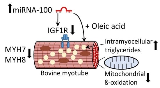
MicroRNA-100 Reduced Fetal Bovine Muscle Satellite Cell Myogenesis and Augmented Intramuscular Lipid Deposition by Modulating IGF1R
by Bilal Ahmad Mir, Elke Albrecht, Asghar Ali, Ola Hansson and Steffen Maak
Cells 2022, 11(3), 451; https://doi.org/10.3390/cells11030451 - 28 Jan 2022
Cited by 9 | Viewed by 3710
Abstract
Previously, microRNA-100 (miR-100) and its putative mRNA target, insulin-like growth factor receptor-1 (IGF1R) were identified as differentially and inversely expressed in bovine longissimus dorsi (LD) muscles with divergent intramuscular fat (IMF) content by our group. While IGF1R signaling is implicated in [...] Read more.
Previously, microRNA-100 (miR-100) and its putative mRNA target, insulin-like growth factor receptor-1 (IGF1R) were identified as differentially and inversely expressed in bovine longissimus dorsi (LD) muscles with divergent intramuscular fat (IMF) content by our group. While IGF1R signaling is implicated in myogenesis and muscle lipid metabolism, the underlying regulatory mechanisms are poorly understood. In the present study, we aimed to investigate the regulation of IGF1R by miR-100 during bovine muscle satellite cell (BMSC) myogenesis and lipid deposition. MiR-100 was confirmed to target the IGF1R 3′-untranslated region (3′-UTR) by luciferase reporter assay. Furthermore, expression of miR-100 and IGF1R was reciprocal during BMSC differentiation, suggesting a crosstalk between the two. Correspondingly, miR-100 mimic (agomiR) suppressed the levels of IGF1R, PI3K/AKT pathway signaling, myogenic gene MYOG, muscle structural components MYH7 and MYH8, whereas the inhibitor (antagomiR) had no clear stimulating effects. The IGF1R inhibitor (BMS-754807) curtailed receptor levels and triggered atrophy in muscle myotubes but did not influence miR-100 expression. AgomiR increased oleic acid-induced lipid deposition in BMSC myotubes supporting its involvement in intramuscular fat deposition, while antagomiR had no effect. Moreover, mitochondrial beta-oxidation and long-chain fatty acid synthesis-related genes were modulated by agomiR addition. Our results demonstrate modulatory roles of miR-100 in BMSC development, lipid deposition, and metabolism and suggest a role of miR-100 in marbling characteristics of meat animals and fat oxidation in muscle. Full article
Show Figures

Graphical abstract

22 pages, 4400 KiB  
Article
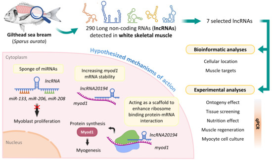
The Emerging Role of Long Non-Coding RNAs in Development and Function of Gilthead Sea Bream (Sparus aurata) Fast Skeletal Muscle
by Isabel García-Pérez, Anna Molsosa-Solanas, Miquel Perelló-Amorós, Elena Sarropoulou, Josefina Blasco, Joaquim Gutiérrez and Daniel Garcia de la serrana
Cells 2022, 11(3), 428; https://doi.org/10.3390/cells11030428 - 26 Jan 2022
Cited by 9 | Viewed by 3399
Abstract
Long non-coding RNAs (lncRNAs) are an emerging group of ncRNAs that can modulate gene expression at the transcriptional or translational levels. In the present work, previously published transcriptomic data were used to identify lncRNAs expressed in gilthead sea bream skeletal muscle, and their [...] Read more.
Long non-coding RNAs (lncRNAs) are an emerging group of ncRNAs that can modulate gene expression at the transcriptional or translational levels. In the present work, previously published transcriptomic data were used to identify lncRNAs expressed in gilthead sea bream skeletal muscle, and their transcription levels were studied under different physiological conditions. Two hundred and ninety lncRNAs were identified and, based on transcriptomic differences between juveniles and adults, a total of seven lncRNAs showed potential to be important for muscle development. Our data suggest that the downregulation of most of the studied lncRNAs might be linked to increased myoblast proliferation, while their upregulation might be necessary for differentiation. However, with these data, as it is not possible to propose a formal mechanism to explain their effect, bioinformatic analysis suggests two possible mechanisms. First, the lncRNAs may act as sponges of myoblast proliferation inducers microRNAs (miRNAs) such as miR-206, miR-208, and miR-133 (binding energy MEF < −25.0 kcal). Secondly, lncRNA20194 had a strong predicted interaction towards the myod1 mRNA (ndG = −0.17) that, based on the positive correlation between the two genes, might promote its function. Our study represents the first characterization of lncRNAs in gilthead sea bream fast skeletal muscle and provides evidence regarding their involvement in muscle development. Full article
Show Figures

Graphical abstract

18 pages, 5250 KiB  
Review
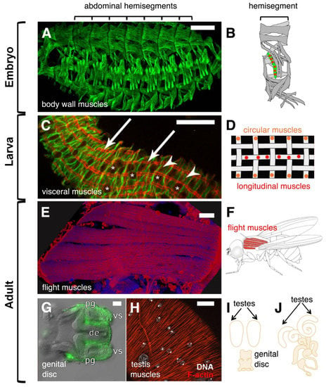
Drosophila melanogaster: A Model System to Study Distinct Genetic Programs in Myoblast Fusion
by Pratiti Rout, Mathieu Preußner and Susanne Filiz Önel
Cells 2022, 11(3), 321; https://doi.org/10.3390/cells11030321 - 19 Jan 2022
Cited by 7 | Viewed by 4467
Abstract
Muscle fibers are multinucleated cells that arise during embryogenesis through the fusion of mononucleated myoblasts. Myoblast fusion is a lifelong process that is crucial for the growth and regeneration of muscles. Understanding the molecular mechanism of myoblast fusion may open the way for [...] Read more.
Muscle fibers are multinucleated cells that arise during embryogenesis through the fusion of mononucleated myoblasts. Myoblast fusion is a lifelong process that is crucial for the growth and regeneration of muscles. Understanding the molecular mechanism of myoblast fusion may open the way for novel therapies in muscle wasting and weakness. Recent reports in Drosophila and mammals have provided new mechanistic insights into myoblast fusion. In Drosophila, muscle formation occurs twice: during embryogenesis and metamorphosis. A fundamental feature is the formation of a cell–cell communication structure that brings the apposing membranes into close proximity and recruits possible fusogenic proteins. However, genetic studies suggest that myoblast fusion in Drosophila is not a uniform process. The complexity of the players involved in myoblast fusion can be modulated depending on the type of muscle that is formed. In this review, we introduce the different types of multinucleated muscles that form during Drosophila development and provide an overview in advances that have been made to understand the mechanism of myoblast fusion. Finally, we will discuss conceptual frameworks in cell–cell fusion in Drosophila and mammals. Full article
Show Figures

Figure 1

2021

Jump to: 2023, 2022

15 pages, 1841 KiB  
Article
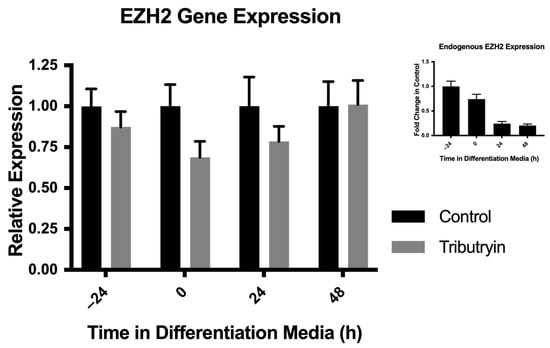
Tributyrin, a Butyrate Pro-Drug, Primes Satellite Cells for Differentiation by Altering the Epigenetic Landscape
by Robert L. Murray, Wei Zhang, Jianan Liu, Jason Cooper, Alex Mitchell, Maria Buman, Jiuzhou Song and Chad H. Stahl
Cells 2021, 10(12), 3475; https://doi.org/10.3390/cells10123475 - 9 Dec 2021
Cited by 6 | Viewed by 3738
Abstract
Satellite cells (SC) are a population of muscle resident stem cells that are responsible for postnatal muscle growth and repair. With investigation into the genomic regulation of SC fate, the role of the epigenome in governing SC myogenesis is becoming clearer. Histone deacetylase [...] Read more.
Satellite cells (SC) are a population of muscle resident stem cells that are responsible for postnatal muscle growth and repair. With investigation into the genomic regulation of SC fate, the role of the epigenome in governing SC myogenesis is becoming clearer. Histone deacetylase (HDAC) inhibitors have been demonstrated to be effective at enhancing the myogenic program of SC, but their role in altering the epigenetic landscape of SC remains undetermined. Our objective was to determine how an HDAC inhibitor, butyrate, promotes myogenic differentiation. SC from tributyrin treated neonatal piglets showed a decrease relative to SC from control animals in the expression of enhance of zeste homologue-2 (EZH2), a chromatin modifier, ex vivo. Chromatin Immunoprecipitation-Sequencing (ChIP-Seq) analysis of SC isolated from tributyrin treated pigs showed a global reduction of the tri-methylation of lysine 27 of histone H3 (H3K27me3) repressive chromatin mark. To determine if reductions in EZH2 was the primary mechanism through which butyrate affects SC behavior, SC were transfected with siRNA targeting EZH2, treated with 0.5 mM butyrate, or both. Treatment with butyrate reduced paired-box-7 (Pax7) and myogenic differentiation-1 (MyoD) gene expression, while siRNA caused reductions in EZH2 had no effect on their expression. EZH2 depletion did result in an increase in differentiating SC, but not in myotube hypertrophy. These results indicate that while EZH2 reduction may force myogenic differentiation, butyrate may operate through a parallel mechanism to enhance the myogenic program. Full article
Show Figures

Figure 1

20 pages, 3741 KiB  
Article
TGF-β Induction of miR-143/145 Is Associated to Exercise Response by Influencing Differentiation and Insulin Signaling Molecules in Human Skeletal Muscle
by Simon I. Dreher, Selina Höckele, Peter Huypens, Martin Irmler, Christoph Hoffmann, Tim Jeske, Maximilian Hastreiter, Anja Moller, Andreas L. Birkenfeld, Hans-Ulrich Häring, Andreas Peter, Johannes Beckers, Martin Hrabě de Angelis and Cora Weigert
Cells 2021, 10(12), 3443; https://doi.org/10.3390/cells10123443 - 7 Dec 2021
Cited by 10 | Viewed by 4122
Abstract
Physical training improves insulin sensitivity and can prevent type 2 diabetes (T2D). However, approximately 20% of individuals lack a beneficial outcome in glycemic control. TGF-β, identified as a possible upstream regulator involved in this low response, is also a potent regulator of microRNAs [...] Read more.
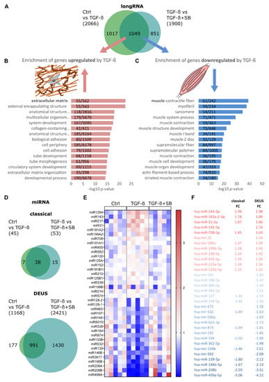
Physical training improves insulin sensitivity and can prevent type 2 diabetes (T2D). However, approximately 20% of individuals lack a beneficial outcome in glycemic control. TGF-β, identified as a possible upstream regulator involved in this low response, is also a potent regulator of microRNAs (miRNAs). The aim of this study was to elucidate the potential impact of TGF-β-driven miRNAs on individual exercise response. Non-targeted long and sncRNA sequencing analyses of TGF-β1-treated human skeletal muscle cells corroborated the effects of TGF-β1 on muscle cell differentiation, the induction of extracellular matrix components, and identified several TGF-β1-regulated miRNAs. qPCR validated a potent upregulation of miR-143-3p/145-5p and miR-181a2-5p by TGF-β1 in both human myoblasts and differentiated myotubes. Healthy subjects who were overweight or obese participated in a supervised 8-week endurance training intervention (n = 40) and were categorized as responder or low responder in glycemic control based on fold change ISIMats (≥+1.1 or <+1.1, respectively). In skeletal muscle biopsies of low responders, TGF-β signaling and miR-143/145 cluster levels were induced by training at much higher rates than among responders. Target-mining revealed HDACs, MYHs, and insulin signaling components INSR and IRS1 as potential miR-143/145 cluster targets. All these targets were down-regulated in TGF-β1-treated myotubes. Transfection of miR-143-3p/145-5p mimics in differentiated myotubes validated MYH1, MYH4, and IRS1 as miR-143/145 cluster targets. Elevated TGF-β signaling and miR-143/145 cluster induction in skeletal muscle of low responders might obstruct improvements in insulin sensitivity by training in two ways: by a negative impact of miR-143-3p on muscle cell fusion and myofiber functionality and by directly impairing insulin signaling via a reduction in INSR by TGF-β and finetuned IRS1 suppression by miR-143-3p. Full article
Show Figures

Figure 1

Back to TopTop