Trends and Advances in Tumor Immunology

A topical collection in Cells (ISSN 2073-4409). This collection belongs to the section "Cellular Immunology".

Viewed by 46476

Editors


E-Mail Website
Collection Editor
Department of Clinical Immunology, Medical University of Lublin, 20-093 Lublin, Poland
Interests: tumor microenvironment;cancer immunology;leukemia;flow cytometry
Special Issues, Collections and Topics in MDPI journals

E-Mail Website
Co-Collection Editor
Independent Laboratory of Cancer Diagnostics and Immunology, I Chair and Department of Oncological Gynaecology and Gynaecology, Medical University in Lublin, Chodźki 1, 20-093 Lublin, Poland
Interests: ovarian cancer microenvironment; immune checkpoints; tumor-infiltrating immune cells
Special Issues, Collections and Topics in MDPI journals

E-Mail Website
Co-Collection Editor
Department of Clinical Immunology, Medical University of Lublin, 20-093 Lublin, Poland.
Interests: flow cytometry; tumour immunology; autoimmunity; γδ T cells
Special Issues, Collections and Topics in MDPI journals

E-Mail Website
Co-Collection Editor
Department of Clinical Immunology, Medical University of Lublin, 20-093 Lublin, Poland
Interests: flow cytometry; tumor microenvironment; cancer immunology; leukemia

Topical Collection Information

Dear Colleagues,

Many cellular and molecular mechanisms are important for the origin and growth of tumors as well as the body's immune defense against cancer. The tumor undergoes continuous remodeling at the genetic, epigenetic, and metabolic level to evade apoptosis. At the same time, it successfully changes all the components of the immune system so as to avoid its antitumor effects. The tumor cells can alter the balance of suppressive versus cytotoxic responses. This complex interaction between immune system and cancer cells can both inhibit and enhance tumor growth. The cellular and noncell components of the tumor immune microenvironment play essential roles in tumor initiation, progression, metastasis, and response to therapies. So far, multiple cellular targets have been examined for preventing or treating cancers including but not limited to transcription factors, epigenetic targeting of oncogenes and tumor suppressor genes, kinase signaling pathways, and immune checkpoint inhibitors. Nevertheless, the clinical efficiency of these therapeutic strategies remains unsatisfactory.

This Topical Collection of Cells will present research articles and reviews that cover the scope of both cellular and molecular mechanisms of tumor immunology. Furthermore, this Topical Collection aims to promote the exchange of ideas, concepts, and findings in any area of cancer and related biomedical science studies, from a molecular point of view. All scientists working in these fields are cordially invited to submit their manuscripts.

Dr. Agnieszka Bojarska-Junak
Prof. Iwona Wertel
Dr. Michal Zarobkiewicz
Dr. Wioleta Kowalska
Collection Editors

Manuscript Submission Information

Manuscripts should be submitted online at www.mdpi.com by registering and logging in to this website. Once you are registered, click here to go to the submission form. Manuscripts can be submitted until the deadline. All submissions that pass pre-check are peer-reviewed. Accepted papers will be published continuously in the journal (as soon as accepted) and will be listed together on the collection website. Research articles, review articles as well as short communications are invited. For planned papers, a title and short abstract (about 100 words) can be sent to the Editorial Office for announcement on this website.

Submitted manuscripts should not have been published previously, nor be under consideration for publication elsewhere (except conference proceedings papers). All manuscripts are thoroughly refereed through a single-blind peer-review process. A guide for authors and other relevant information for submission of manuscripts is available on the Instructions for Authors page. Cells is an international peer-reviewed open access semimonthly journal published by MDPI.

Please visit the Instructions for Authors page before submitting a manuscript. The Article Processing Charge (APC) for publication in this open access journal is 2700 CHF (Swiss Francs). Submitted papers should be well formatted and use good English. Authors may use MDPI's English editing service prior to publication or during author revisions.

Keywords

  • cancer immunology
  • immune cells in cancer
  • molecular pathways in cancer
  • tumor microenvironment
  • cytokines
  • cytokine receptors
  • exosomes and cancer
  • immune checkpoints
  • activating receptors
  • inhibitory receptors
  • inflammation and cancer
  • signaling pathways
  • immune response
  • macrophage
  • myeloid-derived suppressor cells
  • tumor infiltration
  • γδ T cells
  • cytotoxic T cells
  • NKT cells

Published Papers (13 papers)

2024

Jump to: 2023, 2022, 2021

15 pages, 2223 KiB  
Article
High Level of CD8+PD-1+ Cells in Patients with Chronic Myeloid Leukemia Who Experienced Loss of MMR after Imatinib Discontinuation
by Paulina Kwaśnik, Joanna Zaleska, Dorota Link-Lenczowska, Magdalena Zawada, Hubert Wysogląd, Bogdan Ochrem, Grażyna Bober, Ewa Wasilewska, Iwona Hus, Monika Szarejko, Witold Prejzner, Olga Grzybowska-Izydorczyk, Agnieszka Klonowska-Szymczyk, Ewa Mędraś, Michał Kiełbus, Tomasz Sacha and Krzysztof Giannopoulos
Cells 2024, 13(8), 723; https://doi.org/10.3390/cells13080723 - 22 Apr 2024
Viewed by 1416
Abstract
Treatment-free remission (TFR) is achieved in approximately half of chronic myeloid leukemia (CML) patients treated with tyrosine kinase inhibitors. The mechanisms responsible for TFR maintenance remain elusive. This study aimed to identify immune markers responsible for the control of residual CML cells early [...] Read more.
Treatment-free remission (TFR) is achieved in approximately half of chronic myeloid leukemia (CML) patients treated with tyrosine kinase inhibitors. The mechanisms responsible for TFR maintenance remain elusive. This study aimed to identify immune markers responsible for the control of residual CML cells early in the TFR (at 3 months), which may be the key to achieving long-term TFR and relapse-free survival (RFS) after discontinuation of imatinib. Our study included 63 CML patients after imatinib discontinuation, in whom comprehensive analysis of changes in the immune system was performed by flow cytometry, and changes in the BCR::ABL1 transcript levels were assessed by RQ-PCR and ddPCR. We demonstrated a significant increase in the percentage of CD8+PD-1+ cells in patients losing TFR. The level of CD8+PD-1+ cells is inversely related to the duration of treatment and incidence of deep molecular response (DMR) before discontinuation. Analysis of the ROC curve showed that the percentage of CD8+PD-1+ cells may be a significant factor in early molecular recurrence. Interestingly, at 3 months of TFR, patients with the e13a2 transcript had a significantly higher proportion of the PD-1-expressing immune cells compared to patients with the e14a2. Our results suggest the important involvement of CD8+PD-1+ cells in the success of TFR and may help in identifying a group of patients who could successfully discontinue imatinib. Full article
Show Figures

Figure 1

2023

Jump to: 2024, 2022, 2021

18 pages, 3947 KiB  
Article
IRF8 Regulates Intrinsic Ferroptosis through Repressing p53 Expression to Maintain Tumor Cell Sensitivity to Cytotoxic T Lymphocytes
by Dakota B. Poschel, Mercy Kehinde-Ige, John D. Klement, Dafeng Yang, Alyssa D. Merting, Natasha M. Savage, Huidong Shi and Kebin Liu
Cells 2023, 12(2), 310; https://doi.org/10.3390/cells12020310 - 13 Jan 2023
Cited by 5 | Viewed by 3470
Abstract
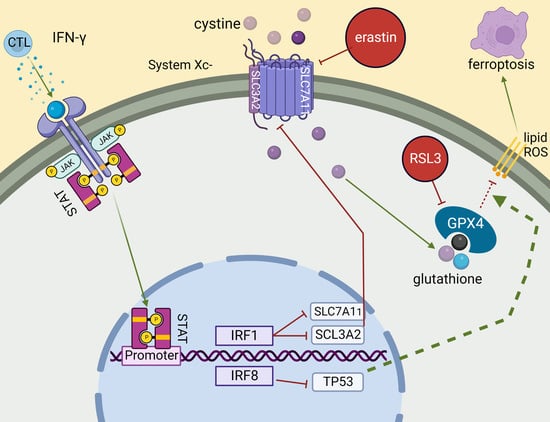
Ferroptosis has emerged as a cytotoxic T lymphocyte (CTL)-induced tumor cell death pathway. The regulation of tumor cell sensitivity to ferroptosis is incompletely understood. Here, we report that interferon regulatory factor 8 (IRF8) functions as a regulator of tumor cell intrinsic ferroptosis. Genome-wide [...] Read more.
Ferroptosis has emerged as a cytotoxic T lymphocyte (CTL)-induced tumor cell death pathway. The regulation of tumor cell sensitivity to ferroptosis is incompletely understood. Here, we report that interferon regulatory factor 8 (IRF8) functions as a regulator of tumor cell intrinsic ferroptosis. Genome-wide gene expression profiling identified the ferroptosis pathway as an IRF8-regulated pathway in tumor cells. IRF8.KO tumor cells acquire resistance to intrinsic ferroptosis induction and IRF8-deficient tumor cells also exhibit decreased ferroptosis in response to tumor-specific CTLs. Irf8 deletion increased p53 expression in tumor cells and knocking out p53 in IRF8.KO tumor cells restored tumor cell sensitivity to intrinsic ferroptosis induction. Furthermore, IRF8.KO tumor cells grew significantly faster than WT tumor cells in immune-competent mice. To restore IRF8 expression in tumor cells, we designed and synthesized codon usage-optimized IRF8-encoding DNA to generate IRF8-encoding plasmid NTC9385R-mIRF8. Restoring IRF8 expression via a lipid nanoparticle-encapsulated NTC9385R-mIRF8 plasmid therapy suppressed established tumor growth in vivo. In human cancer patients, nivolumab responders have a significantly higher IRF8 expression level in their tumor cells as compared to the non-responders. Our data determine that IRF8 represses p53 expression to maintain tumor cell sensitivity to intrinsic ferroptosis. Full article
Show Figures

Graphical abstract

2022

Jump to: 2024, 2023, 2021

17 pages, 10677 KiB  
Article
Immunoscore Combining CD8, FoxP3, and CD68-Positive Cells Density and Distribution Predicts the Prognosis of Head and Neck Cancer Patients
by Sonia Furgiuele, Géraldine Descamps, Jerome R. Lechien, Didier Dequanter, Fabrice Journe and Sven Saussez
Cells 2022, 11(13), 2050; https://doi.org/10.3390/cells11132050 - 28 Jun 2022
Cited by 10 | Viewed by 2283
Abstract
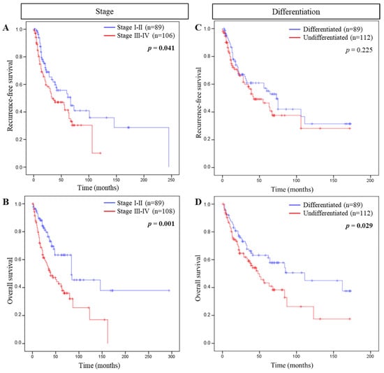
We assessed immune cell infiltrates to develop an immunoscore for prognosis and to investigate its correlation with the clinical data of patients with head and neck cancer. CD8, FoxP3, and CD68 markers were evaluated by immunohistochemistry in 258 carcinoma samples and positive cells [...] Read more.
We assessed immune cell infiltrates to develop an immunoscore for prognosis and to investigate its correlation with the clinical data of patients with head and neck cancer. CD8, FoxP3, and CD68 markers were evaluated by immunohistochemistry in 258 carcinoma samples and positive cells were counted in stromal and intra-tumoral compartments. The RStudio software was used to assess optimal cut-offs to divide the population according to survival while the prognostic value was established by using Kaplan–Meier curves and Cox regression models for each immune marker alone and in combination. We found with univariate analysis that the infiltration of immune cells in both compartments was predictive for recurrence-free survival and overall survival. Multivariate analysis revealed that CD8+ density was an independent prognostic marker. Additionally, the combination of CD8, FoxP3, and CD68 in an immunoscore provided a significant association with overall survival (p = 0.002, HR = 9.87). Such an immunoscore stayed significant (p = 0.018, HR = 11.17) in a multivariate analysis in comparison to tumor stage and histological grade, which had lower prognostic values. Altogether, our analysis indicated that CD8, FoxP3, and CD68 immunoscore was a strong, independent, and significant prognostic marker that could be introduced into the landscape of current tools to improve the clinical management of head and neck cancer patients. Full article
Show Figures

Figure 1

15 pages, 5182 KiB  
Article
An In-Vitro Study of the Expansion and Transcriptomics of CD4+ and CD8+ Naïve and Memory T Cells Stimulated by IL-2, IL-7 and IL-15
by Brooks Hopkins, Justin Fisher, Meiping Chang, Xiaoyan Tang, Zhimei Du, William J. Kelly and Zuyi Huang
Cells 2022, 11(10), 1701; https://doi.org/10.3390/cells11101701 - 20 May 2022
Cited by 3 | Viewed by 2573
Abstract
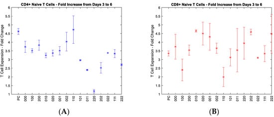
The growth of T cells ex vivo for the purpose of T cell therapies is a rate-limiting step in the overall process for cancer patients to achieve remission. Growing T cells is a fiscally-, time-, and resource-intensive process. Cytokines have been shown to [...] Read more.
The growth of T cells ex vivo for the purpose of T cell therapies is a rate-limiting step in the overall process for cancer patients to achieve remission. Growing T cells is a fiscally-, time-, and resource-intensive process. Cytokines have been shown to accelerate the growth of T cells, specifically IL-2, IL-7, and IL-15. Here a design of experiments was conducted to optimize the growth rate of different naïve and memory T cell subsets using combinations of cytokines. Mathematical models were developed to study the impact of IL-2, IL-7, and IL-15 on the growth of T cells. The results show that CD4+ and CD8+ naïve T cells grew effectively using moderate IL-2 and IL-7 in combination, and IL-7, respectively. CD4+ and CD8+ memory cells favored moderate IL-2 and IL-15 in combination and moderate IL-7 and IL-15 in combination, respectively. A statistically significant interaction was observed between IL-2 and IL-7 in the growth data of CD4+ naïve T cells, while the interaction between IL-7 and IL-15 was found for CD8+ naïve T cells. The important genes and related signaling pathways and metabolic reactions were identified from the RNA sequencing data for each of the four subsets stimulated by each of the three cytokines. This systematic investigation lays the groundwork for studying other T cell subsets. Full article
Show Figures

Figure 1

18 pages, 4478 KiB  
Article
Peripheral Blood Mononuclear Cell Populations Correlate with Outcome in Patients with Metastatic Breast Cancer
by Anna-Maria Larsson, Olle Nordström, Alexandra Johansson, Lisa Rydén, Karin Leandersson and Caroline Bergenfelz
Cells 2022, 11(10), 1639; https://doi.org/10.3390/cells11101639 - 13 May 2022
Cited by 10 | Viewed by 3285
Abstract
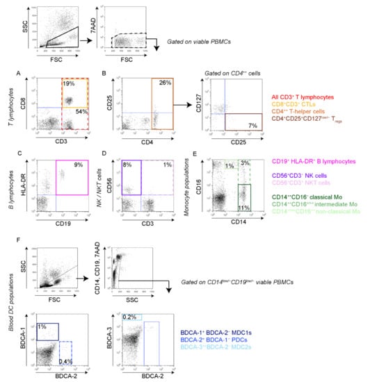
Local tumor-associated immune cells hold prognostic and predictive value in various forms of malignancy. The role of systemic, circulating leukocytes is, however, not well-characterized. In this prospective and explorative study, we aim to delineate the clinical relevance of a broad panel of circulating [...] Read more.
Local tumor-associated immune cells hold prognostic and predictive value in various forms of malignancy. The role of systemic, circulating leukocytes is, however, not well-characterized. In this prospective and explorative study, we aim to delineate the clinical relevance of a broad panel of circulating immune cells in 32 patients with newly diagnosed metastatic breast cancer (MBC) before the start of systemic treatment. Freshly isolated peripheral blood mononuclear cells (PBMCs) were analyzed by flow cytometry and evaluated for potential associations to clinicopathological variables and patient outcome. We show that the levels of specific circulating leukocyte populations are associated with clinical parameters such as hormone receptor status, histological subtype, number of circulating tumor cells (CTCs) and metastatic burden. Importantly, high levels of CD8+ cytotoxic T lymphocytes (CTLs) are significantly linked to improved overall survival (OS). In patients with estrogen receptor (ER)-positive primary tumors, high levels of circulating CTLs and non-classical (CD14+CD16++) monocytes were associated with improved OS, whereas in patients with ER-negative tumors low levels of circulating natural killer (NK) cells potentially associate with improved OS. We propose that the levels of specific circulating immune cell populations, such as CD8+ CTLs, may be used to predict clinical outcomes in MBC patients. Thus, larger studies are warranted to validate these findings. Full article
Show Figures

Figure 1

18 pages, 3158 KiB  
Article
Tissue-Specific Expression of TIGIT, PD-1, TIM-3, and CD39 by γδ T Cells in Ovarian Cancer
by Pauline Weimer, Jasmin Wellbrock, Tabea Sturmheit, Leticia Oliveira-Ferrer, Yi Ding, Stephan Menzel, Marius Witt, Louisa Hell, Barbara Schmalfeldt, Carsten Bokemeyer, Walter Fiedler and Franziska Brauneck
Cells 2022, 11(6), 964; https://doi.org/10.3390/cells11060964 - 11 Mar 2022
Cited by 26 | Viewed by 5477
Abstract
Phenotypic characterization of γδ T cells in the MALs (malignant ascites lymphocytes), TILs (tumor infiltrating lymphocytes), and PBLs (peripheral blood lymphocytes) of ovarian cancer (OvCA) patients is lacking. Therefore, we quantified γδ T cell prevalence in MAL, TIL, and PBL specimens from n [...] Read more.
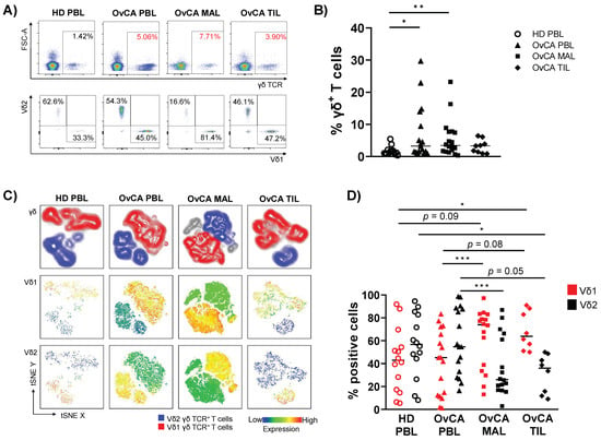
Phenotypic characterization of γδ T cells in the MALs (malignant ascites lymphocytes), TILs (tumor infiltrating lymphocytes), and PBLs (peripheral blood lymphocytes) of ovarian cancer (OvCA) patients is lacking. Therefore, we quantified γδ T cell prevalence in MAL, TIL, and PBL specimens from n = 18 OvCA patients and PBL from age-matched healthy donors (HD, n = 14). Multicolor flow cytometry was performed to evaluate the expression of inhibitory receptors (TIGIT, PD-1 and TIM-3), stimulatory receptors (Ox40), and purinergic ectoenzymes (CD39 and CD73) on γδ T cell subsets. We identified an abundant infiltration of Vδ1 T cells in the MALs and TILs. These cells varied in their differentiation: The majority of Vδ1 TILs displayed an effector memory (EM) phenotype, whereas Vδ1 MALs had a more mature phenotype of terminally differentiated effector memory cells (TEMRA) with high CD45RA expression. TIGIT and TIM-3 were abundantly expressed in both MALs and PBLs, whereas Vδ1 TILs exhibited the highest levels of PD-1, CD39, and Ox40. We also observed specific clusters on mature differentiation stages for the analyzed molecules. Regarding co-expression, Vδ1 TILs showed the highest levels of cells co-expressing TIGIT with PD-1 or CD39 compared to MALs and PBLs. In conclusion, the Vδ1 T cell population showed a high prevalence in the MALs and primary tumors of OvCA patients. Due to their (co-)expression of targetable immune receptors, in particular TIGIT with PD-1 and CD39 in TILs, Vδ1 T cell-based approaches combined with the inhibition of these targets might represent a promising strategy for OvCA. Full article
Show Figures

Figure 1

14 pages, 763 KiB  
Review
The Mysterious Actor—γδ T Lymphocytes in Chronic Lymphocytic Leukaemia (CLL)
by Michał K. Zarobkiewicz and Agnieszka A. Bojarska-Junak
Cells 2022, 11(4), 661; https://doi.org/10.3390/cells11040661 - 14 Feb 2022
Cited by 4 | Viewed by 2988
Abstract
Chronic lymphocytic leukaemia (CLL) is the most common leukaemia among adults. It is the clonal expansion of B cells expressing CD19 and CD5. Despite significant progress in treatment, CLL is still incurable. γδ T cells comprise an important subset of the cytotoxic T [...] Read more.
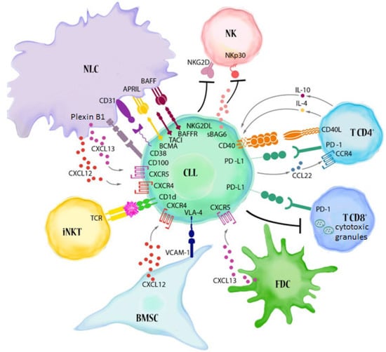
Chronic lymphocytic leukaemia (CLL) is the most common leukaemia among adults. It is the clonal expansion of B cells expressing CD19 and CD5. Despite significant progress in treatment, CLL is still incurable. γδ T cells comprise an important subset of the cytotoxic T cells. Although γδ T cells in CLL are dysfunctional, they still can possibly be used for immunotherapy. The current paper reviews our understanding of γδ T lymphocytes in CLL. Full article
Show Figures

Figure 1

2021

Jump to: 2024, 2023, 2022

10 pages, 1180 KiB  
Article
Immunotherapy in Nonendemic Nasopharyngeal Carcinoma: Real-World Data from Two Nonendemic Regions
by Panagiota Economopoulou, Anastasios Pantazopoulos, Aris Spathis, Ioannis Kotsantis, Anastasios Kyriazoglou, George Kavourakis, Roubini Zakopoulou, Ioannis Chatzidakis, Maria Anastasiou, Maria Prevezanou, Carlo Resteghini, Lisa Licitra, Cristiana Bergamini, Elena Colombo, Francesca Caspani, Nerina Denaro, Stefania Vecchio, Pierluigi Bonomo, Maria Cossu Rocca, Federica Bertolini, Daris Ferrari, Amanda Psyrri and Paolo Bossiadd Show full author list remove Hide full author list
Cells 2022, 11(1), 32; https://doi.org/10.3390/cells11010032 - 23 Dec 2021
Cited by 9 | Viewed by 2837
Abstract
Background: nasopharyngeal carcinoma (NPC) is a complex disease entity that mainly predominates in endemic regions. Real-world data with immunotherapy from nonendemic regions are limited. Methods: we collected data from patients with recurrent/metastatic (R/M) NPC treated at a center in Greece and 8 centers [...] Read more.
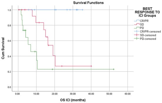
Background: nasopharyngeal carcinoma (NPC) is a complex disease entity that mainly predominates in endemic regions. Real-world data with immunotherapy from nonendemic regions are limited. Methods: we collected data from patients with recurrent/metastatic (R/M) NPC treated at a center in Greece and 8 centers in Italy. Between 2016 and 2021, 46 patients who were treated with at least one cycle of immune checkpoint inhibitors (ICI) were identified. Herein, we present our results and a review of the literature. Results: assessment of response was available in 42 patients. Overall, 11 patients responded to immunotherapy (Overall Response Rate-ORR 26.2%). Three patients had complete response (CR), and 8 patients had partial response (PR). Disease control rate (DCR) was 61.9%. Median Progression Free Survival (PFS) was 5.6 months and median Overall Survival (OS) was 19.1 months. Responders to ICI improved PFS and OS as compared to that of nonresponders. A lower probability of responding to ICI was shown in patients with more than three metastatic sites (p = 0.073), metastatic disease at initial diagnosis, (p = 0.039) or EBV DNA positive before ICI initiation, (p = 0.074). Decline in EBV DNA levels was found to be statistically significant associated with best response to ICI (p = 0.049). Safety was manageable. Conclusions: among 46 patients with R/M NPC treated with immunotherapy in two nonendemic regions, ORR was 26.2% and durable responses were observed. Low disease burden could serve as a biomarker for response to ICI. Full article
Show Figures

Figure 1

14 pages, 1814 KiB  
Article
BTLA Expression in CLL: Epigenetic Regulation and Impact on CLL B Cell Proliferation and Ability to IL-4 Production
by Lidia Karabon, Anna Andrzejczak, Lidia Ciszak, Anna Tomkiewicz, Aleksandra Szteblich, Agnieszka Bojarska-Junak, Jacek Roliński, Dariusz Wołowiec, Tomasz Wróbel and Agata Kosmaczewska
Cells 2021, 10(11), 3009; https://doi.org/10.3390/cells10113009 - 4 Nov 2021
Cited by 6 | Viewed by 2642
Abstract
In our previous study, while chronic lymphocytic leukemia (CLL) cases showed higher levels of B and T lymphocyte attenuator (BTLA) mRNA compared to controls, lower BTLA protein expression was observed in cases compared to controls. Hence we hypothesize that micro RNA (miR) 155-5p [...] Read more.
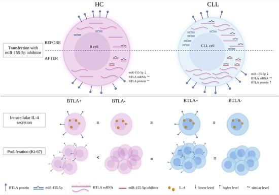
In our previous study, while chronic lymphocytic leukemia (CLL) cases showed higher levels of B and T lymphocyte attenuator (BTLA) mRNA compared to controls, lower BTLA protein expression was observed in cases compared to controls. Hence we hypothesize that micro RNA (miR) 155-5p regulates BTLA expression in CLL. In line with earlier data, expression of BTLA mRNA and miR-155-5p is elevated in CLL (p = 0.034 and p = 0.0006, respectively) as well as in MEC-1 cell line (p = 0.009 and 0.016, respectively). Inhibition of miR-155-5p partially restored BTLA protein expression in CLL patients (p = 0.01) and in MEC-1 cell lines (p = 0.058). Additionally, we aimed to evaluate the significance of BTLA deficiency in CLL cells on proliferation and IL-4 production of B cells. We found that secretion of IL-4 is not dependent on BTLA expression, since fractions of BTLA positive and BTLA negative B cells expressing intracellular IL-4 were similar in CLL patients and controls. We demonstrated that in controls the fraction of proliferating cells is lower in BTLA positive than in BTLA negative B cells (p = 0.059), which was not observed in CLL. However, the frequency of BTLA positive Ki67+ B cells in CLL was higher compared to corresponding cells from controls (p = 0.055) while there were no differences between the examined groups regarding frequency of BTLA negative Ki67+ B cells. Our studies suggest that miR-155-5p is involved in BTLA deficiency, affecting proliferation of CLL B cells, which may be one of the mechanisms responsible for CLL pathogenesis. Full article
Show Figures

Graphical abstract

19 pages, 2885 KiB  
Review
Glucocorticoid and PD-1 Cross-Talk: Does the Immune System Become Confused?
by Sabrina Adorisio, Lorenza Cannarile, Domenico V. Delfino and Emira Ayroldi
Cells 2021, 10(9), 2333; https://doi.org/10.3390/cells10092333 - 6 Sep 2021
Cited by 14 | Viewed by 5029
Abstract
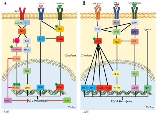
Programmed cell death protein 1 (PD-1) and its ligands, PD-L1/2, control T cell activation and tolerance. While PD-1 expression is induced upon T cell receptor (TCR) activation or cytokine signaling, PD-L1 is expressed on B cells, antigen presenting cells, and on non-immune tissues, [...] Read more.
Programmed cell death protein 1 (PD-1) and its ligands, PD-L1/2, control T cell activation and tolerance. While PD-1 expression is induced upon T cell receptor (TCR) activation or cytokine signaling, PD-L1 is expressed on B cells, antigen presenting cells, and on non-immune tissues, including cancer cells. Importantly, PD-L1 binding inhibits T cell activation. Therefore, the modulation of PD-1/PD-L1 expression on immune cells, both circulating or in a tumor microenvironment and/or on the tumor cell surface, is one mechanism of cancer immune evasion. Therapies that target PD-1/PD-L1, blocking the T cell-cancer cell interaction, have been successful in patients with various types of cancer. Glucocorticoids (GCs) are often administered to manage the side effects of chemo- or immuno-therapy, exerting a wide range of immunosuppressive and anti-inflammatory effects. However, GCs may also have tumor-promoting effects, interfering with therapy. In this review, we examine GC signaling and how it intersects with PD-1/PD-L1 pathways, including a discussion on the potential for GC- and PD-1/PD-L1-targeted therapies to “confuse” the immune system, leading to a cancer cell advantage that counteracts anti-cancer immunotherapy. Therefore, combination therapies should be utilized with an awareness of the potential for opposing effects on the immune system. Full article
Show Figures

Figure 1

17 pages, 2217 KiB  
Article
Metavariables Resuming Host Immune Features and Nodal Involvement Are Associated with Oncological Outcomes in Oral Cavity Squamous Cell Carcinoma
by Francesco Missale, Mattia Bugatti, Davide Mattavelli, Silvia Lonardi, Davide Lombardi, Piero Nicolai, Cesare Piazza, Simonetta Battocchio, Anna Maria Bozzola, Stefano Calza and William Vermi
Cells 2021, 10(9), 2203; https://doi.org/10.3390/cells10092203 - 26 Aug 2021
Cited by 2 | Viewed by 2369
Abstract
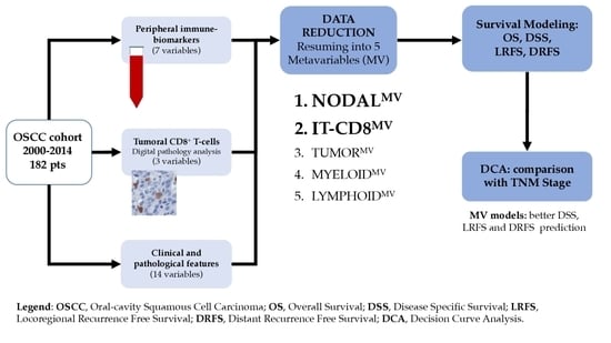
Oral cavity squamous cell carcinoma (OSCC) is a common head and neck cancer characterized by a poor prognosis associated with locoregional or distant failure. Among the predictors of prognosis, a dense infiltration of adaptive immune cells is protective and associated with improved clinical [...] Read more.
Oral cavity squamous cell carcinoma (OSCC) is a common head and neck cancer characterized by a poor prognosis associated with locoregional or distant failure. Among the predictors of prognosis, a dense infiltration of adaptive immune cells is protective and associated with improved clinical outcomes. However, few tools are available to integrate immune contexture variables into clinical settings. By using digital microscopy analysis of a large retrospective OSCC cohort (n = 182), we explored the clinical significance of tumor-infiltrating CD8+ T-cells. To this end, CD8+ T-cells counts were combined with well-established clinical variables and peripheral blood immune cell parameters. Through variable clustering, five metavariables (MV) were obtained and included descriptors of nodal (NODALMV) and primary tumor (TUMORMV) involvement, the frequency of myeloid (MYELOIDMV) or lymphoid (LYMPHOIDMV) peripheral blood immune cell populations, and the density of tumor-infiltrating CD8+ T-cells (TI-CD8MV). The clinical relevance of the MV was evaluated in the multivariable survival models. The NODALMV was significantly associated with all tested outcomes (p < 0.001), the LYMPHOIDMV showed a significant association with the overall, disease-specific and distant recurrence-free survival (p < 0.05) and the MYELOIDMV with the locoregional control only (p < 0.001). Finally, TI-CD8MV was associated with distant recurrence-free survival (p = 0.029). Notably, the performance in terms of survival prediction of the combined effect of NODALMV and immune metavariables (LYMPHOIDMV, MYELOIDMV and TI-CD8MV) was superior to the TNM stage for most of the outcomes analyzed. These findings indicate that the analysis of the baseline host immune features are promising tools to complement clinical features, in stratifying the risk of recurrences. Full article
Show Figures

Graphical abstract

24 pages, 3459 KiB  
Article
Comprehensive Integrative Analysis Reveals the Association of KLF4 with Macrophage Infiltration and Polarization in Lung Cancer Microenvironment
by Shweta Arora, Prithvi Singh, Shaniya Ahmad, Tanveer Ahmad, Ravins Dohare, Saleh A. Almatroodi, Faris Alrumaihi, Arshad Husain Rahmani and Mansoor Ali Syed
Cells 2021, 10(8), 2091; https://doi.org/10.3390/cells10082091 - 14 Aug 2021
Cited by 22 | Viewed by 5868
Abstract
Macrophage polarization and infiltration to the tumor microenvironment (TME) is a critical determining factor for tumor progression. Macrophages are polarized into two states—M1 (pro-inflammatory, anti-tumorigenic and stimulated by LPS or IFN-γ) and M2 (anti-inflammatory pro-tumorigenic and stimulated by IL-4) phenotypes. Specifically, [...] Read more.
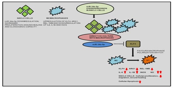
Macrophage polarization and infiltration to the tumor microenvironment (TME) is a critical determining factor for tumor progression. Macrophages are polarized into two states—M1 (pro-inflammatory, anti-tumorigenic and stimulated by LPS or IFN-γ) and M2 (anti-inflammatory pro-tumorigenic and stimulated by IL-4) phenotypes. Specifically, M2 macrophages enhance tumor cell growth and survival. Recent evidences suggest the pivotal role of microRNAs in macrophage polarization during the development of Non-small cell lung cancer (NSCLC), thus proposing a new therapeutic option to target lung cancer. In silico analysis determined cogent upregulation of KLF4, downregulation of IL-1β and miR-34a-5p in NSCLC tissues, consequently worsening the overall survival of NSCLC patients. We observed a significant association of KLF4 with macrophage infiltration and polarization in NSCLC. We found that KLF4 is critically implicated in M2 polarization of macrophages, which, in turn, promotes tumorigenesis. KLF4 expression correlated with miR-34a-5p and IL-1β in a feed-forward loop (FFL), both of which are implicated in immune regulation. Mechanistic overexpression of miR-34a-5p in macrophages (IL-4 stimulated) inhibits KLF4, along with downregulation of ARG1, REL-1MB (M2 macrophage specific markers), and upregulation of IL-1β, IL-6, (M1 macrophage specific markers), demonstrating macrophage polarization switch from M2 to M1 phenotype. Moreover, co-culture of these macrophages with NSCLC cells reduces their proliferation, wound healing, clonogenic capacity and enhanced NO-mediated apoptosis. Further, transfection of miR-34a-5p in NSCLC cells, also degrades KLF4, but enhances the expression of KLF4 regulated genes—IL-1β, IL-6 (pro-inflammatory mediators), which is further enhanced upon co-culture with IL-4 stimulated macrophages. Additionally, we observed a significant increase in i-NOS/NO content upon co-culture, suggesting polarization reversion of macrophages from M2 to M1, and eventually leading to anti-tumor effects. Our findings thus show a significant role of KLF4 in tumorigenesis and TAM polarization of NSCLC. However, miR-34a-5p mediated targeting of these molecular networks will provide a better therapeutic intervention for NSCLC. Full article
Show Figures

Graphical abstract

18 pages, 1277 KiB  
Review
Promising Anti-Mitochondrial Agents for Overcoming Acquired Drug Resistance in Multiple Myeloma
by Vanessa Innao, Vincenzo Rizzo, Andrea Gaetano Allegra, Caterina Musolino and Alessandro Allegra
Cells 2021, 10(2), 439; https://doi.org/10.3390/cells10020439 - 19 Feb 2021
Cited by 18 | Viewed by 3974
Abstract
Multiple myeloma (MM) remains an incurable tumor due to the high rate of relapse that still occurs. Acquired drug resistance represents the most challenging obstacle to the extension of survival and several studies have been conducted to understand the mechanisms of this phenomenon. [...] Read more.
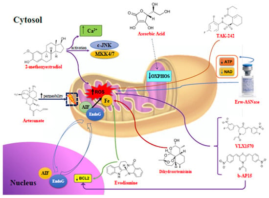
Multiple myeloma (MM) remains an incurable tumor due to the high rate of relapse that still occurs. Acquired drug resistance represents the most challenging obstacle to the extension of survival and several studies have been conducted to understand the mechanisms of this phenomenon. Mitochondrial pathways have been extensively investigated, demonstrating that cancer cells become resistant to drugs by reprogramming their metabolic assessment. MM cells acquire resistance to proteasome inhibitors (PIs), activating protection programs, such as a reduction in oxidative stress, down-regulating pro-apoptotic, and up-regulating anti-apoptotic signals. Knowledge of the mechanisms through which tumor cells escape control of the immune system and acquire resistance to drugs has led to the creation of new compounds that can restore the response by leading to cell death. In this scenario, based on all literature data available, our review represents the first collection of anti-mitochondrial compounds able to overcome drug resistance in MM. Caspase-independent mechanisms, mainly based on increased oxidative stress, result from 2-methoxyestradiol, Artesunate, ascorbic acid, Dihydroartemisinin, Evodiamine, b-AP15, VLX1570, Erw-ASNase, and TAK-242. Other agents restore PIs’ efficacy through caspase-dependent tools, such as CDDO-Im, NOXA-inhibitors, FTY720, GCS-100, LBH589, a derivative of ellipticine, AT-101, KD5170, SMAC-mimetics, glutaminase-1 (GLS1)-inhibitors, and thenoyltrifluoroacetone. Each of these substances improved the efficacy rates when employed in combination with the most frequently used antimyeloma drugs. Full article
Show Figures

Figure 1

Back to TopTop