Progress in Liver Stem Cell Therapy

A topical collection in Cells (ISSN 2073-4409). This collection belongs to the section "Stem Cells".

Viewed by 13890

Editors


E-Mail Website
Collection Editor
Applied Molecular Hepatology Lab., Department of Visceral, Transplant, Thoracic und Vascular Surgery, University of Leipzig Medical Center, Liebigstraße 21, 04103 Leipzig, Germany
Interests: liver diseases; stem cell biology; regenerative medicine; stem cell differentiation; mesenchymal stem cell; stem cell isolation; liver metabolism; liver cell transplantation

E-Mail Website
Collection Editor
Department of Pathology, University of Pittsburgh, S-BST, Room S-404, 200 Lothrop Street, Pittsburgh, PA 15261, USA
Interests: liver stem cells; hepatic cell transplantation; iPS cells; liver regeneration; activin A signaling; senescence

Topical Collection Information

Dear Colleagues,

The transplantation of liver cells to treat liver diseases has a long history. As early as the mid-1970s, donor hepatocytes isolated from rats were transplanted via the portal vein into a recipient rat liver suffering from a defect of the bilirubin-conjugating enzyme uridine diphosphate glucuronosyltransferase. The defect was corrected at least in part for a sustained period of time after transplantation (AJ Matas et al., Science 1976;192:892-4). However, the clinical translation of the method is still awaiting breakthrough because of the rareness of donor livers for hepatocyte isolation. Since the feasibility of hepatocyte transplantation has, nevertheless, been proven in principle, alternative sources of transplantable hepatocytes have been explored. The most promising tools are stem cell-derived hepatocytes. These comprise hepatocyte-like cells derived from adult or embryonic stem cells and from induced pluripotent stem cells, besides others. Additionally, liver-like three-dimensional structures, named liver organoids, have been considered an adequate tissue replacement for diseased liver tissue. Recently, awareness has grown from mesenchymal stromal cells that, not the cells in toto, but even exosomes derived from them were sufficient to correct for liver tissue defects. Therefore, the scope of the Topical Collection “Progress in Liver Stem Cell Therapy” is to compile the foremost developments in stem cell-based approaches to treating liver diseases.

In the context of liver diseases, it is the aim to collect the latest research on novel stem cell resources and their pre-clinical as well as clinical establishment; animal and cell-culture model systems with which to assess stem cells’ therapeutic potential; concepts for the standardization of the isolation, processing and storage of stem cells and stem cell-based hepatocytes; and how stem cells impact liver diseases mechanistically. Contributions including bioinformatics and computational modeling concepts for predicting stem cell-based mechanisms and therapy outcomes are highly appreciated. We invite authors to submit their highly innovative research or comprehensive review articles related to the current and future development of the treatment of liver diseases with stem cell-based approaches.

Prof. Dr. Bruno Christ
Prof. Dr. Michael Oertel
Collection Editors

Manuscript Submission Information

Manuscripts should be submitted online at www.mdpi.com by registering and logging in to this website. Once you are registered, click here to go to the submission form. Manuscripts can be submitted until the deadline. All submissions that pass pre-check are peer-reviewed. Accepted papers will be published continuously in the journal (as soon as accepted) and will be listed together on the collection website. Research articles, review articles as well as short communications are invited. For planned papers, a title and short abstract (about 100 words) can be sent to the Editorial Office for announcement on this website.

Submitted manuscripts should not have been published previously, nor be under consideration for publication elsewhere (except conference proceedings papers). All manuscripts are thoroughly refereed through a single-blind peer-review process. A guide for authors and other relevant information for submission of manuscripts is available on the Instructions for Authors page. Cells is an international peer-reviewed open access semimonthly journal published by MDPI.

Please visit the Instructions for Authors page before submitting a manuscript. The Article Processing Charge (APC) for publication in this open access journal is 2700 CHF (Swiss Francs). Submitted papers should be well formatted and use good English. Authors may use MDPI's English editing service prior to publication or during author revisions.

Keywords

  • liver
  • hepatocyte
  • stem cell
  • cell transplantation
  • organoids
  • liver disease
  • cell therapy
  • experimental models
  • transdifferentiation

Published Papers (4 papers)

2023

Jump to: 2022

15 pages, 2289 KiB  
Review
Therapeutic Cell Repopulation of the Liver: From Fetal Rat Cells to Synthetic Human Tissues
by David A. Shafritz, Mo R. Ebrahimkhani and Michael Oertel
Cells 2023, 12(4), 529; https://doi.org/10.3390/cells12040529 - 6 Feb 2023
Cited by 4 | Viewed by 2493
Abstract
Progenitor cells isolated from the fetal liver can provide a unique cell source to generate new healthy tissue mass. Almost 20 years ago, it was demonstrated that rat fetal liver cells repopulate the normal host liver environment via a mechanism akin to cell [...] Read more.
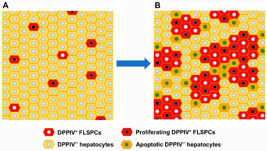
Progenitor cells isolated from the fetal liver can provide a unique cell source to generate new healthy tissue mass. Almost 20 years ago, it was demonstrated that rat fetal liver cells repopulate the normal host liver environment via a mechanism akin to cell competition. Activin A, which is produced by hepatocytes, was identified as an important player during cell competition. Because of reduced activin receptor expression, highly proliferative fetal liver stem/progenitor cells are resistant to activin A and therefore exhibit a growth advantage compared to hepatocytes. As a result, transplanted fetal liver cells are capable of repopulating normal livers. Important for cell-based therapies, hepatic stem/progenitor cells containing repopulation potential can be separated from fetal hematopoietic cells using the cell surface marker δ-like 1 (Dlk-1). In livers with advanced fibrosis, fetal epithelial stem/progenitor cells differentiate into functional hepatic cells and out-compete injured endogenous hepatocytes, which cause anti-fibrotic effects. Although fetal liver cells efficiently repopulate the liver, they will likely not be used for human cell transplantation. Thus, utilizing the underlying mechanism of repopulation and developed methods to produce similar growth-advantaged cells in vitro, e.g., human induced pluripotent stem cells (iPSCs), this approach has great potential for developing novel cell-based therapies in patients with liver disease. The present review gives a brief overview of the classic cell transplantation models and various cell sources studied as donor cell candidates. The advantages of fetal liver-derived stem/progenitor cells are discussed, as well as the mechanism of liver repopulation. Moreover, this article reviews the potential of in vitro developed synthetic human fetal livers from iPSCs and their therapeutic benefits. Full article
Show Figures

Figure 1

2022

Jump to: 2023

15 pages, 2586 KiB  
Article
Label-Free Imaging Analysis of Patient-Derived Cholangiocarcinoma Organoids after Sorafenib Treatment
by Michael Koch, Sandra Nickel, Ruby Lieshout, Susanna M. Lissek, Martina Leskova, Luc J. W. van der Laan, Monique M. A. Verstegen, Bruno Christ and Francesco Pampaloni
Cells 2022, 11(22), 3613; https://doi.org/10.3390/cells11223613 - 15 Nov 2022
Cited by 8 | Viewed by 3125
Abstract
Monitoring tumor growth dynamics is crucial for understanding cancer. To establish an in vitro method for the continuous assessment of patient-specific tumor growth, tumor organoids were generated from patients with intrahepatic CCA (iCCA). Organoid growth was monitored for 48 h by label-free live [...] Read more.
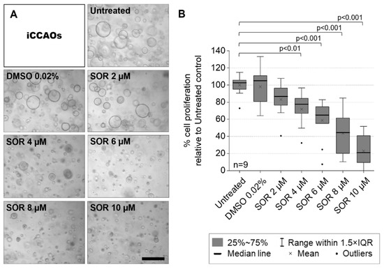
Monitoring tumor growth dynamics is crucial for understanding cancer. To establish an in vitro method for the continuous assessment of patient-specific tumor growth, tumor organoids were generated from patients with intrahepatic CCA (iCCA). Organoid growth was monitored for 48 h by label-free live brightfield imaging. Growth kinetics were calculated and validated by MTS assay as well as immunohistochemistry of Ki67 to determine proliferation rates. We exposed iCCA organoids (iCCAOs) and non-tumor intrahepatic cholangiocyte organoids (ICOs) to sub-therapeutic concentrations of sorafenib. Monitoring the expansion rate of iCCAOs and ICOs revealed that iCCAO growth was inhibited by sorafenib in a time- and dose-dependent fashion, while ICOs were unaffected. Quantification of the proliferation marker Ki67 confirmed inhibition of iCCAO growth by roughly 50% after 48 h of treatment with 4 µM sorafenib. We established a robust analysis pipeline combining brightfield microscopy and a straightforward image processing approach for the label-free growth monitoring of patient-derived iCCAOs. Combined with bioanalytical validation, this approach is suitable for a fast and efficient high-throughput drug screening in tumor organoids to develop patient-specific systemic treatment options. Full article
Show Figures

Figure 1

19 pages, 745 KiB  
Review
Clinical Application of Induced Hepatocyte-like Cells Produced from Mesenchymal Stromal Cells: A Literature Review
by Yanina Bogliotti, Mark Vander Roest, Aras N. Mattis, Robert G. Gish, Gary Peltz, Robin Anwyl, Salah Kivlighn and Eric R. Schuur
Cells 2022, 11(13), 1998; https://doi.org/10.3390/cells11131998 - 22 Jun 2022
Cited by 6 | Viewed by 3275
Abstract
Liver disease is a leading cause of mortality worldwide, resulting in 1.3 million deaths annually. The vast majority of liver disease is caused by metabolic disease (i.e., NASH) and alcohol-induced hepatitis, and to a lesser extent by acute and chronic viral infection. Furthermore, [...] Read more.
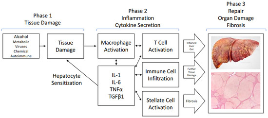
Liver disease is a leading cause of mortality worldwide, resulting in 1.3 million deaths annually. The vast majority of liver disease is caused by metabolic disease (i.e., NASH) and alcohol-induced hepatitis, and to a lesser extent by acute and chronic viral infection. Furthermore, multiple insults to the liver is becoming common due to the prevalence of metabolic and alcohol-related liver diseases. Despite this rising prevalence of liver disease, there are few treatment options: there are treatments for viral hepatitis C and there is vaccination for hepatitis B. Aside from the management of metabolic syndrome, no direct liver therapy has shown clinical efficacy for metabolic liver disease, there is very little for acute alcohol-induced liver disease, and liver transplantation remains the only effective treatment for late-stage liver disease. Traditional pharmacologic interventions have failed to appreciably impact the pathophysiology of alcohol-related liver disease or end-stage liver disease. The difficulties associated with developing liver-specific therapies result from three factors that are common to late-stage liver disease arising from any cause: hepatocyte injury, inflammation, and aberrant tissue healing. Hepatocyte injury results in tissue damage with inflammation, which sensitizes the liver to additional hepatocyte injury and stimulates hepatic stellate cells and aberrant tissue healing responses. In the setting of chronic liver insults, there is progressive scarring, the loss of hepatocyte function, and hemodynamic dysregulation. Regenerative strategies using hepatocyte-like cells that are manufactured from mesenchymal stromal cells may be able to correct this pathophysiology through multiple mechanisms of action. Preclinical studies support their effectiveness and recent clinical studies suggest that cell replacement therapy can be safe and effective in patients with liver disease for whom there is no other option. Full article
Show Figures

Figure 1

21 pages, 13136 KiB  
Article
Human Mesenchymal Stromal Cells Resolve Lipid Load in High Fat Diet-Induced Non-Alcoholic Steatohepatitis in Mice by Mitochondria Donation
by Sandra Nickel, Madlen Christ, Sandra Schmidt, Joanna Kosacka, Hagen Kühne, Martin Roderfeld, Thomas Longerich, Lysann Tietze, Ina Bosse, Mei-Ju Hsu, Peggy Stock, Elke Roeb and Bruno Christ
Cells 2022, 11(11), 1829; https://doi.org/10.3390/cells11111829 - 2 Jun 2022
Cited by 16 | Viewed by 3725
Abstract
Mesenchymal stromal cells (MSC) increasingly emerge as an option to ameliorate non-alcoholic steatohepatitis (NASH), a serious disease, which untreated may progress to liver cirrhosis and cancer. Before clinical translation, the mode of action of MSC needs to be established. Here, we established NASH [...] Read more.
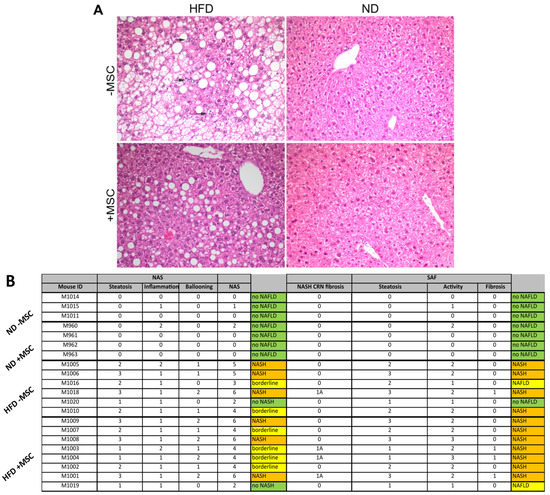
Mesenchymal stromal cells (MSC) increasingly emerge as an option to ameliorate non-alcoholic steatohepatitis (NASH), a serious disease, which untreated may progress to liver cirrhosis and cancer. Before clinical translation, the mode of action of MSC needs to be established. Here, we established NASH in an immune-deficient mouse model by feeding a high fat diet. Human bone-marrow-derived MSC were delivered to the liver via intrasplenic transplantation. As verified by biochemical and image analyses, human mesenchymal stromal cells improved high-fat-diet-induced NASH in the mouse liver by decreasing hepatic lipid content and inflammation, as well as by restoring tissue homeostasis. MSC-mediated changes in gene expression indicated the switch from lipid storage to lipid utilization. It was obvious that host mouse hepatocytes harbored human mitochondria. Thus, it is feasible that resolution of NASH in mouse livers involved the donation of human mitochondria to the mouse hepatocytes. Therefore, human MSC might provide oxidative capacity for lipid breakdown followed by restoration of metabolic and tissue homeostasis. Full article
Show Figures

Figure 1

Back to TopTop