Article

Effect of Photon Hormesis on Dose Responses to Alpha Particles in Zebrafish Embryos

1
Department of Physics and Materials Science, City University of Hong Kong, Hong Kong, China
2
Department of Biomedical Sciences, City University of Hong Kong, Hong Kong, China
3
State Key Laboratory in Marine Pollution, City University of Hong Kong, Hong Kong, China
*
Authors to whom correspondence should be addressed.
Int. J. Mol. Sci. 2017, 18(2), 385; https://doi.org/10.3390/ijms18020385
Submission received: 9 December 2016 / Revised: 3 February 2017 / Accepted: 8 February 2017 / Published: 11 February 2017
(This article belongs to the Special Issue Zebrafish: A Model for Toxicological Research)

Abstract

:
Photon hormesis refers to the phenomenon where the biological effect of ionizing radiation with a high linear energy transfer (LET) value is diminished by photons with a low LET value. The present paper studied the effect of photon hormesis from X-rays on dose responses to alpha particles using embryos of the zebrafish (Danio rerio) as the in vivo vertebrate model. The toxicity of these ionizing radiations in the zebrafish embryos was assessed using the apoptotic counts at 20, 24, or 30 h post fertilization (hpf) revealed through acridine orange (AO) staining. For alpha-particle doses ≥ 4.4 mGy, the additional X-ray dose of 10 mGy significantly reduced the number of apoptotic cells at 24 hpf, which proved the presence of photon hormesis. Smaller alpha-particle doses might not have inflicted sufficient aggregate damages to trigger photon hormesis. The time gap T between the X-ray (10 mGy) and alpha-particle (4.4 mGy) exposures was also studied. Photon hormesis was present when T ≤ 30 min, but was absent when T = 60 min, at which time repair of damage induced by alpha particles would have completed to prevent their interactions with those induced by X-rays. Finally, the drop in the apoptotic counts at 24 hpf due to photon hormesis was explained by bringing the apoptotic events earlier to 20 hpf, which strongly supported the removal of aberrant cells through apoptosis as an underlying mechanism for photon hormesis.

1. Introduction

Linear energy transfer (LET) describes the quality of an ionizing radiation. In general, high-LET radiation, such as alpha particles and heavy ions, generate clusters of damage along their trajectories in a medium, while low-LET radiation, such as X-rays and gamma rays, mainly induce dispersed damage, so high-LET radiation can induce more severe biological effects in cells [1]. It was also established that high- and low-LET radiation damage had different kinetics of induction and repair [2,3].
In fact, all living organisms, including humans, are commonly exposed to more than a single type of radiation in our environment. For instance, the general public is exposed to background gamma radiation together with alpha-particle exposures from indoor radon levels [4,5,6,7,8,9,10]. Airline crew members and astronauts are exposed to cosmic radiation and secondary radiation, which consists of neutrons and gamma rays, produced by interaction of the cosmic rays with the shielding materials of the cabin during airplane and space flights [11,12]. Cancer patients could also be exposed to mixed beams of high- and low-LET radiation during radiotherapy. In boron neutron capture therapy, patients are exposed to radiation fields consisting of a mixture of radiation with different LETs, including the high-LET products generated during the 10B(n,α)7Li reaction and the low-LET gamma rays released during radioactive capture (n,γ) reactions [13].
The biological effects of simultaneous or sequential exposures to high- and low-LET radiations have been studied for more than 40 years. Durand and Olive [14] explored, but did not observe, the repair of sub-lethal damage of combinations of X-rays and neutrons in V79 cells. In contrast, Ngo and Elkind [15] demonstrated the repair of sub-lethal damages of a neutron dose followed by an X-ray dose in V79 cells. McNally et al. [16] and Ngo et al. [17] studied the biological effects of X-rays and neutrons/heavy ions, and the data appeared to imply that X-rays and alpha particles would also interact synergistically to increase cell killing and mutation [18,19]. In contrast, Barendsen et al. [2] reported that the damage from X-rays and alpha particles interacted independently. Brooks et al. [20] showed that in lung epithelial cells, X-rays and alpha particles did not interact to change the cell cycle progression, but interacted to enhance cell killing and micronucleus induction.
More recently, Rithidech and Scott [21] reported an interesting gamma-ray hormetic effect where the biological effect of neutrons was alleviated by a simultaneous gamma-ray exposure. Hormetic responses are generally characterized by biphasic dose-response relationships showing low-dose stimulation and high-dose inhibition [22,23,24,25,26]. Rithidech and Scott [21] confirmed the presence of gamma-ray hormesis in low-dose neutron irradiation through the induction of micronucleated cells in human lymphocytes which had been irradiated with mono-energetic neutron sources with five different energies (i.e., 0.22, 0.44, 1.5, 5.9, and 13.7 MeV) and with 1%, 1%, 2%, 6%, and 6% gamma-ray contamination, respectively. The gamma-ray hormesis in neutron irradiations was further confirmed by Ng et al. [27] who studied the neutron dose response on zebrafish embryos in vivo from 0.6 to 100 mGy with 14% of gamma-ray contamination. Ng et al. [27] demonstrated a suppression of the biological effect of neutrons when the associated gamma-ray contamination reached 10 mGy or above. On the other hand, Scott [28] and Scott et al. [29] investigated the induction of lung cancer on Wistar rats after they inhaled 239Pu (an alpha-particle emitter) in an insoluble dioxide form, with alpha-particle doses up to ~600 mGy. The authors showed that 1 to 2 mGy of gamma-ray doses suppressed lung-cancer induction in Wistar rats. The proposed mechanisms underlying the gamma-ray hormesis included removal of aberrant cells through apoptosis and high-fidelity DNA repair [30,31,32].
This phenomenon was generalized as “photon hormesis” in the present paper to include both X-ray and gamma-ray photons, in which the biological effect of a high-LET ionizing radiation was diminished by a low-LET photon exposure. Photon hormesis was also reported on suppressing the biological effect of alpha-particle radiation. For non-targeted ionizing effect, Ng et al. [33] reported that the onset of photon hormesis could mitigate the neutron-induced damages on zebrafish embryos and thus suppressed the induction of bystander signals to the neighboring naive embryos.
Despite the above interesting findings on interactions between high- and low-LET radiations, some results remained equivocal (e.g., [2,14,15,16,17,18,19,20]). Further studies on these interactions, particularly the underlying mechanisms, would be pertinent. In the present work, zebrafish (Danio rerio) embryos were employed as the in vivo vertebrate model to assess the effect of photon hormesis on dose responses to alpha particles. Zebrafish embryos had been commonly used in research related to toxicology screening and ionizing radiation [27,33,34,35,36,37,38,39,40,41,42,43,44,45,46,47,48,49,50] considering their numerous advantages, including their rapid development process, high fecundity, optically transparency and, most importantly, their genomes sharing considerable homology with human genomes [51]. It was established that different alpha-particle doses would lead to hormetic and toxic effects in zebrafish embryos [49], so a dose response curve for graded alpha-particle dose was established in the present work to fully assess the effect of photon hormesis. In relation, for the study on the effect of photon hormesis on neutron doses in zebrafish embryos, Ng et al. [27] examined the neutron dose response curve using graded neutron doses. For the study on the interaction between X-rays and alpha particles in lung epithelial cells, Brooks et al. [20] also used graded X-ray doses together with two alpha-particle doses. Furthermore, Brooks et al. [20] commented that time gaps between high- and low-LET exposures could weaken their interactions, so we also studied the effect of the time gap between alpha-particle and X-ray irradiations in the present work. Finally, an attempt was made to provide information on the mechanisms underlying the photon hormesis by studying the apoptotic counts at different time points after alpha-particle irradiations with and without additional X-ray irradiation.

2. Results

2.1. Part A: Effect of Alpha-Particle Dose with or without Additional Photon Dose

The dose responses of zebrafish embryos to different alpha-particle doses were first studied. Four alpha-particle doses, namely, 1.1, 2.2, 4.4, and 8.8 mGy, were employed in the present study. For each set of experiment, a total of 50 dechorionated embryos were deployed, which were divided into five groups each having 10 embryos, namely:
(1)
A1 group: embryos received ~1.1 mGy alpha-particle irradiation at 5 hpf;
(2)
A2 group: embryos received ~2.2 mGy alpha-particle irradiation at 5 hpf;
(3)
A4 group: embryos received ~4.4 mGy alpha-particle irradiation at 5 hpf;
(4)
A8 group: embryos received ~8.8 mGy alpha-particle irradiation at 5 hpf; and
(5)
AC group: embryos were sham irradiated with alpha particles at 5 hpf.
To further study the effect of photon hormesis (from X-ray photons in the current study) on the dose response of alpha-particle irradiated zebrafish embryos, an additional X-ray dose of 10 mGy was delivered to the embryos immediately after their alpha-particle irradiation.
For each set of experiment in this section, a total of 50 dechorionated embryos were deployed, which were divided into five groups each having 10 embryos, namely:
(1)
AX1 group: embryos received ~1.1 mGy alpha-particle irradiation and 10 mGy X-ray irradiation at 5 hpf;
(2)
AX2 group: embryos received ~2.2 mGy alpha-particle irradiation and 10 mGy X-ray irradiation at 5 hpf;
(3)
AX4 group: embryos received ~4.4 mGy alpha-particle irradiation and 10 mGy X-ray irradiation at 5 hpf;
(4)
AX8 group: embryos received ~8.8 mGy alpha-particle irradiation and 10 mGy X-ray irradiation at 5 hpf; and
(5)
AXC group: embryos were sham irradiated with alpha particles and X-rays at 5 hpf.
The mean apoptotic counts (N) for the A1, A2, A4, A8, and AC groups were denoted as NA1, NA2, NA4, NA8, and NAc, respectively. The average <NAc> was computed from the NAc values in the corresponding set of experiment. The “net normalized apoptotic counts” for all of the A1, A2, A4, and A8 groups were determined as NAY+ = [(NAY − <NAc>)/<NAc>], where Y could assume values of 1, 2, 4, or 8, which corresponded to exposures to alpha-particle doses of 1.1, 2.2, 4.4, and 8.8 mGy, respectively. In relation, NAc+ = [(NAc − <NAc>)/<NAc>]. Similarly, the “net normalized apoptotic counts” for all the AX1, AX2, AX4, AX8 groups were determined as NAXY+ = [(NAXY − <NAXc>]/[<NAXc>], where <NAXc> was computed from the NAXc values in the corresponding set of experiment. In relation, NAXc+ = [(NAXc − <NAXc>)/<NAXc>]. The net normalized apoptotic counts were grouped for analysis, and the results were shown in Figure 1. The data shown in Figure 1 for (i) alpha-particle dose only, and (ii) alpha-particle dose + X-ray dose were separately analyzed to confirm whether a particular case (dose or a combination of doses) would lead to hormetic or toxic effects, or would lead to effects insignificantly different from the background. ANOVA gave p = 9.69 × 10−39 for (i) alpha-particle dose only and p = 1.43 × 10⁻²⁵ for (ii) alpha-particle dose + X-ray dose. When p ≤ 0.05 from ANOVA, the responses corresponding to at least two cases were significantly different, and post-hoc t-tests would be performed to assess the statistical significance between the AY groups and the AC group for (i) alpha-particle dose only, and between the AXY groups and the AXC group for (ii) alpha-particle dose + X-ray dose. When p ≤ 0.05/10 (i.e., 0.005) from a particular post-hoc t-test, the difference between the tested AY or AXY group and the corresponding AC or AXC group was considered statistically significant, and was asterisked in Figure 1. As revealed in Figure 1, 5-hpf embryos irradiated with 1.1 mGy of alpha particles exhibited hormetic effect and were thus in the hormetic zone (HZ), while those irradiated with ≥2.2 mGy of alpha particles exhibited toxic effect and were, thus, in the toxic zone (TZ) at 24 hpf. Furthermore, 5-hpf embryos irradiated with (1.1 mGy of alpha particles + 10 mGy of X-rays) did not lead to results significantly different from the background signals (i.e., apoptotic counts on AXC groups of embryos), while those irradiated with (≥2.2 mGy of alpha particles + 10 mGy of X-rays) were in the TZ.
To elucidate the effect of the additional X-ray dose on the alpha-particle irradiated embryos, the net normalized data in the three repeated experiments were combined for both parts and compared. The presence of photon hormesis was indicated by NAY+ > NAXY+, while cases with p ≤ 0.05 (assessed through t-tests) were considered statistically significant. The results and interpretations were:
(i)
NA1+ < NAX1+, photon hormesis was absent, p = 3.44 × 10⁻⁵
(ii)
NA2+ < NAX2+, photon hormesis was absent, p = 6.75 × 10⁻¹⁰
(iii)
NA4+ > NAX4+, photon hormesis was present, p = 0.0185
(iv)
NA8+ > NAX8+, photon hormesis was present, p = 1.19 × 10⁻⁸
Apparently, an additional X-ray dose of 10 mGy significantly altered the dose response of embryos at 24 hpf. For alpha-particle doses ≤ 2.2 mGy, the additional X-ray dose led to more apoptotic cells at 24 hpf. Interestingly, the significant hormetic effect originally recorded for the alpha-particle dose of 1.1 mGy no longer existed. In contrast, for alpha-particle doses ≥ 4.4 mGy, the additional X-ray dose significantly reduced the number of apoptotic cells at 24 hpf, which proved the presence of photon hormesis (to diminish the biological effect of alpha particles).

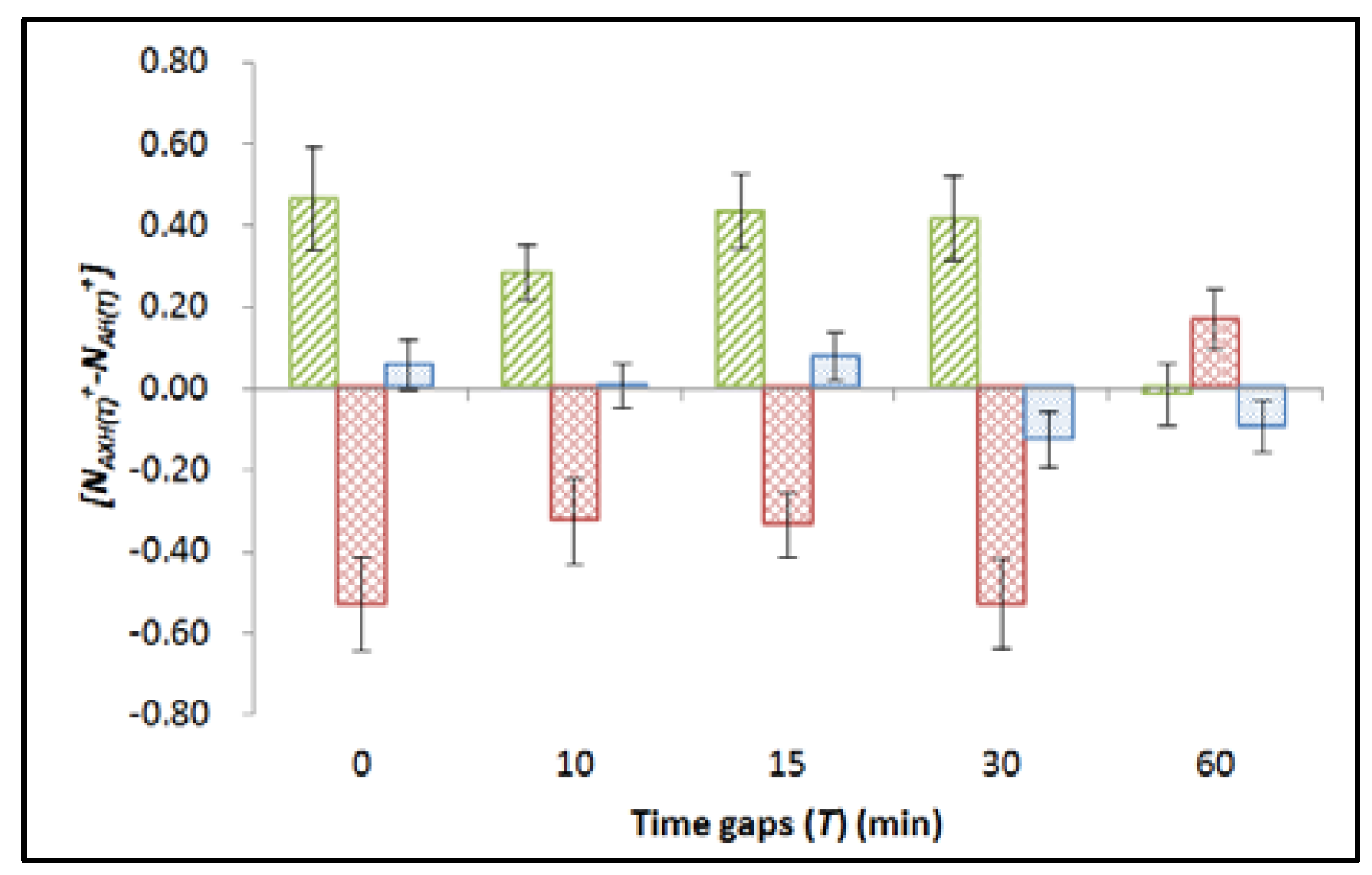
2.2. Part B: Effect of Time Gap between Alpha-Particle and X-ray Irradiations

To study the effect of time gap between alpha-particle and photon irradiation on the photon hormesis, the responses to alpha particles (4.4 mGy) in zebrafish embryos upon their receiving an additional X-ray dose (10 mGy) with time gaps T = 0 (i.e., immediately), 10, 15, 30, or 60 (min) were studied. Alpha-particle irradiation was performed at 5 hpf. Alpha-particle dose of 4.4 mGy and additional X-ray dose of 10 mGy were employed. In this part of the experiment, the apoptotic counts were determined at the time H = 24 (hpf), in contrast to H = 20 and 30 (hpf) in Part C below. A total of 30 dechorionated embryos, which were divided into three groups each having 10 embryos, were deployed in each set of experiment. The three groups were referred to as (with H = 24 in this part):
(1)
AXH(T) group: embryos were irradiated with 4.4 mGy alpha-particle dose and then received 10 mGy X-ray dose with H = 24 (hpf) and T = 0, 10, 15, 30, or 60 (min);
(2)
AH(T) group: embryos were irradiated with 4.4 mGy alpha-particle dose and then sham irradiated with X-rays, with H = 24 (hpf) and T = 0, 10, 15, 30, or 60 (min); and
(3)
CH(T) group: embryos were sham irradiated with alpha particles and X-rays, with H = 24 (hpf) and T = 0, 10, 15, 30, or 60 (min).
The mean apoptotic counts (N) for the AXH(T), AH(T) and CH(T) groups were denoted as NAXH(T), NAH(T), and NCH(T), respectively. The average <NCH(T)> was computed from the NCH(T) values in the corresponding set of experiment. The “net normalized apoptotic counts” for the AXH(T), AH(T) and CH(T) groups could be determined as NAXH(T)+ = [(NAXH(T) − <NCH(T)>)/<NCH(T)>], NAH(T)+ = [(NAH(T) − <NCH(T)>)/ <NCH(T)>] and NCH(T)+ = [(NCH(T) − <NCH(T)>)/<NCH(T)>], respectively. The net normalized apoptotic counts were grouped for analyses. The variations between all groups of embryos were first studied by ANOVA. For cases with p ≤ 0.05, post-hoc t-tests were then performed to assess the statistical significance between different groups. The results were summarized in Figure 2 and Table 1.
Apparently, photon hormesis was effective (i.e., NAX24(T)+NA24(T)+ < 0) when T ≤ 30 min. Here the differences between the net normalized apoptotic counts (NAX24(T)+NA24(T)+) were largely independent of T. When T = 60 min, NAX24(T)+ > NA24(T)+, indicating that the additional X-ray dose could not induce photon hormesis but instead exacerbated the damages in the embryos.

2.3. Part C: Effect of Time Point at Which Apoptotic Counts Were Determined

To study the effect of the time point at which apoptotic counts were determined on the photon hormesis, the experiments in Part B above were repeated, but with H = 20 and 30 (hpf), in contrast to H = 24 (hpf) in Part B above. All embryos in the AX30(T), A30(T) and C30(T) groups that would be analyzed at 30 hpf were treated with 75 µM 1-phenyl 2-thiourea (PTU) during embryogenesis at the 23.5 hpf (28 somite stage) as suggested by Karlsson et al. [52] to block the development of pigment cells within the embryos. A total of 30 dechorionated embryos, which were divided into three groups each having 10 embryos, were deployed in each set of experiment. The three groups were referred to as AXH(T), AH(T) and CH(T) groups (with H = 20 and 30 in this part). The results for (NAXH(T)+NAH(T)+) for H = 20 and 30 were summarized in Figure 2 together with the results for H = 24 for comparison, while the results for NAX20(T)+, NA20(T)+, and NC20(T)+ were also summarized in Table 2. The variations between all groups of embryos were first examined through ANOVA. For cases with p ≤ 0.05, the statistical significances between different groups were further investigated with post-hoc t-tests.
The results indicated that for cases with successful induction of photon hormesis observed at 24 hpf as shown in Part B (i.e., with T ≤ 30 (min)), all NAX20(T)+ were significantly larger than NA20(T)+ at 20 hpf. When T increased to 60 (min), where no photon hormesis was observed at 24 hpf, there was no significant difference between NAX20(T)+ and NA20(T)+. As regards the chosen time point of 30 hpf (25 h after alpha-particle irradiation), ANOVA among NAX30(T)+, NA30(T)+, and NC30(T)+ gave p values of 0.581, 0.874, 0.394, 0.150, and 0.371 for time gaps of 0, 10, 15, 30, and 60 min, respectively. Since p > 0.05 for all cases, no post-hoc t-tests were needed. In other words, the apoptotic counts of all irradiated groups of embryos had returned to the background level. The results are also shown in Figure 2.

3. Discussion

In the present study, we revealed that 5-hpf zebrafish embryos irradiated with 1.1 mGy of alpha particles displayed hormetic effects and were, thus, in the hormetic zone, while those irradiated with ≥2.2 mGy of alpha particles displayed toxic effects and were, thus, in the toxic zone when the apoptotic counts were determined at 24 hpf, which agreed with previous results [49]. It was also interesting to note that the hormetic effect induced by 1.1 mGy of alpha-particle irradiation disappeared when the zebrafish embryos were further irradiated with 10 mGy X-rays. Apparently, an additional X-ray dose of 10 mGy significantly altered the dose response of embryos at 24 hpf. In relation, Ng et al. [27] also demonstrated that photon hormesis was operative on zebrafish embryos for photon doses from 7 to 10 mGy. The present study showed that photon hormesis (with 10 mGy of X-rays) was operative only when the zebrafish embryos were irradiated with alpha particles with doses ≥ 4.4 mGy. The previously proposed mechanisms underlying the photon hormesis included high-fidelity DNA repair and removal of aberrant cells through apoptosis [30,31,32]. In the present study, when the embryos were irradiated with relatively small alpha-particle doses (≤2.2 mGy), the aggregate damages induced together with 10 mGy X-rays were not enough to trigger the high-fidelity DNA repair or removal of aberrant cells through apoptosis, and the additional X-ray dose would instead increase the apoptotic counts within the embryos. On the other hand, when an addition of 10 mGy X-rays was delivered to the embryos which had been irradiated with larger doses of alpha-particle radiation (i.e., 4.4 and 8.8 mGy), the aggregate damage was sufficiently large to induce an effective photon hormesis and, therefore, the apoptotic counts decreased.
In addition, when comparing embryos in the AX4 and AX8 groups, the apoptotic counts were similar (as shown by p = 0.48). This indicated that photon hormesis reduced the apoptotic counts to about the same level in embryos irradiated with alpha-particles doses above a certain threshold. Such observation agreed with the previous results of Ng et al. [27] who also demonstrated that when zebrafish embryos were irradiated with neutron doses of 70 and 100 mGy, where gamma-ray hormesis operated effectively, similar apoptotic counts remained. However, the reason behind these observations remained unclear and further studies would be needed to elucidate the underlying reasons.
As regards the effect of time gap between alpha-particle and X-ray irradiations, we demonstrated that photon hormesis was effective when the time gap was smaller than or equal to 30 min, but became ineffective when the time gap became 60 min. These results agreed with the assertion of Brooks et al. [20] that time gaps between high- and low-LET exposures could weaken their interactions, considering that sublethal damages produced by high-LET exposures were also rapidly repaired [53]. In particular, Ngo et al. [54] found that a time gap of 3 h between neon-ion and X-ray irradiations or between neutron and X-ray irradiations led to complete repair and, therefore, no interactions between the high- and low-LET exposures. In contrast, stronger interactions would occur for simultaneous exposures [20].
As regards the study on apoptotic counts determined at different time points, an interesting finding was revealed in that for cases with successful induction of photon hormesis observed at 24 hpf, all NAX20(T)+ were significantly larger than NA20(T)+ at 20 hpf. Furthermore, for these cases, (NAX20(T)+NA20(T)+) and (NAX24(T)+NA24(T)+) had comparable magnitudes but with opposite signs, and their trends were also commensurate with one another. Apparently, the drop in the apoptotic counts at 24 hpf was explained by bringing the apoptotic events earlier to 20 hpf. These results strongly supported the removal of aberrant cells through apoptosis as an underlying mechanism for photon hormesis. The same mechanism, i.e., removal of aberrant cells through apoptosis at an earlier time point also explained the hormetic effect in zebrafish embryos observed at 24 hpf upon their earlier exposure to a low concentration of uranium [46], in which case the apoptotic counts in the embryos also significantly increased 4 h before reaching 24 hpf.
Together with existing results in the literature, the present findings suggested that at least three conditions would be needed for photon hormesis to operate successfully: (1) the additional dosage of X-ray or gamma-ray photons should be sufficiently large, for example from 1–2 mGy and beyond on Wistar rats to act against alpha-particle irradiation [28,29], and between 7–10 mGy on zebrafish embryos to act against neutron irradiation [27]; (2) the damages inflicted by the primary ionizing radiation (which was the alpha-particle dose in the present study) should be above the threshold; and (3) the photon dose should be applied within a certain period of time after the primary dose.
The present results were obtained using apoptosis as the biological endpoint, which was also employed in previous related works on photon hormesis in zebrafish embryos [27,33,47,55] of which the present work was a continuation. Apoptosis was also used as the biological endpoint in many previous studies on radiobiological effects in zebrafish embryos (see e.g., [27,33,35,36,37,38,42,43,45,46,47,48,49]. As reviewed in [39], the extra apoptotic events developed in irradiated zebrafish embryos likely reflected genomic instability induced in the irradiated cells (at 5 hpf), since these irradiated cells and their earlier progeny, which were unstable and mutation prone, had appeared to be healthy and were able to survive many generations before the apoptosis pathways were initiated (at 20, 24, or 30 hpf). As such, it is anticipated that using other biological endpoints which characterize radiation-induced genomic instability or mutations will give qualitatively similar results.

4. Materials and Methods

4.1. Zebrafish Embryos

These animal studies in City University of Hong Kong were approved by the Department of Health, Government of the Hong Kong Special Administrative Region, with references [7,8,9,10,11,12,13] in DH/HA&P/8/2/5 Pt.1, and were performed in accordance with the guidelines.
Adult zebrafish (Danio rerio), both male and female, were mixed and maintained at 28.5 °C with a 14/10 h light-dark cycle. On the day of an experiment, a specially designed plastic collector was lowered into the fish tanks to collect embryos after the start of the 14-h photoperiod. To obtain a whole batch of embryos with the same developmental stages, the collection time for embryos was restricted to be no longer than 30 min. These collected embryos were then incubated in a 28.5 °C incubator immediately after collection, and were kept until they developed into 4 hpf for dechorionation. Healthy and well developed embryos were transferred into a new Petri dish with a thin agarose gel layer at the bottom and filled with E3 medium (5 mM NaCl, 0.33 mM MgSO4, 0.33 mM CaCl2, 0.17 mM KCl, and 0.1% methylene blue, ( Sigma-Aldrich, Co., St. Louis, MO, USA) for dechorionation under a stereomicroscope ( Nikon, Chi-yoda-ku, Tokyo, Japan). Since alpha particles had weak penetrating power, the chorion of each selected embryo was removed at 4 hpf to avoid the absorption of alpha-particle energy by the chorion and the enclosed embryonic fluid.

4.2. Alpha-Particle Irradiation

The setup for alpha-particle irradiation largely followed the design by Yum et al. [56]. Briefly, a 3.5 μm thick biocompatible Mylar film (Dupont, Hong Kong, China), which was glued to the bottom of a Petri dish having a 35 mm diameter hole, was used as the supporting substrate during the alpha-particle irradiation. In the present study, an 241Am alpha-particle source with alpha-particle energy of 5.49 MeV under vacuum and an activity of 4.26 kBq was employed.

4.3. X-Ray Irradiation

A self-contained X-ray irradiation system (X-RAD 320 irradiator, Precision X-Ray Inc., North Branford, CT, USA) with voltage and current set at 200 kVp and 2 mA, respectively, was employed to irradiate the zebrafish embryos. A 2.5 mm thick beam conditioning filter made of aluminum, copper and tin was employed. Under such a setting, the dose rate of irradiation was ~15 mGy/min. X-ray photons with the same characteristics were also employed in a previous study [47].

4.4. Experimental Protocols

4.4.1. Part A: Effect of Alpha-Particle Dose with or without Additional Photon Dose

To study the effect of photon hormesis on the dose responses of alpha particles in zebrafish, the dose responses of zebrafish embryos to different alpha-particle doses were first studied. As described in Section 2 above, in the first part of the experiments, embryos were divided into five groups, each having 10 embryos, namely: A1, A2, A4, and A8 groups which received ~1.1, ~2.2, ~4.4, and ~8.8 mGy alpha-particle irradiation at 5 hpf, and the AC group which was sham irradiated with alpha particles. In the second part of the experiment, an addition X-ray dose was delivered to the embryos immediately after their alpha-particle irradiation. As Ng et al. [27] showed that gamma-ray hormesis became effective in zebrafish embryos in reducing the apoptotic counts when the neutron dose was increased to above 50 mGy with 7 to 10 mGy of gamma ray contamination, a fixed X-ray dose of 10 mGy was employed in the present work to investigate the effect of photon hormesis. Embryos were divided into five groups each having 10 embryos, namely: AX1, AX2, AX4, and AX8 groups which received ~1.1, ~2.2, ~4.4, and ~8.8 mGy alpha-particle irradiation together with 10 mGy X-ray irradiation at 5 hpf, and the AC group was sham irradiated with alpha particles and X-rays. A volume of 3 mL of E3 medium was used in these 10 agarose dishes and all embryos were incubated in an incubator at 28.5 °C until they reached 24 hpf. A total of three replicates of experiments were carried out independently. The experimental steps involving embryos in the A1, A2, A4, A8, and AC groups, and the AX1, AX2, AX4, AX8, and AXC groups are schematically shown in Figure 3.

4.4.2. Part B: Effect of Time Gap between Alpha-Particle and X-ray Irradiations

To study the effect of the time gap between alpha-particle and photon irradiation on the photon hormesis, the responses to alpha particles (4.4 mGy) in zebrafish embryos upon their receiving an additional X-ray dose (10 mGy) with time gaps T = 0, 10, 15, 30, or 60 (min) were studied. Alpha-particle irradiation was performed at 5 hpf. Alpha-particle dose of 4.4 mGy and additional X-ray dose of 10 mGy were employed in this part of the study. For each time gap, a total of three replicates of experiments were carried out independently. A total of 30 dechorionated embryos, which were divided into three groups each having 10 embryos, were deployed in each set of experiment. As described in Section 2 above, embryos were divided into 3 groups each having 10 embryos, namely: (a) AX24(T), (b) A24(T), and (c) C24(T) groups which were (a) irradiated with 4.4 mGy alpha-particle dose and then received 10 mGy X-ray dose with a time gap T = 0, 10, 15, 30, or 60 (min), (b) irradiated with 4.4 mGy alpha-particle dose and sham irradiated with X-rays with a time gap T, and (c) sham irradiated with alpha particles and X-rays with a time gap T, respectively. A volume of 3 mL of E3 medium was used in these three agarose dishes and all embryos were incubated in an incubator at 28.5 °C until they reached 24 hpf. The presence of photon hormesis could be revealed by comparing NAX24(T) and NA24(T) at 24 hpf. Figure 4 gives the schematic diagram of the experimental steps involving embryos in the AX24(T), A24(T) and C24(T) groups.

4.4.3. Part C: Effect of Time Point at Which Apoptotic Counts Were Determined

High-fidelity DNA repair and removal of aberrant cells through apoptosis were two proposed mechanisms for photon hormesis. Here, an attempt was made to discern between them through examining the apoptotic counts at different time points after alpha-particle irradiations with and without additional X-ray irradiation. Our data demonstrated that the effect of alpha-particle irradiation (4.4 mGy) on the induction of apoptotic cells diminished to a negligible level 25 h after the irradiation (i.e., at 30 hpf since alpha-particle irradiation was performed at 5 hpf) when compared with the sham-irradiated embryos. As such, in the present work, 20 hpf (which was 15 h after alpha-particle irradiation) and 30 hpf (which was 25 h after alpha-particle irradiation) were chosen as the two other time points at which apoptotic counts were determined. The experiments in Part B above were repeated, but with H = 20 and 30 (hpf), in contrast to H = 24 (hpf) in Part B above.
As zebrafish embryos begin to develop pigmentation, which constitute a prominent feature of the embryos, at around 25 hpf at 28.5 °C [57,58], these pigment cells might block the observation of the stained apoptotic cells. Thus, all embryos in the AX30(T), A30(T) and C30(T) groups that would be analyzed at 30 hpf were treated with 75 µM 1-phenyl 2-thiourea (PTU) during embryogenesis at the 23.5 hpf (28 somite stage) as suggested by Karlsson et al. [52] to block the development of pigment cells within embryos. Treating zebrafish embryos at such a concentration of PTU (i.e., 75 µM) at 28 somite stage (i.e., 23.5 hpf) was effective in the generation of transparency without interfering the normal embryonic development of zebrafish embryos [52] and the level of apoptosis [46].
A total of 30 dechorionated embryos, which were divided into three groups each having 10 embryos, were deployed in each set of experiment. The three groups were referred to as AXH(T), AH(T) and CH(T) groups (with H = 20 and 30 in this part). A volume of 3 mL of E3 medium was used in these three agarose dishes and all embryos were incubated at 28.5 °C in an incubator until they reached 20 or 30 hpf. For each time point at which the apoptotic counts were determined, a total of three sets of experiments were carried out independently. The variations between all groups of embryos were first examined through ANOVA. For cases with p ≤ 0.05, the statistical significances between different groups were further investigated with post-hoc t-tests.

4.5. Quantification of Apoptosis by Vital Dye Staining

Apoptosis was chosen as the biological endpoint in the present study. When the embryos developed into 24 (for Parts A and B), 20, or 30 hpf (for Part C), they were transferred into a medium with 2 μg/mL of a vital dye, acridine orange (AO). Embryos were kept in the staining solution in the dark for 60 min, and then washed twice in the culture medium to remove the excess dye. The embryos were anaesthetized using 0.0016 M tricaine (Sigma, St. Louis, MO, USA) before examining under the fluorescent microscope. For each embryo, three images focusing on different sections of the embryo were captured with a magnification of 40×. The images were then combined into a single image for quantification of the apoptotic cells. A computer program “Particle Counting 2.0” (developed by J. Zhang) was employed for apoptotic counts in each embryo.

4.6. Data Analysis

In each part of the experiments (i.e., Parts A, B, and C, as described above), three replicates of experiments were performed independently on different days. The apoptotic counts on each whole embryo were determined as described above. The differences between different groups were assessed first by studying the variations between all the groups of embryos in that part of study by ANOVA. Cases with p values ≤ 0.05 were considered to correspond to statistically significant differences between at least two of the compared groups. In such cases, post-hoc t-tests were then performed to assess the statistical significance between different groups.

5. Conclusions

The present paper examined the effect of X-ray-induced photon hormesis on dose responses to alpha particles in zebrafish embryos, through the revelation of apoptotic events by acridine orange (AO) staining. Photon hormesis was observed at 24 hpf for an X-ray dose of 10 mGy if the alpha-particle dose was ≥4.4 mGy, but not for smaller alpha-particle doses, probably due to insufficient inflicted aggregate damages. Photon hormesis was induced if the time gaps T were ≤30 min between the delivery of the alpha-particle dose (4.4 mGy) and the X-ray dose (10 mGy), but was absent when T = 60 min, at which time repair of damage induced by alpha particles would have completed to prevent their interactions with those induced by X-rays. The reduced apoptotic counts recorded at 24 hpf in the presence of photon hormesis was explained by bringing the apoptotic events earlier to 20 hpf, which supported the removal of aberrant cells through apoptosis as an underlying mechanism for photon hormesis.

Acknowledgments

Funding for covering the cost to publish this article in open access was provided by the State Key Laboratory in Marine Pollution, City University of Hong Kong.

Author Contributions

Candy Yuen Ping Ng and Kwan Ngok Yu conceived and designed the experiments; Candy Yuen Ping Ng performed the experiments; Candy Yuen Ping Ng and Kwan Ngok Yu analyzed the data; Shuk Han Cheng and Kwan Ngok Yu contributed reagents/materials/analysis tools; Candy Yuen Ping Ng and Kwan Ngok Yu wrote the paper.

Conflicts of Interest

The authors declare no conflict of interest.

References

  1. Geetanjali, P.; Sarma, A.; Chatterjee, A. The influence of reduced glutathione on chromosome damage induced by X-rays or heavy ion beams of different LETs and on the interaction of DNA lesions induced by radiations and bleomycin. Mutat. Res. 2010, 696, 154–159. [Google Scholar]
  2. Barendsen, G.N.; Beusker, T.L.J.; Vergroesen, A.J.; Budke, L. Effects of different ionizing radiations on human cells in tissue culture. II. Biological experiments. Radiat. Res. 1960, 13, 841–849. [Google Scholar] [CrossRef]
  3. Geard, C.R. Chromosomal aberration production by “track segment” charged particles as a function of linear energy transfer. Radiat. Prot. Dosim. 1985, 13, 1–4. [Google Scholar]
  4. Hendry, J.H.; Simon, S.L.; Wojcik, A.; Sohrabi, M.; Burkart, W.; Cardis, E.; Laurier, D.; Tirmarche, M.; Hayata, I. Human exposure to high natural background radiation: What can it teach us about radiation risks? J. Radiol. Prot. 2009, 29, A29–A42. [Google Scholar] [CrossRef] [PubMed]
  5. Yu, K.N.; Guan, Z.J.; Stokes, M.J.; Young, E.C.M. The assessment of the natural radiation dose committed to the Hong Kong people. J. Environ. Radioact. 1992, 17, 31–48. [Google Scholar] [CrossRef]
  6. Yu, K.N.; Chan, T.F.; Young, E.C.M. The variation of radon exhalation rates from building surfaces of different ages. Health Phys. 1995, 68, 716–718. [Google Scholar] [CrossRef] [PubMed]
  7. Yu, K.N.; Young, E.C.M.; Li, K.C. A study of factors affecting indoor radon properties. Health Phys. 1996, 71, 179–184. [Google Scholar] [CrossRef] [PubMed]
  8. Yu, K.N.; Young, E.C.M.; Stokes, M.J.; Guan, Z.J.; Cho, K.W. A survey of radon and thoron progeny for dwellings in Hong Kong. Health Phys. 1997, 73, 373–377. [Google Scholar] [CrossRef] [PubMed]
  9. Yu, K.N.; Nikezic, D.; Ng, F.M.F.; Leung, J.K.C. Long-term measurements of radon progeny concentrations with solid-state nuclear track detectors. Radiat. Meas. 2005, 40, 560–568. [Google Scholar] [CrossRef]
  10. Yu, K.N.; Lau, B.M.F.; Nikezic, D. Assessment of environmental radon hazard using human respiratory tract models. J. Hazard. Mater. 2006, 132, 98–110. [Google Scholar] [CrossRef] [PubMed]
  11. Durante, M.; Cucinotta, F.A. Heavy ion carcinogenesis and human space exploration. Nat. Rev. Cancer 2008, 8, 465–472. [Google Scholar] [CrossRef] [PubMed] [Green Version]
  12. Simonsen, L.C.; Wilson, J.W.; Kim, M.H.; Cucinotta, F.A. Radiation exposure for human mars exploration. Health Phys. 2000, 79, 515–525. [Google Scholar] [CrossRef] [PubMed]
  13. Coderre, J.A.; Morris, G.M. The radiation biology of boron neutron capture therapy. Radiat. Res. 1999, 151, 1–18. [Google Scholar] [CrossRef] [PubMed]
  14. Durand, R.E.; Olive, P.L. Irradiation of multi-cell spheroids with fast neutrons versus X-rays: A qualitative difference in sub-lethal damage repair capacity or kinetics. Int. J. Radiat. Biol. Relat. Stud. Phys. Chem. Med. 1976, 30, 589–592. [Google Scholar] [CrossRef] [PubMed]
  15. Ngo, F.Q.H.; Elkind, M.M. Damage interaction in V79 Chinese hamster cells resulting from the combined exposure to X-rays and fast neutrons (Abstract Gg-5). Radiat. Res. 1977, 70, 706–707. [Google Scholar]
  16. McNally, N.J.; Deronde, J.; Hinchliffe, M. The effect of sequential irradiation with X-rays and fast neutrons on the survival of V79 Chinese hamster cells. Int. J. Radiat. Biol. 1984, 45, 301–310. [Google Scholar] [CrossRef]
  17. Ngo, F.Q.H.; Blakely, E.A.; Tobias, C.A. Sequential exposures of mammalian cells to low- and high-LET radiation, I. Lethal effects following X-ray and neon-ion irradiation. Radiat. Res. 1981, 87, 59–78. [Google Scholar] [CrossRef] [PubMed]
  18. Murthy, M.S.S.; Madhvanath, U.; Subrahmanyam, P.; Rao, B.S.; Reddy, N.M.S. Synergistic effect of simultaneous exposure to 60Co gamma rays and 210Po alpha rays in diploid yeast. Radiat. Res. 1975, 63, 185–190. [Google Scholar] [CrossRef] [PubMed]
  19. McNally, N.J.; Deronde, J.; Folkard, M. Interaction between X-rays and alpha-particle damage in V-79 cells. Int. J. Radiat. Biol. 1988, 53, 917–920. [Google Scholar] [CrossRef]
  20. Brooks, A.L.; Newton, G.J.; Shyr, L.J.; Seiler, F.A.; Scott, B.R. The combined effects of alpha-particles and X-rays on cell killing and micronuclei induction in lung epithelial cells. Int. J. Radiat. Biol. 1990, 58, 799–811. [Google Scholar] [CrossRef] [PubMed]
  21. Rithidech, K.N.; Scott, B.R. Evidence for radiation hormesis after in vitro exposure of human lymphocytes to low doses of ionizing radiation. Dose Response 2008, 6, 252–271. [Google Scholar] [CrossRef] [PubMed]
  22. Calabrese, E.J. Hormetic mechanisms. Crit. Rev. Toxicol. 2013, 43, 580–606. [Google Scholar] [CrossRef] [PubMed]
  23. Calabrese, E.J. Hormesis: Why it is important to toxicology and toxicologists. Environ. Toxicol. Chem. 2008, 27, 1452–1474. [Google Scholar] [CrossRef]
  24. Calabrese, E.J.; Linda, A.B. Toxicology rethinks its central belief. Nature 2003, 421, 691–692. [Google Scholar] [CrossRef] [PubMed]
  25. Calabrese, E.J.; Baldwin, L.A. Defining hormesis. Hum. Exp. Toxicol. 2002, 21, 91–97. [Google Scholar] [CrossRef] [PubMed]
  26. Calabrese, E.J. The emergence of the dose–response concept in biology and medicine. Int. J. Mol. Sci. 2016, 17, 2034. [Google Scholar] [CrossRef] [PubMed]
  27. Ng, C.Y.P.; Kong, E.Y.; Konishi, T.; Kobayashi, A.; Suya, N.; Cheng, S.H.; Yu, K.N. Low-dose neutron dose response of zebrafish embryos obtained from the Neutron exposure Accelerator System for Biological Effect Experiments (NASBEE) facility. Radiat. Phys. Chem. 2015, 114, 12–17. [Google Scholar] [CrossRef]
  28. Scott, B.R. Low-dose-radiation stimulated natural chemical and biological protection against lung cancer. Dose Response 2008, 6, 299–318. [Google Scholar] [CrossRef] [PubMed]
  29. Scott, B.R.; Sanders, C.L.; Mitchel, R.E.J.; Boreham, D.R. CT scans may reduce rather than increase the risk of cancer. J. Am. Phys. Surg. 2008, 13, 8–11. [Google Scholar]
  30. Bauer, G. Low dose radiation and intercellular induction of apoptosis: Potential implications for the control of oncogenesis. Int. J. Radiat. Biol. 2007, 83, 873–888. [Google Scholar] [CrossRef] [PubMed]
  31. Portess, D.I.; Bauer, G.; Hill, M.A.; O’Neill, P. Low-dose irradiation of nontransformed cells stimulates the selective removal of precancerous cells via intercellular induction of apoptosis. Cancer Res. 2007, 67, 1246–1253. [Google Scholar] [CrossRef] [PubMed]
  32. Scott, B.R.; Di Palma, J. Sparsely ionizing diagnostic and natural background radiation are likely preventing cancer and other genomic-instability associated diseases. Dose Response 2006, 5, 230–255. [Google Scholar] [CrossRef] [PubMed]
  33. Ng, C.Y.P.; Kong, E.Y.; Kobayashi, A.; Suya, N.; Uchihori, Y.; Cheng, S.H.; Konishi, T.; Yu, K.N. Neutron induced bystander effect among zebrafish embryos. Radiat. Phys. Chem. 2015, 117, 153–159. [Google Scholar] [CrossRef]
  34. Bladen, C.L.; Lam, W.K.; Dynan, W.S.; Kozlowski, D.J. DNA damage response and Ku80 function in the vertebrate embryo. Nucleic Acids Res. 2005, 33, 3002–3010. [Google Scholar] [CrossRef] [PubMed]
  35. Choi, V.W.Y.; Konishi, T.; Oikawa, M.; Cheng, S.H.; Yu, K.N. Threshold number of protons for inducing adaptive response in zebrafish embryos. J. Radiol. Prot. 2013, 33, 91–100. [Google Scholar] [CrossRef] [PubMed]
  36. Choi, V.W.Y.; Ng, C.Y.P.; Kobayashi, A.; Konishi, T.; Suya, N.; Ishikawa, T.; Cheng, S.H.; Yu, K.N. Bystander effect between zebrafish embryos in vivo induced by high-dose X-rays. Environ. Sci. Technol. 2013, 47, 6368–6376. [Google Scholar] [CrossRef] [PubMed]
  37. Choi, V.W.Y.; Ng, C.Y.P.; Kong, M.K.Y.; Cheng, S.H.; Yu, K.N. Adaptive response to ionizing radiation induced by cadmium in zebrafish embryos. J. Radiol. Prot. 2013, 33, 101–112. [Google Scholar] [CrossRef] [PubMed]
  38. Choi, V.W.Y.; Ng, C.Y.P.; Kobayashi, A.; Konishi, T.; Oikawa, M.; Cheng, S.H.; Yu, P.K.N. Exogenous carbon monoxide suppresses adaptive response induced in zebrafish embryos in vivo by microbeam protons. J. Radiat. Res. 2014, 55, i115. [Google Scholar] [CrossRef]
  39. Choi, V.W.Y.; Yu, K.N. Embryos of the zebrafish Danio rerio in studies of non-targeted effects of ionizing radiation. Cancer Lett. 2015, 356, 91–104. [Google Scholar] [CrossRef] [PubMed]
  40. Daroczi, B.; Kari, G.; McAleer, M.F.; Wolf, J.C.; Rodeck, U.; Dicker, A.P. In vivo radioprotection by the fullerene nanoparticle DF-1 as assessed in a zebrafish model. Clin. Cancer Res. 2006, 12, 7086–7091. [Google Scholar] [CrossRef] [PubMed]
  41. Geiger, G.A.; Parker, S.E.; Beothy, A.P.; Tucker, J.A.; Mullins, M.C.; Kao, G.D. Zebrafish as a “Biosensor”? Effects of ionizing radiation and amifostine on embryonic viability and development. Cancer Res. 2006, 66, 8172–8181. [Google Scholar] [CrossRef] [PubMed]
  42. Kong, E.Y.; Cheng, S.H.; Yu, K.N. Biphasic and triphasic dose responses in zebrafish embryos to low-dose 150 kV X-rays with different hardness. J. Radiat. Res. 2016, 57, 363–369. [Google Scholar] [CrossRef] [PubMed]
  43. Kong, E.Y.; Yeung, W.K.; Chan, T.K.Y.; Cheng, S.H.; Yu, K.N. Exogenous nitric oxide suppresses in vivo X-ray-induced targeted and non-targeted effects in zebrafish embryos. Int. J. Mol. Sci. 2016, 17, 1321. [Google Scholar] [CrossRef] [PubMed]
  44. McAleer, M.F.; Davidson, C.; Davidson, W.R.; Yentzer, B.Y.; Farber, S.A.; Rodeck, U.; Dicker, A.P. Novel use of zebrafish as a vertebrate model to screen radiation protectors and sensitizers. Int. J. Radiat. Oncol. Biol. Phys. 2005, 61, 10–13. [Google Scholar] [CrossRef] [PubMed]
  45. Ng, C.Y.P.; Pereira, S.; Cheng, S.H.; Adam-Guillermin, C.; Garnier-Laplace, J.; Yu, K.N. Combined effects of depleted uranium and ionising radiation on zebrafish embryos. Radiat. Prot. Dosim. 2015, 167, 311–315. [Google Scholar] [CrossRef] [PubMed]
  46. Ng, C.Y.P.; Cheng, S.H.; Yu, K.N. Hormetic effect induced by depleted uranium in zebrafish embryos. Aquat. Toxicol. 2016, 175, 184–191. [Google Scholar] [CrossRef] [PubMed]
  47. Ng, C.Y.P.; Kong, E.Y.; Kobayashi, A.; Suya, N.; Uchihori, Y.; Cheng, S.H.; Konishi, T.; Yu, K.N. Non-induction of radioadaptive response in zebrafish embryos by neutrons. J. Radiat. Res. 2016, 57, 210–219. [Google Scholar] [CrossRef] [PubMed]
  48. Ng, C.Y.P.; Pereira, S.; Cheng, S.H.; Adam-Guillermin, C.; Garnier-Laplace, J.; Yu, K.N. Combined effects of alpha particles and depleted uranium on zebrafish (Danio rerio) embryos. J. Radiat. Res. 2016, 57, 343–355. [Google Scholar] [CrossRef] [PubMed]
  49. Yum, E.H.W.; Ng, C.K.M.; Lin, A.C.C.; Cheng, S.H.; Yu, K.N. Effects of alpha particles on zebrafish embryos. Appl. Radiat. Isot. 2010, 68, 714–717. [Google Scholar] [CrossRef] [PubMed]
  50. Kong, E.Y.; Cheng, S.H.; Yu, K.N. Zebrafish as an in vivo model to assess epigenetic effects of ionizing radiation. Int. J. Mol. Sci. 2016, 17, 2108. [Google Scholar] [CrossRef] [PubMed]
  51. Barbazuk, W.B.; Korf, I.; Kadavi, C.; Heyen, J.; Tate, S.; Wun, E.; Bedell, J.A.; McPherson, J.D.; Johnson, S.L. The syntenic relationship of the zebrafish and human genomes. Genome Res. 2000, 10, 1351–1358. [Google Scholar] [CrossRef] [PubMed]
  52. Karlsson, J.; von Hofsten, J.; Olsson, P.E. Generating transparent zebrafish: A refined method to improve detection of gene expression during embryonic development. Mar. Biotechnol. 2001, 10, 522–527. [Google Scholar] [CrossRef] [PubMed]
  53. Fox, J.C.; McNally, N.J. Cell survival and DNA double-strand break repair following X-ray or neutron irradiation of V79 cells. Int. J. Radiat. Biol. 1988, 54, 1021–1030. [Google Scholar] [CrossRef] [PubMed]
  54. Ngo, F.Q.H.; Han, A.; Elkind, M.M. On the repair of sub-lethal damage in V79 Chinese hamster cells resulting from irradiation with fast neutrons or fast neutrons combined with X-rays. Int. J. Radiat. Biol. 1977, 32, 507–511. [Google Scholar] [CrossRef]
  55. Ng, C.Y.P.; Cheng, S.H.; Yu, K.N. Photon hormesis deactivates alpha-particle induced bystander effects between zebrafish embryos. Radiat. Phys. Chem. 2017, 133, 72–80. [Google Scholar] [CrossRef]
  56. Yum, E.H.W.; Ng, C.K.M.; Lin, A.C.C.; Cheng, S.H.; Yu, K.N. Experimental setup for studying the effects of alpha particles on zebrafish embryos. Nucl. Instrum. Methods B 2007, 264, 171–176. [Google Scholar] [CrossRef]
  57. Kimmel, C.B.; Ballard, W.W.; Kimmel, S.R.; Ullmann, B.; Sshilling, T.F. Stages of Embryonic Development of the Zebrafish. Dev. Dyn. 1995, 203, 253–310. [Google Scholar] [CrossRef] [PubMed]
  58. Milos, N.; Dingle, A.D. Dynamics of pigment pattern formation in the zebrafish, BrachyDanio rerio. I. Establishment and regulation of the lateral line melanophore stripe during the first eight days of development. J. Exp. Zool. A 1978, 205, 205–216. [Google Scholar] [CrossRef]
Figure 1. Net normalized apoptotic counts: (i) NAY+ and NAc+, (ii) NAXY+ and NAXc+ on 24-hpf zebrafish embryos which have been irradiated with (i) alpha-particle dose only, and (ii) alpha-particle dose + 10 mGy X-ray at 5 hpf. Error bars represent the standard errors. The lines joining the data points are drawn to guide the eye only. Comparisons are separately made for the five data points within case (i) or case (ii) through ANOVA. When p ≤ 0.05 from ANOVA, post-hoc t-tests are further performed to assess the difference between tested AY or AXY groups and the corresponding AC or AXC group. Significant differences from post-hoc t-tests are asterisked in Figure 1. * Statistically significant differences between the tested AY or AXY group and the corresponding AC or AXC group.
Ijms 18 00385 g001
Figure 2. Differences between the net normalized apoptotic counts (NAXH(T)+NAH(T)+) on zebrafish embryos at 20 hpf (diagonally filled), 24 hpf (filled with diamond), and 30 hpf (filled with dots), which had been irradiated with an alpha-particle dose of 4.4 mGy at 5 hpf and an X-ray dose of 10 mGy, with T = 0, 10, 15, 30, or 60 (min) between the alpha-particle and X-ray irradiations. Error bars represent the standard errors. The error bars represent the standard errors.
Ijms 18 00385 g002
Figure 3. Schematic diagram showing the experimental steps involving embryos in the A1, A2, A4, A8, and AC groups, and AX1, AX2, AX4, AX8, and AXC groups.
Ijms 18 00385 g003
Figure 4. Schematic diagram showing the experimental steps involving embryos in the AX24(T), A24(T), and C24(T) groups, where time gaps T = 0, 10, 15, 30, or 60 (min) between alpha-particle and photon irradiations.
Ijms 18 00385 g004
Table 1. ANOVA and post-hoc t-test results obtained among NAX24(T)+, NA24(T)+, and NC24(T)+, with time gaps T = 0, 10, 15, 30, or 60 (min) between the alpha-particle and X-ray irradiations, and with apoptotic counts determined at H = 24 (hpf).
T (min)ANOVA aPost-Hoc t-TestsPhoton Hormesis
NAX24(T)+ vs. NC24(T)+ bNAX24(T)+ vs. NA24(T)+ c
0p = 1.02 × 10⁻¹¹ *p = 1.37 × 10⁻⁶ **p = 2.58 × 10⁻⁵ **Yes (NAX24(0)+ < NA24(0)+)
10p = 6.74 × 10⁻¹¹ *p = 1.17 × 10⁻⁵ **p = 1.50 × 10⁻³ **Yes (NAX24(10)+ < NA24(10)+)
15p = 2.34 × 10⁻¹³ *p = 3.10 × 10⁻⁶ **p = 4.40 × 10⁻⁵ **Yes (NAX24(15)+ < NA24(15)+)
30p = 4.38 × 10⁻¹⁷ *p = 1.28 × 10⁻⁷ **p = 6.04 × 10⁻⁶ **Yes (NAX24(30)+ < NA24(30)+)
60p = 1.71 × 10⁻¹² *p = 1.09 × 10⁻¹⁰ **p = 0.0113 **No (NAX24(60)+ > NA24(60)+)
a Comparison among NAX24(T)+, NA24(T)+, and NC24(T)+ using ANOVA. Post-hoc t-tests were performed for cases with p ≤ 0.05, which were considered statistically significant and asterisked; b p values obtained in post-hoc t-tests performed between NAX24(T)+ and NC24(T)+. Cases with p ≤ 0.0167 (i.e., 0.05/3) were considered statistically significant and double-asterisked; c p values obtained in post-hoc t-tests performed between NAX24(T)+ and NA24(T)+. Cases with p ≤ 0.0167 (i.e., 0.05/3) were considered statistically significant and double-asterisked.
Table 2. ANOVA and post-hoc t-test results obtained among NAX20(T)+, NA20(T)+, and NC20(T)+, with time gaps T = 0, 10, 15, 30, or 60 (min) between the alpha-particle and X-ray irradiations, and with apoptotic counts determined at H = 20 (hpf).
T (min)ANOVA aPost-Hoc t-TestsPhoton Hormesis
NAX20(T)+ vs. NC20(T)+ bNAX20(T)+ vs. NA20(T)+ c
0p = 1.21 × 10⁻¹⁵ *p = 1.04 × 10⁻¹¹ **p = 2.94 × 10⁻⁴ **No (NAX20(0)+ > NA20(0)+)
10p = 3.22 × 10⁻¹⁵ *p = 2.40 × 10⁻¹³ **p = 6.49 × 10⁻⁵ **No (NAX20(10)+ > NA20(10)+)
15p = 9.23 × 10⁻¹⁷ *p = 9.88 × 10⁻¹⁵ **p = 5.01 × 10⁻⁶ **No (NAX20(15)+ > NA20(15)+)
30p = 5.58 × 10⁻⁹ *p = 8.26 × 10⁻⁹ **p = 1.00 × 10⁻⁴ **No (NAX20(30)+ > NA20(30)+)
60p = 2.98 × 10⁻⁶ *p = 2.28 × 10⁻⁷ **p = 0.414No (NAX20(60)+ ~ NA20(60)+)
a Comparison among NAX20(T)+, NA20(T)+, and NC20(T)+ using ANOVA. Post-hoc t-tests were performed for cases with p ≤ 0.05, which were considered statistically significant and asterisked; b p values obtained in post-hoc t-tests performed between NAX20(T)+ and NC20(T)+. Cases with p ≤ 0.0167 (i.e., 0.05/3) were considered statistically significant and double-asterisked; c p values obtained in post-hoc t-tests performed between NAX20(T)+ and NA20(T)+. Cases with p ≤ 0.0167 (i.e., 0.05/3) were considered statistically significant and double-asterisked.

