Review

Microbial Diversity in Bee Species and Bee Products: Pseudomonads Contribution to Bee Well-Being and the Biological Activity Exerted by Honey Bee Products: A Narrative Review

by
Christina Tsadila
1,
Chiara Amoroso
1,2 and
Dimitris Mossialos
1,*
1
Microbial Biotechnology-Molecular Bacteriology-Virology Laboratory, Department of Biochemistry & Biotechnology, University of Thessaly, 41500 Larissa, Greece
2
Department of Veterinary Medicine and Animal Science, University of Milano, 26900 Lodi, Italy
*
Author to whom correspondence should be addressed.
Diversity 2023, 15(10), 1088; https://doi.org/10.3390/d15101088
Submission received: 18 August 2023 / Revised: 2 October 2023 / Accepted: 13 October 2023 / Published: 16 October 2023
(This article belongs to the Special Issue Pseudomonas Biology and Biodiversity)

Abstract

:
Bees are important pollinators worldwide, promoting sustainability in agriculture and natural ecosystems. Moreover, honey bees produce a variety of honey bee products (beehive products). Honey is the main edible bee product. The consumption of pollen, bee bread, royal jelly, and propolis is becoming more popular nowadays. All these products are characterized by high nutritional value and/or bioactivity. A high microbial diversity has been reported in bees and beehive products, forming distinct microbial communities. The honey bee gut microbiome actively promotes good health and nutrient availability for the host. Furthermore, it prevents food spoilage and contributes to the maintenance of good hygiene conditions in the hive. Pseudomonads are often reported in investigations on bee and bee product microbiomes. Diverse Pseudomonas species demonstrate high metabolic adaptability, producing a wide range of bioactive enzymes and secondary metabolites. Several studies have provided evidence that Pseudomonads might play a role in bee well-being and the bioactivity exerted by honey bee products, though further research is warranted to fully understand the effects and mechanisms. The aim of this narrative review is to highlight the importance of Pseudomonads in the context of up-to-date knowledge regarding the bee and bee product microbiomes.

1. Introduction

Most of the agricultural products (roughly 70%) consumed all over the world depend on pollinators [1,2]. Bees are the most important pollinating insects. As generalist pollinators adapted to a wide range of environments globally, they play an essential role in maintaining the balance in natural ecosystems involving both crops and wild flora [1,3]. Honey bee nutrition consists of rich plant-derived sources [4,5]. Like all perennial social insects, honey bees construct nests, where they raise their broods and store their food supplies [6,7,8]. In addition to providing essential pollination services, honey bees contribute directly, to global agriculture, an estimated amount of €153 billion per year [2,9]. Therefore, from an economical perspective, the contribution of primary honey bee products, also known as beehive products, is very important [10,11,12]. Most well-known bee products include honey, bee-collected pollen, bee bread (naturally fermented pollen), royal jelly, and propolis [13,14]. Honey is the main edible bee product, and there are many studies demonstrating its bioactivity and contribution to human well-being [5,8]. Moreover, pollen, bee bread, royal jelly, and propolis have become increasingly popular in human diets due to their high nutritional value and high bioactivity [14,15].
The large-scale collapse of honey bee populations, caused by biotic and abiotic factors, has been reported worldwide. Microbial bee symbionts are essential for bee well-being, thus preserving innate homeostasis [1,10]. Furthermore, honey bee products are susceptible to temperature, humidity, and foodborne pathogen spoilage [14]. The microbial colonization of bees and beehive products protects and preserves them through the production of bioactive metabolites in these niches [16,17].
Microbiota–host interactions are common in pollinator insects. Often, the gastrointestinal tract is colonized by a plethora of well-conserved bacterial communities. Dietary habits and gut anatomy, as well as social interactions, play key roles in the evolution of honey bee gut microbiota [6,8,18]. The honey bee gut microbiome is actively involved in protection against infections [19] and in the degradation of pollen wall polysaccharides [9,18], as well as in the detoxification of pollutants and toxic plant compounds [6,9]. Furthermore, the honey bee microbiome is essential for honey and bee bread production during maturation [5,18,19,20,21].
Honey bees closely collaborate and communicate in order to produce and store food supplies (e.g., honey, bee bread) in the nest. This behavior affects the transfer and exchange of microbial communities among the bees and in the nest environment [4,6]. Indeed, a wide variety of microbes have been reported in honey bees and beehive products [22]. There is evidence that these microbial communities are formed as combinations of honey bee microbiota and microbes that originate from forage [6,23], thus directly promoting the hygiene within the nest and food preservation [22]. However, every single honey bee product is a different micro-niche associated with distinct microbial communities [24]. Despite the high number of studies investigating the honey bee gut microbiome, much less is known about the microbial ecosystem and its role in diverse bee products [25]. Both culture-dependent and culture-independent methods (based on the direct sequencing and analysis of the 16S rRNA gene and ITS region) have been employed to study honey bee and nest product microbiomes [26,27].
For this narrative review, the following databases were searched: PubMed, Science Direct Scopus (Elsevier, Amsterdam, The Netherlands), and Web of Science. Studies published from 1975 to 2023 were included. Search terms included combinations of the following key words: “honey bee”, “beehive products”, “microbiome”, “Pseudomonas”, “propolis”, “bee bread”, “honey”, “pollen”, and “royal jelly”. Only publications in English were considered. The aim of this review is to highlight the rather neglected role of Pseudomonads in honey bee well-being and the bioactivity exerted by honey bee products in the context of up-to-date knowledge regarding the bee and bee product microbiomes.

2. Bee Species and Their Products

2.1. Bee Species

Honey bees belong to the insect order Hymenoptera, in the suborder Apocrita and the Apidae family, and are eusocial insects that are critically important for the maintenance of diversity in global ecosystems [28,29,30,31]. It is estimated that pollinators contribute at least EUR 22 billion per year to European agriculture [32,33].
Social corbiculate bees comprise honey bees (genus Apis), bumble bees (genus Bombus), and stingless bees (tribe Meliponini) [28,34,35]. There is no doubt that the most important pollinator in agriculture is the European (western) honey bee, Apis mellifera [36,37,38]. Several studies suggest that Apis mellifera was domesticated in Egypt at least 4000 years ago and then spread around the world. It is one out of more than seven-hundred-and-seventy-five species of social bees that contribute as pollinating insects [28,34,39]. The Asian honey bee (Apis cerana) is established in Southeast Asia and recently was introduced in Australia [39]. Together with Apis mellifera, it is one of the two major bee species in Asian apiculture [26,40]. Bumble bees (Bombus) are also important pollinators of many plant species in temperate-to-subarctic and alpine climate zones [41,42]. They count approximately 260 native species and are absent in sub-Saharan Africa, India, Australia, and New Zealand [43]. Stingless bees (Apidae: Meliponini) are the dominant pollinators found in tropical and subtropical regions including Africa [22,39]. They count 50 genera, including 505 species, and they provide honey bee pollination services for plants of the following families: Compositae, Cruciferae, and Leguminosae [44].
Foragers collect nectar as the major source of carbohydrates and pollen in order to satisfy their need for lipids, proteins, vitamins, and other nutrients [45,46]. Recently, Powell et al. (2023) reported that a pollen-free diet in honey bees suppressed the expression of genes related to development, suggesting the necessity of pollen in honey bee nutrition [47]. During foraging, honey bees cover broad areas, thus increasing the chance to be exposed to various contaminants and pathogens [48,49]. The most common contaminants are insecticides, herbicides, and fungicides (the three main classes of pesticides), as well as toxicants (i.e., heavy metals like cadmium), which are carried back to the nest, where they accumulate in honey and bee bread [42,45,48]. For instance, the exposure of honey bees to sub-lethal doses of neonicotinoids negatively affects insect behavior and orientation [1]. Moreover, social interactions might be responsible for spreading pathogens (i.e., Melissococcus plutonius [50] or Paenibacillus larvae [29]). Furthermore, there is an interspecies spread of infectious agents (i.e., Varroa, Nosema ceranae) between honey bee species [28,51].
Honey bees use their mouthparts for social interactions (trophallaxis, cell cleaning) and nutritional purposes (nectar and pollen foraging) [52]. The bee gut is a continuous tube with different compartments. The foregut, also known as the crop, social stomach, or honey stomach, is an actual storage sack used in the transportation of foraged nectar and water to the nest and in nutrient sharing among nest mates [8]. Additionally, the crop serves as a nutritional, social, and microbial interface between the colony, the stored food, and the environment. The crop acts as a filter that selectively allows bacteria to pass into the hive [4,7]. Next to the foregut is located the midgut that contains fewer bacteria because of a peritrophic membrane secreted from its cells. The function of this membrane is to protect the bee from destructive substances found in food and from pathogens [30,39]. The last part of the gut is the hindgut, which comprises the ileum and the rectum. The joint between the midgut and the ileum is a valve called the pylorus [23,53]. The ileum, a narrow tube with six longitudinal folds, is analogous to the small intestine in mammal guts [47]. The rectum is where fecal waste is stored before defecation. Moreover, it is involved in the reabsorption of salts and water [39].

2.2. Honey

The European Union (EU) is the second biggest (after China) honey producer worldwide. Spain, Romania, Greece, Poland, France, and Italy are the major honey-producing EU countries [54]. Both honey bees and stingless bees produce honey as the main carbohydrate nutritional source [5,55]. Honey production starts with the nectar foraging from worker bees. Nectar is water containing dissolved sugars, transferred by foragers into their foreguts. In the hive, the foragers deposit the nectar in the honeycomb cells (honey bees) or in the honey pots (stingless bees), where other workers handle it, in order to reduce the excess water. Through this process, bees enrich the nectar with their own salivary-gland secretions, enzymes (e.g., invertase), and microbes (from the foregut). Water reduction and enzyme activity transform sucrose into glucose and fructose, leading to honey maturation. The complete process takes 1–3 days. By the end, honey is a supersaturated mixture of sugars that prevent the growth of spoilage microorganisms [5,55,56,57]. Honey produced from nectar is known as blossom honey while honeydew honey is produced from plant secretions or the excretions of plant-sucking insects [14,58]. More than 200 different compounds can be detected in honey. The physicochemical characteristics and composition of honey are correlated to the bee species and origin (botanical and geographical) [5,15].
The antibacterial activity of honey has been well studied. Physicochemical characteristics, in combination with honey compounds, exert bacteriostatic and/or bactericidal effects against a wide range of pathogens. Antimicrobial activity is variable and depends on the geographical and botanical origin of honey [59,60]. The main antibacterial constituent in most honeys is hydrogen peroxide (H2O2), enzymatically produced by bee glucose oxidase (GOX) when the concentration of sugars is low (before maturation) and upon honey dilution to 30–50% v/v. Non-peroxide antimicrobial activity occurs due to diverse phytochemicals and proteinaceous compounds. Methylglyoxal (MGO) is a plant-derived substance produced by dihydroxyacetone (DHA), which is found in high concentrations in the nectar of Leptospermum scoparium (Manuka tree), which is native to New Zealand. Manuka honey is one of the major medicinal honeys. MGO concentrations in manuka honey are very high, sometimes being up to 100-fold higher than those of other honeys, and many studies have shown strong antibacterial activity against numerous pathogens including antibiotic-resistant bacteria [59,61,62].
Plant secondary metabolites like flavonoids (i.e., pinacombrin), polyphenolic compounds, and aromatic acids are present in honey [60,63], thus enhancing its antimicrobial potential. Furthermore, antimicrobial peptides such as bee defencin-1 and Major Royal Jelly Proteins (MRJPs) have been identified in honey. Bee defensin-1 (royalisin) has been detected in Revamil, a medical-grade honey which has been approved for clinical use. Several studies have demonstrated the antibacterial activity exerted by bee defensin-1 against Gram-positive bacteria (Bacillus subtilis, Staphylococcus aureus, and Paenibacillus larvae). It also exhibits antibiofilm activity [62,63]. MRJPs comprise up to 90% of the total protein content in royal jelly and honey. Schuh et al. (2019) reported that protein-containing exosome-like vesicles present in honey exerted bacteriostatic, bactericidal, and biofilm-inhibiting effects on S. aureus [64]. Nevertheless, other yet-uncharacterized compounds might be present in honeys, enhancing antimicrobial activity [16,59]. For example, several studies reported the isolation of bacteria from different honey types, demonstrating antimicrobial activity and producing antimicrobial compounds [57,65,66,67,68]. It is plausible that bacteriocin-like compounds and/or peptide antibiotics produced by the honey microbiome contribute to honey’s antimicrobial potential [59,69].

2.3. Bee-Collected Pollen and Bee Bread

Bee-collected pollen and bee bread are honey bee products that are becoming popular among consumers [25]. Both are considered functional foods because they are highly nutritional, containing a range of beneficial compounds such as antioxidants (polyphenols), amino acids, vitamins, and minerals. Furthermore, these products are highly bioactive, exerting antimicrobial, anti-carcinogenic, anti-inflammatory, antioxidant, anti-radiation, and chemoprotective properties. They are implemented as alternative treatments in many conditions, including atherosclerosis (where they reduce blood cholesterol and prevent plaque formation) and hepatitis (hepatoprotective action), as well as against allergies [15,25,26,70]. Furthermore, bee bread and bee-collected pollen are often consumed as dietary supplements exerting probiotic activity [14,15].
Foraging bees use their forelegs to gather pollen grains, which are agglutinated with saliva, nectar, and/or honey to form granules roughly 1.4–4 mm in size [71]. Then, bees pack these sticky granules into the pollen baskets of their hind legs (corbiculae) to bring these pollen loads back to their nests [26,72]. A bee colony might gather 15–40 kg of pollen per year, or 50–250 g per day [71]. Pollen is the major protein source for worker bees [14,47]. Pollen walls are made of a decay-resistant coat (pollen coat), also known as a pollen kit. This coat is structured by two layers: the outer exine layer of sporopollenin, a phenolic biopolymer, and the inner intine, made of cellulose, pectin, and other polysaccharides, resembling the plant cell wall. Complex polysaccharides contained in the outer coat are not digestible by bees, but their gut microbiome helps metabolize them [14,47].
Numerous factors including the botanical origin, geographic location, climate, and harvest period affect the composition and, consequently, the bioactivity of pollen [12,26,71].
The pollen loads are covered with saliva by the foraging bees, who pack them tightly inside the honeycomb wax cells. Subsequently, workers add more glandular secretions, small amounts of nectar and honey, and enzymes (amylase and glucosidase); they inoculate microorganisms and eventually cover the honeycomb cells with a layer of wax, thus creating anaerobic conditions [5,25,73]. Under these conditions, fermentation takes place, converting pollen to bee bread. Bee bread is also designated as ambrosia, perga, fermented pollen, and preserved pollen [72,74,75]. It is the main food source for nurse bees, who convert it in their hypopharyngeal glands to royal jelly [26,76]. The exact mechanism of bee bread production through fermentation in not fully understood [25]. Anaerobic bacteria, especially lactic acid bacteria [44], actively initiate the fermentation of pollen. Bee-derived enzymes and some yeasts complete the fermentation, thus converting pollen to bee bread [5,14].

2.4. Propolis

Another honey bee product is propolis (also known as bee glue), a resinous substance with a sticky waxy texture. Bees use their mandibles to break off parts of plants in order to collect plant resin. Bees then use their forelegs to manipulate the resin before packing it in their hind legs. The resin is partially disintegrated via saliva enzymes after being mixed with them back in the nest [24,77]. Propolis protects the nest against insects and invasive microbes and it is utilized to repair hive damages [78].
Interestingly, it has been shown that in colonies where propolis forms a layer around the nest, the mouthparts of worker bees are characterized by significantly lower bacterial diversity and a higher abundance of beneficial bacteria [52].
More than 300 different compounds have been identified in propolis. Polyphenols (flavonoids, phenolic acids), volatile organic compounds (VOCs—pinene, viridiflorol, eudesmol, and tricosane), essential oils, fatty acids, and minerals are the main ingredients. However, propolis composition is highly variable, depending on environmental factors (geographical origin, season, climate), collection time and method, and plant variability [14,15].
Propolis’s antimicrobial properties against pathogens including bacteria, fungi, and viruses have been well described [14]. Various products containing propolis are available, as wound dressings, ointments, and extracts, for prophylaxis and the treatment of infections [25]. It is also known as an anticancer, anti-inflammatory, antiseptic, and anesthetic agent. Furthermore, propolis exhibits high antioxidant activity due to its high radical scavenging potential [15].

2.5. Royal Jelly

Nurse bees secrete royal jelly from their mandibular and hypopharyngeal glands. During their first three days of life, all bee larvae consume it, but queens are feed with it for the rest of their lives [79].
Traditionally, royal jelly has been used for the treatment of many conditions because it exerts antioxidant, antitumor, antiaging, neurotropic, antimicrobial, and anti-inflammatory properties [80]. Moreover, anti-hypercholesterolemic, vasodilative, hypotensive, and hypoglycaemic effects have been reported for royal jelly [81]. Proteins identified in royal jelly have been proposed as antibacterial factors against various pathogenic microorganisms. These proteins include the MRJPs, royalisin, jelleines, aspimin, and royalactin. Another bioactive compound of royal jelly exhibiting strong immunomodulatory action is the 10-hydroxy-2-decenoic acid (HDA) [15]. Overall, royal jelly has been considered a super food and a valuable raw material in the cosmetic industry [14].

3. Microbiomes of Bee Species and Bee Products
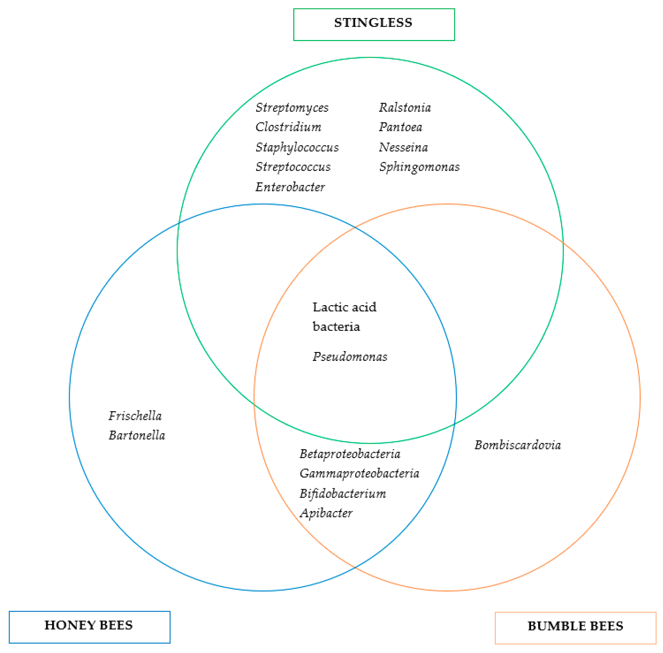
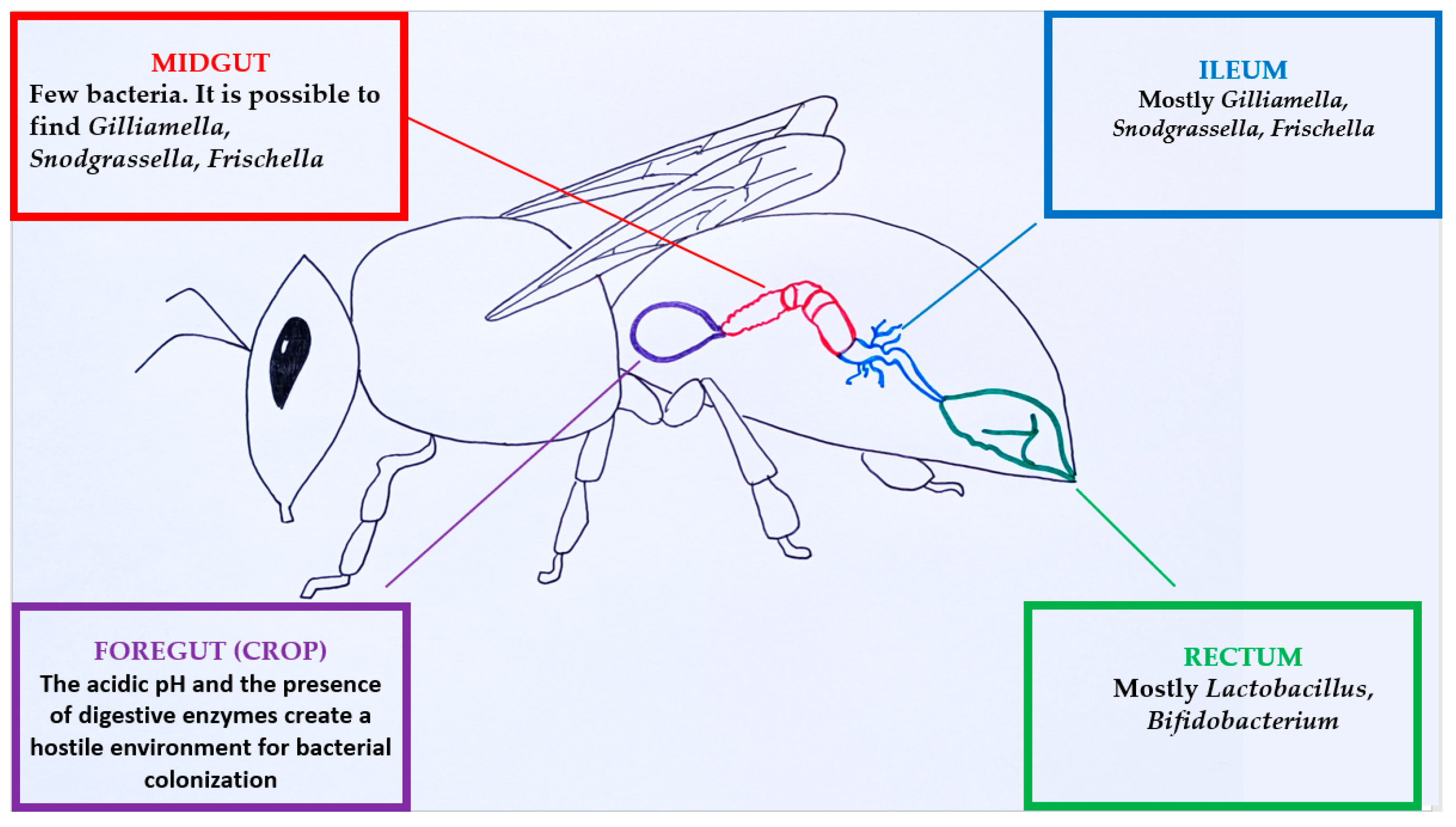
3.1. Bee Microbiome

Eusocial honey bees are colonized by symbiotic bacterial communities, which maintain the homeostasis of the nest and honey bee well-being (Figure 1). Any imbalance of these communities might negatively affect bee health and consequently lead to bee population decline [21]. Honey bee larvae eliminate all the acquired microorganisms before passing to the pupal stage [30,39], and so, pupae and newly emerged workers lack gut microbiota [38,82]. In the first few days after evolving from the pupal stage to adults, before leaving the hive [39], honey bees (Apis mellifera) acquire bacterial symbionts, localized in the hindguts, through social contacts with nurse bees [83] and from parts of the nest milieu such as bee bread, combs, and cell cleaning [30,84]. The typical microbial gut community of worker bees does not change dramatically when workers shift to foragers, suggesting that the adult gut community is stable and distinct [46,82]. A consistent gut microbiome transmission occurs after 3–5 days [35,39] from nurses that stay in the nest to feed the brood [22,37]. Some of the core gut bacteria are detected in royal jelly and the hypopharyngeal glands of the bees [8]. However, social interaction and trophallaxis are not enough to establish the gut microbiome. Several studies have shown that when honey bees are removed from the nest at the pupae stage, they are not colonized by gut bacteria through adulthood. Powell et al. (2014) and Anderson et al. (2021) concluded that exposure to the nest milieu is essential for developing a substantial gut community [23,38]. Moran et al. (2015) reported that larvae are colonized by a limited number of bacteria, mainly environmental Acetobacteraceae (Alpha2.2) and Lactobacillus. These groups are present in nectar and pollen, and so, larvae may acquire them through food consumption [82]. The microbial colonization of newly emerged bees helps them gain weight, to reduce pathogen susceptibility, or to have increased levels of insulin-like peptide I and vitellogenin, two factors implicated in nutrition, bee lifespan, and foraging behavior [18,23]. Anderson et al. (2015) showed that in early gut colonization, Lactobacillus firm 5 is dominant. Moreover, they suggested that colonization by the core bacterium alpha 2.1 is affected by nutrition since they barely detected it in 3- or 7-day-old bees, though it is abundant in queens that exclusively consume royal jelly [8].
The composition of the gut microbial communities, in terms of species diversity and relative abundance, differs among bee castes [27,85]. Differences might be attributed to age, diet, environment, season, and behavioral tasks [11,86]. All these factors shape a caste-specific microbiome for queens and worker bees [81]. Moreover, the abundances of several microbial groups vary significantly between newly emerged bees, nurses, and adult workers [73]. The most stable gut bacterial communities are described in adult bees when compared to queens and drones. Nonetheless, Jones et al. (2018) reported significantly different gut microbial communities in workers of the same age that perform different tasks, (i.e., inside and outside the hive) and follow different nutrition habits. They also reported that the microbial diversity in bees performing tasks inside the hive was higher [27] while other studies reported that those bees had considerably fewer crop bacteria than older foragers [7]. Kešnerová et al. (2020) reported that winter and nurse bees had higher bacterial loads compared to foragers [37]. Moreover, older foragers may exhibit a lower abundance of core bacterial species compared to younger ones [39]. According to Kwong and Moran (2016), the core bacteriome of the honey bee gut consists of Snodgrassella alvi (Betaproteobacteria), Gilliamella apicola (γ-proteobacteria), Bifidobacterium spp., Lactobacillus Firm-4, and Lactobacillus Firm-5. These five phylotypes are virtually present in all adult workers, as well as in closely related bee species like bumble bees [39]. Several other species have been identified in A. mellifera, but they are not detected in all workers of a single colony [73]. Other bacteria (auxiliary bacteriome) include Frischella perrara, Bartonella apis, and certain Acetobacteraceae (Alpha 2.2, known as Parasaccharibacter apium, and Alpha 2.1, known as Commensalibacter sp.). Bacteroidetes species appear to be associated with bees; however, their abundance may vary depending on the host or ecological niche. Collectively, these bacteria form a specialized microbial community that co-evolved over millions of years together with the bee hosts [39]. Martinson et al. (2011) showed that Apis mellifera communities were dominated by the Firm-5 phylotype [87]. On the contrary, Cox-Foster et al. (2007) demonstrated that A. mellifera microbiota were dominated by Gamma-1 [88]. Anderson et al. (2018) reported that Firm-4 (Lactobacillus mellis), Bifidobacteriacea, and Firm-5 (Lactobacillus melliventris) were detected in significantly higher relative abundance in both nurse and food processing bees compared to foragers, while Bartonella apis abundance was significantly higher in food-processing workers [81]. Powell et al. (2014) suggested that Snodgrassella alvi, Gilliamella apicola, and Frischella perrara (Gram-negative bacteria) colonization depends on the presence of nurses, whereas workers are colonized by Gram-positive species through contact to the nest milieu [38]. Kešnerová et al. (2020) found that the gut microbiome of winter bees was different when it came to the total bacterial abundance and the levels of individual phylotypes compared to that of foragers, being largely dominated by Lactobacillus Firm-5 and Bartonella. They suggested that aged pollen consumption leads to increased gut weight and overall bacterial load in winter bees. Increased levels of Bartonella and Commensalibacter in the guts of winter bees may be age-related [37]. The drone gut microbiome composition is closer to that of workers, being more variable and with a higher abundance of Lactobacillus spp. [39].
Worker bees and queens share many core gut bacterial species; nonetheless, the queen microbiome is highly variable in size and often lacks certain species present in workers’ guts [35,39]. The queen’s bacteriome is represented by nectar-, larvae-, and nest-related bacteria [89]. It has been found that Lactobacillus kunkeei (considered a probiotic) and Parasaccharibacter apium are prevalent in the queen’s gut, thus being part of the core gut bacteriomes of A. mellifera queens. These bacterial species are detected sporadically in the guts of workers that suffer dysbiosis and oxidative stress and may be associated with the royal jelly diet of the queen. Moreover, B. apis was extremely rare and F. perrara not detected in queens, suggesting that these Proteobacteria may not tolerate the royal jelly antimicrobial activity, while worker-specific G. apicola and S. alvi were detected at low relative abundances in queen guts. It was concluded that aging in queens was significantly associated with increased abundances of Lactobacillus and Bifidobacterium (probiotics) and the depletion of various Proteobacteria, in contrast to what was found in workers. As workers proceed to foragers (aging), the hindgut microbiota shifts and there is a decrease in vitellogenin and life expectancy. Vitellogenin hemolymph concentration, the royal jelly diet, and microbiota may contribute to antioxidant function in long-lived queens [81]. In another study, L. kunkeei and Bombella apis, two abundant species in the nest, were reported as core species in the queen’s ileum [23]. Powell et al. (2018) found queen gut microbial communities to be dominated by Acetobacteraceae and Lactobacilli in early development and then shift, mainly to Acetobacteraceae (mostly Alpha-2.1), as the queens grew older [89]. Bacillus, Lactococcus, and Pseudomonas species in mated and older unmated bumble bee queen guts were detected. Cold-loving and tolerant bacteria such as Acinetobacter, Chryseobacterium, Hafnia, Psychrobacter, and Pseudomonas were detected before the hibernation period, suggesting that these species might protect the queens from environmental changes that take part during hibernation. Differences in the gut microbiomes of bumble bee queens, compared to Apis species, may exist due to the fact that bumble bee queens go through diapause and through a solitary founding stage [41].
Bee microbiota appear to metabolically adapt to host nutrition since core bacterial genera such as Gilliamella, Lactobacillus, and Bifidobacterium are capable of enzymatically breaking down and fermenting complex macromolecules (polysaccharides and polypeptides) found in pollen, honey, and nectar [5,34]. Several studies have demonstrated that different Apis species harbor all five core gut bacteria but demonstrate different relative abundances and microbiota structures [26,29,40,90]. A. mellifera shows a much higher diversity of gut-associated strains than A. cerana [40]. It has been reported that Apis mellifera and Apis cerana harvesting different floral sources possessed similar core microbial communities, but not with the same structure, and it has been concluded that pollen from different geographical locations might affect microbial communities [26]. Yoshiyama et al. (2009) studied the Japanese honey bee (Apis cerana japonica) gut microbiome and suggested that some isolated strains, mostly Bacilli, showed strong inhibitory activity against Paenibacillus larvae, and so, they might play a protective role against pathogens in honey bees. Moreover, they did not detect any Lactobacillus species, but detected many gut bacteria species that were reported for the first time in the Apis genus [90]. Apis florea and Apis dorsata, two honey bees native to Thailand, despite foraging in the same environment, demonstrated differences in bacterial diversity and the abundance of core gut microbes. Lactobacillus and Bifidobacterium were predominantly associated with Apis florea while Gilliamella and other non-core members (Frischella, Apibacter, Commensalibacter) were dominant in Apis dorsata [91]. Interestingly, Asian honey bees host Apibacter sp., which is rarely reported in European honey bees and sporadically in bumble bees [2].
Bumble bees are closely related to Apis species, and so, they share distinct lineages of the same gut bacteria [82,92,93]. However, Kwong et al. (2017) reported that bumble bee gut communities are less diverse and more erratic than those of honey bees [34]. Several studies have detected phylotypes of Snodgrassella, Gilliamella, Bartonella apis, Serratia sp., and Lactobacillus [35,92,94]. Related Bombus species tend to host similar strains of S. alvi and G. apicola that putatively protect them against bee parasites [38,41]. It has been noticed that bumble bee workers colonized by reduced abundances of these two species are more likely to be colonized by enteric pathogens [6]. Other isolated species such as Rosenbergiella nectarea, Streptomyces spp., Bacillus spp., Micrococcus luteus, Kocuria rhizophila, and Pseudomonas spp. are associated with environmental sources [92].
In comparison to Apis and Bombus microbiomes, the stingless bee microbiome appears highly variable in terms of composition and richness [19,34]. Tang et al. (2021) studied three common stingless bee species (Lepidotrigona terminata, Lepidotrigona ventralis, and Tetragonula pagdeni). They observed that all three species shared some dominant phylotypes, but their overall gut microbial communities were different. L. terminus and L. ventralis showed more similar microbial structures than T. pagdeni [44]. The major bacterial genera associated with stingless bees are Lactobacillus, Bacillus, Streptomyces, Clostridium, Staphylococcus, Streptococcus, Enterobacter, Ralstonia, Pantoea, Pseudomonas, Fructobacillus, Lysinibacillus, and Neisseria [95]. It has been demonstrated that the microbiomes of two Brazilian stingless bee species (Frieseomelitta varia and Tetragonisca angustula) that build their nests using plant materials are dominated by Pseudomonas spp. and Sphingomonas spp., commonly present in plants. Another stingless bee species (Melipona quadrifasciata) has demonstrated a similar colonization pattern by Methylobacterium cerastii, Sphingomonas cynarae, Acinetobacter nectaris, Agrobacterium tumefaciens, Erwinia billingiae, Pantoea ananatis, Pseudomonas putida, and Sphingomonas spp. [19]. Other researchers have reported that the stingless bee gut microbiome is dominated by plant-derived bacteria [21,44]. A study on two phylogenetically distant genera of Australian stingless bees showed common plant bacteria, as well as Lactic Acid Bacteria (LAB), to be members of their gut microbiomes [21].
The highest microbial load in honey bees is detected inside the body rather than in the external parts [31]. The gut bacterial community of an adult worker bee consists of approximately one billion bacteria [82] that are adapted to the different gut compartments [38,39,84]. The vast majority of these bacteria (~95–99%) are located in the hindgut, ileum, and rectum [23,96], and each of these three parts is colonized by a distinct microbial community [39,82] (Figure 2). The hindgut bacteria closely interact with the host tissue [52] as they can selectively adhere to the surface of the ileum. These communities degrade nondigested nutrients that accumulate in the digestive tract. Such compounds are the main energy source for the hindgut microbiota [18,46,52,93]. Furthermore, bacterial strains produce enzymes used to metabolize hard-to-digest bee food like pollen [18].
Other niches that harbor bacterial species in honey bees are the mouthparts and the hypopharyngeal glands. The mouthparts are important for the bees’ social interactions, taking part in horizontal bacterial transmission in the nest. These bacteria may be beneficial to the bees due to pathogen competition [2,52,84]. Dalenberg et al. (2020) reported the high abundance of Bombella apis in the mouthparts of worker bees and suggested that this species might be a core member of the mouthpart microbiome. This bacterium produces antifungal compounds that could protect larvae from infection [52].
The foregut harbors distinct microbiota from the gut, dominated by Lactobacillus kunkeei, Bombella apis (Acetobacteraceae Alpha 2.2, Parasaccharibacter apium), and Fructobacillus fructosus [7,52,82]. L. kunkeei has been often isolated from the foreguts of A. mellifera and A. dorsata [29]. Alpha 2.2, Lactobacillus kunkeei, and other environmental Lactobacilli have also been identified in honey and bee bread. This might indicate the adaptation of these species to the nest milieu and vertical transmission [4,82]. The presence of novel lactic acid bacteria (LAB) in the honey stomach has been reported. In one study, these species were not detected in floral nectar, but were identified in stored food [97]. Honey stomach LABs participate in the fermentation of pollen to bee bread and are detected in fresh bee bread (2-week-old). The antimicrobial compounds produced by these bacteria in bee bread may protect the bees and bee bread from infections and spoilage microorganisms, respectively [20]. Studies have suggested that antimicrobial compounds detected in the foregut are produced by bacteria of the genera Lactobacillus and Bifidobacterium that reside symbiotically [30,38]. It has been demonstrated that the LABs in the honey crop positively affect bee health and protect bees against microbial infections. In particular, their results suggest that LABs linked to the honey bee crop might protect against brood diseases such as American foulbrood (AFB) and European foulbrood (EFB) [98].
It has been studied that B. apis strains inhibit the growth of two insect fungal pathogens, Beauveria bassiana and Aspergillus flavus, both in vitro and in vivo. Moreover, in a small number of infected bees, B. apis has caused diminished A. flavus sporulation. An antifungal metabolite has been demonstrated to be responsible for the inhibition. Biosynthetic gene cluster analysis suggests a novel type-I polyketide synthase. Gene annotation shows additional domains (dehydrogenase, enoyl reductase, and ketoreductase) that might modify the polyketide activity by altering the chemical structure of the product [51].
Gut microbial communities could affect host health by modulating the host immune response. Kwong et al. (2017) reported that when the microbiota is present, the expressions of the antimicrobial peptitides (AMPs) apidaecin and hymenoptaecin are upregulated in gut tissue. On the contrary, in microbiota-free bees, gut apidaecin levels are lower in the gut and the hemolymph than in bees bearing normal gut microbiomes. In support to these findings, honey bees bearing the typical microbiome have demonstrated higher survival after being inoculated with E. coli [34].
Furthermore, the gut microbiota modulates the virulence of the Deformed Wing Virus (DWV), a major honey bee pathogen. The honey bee survival rates after oral virus exposure have been compared among bees bearing experimentally established normal gut microbiota and control bees with perturbed (dysbiotic) gut microbiota. The viral titer in the first case was found to be lower, indicating higher tolerance against DWV in bees with normal gut microbiota [99]. Similarly, it has been suggested that Snodgrassella and Lactobacillus might play a key role in protection against Israeli acute paralysis virus infection [40].
Several studies have described the implication of the honey bee gut microbiome in the detoxification of heavy metals and xenobiotics. Rothman et al. (2019) described how the bee microbiome composition was slightly altered on selenate and cadmium exposure. The growth of gut bacteria in media containing selenate or cadmium led to the removal of cadmium from the media in variable quantities by all strains while selenate was removed only by Lactobacillus Firm-5. These results suggest that bee-associated bacterial strains bioaccumulate these toxic substances. Lactobacillus spp. can accumulate copper, cadmium, aluminum, and chromium, suggesting that some of these strains have the ability to reduce the metal load in their host [100]. Recently, Almasri et al. (2022) showed that bees colonized by core gut bacteria are more resilient to oxidative stress and demonstrated the increased detoxification of pesticides. It has been showed that the FE4-like esterase is implicated in the oxidative response during exposure to the pesticide imidacloprid and herbicide paraquat in Apis cerana while the CYP6B gene family is involved in Culex pipiens pallens’ insecticide resistance [48]. In a co-evolving process, it has been suggested that herbivorous insects have developed counter adaptations to overcome chemical defenses in their host plants. In order to break down furanocoumarins in Rutaceae or Apiceae host plants, Papillo butterflies induce the expression of the CYP6B gene family. Diverse types of furanocoumarins can be metabolized by CYP6Bs thanks to functional divergence across Papillo species [101]. It is plausible that honey bees adapt to harmful metabolites produced by plants in a similar way.

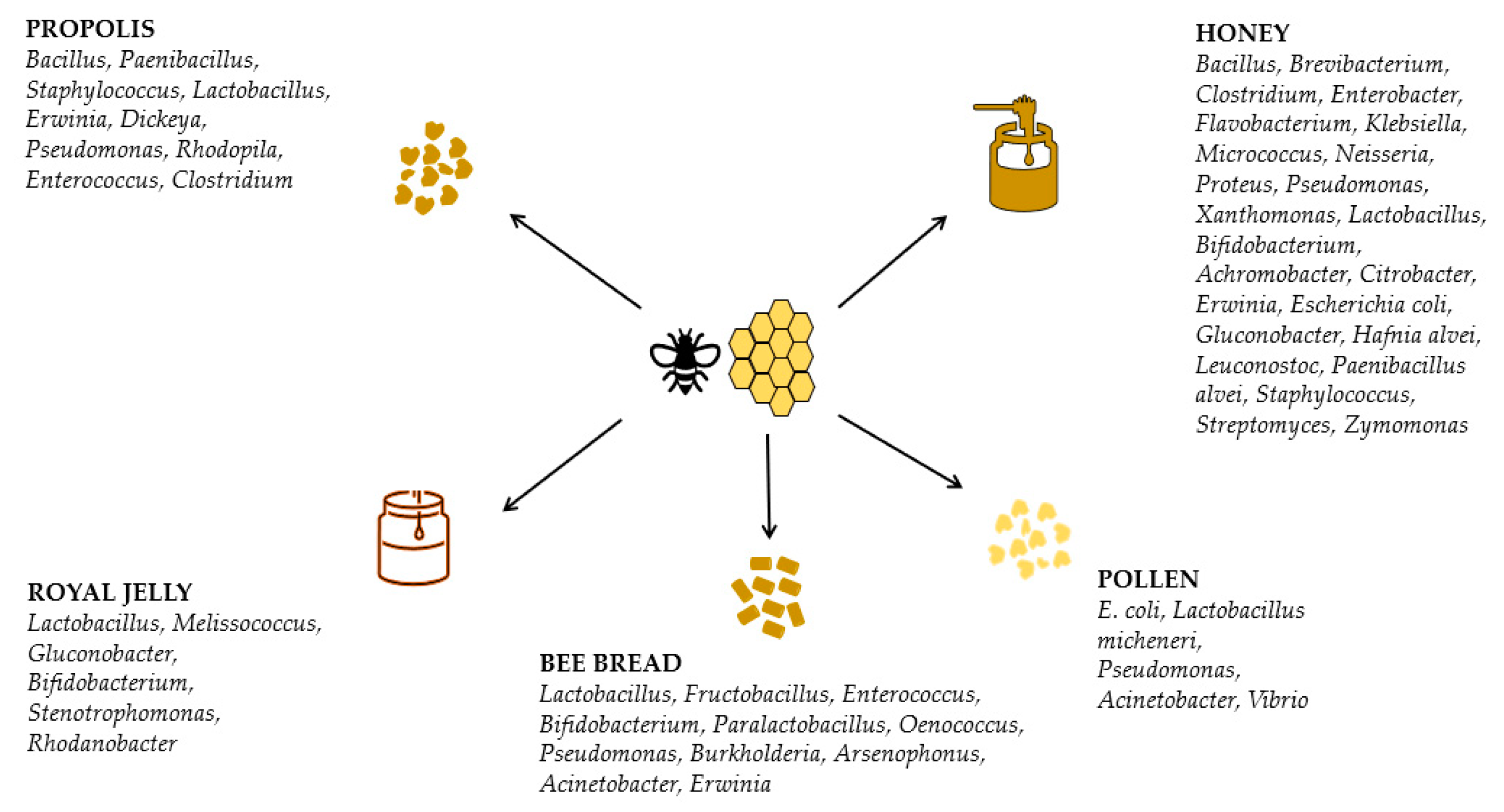
3.2. Honey Microbiome

High-quality honey is characterized by a low number of microorganisms that are not harmful to the consumer [58,62]. However, microbial contamination might impose a safety hazard to consumers. Therefore, the early detection of deleterious microorganisms in honey is paramount [16,102]. Furthermore, microbial contaminants such as bacterial and fungal spores might prohibit the use of honey in medicine, cosmetics, and biotechnology [103]. The most prominent health risk related to honey consumption is infant botulism caused by Clostridium botulinum. The latter is a bacterium frequently present in soil and aquatic environments. Eight distinct serotypes (A-G and X) and four physiological groups (I-IV) that produce botulinum neurotoxins (BoNTs) constitute this species [104]. Khan et al. (2019) detected only low quantities of Clostridium botulinum spores in some of the tested honey samples whereas they did not report any vegetative pathogenic bacteria [30]. In addition to C. botulinum, some Bacillus spp., like B. cereus, are potentially pathogenic since they are implicated in food spoilage as well as in foodborne outbreaks [60]. Nevertheless, not all microorganisms present in honey are harmful. In fact, the honey microbiome is reported as a potential source of beneficial compounds [57]. Furthermore, most microorganisms cannot survive in honey in their metabolically active forms due to the unfavorable milieu of this bee product. Actually, vegetative bacteria might not survive in the honey matrix for more than 30 days [58,105]. Microorganisms in honey only survive in non-vegetative forms and as non-metabolically active endospores [58,62]. The spore-forming microorganisms survive in honey by remaining dormant [56].
In the initial stages of honey ripening, bacteria transferred by nectar, along with microbial populations added by honey bees (from their gastrointestinal tracts and especially from the honey stomachs, such as Lactobacillus spp., Bacillus spp., and Bifidobacteria), are present, and some of them take part in nectar’s maturation to honey [58,106]. Snowdon and Cliverb (1996) reported the following bacterial genera: Alcaligenes, Bacillus, Bacterium, Brevibacterium, Clostridium, Enterobacter, Flavobacterium, Klebsiella, Micrococcus, Neisseria, Proteus, Pseudomonas, and Xanthomonas [107]. Khan et al. (2019) detected Gluconobacter, Lactobacillus, and Zymomonas (Figure 3), as well as some yeasts, to be present during honey maturation. During honey maturation, a decrease in the bacterial load was observed, and finally, Lactobacillus and Gluconobacter were no longer detectable [30]. Physicochemical characteristics might favor microorganisms adaptable to low pH and a high osmotic milieu in order to shape the core honey microbiome [16].
The sources of honey microbiota might be divided into primary and secondary sources. Air, water, dust, soil, pollination environment (nectar, pollen, flowers, honeydew secretions), the nest, and honey bees (or other insects as well) [62,63,108] comprise the primary sources of microbial horizontal transfer in honey, which are actually difficult to control [16,55,58]. Honey bees not only inoculate honey with microbial communities [30] but are also involved in microbial spreading when conducting foraging activities [16]. Laconi et al. (2022) suggested that the honey microbiome is affected by both the honey bee gut and pollen [49]. The secondary sources of the honey microbiome might be considered the harvesting and post-harvesting manipulations of the product, as well as the hygiene conditions. Factors such as the storage environment (containers, warehouses) and equipment might be controlled and managed easier than the primary sources [56,60,68,108].
Olofsson and Vasquez (2008) reported a novel honey microbiota, composed of LABs belonging to the Lactobacillus and Bifidobacterium genera, present in the stomachs of honey bees [109]. The antimicrobial potential of LABs isolated from honey against bee and human pathogens has been reported. They also produce antifungal compounds and contribute to fungal mycotoxin inactivation [16].
Grabowski and Klein (2015) reported a more diverse microbiome in honey including Achromobacter spp., Citrobacter spp., Erwinia spp., Escherichia coli, Gluconobacter spp., Hafnia alvei, Leuconostoc spp., Paenibacillus alvei, Staphylococcus spp., and Streptomyces spp. [110]. Lactobacillus and Acinetobacter were the most abundant genera detected in both honey and pollen by Laconi et al. (2022) [49].
It has been reported that the honey microbiome is different and richer compared to pollen. Twenty-one honey-associated taxa have been identified, compared to nine pollen-associated taxa (Mycoplasma, Pseudomonas, and Vibrio genera). The Melissococcus genus (M. plutonius causes European foulbrood) is more abundant in honey than in pollen [49]. Anderson et al. (2013) reported L. kunkeei and Alpha 2.2 to be abundant in bee bread and honey. Alpha 2.2 is frequently detected in the honey stomach, suggesting a horizontal transmission in the nest [4].
A study on honey microbial communities in Apis mellifera and Melipona beecheii from different locations (Mexico, Jamaica, and Costa Rica) revealed that microbiota in A. mellifera honey were more similar to those in A. mellifera honeys produced in other countries than to those present in M. beecheii honey. Apis mellifera honey microbiota demonstrated higher diversity and species richness than those in M. beecheii honey. Nevertheless, the most abundant species in both honeys were of the Lactobacillaeae family. These results suggest that honey bee species shape the honey microbiome. However, different physicochemical characteristics (M. beecheii honey has higher moisture and lower pH than A. mellifera honey) and antibacterial compounds impose selective pressure on the honey microbiome [111].
It has been assessed that the culturable microbiota of honeydew and floral honeys are of different geographical origin. Researchers identified, for the first time, K. pneumoniae, L. lactis, E. faecalis, and A. viridans present in floral honeys and Bacillus spp. in honeydew honeys. Most of the isolates were identified as B. amyloliquefaciens. There was no correlation between botanical/geographical origin and identified species, but several strains were detected in different samples, suggesting high adaptation. The multifloral honey microbiome was characterized by higher richness and diversity [102].
The dynamics of microbiota during Vitex honey ripening were investigated in another study. The dominant bacterial species were identified as Bacillus spp., Lactococcus spp., Oceanobacillus spp., Enterococcus spp., Pseudomonas spp., Psychrobacter spp., and Arthrobacter spp. During honey ripening, no significant changes were observed among the most abundant bacteria, suggesting their high adaptation [55].
Olivieri et al. (2012) identified six orders of bacteria, Sphingomonadales, Burkholderiales, Pseudomonadales, Enterobacteriales, Actinomycetales, and Bifidobacteriales, in a honey sample. The most represented order was indeed Pseudomonadales [63].
Other researchers identified Bacillus spp, Staphylococcus spp., Lysinibacillus spp., Micrococcus spp., and Brevibacillus spp in honey samples. Micrococcus luteus and Staphylococcus spp. have been identified in the honey bee gut, too. Most of the isolates have been identified as Bacillus species, predominantly B. subtilis and B. cereus [60].
Recently, the microbiota of Greek honeys of diverse botanical and geographical origin were investigated, implementing metataxonomics. The researchers identified 14 taxa, including bacilli/anoxybacilli, paracocci, lysobacters, pseudomonads, and sphingomonads, in all tested honeys. They suggested that low oxygen levels in honey might affect the structure of the microbial community. Overall, the investigated honey microbiota comprised bacteria present in the bee gut, both beneficial bacteria (probiotics) as well as pathogenic microbes of botanical origin [54].
In one of our previous studies (Tsadila et al., 2021), two-thousand-and-fourteen bacterial isolates obtained from diverse Greek honey samples were tested for their antimicrobial potential against five nosocomial and foodborne pathogens (S. aureus, Citrobacter freundii, P. aeruginosa, A. baumannii, and Salmonella enterica ser. Typhimurium). Overall, 31.5% of the isolates inhibited the growth of at least one of the tested pathogens. Most of them (62%) were identified as Bacillus spp. Moreover, Staphylococcus spp., Lysinbacillus fusiformis, Microbacterium imperiale, Micrococcus yunnanensis, Paenibacillus profundus, and Terribacillus saccharophilus were identified. Ten percent of the identified strains were Gram-negative bacteria, Pseudomonas spp. (P. fulva and P. coleopterum) and Acinetobacter lwoffii. The species B. paramycoides, L. fusiformis, M. imperiale, M. yunnanensis, P. profundus, T. saccharophilus, Staphylococcus arlettae, Staphylococcus cohnii, P. fulva, and P. coleopterorum, which have demonstrated antibacterial potential, were detected in homey for the first time [57].
Importantly, genes encoding non-ribosomal peptide synthetase (NRPS) and polyketide synthase (PKS) were detected in several strains, indicating the high biosynthetic potential of antimicrobial compounds [57]. Non-ribosomal peptides (NRPs) and polyketides (PKs) are of high pharmaceutical and biotechnological interest (being secondary metabolites with antimicrobial, anticancer, and immunosuppressive activities) [16].
In conclusion, the honey microbiome consists of Lactobacillales (Lactobacillus) and Bacillales (Bacillus and Paenibacillus), as well as Actinobacteria, Firmicutes, and Proteobacteria (Alpha- and γ-). These microorganisms produce a plethora of secondary metabolites (antibiotics, antimicrobial peptides, bacteriocins, surfactants, etc.) that might contribute to the bioactivity exerted by honeys, especially the strong antimicrobial activity [16].

3.3. Bee-Collected Pollen and Bee Bread Microbiome

The investigation of microbial communities associated with bee-collected pollen (BCP) and bee bread is important for their production and preservation. Both BCP and BB are considered safe from a microbiological perspective [25]. Manirajan et al. (2018) have demonstrated that the pollen microbiome is closely associated with the plant species and the type of pollination. The core pollen microbiome consists of 12 bacterial genera, of which the most abundant are Pseudomonas, Rosenbergiella, and Bradyrhizobium. Rosenbergiella nectarea is often isolated from flower organs, and it produces several metabolites (i.e., D-lactic acid, acetic acid) [112]. It seems that differences regarding the pollen coat structure in insect- and wind-pollinated plants, as well as in the variety of nutrients, are the main factors affecting bacterial colonization [112,113]. Environmental factors like climate affect the microbiome in BCP and BB. Furthermore, honey bees shape the structures of microbial communities present in these bee products. Nevertheless, bacterial species characterized by high adaptation might heavily colonize and utilize nutrients available in these products [26].
It was demonstrated that mesophilic anaerobic and aerobic microorganisms, coliform bacteria, and fungi were present in BCP samples collected in Slovakia during different seasons [108].
McFrederick et al. (2017) investigated whether diverse flower species and wild megachilid bee species are colonized by common microorganisms, implying that flowers serve as hubs of microbial transmission through pollen. According to qPCR results, Lactobacillus micheneri was abundant in adult and larval bee guts, and in pollen provisions as well, thus supporting the assumption that microbes spread through flowers in bee populations [114].
Furthermore, McFrederick and Rehan (2019) studied the pollen collection by Ceratina australensins across diverse landscapes in correlation with flower microbial populations. They demonstrated that pollen collected in Queensland had the highest diversity of bacterial and fungal species and that actually, fungal communities were strongly correlated with pollen plant origins. These data provided evidence that pollen is not necessarily a bacteria-rich habitat, suggesting that fungi might be more prevalent in certain environments [115].
Laconi et al. (2022) reported Pseudomonas, Lactobacillus, and Acinetobacter, which are commonly detected in the pollen of plants visited by pollinating insects, as well as Vibrio, to be the most abundant genera in BCP [49].
Similarly, Harris et al. (2014) reported that the majority of identified bacteria in corbicular pollen had originated in the pollination environment (γ-proteobacteria; Pseudomonadaceae, Xanthomonadaceae, Acinetobacter, Actinomycetales, and Enterobacteriaceae) whereas less than 4% were matched to core gut bacteria. These bacteria may protect corbicular pollen, bee bread, or even the bees from fungal infection. It was concluded that corbicular pollen is a highly diverse microbial habitat dominated by bacteria associated with flowers and the pollination environment [7].
Recently, the BB microbiome has been intensively investigated. It has been documented that only 10% of bacterial species are shared between the bee gut and bee bread [116].
The maturation of BCP to BB takes place in distinct phases, the first three of which last about 7 days. In the first 12 h, a heterogeneous microbial population including yeasts is present. During the second phase, anaerobic LABs produce lactic acid and lower the pH, while in the third phase, Lactobacillus spp. are increased. Interestingly, Pseudomonads are present and could be implicated in pollen wall breakdown. The lactic acid fermentation follows, lasting about 15 days [20,26].
In addition to performing fermentation, LABs produce substances (bacteriocins and aliphatic acids) that could inhibit the growth of spoilage microorganisms [25]. Important roles during the first steps of BB maturation are played by yeasts such as Saccharomyces, which metabolize residual sugars, and Pseudomonads, which consume oxygen, thus creating anaerobic conditions. Under these conditions, Lactobacilli ferment carbohydrates into lactic acid [74].
In a study conducted by Mattila et al., 16S rRNA pyrosequencing was performed in order to characterize the bacterial communities present in BB. The authors assumed that oxygen tension in BB may be stratified and so it could sustain a variety of bacteria regarding oxygen demand. Indeed, they identified LABs including Oenococcus, Bifidobacterium, and Paralactobacillus, all of which are facultative anaerobes. The abundance of these LABs in the bee gut and bee bread demonstrated that under certain circumstances, these environments might be anaerobic. According to the authors, osmotic pressure helps break down hard pollen walls during BB fermentation. Hemicellulose is easily hydrolyzed by acid, and so, BB acidification during LAB fermentation could disintegrate complex plant polysaccharides into disaccharides that, subsequently, could be further digested by fermentative microorganisms [116].
In addition to LABs, Anderson et al. (2014) identified Bradyrhizobiaceae, Xanthomonadaceae, Enterobacteriaceae, Rhodobacterales, Pseudomonadales, Bacteriodetes, and many groups of Actinobacteria in BB samples [76]. Similarly, in another study, culturable fructophilic lactic acid bacteria (FLAB) were isolated from bee bread samples. Four different bacterial genera were detected in bee bread samples. Lactobacillus was the most abundant genus whereas L. kunkeei was the dominant species. The less abundant genera were Fructobacillus, Enterococcus, and Bifidobacterium [117].
A similar outcome was reported by Donkersley et al. (2018) that reported 13 bacterial phyla, with the most abundant being Bacteroidetes, Firmicutes, Alpha-proteobacteria, Beta-proteobacteria, and γ-proteobacteria. Regarding bacterial genera, Pseudomonas, Arsenophonus, Lactobacillus, Orbus, Erwinia, and Acinetobacter were the most dominant. It has been suggested that the BB microbiome somehow affects the ability of the bee gut microbiome to promote bee health, thus suggesting an indirect link between the pollinating environment and bee well-being [6]. The antimicrobial activity of mainly isolated Bacilli (B. subtilis, B. licheniformis, B. pumilus, B. altitudinis, and B. safensis) bacteria from BB and BCP against staphylococci, E. coli, and P. aeruginosa was reported in a study [25].
BCB and BB are expected to support different microbiota due to their different (bio)chemical compositions. Ghosh et al. (2022) reported that the BCB microbiome was clearly different from that in BB, which is more diverse and richer. Most bacteria in BB belong to the Actinobacteria, Bacteroidetes, Cyanobacteria, Firmicutes, and Proteobacteria groups. Less abundant bacterial groups are Acidobacteria, Armatimonadetes, Chloroflexi, Deinococcus-Thermus, Dependentiae, Epsilonbacteraeota, Fusobacteria, Gemmatimonadates, Nitrospira, Patescibacteria, Planctomycetes, Tenericutes, and Verrucomicrobia. Firmicutes (Lactobacillales) is the most abundant phylum in bee pollen. Bacteroidetes bacteria (Flavobacteriales and Sphingobacteriales), typically found in soil, were present both in BCP and BB [46].
Some researchers have also reported differences regarding BCP and BB microbiota in different bee species. The bacterial communities in the corbicular pollen and hive-stored bee bread of Apis mellifera and Apis cerana in China were studied. Community structures were not different in these species, but the relative abundances were. The corbicular pollen of both species was colonized by populations of Escherichia-Shiga, Panteoa, and Pseudomonas that are usually present in plant material. A shift in the bacterial communities was observed in 48–72 h stored bee bread. Escherichia-Shiga, Pseudomonas, and Paracoccus decreased in all samples whereas Acinetobacter loads rapidly increased in BB compared to corbicular pollen, suggesting that Acinetobacter could adapt in protein-rich bee bread. Apis mellifera bee bread was more abundant in core gut bacteria than A. cerana while Panteoa and Rosenbergiella loads were higher in A. cerana bee bread [26]. Moreover, Tang et al. (2021) investigated the bacterial community in BB produced by the stingless bee T. pagdeni. The predominant bacterial genera were Lactobacillus, Carnimonas, Escherichia-Shigella, and Acinetobacter. The most abundant genus was reported to be Lactobacillus, indicating that BB samples had been stored for a few weeks. The authors suggested that Escherichia-Shigella may originate from soil or animal feces collected by stingless bees [44].
Conclusively, the BB core bacteriome appears to consist mainly of LABs (Lactobacillus, Fructobacillus, Bifidobacterium, Paralactobacillus, and Oenoccoccus) and Enterobacteriaceae members. Pseudomonas, Burkholderia, Arsenophonus, Acinetobacter, Erwinia, Actinobacteria, Bradyrhizobiaceae, Bacteriodetes, Rhodobacterales, and Xanthomonadaceae have been reported in several studies (Figure 3). Overall, the bacterial communities present in various bee bread samples are characterized by high diversity and may play essential roles in providing nutrients and promoting honey bee well-being.

3.4. Propolis Microbiome

Propolis was thought to be sterile due to its high antimicrobial activity [24]. Grubbs et al. (2015) conducted the first study on the propolis microbiome. They characterized the microbial community associated with propolis by implementing a combination of Fatty Acid Methyl Ester (FAME) and Phospholipid-Derived Fatty Acid (PLFA) analysis. Interestingly, they reported a unique microbial community. Nevertheless, it was a preliminary study that provided the first clues regarding the propolis microbiota [118].
A further study on the microbiome was conducted by Casalone et al. (2020) in a sample of propolis derived from a A. mellifera nest based on culture-dependent and culture-independent methods (Next-Generation Sequencing). Bacillus, Paenibacillus, and Staphylococcus were the most represented genera found by culture-dependent methods, demonstrating a notable diversity (14 genera and 20 species). The structure of bacterial communities was further investigated by 16S rDNA gene high-throughput sequencing, which showed that the most dominant bacteria at the phylum level were Proteobacteria (84.9%), mainly γ-proteobacteria (77.4%), followed by Firmicutes (8.7%) and Actinobacteria (4.9%). Moreover, Lactobacillus kunkeei was the most abundant species (3.4%). Proteobacteria were the most diversified phylum, represented by the Erwinia, Dickeya, and Pseudomonas genera. The authors showed that propolis harbors diverse bacteria from taxa already reported from the nest milieu (Figure 3) [24].
On the contrary, Omeroglu et al. (2023) identified Firmicutes as the most abundant phylum, followed by Proteobacteria, Actinobacteria, Tenericutes, and Spirochaetes, while the most common bacterial families were found to be Bacillaceae, Enterobacteriaceae, and Enterococcaceae. Bacillus (represented mainly by B. badius and B. thermolactis) was the most abundant genus, followed by Enterococcus and Clostridium sensu stricto [119].
Garcia et al. (2019) extracted total genomic DNA from Apis mellifera propolis samples from four locations in Mexico and then amplified, using PCR, the V4 region of the 16S rRNA gene. Analysis of the obtained sequences revealed extensive microbial diversity among the propolis samples. The most abundant bacterial group was Rhodopila spp. Other abundant genera include Corynebacterium spp. and Sphingomonas spp. Of note, Bacillus and Prevotella spp. showed a relative abundance as high as 88% (53% and 35%, respectively) in one sample. Rhodopila spp. had never been identified before in honey-bee-related products, suggesting that propolis promotes the growth of certain microorganisms not frequently present in plant material, the bee host, or the nest [120].

3.5. Royal Jelly Microbiome

The first study on the royal jelly microbiome was conducted by Corby-Harris et al. (2014). They demonstrated that Acetobacteraceae Alpha 2.2 is frequently present in royal jelly but not in the bee gut [121].
Asama et al., in 2015, implemented pyrosequencing to assess the diversity and relative abundances of bacterial groups in royal jelly. Lactobacillus was the most dominant genus (93.3%), followed by Melissococcus (3.2%), Gluconobacter (2.1%), and Bifdobacterium (0.1%). They confirmed that among the Lactobacillus spp., Lactobacillus kunkeei was the most prevalent species (99.7%) (Figure 3) [122].
Stenotrophomonas spp., Rhodanobacter spp., and bee gut bacteria (Xanthomonadaceae, Actinobacteria, Enterobacteriaceae, and Lactobacillus spp.) are members of the royal jelly microbiome, though in low relative abundance. These bacteria perform important functions. Rhodanobacter spp. degrade pesticides and generate β-galactosidase that might provide nutrients to larvae through glycolipid and glycoprotein hydrolysis [15].
Xanthomonadaceae are potentially significant microbiome members of the nest environment that are transferred from nurses to larvae, while Stenotrophomonas spp. might protect larvae by repelling the important pathogen Paenibacillus larvae [123].

4. Contribution of Pseudomonads to Bee Well-Being and the Biological Activity Exerted by Honey Bee Products

Pseudomonas is a Gram-negative bacterial genus belonging to the Pseudomonadaceae family in the class of γ-proteobacteria [124]. It is highly diverse and includes 272 species, which are widely distributed across various niches [57,125]. Several Pseudomonas species exert a high degree of metabolic adaptability because they produce enzymes involved in denitrification and the degradation of aromatic compounds, thus allowing these bacteria to utilize numerous nutrients. Furthermore, they can produce a wide range of secondary metabolites such as antimicrobial peptides, surfactants, siderophores, and cell-wall-degrading enzymes [25,126,127].
Pseudomonads present in bees are of environmental origin (mainly from water and plants) and might be transient members of mouth and/or surface microbiota [2,108].
However, in a study, Pseudomonads in solitary bees were the most represented bacteria, with Lasioglossium, Xylocopa augusti, and Eucera fervens being prevalent in the gut microbiome, with average relative abundances of 39.84%, 36.05%, and 50.91%, respectively [128].
Nevertheless, in honey bees, Pseudomonas is one of the five most common bacterial genera that may have shaped the core bee gut microbiome [129].
Interestingly, in honey bee larvae guts, Pseudomonas was found to be the third most abundant bacterial genus (7.73%) [130].
Bosmans et al. (2018) implemented deep sequencing to investigate the gut microbial communities in Bombus terrestris queens collected from forests and urbanized habitats. Forest specimens contained a higher relative abundance of bacteria including Pseudomonaceae members [131].
Dominant bacterial genera in the stingless bee gut microbiome include the Acetobacter-like, Snodgrassella, Lactobacillus, Psychrobacter, Pseudomonas, and Bifidobacterium genera [44].
Interestingly, the microbiota of Brazilian stingless bee species who build their nests primarily of plant material are dominated by Pseudomonas spp. and Sphingomonas spp., which are frequently present in phylloplane and flowers [19].
Pseudomonas spp. might colonize the bee gut when the intrinsic bacterial community declines due to their metabolic versatility and high adaptability [69].
Table 1 presents the studies reporting the identification of Pseudomonas and the relevant abundances (%) in social bees and honey bee products
The putative biological functions of Pseudomonads present in the gut microbial communities of bee species have been rather under-investigated (Table 2). However, it has been demonstrated that Pseudomonas spp. strains GA07, GA09, and GC04 catabolize glyphosate and use it as an additional carbon source. Glyphosate is a notorious non-selective herbicide frequently applied in agriculture to control weedy plants. The extensive use of glyphosate leads to soil accumulation and negatively affects the native microbiota. Therefore, it is plausible that glyphosate degradation by Pseudomonads keeps the bee gut microbiome healthy [141].
Further support regarding the role of insect gut bacteria in xenobiotic degradation has been provided by a study on Nasonia vitripennis wasps. It has been shown that the gut bacteria Serratia and Pseudomonas contribute to atrazine (herbicide) degradation. Both of these bacteria have atrazine-degradation-related genes, which conferred resistance in an experimentally exposed wasp population [128,142]. The genes and corresponding enzymes involved in atrazine degradation by a Pseudomonas sp. strain have been studied by De Souza et al. Genes encoding these enzymes (atzA, -B, and -C) have been cloned and sequenced. The hydrolytic dichlorination of atrazine is catalyzed by the first enzyme AtzA, producing hydroxyatrazine. AtzB is involved in the catalyzation of hydroxyatrazine deamidation, producing N-isopropilammelide. The former is transformed by the enzyme AtzC into cyanuric acid and isopropylamine [143].
The honey bee gut microbiome might provide resistance not only against herbicides but also against insecticides such as flumethrin. Flumethrin is widely used to manage mites in apiculture. Flumethrin residues negatively affect honey bees, especially larvae. Recently, it has been shown that honey bee gut bacteria act as a barrier, thus protecting honey bee larvae to some extent. Once the flumethrin concentration rises, the immune and detoxifying systems are activated to protect honey bee larvae against any potential flumethrin risks. It is reasonable to assume that Pseudomonads might be involved in flumethrin detoxification because they were one of the most abundant bacterial groups in honey bee larvae guts exposed to flumethrin (relative abundance: 7.73%). Nevertheless, further research to elucidate the mechanism of flumethrin detoxification and honey bee larvae protection by diverse bacteria (including Pseudomonads) is warranted [130].
Several psychrophilic and cold-tolerant bacteria, among them Pseudomonads, have been detected in samples of mated queens and older unmated queens before hibernation. There is circumstantial evidence that Pseudomonads could assist the queens in enduring harsh environmental conditions and prepare them for hibernation, thus allowing them to reproduce [41].
Moreover, it is plausible that Pseudomonads could protect honey bees against pathogens by producing secondary metabolites.
Researchers developed the Pseudomonas strain AN5 initially as a biocontrol agent for the wheat “take all” fungal pathogen. However, they demonstrated that the same strain strongly inhibited the chalkbrood fungus via gluconic acid (a known antifungal metabolite) production [22].
Given the plethora of metabolites produced by Pseudomonads, we suggest that Pseudomonas strains present in bees and honey bee products may suppress bee pathogens (including bacterial honey bee pathogens such as Paenibacillus larvae and Melissococcus plutonius) and preserve honey bee products against spoilage microorganisms. Data supporting this hypothesis were reported in our previous study [57], where P. fulva and P.coleopterum isolated from honey exerted strong antibacterial activity, presumably attributed to NRPs and PKs since corresponding encoding genes were detected. Other Pseudomonads present in honey include both plant-associated species, e.g., Pseudomonas syringae, as well as ubiquitous and specialized species, e.g., Pseudomonas agglomerans [58]. It seems that the microbiome contributes to the strong antimicrobial activity exerted by different types of honey.
Another study that supports our hypothesis was conducted by Indiragandhi et al. [144], demonstrating the antagonistic activity of Pseudomonas spp. against pathogenic fungi in the diamondback moth, attributed to pyoverdin, the major siderophore produced by fluorescent Pseudomonads [145,146]. The high affinity for iron and the antagonism against phytopathogens exerted by pyoverdine have been well documented [147].
The bee bread core bacterial microbiome is highly represented by members of the Pseudomonadaceae family. Pseudomonas is the most dominant genus (32.4%) in terms of relative abundance [6]. Pseudomonads might be involved in the conversion of bee pollen to bee bread via lactic acid fermentation because they facilitate pollen wall degradation though enzyme activity [26,148]. The degradation of the pollen wall is directly linked to higher nutrient bioavailability in pollen grains [149].
Table 2. Biological functions of Pseudomonas in bees and honey bee products.
Pseudomonas Function in Bees and Honey Bee ProductsAuthors
Social beesCatabolize glyphosate and use it as an additional carbon sourceZhao et al., 2015 [141]
Contributing to resistance against insecticides such as flumethrinYu et al., 2021 [130]
Fight pathogens by producing secondary metabolitesKhan et al., 2020 [30]
Bumble bee queensAssist queens in enduring harsh environmental conditions and prepare them for hibernation, thus allowing them to reproduceWang et al., 2019 [41]
Honey bee productsPreserve honey bee products against spoilage microorganismsTsadila et al., 2021 [57]
Pseudomonads are implicated in the conversion of bee pollen to bee breadBarta et al., 2022 [148]
Disayathanoowat et al., 2020 [26]
Conclusively, Pseudomonads are often relatively abundant and metabolic versatile bacteria reported in investigations on honey bee and bee product microbiomes. Although further research is required, a strong body of evidence supports the hypothesis that these γ- proteobacteria significantly contribute to bee well-being through detoxification and by providing protection against pathogens. Furthermore, Pseudomonads might contribute towards the bioactivity (especially antimicrobial) exerted by honey bee products like honey and bee bread (including increased nutrient bioavailability) [149].

Author Contributions

Conceptualization, D.M.; writing—original draft preparation, C.T., C.A. and D.M.; writing—review and editing, D.M., C.T. and C.A. All authors have read and agreed to the published version of the manuscript.

Funding

This research received no external funding.

Institutional Review Board Statement

Not applicable.

Data Availability Statement

No new data were created.

Acknowledgments

C.T. received a scholarship from the State Scholarship Foundation [Sub-action 2: IKY Scholarship Programme for PhD candidates in the Greek Universities, grant no. 52511.

Conflicts of Interest

The authors declare no conflict of interest.

References

  1. Crotti, E.; Sansonno, L.; Prosdocimi, E.M.; Vacchini, V.; Hamdi, C.; Cherif, A.; Gonella, E.; Marzorati, M.; Balloi, A. Microbial Symbionts of Honeybees: A Promising Tool to Improve Honeybee Health. N. Biotechnol. 2013, 30, 716–722. [Google Scholar] [CrossRef] [PubMed]
  2. Ribière, C.; Hegarty, C.; Stephenson, H.; Whelan, P.; O’Toole, P.W. Gut and Whole-Body Microbiota of the Honey Bee Separate Thriving and Non-Thriving Hives. Microb. Ecol. 2019, 78, 195–205. [Google Scholar] [CrossRef] [PubMed]
  3. Castelli, L.; Branchiccela, B.; Romero, H.; Zunino, P.; Antúnez, K. Seasonal Dynamics of the Honey Bee Gut Microbiota in Colonies Under Subtropical Climate: Seasonal Dynamics of Honey Bee Gut Microbiota. Microb. Ecol. 2022, 83, 492–500. [Google Scholar] [CrossRef] [PubMed]
  4. Anderson, K.E.; Sheehan, T.H.; Mott, B.M.; Maes, P.; Snyder, L.; Schwan, M.R.; Walton, A.; Jones, B.M.; Corby-Harris, V. Microbial Ecology of the Hive and Pollination Landscape: Bacterial Associates from Floral Nectar, the Alimentary Tract and Stored Food of Honey Bees (Apis mellifera). PLoS ONE 2013, 8, e83125. [Google Scholar] [CrossRef] [PubMed]
  5. Lee, F.J.; Rusch, D.B.; Stewart, F.J.; Mattila, H.R.; Newton, I.L.G. Saccharide Breakdown and Fermentation by the Honey Bee Gut Microbiome. Environ. Microbiol. 2015, 17, 796–815. [Google Scholar] [CrossRef]
  6. Donkersley, P.; Rhodes, G.; Pickup, R.W.; Jones, K.C.; Wilson, K. Bacterial Communities Associated with Honeybee Food Stores Are Correlated with Land Use. Ecol. Evol. 2018, 8, 4743–4756. [Google Scholar] [CrossRef]
  7. Corby-Harris, V.; Maes, P.; Anderson, K.E. The Bacterial Communities Associated with Honey Bee (Apis mellifera) Foragers. PLoS ONE 2014, 9, e95056. [Google Scholar] [CrossRef]
  8. Anderson, K.E.; Rodrigues, P.A.P.; Mott, B.M.; Maes, P.; Corby-Harris, V. Ecological Succession in the Honey Bee Gut: Shift in Lactobacillus Strain Dominance during Early Adult Development. Microb. Ecol. 2016, 71, 1008–1019. [Google Scholar] [CrossRef]
  9. Papp, M.; Békési, L.; Farkas, R.; Makrai, L.; Judge, M.F.; Maróti, G.; Tozsér, D.; Solymosi, N. Natural Diversity of the Honey Bee (Apis mellifera) Gut Bacteriome in Various Climatic and Seasonal States. PLoS ONE 2022, 17, e0273844. [Google Scholar] [CrossRef]
  10. Romero, S.; Nastasa, A.; Chapman, A.; Kwong, W.K.; Foster, L.J. The Honey Bee Gut Microbiota: Strategies for Study and Characterization. Insect Mol. Biol. 2019, 28, 455–472. [Google Scholar] [CrossRef]
  11. Dai, P.; Yan, Z.; Ma, S.; Yang, Y.; Wang, Q.; Hou, C.; Wu, Y.; Liu, Y.; Diao, Q. The Herbicide Glyphosate Negatively Affects Midgut Bacterial Communities and Survival of Honey Bee during Larvae Reared in Vitro. J. Agric. Food Chem. 2018, 66, 7786–7793. [Google Scholar] [CrossRef]
  12. Ricigliano, V.A.; Williams, S.T.; Oliver, R. Effects of Different Artificial Diets on Commercial Honey Bee Colony Performance, Health Biomarkers, and Gut Microbiota. BMC Vet. Res. 2022, 18, 52. [Google Scholar] [CrossRef] [PubMed]
  13. Yun, B.R.; Truong, A.T.; Choi, Y.S.; Lee, M.Y.; Kim, B.Y.; Seo, M.; Yoon, S.S.; Yoo, M.S.; Van Quyen, D.; Cho, Y.S. Comparison of the Gut Microbiome of Sacbrood Virus-Resistant and -Susceptible Apis cerana from South Korea. Sci. Rep. 2022, 12, 1–9. [Google Scholar] [CrossRef] [PubMed]
  14. Luo, X.; Dong, Y.; Gu, C.; Zhang, X.; Ma, H. Processing Technologies for Bee Products: An Overview of Recent Developments and Perspectives. Front. Nutr. 2021, 8, 834. [Google Scholar] [CrossRef] [PubMed]
  15. Kafantaris, I.; Amoutzias, G.D.; Mossialos, D. Foodomics in Bee Product Research: A Systematic Literature Review. Eur. Food Res. Technol. 2020, 247, 309–331. [Google Scholar] [CrossRef]
  16. Brudzynski, K. Honey as an Ecological Reservoir of Antibacterial Compounds Produced by Antagonistic Microbial Interactions in Plant Nectars, Honey and Honey Bee. Antibiotics 2021, 10, 551. [Google Scholar] [CrossRef]
  17. Saccà, M.L.; Bianchi, G.; Scalzo, R. Lo Biosynthesis of 2-Heptanone, a Volatile Organic Compound with a Protective Role against Honey Bee Pathogens, by Hive Associated Bacteria. Microorganisms 2021, 9, 2218. [Google Scholar] [CrossRef]
  18. Bonilla-Rosso, G.; Engel, P. Functional Roles and Metabolic Niches in the Honey Bee Gut Microbiota. Curr. Opin. Microbiol. 2018, 43, 69–76. [Google Scholar] [CrossRef]
  19. de Sousa, L.P. Bacterial Communities of Indoor Surface of Stingless Bee Nests. PLoS ONE 2021, 16, e0252933. [Google Scholar] [CrossRef]
  20. Degrandi-Hoff Man, G.; Eckholm, B.; Anderson, K.E. Honey Bee Health: The Potential Role of Microbes. In Honey Bee Colony Health: Challenges and Sustainable Solutions; CRC Press: Boca Raton, FL, USA, 2011; pp. 1–12. [Google Scholar] [CrossRef]
  21. Leonhardt, S.D.; Kaltenpoth, M. Microbial Communities of Three Sympatric Australian Stingless Bee Species. PLoS ONE 2014, 9, e105718. [Google Scholar] [CrossRef]
  22. Khan, S.; Somerville, D.; Frese, M.; Nayudu, M. Environmental Gut Bacteria in European Honey Bees (Apis mellifera) from Australia and Their Relationship to the Chalkbrood Disease. PLoS ONE 2020, 15, e0238252. [Google Scholar] [CrossRef] [PubMed]
  23. Anderson, K.E.; Ricigliano, V.A.; Copeland, D.C.; Mott, B.M.; Maes, P. Social Interaction Is Unnecessary for Hindgut Microbiome Transmission in Honey Bees: The Effect of Diet and Social Exposure on Tissue-Specific Microbiome Assembly. Invertebr. Microbiol. 2023, 85, 1498–1513. [Google Scholar] [CrossRef] [PubMed]
  24. Casalone, E.; Cavalieri, D.; Daly, G.; Vitali, F.; Perito, B. Propolis Hosts a Diverse Microbial Community. World J. Microbiol. Biotechnol. 2020, 36, 50. [Google Scholar] [CrossRef] [PubMed]
  25. Pełka, K.; Worobo, R.W.; Walkusz, J.; Szweda, P. Bee Pollen and Bee Bread as a Source of Bacteria Producing Antimicrobials. Antibiotics 2021, 10, 713. [Google Scholar] [CrossRef]
  26. Disayathanoowat, T.; Li, H.; Supapimon, N.; Suwannarach, N.; Lumyong, S.; Chantawannakul, P.; Guo, J. Different Dynamics of Bacterial and Fungal Communities in Hive-Stored Bee Bread and Their Possible Roles: A Case Study from Two Commercial Honey Bees in China. Microorganisms 2020, 8, 264. [Google Scholar] [CrossRef]
  27. Jones, J.C.; Fruciano, C.; Marchant, J.; Hildebrand, F.; Forslund, S.; Bork, P.; Engel, P.; Hughes, W.O.H. The Gut Microbiome Is Associated with Behavioural Task in Honey Bees. Insectes Soc. 2018, 65, 419–429. [Google Scholar] [CrossRef]
  28. Alberoni, D.; Gaggìa, F.; Baffoni, L.; Di Gioia, D. Beneficial Microorganisms for Honey Bees: Problems and Progresses. Appl. Microbiol. Biotechnol. 2016, 100, 9469–9482. [Google Scholar] [CrossRef]
  29. Arredondo, D.; Castelli, L.; Porrini, M.P.; Garrido, P.M.; Eguaras, M.J.; Zunino, P.; Antúnez, K. Lactobacillus kunkeei Strains Decreased the Infection by Honey Bee Pathogens Paenibacillus larvae and Nosema Ceranae. Benef. Microbes 2018, 9, 279–290. [Google Scholar] [CrossRef]
  30. Khan, K.A.; Al-Ghamdi, A.A.; Ghramh, H.A.; Ansari, M.J.; Ali, H.; Alamri, S.A.; Al-Kahtani, S.N.; Adgaba, N.; Qasim, M.; Hafeez, M. Structural Diversity and Functional Variability of Gut Microbial Communities Associated with Honey Bees. Microb. Pathog. 2020, 138, 103793. [Google Scholar] [CrossRef]
  31. Subotic, S.; Boddicker, A.M.; Nguyen, V.M.; Rivers, J.; Briles, C.E.; Mosier, A.C. Honey Bee Microbiome Associated with Different Hive and Sample Types over a Honey Production Season. PLoS ONE 2019, 14, e0223834. [Google Scholar] [CrossRef]
  32. EU Overview. Detailed Information on Honey Bees. Available online: https://food.ec.europa.eu/animals/live-animal-movements/honey-bees_en (accessed on 9 August 2023).
  33. Gkantiragas, A.G.; Gabrielli, J. A Meta-Analysis of the 16S-RRNA Gut Microbiome Data in Honeybees (Apis mellifera). bioRxiv 2021. [Google Scholar] [CrossRef]
  34. Kwong, W.K.; Medina, L.A.; Koch, H.; Sing, K.W.; Soh, E.J.Y.; Ascher, J.S.; Jaffé, R.; Moran, N.A. Dynamic Microbiome Evolution in Social Bees. Sci. Adv. 2017, 3, e1600513. [Google Scholar] [CrossRef] [PubMed]
  35. Raymann, K.; Moran, N.A. The Role of the Gut Microbiome in Health and Disease of Adult Honey Bee Workers. Curr. Opin. Insect Sci. 2018, 26, 97–104. [Google Scholar] [CrossRef] [PubMed]
  36. Kakumanu, M.L.; Reeves, A.M.; Anderson, T.D.; Rodrigues, R.R.; Williams, M.A. Honey Bee Gut Microbiome Is Altered by In-Hive Pesticide Exposures. Front. Microbiol. 2016, 7, 1255. [Google Scholar] [CrossRef]
  37. Kešnerová, L.; Emery, O.; Troilo, M.; Liberti, J.; Erkosar, B.; Engel, P. Gut Microbiota Structure Differs between Honeybees in Winter and Summer. ISME J. 2020, 14, 801–814. [Google Scholar] [CrossRef]
  38. Powell, J.E.; Martinson, V.G.; Urban-Mead, K.; Moran, N.A. Routes of Acquisition of the Gut Microbiota of the Honey Bee Apis mellifera. Appl. Environ. Microbiol. 2014, 80, 7378–7387. [Google Scholar] [CrossRef]
  39. Kwong, W.K.; Moran, N.A. Gut Microbial Communities of Social Bees. Nat. Rev. Microbiol. 2016, 14, 374–384. [Google Scholar] [CrossRef]
  40. Deng, Y.; Yang, S.; Zhao, H.; Luo, J.; Yang, W.; Hou, C. Antibiotics-Induced Changes in Intestinal Bacteria Result in the Sensitivity of Honey Bee to Virus. Environ. Pollut. 2022, 314, 120278. [Google Scholar] [CrossRef]
  41. Wang, L.; Wu, J.; Li, K.; Sadd, B.M.; Guo, Y.; Zhuang, D.; Zhang, Z.; Chen, Y.; Evans, J.D.; Guo, J.; et al. Dynamic Changes of Gut Microbial Communities of Bumble Bee Queens through Important Life Stages. mSystems 2019, 4, 10–1128. [Google Scholar] [CrossRef]
  42. Rothman, J.A.; Leger, L.; Graystock, P.; Russell, K.; McFrederick, Q.S. The Bumble Bee Microbiome Increases Survival of Bees Exposed to Selenate Toxicity. Environ. Microbiol. 2019, 21, 3417–3429. [Google Scholar] [CrossRef]
  43. Cameron, S.A.; Sadd, B.M. Annual Review of Entomology Global Trends in Bumble Bee Health. Annu. Rev. Entomol. 2020, 65, 209–232. [Google Scholar] [CrossRef] [PubMed]
  44. Tang, Q.H.; Miao, C.H.; Chen, Y.F.; Dong, Z.X.; Cao, Z.; Liao, S.Q.; Wang, J.X.; Wang, Z.W.; Guo, J. The Composition of Bacteria in Gut and Beebread of Stingless Bees (Apidae: Meliponini) from Tropics Yunnan, China. Antonie Van Leeuwenhoek Int. J. General. Mol. Microbiol. 2021, 114, 1293–1305. [Google Scholar] [CrossRef] [PubMed]
  45. Cunningham, M.M.; Tran, L.; McKee, C.G.; Ortega Polo, R.; Newman, T.; Lansing, L.; Griffiths, J.S.; Bilodeau, G.J.; Rott, M.; Marta Guarna, M. Honey Bees as Biomonitors of Environmental Contaminants, Pathogens, and Climate Change. Ecol. Indic. 2022, 134, 108457. [Google Scholar] [CrossRef]
  46. Ghosh, S.; Namin, S.M.; Jung, C. Differential Bacterial Community of Bee Bread and Bee Pollen Revealed by 16s RRNA High-Throughput Sequencing. Insects 2022, 13, 863. [Google Scholar] [CrossRef]
  47. Powell, J.E.; Lau, P.; Rangel, J.; Arnott, R.; De Jong, T.; Moran, N.A. The Microbiome and Gene Expression of Honey Bee Workers Are Affected by a Diet Containing Pollen Substitutes. PLoS ONE 2023, 18, e0286070. [Google Scholar] [CrossRef]
  48. Almasri, H.; Liberti, J.; Brunet, J.L.; Engel, P.; Belzunces, L.P. Mild Chronic Exposure to Pesticides Alters Physiological Markers of Honey Bee Health without Perturbing the Core Gut Microbiota. Sci. Rep. 2022, 12, 4281. [Google Scholar] [CrossRef]
  49. Laconi, A.; Tolosi, R.; Mughini-Gras, L.; Mazzucato, M.; Ferrè, N.; Carraro, L.; Cardazzo, B.; Capolongo, F.; Merlanti, R.; Piccirillo, A. Beehive Products as Bioindicators of Antimicrobial Resistance Contamination in the Environment. Sci. Total Environ. 2022, 823, 151131. [Google Scholar] [CrossRef]
  50. Erban, T.; Ledvinka, O.; Kamler, M.; Hortova, B.; Nesvorna, M.; Tyl, J.; Titera, D.; Markovic, M.; Hubert, J. Bacterial Community Associated with Worker Honeybees (Apis mellifera) Affected by European Foulbrood. PeerJ 2017, 5, e3816. [Google Scholar] [CrossRef]
  51. Miller, D.L.; Smith, E.A.; Newton, I.L.G. A Bacterial Symbiont Protects Honey Bees from Fungal Disease. ASM J. 2021, 12, 3. [Google Scholar] [CrossRef]
  52. Dalenberg, H.; Maes, P.; Mott, B.; Anderson, K.E.; Spivak, M. Propolis Envelope Promotes Beneficial Bacteria in the Honey Bee (Apis mellifera) Mouthpart Microbiome. Insects 2020, 11, 453. [Google Scholar] [CrossRef]
  53. De Paula, J.C.; Doello, K.; Mesas, C.; Kapravelou, G.; Cornet-Gómez, A.; Orantes, F.J.; Martínez, R.; Linares, F.; Prados, J.C.; Porres, J.M.; et al. Exploring Honeybee Abdominal Anatomy through Micro-CT and Novel Multi-Staining Approaches. Insects 2022, 13, 556. [Google Scholar] [CrossRef]
  54. Stavropoulou, E.; Remmas, N.; Voidarou, C.; Vrioni, G.; Konstantinidis, T.; Ntougias, S.; Tsakris, A. Microbial Community Structure among Honey Samples of Different Pollen Origin. Antibiotics 2023, 12, 101. [Google Scholar] [CrossRef] [PubMed]
  55. Wen, Y.; Wang, L.; Jin, Y.; Zhang, J.; Su, L.; Zhang, X.; Zhou, J.; Li, Y. The Microbial Community Dynamics during the Vitex Honey Ripening Process in the Honeycomb. Front. Microbiol. 2017, 8, 1649. [Google Scholar] [CrossRef] [PubMed]
  56. Olaitan, P.B.; Adeleke, O.E.; OOla, I. Honey: A Reservoir for Microorganisms and an Inhibitory Agent for Microbes. Afr. Health Sci. 2007, 7, 159–165. [Google Scholar] [PubMed]
  57. Tsadila, C.; Nikolaidis, M.; Dimitriou, T.G.; Kafantaris, I.; Amoutzias, G.D.; Pournaras, S.; Mossialos, D. Antibacterial Activity and Characterization of Bacteria Isolated from Diverse Types of Greek Honey against Nosocomial and Foodborne Pathogens. Appl. Sci. 2021, 11, 5801. [Google Scholar] [CrossRef]
  58. Kňazovická, V.; Gábor, M.; Miluchová, M.; Bobko, M.; Medo, J. Diversity of bacteria in slovak and foreign honey, with assessment of its physico-chemical quality and counts of cultivable microorganisms. J. Microbiol. Biotechnol. Food Sci. 2019, 9, 414–421. [Google Scholar] [CrossRef]
  59. Lee, H.; Churey, J.J.; Worobo, R.W. Antimicrobial Activity of Bacterial Isolates from Different Floral Sources of Honey. Int. J. Food Microbiol. 2008, 126, 240–244. [Google Scholar] [CrossRef]
  60. Pomastowski, P.; Złoch, M.; Rodzik, A.; Ligor, M.; Kostrzewa, M.; Buszewski, B. Analysis of Bacteria Associated with Honeys of Different Geographical and Botanical Origin Using Two Different Identification Approaches: MALDI-TOF MS and 16S RDNA PCR Technique. PLoS ONE 2019, 14, e0217078. [Google Scholar] [CrossRef]
  61. Kwakman, P.H.S.; Zaat, S.A.J. Antibacterial Components of Honey. IUBMB Life 2012, 64, 48–55. [Google Scholar] [CrossRef]
  62. Majtan, J.; Bucekova, M.; Kafantaris, I.; Szweda, P.; Hammer, K.; Mossialos, D. Honey Antibacterial Activity: A Neglected Aspect of Honey Quality Assurance as Functional Food. Trends Food Sci. Technol. 2021, 118, 870–886. [Google Scholar] [CrossRef]
  63. Olivieri, C.; Marota, I.; Rollo, F.; Luciani, S. Tracking Plant, Fungal, and Bacterial DNA in Honey Specimens*. J. Forensic Sci. 2012, 57, 222–227. [Google Scholar] [CrossRef] [PubMed]
  64. Schuh, C.M.A.P.; Aguayo, S.; Zavala, G.; Khoury, M. Exosome-like Vesicles in Apis mellifera Bee Pollen, Honey and Royal Jelly Contribute to Their Antibacterial and pro-Regenerative Activity. J. Exp. Biol. 2019, 222, jeb208702. [Google Scholar] [CrossRef]
  65. Lee, H.; Churey, J.J.; Worobo, R.W. Purification and Structural Characterization of Bacillomycin F Produced by a Bacterial Honey Isolate Active against Byssochlamys fulva H25. J. Appl. Microbiol. 2008, 105, 663–673. [Google Scholar] [CrossRef] [PubMed]
  66. Ngalimat, M.S.; Raja Abd. Rahman, R.N.Z.; Yusof, M.T.; Syahir, A.; Sabri, S. Characterisation of Bacteria Isolated from the Stingless Bee, Heterotrigona itama, Honey, Bee Bread and Propolis. PeerJ 2019, 7, e7478. [Google Scholar] [CrossRef]
  67. Pajor, M.; Worobo, R.W.; Milewski, S.; Szweda, P. The Antimicrobial Potential of Bacteria Isolated from Honey Samples Produced in the Apiaries Located in Pomeranian Voivodeship in Northern Poland. Int. J. Environ. Res. Public. Health 2018, 15, 2002. [Google Scholar] [CrossRef] [PubMed]
  68. Vázquez-Quiñones, C.R.; Moreno-Terrazas, R.; Natividad-Bonifacio, I.; Quiñones-Ramírez, E.I.; Vázquez-Salinas, C. Microbiological Assessment of Honey in México. Rev. Argent. Microbiol. 2018, 50, 75–80. [Google Scholar] [CrossRef] [PubMed]
  69. D’alvise, P.; Böhme, F.; Codrea, M.C.; Seitz, A.; Nahnsen, S.; Binzer, M.; Rosenkranz, P.; Hasselmann, M. The Impact of Winter Feed Type on Intestinal Microbiota and Parasites in Honey Bees. Apidologie 2018, 49, 252–264. [Google Scholar] [CrossRef]
  70. Didaras, N.A.; Kafantaris, I.; Dimitriou, T.G.; Mitsagga, C.; Karatasou, K.; Giavasis, I.; Stagos, D.; Amoutzias, G.D.; Hatjina, F.; Mossialos, D. Biological Properties of Bee Bread Collected from Apiaries Located across Greece. Antibiotics 2021, 10, 555. [Google Scholar] [CrossRef]
  71. Giampieri, F.; Quiles, J.L.; Cianciosi, D.; Forbes-Hernández, T.Y.; Orantes-Bermejo, F.J.; Alvarez-Suarez, J.M.; Battino, M. Bee Products: An Emblematic Example of Underutilized Sources of Bioactive Compounds. J. Agric. Food Chem. 2022, 70, 6833–6848. [Google Scholar] [CrossRef]
  72. Oladipupo-Alade, E.O.; Lawal, O.A.; Oyewo, I.O.; Odiaka, I.E.; Haastrup, N.O.; Oyedele, M.D.; Ganiyu, O.A. Molecular Characterization of Gut Bacterial Flora of Honeybee (Apis mellifera Adansonii) from Some Selected Apiaries in Ogun State, Nigeria. J. Appl. Sci. Environ. Manag. 2021, 25, 605–608. [Google Scholar] [CrossRef]
  73. Georgi, I.; Asoutis Didaras, N.A.; Nikolaidis, M.; Dimitriou, T.G.; Charistos, L.; Hatjina, F.; Amoutzias, G.D.; Mossialos, D. The Impact of Vairimorpha (Nosema) Ceranae Natural Infection on Honey Bee (Apis mellifera) and Bee Bread Microbiota. Appl. Sci. 2022, 12, 11476. [Google Scholar] [CrossRef]
  74. El Ghouizi, A.; Bakour, M.; Laaroussi, H.; Ousaaid, D.; El Menyiy, N.; Hano, C.; Lyoussi, B. Bee Pollen as Functional Food: Insights into Its Composition and Therapeutic Properties. Antioxidants 2023, 12, 557. [Google Scholar] [CrossRef] [PubMed]
  75. Mohammad, S.M.; Mahmud-Ab-Rashid, N.K.; Zawawi, N. Stingless Bee-Collected Pollen (Bee Bread): Chemical and Microbiology Properties and Health Benefits. Molecules 2021, 26, 957. [Google Scholar] [CrossRef] [PubMed]
  76. Anderson, K.E.; Carroll, M.J.; Sheehan, T.; Mott, B.M.; Maes, P.; Corby-Harris, V. Hive-Stored Pollen of Honey Bees: Many Lines of Evidence Are Consistent with Pollen Preservation, Not Nutrient Conversion. Mol. Ecol. 2014, 23, 5904–5917. [Google Scholar] [CrossRef]
  77. Santos, L.M.; Fonseca, M.S.; Sokolonski, A.R.; Deegan, K.R.; Araújo, R.P.C.; Umsza-Guez, M.A.; Barbosa, J.D.V.; Portela, R.D.; Machado, B.A.S. Propolis: Types, Composition, Biological Activities, and Veterinary Product Patent Prospecting. J. Sci. Food Agric. 2020, 100, 1369–1382. [Google Scholar] [CrossRef]
  78. Rufatto, L.C.; dos Santos, D.A.; Marinho, F.; Henriques, J.A.P.; Roesch Ely, M.; Moura, S. Red Propolis: Chemical Composition and Pharmacological Activity. Asian Pac. J. Trop. Biomed. 2017, 7, 591–598. [Google Scholar] [CrossRef]
  79. Kunugi, H.; Ali, A.M. Royal Jelly and Its Components Promote Healthy Aging and Longevity: From Animal Models to Humans. Int. J. Mol. Sci. 2019, 20, 4662. [Google Scholar] [CrossRef]
  80. Pasupuleti, V.R.; Sammugam, L.; Ramesh, N.; Gan, S.H. Honey, Propolis, and Royal Jelly: A Comprehensive Review of Their Biological Actions and Health Benefits. Oxid. Med. Cell Longev. 2017, 2017, 1259510. [Google Scholar] [CrossRef]
  81. Anderson, K.E.; Ricigliano, V.A.; Mott, B.M.; Copeland, D.C.; Floyd, A.S.; Maes, P. The Queen’s Gut Refines with Age: Longevity Phenotypes in a Social Insect Model. Microbiome 2018, 6, 108. [Google Scholar] [CrossRef]
  82. Moran, N.A. Genomics of the Honey Bee Microbiome. Curr. Opin. Insect Sci. 2015, 10, 22–28. [Google Scholar] [CrossRef]
  83. Engel, P.; Moran, N.A. The Gut Microbiota of Insects—Diversity in Structure and Function. FEMS Microbiol. Rev. 2013, 37, 699–735. [Google Scholar] [CrossRef] [PubMed]
  84. Jabal-Uriel, C.; Alba, C.; Higes, M.; Rodríguez, J.M.; Martín-Hernández, R. Effect of Nosema Ceranae Infection and Season on the Gut Bacteriome Composition of the European Honeybee (Apis mellifera). Sci. Rep. 2022, 12, 9326. [Google Scholar] [CrossRef] [PubMed]
  85. DeGrandi-Hoffman, G.; Corby-Harris, V.; DeJong, E.W.; Chambers, M.; Hidalgo, G. Honey Bee Gut Microbial Communities Are Robust to the Fungicide Pristine® Consumed in Pollen. Apidologie 2017, 48, 340–352. [Google Scholar] [CrossRef]
  86. Ellegaard, K.M.; Engel, P. Genomic Diversity Landscape of the Honey Bee Gut Microbiota. Nat. Commun. 2019, 10, 446. [Google Scholar] [CrossRef]
  87. Martinson, V.G.; Danforth, B.N.; Minckley, R.L.; Rueppell, O.; Tingek, S.; Moran, N.A. A Simple and Distinctive Microbiota Associated with Honey Bees and Bumble Bees. Mol. Ecol. 2011, 20, 619–628. [Google Scholar] [CrossRef]
  88. Cox-Foster, D.L.; Conlan, S.; Holmes, E.C.; Palacios, G.; Evans, J.D.; Moran, N.A.; Quan, P.L.; Briese, T.; Hornig, M.; Geiser, D.M.; et al. A Metagenomic Survey of Microbes in Honey Bee Colony Collapse Disorder. Science 2007, 318, 283–287. [Google Scholar] [CrossRef]
  89. Elijah Powell, J.; Eiri, D.; Moran, N.A.; Rangel, J. Modulation of the Honey Bee Queen Microbiota: Effects of Early Social Contact. PLoS ONE 2018, 13, e0200527. [Google Scholar] [CrossRef]
  90. Yoshiyama, M.; Kimura, K. Bacteria in the Gut of Japanese Honeybee, Apis cerana Japonica, and Their Antagonistic Effect against Paenibacillus larvae, the Causal Agent of American Foulbrood. J. Invertebr. Pathol. 2009, 102, 91–96. [Google Scholar] [CrossRef]
  91. Gruneck, L.; Khongphinitbunjong, K.; Popluechai, S. Gut Microbiota Associated with Two Species of Domesticated Honey Bees from Thailand. Symbiosis 2021, 83, 335–345. [Google Scholar] [CrossRef]
  92. Praet, J.; Parmentier, A.; Schmid-Hempel, R.; Meeus, I.; Smagghe, G.; Vandamme, P. Large-Scale Cultivation of the Bumblebee Gut Microbiota Reveals an Underestimated Bacterial Species Diversity Capable of Pathogen Inhibition. Environ. Microbiol. 2018, 20, 214–227. [Google Scholar] [CrossRef]
  93. Schmidt, K.; Engel, P. Mechanisms Underlying Gut Microbiota-Host Interactions in Insects. J. Exp. Biol. 2021, 224, jeb207696. [Google Scholar] [CrossRef] [PubMed]
  94. Leonard, S.P.; Perutka, J.; Powell, J.E.; Geng, P.; Richhart, D.D.; Byrom, M.; Kar, S.; Davies, B.W.; Ellington, A.D.; Moran, N.A.; et al. Genetic Engineering of Bee Gut Microbiome Bacteria with a Toolkit for Modular Assembly of Broad-Host-Range Plasmids. ACS Synth. Biol. 2018, 7, 1279–1290. [Google Scholar] [CrossRef] [PubMed]
  95. de Paula, G.T.; Menezes, C.; Pupo, M.T.; Rosa, C.A. Stingless Bees and Microbial Interactions. Curr. Opin. Insect Sci. 2021, 44, 41–47. [Google Scholar] [CrossRef] [PubMed]
  96. Zheng, H.; Powell, J.E.; Steele, M.I.; Dietrich, C.; Moran, N.A. Honeybee Gut Microbiota Promotes Host Weight Gain via Bacterial Metabolism and Hormonal Signaling. Proc. Natl. Acad. Sci. USA 2017, 114, 4775–4780. [Google Scholar] [CrossRef] [PubMed]
  97. Olofsson, T.C.; Butler, È.; Markowicz, P.; Lindholm, C.; Larsson, L.; Vásquez, A. Lactic Acid Bacterial Symbionts in Honeybees—An Unknown Key to Honey’s Antimicrobial and Therapeutic Activities. Int. Wound J. 2016, 13, 668–679. [Google Scholar] [CrossRef] [PubMed]
  98. Vásquez, A.; Forsgren, E.; Fries, I.; Paxton, R.J.; Flaberg, E.; Szekely, L.; Olofsson, T.C. Symbionts as Major Modulators of Insect Health: Lactic Acid Bacteria and Honeybees. PLoS ONE 2012, 7, 33188. [Google Scholar] [CrossRef]
  99. Dosch, C.; Manigk, A.; Streicher, T.; Tehel, A.; Paxton, R.J.; Tragust, S. The Gut Microbiota Can Provide Viral Tolerance in the Honey Bee. Microorganisms 2021, 9, 871. [Google Scholar] [CrossRef]
  100. Rothman, J.A.; Leger, L.; Kirkwood, J.S.; Mcfrederick, Q.S.; Stabb, E.V. Cadmium and Selenate Exposure Affects the Honey Bee Microbiome and Metabolome, and Bee-Associated Bacteria Show Potential for Bioaccumulation. Appl. Environ. Microbiol. 2019, 85, e01411-19. [Google Scholar] [CrossRef]
  101. Sato, A.; Okamura, Y.; Murakami, M. Diversification and Selection Pattern of CYP6B Genes in Japanese Papilio Butterflies and Their Association with Host Plant Spectra. PeerJ 2020, 8, e10625. [Google Scholar] [CrossRef]
  102. Sinacori, M.; Francesca, N.; Alfonzo, A.; Cruciata, M.; Sannino, C.; Settanni, L.; Moschetti, G. Cultivable Microorganisms Associated with Honeys of Different Geographical and Botanical Origin. Food Microbiol. 2014, 38, 284–294. [Google Scholar] [CrossRef]
  103. Scepankova, H.; Pinto, C.A.; Paula, V.; Estevinho, L.M.; Saraiva, J.A. Conventional and Emergent Technologies for Honey Processing: A Perspective on Microbiological Safety, Bioactivity, and Quality. Compr. Rev. Food Sci. Food Saf. 2021, 20, 5393–5420. [Google Scholar] [CrossRef] [PubMed]
  104. Carter, A.T.; Peck, M.W. Genomes, Neurotoxins and Biology of Clostridium Botulinum Group I and Group II. Res. Microbiol. 2015, 166, 303–317. [Google Scholar] [CrossRef] [PubMed]
  105. Brudzynski, K.; Flick, R. Accumulation of Soluble Menaquinones MK-7 in Honey Coincides with Death of Bacillus Spp. Present in Honey. Food Chem. X 2019, 1, 100008. [Google Scholar] [CrossRef] [PubMed]
  106. Ruiz-Argueso, T.; Rodriguez-Navarro, A. Microbiology of Ripening Honey. Appl. Microbiol. 1975, 30, 893–896. [Google Scholar] [CrossRef] [PubMed]
  107. Snowdon, J.A.; Cliver, D.O. Microorganisms in Honey. Int. J. Food Microbiol. 1996, 31, 1–26. [Google Scholar] [CrossRef]
  108. Kacániová, M.; Pavličová, S.; Haščík, P.; Kociubinski, G.; Kńazovická, V.; Sudzina, M.; Sudzinová, J.; Fikselová, M. Microbial Communities in Bees, Pollen and Honey from Slovakia. Acta Microbiol. Immunol. Hung. 2009, 56, 285–295. [Google Scholar] [CrossRef] [PubMed]
  109. Olofsson, T.C.; Vásquez, A. Detection and Identification of a Novel Lactic Acid Bacterial Flora within the Honey Stomach of the Honeybee Apis mellifera. Curr. Microbiol. 2008, 57, 356–363. [Google Scholar] [CrossRef]
  110. Grabowski, N.T.; Klein, G. Microbiology and Food-Borne Pathogens in Honey. Crit. Rev. Food Sci. Nutr. 2015, 57, 1852–1862. [Google Scholar] [CrossRef]
  111. Jacinto-Castillo, D.F.; Canto, A.; Medina-Medina, L.A.; O’Connor-Sánchez, A. Living in Honey: Bacterial and Fungal Communities in Honey of Sympatric Populations of Apis mellifera and the Stingless Bee Melipona Beecheii, in Yucatan, Mexico. Arch. Microbiol. 2022, 204, 718. [Google Scholar] [CrossRef]
  112. Ambika Manirajan, B.; Ratering, S.; Rusch, V.; Schwiertz, A.; Geissler-Plaum, R.; Cardinale, M.; Schnell, S. Bacterial Microbiota Associated with Flower Pollen Is Influenced by Pollination Type, and Shows a High Degree of Diversity and Species-Specificity. Environ. Microbiol. 2016, 18, 5161–5174. [Google Scholar] [CrossRef]
  113. Manirajan, B.A.; Maisinger, C.; Ratering, S.; Rusch, V.; Schwiertz, A.; Cardinale, M.; Schnell, S. Diversity, Specificity, Co-Occurrence and Hub Taxa of the Bacterial–Fungal Pollen Microbiome. FEMS Microbiol. Ecol. 2018, 94, 112. [Google Scholar] [CrossRef] [PubMed]
  114. McFrederick, Q.S.; Thomas, J.M.; Neff, J.L.; Vuong, H.Q.; Russell, K.A.; Hale, A.R.; Mueller, U.G. Flowers and Wild Megachilid Bees Share Microbes. Microb. Ecol. 2017, 73, 188–200. [Google Scholar] [CrossRef] [PubMed]
  115. McFrederick, Q.S.; Rehan, S.M. Wild Bee Pollen Usage and Microbial Communities Co-Vary Across Landscapes. Microb. Ecol. 2019, 77, 513–522. [Google Scholar] [CrossRef]
  116. Mattila, H.R.; Rios, D.; Walker-Sperling, V.E.; Roeselers, G.; Newton, I.L.G. Characterization of the Active Microbiotas Associated with Honey Bees Reveals Healthier and Broader Communities When Colonies Are Genetically Diverse. PLoS ONE 2012, 7, e32962. [Google Scholar] [CrossRef] [PubMed]
  117. Janashia, I.; Carminati, D.; Rossetti, L.; Zago, M.; Fornasari, M.E.; Haertlé, T.; Chanishvili, N.; Giraffa, G. Characterization of Fructophilic Lactic Microbiota of Apis mellifera from the Caucasus Mountains. Ann. Microbiol. 2016, 66, 1387–1395. [Google Scholar] [CrossRef]
  118. Grubbs, K.J.; Scott, J.J.; Budsberg, K.J.; Read, H.; Balser, T.C.; Currie, C.R. Unique Honey Bee (Apis mellifera) Hive Component-Based Communities as Detected by a Hybrid of Phospholipid Fatty-Acid and Fatty-Acid Methyl Ester Analyses. PLoS ONE 2015, 10, e0121697. [Google Scholar] [CrossRef]
  119. Ersoy Omeroglu, E.; Keriman Arserim-Uçar, D.; Yegin, Z.; Çağlayan, N.; Nur Zafer Yurt, M.; Busra Tasbasi, B.; Esma Acar, E.; Ucak, S.; Cengiz Ozalp, V.; Sudagidan, M. Determination of Bacterial Diversity of Propolis Microbiota. Chem. Biodivers. 2023, 20, e202201182. [Google Scholar] [CrossRef]
  120. Garcia-Mazcorro, J.F.; Kawas, J.R.; Marroquin-Cardona, A.G. Descriptive Bacterial and Fungal Characterization of Propolis Using Ultra-High-Throughput Marker Gene Sequencing. Insects 2019, 10, 402. [Google Scholar] [CrossRef]
  121. Corby-Harris, V.; Snyder, L.A.; Schwan, M.R.; Maes, P.; McFrederick, Q.S.; Anderson, K.E. Origin and Effect of Alpha 2.2 Acetobacteraceae in Honey Bee Larvae and Description of Parasaccharibacter apium Gen. Nov., Sp. Nov. Appl. Environ. Microbiol. 2014, 80, 7460. [Google Scholar] [CrossRef]
  122. Asama, T.; Arima, T.H.; Gomi, T.; Keishi, T.; Tani, H.; Kimura, Y.; Tatefuji, T.; Hashimoto, K. Lactobacillus kunkeei YB38 from Honeybee Products Enhances IgA Production in Healthy Adults. J. Appl. Microbiol. 2015, 119, 818–826. [Google Scholar] [CrossRef]
  123. Evans, J.D.; Armstrong, T.N. Antagonistic Interactions between Honey Bee Bacterial Symbionts and Implications for Disease. BMC Ecol. 2006, 6, 4. [Google Scholar] [CrossRef] [PubMed]
  124. Lalucat, J.; Gomila, M.; Mulet, M.; Zaruma, A.; García-Valdés, E. Past, Present and Future of the Boundaries of the Pseudomonas Genus: Proposal of Stutzerimonas Gen. Nov. Syst. Appl. Microbiol. 2022, 45, 126289. [Google Scholar] [CrossRef] [PubMed]
  125. Spiers, A.J.; Buckling, A.; Rainey, P.B. The Causes of Pseudomonas Diversity. Microbiology (Reading) 2000, 146 Pt 10, 2345–2350. [Google Scholar] [CrossRef] [PubMed]
  126. Molina, G.; Pimentel, M.R.; Pastore, G.M. Pseudomonas: A Promising Biocatalyst for the Bioconversion of Terpenes. Appl. Microbiol. Biotechnol. 2013, 97, 1851–1864. [Google Scholar] [CrossRef]
  127. Silby, M.W.; Winstanley, C.; Godfrey, S.A.C.; Levy, S.B.; Jackson, R.W. Pseudomonas Genomes: Diverse and Adaptable. FEMS Microbiol. Rev. 2011, 35, 652–680. [Google Scholar] [CrossRef]
  128. Fernandez De Landa, G.; Alberoni, D.; Baffoni, L.; Fernandez De Landa, M.; Revainera, P.D.; Porrini, L.P.; Brasesco, C.; Quintana, S.; Zumpano, F.; Eguaras, M.J.; et al. The Gut Microbiome of Solitary Bees Is Mainly Affected by Pathogen Assemblage and Partially by Land Use. Environ. Microbiome 2023, 18, 38. [Google Scholar] [CrossRef]
  129. Anjum, S.I.; Aldakheel, F.; Shah, A.H.; Khan, S.; Ullah, A.; Hussain, R.; Khan, H.; Ansari, M.J.; Mahmoud, A.H.; Mohammed, O.B. Honey Bee Gut an Unexpected Niche of Human Pathogen. J. King Saud. Univ. Sci. 2021, 33, 101247. [Google Scholar] [CrossRef]
  130. Yu, L.; Yang, H.; Cheng, F.; Wu, Z.; Huang, Q.; He, X.; Yan, W.; Zhang, L.; Wu, X. Honey Bee Apis mellifera Larvae Gut Microbial and Immune, Detoxication Responses towards Flumethrin Stress. Environ. Pollut. 2021, 290, 118107. [Google Scholar] [CrossRef]
  131. Bosmans, L.; Pozo, M.I.; Verreth, C.; Crauwels, S.; Wilberts, L.; Sobhy, I.S.; Wäckers, F.; Jacquemyn, H.; Lievens, B. Habitat-Specific Variation in Gut Microbial Communities and Pathogen Prevalence in Bumblebee Queens (Bombus terrestris). PLoS ONE 2018, 13, e0204612. [Google Scholar] [CrossRef]
  132. Tola, Y.H.; Waweru, J.W.; Hurst, G.D.D.; Slippers, B.; Paredes, J.C. Characterization of the Kenyan Honey Bee (Apis mellifera) Gut Microbiota: A First Look at Tropical and Sub-Saharan African Bee Associated Microbiomes. Microorganisms 2020, 8, 1721. [Google Scholar] [CrossRef]
  133. Ahn, J.H.; Hong, I.P.; Bok, J.I.; Kim, B.Y.; Song, J.; Weon, H.Y. Pyrosequencing Analysis of the Bacterial Communities in the Guts of Honey Bees Apis cerana and Apis mellifera in Korea. J. Microbiol. 2012, 50, 735–745. [Google Scholar] [CrossRef] [PubMed]
  134. Ma, S.; Yang, Y.; Fu, Z.; Diao, Q.; Wang, M.; Luo, Q.; Wang, X.; Dai, P. A Combination of Tropilaelaps Mercedesae and Imidacloprid Negatively Affects Survival, Pollen Consumption and Midgut Bacterial Composition of Honey Bee. Chemosphere 2021, 268, 129368. [Google Scholar] [CrossRef] [PubMed]
  135. Gaggìa, F.; Jakobsen, R.R.; Alberoni, D.; Baffoni, L.; Cutajar, S.; Mifsud, D.; Nielsen, D.S.; Di Gioia, D. Environment or Genetic Isolation? An Atypical Intestinal Microbiota in the Maltese Honey Bee Apis mellifera Spp. Ruttneri. Front. Microbiol. 2023, 14, 1127717. [Google Scholar] [CrossRef]
  136. Kačániová, M.; Terentjeva, M.; Žiarovská, J.; Kowalczewski, P.Ł. In Vitro Antagonistic Effect of Gut Bacteriota Isolated from Indigenous Honey Bees and Essential Oils against Paenibacillus larvae. Int. J. Mol. Sci. 2020, 21, 6736. [Google Scholar] [CrossRef] [PubMed]
  137. Topal, E.; Ceylan; Tunca, R.; Bay, V.; Aldemir, S.; İnci, H.; Topçuoğlu, U.; Kösoğlu, M. The Effect of Different Feeding Strategies on Honey Bee Gut Microbiota and the Presence of Nosema. S. Afr. J. Anim. Sci. 2022, 52, 577–590. [Google Scholar] [CrossRef]
  138. Oladipupo-Alade, E.O.; Lawal, O.A.; Owolola, O.I.; Emmanuel, I.B.; Oyedele, M.D.; Odiaka, I.E.; Haastrup, N.O. Phylogenetic Relationship Between The Gut Bacterial Flora of Honeybee (Apis mellifera) from Apiary in Ogun State, Nigeria. J. Res. For. Wildl. Environ. 2021, 13, 123–134. [Google Scholar]
  139. Santos, A.C.C.; Borges, L.D.F.; Rocha, N.D.C.; de Carvalho Azevedo, V.A.; Bonetti, A.M.; dos Santos, A.R.; da Rocha Fernandes, G.; Dantas, R.C.C.; Ueira-Vieira, C. Bacteria, Yeasts, and Fungi Associated with Larval Food of Brazilian Native Stingless Bees. Sci. Rep. 2023, 13, 5147. [Google Scholar] [CrossRef]
  140. Praet, J. The Gut Microbiota of Bumblebees: A Treasure Chest of Biodiversity and Functionality. Ph.D. Dissertation, Ghent University, Ghent, Belgium, 2017. [Google Scholar]
  141. Zhao, H.; Tao, K.; Zhu, J.; Liu, S.; Gao, H.; Zhou, X. Bioremediation Potential of Glyphosate-Degrading Pseudomonas Spp. Strains Isolated from Contaminated Soil. J. Gen. Appl. Microbiol. 2015, 61, 165–170. [Google Scholar] [CrossRef]
  142. Wang, G.H.; Berdy, B.M.; Velasquez, O.; Jovanovic, N.; Alkhalifa, S.; Minbiole, K.P.C.; Brucker, R.M. Changes in Microbiome Confer Multigenerational Host Resistance after Sub-Toxic Pesticide Exposure. Cell Host Microbe 2020, 27, 213–224.e7. [Google Scholar] [CrossRef]
  143. De Souza, M.L.; Wackett, L.P.; Sadowsky, M.J. The atzABC genes encoding atrazine catabolism are located on a self-transmissible plasmid in Pseudomonas sp. strain ADP. Appl. Environ. Microbiol. 1998, 64, 2323–2326. [Google Scholar] [CrossRef]
  144. Indiragandhi, P.; Anandham, R.; Madhaiyan, M.; Poonguzhali, S.; Kim, G.H.; Saravanan, V.S.; Sa, T. Cultivable Bacteria Associated with Larval Gut of Prothiofos-Resistant, Prothiofos-Susceptible and Field-Caught Populations of Diamondback Moth, Plutella Xylostella and Their Potential for, Antagonism towards Entomopathogenic Fungi and Host Insect Nutrition. J. Appl. Microbiol. 2007, 103, 2664–2675. [Google Scholar] [CrossRef] [PubMed]
  145. Schalk, I.J.; Perraud, Q. Pseudomonas Aeruginosa and Its Multiple Strategies to Access Iron. Environ. Microbiol. 2023, 25, 811–831. [Google Scholar] [CrossRef] [PubMed]
  146. Mossialos, D.; Amoutzias, G.D. Role of Siderophores in Cystic Fibrosis Pathogenesis: Foes or Friends? Int. J. Med. Microbiol. 2009, 299, 87–98. [Google Scholar] [CrossRef]
  147. Mossialos, D.; Amoutzias, G.D. Siderophores in Fluorescent Pseudomonads: New Tricks from an Old Dog. Future Microbiol. 2007, 2, 387–395. [Google Scholar] [CrossRef] [PubMed]
  148. Barta, D.G.; Cornea-Cipcigan, M.; Margaoan, R.; Vodnar, D.C. Biotechnological Processes Simulating the Natural Fermentation Process of Bee Bread and Therapeutic Properties—An Overview. Front. Nutr. 2022, 9, 871896. [Google Scholar] [CrossRef] [PubMed]
  149. Didaras, N.A.; Karatasou, K.; Dimitriou, T.G.; Amoutzias, G.D.; Mossialos, D. Antimicrobial Activity of Bee-Collected Pollen and Beebread: State of the Art and Future Perspectives. Antibiotics 2020, 9, 811. [Google Scholar] [CrossRef] [PubMed]
Figure 1. Venn diagram depicting common bacterial groups in honey bees, bumble bees, and stingless bees. For more details, refer to text.
Diversity 15 01088 g001
Figure 2. Bacterial genera colonizing different parts of honey bee gastrointestinal tract. For more details refer to text. Adapted from Raymann K. and Moran N. (2018) [35].
Diversity 15 01088 g002
Figure 3. An overview of bacterial groups, genera, and species detected in major bee products. For more details, refer to text.
Diversity 15 01088 g003
Table 1. Pseudomonas in social bees and honey bee products.
Detection and Relative Abundance of PseudomonadsReported Pseudomonas SpeciesAuthors
Honey beesApis mellifera (larvae)7.73%-Yu et al., 2021 [130]
Apis mellifera
Summer samples
(0.002–18.0%) Mean:0.01%-D’Alvise et al., 2017 [69]
Apis mellifera
winter samples
0–0.19%
Mean: 0.002%
-
Apis dorsata
(giant honey bee)
Detected-Gruneck et al., 2021 [91]
Apis mellifera scutellata2.3%-Yosef Hamba Tola et al., 2020 [132]
Apis mellifera (adult)0.6%-Jae-Hyung Ahn et al., 2012 [133]
Apis mellifera2.27%-Shilong Ma et al., 2021 [134]
Apis mellifera ruttneri1–3%-Gaggìa et al., 2023 [135]
Apis mellifera-Pseudomonas marginalisKačániová et al., 2020 [136]
-Pseudomonas oryzihabitans
-Pseudomonas putida
Apis mellifera anatoliaca-Pseudomonas luteolaTopal et al., 2022 [137]
-Pseudomonas alcaligenes
Apismellifera adansonii-Pseudomonas aeruginosaOladipupo-Alade et al., 2021 [138]
-Pseudomonas plecoglossicida
Stingless beeFrieseomelitta variaDetectedPseudomonas syringaeDe Sousa 2021 [19]
Melipona quadrifasciata Pseudomonas putida
Lepidotrigonaterminata3.35%-Qi-Hi Tang et al., 2021 [44]
Lepidotrigona ventralis3.18%-
Tetragnula pagdeni4.63%-
Melipona quadrifasciataDetected Santos et al., 2023 [139]
Bumble beesBombus lapidarius5.98%-Jessy Praet 2017 [140]
Bombuslucorum0.48%-
Bombusterrestris0.24%-
Honey bee productsBee pollenDetected-Disayathanoowat et al., 2020 [26]
Manirajan et al., 2018 [113]
Laconi et al., 2022 [49]
Harris et al., 2014 [7]
Bee breadDetected-Donkersley et al., 2018 [6]
Anderson et al., 2014 [76]
Honey-Pseudomonas sp.
Pseudomonas coleopterorum
Pseudomonas fulva
Pseudomonas stutzeri
Tsadila et al., 2021 [57]
Detected-Stavropoulou et al., 2023 [54]
-Pseudomonas sp.Olivieri et al., 2012 [63]
-Pseudomonas migulae
-Pseudomonas flectens
PropolisDetected-Casalone et al., 2020 [24]
Disclaimer/Publisher’s Note: The statements, opinions and data contained in all publications are solely those of the individual author(s) and contributor(s) and not of MDPI and/or the editor(s). MDPI and/or the editor(s) disclaim responsibility for any injury to people or property resulting from any ideas, methods, instructions or products referred to in the content.

