Next Issue
Volume 9, November
Previous Issue
Volume 9, September


Toxins, Volume 9, Issue 10 (October 2017) – 43 articles

Cover Story (view full-size image): 
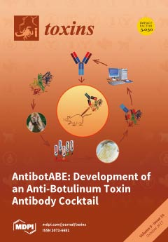
The aim of the AntiBotABE Program was the development of recombinant antibodies neutralizing botulinum neurotoxins (BoNT) A, B and E, responsible for most human botulism cases. Macaques were immunized with the recombinant non-toxic BoNT/A, B or E, heavy and light chains, followed by the generation of immune phage-display libraries. Selected antibodies were analyzed in in vitro and ex vivo assays. The best neutralizing antibodies were germline-humanized, expressed as IgGs and protection was analzyed in two different in vivo models showing a synergistic effect when combining anti-heavy and anti-light chains antibodies. These germline humanized antibodies are promising candidates for further regulatory and clinical development. View Paper here
  • Issues are regarded as officially published after their release is announced to the table of contents alert mailing list.
  • You may sign up for e-mail alerts to receive table of contents of newly released issues.
  • PDF is the official format for papers published in both, html and pdf forms. To view the papers in pdf format, click on the "PDF Full-text" link, and use the free Adobe Reader to open them.
Order results
Result details
Section
Select all
Export citation of selected articles as:
10486 KiB  
Article
Effect of Clostridium perfringens β-Toxin on Platelets
by Anne Thiel, Helga Mogel, Julia Bruggisser, Arnaud Baumann, Marianne Wyder, Michael H. Stoffel, Artur Summerfield and Horst Posthaus
Toxins 2017, 9(10), 336; https://doi.org/10.3390/toxins9100336 - 24 Oct 2017
Cited by 7 | Viewed by 5643
Abstract
Clostridium perfringens β-toxin (CPB) is the major virulence factor of C. perfringens type C causing a hemorrhagic enteritis in animals and humans. In experimentally infected pigs, endothelial binding of CPB was shown to be associated with early vascular lesions and hemorrhage but [...] Read more.
Clostridium perfringens β-toxin (CPB) is the major virulence factor of C. perfringens type C causing a hemorrhagic enteritis in animals and humans. In experimentally infected pigs, endothelial binding of CPB was shown to be associated with early vascular lesions and hemorrhage but without obvious thrombosis of affected vessels, suggesting altered hemostasis in the early phase of the disease. The objective of the present study was to investigate the effect of CPB on platelets, with respect to primary hemostasis. Our results demonstrate that CPB binds to porcine and human platelets and forms oligomers resulting in a time- and dose-dependent cell death. Platelets showed rapid ultrastructural changes, significantly decreased aggregation and could no longer be activated by thrombin. This indicates that CPB affects the physiological function of platelets and counteracts primary hemostasis. Our results add platelets to the list of target cells of CPB and extend the current hypothesis of its role in the pathogenesis of C. perfringens type C enteritis. Full article
Show Figures

Figure 1

1222 KiB  
Article
Rapid Assessment of the Toxicity of Fungal Compounds Using Luminescent Vibrio qinghaiensis sp. Q67
by Qijie Jian, Liang Gong, Taotao Li, Yong Wang, Yu Wu, Feng Chen, Hongxia Qu, Xuewu Duan and Yueming Jiang
Toxins 2017, 9(10), 335; https://doi.org/10.3390/toxins9100335 - 21 Oct 2017
Cited by 11 | Viewed by 4737
Abstract
Most tropical fruits after harvest are very perishable because of fungal infection. Since some pathogenic fungi can produce hazardous compounds such as mycotoxins, novel rapid and effective methods to assess those hazardous compounds are urgently needed. Herein we report that Vibrio qinghaiensis sp. [...] Read more.
Most tropical fruits after harvest are very perishable because of fungal infection. Since some pathogenic fungi can produce hazardous compounds such as mycotoxins, novel rapid and effective methods to assess those hazardous compounds are urgently needed. Herein we report that Vibrio qinghaiensis sp. Q67, a luminescent bacterium, can be used to rapidly assess the toxicities of mycotoxins and cultures from mycotoxin-producing pathogens. A good correlation (R2 > 0.98) between concentrations of the mycotoxins (fumonisin B1, deoxynivalenol, zearalenone, ochratoxin A, patulin, and citrinin) and the luminous intensity of V. qinghaiensis sp. Q67 was obtained. Furthermore, significant correlations (R2 > 0.96) between the amount of mycotoxin and the luminous intensity from the cultures of 10 major mycotoxin-producing pathogens were also observed. In addition, Fusarium proliferatum (half-maximal inhibitory concentration (IC50) = 17.49%) exhibited greater luminescence suppression than Fusarium semitectum (IC50 = 92.56%) or Fusarium oxysporum (IC50 = 28.61%), which was in agreement with the existing higher levels of fumonisin B1, fumonisin B2, and deoxynivalenol, which were measured by high-performance liquid chromatography-tandem mass spectrometry. These results suggest that V. qinghaiensis sp. Q67 is a promising alternative for the rapid evaluation of the toxicity of fungal mycotoxins. Full article
(This article belongs to the Section Mycotoxins)
Show Figures

Figure 1

4379 KiB  
Article
Chronic Exposure to the Fusarium Mycotoxin Deoxynivalenol: Impact on Performance, Immune Organ, and Intestinal Integrity of Slow-Growing Chickens
by Stephanie S. Chen, Yi-Hung Li and Mei-Fong Lin
Toxins 2017, 9(10), 334; https://doi.org/10.3390/toxins9100334 - 20 Oct 2017
Cited by 50 | Viewed by 6463
Abstract
This study investigates the long-term effects of deoxynivalenol (DON) consumption on avian growth performance, on the proliferation, apoptosis, and DNA damage of spleen cells, and on intestinal integrity. Two hundred and eight 5-day-old black-feathered Taiwan country chickens were fed diets containing 0, 2, [...] Read more.
This study investigates the long-term effects of deoxynivalenol (DON) consumption on avian growth performance, on the proliferation, apoptosis, and DNA damage of spleen cells, and on intestinal integrity. Two hundred and eight 5-day-old black-feathered Taiwan country chickens were fed diets containing 0, 2, 5, and 10 mg/kg of DON for 16 weeks. Body weight gain of male birds in the 2 mg/kg group was significantly lower than that in the 5 mg/kg group. At the end of trial, feeding DON-contaminated diets of 5 mg/kg resulted in heavier spleens. Moreover, the increase in DON induced cellular proliferation, apoptosis, and DNA damage signals in the spleen, the exception being female birds fed 10 mg/kg of DON showing reduced proliferation. Expression of claudin-5 was increased in jejunum of female birds fed 2 and 5 mg/kg of DON, whereas decreased expression levels were found in male birds. In conclusion, our results verified that DON may cause a disturbance to the immune system and alter the intestinal barrier in Taiwan country chickens, and may also lead to discrepancies in growth performances in a dose- and sex-dependent manner. Full article
(This article belongs to the Collection Fusarium Toxins – Relevance for Human and Animal Health)
Show Figures

Figure 1

4121 KiB  
Review
Yeast Killer Toxin K28: Biology and Unique Strategy of Host Cell Intoxication and Killing
by Björn Becker and Manfred J. Schmitt
Toxins 2017, 9(10), 333; https://doi.org/10.3390/toxins9100333 - 20 Oct 2017
Cited by 28 | Viewed by 7851 | Correction
Abstract
The initial discovery of killer toxin-secreting brewery strains of Saccharomyces cerevisiae (S. cerevisiae) in the mid-sixties of the last century marked the beginning of intensive research in the yeast virology field. So far, four different S. cerevisiae killer toxins (K28, K1, [...] Read more.
The initial discovery of killer toxin-secreting brewery strains of Saccharomyces cerevisiae (S. cerevisiae) in the mid-sixties of the last century marked the beginning of intensive research in the yeast virology field. So far, four different S. cerevisiae killer toxins (K28, K1, K2, and Klus), encoded by cytoplasmic inherited double-stranded RNA viruses (dsRNA) of the Totiviridae family, have been identified. Among these, K28 represents the unique example of a yeast viral killer toxin that enters a sensitive cell by receptor-mediated endocytosis to reach its intracellular target(s). This review summarizes and discusses the most recent advances and current knowledge on yeast killer toxin K28, with special emphasis on its endocytosis and intracellular trafficking, pointing towards future directions and open questions in this still timely and fascinating field of killer yeast research. Full article
(This article belongs to the Special Issue Yeast Killer Toxins)
Show Figures

Figure 1

7397 KiB  
Article
P2X-Receptor Antagonists Inhibit the Interaction of S. aureus Hemolysin A with Membranes
by Markus Schwiering, Matthias Husmann and Nadja Hellmann
Toxins 2017, 9(10), 332; https://doi.org/10.3390/toxins9100332 - 19 Oct 2017
Cited by 10 | Viewed by 4883
Abstract
The pore forming hemolysin A, Hla, is a major virulence factor of Staphylococcus aureus. Apparently, 1–2 pore(s) per cell suffice(s) to cause cell death. Accumulated experimental evidence points towards a major role of ATP-gated purinergic receptors (P2XR) for hemolysis caused by Hla, [...] Read more.
The pore forming hemolysin A, Hla, is a major virulence factor of Staphylococcus aureus. Apparently, 1–2 pore(s) per cell suffice(s) to cause cell death. Accumulated experimental evidence points towards a major role of ATP-gated purinergic receptors (P2XR) for hemolysis caused by Hla, complement and other pore forming proteins, presumably by increasing membrane permeability. Indeed, in experiments employing rabbit erythrocytes, inhibitory concentrations of frequently employed P2XR-antagonists were in a similar range as previously reported for erythrocytes of other species and other toxins. However, Hla-dependent hemolysis was not enhanced by extracellular ATP, and oxidized adenosinetriphosphate (oxATP) had only a minor inhibitory effect. Unexpectedly, P2XR-inhibitors also prevented Hla-induced lysis of pure lipid membranes, demonstrating that the inhibition did not even depend on the presence of P2XR. Fluorescence microscopy and gel-electrophoresis clearly revealed that P2XR-inhibitors interfere with binding and subsequent oligomerisation of Hla with membranes. Similar results were obtained employing HaCaT-cells. Furthermore, calorimetric data and hemolysis experiments with Hla pre-treated with pyridoxal phosphate-6-azophenyl-2′,4′-disulfonic acid (PPADS) showed that this compound directly binds to Hla. Our results call for a critical re-assessment of the appealing concept, which suggests that P2XR are general amplifiers of damage by pore-forming proteins. Full article
(This article belongs to the Special Issue Toxins and Ion Channels)
Show Figures

Graphical abstract

1290 KiB  
Article
Mycotoxin Analysis of Human Urine by LC-MS/MS: A Comparative Extraction Study
by Laura Escrivá, Lara Manyes, Guillermina Font and Houda Berrada
Toxins 2017, 9(10), 330; https://doi.org/10.3390/toxins9100330 - 19 Oct 2017
Cited by 31 | Viewed by 5965
Abstract
The lower mycotoxin levels detected in urine make the development of sensitive and accurate analytical methods essential. Three extraction methods, namely salting-out liquid–liquid extraction (SALLE), miniQuEChERS (quick, easy, cheap, effective, rugged, and safe), and dispersive liquid–liquid microextraction (DLLME), were evaluated and compared based [...] Read more.
The lower mycotoxin levels detected in urine make the development of sensitive and accurate analytical methods essential. Three extraction methods, namely salting-out liquid–liquid extraction (SALLE), miniQuEChERS (quick, easy, cheap, effective, rugged, and safe), and dispersive liquid–liquid microextraction (DLLME), were evaluated and compared based on analytical parameters for the quantitative LC-MS/MS measurement of 11 mycotoxins (AFB1, AFB2, AFG1, AFG2, OTA, ZEA, BEA, EN A, EN B, EN A1 and EN B1) in human urine. DLLME was selected as the most appropriate methodology, as it produced better validation results for recovery (79–113%), reproducibility (RSDs < 12%), and repeatability (RSDs < 15%) than miniQuEChERS (71–109%, RSDs <14% and <24%, respectively) and SALLE (70–108%, RSDs < 14% and < 24%, respectively). Moreover, the lowest detection (LODS) and quantitation limits (LOQS) were achieved with DLLME (LODs: 0.005–2 μg L−1, LOQs: 0.1–4 μg L−1). DLLME methodology was used for the analysis of 10 real urine samples from healthy volunteers showing the presence of ENs B, B1 and A1 at low concentrations. Full article
(This article belongs to the Section Mycotoxins)
Show Figures

Figure 1

1777 KiB  
Article
Production, Characterisation and Testing of an Ovine Antitoxin against Ricin; Efficacy, Potency and Mechanisms of Action
by Sarah J. C. Whitfield, Gareth D. Griffiths, Dominic C. Jenner, Robert J. Gwyther, Fiona M. Stahl, Lucy J. Cork, Jane L. Holley, A. Christopher Green and Graeme C. Clark
Toxins 2017, 9(10), 329; https://doi.org/10.3390/toxins9100329 - 18 Oct 2017
Cited by 13 | Viewed by 5268
Abstract
Ricin is a type II ribosome-inactivating toxin that catalytically inactivates ribosomes ultimately leading to cell death. The toxicity of ricin along with the prevalence of castor beans (its natural source) has led to its increased notoriety and incidences of nefarious use. Despite these [...] Read more.
Ricin is a type II ribosome-inactivating toxin that catalytically inactivates ribosomes ultimately leading to cell death. The toxicity of ricin along with the prevalence of castor beans (its natural source) has led to its increased notoriety and incidences of nefarious use. Despite these concerns, there are no licensed therapies available for treating ricin intoxication. Here, we describe the development of a F(ab’)2 polyclonal ovine antitoxin against ricin and demonstrate the efficacy of a single, post-exposure, administration in an in vivo murine model of intoxication against aerosolised ricin. We found that a single dose of antitoxin afforded a wide window of opportunity for effective treatment with 100% protection observed in mice challenged with aerosolised ricin when given 24 h after exposure to the toxin and 75% protection when given at 30 h. Treated mice had reduced weight loss and clinical signs of intoxication compared to the untreated control group. Finally, using imaging flow cytometry, it was found that both cellular uptake and intracellular trafficking of ricin toxin to the Golgi apparatus was reduced in the presence of the antitoxin suggesting both actions can contribute to the therapeutic mechanism of a polyclonal antitoxin. Collectively, the research highlights the significant potential of the ovine F(ab’)2 antitoxin as a treatment for ricin intoxication. Full article
(This article belongs to the Special Issue Ribosome Inactivating Toxins)
Show Figures

Figure 1

2185 KiB  
Article
A Monoclonal–Monoclonal Antibody Based Capture ELISA for Abrin
by Christina C. Tam, Luisa W. Cheng, Xiaohua He, Paul Merrill, David Hodge and Larry H. Stanker
Toxins 2017, 9(10), 328; https://doi.org/10.3390/toxins9100328 - 18 Oct 2017
Cited by 5 | Viewed by 5570
Abstract
Abrin, one of the most highly potent toxins in the world, is derived from the plant, Abrus precatorius. Because of its high toxicity, it poses potential bioterror risks. Therefore, a need exists for new reagents and technologies that would be able to [...] Read more.
Abrin, one of the most highly potent toxins in the world, is derived from the plant, Abrus precatorius. Because of its high toxicity, it poses potential bioterror risks. Therefore, a need exists for new reagents and technologies that would be able to rapidly detect abrin contamination as well as lead to new therapeutics. We report here a group of abrin-specific monoclonal antibodies (mAbs) that recognize abrin A-chain, intact A–B chain toxin, and agglutinin by Western blot. Additionally, these mAbs were evaluated for their ability to serve as capture antibodies for a sandwich (capture) ELISA. All possible capture–detector pairs were evaluated and the best antibody pair identified and optimized for a capture ELISA. The capture ELISA based on this capture–detector mAb pair had a limit of detection (L.O.D) of ≈1 ng/mL measured using three independent experiments. The assay did not reveal any false positives with extracts containing other potential ribosome-inactivating proteins (RIPs). Thus, this new capture ELISA uses mAbs for both capture and detection; has no cross-reactivity against other plant RIPs; and has a sensitivity comparable to other reported capture ELISAs using polyclonal antibodies as either capture or detector. Full article
(This article belongs to the Special Issue Ribosome Inactivating Toxins)
Show Figures

Graphical abstract

2098 KiB  
Article
Risk Levels of Toxic Cyanobacteria in Portuguese Recreational Freshwaters
by Carina Menezes, Catarina Churro and Elsa Dias
Toxins 2017, 9(10), 327; https://doi.org/10.3390/toxins9100327 - 18 Oct 2017
Cited by 10 | Viewed by 5130
Abstract
Portuguese freshwater reservoirs are important socio-economic resources, namely for recreational use. National legislation concerning bathing waters does not include mandatory levels or guidelines for cyanobacteria and cyanotoxins. This is an issue of concern since cyanotoxin-based evidence is insufficient to change the law, and [...] Read more.
Portuguese freshwater reservoirs are important socio-economic resources, namely for recreational use. National legislation concerning bathing waters does not include mandatory levels or guidelines for cyanobacteria and cyanotoxins. This is an issue of concern since cyanotoxin-based evidence is insufficient to change the law, and the collection of scientific evidence has been hampered by the lack of regulatory levels for cyanotoxins in bathing waters. In this work, we evaluate the profile of cyanobacteria and microcystins (MC) in eight freshwater reservoirs from the center of Portugal, used for bathing/recreation, in order to determine the risk levels concerning toxic cyanobacteria occurrence. Three of the reservoirs did not pose a risk of MC contamination. However, two reservoirs presented a high risk in 7% of the samples according to the World Health Organization (WHO) guidelines for MC in bathing waters (above 20 µg/L). In the remaining three reservoirs, the risk concerning microcystins occurrence was low. However, they exhibited recurrent blooms and persistent contamination with MC up to 4 µg/L. Thus, the risk of exposure to MC and potential acute and/or chronic health outcomes should not be disregarded in these reservoirs. These results contribute to characterize the cyanobacterial blooms profile and to map the risk of toxic cyanobacteria and microcystins occurrence in Portuguese inland waters. Full article
(This article belongs to the Special Issue Selected Papers from the 5th Iberoamerican Cyanotoxins Meeting)
Show Figures

Figure 1

1715 KiB  
Review
Animal Toxins Providing Insights into TRPV1 Activation Mechanism
by Matan Geron, Adina Hazan and Avi Priel
Toxins 2017, 9(10), 326; https://doi.org/10.3390/toxins9100326 - 16 Oct 2017
Cited by 34 | Viewed by 7321
Abstract
Beyond providing evolutionary advantages, venoms offer unique research tools, as they were developed to target functionally important proteins and pathways. As a key pain receptor in the nociceptive pathway, transient receptor potential vanilloid 1 (TRPV1) of the TRP superfamily has been shown to [...] Read more.
Beyond providing evolutionary advantages, venoms offer unique research tools, as they were developed to target functionally important proteins and pathways. As a key pain receptor in the nociceptive pathway, transient receptor potential vanilloid 1 (TRPV1) of the TRP superfamily has been shown to be a target for several toxins, as a way of producing pain to deter predators. Importantly, TRPV1 is involved in thermoregulation, inflammation, and acute nociception. As such, toxins provide tools to understand TRPV1 activation and modulation, a critical step in advancing pain research and the development of novel analgesics. Indeed, the phytotoxin capsaicin, which is the spicy chemical in chili peppers, was invaluable in the original cloning and characterization of TRPV1. The unique properties of each subsequently characterized toxin have continued to advance our understanding of functional, structural, and biophysical characteristics of TRPV1. By building on previous reviews, this work aims to provide a comprehensive summary of the advancements made in TRPV1 research in recent years by employing animal toxins, in particular DkTx, RhTx, BmP01, Echis coloratus toxins, APHCs and HCRG21. We examine each toxin’s functional aspects, behavioral effects, and structural features, all of which have contributed to our current knowledge of TRPV1. We additionally discuss the key features of TRPV1’s outer pore domain, which proves to be the target of the currently discussed toxins. Full article
(This article belongs to the Special Issue Toxins in Drug Discovery and Pharmacology)
Show Figures

Figure 1

215 KiB  
Editorial
Introduction to the Toxins Special Issue on LC-MS/MS Methods for Mycotoxin Analysis
by Aldo Laganà
Toxins 2017, 9(10), 325; https://doi.org/10.3390/toxins9100325 - 16 Oct 2017
Cited by 9 | Viewed by 4475
Abstract
Various filamentous fungi can produce secondary metabolites, whose biochemical significance in fungal growth and development has not always been fully clarified; however, some of these metabolites can cause deleterious effects on other organisms and are classified as mycotoxins [...]
Full article
(This article belongs to the Special Issue LC-MS/MS Method for Mycotoxin Analysis)
1813 KiB  
Review
Recent Advances in Mycotoxin Determination for Food Monitoring via Microchip
by Yan Man, Gang Liang, An Li and Ligang Pan
Toxins 2017, 9(10), 324; https://doi.org/10.3390/toxins9100324 - 14 Oct 2017
Cited by 39 | Viewed by 7845
Abstract
Mycotoxins are one of the main factors impacting food safety. Mycotoxin contamination has threatened the health of humans and animals. Conventional methods for the detection of mycotoxins are gas chromatography (GC) or liquid chromatography (LC) coupled with mass spectrometry (MS), or enzyme-linked immunosorbent [...] Read more.
Mycotoxins are one of the main factors impacting food safety. Mycotoxin contamination has threatened the health of humans and animals. Conventional methods for the detection of mycotoxins are gas chromatography (GC) or liquid chromatography (LC) coupled with mass spectrometry (MS), or enzyme-linked immunosorbent assay (ELISA). However, all these methods are time-consuming, require large-scale instruments and skilled technicians, and consume large amounts of hazardous regents and solvents. Interestingly, a microchip requires less sample consumption and short analysis time, and can realize the integration, miniaturization, and high-throughput detection of the samples. Hence, the application of a microchip for the detection of mycotoxins can make up for the deficiency of the conventional detection methods. This review focuses on the application of a microchip to detect mycotoxins in foods. The toxicities of mycotoxins and the materials of the microchip are firstly summarized in turn. Then the application of a microchip that integrates various kinds of detection methods (optical, electrochemical, photo-electrochemical, and label-free detection) to detect mycotoxins is reviewed in detail. Finally, challenges and future research directions in the development of a microchip to detect mycotoxins are previewed. Full article
(This article belongs to the Collection Biorecognition Assays for Mycotoxins)
Show Figures

Figure 1

4235 KiB  
Article
Combined Venom Gland Transcriptomic and Venom Peptidomic Analysis of the Predatory Ant Odontomachus monticola
by Kohei Kazuma, Keiichi Masuko, Katsuhiro Konno and Hidetoshi Inagaki
Toxins 2017, 9(10), 323; https://doi.org/10.3390/toxins9100323 - 13 Oct 2017
Cited by 29 | Viewed by 8222
Abstract
Ants (hymenoptera: Formicidae) have adapted to many different environments and have become some of the most prolific and successful insects. To date, 13,258 ant species have been reported. They have been classified into 333 genera and 17 subfamilies. Except for a few Formicinae, [...] Read more.
Ants (hymenoptera: Formicidae) have adapted to many different environments and have become some of the most prolific and successful insects. To date, 13,258 ant species have been reported. They have been classified into 333 genera and 17 subfamilies. Except for a few Formicinae, Dolichoderinae, and members of other subfamilies, most ant species have a sting with venom. The venoms are composed of formic acid, alkaloids, hydrocarbons, amines, peptides, and proteins. Unlike the venoms of other animals such as snakes and spiders, ant venoms have seldom been analyzed comprehensively, and their compositions are not yet completely known. In this study, we used both transcriptomic and peptidomic analyses to study the composition of the venom produced by the predatory ant species Odontomachus monticola. The transcriptome analysis yielded 49,639 contigs, of which 92 encoded toxin-like peptides and proteins with 18,106,338 mapped reads. We identified six pilosulin-like peptides by transcriptomic analysis in the venom gland. Further, we found intact pilosulin-like peptide 1 and truncated pilosulin-like peptides 2 and 3 by peptidomic analysis in the venom. Our findings related to ant venom peptides and proteins may lead the way towards development and application of novel pharmaceutical and biopesticidal resources. Full article
(This article belongs to the Section Animal Venoms)
Show Figures

Figure 1

1316 KiB  
Article
Temperature Influences the Production and Transport of Saxitoxin and the Expression of sxt Genes in the Cyanobacterium Aphanizomenon gracile
by Samuel Cirés, Adrián Delgado, Miguel González-Pleiter and Antonio Quesada
Toxins 2017, 9(10), 322; https://doi.org/10.3390/toxins9100322 - 13 Oct 2017
Cited by 23 | Viewed by 6932
Abstract
The cyanobacterium Aphanizomenon gracile is the most widely distributed producer of the potent neurotoxin saxitoxin in freshwaters. In this work, total and extracellular saxitoxin and the transcriptional response of three genes linked to saxitoxin biosynthesis (sxtA) and transport (sxtM, [...] Read more.
The cyanobacterium Aphanizomenon gracile is the most widely distributed producer of the potent neurotoxin saxitoxin in freshwaters. In this work, total and extracellular saxitoxin and the transcriptional response of three genes linked to saxitoxin biosynthesis (sxtA) and transport (sxtM, sxtPer) were assessed in Aphanizomenon gracile UAM529 cultures under temperatures covering its annual cycle (12 °C, 23 °C, and 30 °C). Temperature influenced saxitoxin production being maximum at high temperatures (30 °C) above the growth optimum (23 °C), concurring with a 4.3-fold increased sxtA expression at 30 °C. Extracellular saxitoxin transport was temperature-dependent, with maxima at extremes of temperature (12 °C with 16.9% extracellular saxitoxin; and especially 30 °C with 53.8%) outside the growth optimum (23 °C), coinciding with a clear upregulation of sxtM at both 12 °C and 30 °C (3.8–4.1 fold respectively), and yet with just a slight upregulation of sxtPer at 30 °C (2.1-fold). Nitrate depletion also induced a high extracellular saxitoxin release (51.2%), although without variations of sxtM and sxtPer transcription, and showing evidence of membrane damage. This is the first study analysing the transcriptional response of sxtPer under environmental gradients, as well as the effect of temperature on putative saxitoxin transporters (sxtM and sxtPer) in cyanobacteria in general. Full article
(This article belongs to the Special Issue Selected Papers from the 5th Iberoamerican Cyanotoxins Meeting)
Show Figures

Figure 1

2454 KiB  
Article
Anti-Salmonella Activity Modulation of Mastoparan V1—A Wasp Venom Toxin—Using Protease Inhibitors, and Its Efficient Production via an Escherichia coli Secretion System
by Yeon Jo Ha, Sam Woong Kim, Chae Won Lee, Chang-Hwan Bae, Joo-Hong Yeo, Il-Suk Kim, Sang Wan Gal, Jin Hur, Ho-Kyoung Jung, Min-Ju Kim and Woo Young Bang
Toxins 2017, 9(10), 321; https://doi.org/10.3390/toxins9100321 - 13 Oct 2017
Cited by 8 | Viewed by 5129
Abstract
A previous study highlighted that mastoparan V1 (MP-V1), a mastoparan from the venom of the social wasp Vespula vulgaris, is a potent antimicrobial peptide against Salmonella infection, which causes enteric diseases. However, there exist some limits for its practical application due to [...] Read more.
A previous study highlighted that mastoparan V1 (MP-V1), a mastoparan from the venom of the social wasp Vespula vulgaris, is a potent antimicrobial peptide against Salmonella infection, which causes enteric diseases. However, there exist some limits for its practical application due to the loss of its activity in an increased bacterial density and the difficulty of its efficient production. In this study, we first modulated successfully the antimicrobial activity of synthetic MP-V1 against an increased Salmonella population using protease inhibitors, and developed an Escherichia coli secretion system efficiently producing active MP-V1. The protease inhibitors used, except pepstatin A, significantly increased the antimicrobial activity of the synthetic MP-V1 at minimum inhibitory concentrations (determined against 106 cfu/mL of population) against an increased population (108 cfu/mL) of three different Salmonella serotypes, Gallinarum, Typhimurium and Enteritidis. Meanwhile, the E. coli strain harboring OmpA SS::MP-V1 was identified to successfully secrete active MP-V1 into cell-free supernatant, whose antimicrobial activity disappeared in the increased population (108 cfu/mL) of Salmonella Typhimurium recovered by adding a protease inhibitor cocktail. Therefore, it has been concluded that our challenge using the E. coli secretion system with the protease inhibitors is an attractive strategy for practical application of peptide toxins, such as MP-V1. Full article
(This article belongs to the Special Issue Toxins in Drug Discovery and Pharmacology)
Show Figures

Figure 1

2080 KiB  
Article
Abrin Toxicity and Bioavailability after Temperature and pH Treatment
by Christina C. Tam, Thomas D. Henderson, Larry H. Stanker, Xiaohua He and Luisa W. Cheng
Toxins 2017, 9(10), 320; https://doi.org/10.3390/toxins9100320 - 13 Oct 2017
Cited by 10 | Viewed by 8303
Abstract
Abrin, one of most potent toxins known to man, is derived from the rosary pea (jequirity pea), Abrus precatorius and is a potential bioterror weapon. The temperature and pH stability of abrin was evaluated with an in vitro cell free translation (CFT) assay, [...] Read more.
Abrin, one of most potent toxins known to man, is derived from the rosary pea (jequirity pea), Abrus precatorius and is a potential bioterror weapon. The temperature and pH stability of abrin was evaluated with an in vitro cell free translation (CFT) assay, a Vero cell culture cytotoxicity assay, and an in vivo mouse bioassay. pH treatment of abrin had no detrimental effect on its stability and toxicity as seen either in vitro or in vivo. Abrin exposure to increasing temperatures did not completely abrogate protein translation. In both the cell culture cytotoxicity model and the mouse bioassay, abrin’s toxic effects were completely abrogated if the toxin was exposed to temperatures of 74 °C or higher. In the cell culture model, 63 °C-treated abrin had a 30% reduction in cytotoxicity which was validated in the in vivo mouse bioassay with all mice dying but with a slight time-to-death delay as compared to the non-treated abrin control. Since temperature inactivation did not affect abrin’s ability to inhibit protein synthesis (A-chain), we hypothesize that high temperature treatment affected abrin’s ability to bind to cellular receptors (affecting B-chain). Our results confirm the absolute need to validate in vitro cytotoxicity assays with in vivo mouse bioassays. Full article
(This article belongs to the Special Issue Ribosome Inactivating Toxins)
Show Figures

Graphical abstract

5219 KiB  
Article
Shiga Toxins Induce Apoptosis and ER Stress in Human Retinal Pigment Epithelial Cells
by Jun-Young Park, Yu-Jin Jeong, Sung-Kyun Park, Sung-Jin Yoon, Song Choi, Dae Gwin Jeong, Su Wol Chung, Byung Joo Lee, Jeong Hun Kim, Vernon L. Tesh, Moo-Seung Lee and Young-Jun Park
Toxins 2017, 9(10), 319; https://doi.org/10.3390/toxins9100319 - 13 Oct 2017
Cited by 25 | Viewed by 6987
Abstract
Shiga toxins (Stxs) produced by Shiga toxin-producing bacteria Shigella dysenteriae serotype 1 and select serotypes of Escherichia coli are the most potent known virulence factors in the pathogenesis of hemorrhagic colitis progressing to potentially fatal systemic complications such as acute renal failure, blindness [...] Read more.
Shiga toxins (Stxs) produced by Shiga toxin-producing bacteria Shigella dysenteriae serotype 1 and select serotypes of Escherichia coli are the most potent known virulence factors in the pathogenesis of hemorrhagic colitis progressing to potentially fatal systemic complications such as acute renal failure, blindness and neurological abnormalities. Although numerous studies have defined apoptotic responses to Shiga toxin type 1 (Stx1) or Shiga toxin type 2 (Stx2) in a variety of cell types, the potential significance of Stx-induced apoptosis of photoreceptor and pigmented cells of the eye following intoxication is unknown. We explored the use of immortalized human retinal pigment epithelial (RPE) cells as an in vitro model of Stx-induced retinal damage. To the best of our knowledge, this study is the first report that intoxication of RPE cells with Stxs activates both apoptotic cell death signaling and the endoplasmic reticulum (ER) stress response. Using live-cell imaging analysis, fluorescently labeled Stx1 or Stx2 were internalized and routed to the RPE cell endoplasmic reticulum. RPE cells were significantly sensitive to wild type Stxs by 72 h, while the cells survived challenge with enzymatically deficient mutant toxins (Stx1A or Stx2A). Upon exposure to purified Stxs, RPE cells showed activation of a caspase-dependent apoptotic program involving a reduction of mitochondrial transmembrane potential (Δψm), increased activation of ER stress sensors IRE1, PERK and ATF6, and overexpression CHOP and DR5. Finally, we demonstrated that treatment of RPE cells with Stxs resulted in the activation of c-Jun N-terminal kinase (JNK) and p38 mitogen-activated protein kinase (p38MAPK), suggesting that the ribotoxic stress response may be triggered. Collectively, these data support the involvement of Stx-induced apoptosis in ocular complications of intoxication. The evaluation of apoptotic responses to Stxs by cells isolated from multiple organs may reveal unique functional patterns of the cytotoxic actions of these toxins in the systemic complications that follow ingestion of toxin-producing bacteria. Full article
(This article belongs to the Special Issue Ribosome Inactivating Toxins)
Show Figures

Figure 1

3667 KiB  
Article
Palladium Nanoparticles-Based Fluorescence Resonance Energy Transfer Aptasensor for Highly Sensitive Detection of Aflatoxin M1 in Milk
by Hui Li, Daibin Yang, Peiwu Li, Qi Zhang, Wen Zhang, Xiaoxia Ding, Jin Mao and Jing Wu
Toxins 2017, 9(10), 318; https://doi.org/10.3390/toxins9100318 - 13 Oct 2017
Cited by 31 | Viewed by 6332
Abstract
A highly sensitive aptasensor for aflatoxin M1 (AFM1) detection was constructed based on fluorescence resonance energy transfer (FRET) between 5-carboxyfluorescein (FAM) and palladium nanoparticles (PdNPs). PdNPs (33 nm) were synthesized through a seed-mediated growth method and exhibited broad and strong [...] Read more.
A highly sensitive aptasensor for aflatoxin M1 (AFM1) detection was constructed based on fluorescence resonance energy transfer (FRET) between 5-carboxyfluorescein (FAM) and palladium nanoparticles (PdNPs). PdNPs (33 nm) were synthesized through a seed-mediated growth method and exhibited broad and strong absorption in the whole ultraviolet-visible (UV-Vis) range. The strong coordination interaction between nitrogen functional groups of the AFM1 aptamer and PdNPs brought FAM and PdNPs in close proximity, which resulted in the fluorescence quenching of FAM to a maximum extent of 95%. The non-specific fluorescence quenching caused by PdNPs towards fluorescein was negligible. After the introduction of AFM1 into the FAM-AFM1 aptamer-PdNPs FRET system, the AFM1 aptamer preferentially combined with AFM1 accompanied by conformational change, which greatly weakened the coordination interaction between the AFM1 aptamer and PdNPs. Thus, fluorescence recovery of FAM was observed and a linear relationship between the fluorescence recovery and the concentration of AFM1 was obtained in the range of 5–150 pg/mL in aqueous buffer with the detection limit of 1.5 pg/mL. AFM1 detection was also realized in milk samples with a linear detection range from 6 pg/mL to 150 pg/mL. The highly sensitive FRET aptasensor with simple configuration shows promising prospect in detecting a variety of food contaminants. Full article
(This article belongs to the Collection Aflatoxins)
Show Figures

Figure 1

2209 KiB  
Article
Response of Intestinal Bacterial Flora to the Long-term Feeding of Aflatoxin B1 (AFB1) in Mice
by Xiai Yang, Liangliang Liu, Jing Chen and Aiping Xiao
Toxins 2017, 9(10), 317; https://doi.org/10.3390/toxins9100317 - 12 Oct 2017
Cited by 36 | Viewed by 5633
Abstract
In order to investigate the influence of aflatoxin B1 (AFB1) on intestinal bacterial flora, 24 Kunming mice (KM mice) were randomly placed into four groups, which were labeled as control, low-dose, medium-dose, and high-dose groups. They were fed intragastrically with 0.4 mL of [...] Read more.
In order to investigate the influence of aflatoxin B1 (AFB1) on intestinal bacterial flora, 24 Kunming mice (KM mice) were randomly placed into four groups, which were labeled as control, low-dose, medium-dose, and high-dose groups. They were fed intragastrically with 0.4 mL of 0 mg/L, 2.5 mg/L, 4 mg/L, or 10 mg/L of AFB1 solutions, twice a day for 2 months. The hypervariable region V3 + V4 on 16S rDNA of intestinal bacterial flora was sequenced by the use of a high-flux sequencing system on a Miseq Illumina platform; then, the obtained sequences were analyzed. The results showed that, when compared with the control group, both genera and phyla of intestinal bacteria in the three treatment groups decreased. About one third of the total genera and one half of the total phyla remained in the high-dose group. The dominant flora were Lactobacillus and Bacteroides in all groups. There were significant differences in the relative abundance of intestinal bacterial flora among groups. Most bacteria decreased as a whole from the control to the high-dose groups, but several beneficial and pathogenic bacterial species increased significantly with increasing dose of AFB1. Thus, the conclusion was that intragastric feeding with 2.5~10 mg/mL AFB1 for 2 months could decrease the majority of intestinal bacterial flora and induce the proliferation of some intestinal bacteria flora. Full article
(This article belongs to the Special Issue Effects of Mycotoxins on the Intestine)
Show Figures

Figure 1

937 KiB  
Review
Helicobacter pylori Vacuolating Toxin and Gastric Cancer
by Mark S. McClain, Amber C. Beckett and Timothy L. Cover
Toxins 2017, 9(10), 316; https://doi.org/10.3390/toxins9100316 - 12 Oct 2017
Cited by 100 | Viewed by 9412
Abstract
Helicobacter pylori VacA is a channel-forming toxin unrelated to other known bacterial toxins. Most H. pylori strains contain a vacA gene, but there is marked variation among strains in VacA toxin activity. This variation is attributable to strain-specific variations in VacA amino acid [...] Read more.
Helicobacter pylori VacA is a channel-forming toxin unrelated to other known bacterial toxins. Most H. pylori strains contain a vacA gene, but there is marked variation among strains in VacA toxin activity. This variation is attributable to strain-specific variations in VacA amino acid sequences, as well as variations in the levels of VacA transcription and secretion. In this review, we discuss epidemiologic studies showing an association between specific vacA allelic types and gastric cancer, as well as studies that have used animal models to investigate VacA activities relevant to gastric cancer. We also discuss the mechanisms by which VacA-induced cellular alterations may contribute to the pathogenesis of gastric cancer. Full article
(This article belongs to the Special Issue H. pylori Virulence Factors in the Induction of Gastric Cancer)
Show Figures

Figure 1

4263 KiB  
Article
The Aspergillus flavus Homeobox Gene, hbx1, Is Required for Development and Aflatoxin Production
by Jeffrey W. Cary, Pamela Harris-Coward, Leslie Scharfenstein, Brian M. Mack, Perng-Kuang Chang, Qijian Wei, Matthew Lebar, Carol Carter-Wientjes, Rajtilak Majumdar, Chandrani Mitra, Sourav Banerjee and Anindya Chanda
Toxins 2017, 9(10), 315; https://doi.org/10.3390/toxins9100315 - 12 Oct 2017
Cited by 35 | Viewed by 8773
Abstract
Homeobox proteins, a class of well conserved transcription factors, regulate the expression of targeted genes, especially those involved in development. In filamentous fungi, homeobox genes are required for normal conidiogenesis and fruiting body formation. In the present study, we identified eight homeobox ( [...] Read more.
Homeobox proteins, a class of well conserved transcription factors, regulate the expression of targeted genes, especially those involved in development. In filamentous fungi, homeobox genes are required for normal conidiogenesis and fruiting body formation. In the present study, we identified eight homeobox (hbx) genes in the aflatoxin-producing ascomycete, Aspergillus flavus, and determined their respective role in growth, conidiation and sclerotial production. Disruption of seven of the eight genes had little to no effect on fungal growth and development. However, disruption of the homeobox gene AFLA_069100, designated as hbx1, in two morphologically different A. flavus strains, CA14 and AF70, resulted in complete loss of production of conidia and sclerotia as well as aflatoxins B1 and B2, cyclopiazonic acid and aflatrem. Microscopic examination showed that the Δhbx1 mutants did not produce conidiophores. The inability of Δhbx1 mutants to produce conidia was related to downregulation of brlA (bristle) and abaA (abacus), regulatory genes for conidiophore development. These mutants also had significant downregulation of the aflatoxin pathway biosynthetic genes aflC, aflD, aflM and the cluster-specific regulatory gene, aflR. Our results demonstrate that hbx1 not only plays a significant role in controlling A. flavus development but is also critical for the production of secondary metabolites, such as aflatoxins. Full article
Show Figures

Figure 1

3265 KiB  
Review
Plant Ribosome-Inactivating Proteins: Progesses, Challenges and Biotechnological Applications (and a Few Digressions)
by Maria Serena Fabbrini, Miku Katayama, Ikuhiko Nakase and Riccardo Vago
Toxins 2017, 9(10), 314; https://doi.org/10.3390/toxins9100314 - 12 Oct 2017
Cited by 51 | Viewed by 9365
Abstract
Plant ribosome-inactivating protein (RIP) toxins are EC3.2.2.22 N-glycosidases, found among most plant species encoded as small gene families, distributed in several tissues being endowed with defensive functions against fungal or viral infections. The two main plant RIP classes include type I (monomeric) and [...] Read more.
Plant ribosome-inactivating protein (RIP) toxins are EC3.2.2.22 N-glycosidases, found among most plant species encoded as small gene families, distributed in several tissues being endowed with defensive functions against fungal or viral infections. The two main plant RIP classes include type I (monomeric) and type II (dimeric) as the prototype ricin holotoxin from Ricinus communis that is composed of a catalytic active A chain linked via a disulphide bridge to a B-lectin domain that mediates efficient endocytosis in eukaryotic cells. Plant RIPs can recognize a universally conserved stem-loop, known as the α-sarcin/ ricin loop or SRL structure in 23S/25S/28S rRNA. By depurinating a single adenine (A4324 in 28S rat rRNA), they can irreversibly arrest protein translation and trigger cell death in the intoxicated mammalian cell. Besides their useful application as potential weapons against infected/tumor cells, ricin was also used in bio-terroristic attacks and, as such, constitutes a major concern. In this review, we aim to summarize past studies and more recent progresses made studying plant RIPs and discuss successful approaches that might help overcoming some of the bottlenecks encountered during the development of their biomedical applications. Full article
(This article belongs to the Special Issue Ribosome Inactivating Toxins)
Show Figures

Figure 1

3207 KiB  
Article
Variation and Distribution of L-A Helper Totiviruses in Saccharomyces sensu stricto Yeasts Producing Different Killer Toxins
by Nieves Rodríguez-Cousiño, Pilar Gómez and Rosa Esteban
Toxins 2017, 9(10), 313; https://doi.org/10.3390/toxins9100313 - 11 Oct 2017
Cited by 23 | Viewed by 5069
Abstract
Yeasts within the Saccharomyces sensu stricto cluster can produce different killer toxins. Each toxin is encoded by a medium size (1.5–2.4 Kb) M dsRNA virus, maintained by a larger helper virus generally called L-A (4.6 Kb). Different types of L-A are found associated [...] Read more.
Yeasts within the Saccharomyces sensu stricto cluster can produce different killer toxins. Each toxin is encoded by a medium size (1.5–2.4 Kb) M dsRNA virus, maintained by a larger helper virus generally called L-A (4.6 Kb). Different types of L-A are found associated to specific Ms: L-A in K1 strains and L-A-2 in K2 strains. Here, we extend the analysis of L-A helper viruses to yeasts other than S. cerevisiae, namely S. paradoxus, S. uvarum and S. kudriavzevii. Our sequencing data from nine new L-A variants confirm the specific association of each toxin-producing M and its helper virus, suggesting co-evolution. Their nucleotide sequences vary from 10% to 30% and the variation seems to depend on the geographical location of the hosts, suggesting cross-species transmission between species in the same habitat. Finally, we transferred by genetic methods different killer viruses from S. paradoxus into S. cerevisiae or viruses from S. cerevisiae into S. uvarum or S. kudriavzevii. In the foster hosts, we observed no impairment for their stable transmission and maintenance, indicating that the requirements for virus amplification in these species are essentially the same. We also characterized new killer toxins from S. paradoxus and constructed “superkiller” strains expressing them. Full article
(This article belongs to the Special Issue Yeast Killer Toxins)
Show Figures

Figure 1

292 KiB  
Article
Target-Specificity in Scorpions; Comparing Lethality of Scorpion Venoms across Arthropods and Vertebrates
by Arie Van der Meijden, Bjørn Koch, Tom Van der Valk, Leidy J. Vargas-Muñoz and Sebastian Estrada-Gómez
Toxins 2017, 9(10), 312; https://doi.org/10.3390/toxins9100312 - 04 Oct 2017
Cited by 17 | Viewed by 6484
Abstract
Scorpions use their venom in defensive situations as well as for subduing prey. Since some species of scorpion use their venom more in defensive situations than others, this may have led to selection for differences in effectiveness in defensive situations. Here, we compared [...] Read more.
Scorpions use their venom in defensive situations as well as for subduing prey. Since some species of scorpion use their venom more in defensive situations than others, this may have led to selection for differences in effectiveness in defensive situations. Here, we compared the LD50 of the venom of 10 species of scorpions on five different species of target organisms; two insects and three vertebrates. We found little correlation between the target species in the efficacy of the different scorpion venoms. Only the two insects showed a positive correlation, indicating that they responded similarly to the panel of scorpion venoms. We discuss the lack of positive correlation between the vertebrate target species in the light of their evolution and development. When comparing the responses of the target systems to individual scorpion venoms pairwise, we found that closely related scorpion species tend to elicit a similar response pattern across the target species. This was further reflected in a significant phylogenetic signal across the scorpion phylogeny for the LD50 in mice and in zebrafish. We also provide the first mouse LD50 value for Grosphus grandidieri. Full article
(This article belongs to the Special Issue Scorpion Toxins)
1161 KiB  
Review
Treatments for Pulmonary Ricin Intoxication: Current Aspects and Future Prospects
by Yoav Gal, Ohad Mazor, Reut Falach, Anita Sapoznikov, Chanoch Kronman and Tamar Sabo
Toxins 2017, 9(10), 311; https://doi.org/10.3390/toxins9100311 - 03 Oct 2017
Cited by 41 | Viewed by 7049
Abstract
Ricin, a plant-derived toxin originating from the seeds of Ricinus communis (castor beans), is one of the most lethal toxins known, particularly if inhaled. Ricin is considered a potential biological threat agent due to its high availability and ease of production. The clinical [...] Read more.
Ricin, a plant-derived toxin originating from the seeds of Ricinus communis (castor beans), is one of the most lethal toxins known, particularly if inhaled. Ricin is considered a potential biological threat agent due to its high availability and ease of production. The clinical manifestation of pulmonary ricin intoxication in animal models is closely related to acute respiratory distress syndrome (ARDS), which involves pulmonary proinflammatory cytokine upregulation, massive neutrophil infiltration and severe edema. Currently, the only post-exposure measure that is effective against pulmonary ricinosis at clinically relevant time-points following intoxication in pre-clinical studies is passive immunization with anti-ricin neutralizing antibodies. The efficacy of this antitoxin treatment depends on antibody affinity and the time of treatment initiation within a limited therapeutic time window. Small-molecule compounds that interfere directly with the toxin or inhibit its intracellular trafficking may also be beneficial against ricinosis. Another approach relies on the co-administration of antitoxin antibodies with immunomodulatory drugs, thereby neutralizing the toxin while attenuating lung injury. Immunomodulators and other pharmacological-based treatment options should be tailored according to the particular pathogenesis pathways of pulmonary ricinosis. This review focuses on the current treatment options for pulmonary ricin intoxication using anti-ricin antibodies, disease-modifying countermeasures, anti-ricin small molecules and their various combinations. Full article
(This article belongs to the Special Issue Ribosome Inactivating Toxins)
Show Figures

Figure 1

2500 KiB  
Article
The Influence of Resiniferatoxin (RTX) and Tetrodotoxin (TTX) on the Distribution, Relative Frequency, and Chemical Coding of Noradrenergic and Cholinergic Nerve Fibers Supplying the Porcine Urinary Bladder Wall
by Ewa Lepiarczyk, Agnieszka Bossowska, Jerzy Kaleczyc, Agnieszka Skowrońska, Marta Majewska, Michal Majewski and Mariusz Majewski
Toxins 2017, 9(10), 310; https://doi.org/10.3390/toxins9100310 - 03 Oct 2017
Cited by 10 | Viewed by 5321
Abstract
The present study investigated the influence of intravesically instilled resiniferatoxin (RTX) or tetrodotoxin (TTX) on the distribution, number, and chemical coding of noradrenergic and cholinergic nerve fibers (NF) supplying the urinary bladder in female pigs. Samples from the bladder wall were processed for [...] Read more.
The present study investigated the influence of intravesically instilled resiniferatoxin (RTX) or tetrodotoxin (TTX) on the distribution, number, and chemical coding of noradrenergic and cholinergic nerve fibers (NF) supplying the urinary bladder in female pigs. Samples from the bladder wall were processed for double-labelling immunofluorescence with antibodies against cholinergic and noradrenergic markers and some other neurotransmitter substances. Both RTX and TTX caused a significant decrease in the number of cholinergic NF in the urinary bladder wall (in the muscle coat, submucosa, and beneath the urothelium). RTX instillation resulted in a decrease in the number of noradrenergic NF in the submucosa and urothelium, while TTX treatment caused a significant increase in the number of these axons in all the layers. The most remarkable changes in the chemical coding of the NF comprised a distinct decrease in the number of the cholinergic NF immunoreactive to CGRP (calcitonin gene-related peptide), nNOS (neuronal nitric oxide synthase), SOM (somatostatin) or VIP (vasoactive intestinal polypeptide), and an increase in the number of noradrenergic NF immunopositive to GAL (galanin) or nNOS, both after RTX or TTX instillation. The present study is the first to suggest that both RTX and TTX can modify the number of noradrenergic and cholinergic NF supplying the porcine urinary bladder. Full article
(This article belongs to the Special Issue Toxins in Drug Discovery and Pharmacology)
Show Figures

Figure 1

3741 KiB  
Review
The European AntibotABE Framework Program and Its Update: Development of Innovative Botulinum Antibodies
by Christine Rasetti-Escargueil, Arnaud Avril, Sebastian Miethe, Christelle Mazuet, Yagmur Derman, Katja Selby, Philippe Thullier, Thibaut Pelat, Remi Urbain, Alexandre Fontayne, Hannu Korkeala, Dorothea Sesardic, Michael Hust and Michel R. Popoff
Toxins 2017, 9(10), 309; https://doi.org/10.3390/toxins9100309 - 02 Oct 2017
Cited by 24 | Viewed by 6675
Abstract
The goal of the AntiBotABE Program was the development of recombinant antibodies that neutralize botulinum neurotoxins (BoNT) A, B and E. These serotypes are lethal and responsible for most human botulinum cases. To improve therapeutic efficacy, the heavy and light chains (HC and [...] Read more.
The goal of the AntiBotABE Program was the development of recombinant antibodies that neutralize botulinum neurotoxins (BoNT) A, B and E. These serotypes are lethal and responsible for most human botulinum cases. To improve therapeutic efficacy, the heavy and light chains (HC and LC) of the three BoNT serotypes were targeted to achieve a synergistic effect (oligoclonal antibodies). For antibody isolation, macaques were immunized with the recombinant and non-toxic BoNT/A, B or E, HC or LC, followed by the generation of immune phage-display libraries. Antibodies were selected from these libraries against the holotoxin and further analyzed in in vitro and ex vivo assays. For each library, the best ex vivo neutralizing antibody fragments were germline-humanized and expressed as immunoglobulin G (IgGs). The IgGs were tested in vivo, in a standardized model of protection, and challenged with toxins obtained from collections of Clostridium strains. Protective antibody combinations against BoNT/A and BoNT/B were evidenced and for BoNT/E, the anti-LC antibody alone was found highly protective. The combination of these five antibodies as an oligoclonal antibody cocktail can be clinically and regulatorily developed while their high “humanness” predicts a high tolerance in humans. Full article
(This article belongs to the Special Issue Botulinum Neurotoxins Antibody and Vaccine)
Show Figures

Figure 1

1951 KiB  
Article
Profiling of Extracellular Toxins Associated with Diarrhetic Shellfish Poison in Prorocentrum lima Culture Medium by High-Performance Liquid Chromatography Coupled with Mass Spectrometry
by Lei Pan, Junhui Chen, Huihui Shen, Xiuping He, Guangjiu Li, Xincheng Song, Deshan Zhou and Chengjun Sun
Toxins 2017, 9(10), 308; https://doi.org/10.3390/toxins9100308 - 30 Sep 2017
Cited by 25 | Viewed by 5760
Abstract
Extracellular toxins released by marine toxigenic algae into the marine environment have attracted increasing attention in recent years. In this study, profiling, characterization and quantification of extracellular toxin compounds associated with diarrhetic shellfish poison (DSP) in the culture medium of toxin-producing dinoflagellates were [...] Read more.
Extracellular toxins released by marine toxigenic algae into the marine environment have attracted increasing attention in recent years. In this study, profiling, characterization and quantification of extracellular toxin compounds associated with diarrhetic shellfish poison (DSP) in the culture medium of toxin-producing dinoflagellates were performed using high-performance liquid chromatography–high-resolution mass spectrometry/tandem mass spectrometry for the first time. Results showed that solid-phase extraction can effectively enrich and clean the DSP compounds in the culture medium of Prorocentrum lima (P. lima), and the proposed method achieved satisfactory recoveries (94.80%–100.58%) and repeatability (relative standard deviation ≤9.27%). Commercial software associated with the accurate mass information of known DSP toxins and their derivatives was used to screen and identify DSP compounds. Nine extracellular DSP compounds were identified, of which seven toxins (including OA-D7b, OA-D9b, OA-D10a/b, and so on) were found in the culture medium of P. lima for the first time. The results of quantitative analysis showed that the contents of extracellular DSP compounds in P. lima culture medium were relatively high, and the types and contents of intracellular and extracellular toxins apparently varied in the different growth stages of P. lima. The concentrations of extracellular okadaic acid and dinophysistoxin-1 were within 19.9–34.0 and 15.2–27.9 μg/L, respectively. The total concentration of the DSP compounds was within the range of 57.70–79.63 μg/L. The results showed that the proposed method is an effective tool for profiling the extracellular DSP compounds in the culture medium of marine toxigenic algae. Full article
(This article belongs to the Collection Marine and Freshwater Toxins)
Show Figures

Graphical abstract

3534 KiB  
Article
Alternagin-C (ALT-C), a Disintegrin-Like Cys-Rich Protein Isolated from the Venom of the Snake Rhinocerophis alternatus, Stimulates Angiogenesis and Antioxidant Defenses in the Liver of Freshwater Fish, Hoplias malabaricus
by Diana Amaral Monteiro, Heloisa Sobreiro Selistre-de-Araújo, Driele Tavares, Marisa Narciso Fernandes, Ana Lúcia Kalinin and Francisco Tadeu Rantin
Toxins 2017, 9(10), 307; https://doi.org/10.3390/toxins9100307 - 28 Sep 2017
Cited by 4 | Viewed by 4767
Abstract
Alternagin-C (ALT-C) is a disintegrin-like protein isolated from Rhinocerophis alternatus snake venom, which induces endothelial cell proliferation and angiogenesis. The aim of this study was to evaluate the systemic effects of a single dose of alternagin-C (0.5 mg·kg−1, via intra-arterial) on [...] Read more.
Alternagin-C (ALT-C) is a disintegrin-like protein isolated from Rhinocerophis alternatus snake venom, which induces endothelial cell proliferation and angiogenesis. The aim of this study was to evaluate the systemic effects of a single dose of alternagin-C (0.5 mg·kg−1, via intra-arterial) on oxidative stress biomarkers, histological alterations, vascular endothelial growth factor (VEGF) production, and the degree of vascularization in the liver of the freshwater fish traíra, Hoplias malabaricus, seven days after the initiation of therapy. ALT-C treatment increased VEGF levels and hepatic angiogenesis. ALT-C also enhanced hepatic antioxidant enzymes activities such as superoxide dismutase, catalase, glutathione peroxidase, and glutathione reductase, decreasing the basal oxidative damage to lipids and proteins in the fish liver. These results indicate that ALT-C improved hepatic tissue and may play a crucial role in tissue regeneration mechanisms. Full article
(This article belongs to the Special Issue Venom and Toxin as Targeted Therapy)
Show Figures

Figure 1

1361 KiB  
Article
Responses of Microcystis Colonies of Different Sizes to Hydrogen Peroxide Stress
by Mixue Liu, Xiaoli Shi, Chao Chen, Li Yu and Chuang Sun
Toxins 2017, 9(10), 306; https://doi.org/10.3390/toxins9100306 - 27 Sep 2017
Cited by 39 | Viewed by 4603
Abstract
Microcystis blooms have become a ubiquitous phenomenon in freshwater ecosystems, and the size of Microcystis colonies varies widely throughout the year. In the present study, hydrogen peroxide (H2O2) was applied to test the effect of this algaecide on Microcystis [...] Read more.
Microcystis blooms have become a ubiquitous phenomenon in freshwater ecosystems, and the size of Microcystis colonies varies widely throughout the year. In the present study, hydrogen peroxide (H2O2) was applied to test the effect of this algaecide on Microcystis colonies of different sizes and to evaluate the colonies' antioxidant strategy. The results showed that Microcystis populations collapsed under treatment with 5 mg/L H2O2 at colony sizes smaller than 25 μm. A dosage of 20 mg/L H2O2 was necessary to efficiently control Microcystis colonies larger than 25 μm. The enzymatic and non-enzymatic antioxidant systems of different colonies exhibited various strategies to mitigate oxidative stress. In small colonies, superoxide dismutase (SOD) activity was readily stimulated and operated with catalase (CAT) activity to eliminate reactive oxygen species (ROS). In colonies larger than 25 μm, the antioxidant enzyme CAT and antioxidant substance glutathione (GSH) played major roles in mitigating oxidative stress at H2O2 concentrations below 20 mg/L. In addition, application of the algaecide led to the release of intracellular-microcystins (MCs), and oxidatively-driven MCs reached high concentrations when colony size was larger than 100 μm. Algaecide control measures should be implemented before the formation of large colonies to limit the algaecide dosage and MC release. Full article
Show Figures

Figure 1

Previous Issue
Next Issue
Back to TopTop