Next Issue
Volume 12, October-1
Previous Issue
Volume 12, September-1


Cells, Volume 12, Issue 18 (September-2 2023) – 126 articles

Cover Story (view full-size image): 
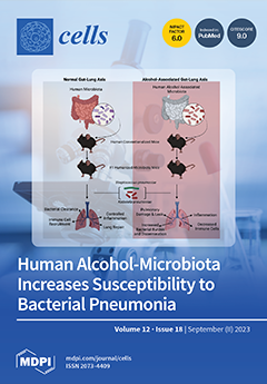
Chronic alcohol abuse leads to alterations in the gastrointestinal microbiota that are associated with behavioral, physiological, and immunological effects. However, the direct effects of alcohol-associated changes in the microbiome are ill-defined. To address this, we developed a humanized alcohol-microbiota mouse model, which allows us to systematically evaluate the immunological effects of chronic alcohol abuse mediated by changes in the intestinal microbiota. In this study, we discuss the effects of human alcohol-associated microbiota on pulmonary host defense against bacterial pneumonia. Our findings highlight the importance of considering both the direct effects of alcohol and alcohol-induced changes in the microbiota when investigating the mechanisms behind alcohol-related disorders and treatment strategies. View this paper
  • Issues are regarded as officially published after their release is announced to the table of contents alert mailing list.
  • You may sign up for e-mail alerts to receive table of contents of newly released issues.
  • PDF is the official format for papers published in both, html and pdf forms. To view the papers in pdf format, click on the "PDF Full-text" link, and use the free Adobe Reader to open them.
Order results
Result details
Section
Select all
Export citation of selected articles as:
20 pages, 17068 KiB  
Article
Cognitive Functions, Neurotransmitter Alterations, and Hippocampal Microstructural Changes in Mice Caused by Feeding on Western Diet
by Raly James Perez Custodio, Zaynab Hobloss, Maiju Myllys, Reham Hassan, Daniela González, Jörg Reinders, Julia Bornhorst, Ann-Kathrin Weishaupt, Abdel-latif Seddek, Tahany Abbas, Adrian Friebel, Stefan Hoehme, Stephan Getzmann, Jan G. Hengstler, Christoph van Thriel and Ahmed Ghallab
Cells 2023, 12(18), 2331; https://doi.org/10.3390/cells12182331 - 21 Sep 2023
Cited by 3 | Viewed by 2217
Abstract
Metabolic Dysfunction Associated Steatotic Liver Disease (MASLD) is the most common chronic liver disease in Western countries. It is becoming increasingly evident that peripheral organ-centered inflammatory diseases, including liver diseases, are linked with brain dysfunctions. Therefore, this study aims to unravel the effect [...] Read more.
Metabolic Dysfunction Associated Steatotic Liver Disease (MASLD) is the most common chronic liver disease in Western countries. It is becoming increasingly evident that peripheral organ-centered inflammatory diseases, including liver diseases, are linked with brain dysfunctions. Therefore, this study aims to unravel the effect of MASLD on brain histology, cognitive functions, and neurotransmitters. For this purpose, mice fed for 48 weeks on standard (SD) or Western diet (WD) were evaluated by behavioral tests, followed by sacrifice and analysis of the liver-brain axis including histopathology, immunohistochemistry, and biochemical analyses. Histological analysis of the liver showed features of Metabolic Dysfunction-Associated Steatohepatitis (MASH) in the WD-fed mice including lipid droplet accumulation, inflammation, and fibrosis. This was accompanied by an elevation of transaminase and alkaline phosphatase activities, increase in inflammatory cytokine and bile acid concentrations, as well as altered amino acid concentrations in the blood. Interestingly, compromised blood capillary morphology coupled with astrogliosis and microgliosis were observed in brain hippocampus of the WD mice, indicating neuroinflammation or a disrupted neurovascular unit. Moreover, attention was impaired in WD-fed mice along with the observations of impaired motor activity and balance, enhanced anxiety, and stereotyped head-twitch response (HTR) behaviors. Analysis of neurotransmitters and modulators including dopamine, serotonin, GABA, glutamate, and acetylcholine showed region-specific dysregulation in the brain of the WD-fed mice. In conclusion, the induction of MASH in mice is accompanied by the alteration of cellular morphology and neurotransmitter expression in the brain, associated with compromised cognitive functions. Full article
Show Figures

Graphical abstract

69 pages, 4604 KiB  
Review
Insight and Recommendations for Fragile X-Premutation-Associated Conditions from the Fifth International Conference on FMR1 Premutation
by Flora Tassone, Dragana Protic, Emily Graves Allen, Alison D. Archibald, Anna Baud, Ted W. Brown, Dejan B. Budimirovic, Jonathan Cohen, Brett Dufour, Rachel Eiges, Nicola Elvassore, Lidia V. Gabis, Samantha J. Grudzien, Deborah A. Hall, David Hessl, Abigail Hogan, Jessica Ezzell Hunter, Peng Jin, Poonnada Jiraanont, Jessica Klusek, R. Frank Kooy, Claudine M. Kraan, Cecilia Laterza, Andrea Lee, Karen Lipworth, Molly Losh, Danuta Loesch, Reymundo Lozano, Marsha R. Mailick, Apostolos Manolopoulos, Veronica Martinez-Cerdeno, Yingratana McLennan, Robert M. Miller, Federica Alice Maria Montanaro, Matthew W. Mosconi, Sarah Nelson Potter, Melissa Raspa, Susan M. Rivera, Katharine Shelly, Peter K. Todd, Katarzyna Tutak, Jun Yi Wang, Anne Wheeler, Tri Indah Winarni, Marwa Zafarullah and Randi J. Hagermanadd Show full author list remove Hide full author list
Cells 2023, 12(18), 2330; https://doi.org/10.3390/cells12182330 - 21 Sep 2023
Cited by 11 | Viewed by 5164
Abstract
The premutation of the fragile X messenger ribonucleoprotein 1 (FMR1) gene is characterized by an expansion of the CGG trinucleotide repeats (55 to 200 CGGs) in the 5’ untranslated region and increased levels of FMR1 mRNA. Molecular mechanisms leading to fragile [...] Read more.
The premutation of the fragile X messenger ribonucleoprotein 1 (FMR1) gene is characterized by an expansion of the CGG trinucleotide repeats (55 to 200 CGGs) in the 5’ untranslated region and increased levels of FMR1 mRNA. Molecular mechanisms leading to fragile X-premutation-associated conditions (FXPAC) include cotranscriptional R-loop formations, FMR1 mRNA toxicity through both RNA gelation into nuclear foci and sequestration of various CGG-repeat-binding proteins, and the repeat-associated non-AUG (RAN)-initiated translation of potentially toxic proteins. Such molecular mechanisms contribute to subsequent consequences, including mitochondrial dysfunction and neuronal death. Clinically, premutation carriers may exhibit a wide range of symptoms and phenotypes. Any of the problems associated with the premutation can appropriately be called FXPAC. Fragile X-associated tremor/ataxia syndrome (FXTAS), fragile X-associated primary ovarian insufficiency (FXPOI), and fragile X-associated neuropsychiatric disorders (FXAND) can fall under FXPAC. Understanding the molecular and clinical aspects of the premutation of the FMR1 gene is crucial for the accurate diagnosis, genetic counseling, and appropriate management of affected individuals and families. This paper summarizes all the known problems associated with the premutation and documents the presentations and discussions that occurred at the International Premutation Conference, which took place in New Zealand in 2023. Full article
Show Figures

Figure 1

14 pages, 1088 KiB  
Review
Emerging Roles of Ubiquitination in Biomolecular Condensates
by Peigang Liang, Jiaqi Zhang and Bo Wang
Cells 2023, 12(18), 2329; https://doi.org/10.3390/cells12182329 - 21 Sep 2023
Cited by 1 | Viewed by 1436
Abstract
Biomolecular condensates are dynamic non-membrane-bound macromolecular high-order assemblies that participate in a growing list of cellular processes, such as transcription, the cell cycle, etc. Disturbed dynamics of biomolecular condensates are associated with many diseases, including cancer and neurodegeneration. Extensive efforts have been devoted [...] Read more.
Biomolecular condensates are dynamic non-membrane-bound macromolecular high-order assemblies that participate in a growing list of cellular processes, such as transcription, the cell cycle, etc. Disturbed dynamics of biomolecular condensates are associated with many diseases, including cancer and neurodegeneration. Extensive efforts have been devoted to uncovering the molecular and biochemical grammar governing the dynamics of biomolecular condensates and establishing the critical roles of protein posttranslational modifications (PTMs) in this process. Here, we summarize the regulatory roles of ubiquitination (a major form of cellular PTM) in the dynamics of biomolecular condensates. We propose that these regulatory mechanisms can be harnessed to combat many diseases. Full article
(This article belongs to the Special Issue Advances in Ubiquitination and Deubiquitination Research)
Show Figures

Figure 1

13 pages, 2693 KiB  
Article
Novel Variant in CEP250 Causes Protein Mislocalization and Leads to Nonsyndromic Autosomal Recessive Type of Progressive Hearing Loss
by Minjin Kang, Jung Ah Kim, Mee Hyun Song, Sun Young Joo, Se Jin Kim, Seung Hyun Jang, Ho Lee, Je Kyung Seong, Jae Young Choi, Heon Yung Gee and Jinsei Jung
Cells 2023, 12(18), 2328; https://doi.org/10.3390/cells12182328 - 21 Sep 2023
Cited by 1 | Viewed by 1054
Abstract
Genetic hearing loss is the most common hereditary sensorial disorder. Though more than 120 genes associated with deafness have been identified, unveiled causative genes and variants of diverse types of hearing loss remain. Herein, we identified a novel nonsense homozygous variant in CEP250 [...] Read more.
Genetic hearing loss is the most common hereditary sensorial disorder. Though more than 120 genes associated with deafness have been identified, unveiled causative genes and variants of diverse types of hearing loss remain. Herein, we identified a novel nonsense homozygous variant in CEP250 (c.3511C>T; p.Gln1171Ter) among the family members with progressive moderate sensorineural hearing loss in nonsyndromic autosomal recessive type but without retinal degeneration. CEP250 encodes C-Nap1 protein belonging to the CEP protein family, comprising 30 proteins that play roles in centrosome aggregation and cell cycle progression. The nonsense variant in CEP250 led to the early truncating protein of C-Nap1, which hindered centrosome localization; heterologous expression of CEP250 (c.3511C>T) in NIH3T3 cells within cilia expression condition revealed that the truncating C-Nap1 (p.Gln1171Ter) was not localized at the centrosome but was dispersed in the cytosol. In the murine adult cochlea, Cep250 was expressed in the inner and outer hair cells. Knockout mice of Cep250 showed significant hair cell degeneration and progressive hearing loss in auditory brainstem response. In conclusion, a nonsense variant in CEP250 results in a deficit of centrosome localization and hair cell degeneration in the cochlea, which is associated with the progression of hearing loss in humans and mice. Full article
(This article belongs to the Special Issue Cell Death in Health and Disease)
Show Figures

Graphical abstract

15 pages, 1752 KiB  
Review
NLRP3 Inflammasome as a Potentially New Therapeutic Target of Mesenchymal Stem Cells and Their Exosomes in the Treatment of Inflammatory Eye Diseases
by Carl Randall Harrell, Valentin Djonov, Ana Antonijevic and Vladislav Volarevic
Cells 2023, 12(18), 2327; https://doi.org/10.3390/cells12182327 - 21 Sep 2023
Cited by 4 | Viewed by 1322
Abstract
Due to their potent immunoregulatory and angio-modulatory properties, mesenchymal stem cells (MSCs) and their exosomes (MSC-Exos) have emerged as potential game-changers in regenerative ophthalmology, particularly for the personalized treatment of inflammatory diseases. MSCs suppress detrimental immune responses in the eyes and alleviate ongoing [...] Read more.
Due to their potent immunoregulatory and angio-modulatory properties, mesenchymal stem cells (MSCs) and their exosomes (MSC-Exos) have emerged as potential game-changers in regenerative ophthalmology, particularly for the personalized treatment of inflammatory diseases. MSCs suppress detrimental immune responses in the eyes and alleviate ongoing inflammation in ocular tissues by modulating the phenotype and function of all immune cells that play pathogenic roles in the development and progression of inflammatory eye diseases. MSC-Exos, due to their nano-sized dimension and lipid envelope, easily bypass all barriers in the eyes and deliver MSC-sourced bioactive compounds directly to target cells. Although MSCs and their exosomes offer a novel approach to treating immune cell-driven eye diseases, further research is needed to optimize their therapeutic efficacy. A significant number of experimental studies is currently focused on the delineation of intracellular targets, which crucially contribute to the immunosuppressive and anti-inflammatory effects of MSCs and MSC-Exos. The activation of NLRP3 inflammasome induces programmed cell death of epithelial cells, induces the generation of inflammatory phenotypes in eye-infiltrated immune cells, and enhances the expression of adhesion molecules on ECs facilitating the recruitment of circulating leukocytes in injured and inflamed eyes. In this review article, we summarize current knowledge about signaling pathways that are responsible for NLRP3 inflammasome-driven intraocular inflammation and we emphasize molecular mechanisms that regulate MSC-based modulation of NLRP3-driven signaling in eye-infiltrated immune cells, providing evidence that NLRP3 inflammasome should be considered a potentially new therapeutic target for MSCs and MSC-Exo-based treatment of inflammatory eye diseases. Full article
(This article belongs to the Special Issue Updates on Mesenchymal Stem Cells-Derived Extracellular Vesicles)
Show Figures

Figure 1

20 pages, 7512 KiB  
Article
Human Preadipocytes Differentiated under Hypoxia following PCB126 Exposure during Proliferation: Effects on Differentiation, Glucose Uptake and Adipokine Profile
by Zeinab El Amine, Jean-François Mauger and Pascal Imbeault
Cells 2023, 12(18), 2326; https://doi.org/10.3390/cells12182326 - 21 Sep 2023
Viewed by 970
Abstract
Persistent organic pollutants (POPs) accumulation and hypoxia are two factors proposed to adversely alter adipose tissue (AT) functions in the context of excess adiposity. Studies have shown that preadipocytes exposure to dioxin and dioxin-like POPs have the greatest deleterious impact on rodent and [...] Read more.
Persistent organic pollutants (POPs) accumulation and hypoxia are two factors proposed to adversely alter adipose tissue (AT) functions in the context of excess adiposity. Studies have shown that preadipocytes exposure to dioxin and dioxin-like POPs have the greatest deleterious impact on rodent and immortalized human preadipocyte differentiation, but evidence on human preadipocytes is lacking. Additionally, hypoxia is known to strongly interfere with the dioxin-response pathway. Therefore, we tested the effects of pre-differentiation polychlorinated biphenyl (PCB)126 exposure at 10 µM for 3 days and subsequent differentiation under hypoxia on human subcutaneous adipocytes (hSA) differentiation, glucose uptake and expression of selected metabolism- and inflammation-related genes. Pre-differentiation PCB126 exposure lowered the adenosine triphosphate (ATP) content, glucose uptake and leptin expression of mature adipocytes but had limited effects on differentiation under normoxia (21% O2). Under hypoxia (3% O2), preadipocytes ability to differentiate was significantly reduced as reflected by significant decreased lipid accumulation and downregulation of key adipocyte genes such as peroxisome proliferator-activated receptor gamma (PPARγ) and adiponectin. Hypoxia increased glucose uptake and glucose transporter 1 (GLUT1) expression but abolished the adipocytes insulin response and GLUT4 expression. The expression of pro-inflammatory adipokine interleukin-6 (IL-6) was slightly increased by both PCB126 and hypoxia, while IL-8 expression was significantly increased only following the PCB126-hypoxia sequence. These observations suggest that PCB126 does not affect human preadipocyte differentiation, but does affect the subsequent adipocytes population, as reflected by lower ATP levels and absolute glucose uptake. On the other hand, PCB126 and hypoxia exert additive effects on AT inflammation, an important player in the development of chronic diseases such as type 2 diabetes and cardiovascular diseases. Full article
(This article belongs to the Special Issue The Adipose Tissue: From “Cinderella” to “Lion King” Organ)
Show Figures

Graphical abstract

15 pages, 2989 KiB  
Article
Establishment and Characterization of Continuous Satellite Muscle Cells from Olive Flounder (Paralichthys olivaceus): Isolation, Culture Conditions, and Myogenic Protein Expression
by Sathish Krishnan, Selvakumari Ulagesan, Josel Cadangin, Ji-Hye Lee, Taek-Jeong Nam and Youn-Hee Choi
Cells 2023, 12(18), 2325; https://doi.org/10.3390/cells12182325 - 21 Sep 2023
Viewed by 1400
Abstract
Olive flounder (Paralichthys olivaceus) muscle satellite cells (OFMCs) were obtained by enzymatic primary cell isolation and the explant method. Enzymatic isolation yielded cells that reached 80% confluence within 8 days, compared to 15 days for the explant method. Optimal OFMC growth [...] Read more.
Olive flounder (Paralichthys olivaceus) muscle satellite cells (OFMCs) were obtained by enzymatic primary cell isolation and the explant method. Enzymatic isolation yielded cells that reached 80% confluence within 8 days, compared to 15 days for the explant method. Optimal OFMC growth was observed in 20% fetal bovine serum at 28 °C with 0.8 mM CaCl2 and the basic fibroblast growth factor (BFGF) to enhance cell growth. OFMCs have become permanent cell lines through the spontaneous immortalization crisis at the 20th passage. Olive flounder skeletal muscle myoblasts were induced into a mitogen-poor medium containing 2% horse serum for differentiation; they fused to form multinucleate myotubes. The results indicated complete differentiation of myoblasts into myotubes; we also detected the expression of the myogenic regulatory factors myoD, myogenin, and desmin. Upregulation (Myogenin, desmin) and downregulation (MyoD) of muscle regulation factors confirmed the differentiation in OFMCs. Full article
Show Figures

Figure 1

21 pages, 1044 KiB  
Review
Regulatory Mechanisms That Guide the Fetal to Postnatal Transition of Cardiomyocytes
by Patrick G. Burgon, Jonathan J. Weldrick, Omar Mohamed Sayed Ahmed Talab, Muhammad Nadeer, Michail Nomikos and Lynn A. Megeney
Cells 2023, 12(18), 2324; https://doi.org/10.3390/cells12182324 - 21 Sep 2023
Cited by 1 | Viewed by 1353
Abstract
Heart disease remains a global leading cause of death and disability, necessitating a comprehensive understanding of the heart’s development, repair, and dysfunction. This review surveys recent discoveries that explore the developmental transition of proliferative fetal cardiomyocytes into hypertrophic postnatal cardiomyocytes, a process yet [...] Read more.
Heart disease remains a global leading cause of death and disability, necessitating a comprehensive understanding of the heart’s development, repair, and dysfunction. This review surveys recent discoveries that explore the developmental transition of proliferative fetal cardiomyocytes into hypertrophic postnatal cardiomyocytes, a process yet to be well-defined. This transition is key to the heart’s growth and has promising therapeutic potential, particularly for congenital or acquired heart damage, such as myocardial infarctions. Although significant progress has been made, much work is needed to unravel the complex interplay of signaling pathways that regulate cardiomyocyte proliferation and hypertrophy. This review provides a detailed perspective for future research directions aimed at the potential therapeutic harnessing of the perinatal heart transitions. Full article
Show Figures

Figure 1

16 pages, 2284 KiB  
Review
Neuroimmune Interactions in Fetal Alcohol Spectrum Disorders: Potential Therapeutic Targets and Intervention Strategies
by Sayani Mukherjee, Prashant Tarale and Dipak K. Sarkar
Cells 2023, 12(18), 2323; https://doi.org/10.3390/cells12182323 - 21 Sep 2023
Viewed by 1564
Abstract
Fetal alcohol spectrum disorders (FASD) are a set of abnormalities caused by prenatal exposure to ethanol and are characterized by developmental defects in the brain that lead to various overt and non-overt physiological abnormalities. Growing evidence suggests that in utero alcohol exposure induces [...] Read more.
Fetal alcohol spectrum disorders (FASD) are a set of abnormalities caused by prenatal exposure to ethanol and are characterized by developmental defects in the brain that lead to various overt and non-overt physiological abnormalities. Growing evidence suggests that in utero alcohol exposure induces functional and structural abnormalities in gliogenesis and neuron–glia interactions, suggesting a possible role of glial cell pathologies in the development of FASD. However, the molecular mechanisms of neuron–glia interactions that lead to the development of FASD are not clearly understood. In this review, we discuss glial cell pathologies with a particular emphasis on microglia, primary resident immune cells in the brain. Additionally, we examine the involvement of several neuroimmune molecules released by glial cells, their signaling pathways, and epigenetic mechanisms responsible for FASD-related alteration in brain functions. Growing evidence suggests that extracellular vesicles (EVs) play a crucial role in the communication between cells via transporting bioactive cargo from one cell to the other. This review emphasizes the role of EVs in the context of neuron–glia interactions during prenatal alcohol exposure. Finally, some potential applications involving nutritional, pharmacological, cell-based, and exosome-based therapies in the treatment of FASD are discussed. Full article
(This article belongs to the Special Issue Alcohol and Neuroimmunology)
Show Figures

Figure 1

22 pages, 7521 KiB  
Article
TRPV1 Channels Are New Players in the Reticulum–Mitochondria Ca2+ Coupling in a Rat Cardiomyoblast Cell Line
by Nolwenn Tessier, Mallory Ducrozet, Maya Dia, Sally Badawi, Christophe Chouabe, Claire Crola Da Silva, Michel Ovize, Gabriel Bidaux, Fabien Van Coppenolle and Sylvie Ducreux
Cells 2023, 12(18), 2322; https://doi.org/10.3390/cells12182322 - 20 Sep 2023
Cited by 3 | Viewed by 1683
Abstract
The Ca2+ release in microdomains formed by intercompartmental contacts, such as mitochondria-associated endoplasmic reticulum membranes (MAMs), encodes a signal that contributes to Ca2+ homeostasis and cell fate control. However, the composition and function of MAMs remain to be fully defined. Here, [...] Read more.
The Ca2+ release in microdomains formed by intercompartmental contacts, such as mitochondria-associated endoplasmic reticulum membranes (MAMs), encodes a signal that contributes to Ca2+ homeostasis and cell fate control. However, the composition and function of MAMs remain to be fully defined. Here, we focused on the transient receptor potential vanilloid 1 (TRPV1), a Ca2+-permeable ion channel and a polymodal nociceptor. We found TRPV1 channels in the reticular membrane, including some at MAMs, in a rat cardiomyoblast cell line (SV40-transformed H9c2) by Western blotting, immunostaining, cell fractionation, and proximity ligation assay. We used chemical and genetic probes to perform Ca2+ imaging in four cellular compartments: the endoplasmic reticulum (ER), cytoplasm, mitochondrial matrix, and mitochondrial surface. Our results showed that the ER Ca2+ released through TRPV1 channels is detected at the mitochondrial outer membrane and transferred to the mitochondria. Finally, we observed that prolonged TRPV1 modulation for 30 min alters the intracellular Ca2+ equilibrium and influences the MAM structure or the hypoxia/reoxygenation-induced cell death. Thus, our study provides the first evidence that TRPV1 channels contribute to MAM Ca2+ exchanges. Full article
Show Figures

Figure 1

14 pages, 643 KiB  
Review
What Does the Future Hold for Biomarkers of Response to Extracorporeal Photopheresis for Mycosis Fungoides and Sézary Syndrome?
by Oleg E. Akilov
Cells 2023, 12(18), 2321; https://doi.org/10.3390/cells12182321 - 20 Sep 2023
Viewed by 845
Abstract
Extracorporeal photopheresis (ECP) is an FDA-approved immunotherapy for cutaneous T-cell lymphoma, which can provide a complete response in some patients. However, it is still being determined who will respond well, and predictive biomarkers are urgently needed to target patients for timely treatment and [...] Read more.
Extracorporeal photopheresis (ECP) is an FDA-approved immunotherapy for cutaneous T-cell lymphoma, which can provide a complete response in some patients. However, it is still being determined who will respond well, and predictive biomarkers are urgently needed to target patients for timely treatment and to monitor their response over time. The aim of this review is to analyze the current state of the diagnostic, prognostic, and disease state-monitoring biomarkers of ECP, and outline the future direction of the ECP biomarker discovery. Specifically, we focus on biomarkers of response to ECP in mycosis fungoides and Sézary syndrome. The review summarizes the current knowledge of ECP biomarkers, including their limitations and potential applications, and identifies key challenges in ECP biomarker discovery. In addition, we discuss emerging technologies that could revolutionize ECP biomarker discovery and accelerate the translation of biomarker research into clinical practice. This review will interest researchers and clinicians seeking to optimize ECP therapy for cutaneous T-cell lymphoma. Full article
Show Figures

Figure 1

0 pages, 3403 KiB  
Article
Effect of Acute Enriched Environment Exposure on Brain Oscillations and Activation of the Translation Initiation Factor 4E-BPs at Synapses across Wakefulness and Sleep in Rats
by José Lucas Santos, Evlalia Petsidou, Pallavi Saraogi, Ullrich Bartsch, André P. Gerber and Julie Seibt
Cells 2023, 12(18), 2320; https://doi.org/10.3390/cells12182320 - 20 Sep 2023
Viewed by 1281
Abstract
Brain plasticity is induced by learning during wakefulness and is consolidated during sleep. But the molecular mechanisms involved are poorly understood and their relation to experience-dependent changes in brain activity remains to be clarified. Localised mRNA translation is important for the structural changes [...] Read more.
Brain plasticity is induced by learning during wakefulness and is consolidated during sleep. But the molecular mechanisms involved are poorly understood and their relation to experience-dependent changes in brain activity remains to be clarified. Localised mRNA translation is important for the structural changes at synapses supporting brain plasticity consolidation. The translation mTOR pathway, via phosphorylation of 4E-BPs, is known to be activate during sleep and contributes to brain plasticity, but whether this activation is specific to synapses is not known. We investigated this question using acute exposure of rats to an enriched environment (EE). We measured brain activity with EEGs and 4E-BP phosphorylation at cortical and cerebellar synapses with Western blot analyses. Sleep significantly increased the conversion of 4E-BPs to their hyperphosphorylated forms at synapses, especially after EE exposure. EE exposure increased oscillations in the alpha band during active exploration and in the theta-to-beta (4–30 Hz) range, as well as spindle density, during NREM sleep. Theta activity during exploration and NREM spindle frequency predicted changes in 4E-BP hyperphosphorylation at synapses. Hence, our results suggest a functional link between EEG and molecular markers of plasticity across wakefulness and sleep. Full article
Show Figures

Graphical abstract

14 pages, 17035 KiB  
Article
Modeling of FAN1-Deficient Kidney Disease Using a Human Induced Pluripotent Stem Cell-Derived Kidney Organoid System
by Sun Woo Lim, Dohyun Na, Hanbi Lee, Xianying Fang, Sheng Cui, Yoo Jin Shin, Kang In Lee, Jae Young Lee, Chul Woo Yang and Byung Ha Chung
Cells 2023, 12(18), 2319; https://doi.org/10.3390/cells12182319 - 20 Sep 2023
Viewed by 1277
Abstract
Karyomegalic interstitial nephritis (KIN) is a genetic kidney disease caused by mutations in the FANCD2/FANCI-Associated Nuclease 1 (FAN1) gene on 15q13.3, which results in karyomegaly and fibrosis of kidney cells through the incomplete repair of DNA damage. The aim of this [...] Read more.
Karyomegalic interstitial nephritis (KIN) is a genetic kidney disease caused by mutations in the FANCD2/FANCI-Associated Nuclease 1 (FAN1) gene on 15q13.3, which results in karyomegaly and fibrosis of kidney cells through the incomplete repair of DNA damage. The aim of this study was to explore the possibility of using a human induced pluripotent stem cell (hiPSC)-derived kidney organoid system for modeling FAN1-deficient kidney disease, also known as KIN. We generated kidney organoids using WTC-11 (wild-type) hiPSCs and FAN1-mutant hiPSCs which include KIN patient-derived hiPSCs and FAN1-edited hiPSCs (WTC-11 FAN1+/−), created using the CRISPR/Cas9 system in WTC-11-hiPSCs. Kidney organoids from each group were treated with 20 nM of mitomycin C (MMC) for 24 or 48 h, and the expression levels of Ki67 and H2A histone family member X (H2A.X) were analyzed to detect DNA damage and assess the viability of cells within the kidney organoids. Both WTC-11-hiPSCs and FAN1-mutant hiPSCs were successfully differentiated into kidney organoids without structural deformities. MMC treatment for 48 h significantly increased the expression of DNA damage markers, while cell viability in both FAN1-mutant kidney organoids was decreased. However, these findings were observed in WTC-11-kidney organoids. These results suggest that FAN1-mutant kidney organoids can recapitulate the phenotype of FAN1-deficient kidney disease. Full article
Show Figures

Figure 1

20 pages, 3016 KiB  
Review
Biochemical and Molecular Pathways in Neurodegenerative Diseases: An Integrated View
by Nitesh Sanghai and Geoffrey K. Tranmer
Cells 2023, 12(18), 2318; https://doi.org/10.3390/cells12182318 - 20 Sep 2023
Cited by 6 | Viewed by 4588
Abstract
Neurodegenerative diseases (NDDs) like Alzheimer’s disease (AD), Parkinson’s disease (PD), and amyotrophic lateral sclerosis (ALS) are defined by a myriad of complex aetiologies. Understanding the common biochemical molecular pathologies among NDDs gives an opportunity to decipher the overlapping and numerous cross-talk mechanisms of [...] Read more.
Neurodegenerative diseases (NDDs) like Alzheimer’s disease (AD), Parkinson’s disease (PD), and amyotrophic lateral sclerosis (ALS) are defined by a myriad of complex aetiologies. Understanding the common biochemical molecular pathologies among NDDs gives an opportunity to decipher the overlapping and numerous cross-talk mechanisms of neurodegeneration. Numerous interrelated pathways lead to the progression of neurodegeneration. We present evidence from the past pieces of literature for the most usual global convergent hallmarks like ageing, oxidative stress, excitotoxicity-induced calcium butterfly effect, defective proteostasis including chaperones, autophagy, mitophagy, and proteosome networks, and neuroinflammation. Herein, we applied a holistic approach to identify and represent the shared mechanism across NDDs. Further, we believe that this approach could be helpful in identifying key modulators across NDDs, with a particular focus on AD, PD, and ALS. Moreover, these concepts could be applied to the development and diagnosis of novel strategies for diverse NDDs. Full article
Show Figures

Graphical abstract

27 pages, 4896 KiB  
Article
Differential Effects of Regulatory T Cells in the Meninges and Spinal Cord of Male and Female Mice with Neuropathic Pain
by Nathan T. Fiore, Brooke A. Keating, Yuting Chen, Sarah I. Williams and Gila Moalem-Taylor
Cells 2023, 12(18), 2317; https://doi.org/10.3390/cells12182317 - 20 Sep 2023
Cited by 3 | Viewed by 1453
Abstract
Immune cells play a critical role in promoting neuroinflammation and the development of neuropathic pain. However, some subsets of immune cells are essential for pain resolution. Among them are regulatory T cells (Tregs), a specialised subpopulation of T cells that limit excessive immune [...] Read more.
Immune cells play a critical role in promoting neuroinflammation and the development of neuropathic pain. However, some subsets of immune cells are essential for pain resolution. Among them are regulatory T cells (Tregs), a specialised subpopulation of T cells that limit excessive immune responses and preserve immune homeostasis. In this study, we utilised intrathecal adoptive transfer of activated Tregs in male and female mice after peripheral nerve injury to investigate Treg migration and whether Treg-mediated suppression of pain behaviours is associated with changes in peripheral immune cell populations in lymphoid and meningeal tissues and spinal microglial and astrocyte reactivity and phenotypes. Treatment with Tregs suppressed mechanical pain hypersensitivity and improved changes in exploratory behaviours after chronic constriction injury (CCI) of the sciatic nerve in both male and female mice. The injected Treg cells were detected in the choroid plexus and the pia mater and in peripheral lymphoid organs in both male and female recipient mice. Nonetheless, Treg treatment resulted in differential changes in meningeal and lymph node immune cell profiles in male and female mice. Moreover, in male mice, adoptive transfer of Tregs ameliorated the CCI-induced increase in microglia reactivity and inflammatory phenotypic shift, increasing M2-like phenotypic markers and attenuating astrocyte reactivity and neurotoxic astrocytes. Contrastingly, in CCI female mice, Treg injection increased astrocyte reactivity and neuroprotective astrocytes. These findings show that the adoptive transfer of Tregs modulates meningeal and peripheral immunity, as well as spinal glial populations, and alleviates neuropathic pain, potentially through different mechanisms in males and females. Full article
(This article belongs to the Special Issue Role of Glial Cells in Neuropathic Pain)
Show Figures

Figure 1

19 pages, 2940 KiB  
Article
Mutant Cytochrome C as a Potential Detector of Superoxide Generation: Effect of Mutations on the Function and Properties
by Rita V. Chertkova, Ilya P. Oleynikov, Alexey A. Pakhomov, Roman V. Sudakov, Victor N. Orlov, Marina A. Semenova, Alexander M. Arutyunyan, Vasily V. Ptushenko, Mikhail P. Kirpichnikov, Dmitry A. Dolgikh and Tatiana V. Vygodina
Cells 2023, 12(18), 2316; https://doi.org/10.3390/cells12182316 - 19 Sep 2023
Viewed by 1142
Abstract
Cytochrome c (CytC) is a single-electron carrier between complex bc1 and cytochrome c-oxidase (CcO) in the electron transport chain (ETC). It is also known as a good radical scavenger but its participation in electron flow through the ETC makes it impossible to use [...] Read more.
Cytochrome c (CytC) is a single-electron carrier between complex bc1 and cytochrome c-oxidase (CcO) in the electron transport chain (ETC). It is also known as a good radical scavenger but its participation in electron flow through the ETC makes it impossible to use CytC as a radical sensor. To solve this problem, a series of mutants were constructed with substitutions of Lys residues in the universal binding site (UBS) which interact electrostatically with negatively charged Asp and Glu residues at the binding sites of CytC partners, bc1 complex and CcO. The aim of this study was to select a mutant that had lost its function as an electron carrier in the ETC, retaining the structure and ability to quench radicals. It was shown that a mutant CytC with substitutions of five (8Mut) and four (5Mut) Lys residues in the UBS was almost inactive toward CcO. However, all mutant proteins kept their antioxidant activity sufficiently with respect to the superoxide radical. Mutations shifted the dipole moment of the CytC molecule due to seriously changed electrostatics on the surface of the protein. In addition, a decrease in the redox potential of the protein as revealed by the redox titrations of 8Mut was detected. Nevertheless, the CD spectrum and dynamic light scattering suggested no significant changes in the secondary structure or aggregation of the molecules of CytC 8Mut. Thus, a variant 8Mut with multiple mutations in the UBS which lost its ability to electron transfer and saved most of its physico-chemical properties can be effectively used as a detector of superoxide generation both in mitochondria and in other systems. Full article
Show Figures

Figure 1

21 pages, 4862 KiB  
Article
Characterisation of Lipoma-Preferred Partner as a Novel Mechanotransducer in Vascular Smooth Muscle Cells
by Alexandra Sporkova, Taslima Nahar, Mingsi Cao, Subhajit Ghosh, Carla Sens-Albert, Prisca Amayi Patricia Friede, Anika Nagel, Jaafar Al-Hasani and Markus Hecker
Cells 2023, 12(18), 2315; https://doi.org/10.3390/cells12182315 - 19 Sep 2023
Viewed by 3122
Abstract
In arteries and arterioles, a chronic increase in blood pressure raises wall tension. This continuous biomechanical strain causes a change in gene expression in vascular smooth muscle cells (VSMCs) that may lead to pathological changes. Here we have characterised the functional properties of [...] Read more.
In arteries and arterioles, a chronic increase in blood pressure raises wall tension. This continuous biomechanical strain causes a change in gene expression in vascular smooth muscle cells (VSMCs) that may lead to pathological changes. Here we have characterised the functional properties of lipoma-preferred partner (LPP), a Lin11–Isl1–Mec3 (LIM)-domain protein, which is most closely related to the mechanotransducer zyxin but selectively expressed by smooth muscle cells, including VSMCs in adult mice. VSMCs isolated from the aorta of LPP knockout (LPP-KO) mice displayed a higher rate of proliferation than their wildtype (WT) counterparts, and when cultured as three-dimensional spheroids, they revealed a higher expression of the proliferation marker Ki 67 and showed greater invasion into a collagen gel. Accordingly, the gelatinase activity was increased in LPP-KO but not WT spheroids. The LPP-KO spheroids adhering to the collagen gel responded with decreased contraction to potassium chloride. The relaxation response to caffeine and norepinephrine was also smaller in the LPP-KO spheroids than in their WT counterparts. The overexpression of zyxin in LPP-KO VSMCs resulted in a reversal to a more quiescent differentiated phenotype. In native VSMCs, i.e., in isolated perfused segments of the mesenteric artery (MA), the contractile responses of LPP-KO segments to potassium chloride, phenylephrine or endothelin-1 did not vary from those in isolated perfused WT segments. In contrast, the myogenic response of LPP-KO MA segments was significantly attenuated while zyxin-deficient MA segments displayed a normal myogenic response. We propose that LPP, which we found to be expressed solely in the medial layer of different arteries from adult mice, may play an important role in controlling the quiescent contractile phenotype of VSMCs. Full article
(This article belongs to the Special Issue Role of Vascular Smooth Muscle Cells in Cardiovascular Disease)
Show Figures

Figure 1

44 pages, 552 KiB  
Review
Inborn Errors of Metabolism with Ataxia: Current and Future Treatment Options
by Tatiana Bremova-Ertl, Jan Hofmann, Janine Stucki, Anja Vossenkaul and Matthias Gautschi
Cells 2023, 12(18), 2314; https://doi.org/10.3390/cells12182314 - 19 Sep 2023
Viewed by 1568
Abstract
A number of hereditary ataxias are caused by inborn errors of metabolism (IEM), most of which are highly heterogeneous in their clinical presentation. Prompt diagnosis is important because disease-specific therapies may be available. In this review, we offer a comprehensive overview of metabolic [...] Read more.
A number of hereditary ataxias are caused by inborn errors of metabolism (IEM), most of which are highly heterogeneous in their clinical presentation. Prompt diagnosis is important because disease-specific therapies may be available. In this review, we offer a comprehensive overview of metabolic ataxias summarized by disease, highlighting novel clinical trials and emerging therapies with a particular emphasis on first-in-human gene therapies. We present disease-specific treatments if they exist and review the current evidence for symptomatic treatments of these highly heterogeneous diseases (where cerebellar ataxia is part of their phenotype) that aim to improve the disease burden and enhance quality of life. In general, a multimodal and holistic approach to the treatment of cerebellar ataxia, irrespective of etiology, is necessary to offer the best medical care. Physical therapy and speech and occupational therapy are obligatory. Genetic counseling is essential for making informed decisions about family planning. Full article
(This article belongs to the Special Issue Emerging Therapies for Hereditary Ataxia)
26 pages, 4596 KiB  
Article
Let-7g Upregulation Attenuated the KRAS–PI3K–Rac1–Akt Axis-Mediated Bioenergetic Functions
by Kuang-Chen Hung, Ni Tien, Da-Tian Bau, Chun-Hsu Yao, Chan-Hung Chen, Jiun-Long Yang, Meng-Liang Lin and Shih-Shun Chen
Cells 2023, 12(18), 2313; https://doi.org/10.3390/cells12182313 - 19 Sep 2023
Viewed by 1344
Abstract
The aberrant activation of signaling pathways contributes to cancer cells with metabolic reprogramming. Thus, targeting signaling modulators is considered a potential therapeutic strategy for cancer. Subcellular fractionation, coimmunoprecipitation, biochemical analysis, and gene manipulation experiments revealed that decreasing the interaction of kirsten rat sarcoma [...] Read more.
The aberrant activation of signaling pathways contributes to cancer cells with metabolic reprogramming. Thus, targeting signaling modulators is considered a potential therapeutic strategy for cancer. Subcellular fractionation, coimmunoprecipitation, biochemical analysis, and gene manipulation experiments revealed that decreasing the interaction of kirsten rat sarcoma viral oncogene homolog (KRAS) with p110α in lipid rafts with the use of naringenin (NGN), a citrus flavonoid, causes lipid raft-associated phosphatidylinositol 3-kinase (PI3K)−GTP-ras-related C3 botulinum toxin substrate 1 (Rac1)−protein kinase B (Akt)-regulated metabolic dysfunction of glycolysis and mitochondrial oxidative phosphorylation (OXPHOS), leading to apoptosis in human nasopharyngeal carcinoma (NPC) cells. The use of lethal-7g (let-7g) mimic and let-7g inhibitor confirmed that elevated let-7g resulted in a decrease in KRAS expression, which attenuated the PI3K−Rac1−Akt−BCL-2/BCL-xL-modulated mitochondrial energy metabolic functions. Increased let-7g depends on the suppression of the RNA-specificity of monocyte chemoattractant protein-induced protein-1 (MCPIP1) ribonuclease since NGN specifically blocks the degradation of pre-let-7g by NPC cell-derived immunoprecipitated MCPIP1. Converging lines of evidence indicate that the inhibition of MCPIP1 by NGN leads to let-7g upregulation, suppressing oncogenic KRAS-modulated PI3K–Rac1–Akt signaling and thereby impeding the metabolic activities of aerobic glycolysis and mitochondrial OXPHOS. Full article
(This article belongs to the Section Intracellular and Plasma Membranes)
Show Figures

Graphical abstract

32 pages, 5221 KiB  
Review
The Chemical Inhibitors of Endocytosis: From Mechanisms to Potential Clinical Applications
by Olga Klaudia Szewczyk-Roszczenko, Piotr Roszczenko, Anna Shmakova, Nataliya Finiuk, Serhii Holota, Roman Lesyk, Anna Bielawska, Yegor Vassetzky and Krzysztof Bielawski
Cells 2023, 12(18), 2312; https://doi.org/10.3390/cells12182312 - 19 Sep 2023
Cited by 5 | Viewed by 4233
Abstract
Endocytosis is one of the major ways cells communicate with their environment. This process is frequently hijacked by pathogens. Endocytosis also participates in the oncogenic transformation. Here, we review the approaches to inhibit endocytosis, discuss chemical inhibitors of this process, and discuss potential [...] Read more.
Endocytosis is one of the major ways cells communicate with their environment. This process is frequently hijacked by pathogens. Endocytosis also participates in the oncogenic transformation. Here, we review the approaches to inhibit endocytosis, discuss chemical inhibitors of this process, and discuss potential clinical applications of the endocytosis inhibitors. Full article
(This article belongs to the Section Cellular Pathology)
Show Figures

Graphical abstract

23 pages, 7211 KiB  
Article
Unraveling the Role of Hepatic PGC1α in Breast Cancer Invasion: A New Target for Therapeutic Intervention?
by Kumar Ganesan, Cong Xu, Qingqing Liu, Yue Sui and Jianping Chen
Cells 2023, 12(18), 2311; https://doi.org/10.3390/cells12182311 - 19 Sep 2023
Cited by 3 | Viewed by 980
Abstract
Breast cancer (BC) is the most common cancer among women worldwide and the main cause of cancer deaths in women. Metabolic components are key risk factors for the development of non-alcoholic fatty liver disease (NAFLD), which may promote BC. Studies have reported that [...] Read more.
Breast cancer (BC) is the most common cancer among women worldwide and the main cause of cancer deaths in women. Metabolic components are key risk factors for the development of non-alcoholic fatty liver disease (NAFLD), which may promote BC. Studies have reported that increasing PGC1α levels increases mitochondrial biogenesis, thereby increasing cell proliferation and metastasis. Moreover, the PGC1α/ERRα axis is a crucial regulator of cellular metabolism in various tissues, including BC. However, it remains unclear whether NAFLD is closely associated with the risk of BC. Therefore, the present study aimed to determine whether hepatic PGC1α promotes BC cell invasion via ERRα. Various assays, including ELISA, western blotting, and immunoprecipitation, have been employed to explore these mechanisms. According to the KM plot and TCGA data, elevated PGC1α expression was highly associated with a shorter overall survival time in patients with BC. High concentrations of palmitic acid (PA) promoted PGC1α expression, lipogenesis, and inflammatory processes in hepatocytes. Conditioned medium obtained from PA-treated hepatocytes significantly increased BC cell proliferation. Similarly, recombinant PGC1α in E0771 and MCF7 cells promoted cell proliferation, migration, and invasion in vitro. However, silencing PGC1α in both BC cell lines resulted in a decrease in this trend. As determined by immunoprecipitation assay, PCG1a interacted with ERRα, thereby facilitating the proliferation of BC cells. This outcome recognizes the importance of further investigations in exploring the full potential of hepatic PGC1α as a prognostic marker for BC development. Full article
Show Figures

Figure 1

26 pages, 8216 KiB  
Article
Syringin Prevents 6-Hydroxydopamine Neurotoxicity by Mediating the MiR-34a/SIRT1/Beclin-1 Pathway and Activating Autophagy in SH-SY5Y Cells and the Caenorhabditis elegans Model
by Ru-Huei Fu, Syuan-Yu Hong and Hui-Jye Chen
Cells 2023, 12(18), 2310; https://doi.org/10.3390/cells12182310 - 19 Sep 2023
Viewed by 1301
Abstract
Defective autophagy is one of the cellular hallmarks of Parkinson’s disease (PD). Therefore, a therapeutic strategy could be a modest enhancement of autophagic activity in dopamine (DA) neurons to deal with the clearance of damaged mitochondria and abnormal protein aggregates. Syringin (SRG) is [...] Read more.
Defective autophagy is one of the cellular hallmarks of Parkinson’s disease (PD). Therefore, a therapeutic strategy could be a modest enhancement of autophagic activity in dopamine (DA) neurons to deal with the clearance of damaged mitochondria and abnormal protein aggregates. Syringin (SRG) is a phenolic glycoside derived from the root of Acanthopanax senticosus. It has antioxidant, anti-apoptotic, and anti-inflammatory properties. However, whether it has a preventive effect on PD remains unclear. The present study found that SRG reversed the increase in intracellular ROS-caused apoptosis in SH-SY5Y cells induced by neurotoxin 6-OHDA exposure. Likewise, in C. elegans, degeneration of DA neurons, DA-related food-sensitive behaviors, longevity, and accumulation of α-synuclein were also improved. Studies of neuroprotective mechanisms have shown that SRG can reverse the suppressed expression of SIRT1, Beclin-1, and other autophagy markers in 6-OHDA-exposed cells. Thus, these enhanced the formation of autophagic vacuoles and autophagy activity. This protective effect can be blocked by pretreatment with wortmannin (an autophagosome formation blocker) and bafilomycin A1 (an autophagosome–lysosome fusion blocker). In addition, 6-OHDA increases the acetylation of Beclin-1, leading to its inactivation. SRG can induce the expression of SIRT1 and promote the deacetylation of Beclin-1. Finally, we found that SRG reduced the 6-OHDA-induced expression of miR-34a targeting SIRT1. The overexpression of miR-34a mimic abolishes the neuroprotective ability of SRG. In conclusion, SRG induces autophagy via partially regulating the miR-34a/SIRT1/Beclin-1 axis to prevent 6-OHDA-induced apoptosis and α-synuclein accumulation. SRG has the opportunity to be established as a candidate agent for the prevention and cure of PD. Full article
(This article belongs to the Special Issue Autophagy in Parkinson's Disease)
Show Figures

Graphical abstract

15 pages, 3303 KiB  
Article
Lymphatic Defects in Zebrafish sox18 Mutants Are Exacerbated by Perturbed VEGFC Signaling, While Masked by Elevated sox7 Expression
by Silvia Moleri, Sara Mercurio, Alex Pezzotta, Donatella D’Angelo, Alessia Brix, Alice Plebani, Giulia Lini, Marialaura Di Fuorti and Monica Beltrame
Cells 2023, 12(18), 2309; https://doi.org/10.3390/cells12182309 - 19 Sep 2023
Cited by 2 | Viewed by 1108
Abstract
Mutations in the transcription factor-coding gene SOX18, the growth factor-coding gene VEGFC and its receptor-coding gene VEGFR3/FLT4 cause primary lymphedema in humans. In mammals, SOX18, together with COUP-TFII/NR2F2, activates the expression of Prox1, a master regulator in lymphatic identity and development. [...] Read more.
Mutations in the transcription factor-coding gene SOX18, the growth factor-coding gene VEGFC and its receptor-coding gene VEGFR3/FLT4 cause primary lymphedema in humans. In mammals, SOX18, together with COUP-TFII/NR2F2, activates the expression of Prox1, a master regulator in lymphatic identity and development. Knockdown studies have also suggested an involvement of Sox18, Coup-tfII/Nr2f2, and Prox1 in zebrafish lymphatic development. Mutants in the corresponding genes initially failed to recapitulate the lymphatic defects observed in morphants. In this paper, we describe a novel zebrafish sox18 mutant allele, sa12315, which behaves as a null. The formation of the lymphatic thoracic duct is affected in sox18 homozygous mutants, but defects are milder in both zygotic and maternal-zygotic sox18 mutants than in sox18 morphants. Remarkably, in sox18 mutants, the expression of the closely related sox7 gene is elevated where lymphatic precursors arise. Sox7 could thus mask the absence of a functional Sox18 protein and account for the mild lymphatic phenotype in sox18 mutants, as shown in mice. Partial knockdown of vegfc exacerbates lymphatic defects in sox18 mutants, making them visible in heterozygotes. Our data thus reinforce the genetic interaction between Sox18 and Vegfc in lymphatic development, previously suggested by knockdown studies, and highlight the ability of Sox7 to compensate for Sox18 lymphatic dysfunction. Full article
(This article belongs to the Special Issue Modeling Developmental Processes and Disorders in Zebrafish)
Show Figures

Figure 1

24 pages, 2343 KiB  
Article
Altered Epigenetic Marks and Gene Expression in Fetal Brain, and Postnatal Behavioural Disorders, Following Prenatal Exposure of Ogg1 Knockout Mice to Saline or Ethanol
by Shama Bhatia, David Bodenstein, Ashley P. Cheng and Peter G. Wells
Cells 2023, 12(18), 2308; https://doi.org/10.3390/cells12182308 - 19 Sep 2023
Viewed by 1121
Abstract
Oxoguanine glycosylase 1 (OGG1) is widely known to repair the reactive oxygen species (ROS)-initiated DNA lesion 8-oxoguanine (8-oxoG), and more recently was shown to act as an epigenetic modifier. We have previously shown that saline-exposed Ogg1 −/− knockout progeny exhibited learning and memory [...] Read more.
Oxoguanine glycosylase 1 (OGG1) is widely known to repair the reactive oxygen species (ROS)-initiated DNA lesion 8-oxoguanine (8-oxoG), and more recently was shown to act as an epigenetic modifier. We have previously shown that saline-exposed Ogg1 −/− knockout progeny exhibited learning and memory deficits, which were enhanced by in utero exposure to a single low dose of ethanol (EtOH) in both Ogg1 +/+ and −/− progeny, but more so in Ogg1 −/− progeny. Herein, OGG1-deficient progeny exposed in utero to a single low dose of EtOH or its saline vehicle exhibited OGG1- and/or EtOH-dependent alterations in global histone methylation and acetylation, DNA methylation and gene expression (Tet1 (Tet Methylcytosine Dioxygenase 1), Nlgn3 (Neuroligin 3), Hdac2 (Histone Deacetylase 2), Reln (Reelin) and Esr1 (Estrogen Receptor 1)) in fetal brains, and behavioural changes in open field activity, social interaction and ultrasonic vocalization, but not prepulse inhibition. OGG1- and EtOH-dependent changes in Esr1 and Esr2 mRNA and protein levels were sex-dependent, as was the association of Esr1 gene expression with gene activation mark histone H3 lysine 4 trimethylation (H3K4me3) and gene repression mark histone H3 lysine 27 trimethylation (H3K27me3) measured via ChIP-qPCR. The OGG1-dependent changes in global epigenetic marks and gene/protein expression in fetal brains, and postnatal behavioural changes, observed in both saline- and EtOH-exposed progeny, suggest the involvement of epigenetic mechanisms in developmental disorders mediated by 8-oxoG and/or OGG1. Epigenetic effects of OGG1 may be involved in ESR1-mediated gene regulation, which may be altered by physiological and EtOH-enhanced levels of ROS formation, possibly contributing to sex-dependent developmental disorders observed in Ogg1 knockout mice. The OGG1- and EtOH-dependent associations provide a basis for more comprehensive mechanistic studies to determine the causal involvement of oxidative DNA damage and epigenetic changes in ROS-mediated neurodevelopmental disorders. Full article
Show Figures

Figure 1

19 pages, 1623 KiB  
Review
Astrocytes Are a Key Target for Neurotropic Viral Infection
by Maja Potokar, Robert Zorec and Jernej Jorgačevski
Cells 2023, 12(18), 2307; https://doi.org/10.3390/cells12182307 - 19 Sep 2023
Cited by 1 | Viewed by 1241
Abstract
Astrocytes are increasingly recognized as important viral host cells in the central nervous system. These cells can produce relatively high quantities of new virions. In part, this can be attributed to the characteristics of astrocyte metabolism and its abundant and dynamic cytoskeleton network. [...] Read more.
Astrocytes are increasingly recognized as important viral host cells in the central nervous system. These cells can produce relatively high quantities of new virions. In part, this can be attributed to the characteristics of astrocyte metabolism and its abundant and dynamic cytoskeleton network. Astrocytes are anatomically localized adjacent to interfaces between blood capillaries and brain parenchyma and between blood capillaries and brain ventricles. Moreover, astrocytes exhibit a larger membrane interface with the extracellular space than neurons. These properties, together with the expression of various and numerous viral entry receptors, a relatively high rate of endocytosis, and morphological plasticity of intracellular organelles, render astrocytes important target cells in neurotropic infections. In this review, we describe factors that mediate the high susceptibility of astrocytes to viral infection and replication, including the anatomic localization of astrocytes, morphology, expression of viral entry receptors, and various forms of autophagy. Full article
(This article belongs to the Special Issue Astrocytes in CNS Disorders)
Show Figures

Figure 1

23 pages, 2332 KiB  
Article
Impact of Elevated Brain IL-6 in Transgenic Mice on the Behavioral and Neurochemical Consequences of Chronic Alcohol Exposure
by Donna L. Gruol, Delilah Calderon, Salvador Huitron-Resendiz, Chelsea Cates-Gatto and Amanda J. Roberts
Cells 2023, 12(18), 2306; https://doi.org/10.3390/cells12182306 - 19 Sep 2023
Cited by 1 | Viewed by 915
Abstract
Alcohol consumption activates the neuroimmune system of the brain, a system in which brain astrocytes and microglia play dominant roles. These glial cells normally produce low levels of neuroimmune factors, which are important signaling factors and regulators of brain function. Alcohol activation of [...] Read more.
Alcohol consumption activates the neuroimmune system of the brain, a system in which brain astrocytes and microglia play dominant roles. These glial cells normally produce low levels of neuroimmune factors, which are important signaling factors and regulators of brain function. Alcohol activation of the neuroimmune system is known to dysregulate the production of neuroimmune factors, such as the cytokine IL-6, thereby changing the neuroimmune status of the brain, which could impact the actions of alcohol. The consequences of neuroimmune–alcohol interactions are not fully known. In the current studies we investigated this issue in transgenic (TG) mice with altered neuroimmune status relative to IL-6. The TG mice express elevated levels of astrocyte-produced IL-6, a condition known to occur with alcohol exposure. Standard behavioral tests of alcohol drinking and negative affect/emotionality were carried out in homozygous and heterozygous TG mice and control mice to assess the impact of neuroimmune status on the actions of chronic intermittent alcohol (ethanol) (CIE) exposure on these behaviors. The expressions of signal transduction and synaptic proteins were also assessed by Western blot to identify the impact of alcohol–neuroimmune interactions on brain neurochemistry. The results from these studies show that neuroimmune status with respect to IL-6 significantly impacts the effects of alcohol on multiple levels. Full article
(This article belongs to the Special Issue Alcohol and Neuroimmunology)
Show Figures

Figure 1

21 pages, 21543 KiB  
Article
scRNA-seq Reveals the Mechanism of Fatty Acid Desaturase 2 Mutation to Repress Leaf Growth in Peanut (Arachis hypogaea L.)
by Puxuan Du, Quanqing Deng, Wenyi Wang, Vanika Garg, Qing Lu, Lu Huang, Runfeng Wang, Haifen Li, Dongxin Huai, Xiaoping Chen, Rajeev K. Varshney, Yanbin Hong and Hao Liu
Cells 2023, 12(18), 2305; https://doi.org/10.3390/cells12182305 - 19 Sep 2023
Cited by 1 | Viewed by 1385
Abstract
Fatty Acid Desaturase 2 (FAD2) controls the conversion of oleic acids into linoleic acids. Mutations in FAD2 not only increase the high-oleic content, but also repress the leaf growth. However, the mechanism by which FAD2 regulates the growth pathway has not [...] Read more.
Fatty Acid Desaturase 2 (FAD2) controls the conversion of oleic acids into linoleic acids. Mutations in FAD2 not only increase the high-oleic content, but also repress the leaf growth. However, the mechanism by which FAD2 regulates the growth pathway has not been elucidated in peanut leaves with single-cell resolution. In this study, we isolated fad2 mutant leaf protoplast cells to perform single-cell RNA sequencing. Approximately 24,988 individual cells with 10,249 expressed genes were classified into five major cell types. A comparative analysis of 3495 differentially expressed genes (DEGs) in distinct cell types demonstrated that fad2 inhibited the expression of the cytokinin synthesis gene LOG in vascular cells, thereby repressing leaf growth. Further, pseudo-time trajectory analysis indicated that fad2 repressed leaf cell differentiation, and cell-cycle evidence displayed that fad2 perturbed the normal cell cycle to induce the majority of cells to drop into the S phase. Additionally, important transcription factors were filtered from the DEG profiles that connected the network involved in high-oleic acid accumulation (WRKY6), activated the hormone pathway (WRKY23, ERF109), and potentially regulated leaf growth (ERF6, MYB102, WRKY30). Collectively, our study describes different gene atlases in high-oleic and normal peanut seedling leaves, providing novel biological insights to elucidate the molecular mechanism of the high-oleic peanut-associated agronomic trait at the single-cell level. Full article
(This article belongs to the Collection Feature Papers in Plant, Algae and Fungi Cell Biology)
Show Figures

Figure 1

14 pages, 1015 KiB  
Review
Riboflavin and Its Derivates as Potential Photosensitizers in the Photodynamic Treatment of Skin Cancers
by Małgorzata Insińska-Rak, Marek Sikorski and Agnieszka Wolnicka-Glubisz
Cells 2023, 12(18), 2304; https://doi.org/10.3390/cells12182304 - 19 Sep 2023
Cited by 1 | Viewed by 1719
Abstract
Riboflavin, a water-soluble vitamin B2, possesses unique biological and physicochemical properties. Its photosensitizing properties make it suitable for various biological applications, such as pathogen inactivation and photodynamic therapy. However, the effectiveness of riboflavin as a photosensitizer is hindered by its degradation upon exposure [...] Read more.
Riboflavin, a water-soluble vitamin B2, possesses unique biological and physicochemical properties. Its photosensitizing properties make it suitable for various biological applications, such as pathogen inactivation and photodynamic therapy. However, the effectiveness of riboflavin as a photosensitizer is hindered by its degradation upon exposure to light. The review aims to highlight the significance of riboflavin and its derivatives as potential photosensitizers for use in photodynamic therapy. Additionally, a concise overview of photodynamic therapy and utilization of blue light in dermatology is provided, as well as the photochemistry and photobiophysics of riboflavin and its derivatives. Particular emphasis is given to the latest findings on the use of acetylated 3-methyltetraacetyl-riboflavin derivative (3MeTARF) in photodynamic therapy. Full article
Show Figures

Figure 1

19 pages, 1217 KiB  
Review
Multiple Roles of the RUNX Gene Family in Hepatocellular Carcinoma and Their Potential Clinical Implications
by Milena Krajnović, Bojana Kožik, Ana Božović and Snežana Jovanović-Ćupić
Cells 2023, 12(18), 2303; https://doi.org/10.3390/cells12182303 - 19 Sep 2023
Cited by 1 | Viewed by 1054
Abstract
Hepatocellular carcinoma (HCC) is one of the most frequent cancers in humans, characterised by a high resistance to conventional chemotherapy, late diagnosis, and a high mortality rate. It is necessary to elucidate the molecular mechanisms involved in hepatocarcinogenesis to improve diagnosis and treatment [...] Read more.
Hepatocellular carcinoma (HCC) is one of the most frequent cancers in humans, characterised by a high resistance to conventional chemotherapy, late diagnosis, and a high mortality rate. It is necessary to elucidate the molecular mechanisms involved in hepatocarcinogenesis to improve diagnosis and treatment outcomes. The Runt-related (RUNX) family of transcription factors (RUNX1, RUNX2, and RUNX3) participates in cardinal biological processes and plays paramount roles in the pathogenesis of numerous human malignancies. Their role is often controversial as they can act as oncogenes or tumour suppressors and depends on cellular context. Evidence shows that deregulated RUNX genes may be involved in hepatocarcinogenesis from the earliest to the latest stages. In this review, we summarise the topical evidence on the roles of RUNX gene family members in HCC. We discuss their possible application as non-invasive molecular markers for early diagnosis, prognosis, and development of novel treatment strategies in HCC patients. Full article
(This article belongs to the Special Issue Emerging Therapeutic Approaches for Chronic Liver Diseases)
Show Figures

Figure 1

19 pages, 13972 KiB  
Article
Pancreatic Islet Viability Assessment Using Hyperspectral Imaging of Autofluorescence
by Jared M. Campbell, Stacey N. Walters, Abbas Habibalahi, Saabah B. Mahbub, Ayad G. Anwer, Shannon Handley, Shane T. Grey and Ewa M. Goldys
Cells 2023, 12(18), 2302; https://doi.org/10.3390/cells12182302 - 19 Sep 2023
Viewed by 1204
Abstract
Islets prepared for transplantation into type 1 diabetes patients are exposed to compromising intrinsic and extrinsic factors that contribute to early graft failure, necessitating repeated islet infusions for clinical insulin independence. A lack of reliable pre-transplant measures to determine islet viability severely limits [...] Read more.
Islets prepared for transplantation into type 1 diabetes patients are exposed to compromising intrinsic and extrinsic factors that contribute to early graft failure, necessitating repeated islet infusions for clinical insulin independence. A lack of reliable pre-transplant measures to determine islet viability severely limits the success of islet transplantation and will limit future beta cell replacement strategies. We applied hyperspectral fluorescent microscopy to determine whether we could non-invasively detect islet damage induced by oxidative stress, hypoxia, cytokine injury, and warm ischaemia, and so predict transplant outcomes in a mouse model. In assessing islet spectral signals for NAD(P)H, flavins, collagen-I, and cytochrome-C in intact islets, we distinguished islets compromised by oxidative stress (ROS) (AUC = 1.00), hypoxia (AUC = 0.69), cytokine exposure (AUC = 0.94), and warm ischaemia (AUC = 0.94) compared to islets harvested from pristine anaesthetised heart-beating mouse donors. Significantly, with unsupervised assessment we defined an autofluorescent score for ischaemic islets that accurately predicted the restoration of glucose control in diabetic recipients following transplantation. Similar results were obtained for islet single cell suspensions, suggesting translational utility in the context of emerging beta cell replacement strategies. These data show that the pre-transplant hyperspectral imaging of islet autofluorescence has promise for predicting islet viability and transplant success. Full article
(This article belongs to the Special Issue Islet Transplantation)
Show Figures

Figure 1

Previous Issue
Back to TopTop